Embed Size (px)
Citation preview
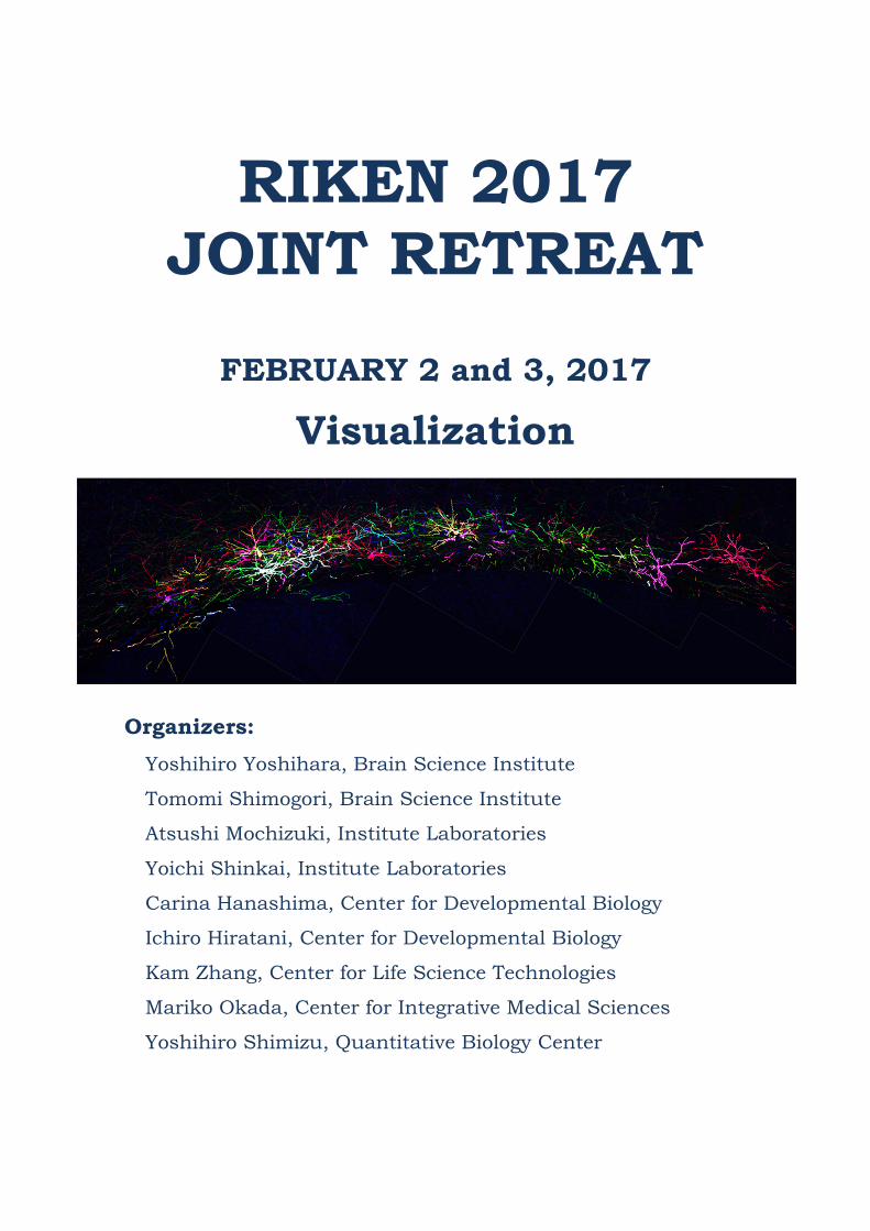
RIKEN 2017 JOINT RETREAT
FEBRUARY 2 and 3, 2017
Visualization
Organizers:
Yoshihiro Yoshihara, Brain Science Institute
Tomomi Shimogori, Brain Science Institute
Atsushi Mochizuki, Institute Laboratories
Yoichi Shinkai, Institute Laboratories
Carina Hanashima, Center for Developmental Biology
Ichiro Hiratani, Center for Developmental Biology
Kam Zhang, Center for Life Science Technologies
Mariko Okada, Center for Integrative Medical Sciences
Yoshihiro Shimizu, Quantitative Biology Center
Notice
Joint Retreat is a closed meeting in which active discussion on
unpublished results and ideas is encouraged. All information
presented should be treated as confidential by attendees.
General Information ························································ 3
Program ····································································· 11
Abstracts for Session I Oral Presentation ·························· 13
Abstract for Educational Lecture ······································ 16
Abstract for Guest Speaker’s Presentation ························ 17
Abstracts for Session II Young Investigators’ Talks ·············· 18
Abstracts for Session III Oral Presentation ························ 23
Order of Poster Short Talks ············································ 26
Poster Session Layout ·················································· 28
Authors and Titles for Poster Presentations ······················· 29
Participant List ····························································· 46
Contents
-1-

-2-
Shinkansen (Hikari/Kodama)
nkansenShinkansen
Shinkansen (Hikari)
About 1 hour 40min. About 2 hours from Tokyo
※ by bus about 30 min.
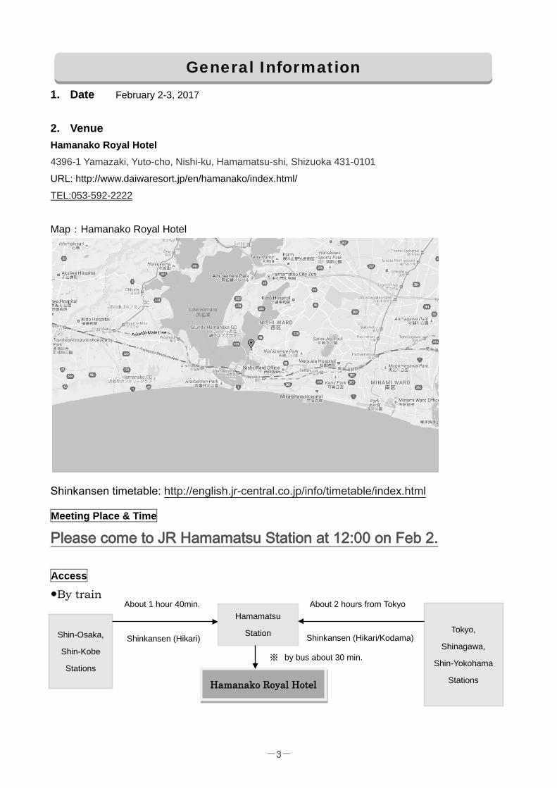
1. Date February 2-3, 2017
2. Venue Hamanako Royal Hotel
4396-1 Yamazaki, Yuto-cho, Nishi-ku, Hamamatsu-shi, Shizuoka 431-0101
URL: http://www.daiwaresort.jp/en/hamanako/index.html/
TEL:053-592-2222
Map:Hamanako Royal Hotel
Shinkansen timetable: http://english.jr-central.co.jp/info/timetable/index.html
Meeting Place & Time
Please come to JR Hamamatsu Station at 12:00 on Feb 2. Access
●By train
General Information
Shin-Osaka,
Shin-Kobe
Stations
Hamamatsu
Station Tokyo,
Shinagawa,
Shin-Yokohama
Stations Hamanako Royal Hotel
-3-
Meeting Place&Time at Hamamatsu station
If you arrive there later than at around 12:15, you should take JR Tokaido Honsen* (see time table next page) local train to JR Maisaka Station and then take a taxi out of your own pocket (or take a hotel shuttle bus, limited operation)
Hotel Shuttle Bus timetable
Lv. JR Maisaka 11:15 14:00 15:00 16:00
After the retreat, the busses will take you back to JR Hamamatsu Station.
To Yokohama / Tokyo
To Osaka / Kobe
Hamamatsu station
North Exit
★HERE! 12:00 In front of ACT city
South Exit
-4-
*JR Tokaido Honsen Timetable
●For participants from Yokohama/Shinagawa/Tokyo Hamamatsu and Maisaka Station JR Tokaido Honsen Timetable Outward (bound for Toyohashi / Gifu) Return (bound for Hamamatsu / Kakegawa)
Hamamatsu Sta. Maisaka Sta. Maisaka Sta. Hamamatsu Sta.
11:03 11:13 15:05 15:15
11:20 11:30 15:29 15:39
11:44 11:54 15:49 15:59
12:03 12:13 16:05 16:15
12:20 12:30 16:24 16:34
12:43 12:53 16:48 16:58
●For participants from Kobe/Osaka Toyohashi and Maisaka Station JR Tokaido Honsen Timetable Outward (bound for Hamamatsu / Kakegawa) Return (bound for Toyohashi / Gifu)
Toyohashi Sta. Maisaka Sta. Maisaka Sta. Toyohashi Sta.
10:42 11:06 15:13 15:39
11:03 11:27 15:30 15:56
11:23 11:47 15:52 16:18
11:42 12:06 16:13 16:39
12:03 12:27 16:30 16:56
12:23 12:47 16:50 17:16
●Taxi Reservation from Maisaka Station ⇒ Entetsu Taxi TEL: 053-412-7777
-5-
3. Official language: English
4. Registration 11:30 to 13:00 on February 2nd. The registration desk is located in front of the SWAN. All participants
are requested to sign in, pay the registration fee and pick up their name card.
Participants are required to wear the Name card throughout the Joint Retreat. 5. Registration fee Registration fee will be collected on the event day. Please prepare cash. Participation fees include accommodation, breakfast, banquet and lunch (only 2nd day).
Please bring cash with you!!
Management and higher rank employees (Kanri-shoku): 17,150 JPY Others: 15,150 JPY (150 JPY is hot spring tax) ※ Additional 4,000JPY is required for a single room request.
Note: Expense for accommodation, meals and travelling will be covered by each lab budget. Please follow the business trip procedures of your centers. 6. Hotel facilities Hotel Floor Plan
13F Restaurant, Guest Rooms 5-12F Guest Rooms 4F Conference Rooms Poster Session 3F Japanese-style Banquet Halls, Game Corner 2F Restaurant “Shiki”, Royal Hall (SWAN), Western-style Banquet
Rooms, Seminar Hall, Dining Room, Wedding Hall 1F Front, Lobby, Restaurant, Shopping Plaza, Hot Spring Large
Japanese Bath, etc. For detail, please see the hotel official website: http://www.daiwaresort.jp/en/hamanako/facilities/index.html/#a04 (1) Internet environment The hotel provides free Wi-Fi service for guests. Choose a network name “hamanakorh”. (2) Shopping Plaza Regional specialty products, fresh seafood, locally-brewed sake, crafts, and other souvenir goods fill this large shop. There is also a selection of soft drinks, alcohol, and sweets for enjoying in your room. Floor: 1F Open: 7:00-21:00 (3) Vending machines
-6-
Vending machines are located on Floor 1 and Floors 5-12 near Room 30. (4) Bar, Lounge A menu of drinks and light meals is available. - Tea lounge "Ripple"
Floor: 1F Open: 8:00-18:00 - Sky bar "Séduire"
Floor: 13F Open: 20:00-22:00 For detail, please see the hotel official website: http://www.daiwaresort.jp/en/hamanako/facilities/index.html/#a02 (5) Hot springs - Open-air hot spring rock bath
Floor: 1F Open: 5:00-10:30 / 11:30-25:00 - Indoor hot spring communal bath
Floor: 1F Open: 5:00-10:30/11:30-25:00 For detail, please see the hotel official website: http://www.daiwaresort.jp/en/hamanako/facilities/index.html/#a03 7. Accommodation a) Check in Only 1 room key is available per room. A group leader is assigned per each room. The leader must pick up the room key at the registration desk at SWAN after the poster session on February 2 and open the room. All participants should drop off your bags and meet your room mates between 19:00 to 19:30. b) Check out Check out the room by 09:00 on February 3. Keys should be returned directly to the front desk. Payment of the hotel room is NOT required. However, the cost of personal expenses such as use of phones, must be paid for separately at the time of check out by the individuals. c) Amenities Every guest room has its own bath and toilet and comes complete with various amenities, including bath towels, face towels, yukata robes, tooth brush (tooth paste), shampoo, conditioner, body soap, hair dryer among other things.
8. Meals
Lunch (Feb. 2) Participants must arrange their own lunch on Feb 2.
Banquet (Feb. 2) Banquet starts at 19:30 at “Shiki”
Breakfast (Feb. 3) Breakfast is served from 7:30 to 9:00 at “Shiki”
Lunch (Feb. 3) Lunch starts at 11:55 at "Shiki" on the 2nd floor
-7-
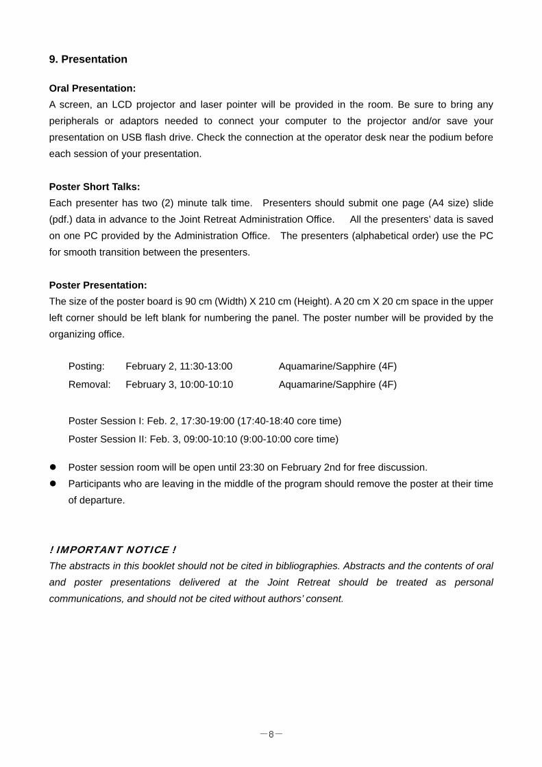
9. Presentation Oral Presentation: A screen, an LCD projector and laser pointer will be provided in the room. Be sure to bring any peripherals or adaptors needed to connect your computer to the projector and/or save your presentation on USB flash drive. Check the connection at the operator desk near the podium before each session of your presentation. Poster Short Talks: Each presenter has two (2) minute talk time. Presenters should submit one page (A4 size) slide (pdf.) data in advance to the Joint Retreat Administration Office. All the presenters’ data is saved on one PC provided by the Administration Office. The presenters (alphabetical order) use the PC for smooth transition between the presenters. Poster Presentation: The size of the poster board is 90 cm (Width) X 210 cm (Height). A 20 cm X 20 cm space in the upper left corner should be left blank for numbering the panel. The poster number will be provided by the organizing office.
Posting: February 2, 11:30-13:00 Aquamarine/Sapphire (4F)
Removal: February 3, 10:00-10:10 Aquamarine/Sapphire (4F)
Poster Session I: Feb. 2, 17:30-19:00 (17:40-18:40 core time)
Poster Session II: Feb. 3, 09:00-10:10 (9:00-10:00 core time) Poster session room will be open until 23:30 on February 2nd for free discussion. Participants who are leaving in the middle of the program should remove the poster at their time
of departure.
! IMPORTANT NOTICE ! The abstracts in this booklet should not be cited in bibliographies. Abstracts and the contents of oral and poster presentations delivered at the Joint Retreat should be treated as personal communications, and should not be cited without authors’ consent.
-8-
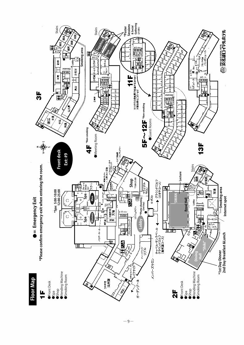
Floor Map
Emergency Exit
*Please confirm emergency exit when entering the room.
Front desk
Ext: #9
Spa
Spa
Women
Men
Shop
*7:00-21:00
*Spa: 5:00-10:00
12:00-25:00
SWAN
Dining
Hall
Internet spot
Smoking area
*1st Day Dinner
2nd Day Breakfast &Lunch
Stairs
Stairs
*Poster
Sessions
& Social
Gathering
(21:00-)
Smoking area
*Non-smoking
Lobby
*Photo shooting
(2nd day)
Lecture
●Front Desk
●Spa
●Shop
●Vending Machine
●Smoking Room
●Front Desk
●Spa
●Shop
●Vending Machine
●Smoking Room
Lobby
●Meeting Room ●Guest Rooms*Non-smoking
Lobby
Elevator
Elevator
Elevator
Elevator
Royal Hall
“Shiki”
Stairs
-9-
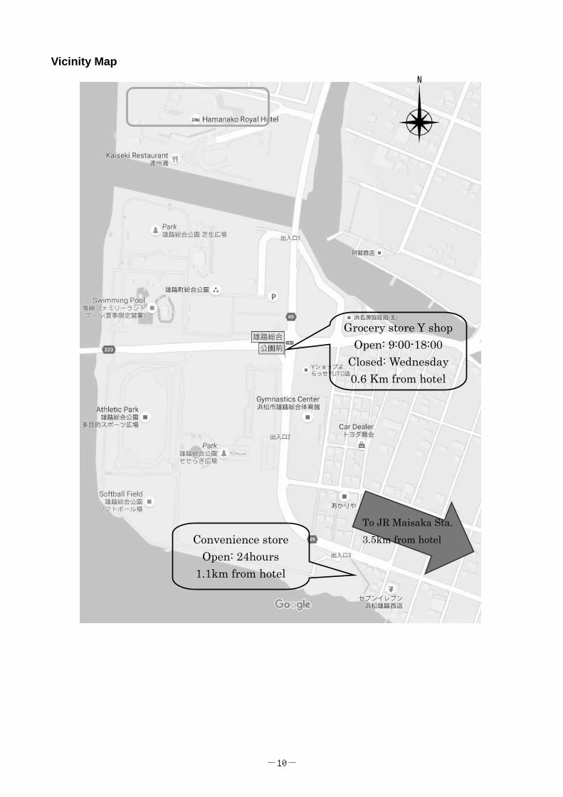
Vicinity Map
To JR Maisaka Sta. 3.5km from hotel
Grocery store Y shop Open: 9:00-18:00
Closed: Wednesday 0.6 Km from hotel
Convenience store Open: 24hours
1.1km from hotel
-10-
Time Event Place
11:30- Registration and Poster setup Lobby in front of SWAN (2F)
13:00-13:10 Opening Remarks Yoshihiro Yoshihara (BSI)
SWAN(2F)
Session I Oral presentation (Presentation 20min+ Q&A 10min) Chairperson: Yoshihiro Shimizu (QBiC)
13:10-13:40
Title Involvement of PD1 in antibody diversification and immune homeostasis
Speaker Sidonia Fagarasan (Abs. No. SI-1)
Lab Lab. for Mucosal Immunity, IMS
13:40-14:10
Title Visualization of biological complexes by electron cryo-microscopy
Speaker Hideki Shigematsu (Abs. No. SI-2)
Lab Protein Function and Structural Biology Team, CLST
14:10-14:40
Title Endothelial actin dynamics during blood vessel morphogenesis
Speaker Li-Kun Phng (Abs. No. SI-3)
Lab Lab. for Vascular Morphogenesis, CDB
Educational Lecture Chairperson: Tomomi Shimogori (BSI)
14:40-15:10
Title Visualization approaches for scientific manuscript writing
Speaker Charles Yokoyama (Abs. No. SI-4)
Lab Director’s Office, BSI
15:10-15:30 Coffee Break Lobby in front of SWAN
Guest speaker’s presentation (Presentation 40min+Q&A 10min) Chairperson: Mariko Okada (IMS)
15:30-16:20
Title Studies on biomolecules using single-molecule imaging techniques
SWAN(2F) Speaker Yoshie Harada
Affiliation Institute for Protein Research, Osaka University
16:20-17:10 Poster Short Talks
17:10-19:00 Poster Session (17:40-18:40 Odd Numbers Core Time) Aquamarine, Sapphire (4F)
19:00-19:30 Room Key Pick-up Lobby in front
of SWAN(2F)
Check in 5F-12F Guest rooms
19:30-21:00 Dinner Shiki (2F)
21:00-23:30 room close Social Gathering Aquamarine,
Sapphire (4F)
Program Day 1, Feb. 2, 2017
-11-
Program Day 1, Feb. 2, 2017
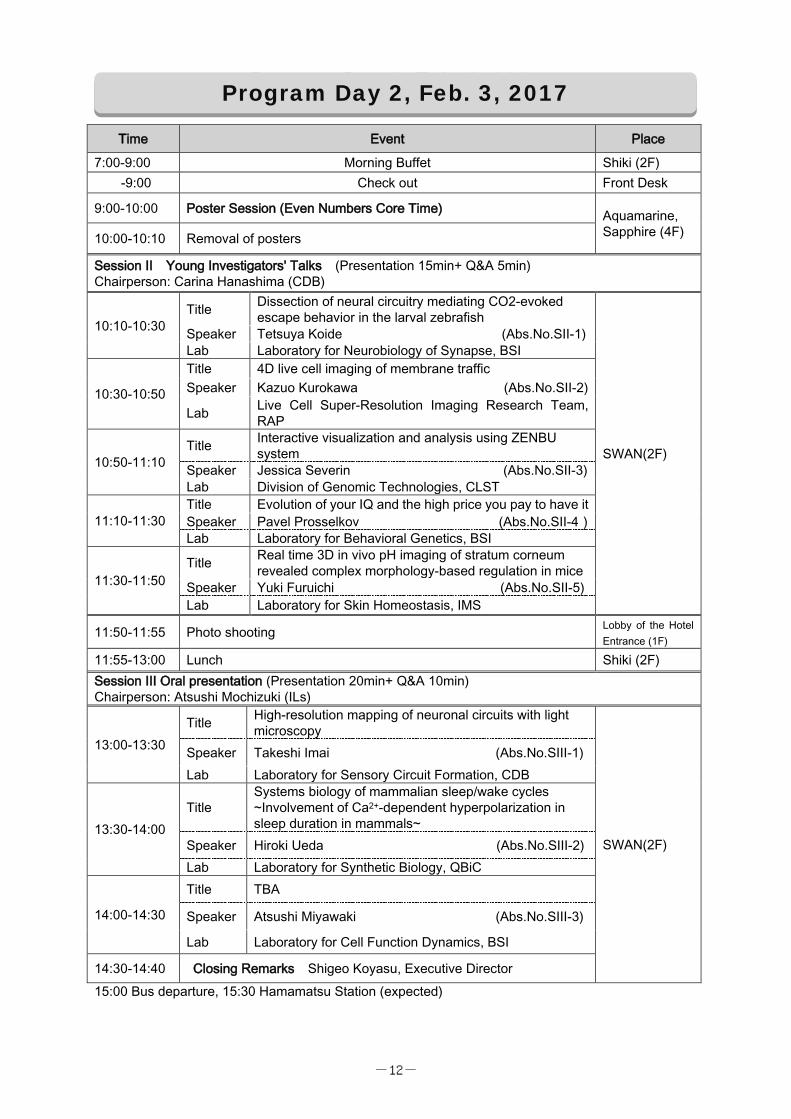
Time Event Place
7:00-9:00 Morning Buffet Shiki (2F) -9:00 Check out Front Desk
9:00-10:00 Poster Session (Even Numbers Core Time) Aquamarine, Sapphire (4F) 10:00-10:10 Removal of posters
Session II Young Investigators' Talks (Presentation 15min+ Q&A 5min) Chairperson: Carina Hanashima (CDB)
10:10-10:30 Title Dissection of neural circuitry mediating CO2-evoked
escape behavior in the larval zebrafish
SWAN(2F)
Speaker Tetsuya Koide (Abs.No.SII-1) Lab Laboratory for Neurobiology of Synapse, BSI
10:30-10:50
Title 4D live cell imaging of membrane traffic Speaker Kazuo Kurokawa (Abs.No.SII-2)
Lab Live Cell Super-Resolution Imaging Research Team, RAP
10:50-11:10 Title Interactive visualization and analysis using ZENBU
system Speaker Jessica Severin (Abs.No.SII-3) Lab Division of Genomic Technologies, CLST
11:10-11:30 Title Evolution of your IQ and the high price you pay to have it Speaker Pavel Prosselkov (Abs.No.SII-4) Lab Laboratory for Behavioral Genetics, BSI
11:30-11:50 Title Real time 3D in vivo pH imaging of stratum corneum
revealed complex morphology-based regulation in mice Speaker Yuki Furuichi (Abs.No.SII-5) Lab Laboratory for Skin Homeostasis, IMS
11:50-11:55 Photo shooting Lobby of the Hotel Entrance (1F)
11:55-13:00 Lunch Shiki (2F) Session III Oral presentation (Presentation 20min+ Q&A 10min) Chairperson: Atsushi Mochizuki (ILs)
13:00-13:30 Title High-resolution mapping of neuronal circuits with light
microscopy
SWAN(2F)
Speaker Takeshi Imai (Abs.No.SIII-1) Lab Laboratory for Sensory Circuit Formation, CDB
13:30-14:00 Title
Systems biology of mammalian sleep/wake cycles ~Involvement of Ca2+-dependent hyperpolarization in sleep duration in mammals~
Speaker Hiroki Ueda (Abs.No.SIII-2) Lab Laboratory for Synthetic Biology, QBiC
14:00-14:30
Title TBA
Speaker Atsushi Miyawaki (Abs.No.SIII-3)
Lab Laboratory for Cell Function Dynamics, BSI
14:30-14:40 Closing Remarks Shigeo Koyasu, Executive Director 15:00 Bus departure, 15:30 Hamamatsu Station (expected)
Program Day 2, Feb. 3, 2017
-12-
No.SI-1 February 2nd
13:10-13:40
Session I Oral Presentation
Involvement of PD1 in antibody diversification and immune homeostasis
FAGARASAN Sidonia
Laboratory for Mucosal Immunity, RIKEN Center for Integrative Medical Sciences (IMS)
The main function of the immune system is to protect the host against pathogens, such as bacteria or viruses. However, unlike the systemic immune system, the gut immune system does not eliminate microorganisms but instead nourishes rich bacterial communities and establishes advanced symbiotic relationships. Not only that the gut bacteria are essential for nutrient processing, production of vitamins and protection against pathogens (through competition for space and nutrients) but the development and maturation of the immune system depends on these bacteria.
Our previous studies demonstrated that the absence of immunoglobulin A (IgA) (the major effector molecule of the adaptive immunity in the gut), or the impaired IgA selection in germinal centers (GC) due to deregulated T cell control, severely affects the balance of gut bacterial communities, resulting in massive activation of the whole body immune system. Together the results point to an important role played by the adaptive immune system in regulating the microbial communities in the gut. I will discuss the role of the inhibitory co-receptor programmed cell death (PD)-1 in IgA diversification required for the maintenance of gut homeostasis and prevention of autoimmunity.
-13-
No.SI-2 February 2nd
13:40-14:10
Visualization of biological complexes by electron cryo-microscopy Hideki Shigematsu
Protein Functional and Structural Biology Team, Center for Life Science Technologies
Electron cryo-microscopy (cryoEM) is the way to visualize biological macromolecular
complexes embedded in ice by using transmission electron microscopy (TEM). TEM is a
powerful tool to magnify and visualize specimens at sub-angstrom resolution. The
method so called single particle analysis in cryoEM utilizes such a powerful tool to
depict individual molecules embedded in ice as particles. We utilize purified biological
complexes as a specimen for this analysis, therefore we assume all the particles appeared
in the data corresponds to the same three-dimensional (3D) shape. This is the reason why
we call this method as single particle analysis. The recent advances in development of
detectors and software algorithm allow us to resolve the 3D structures of biological
complexes in near atomic resolution. This was called resolution revolution in a review
article on Science and the cryoEM was selected as a “method of the year” in 2015 by
Nature Methods. Nowadays, you will see many of articles on the top journals every
week for the cryoEM single particle analysis and still the field is growing.
We have introduced state of the art cryoEMs in Yokohama campus at the end of fiscal
year of 2013 and have been working on collaborative research with inside and outside
RIKEN researchers. I will present some results already published and will talk about
recent progress in methodology development in single particle cryoEM to visualize
membrane protein structure embedded in liposomes. Our goal for the current project is
to understand voltage gating mechanism of ion channels. For this purpose, we employed
liposome as a lipid environment for voltage gating ion channels to be embedded.
Because of its spherical shape, we can apply membrane potential by changing the
outside buffer with ion channels. Therefore, our aim is to depict structural differences
with and without membrane potential to observe conformational changes in voltage
gating sensors.
-14-
No.SI-3 February 2nd
14:10-14:40
Endothelial actin dynamics during blood vessel morphogenesis Li-Kun Phng
RIKEN Center for Developmental Biology, Laboratory for Vascular Morphogenesis
Blood vessels, together with the heart, have a fundamental role in transporting
oxygen and nutrients to support the metabolic demands of tissues and organs not only
during development, but also in adults. In order to serve this function, blood vessels
must form and maintain lumen so that an interconnected network of tubules spans the
entire body to infiltrate organs and tissues. Many blood vessels form through the process
of sprouting angiogenesis, when endothelial cells lining pre-existing vessels become
activated to form new vascular sprouts. The new vascular sprout grows through
polarised collective cell migration, cell elongation and cell proliferation, and, in order to
generate a continuous network of vessels, endothelial tip cells fuse through the process
of anastomosis. Concomitantly, the process of lumen formation takes place to render
blood vessels functional. Thus, endothelial cells exhibit a multitude of cellular processes
that are tightly coordinated during vessel development.
Endothelial cells are highly plastic in their ability to change their morphology to
drive specific cellular processes. In order to understand how this plasticity is achieved,
we investigated of how the actin cytoskeleton regulates endothelial membrane dynamics
during angiogenesis in the zebrafish embryo. During the expansion of blood vessels, the
generation of actin bundles in filopodia facilitates efficient collective cell migration and
anastomosis. During lumen formation, transient polymerization of F-actin at the apical
membranes controls lumen expansion, while a stable pool of actin cables at endothelial
cell junctions stabilizes nascent lumens to produce a functional vascular network. Thus,
actin cytoskeleton of different subcellular localization and dynamics drive distinct steps
of vessel morphogenesis.
-15-
February 2nd
14:40-15:10
Educational Lecture
Visualization Approaches for Scientific Manuscript Writing Charles Yokoyama, Ph.D. Director for Research Administration, Brain Science Institute A single piece of scientific knowledge, such as a paper, is defined by a lineage and
coherence. Similar pieces aggregate into knowledge domains that in turn form a
conceptual landscape serving as a comprehensive ontology of scientific history and a
“road map” for future investigation. Two practical challenges for researchers navigating
at the frontiers of this knowledge landscape are (1) How to read existing conceptual maps
and choose important and interesting directions to pursue within a limited career span (2)
How to write effective and useful research manuscripts that add significant knowledge
and value to the conceptual map. In this lecture, we will discuss the structures of
scientific concepts, and how the resultant projects and papers interact with the publication
process. We will explore how visualization approaches in manuscript writing can help to
facilitate knowledge formation and provide the basis for a future ontology-based science.
-16-
February 2nd
15:30-16:20
Guest Speaker’s Presentation
Studies on Biomolecules Using Single-Molecule Imaging Techniques Yoshie Harada
Institute for Protein Research, Laboratory of Nanobiology, Osaka University
The best way to obtain unambiguous information about the function of biomolecules is to
study their function at the single-molecule level. Twenty years ago, we developed single
fluorescent dye molecule imaging technique using fluorescence microscope which reduced
background noise dramatically by installing evanescent illumination. Using this technique,
we observed the sliding movement of single kinesin molecules along a microtubule and
also visualized binding of single RNA polymerase molecules to a single DNA molecule.
However, due to the background noise, even if using the evanescent illumination, we are
not able to observe a single fluorescent dye molecule under higher than 50 nM fluorescent
dye concentration conditions. To investigate dynamic processes of DNA-protein
interactions related to genome DNA maintenance we are employing zero-mode
waveguides (ZMWs). ZMWs are nano-hole (100 nm in both depth and diameter) arrays on
an aluminum coated fused silica substrate. ZMWs allow single-molecule observation under
several μM fluorescent dye concentration conditions due to its extremely small excitation
volume (~ 10-21 L). Using ZMWs, we focus on characterizations of proteins involved in
homologous recombination or epigenetics such as RuvAB protein complex or nucleosome
binding proteins. Currently we are also developing a new single-molecule imaging
technique using fluorescent diamond nanoparticles. Fluorescence of nitrogen-vacancy
centers (NVC) embedded in nanodiamonds exhibits neither photobleaching nor blinking,
superior to the fluorescence properties of widely-used organic dyes and quantum dots.
Another uniqueness of NVC is that the fluorescence intensity can be modulated by
magnetic resonance technique. It is based upon a presence of energy levels partly shared by
both processes of the fluorescence-emission and of electron spin resonance. By using the
property we developed a technique that can selectively detect single NVC out of an
autofluorescence.
-17-
Dissection of Neural Circuitry Mediating CO2-evoked Escape Behavior in the Larval Zebrafish Tetsuya Koide and Yoshihiro Yoshihara
RIKEN Brain Science Institute, Japan
Avoidance of carbon dioxide (CO2) is a vital reaction commonly preserved in various
animal species from worms to mammals for their survival. In this study, we aim to
elucidate the neural circuit mechanism underlying CO2 avoidance in larval zebrafish.
First, we observed that the application of CO2 to larval zebrafish elicits a robust escape
response. Next, we performed in vivo whole-brain Ca2+ imaging to map neural
substrates governing CO2-evoked escape response. Upon CO2 application to the nose,
ensembles of neural activation were observed in multiple brain regions, including the
olfactory bulb (OB), the trigeminal ganglion (TG), and the hindbrain. Because the fish
nose receives two distinct types sensory information, olfactory and trigeminal, we
laser-ablated either the OB or the TG and performed Ca2+ imaging and behavioral
analysis to examine each contribution to the escape response. As a result, the
CO2-evoked neural activation in the hindbrain and escape behavior were markedly
decreased in the TG-ablated fish, but not in the OB-ablated fish. These findings
demonstrate that the nasal trigeminal pathway is responsible for CO2-evoked escape in
larval zebrafish.
Young Investigators’ Talks
-18-
No. SII-2 February 3rd 10:30-10:50
4D Live Cell Imaging of Membrane Traffic Kazuo Kurokawa1), Midori Ishii1) 2), Akihiko Nakano1)2)
1) Live Cell Super-Resolution Imaging Research Team, RIKEN, Japan, 2) Laboratory
of Developmental Cell Biology, University of Tokyo, Japan
Membrane traffic is an essential mechanism for transport of proteins and other
macromolecules to various destinations inside or outside of the cells. It also underlies
the fundamental need for cells to maintain cellular homeostasis. Membrane traffic is
mediated via membrane-surrounded vesicles and carrier intermediates. The basic
mechanism of membrane traffic is conserved in all eukaryotic cells, from yeast to higher
animals and plants. The Golgi apparatus is a central station of this traffic. Newly
synthesized proteins in the ER are delivered to the Golgi apparatus in which they are
modified and selectively sorted to their final destination.
We have conducted 4D live cell imaging at high spatiotemporal resolution, which is a
promising approach to resolve many remaining problems of membrane traffic. For this
purpose, we have developed SCLIM (super-resolution confocal live imaging
microscopy) by combining inverted fluorescence microscope with a high-speed and
high-signal-to noise-ratio spinning-disk confocal scanner, a custom-made spectroscopic
unit, image intensifiers, and multiple EM-CCD cameras for multiple color observation.
Using this system, we have investigated 1) ER-Golgi protein traffic, 2) the mechanism
of the Golgi maturation, 3) intra-Golgi protein traffic in living Saccharomyces
cerevisiae.
-19-
No. SII-3 February 3rd
10:50-11:10
Interactive visualization and analysis using ZENBU system
Jessica Severin, Michiel De Hoon, Alistair Forrest, Piero Carninci
RIKEN CLST
Genome-wide compendium studies, such as FANTOM, ENCODE, Roadmap, TCGA
and single-cell studies are providing new challenges for visualization, analysis and
secured data sharing due to their unprecedented breadth and scale. The ZENBU system
(Severin et.al. Nature Biotechnology, 2014) enhances the genome browser concept by
integrating advanced, on-demand data processing and analysis with interactive
visualization optimized for comparison across 1000s of secured experimental samples.
Any user of the system can create secured collaborations, manage membership, and
upload 1000s of experimental data files with descriptive metadata. Through the
visualization interface, researchers can choose experimental data, modify analysis
parameters and scripts, quickly see the newly processed result on a genomic region of
interest, apply the analysis genome wide, and share the visualization and data export
results with their collaborators.
ZENBU‘s interactive visualization can dynamically combine thousands of experimental
datasets in a multi-dimensional matrix allowing the researchers to visually slice and
group the dataset through linked genome location, experimental signal and metadata
views. ZENBU is open-source and freely available for use on the web
http://fantom.gsc.riken.jp/zenbu and for installation in individual laboratories.
-20-
No. SII-4 February 3rd 11:10-11:30
Evolution of your IQ and the High Price you pay to have it Pavel Prosselkov1)2), Ryota Hashimoto3)4), Denis Polygalov5), Kazutaka Ohi3), Qi Zhang1), Hiromichi Goto1), Thomas J. McHugh5), Masatoshi Takeda3)4), and Shigeyoshi Itohara1)
1) Laboratory for Behavioral Genetics, RIKEN Brain Science Institute, Japan 2) Grad School, Department of Veterinary Medicine, Tokyo University, Japan 3) Department of Psychiatry, Osaka Univ Graduate School of Medicine, Japan 4) United Graduate School of Child Development, Osaka University, Japan 5) Lab for Circuit and Behav Physiol, RIKEN Brain Science Institute, Japan
Around 500 million years ago a massive genome duplication event, termed the
Cambrian explosion, took place. Many genomes underwent a gene duplication event,
subfunctionalization, and de novo functional elaboration. This provided animals with a
capacity to occupy challenging ecological niches (e.g. the land) through the positive
selection of advantageous anatomical and cognitive traits. Among the duplicated gene
paralogs were NTNG1 and NTNG2, brain-specific presynaptic proteins found only in
vertebrates. These genes have been implicated in IQ determination in human subjects,
with a unique labor division, such that NTNG1 affects verbal comprehension and
processing speed while NTNG2 impacts working memory and perceptual organization.
Non-coding gene loci responsible for this effect underwent accelerated evolution from
primates through Neanderthals/Denisovans to modern humans. Simultaneously, both
NTNG genes became associated with a variety of neurocognitive disorders, co-
segregating with some complex etiologies (e.g. schizophrenia and autism), while
individually and exclusively segregating within others (e.g. Alzheimer and lupus). In
mice both genes affect executive function, contributing to attention and working
memory, top-down (Ntng2, cognition) and bottom-up (Ntng1, perception) information
flow, in a manner similar to the human NTNG gene orthologs. Such “cognitive
complementarity” underscores an evolutionary gain-dependent link between advanced
cognition and concomitant propensity to develop neuropathologies. This risk/reward
trade-off implies that as living creatures we may have reached the maximum threshold
of our brain capacity.
-21-
No. SII-5
February 3rd 11:30-11:50
Real Time 3D in vivo pH Imaging of Stratum Corneum Revealed Complex
Morphology-based Regulation in Mice
Yuki Furuichi 1) 2), Takeshi Matsui 2), Masayuki Amagai 1) 2)
1) Department of Dermatology, Keio University School of Medicine, Japan, 2)
Laboratory for Skin Homeostasis, RIKEN, Japan
The regulation of pH is an important element in various biological functions. Stratum
corneum (SC) pH is considered as acidic in general while its precise distribution
remains to be clarified. The purpose of this study is to produce a transgenic mouse for
visualizing in vivo SC pH to enhance our understanding of the SC. We generated a
ratiometric pH biosensor with pH sensitive fluorescent protein, VenusH148G, and pH less
sensitive mCherry, expressed specifically in the uppermost layer of stratum granulosum
(SG1) by modified CRISPR/Cas9 system. Confocal microscopic analysis of living pH
imaging demonstrated that SC has at least two distinct middle-acidic and upper-neutral
layers with cross section view, rather than gradual pH changes across the layers. These
findings together indicated the dynamic and complex nature of SC in terms of pH
regulation and that the 3D in vivo pH imaging mice will provide a valuable tool to
dissect the homeostatic mechanisms of SC by pH regulation.
-22-
No.SIII-1 February 3rd
13:00-13:30
Session III Oral Presentation
High-resolution mapping of neuronal circuits with light microscopy Takeshi Imai
Laboratory for Sensory Circuit Formation, RIKEN Center for Developmental Biology
Neurons receive excitatory and inhibitory synaptic inputs from compartmentalized
structures, known as synapses. Although their geometrical distribution is crucial to
understand synaptic integration mechanisms within a neuron, its distribution at a
whole-neuron scale has not been fully established. Previously, the precise distribution of
synapses has been best studied by time-consuming serial section electron microscopy.
Due to the diffraction limit of light, conventional light microscopy could not fully
resolve the fine structure of synapses. To overcome these limitations, we recently
established a tissue clearing agent, SeeDB2, which is optimized for deep-tissue
high-resolution fluorescence imaging. Using super-resolution microscopy of
SeeDB2-cleared brain tissues, we performed a comprehensive and quantitative mapping
of dendritic spines, where excitatory synapses are formed in cortical pyramidal neurons.
We found that dendritic spines are highly enriched in a particular domain of the
dendrites, and that the biased spine distribution is established at a later stage of postnatal
development in an activity-dependent manner. Our results provide an important platform
toward our understanding of synaptic integration mechanisms in different types and
stages of dendrites in cortical pyramidal neurons. In this talk, I will also introduce our
ongoing efforts toward dense reconstruction of neuronal circuits using light microscopy.
References:
1. Ke MT, Nakai Y, Fujimoto S, Takayama R, Yoshida S, Kitajima TS, Sato M, Imai T.,
(2016) Cell Rep 14 (11), 2718-32.
2. Ke MT, Fujimoto S, Imai T., (2013) Nat Neurosci 16 (8), 1154-61.
-23-
No.SIII-2 February 3rd
13:30-14:00
Systems Biology of Mammalian Sleep/wake Cycles ~Involvement of Ca2+-Dependent Hyperpolarization in Sleep Duration in Mammals~
Hiroki R. Ueda
Laboratory for Synthetic Biology, Quantitative Biology Center, RIKEN
The detailed molecular mechanisms underlying the regulation of sleep duration in
mammals are still elusive. To address this challenge, we constructed a simple
computational model, which recapitulates the electrophysiological characteristics of the
slow-wave sleep and awake states. Comprehensive bifurcation analysis predicted that a
Ca2+-dependent hyperpolarization pathway may play a role in slow-wave sleep and
hence in the regulation of sleep duration. To experimentally validate the prediction, we
generate and analyze 26 KO mice. Here we found that impaired Ca2+-dependent K+
channels (Kcnn2 and Kcnn3), voltage-gated Ca2+ channels (Cacna1g and Cacna1h), or
Ca2+/calmodulin-dependent kinases (Camk2a and Camk2b) decrease sleep duration,
while impaired plasma membrane Ca2+ ATPase (Atp2b3) increases sleep duration.
Genetical (Nr3a) and pharmacological intervention (PCP, MK-801) and whole-brain
imaging validated that impaired NMDA receptors reduce sleep duration and directly
increase the excitability of cells. Based on these results, we propose a hypothesis that a
Ca2+-dependent hyperpolarization pathway underlies the regulation of sleep duration in
mammals.
References 1. Tatsuki et al. Neuron, 90(1) : 70–85 (2016).
2. Sunagawa et al, Cell Reports, 14(3):662-77 (2016).
3. Susaki et al. Cell, 157(3): 726–39, (2014).
4. Tainaka et al. Cell, 159(6):911-24(2014).
5. Susaki et al. Nature Protocols, 10(11):1709-27(2015).
6. Susaki and Ueda. Cell Chemical Biology, 23(1):137-57 (2016).
7. Tainaka et al. Ann. Rev. of Cell and Devel. Biol. 32: 713-741 (2016).
8. Ode et al. Mol. Cell, in press
-24-
No.SIII-3 February 3rd
14:00-14:30
CRUSING INSIDE CELLS
Atsushi Miyawaki
Laboratory for Cell Function Dynamics, RIKEN Brain Science Institute
The behavior of biochemical molecules moving around in cells makes me think of a
school of whales wandering in the ocean, captured by the Argus system on the artificial
satellite. When bringing a whale back into the sea --- with a transmitter on its dorsal fin,
every staff member hopes that it will return safely to a school of its species. A
transmitter is now minute in size, but it was not this way before. There used to be some
concern that a whale fitted with a transmitter could be given the cold shoulder and thus
ostracized by other whales for “wearing something annoying.” How is whale’s
wandering related to the tide or a shoal of small fish? What kind of interaction is there
among different species of whales? We human beings have attempted to fully
understand this fellow creature in the sea both during and since the age of whale fishing.
In a live cell imaging experiment, a luminescent probe replaces a transmitter. We
label a luminescent probe on a specific region of a biological molecule and bring it back
into a cell. We can then visualize how the biological molecule behaves in response to
external stimulation. Since luminescence is a physical phenomenon, we can extract
various kinds of information by making full use of its characteristics. For example, the
excited energy of a fluorescent molecule donor transfers to an acceptor relative to the
distance and orientation between the two fluorophores. This phenomenon can be used to
identify interaction between biological molecules or structural change in biological
molecules.
Cruising inside cells in a supermicro corps, gliding down in a microtubule like a
roller coaster, pushing our ways through a jungle of chromatin while hoisting a flag of
nuclear localization signal --- we are reminded to retain a playful and adventurous
perspective at all times. What matters is mobilizing all capabilities of science and giving
full play to our imagination. We believe that such serendipitous findings can arise out of
such a sportive mind, a frame of mind that prevails when enjoying whale-watching.
-25-
Poster Short Talks
February 2nd 16:20-17:10
2min.each 1. P-9 : Sufeng Chiang
Laboratory for Integrated Cellular Systems, Center for Integrative Medical Sciences
2. P-12 : Shingo Gibo Theoretical Biology Laboratory, Chief Scientist Laboratories
3. P-14 : Takeshi Hanami Genetic Diagnosis Technology Unit and Ultra-Sensitive Biomolecule Detection Laboratory, Center for Life Science Technologies
4. P-16 : Maxime Hebrard Laboratory for Integrated Bioinformatics, Center for Integrative Medical Sciences
5. P-19 : Yoko Ito Live Cell Super-Resolution Imaging Research Team, Center for Advanced Photonics
6. P-21 : Bogumil Kaczkowski Division of Genomic Technologies, Center for Life Science Technologies
7. P-25 : Toshimori Kitami Young Chief Investigator Program, Center for Integrative Medical Sciences
8. P-33 : Marcus Leiwe Laboratory for Sensory Circuit Formation, Center for Developmental Biology
9. P-34 : Chia-Wen Lin Laboratory of Mental Biology, Brain Science Institute
10. P-35 : Takeshi Matsui Laboratory for Skin Homeostasis, Center for Integrative Medical Sciences
11. P-38 : Nobuhiko Miyasaka
Laboratory for Neurobiology of Synapse, Brain Science Institute
-26-
February 2nd 16:20-17:10
2min.each 12. P-40 : Fumito Mori
Theoretical Biology Laboratory, Chief Scientist Laboratories
13. P-41 : Ritsuko Morita Laboratory for Tissue Microenvironment, Center for Developmental Biology
14. P-43 : Masanori Murayama Laboratory for Behavioral Neurophysiology, Brain Science Institute
15. P-44 : Nobuhiro Nakai Laboratory for Mental Biology, Brain Science Institute
16. P-47 : Noriaki Ogawa Hatsuda Quantum Hadron Physics Laboratory, Nishina Center for Accelerator-Based Science
17. P-49 : Charles Plessy Genomics Miniaturization Technology UNit, Center for Life Science Technologies
18. P-64 : Go Shioi Genetic Engineering Unit, Center for Life Science Technologies
19. P-67 : Masaru Tamura
Technology and Development Team for Mouse Phenotype Analysis, BioResource Center
20. P-68 : Todd Taylor Laboratory for Integrated Bioinformatics, Center for Integrative Medical Sciences
21. P-71 : Bo Thomsen
Theoretical Molecular Science Laboratory, Chief Scientist Laboratories
22. P-75 : Kiminori Toyooka Mass Spectrometry and Microscopy Unit, Center for Sustainable Resource Science
23. P-8 : Arno Germond
Linking gene expression with Raman spectral analysis
-27-
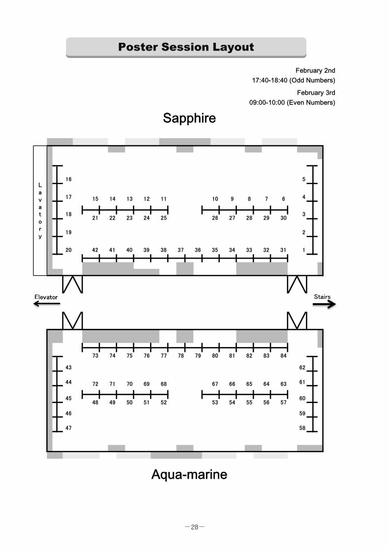
16 5
17 4
19 2
20 42 41 40 39 38 37 36 35 34 33 32 31 1
Elevator
43 62
44 61
46 59
47 58
Stairs
Lavatory
February 3rd09:00-10:00 (Even Numbers)
February 2nd17:40-18:40 (Odd Numbers)
21 22 23 24 2518
15 14 13 12 11 10 6
26 27 28 29 303
72 71 70 69 68 67 66 65 64 63
45 6048 49 50 51 52 53 56 5754
73 74 75 76 77 78 79 84
Sapphire
Aqua-marine
80 81 82 83
55
9 8 7
Poster Session Layout
-28-
(Alphabetical orders, in principle, some irregular arrangement due to late application, cancellation and request)
P-1
Harnessing the CRISPR/Cas9 system in mouse genetic engineering @ LARGE Takaya Abe
Takaya Abe1), Kiyonari Hiroshi1)2), Yashide Furuta1)2)
1) Genetic Engineering Team, and 2) Animal Resource Development Unit, RIKEN CLST
Laboratories for Animal Resource Development and Genetic Engineering, Center for Life
Science Technologies
P-2
Proteomic analysis of E. coli based cell-free protein synthesis Jiro Adachi
Yoshihiro Shimizu (QBiC, RIKEN)
Laboratory for cell-free protein synthesis, Quantitative Biology Center
P-3
Electrophysiological and Optogenetic Analysis of Neurons in the Mouse Claustrum Ayako Ajima
Momoko Shiozaki (RIKEN BSI), Yoshihiro Yoshihara (RIKEN BSI)
Lab for Neurobiology of Synapse, Brain Science Institute
P-4
Genetic Neuroanatomical Analysis of the Claustrum Momoko Shiozaki
Rumiko Mizuguchi(RIKEN), Sachiko Mitsui(RIKEN), Hiroki Hamanaka(RIKEN), Joshua P.
Johansen(RIKEN), Yoshihiro Yoshihara(RIKEN)
Neurobiology of Synapse, Brain Science Institute
P-5
Decoding context-dependent olfactory valence in Drosophila Laurent Badel
Kazumi Ohto (RIKEN BSI), Yoshiko Tsuchimoto (RIKEN BSI), Hokto Kazama (RIKEN BSI)
Circuit Mechanisms of Sensory Perception , Brain Science Institute
Authors and Titles for Poster Presentations
-29-
P-6
Fine mapping of mouse chromosome 10 prepulse inhibition-quantitative trait loci (PPI-QTL) points to Cdh23 Shabeesh Balan
Tetsuo Ohnishi (RIKEN-BSI), Akiko Watanabe (RIKEN-BSI), Hisako Ohba (RIKEN-BSI),
Yoshimi Iwayama (RIKEN-BSI), Yasuko Hisano (RIKEN-BSI), Takeshi Hayashi (National
Agricultural Research Centre, Tsukuba),Yoshiaki Kikkawa (Tokyo Metropolitan Institute of
Medical Science, Tokyo), Tomomi Shimogori (RIKEN-BSI), Takeo Yoshikawa (RIKEN-BSI)
Laboratory for Molecular Psychiatry, Brain Science Institute
P-7
Role of insulin/IGF signaling on body growth and adult physiology in Drosophila Kota Banzai
Laboratory for Growth Control Signaling, Center for Developmental Biology
*P-8
Linking gene expression with Raman spectral analysis Arno Germond
Taro Ichimura, Takaki Horinouchi, Hideaki Fujita, Chikara Furusawa, Tomonobu Watanabe,
(affiliation: QBIC, RIKEN)
Laboratory for Comprehensive Bioimaing
*P-9
Dissecting the regulatory mechanism and transcriptional responses triggered by the NF-kappaB and ERK signaling pathways in the antigen-stimulated B cells Sufeng Chiang
Hisaaki Shinohara (RIKEN), Huai-Kuang Tsai (Academia Sinica, TAIWAN), Mariko Okada
(RIKEN)
Laboratory for Integrated Cellular Systems, Center for Integrative Medical Sciences
P-10
A role of sumoylation in maintenance of centromeric cohesion at telophase I in meiosis Yi Ding
Masako Kaido(RIKEN)
-30-
Laboratory for Chromosome Segregation, Center for Developmental Biology
P-11
Real time 3D in vivo pH imaging of stratum corneum revealed complex morphology-based regulation in mice Yuki Furuichi
Takeshi Matsui (RIKEN), Masayuki Amagai (RIKEN, Keio)
Laboratory for Skin Homeostasis, Center for Integrative Medical Sciences
*P-12
Prediction of essential intersections between RNA methylation and circadian rhythm using models Shingo Gibo
Gen Kurosawa (RIKEN)
Theoretical Biology Laboratory, Chief Scientist Laboratories
P-13
Optimized Sequential Techniques for Marmoset Neural Circuit Mapping Mitsutoshi Hanada
Kevin Weber (RIKEN)
Laboratory for Marmoset Neural Architecture, Brain Science Institute
*P-14
Ultra-sensitive detection for miRNA by the combination of scanning single-molecule counting (SSMC) and Eprobe without amplification Takeshi Hanami
Genetic Diagnosis Technology Unit and Ultra-Sensitive Biomolecule Detection Laboratory,
Center for Life Science Technologies
P-15
Madagascar ground gecko, yet another model animal for amniotes Yuichiro Hara
Phyloinformatics Unit, Center for Life Science Technologies
-31-
*P-16
Visualization Platforms of the Laboratory for Integrated Bioinformatics Maxime Hebrard
Hidehiro Fukuyama(RIKEN), Jayson Harshbarger(RIKEN), Todd D. Taylor(RIKEN)
Laboratory for Integrated Bioinformatics, Center for Integrative Medical Sciences
P-17
Dynamic changes in replication timing mirror subnuclear compartment dynamics during differentiation Ichiro Hiratani
Hisashi Miura (RIKEN CDB) (First Author)
Laboratory for Developmental Epigenetics, Center for Developmental Biology
P-18
Dynamic transcriptional and epigenetic regulation that determines B cell fate Tomokatsu Ikawa
Tomohiro Miyai, and Junichiro Takano (YCI laboratory for Immune Regeneration)
Laboratory for Immune Regeneration, Center for Integrative Medical Sciences
*P-19
Biogenesis and maintenance of the Golgi apparatus in plant cells Yoko Ito
Tomohiro Uemura (Univ. Tokyo), Kei H Kojo (Sophia Univ.), Seiichiro Hasezawa (Univ.
Tokyo), Takashi Ueda (NIBB), Akihiko Nakano (RIKEN)
Live Cell Super-Resolution Imaging Research Team, Center for Advanced Photonics
P-20
In silico investigation of Deoxyhypusine Synthase and Deoxyhypusine Hydroxylase Matej Janezic
Akihiro Ito (RIKEN), Minoru Yoshida (RIKEN), Kam Y. J. Zhang (RIKEN)
Division of Structural and Synthetic Biology, Structural Bioinformatics Team, Center for Life
Science Technologies
*P-21
Pan cancer biomarkers from integrative analysis of transcriptome and epigenome
-32-
Bogumil Kaczkowski
Tanaka Yuji (RIKEN ACCC & PMI), Hideya Kawaji (RIKEN DGT & PMI), Yoshihide
Hayashizaki (RIKEN PMI), Alistair Forrest (Perkins Institute, Australia), Piero Carninci
(RIKEN DGT)
Division of Genomic Technologies, Center for Life Science Technologies
P-22
Fast polarization converter for microscopic analysis of protein macromolecular structures Junichi Kaneshiro
Tomonobu M. Watanabe(RIKEN QBiC), Taro Ichimura(RIKEN QBiC)
Laboratory for comprehensive bioimaging, Quantitative Biology Center
P-23
Precise analysis of microbiota dynamics using high-resolution time-series mouse fecal sampling method Seok-Won Kim
Todd D. Taylor(RIKEN)
Laboratory for Integrated Bioinformatics, Center for Integrative Medical Sciences
P-24
Development of the gene expression atlas for the marmoset brain Yoshiaki Kita
Satomi S Kikuchi(RIKEN), ChIhiro Yoshida(RIKEN), Mami U(RIKEN), Karl Windak(RIKEN),
Hirozumi Nishibe(RIKEN), Yasuhiro Go(NINS), Tomomi Shimogori(RIKEN)
Laboratory for Molecular Mechanisms of Thalamus Development, Brain Science Institute
*P-25
Dissecting the role of mitochondria in NLRP3 inflammasome activation via chemical genetics Toshimori Kitami
Uyen Thi Tran (RIKEN Center for Integrative Medical Sciences, Young Chief Investigator
Program)
Young Chief Investigator Program, Center for Integrative Medical Sciences
-33-
P-26
Neural activity measurement by the head-mounted imager developed to obtain a vast field of view in freely-moving animal Takuma Kobayashi
Hitoshi Okamoto(RIKEN)
Laboratory for Developmental Gene Regulation, Brain Science Institute
P-27
Dissection of Neural Circuitry Mediating CO2-evoked Escape Behavior in the Larval Zebrafish Tetsuya Koide
Yoshihiro Yoshihara (RIKEN)
Laboratory for Neurobiology of Synapse, Brain Science Institute
P-28
Scientific media annotation using the iCLiKVAL offline-first browser extension Naveen Kumar
Todd Taylor (RIKEN)
Laboratory for Integrated Bioinformatics, Center for Integrative Medical Sciences
P-29
A Novel Scaffold for Developing Specific or Broad-Spectrum Chitinase Inhibitors Ashutosh Kumar
Xi Jiang(Dalian University of Technology, China), Tian Liu(Dalian University of Technology,
China), Kam Y. J. Zhang(RIKEN), and Qing Yang(Dalian University of Technology, China)
Structural Bioinformatics Team, Division of Structural and Synthetic Biology,
Center for Life Science Technologies
P-31
Synthesis and transport of ciliary protein Cluap1 within a cell Lynda Lamri
Laboratoy for Organismal Patterning, Center for Developmental Biology
P-32
Extended depth-of-field microscopy for long-term imaging of synaptic plasticity.
-34-
Thomas Launey
Lab. for Synaptic Molecules of Memory Persistence, Brain Science Institute
*P-33
Spontaneous Network Activity in the Neonatal Mouse Olfactory Bulb Regulates Dendrite Pruning of Mitral Cells. Marcus Leiwe
Satoshi Fujimoto (RIKEN), Takeshi Imai (RIKEN)
Laboratory for Sensory Circuit Formation, Center for Developmental Biology
*P-34
Abnormal Development of Immune Cells Explains Brain Inflammation and GI Symptom in ASD Chia-Wen Lin
Laboratory of Mental Biology, Brain Science Institute
*P-35
Dissecting the adaptive evolution of skin by using isolated stratum granulosum cells from terrestrial vertebrates Takeshi Matsui
Masayuki Amagai (RIKEN-IMS/Keio Univ Sch of Med)
Laboratory for Skin Homeostasis, Center for Integrative Medical Sciences
P-36
Activities in the CLST Epigenome Technology Exploration Unit Aki Minoda
Epigeoome Technology Exploration Unit, Center for Life Science Technologies
P-37
Visualization and characterization of the BCR induced oscillation-like NF-κB kinetics in a B cell. Yu Miyamoto
Kazunari Iwamoto (RIKEN), Mariko Okada (RIKEN)
Laboratory for integrated cellular systems, Center for Integrative Medical Sciences
-35-
*P-38
Appetitive olfactory learning in zebrafish Nobuhiko Miyasaka
Yoshihiro Yoshihara (RIKEN)
Laboratory for Neurobiology of Synapse, Brain Science Institute
P-39
Mammalian expression of Gas Vesicle nanoparticles and its application to genetically-encoded UI/MRI multimodal contrast agent. Ryota Mizushima
Kanako Inoue (Osaka Univ.), Atsuko Iwane (RIKEN), Tomonobu Watanabe (RIKEN)
Comprehensive Bioimaging Team, Quantitative Biology Center
*P-40
Origin of cell diversity: Analysis of fixed points in Boolean networks Fumito Mori
Atsushi Mochizuki(RIKEN)
Theoretical Biology Laboratory, Chief Scientist Laboratories
*P-41
Deciphering the molecular and cellular basis of the induction of hair follicle stem cells at single cell resolution Ritsuko Morita
Ritsuko Morita1, Tetsutaro Hayashi2, Itoshi Nikaido2, Takaya Abe3, Hiroshi Kiyonari3, 4,
Yasuhide Furuta3, 4, Hironobu Fujiwara1 1 Laboratory for Tissue Microenvironment, RIKEN Center for Developmental Biology, Kobe 650–0047,
Japan
2 Bioinformatics Research Unit, RIKEN Advanced Center for Computing and Communication, Wako
351-0198, Japan
3 Animal Resource Development Unit, Division of Bio-Function Dynamics Imaging, RIKEN Center for Life
Science Technologies, Kobe 650–0047, Japan
4 Genetic Engineering Team, Division of Bio-Function Dynamics Imaging, RIKEN Center for Life Science
Technologies, Kobe 650–0047, Japan
Laboratory for Tissue Microenvironment., Center for Developmental Biology
-36-
P-42
Elucidating the role of lncRNAs in pluripotency maintenance and differentiation in human iPSCs Divya Mundackal Sivaraman
Joachim Luginbuehl (CLST RIKEN), Jordan Ramilowski(CLST RIKEN), Tsukasa
Kouno(CLST RIKEN), Suzannah Szumowski(CLST,RIKEN), Jay W Shin(CLST RIKEN)
Cell Conversion Technology Unit, Center for Life Science Technologies
*P-43
Fast-Scanning Two-Photon Microscopy for Megapixel Imaging Masanori Murayama
Keisuke Ota, BSI, Laboratory for Behavioral Neurophysiology
Laboratory for Behavioral Neurophysiology, Brain Science Institute
*P-44
Cortical Network Analysis for Behaving Mice in Virtual Reality Environment Nobuhiro Nakai
Masaaki Sato, Yasunori Hayashi, Hajime Hirase, Shigeyoshi Itohara, Toru Takumi (RIKEN
BSI)
Laboratory for Mental Biology, Brain Science Institute
P-45
Analysis of habenula neural circuit which encodes aversive expectation value Haruna Nakajo
Developmental Gene Regulation, Brain Science Institute
P-46
Respiration-Phased and Layer-Specific Operation Modes in the Olfactory Cortex Kimiya Narikiyo
Hiroyuki Manabe(Doshisha Univ.), Yoshihiro Yoshihara(RIKEN), Kensaku Mori(Univ. Tokyo)
Laboratory for Neurobiology of Synapse, Brain Science Institute
*P-47
Dynamical Pattern Selection of Growing Cellular Mosaic in Fish Retina Noriaki Ogawa
-37-
Tetsuo Hatsuda(RIKEN), Atsushi Mochizuki(RIKEN), Masashi Tachikawa(RIKEN)
Hatsuda Quantum Hadron Physics Laboratory, Nishina Center for Accelerator-Based
Science
P-48
Computational Investigation of the Role of AIOLOS Mutations in B-cell Deficiency and Familial Lymphoma Aditya Kumar Padhi
Motoi Yamashita2, Ichiro Taniuchi2 and Kam Y.J. Zhang1 (1 Structural Bioinformatics Team,
CLST, DSSB, RIKEN, Yokohama, Japan, 2 Laboratory for Transcriptional Regulation, CIMS,
RIKEN, Yokohama, Japan)
Structural Bioinformatics Team, Center for Life Science Technologies
*P-49
Single-molecule transcript assembly with unique molecular identifiers and tagmentation Charles Plessy
Genomics Miniaturization Technology Unit, Center for Life Science Technologies
P-50
Screening for novel regulators of the inactive X chromosome organization Rawin Poonperm
Ichiro Hiratani (RIKEN CDB)
Laboratory for Developmental Epigenetics, Center for Developmental Biology
P-51
Transcriptome analysis of FACS-sorted single cells with nanoCAGE Stephane Poulain
Arnaud, Ophélie ; Kato, Sachi ; Mendez, Mickaël ; Plessy, Charles (RIKEN)
Genomic Miniaturization Technology Unit, Center for Life Science Technologies
P-52
Capsule Hotel. A convenient device for imaging thousands of E. coli cells. David Geoffrey Priest
Nobuyuki Tanaka, Laboratory for Integrated Biodevice, QBiC. Yuichi Taniguchi, Head,
-38-
Laboratory for Single Cell Gene Dynamics, QBiC.
Laboratory for Single Cell Gene Dynamics, Quantitative Biology Center
P-53
Evolution of your IQ and the high price you pay for having it Pavel Prosselkov
Ryota Hashimoto (Osaka), Denis Polygalov (RIKEN), Kazutaka Ohi (Osaka), Qi Zhang
(RIKEN), Thomas J McHugh (RIKEN), Masatoshi Takeda (Osaka), Shigeyoshi Itohara
(RIKEN)
Laboratory for Behavioral Genetics, Brain Science Institute
P-54
Visualising single endocytic vesicles in neurons Morgane Rosendale
Damien Jullie (UCSF-USA), Daniel Choquet (IINS/CNRS-France), David Perrais
(IINS/CNRS-France)
Laboratory for Memory Mechanisms, Brain Science Institute
P-55
Identification of fully reprogrammed iPS cells by Raman spectroscopy Yulia Rozanova
Arnaud Germond (RIKEN), Taro Ichimura(RIKEN), Tomonobu Watanabe(RIKEN)
Laboratory for Comprehensive Bioimaging, Quantitative Biology Center
P-56
Two-photon calcium imaging using genetically-encoded calcium indicator in primate brain Osamu Sadakane
Mitsuhito Ueda(RIKEN), Akiya Watakabe(RIKEN), Hiroaki Mizukami(Jichi Medical
University), Tetsuo Yamamori(RIKEN)
Laboratory for Molecular Analysis of Higher Brain Function, Brain Science Institute
P-57
Super-multicolor Labeling and Automatic Reconstruction of Neuronal Circuits Richi Sakaguchi
-39-
Marcus Leiwe(Laboratory for Sensory Circuit Formation), Takeshi Imai(Laboratory for
Sensory Circuit Formation)
Laboratory for Sensory Circuit Formation, Center for Developmental Biology
P-58
Effects of condensin functions on chromosome organization and segregation Yuji Sakai
Masashi Tachikawa(RIKEN), Atsushi Mochizuki(RIKEN), Kazuhisa Kinoshita(RIKEN),
Tatsuya Hirano(RIKEN)
Theoretical Biology Laboratory, Chief Scientist Laboratories
P-59
Heterogeneity of pre and postsynaptic strengths in hippocampal networks during astrocyte function knockdown Abhilash Arun Sawant
Yukiko Goda, RIKEN
Lab for synaptic plasticity and connectivity, Brain Science Institute
P-60
Interactive visualization and analysis of large-scale sequencing datasets with ZENBU genome browser system Jessica Severin
Marina Lizio(RIKEN), Jayson Harshbarger(RIKEN), Hideya Kawaji(RIKEN), Michiel De
Hoon(RIKEN), Carsten Daub(Karolinska), Yoshihide Hayashizaki(RIKEN), Piero
Carninci(RIKEN), Nicolas Bertin(Cancer Science Institute Singapore), Alistair RR
Forrest(Harry Perk ins Institute)
Division of Genomic Technologies, Center for Life Science Technologies
P-61
Single molecule imaging of long noncoding RNAs Youtaro Shibayama
Divya Sivaraman (RIKEN), Jen-Chien Chang (RIKEN), Takeshi Hanami (RIKEN), Musa
Mhlanga (CSIR), Jay Shin (RIKEN)
Cell Conversion Technology Unit, Center for Life Science Technologies
-40-
P-62
Body time detection by a novel protein quantification method using mass spectrometry and cell-free protein synthesis system Yoshihiro Shimizu
Ryohei Narumi(RIKEN), Hiroki R. Ueda(RIKEN)
Laboratory for Cell-Free Protein Synthesis, Quantitative Biology Center
P-63
FANTOM6: Functional characterization of long non-coding RNAs Jay W. Shin
Cell Conversion Technology Unit, Center for Integrative Medical Sciences
*P-64
Live Imaging of Cell Behavior and Morphological Change in a Mouse Embryo during the A-P axis Formation Go Shioi
Hideharu Hoshino (RIKEN), Takaya Abe (RIKEN), Hiroshi Kiyonari (RIKEN), Kazuki Nakao
(Tokyo Univ.), Yasuhide Furuta (RIKEN), Toshihiko Fujimori (NIBB), and Shinichi Aizawa
(RIKEN)
Genetic Engineering Unit, Center for Life Science Technologies
P-65
Generation of thalamic neurons from mouse ES cells Atsushi Shiraishi
Laboratory for Cell Asymmetry, Center for Developmental Biology
P-66
Modelling human development in pluripotent stem cells generates kidney organoids with self-organizing nephrons. Minoru Takasato
Laboratory for Human Organogenesis, Center for Developmental Biology
*P-67
Visualization of non-mineralized soft tissues by X-ray: Micro-CT imaging for mouse embryonic phenotyping
-41-
Masaru Tamura
Technology and Development Team for Mouse Phenotype Analysis
BioResource Center
*P-68
Turning 'big data' into 'small data' through crowdsourced curation: integrating all types of scientific knowledge Todd D.Taylor
Naveen Kumar (RIKEN), Maxime Hebrard (RIKEN)
Laboratory for Integrated Bioinformatics, Center for Integrative Medical Sciences
P-69
Computational design of a symmetrical β-trefoil protein with galactose binding and recognition of cancer cell Daiki Terada
Arnout R. D. Voet(KU. Leuven), Kenichi Kamata(YCU), Yasuhiro Ozeki(YCU), Jeremy R.H.
Tame(YCU), and Kam Y. J. Zhang(Riken)
Structural Bioinformatics Team, Center for Life Science Technologies
P-70
Creation of an objective score for the assessment of the quality of protein design before experimental validation Pierre Guillaume Gregory THEVENET
Kam ZHANG (RIKEN)
Division of Structural and Synthetic Biology, Center for Life Science Technologies
*P-71
Visualizing Hydration Structures of Nylon-6 by Anharmonic Vibrational Calculations Bo Thomsen
Kiyoshi Yagi(RIKEN), Yuji Sugita(RIKEN)
Theoretical Molecular Science Laboratory, Chief Scientist Laboratories
P-72
Spatio-temporal dynamics of coat and adaptor proteins on the trans-Golgi network TakuroTojima
-42-
Yasuyuki Suda (Tsukuba Univ), Midori Ishii (Tokyo Univ), Kazuo Kurokawa (RIKEN), Akihiko
Nakano (RIKEN)
Live Cell Super-Resolution Imaging Reseach Team, Center for Advanced Photonics
P-73
In-vivo imaging of telencephalic neural activities in adult zebrafish in the closed-loop virtual reality environment MakioTorigoe
Tanvir Islam, Hisaya Kakinuma, Hideaki Shimazaki, Tazu Aoki, Taro Toyoizumi and Hitoshi
Okamoto (RIKEN)
Developmental Gene Regulation, Brain Science Institute
P-74
Necdin Facilitates Spine Regulation In 15q Duplication Model Mice Tsuyoshi Toya
Keita Fukumoto(RIKEN BSI, Grad. Sch. Biomed. Sci., Hiroshima Univ.), Kota
Tamada(RIKEN BSI), Shinji Tanaka(Grad. Sch. Med., Univ. Tokyo), Hidemi Misawa(Dept.
Pharmacy, Keio Univ.), Shigeo Okabe(Grad. Sch. Med., Univ. Tokyo), Toru Takumi(RIKEN
BSI, Grad. Sch. Biomed. Sci., Hiroshima Univ.), Laboratory for Mental Biology, Brain
Science Institute
*P-75
Development of correlative light and electron microscopy to observe GFP-labeled organelles embedded in resin using FE-SEM Kiminori Toyooka
Mass Spectrometry and Microscopy Unit, Center for Sustainable Resource Science
P-76
Caregiver-Infant Interactions’ Quality in Marmosets: The Role of Parental Behaviors Anna Truzzi
Kazutaka Shinozuka(RIKEN), Saori Yano-Nashimoto(RIKEN), Sayaka Shindo(RIKEN),
Atsuko Saito(RIKEN), Gianluca Esposito(University of Trento; Nanyang Technological
University), Kumi O. Kuroda(RIKEN)
Affiliative and Social Behavior Laboratory, Brain Science Institute
-43-
P-77
Mathematical model for differentiation of pre-stalk/spore cells in a social amoeba through chemical signal Kouki Uchinomiya
Yoh Iwasa (Kyushu University)
Theoretical Biology Laboratory, Other
P-78
Imaging of Hes1 Oscillation Dynamics in Mouse Embryonic Cortical Neural Stem Cells Fatma Rabia Urun
Adrian W. Moore (RIKEN)
Laboratory for Genetic Control of Neuronal Architecture, Brain Science Institute
P-79
Barp, a new neuronal regulator of voltage-gated calcium channels Rejan Vigot
Atsuko Matsunaga (RIKEN), Pascal Beguin (RIKEN), Thomas Launey (RIKEN)
Team for Synaptic Molecules of Memory Persistence, Brain Science Institute
P-80
Structure based design of peptide inhibitors against human glutaminyl cyclase Dileep Kalarickal Vijayan
Kam Zhang (RIKEN)
Structural Bioinformatics Team, Center for Life Science Technologies
P-81
An Attractive Scent of ATP: Fishes Equip a Unique Adenosine Receptor for Olfaction Noriko Wakisaka
Nobuhiko Miyasaka (RIKEN), Tetsuya Koide (RIKEN), Miwa Masuda (RIKEN), Yoshihiro
Yoshihara (RIKEN)
Laboratory for Neurobiology of Synapse, Brain Science Institute
P-82
Exploring prefrontal projection of common marmoset
-44-
Akiya Watakabe
Lab for Molecular analysis of Higher Brain Function, Brain Science Institute
P-83
Degradation Mechanism of Tbx6 during Mouse Somitogenesis Wei Zhao
Human Organogenesis, Center for Developmental Biology
-45-
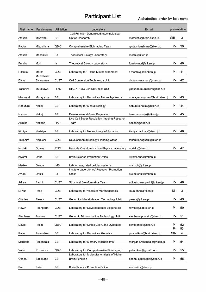
Participant List Alphabetical order by last name
First name Family name Affiliation Laboratory E-mail
Mohammad Abdolrahmani BSI Neural Circuits and Behavior [email protected]
Takaya Abe CLSTLaboratories for Animal Resource Developmentand Genetic Engineering [email protected] P- 1
Jiro Adachi QBiC Laboratory for cell-free protein synthesis [email protected] P- 2
Ayako Ajima BSI Lab for Neurobiology of Synapse [email protected] P- 3
Takaaki Aoki QBiC Science Communication Room [email protected]
Laurent Badel BSILaboratory for Circuit Mechanisms of SensoryPerception [email protected] P- 5
Shabeesh Balan BSI Laboratory for Molecular Psychiatry [email protected] P- 6
Kota Banzai CDB Laboratory for Growth Control Signaling [email protected] P- 7
Sufeng Chiang IMS Laboratory for Integrated Cellular Systems [email protected] P- 9
Yi Ding CDB Laboratory for Chromosome Segregation [email protected] P- 10
Sidonia Fagarasan IMS Laboratory for Mucosal Immunity [email protected] SI- 1
Emanuele Frandi BSI Lab for Neural Circuits and Behavior [email protected]
Yuki Furuichi IMS Laboratory for Skin Homeostasis [email protected]
SII-115
Arno GERMOND QBiC Laboratory for Comprehensive Bioimaing [email protected] P- 8
Shingo Gibo Ils Theoretical Biology Laboratory [email protected] P- 12
Alireza Goudarzi BSI Neural Computation and Adaptation [email protected]
Anne Guenther BSILab. Synaptic Molecules of MemoryPersistence [email protected]
Mitsutoshi Hanada BSI Laboratory for Marmoset Neural Architecture mitsutoshi.hanada@riken.jp P- 13
Takeshi Hanami CLSTGenetic Diagnosis Technology Unit and Ultra-Sensitive Biomolecule Detection Laboratory [email protected] P- 14
Carina Hanashima CDB Neocortical Development [email protected]
Yuichiro Hara CLST Phyloinformatics Unit [email protected] P- 15
Yoshie Harada Osaka UGuest SpeakerOsaka University
Yoshihide Hayashizaki PMIPreventive Medicine and Diagnosis InnovationProgram [email protected]
Linmeng He BSI Circuit and Behavioral Physiology [email protected]
Maxime Hebrard IMS Laboratory for Integrated Bioinformatics [email protected] P- 16
Ichiro Hiratani CDB Laboratory for Developmental Epigenetics [email protected] P- 17
Tomokatsu Ikawa IMS Laboratory for Immune Regeneration [email protected] P- 18
Takeshi Imai CDB Laboratory for Sensory Circuit Formation [email protected] SIII- 1
presentation
-46-
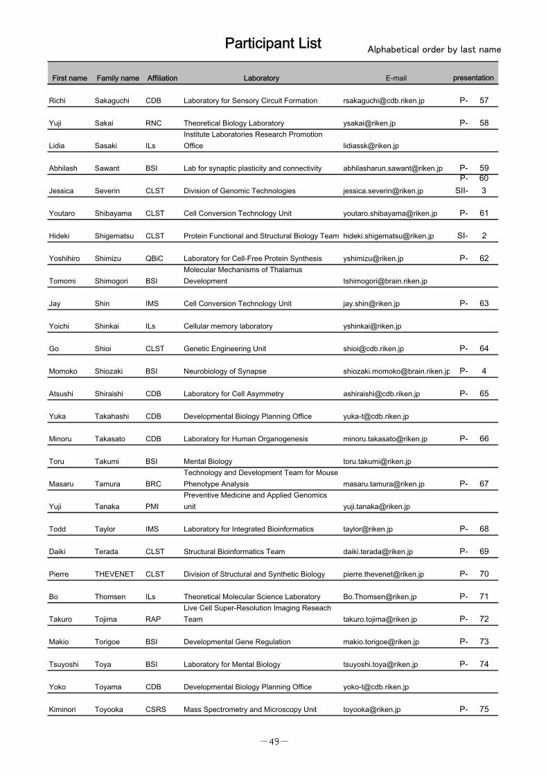
Participant List Alphabetical order by last name
First name Family name Affiliation Laboratory E-mail presentation
Yoko Ito RAPLive Cell Super-Resolution Imaging ResearchTeam [email protected] P- 19
Matej Janezic CLSTDivision of Structural and Synthetic Biology,Structural Bioinformatics Team [email protected] P- 20
Worapoj Jinda Laboratory for Integrated Bioinformatics [email protected]
Bogumil Kaczkowski CLST Division of Genomic Technologies [email protected] P- 21
Junichi Kaneshiro QBiC Laboratory for comprehensive bioimaging [email protected] P- 22
Takanori Kigawa QBiCLaboratory for Biomolecular Structure andDynamics [email protected]
Seok-Won Kim IMS Laboratory for Integrated Bioinformatics [email protected] P- 23
Yoshiaki Kita BSILaboratory for Molecular Mechanisms ofThalamus Development [email protected] P- 24
Toshimori Kitami IMS Young Chief Investigator Program [email protected] P- 25
Takuma Kobayashi BSI Laboratory for Developmental Gene Regulation [email protected] P- 26
Yasushi Kogo PMIPreventive Medicine and DiagnosisInnovation Program [email protected]
Tetsuya Koide BSI Laboratory for Neurobiology of Synapse [email protected]
SII-272
Shigeo Koyasu RIKEN [email protected]
Naveen Kumar IMS Laboratory for Integrated Bioinformatics [email protected] P- 28
Ashutosh Kumar CLSTStructural Bioinformatics Team, Division ofStructural and Synthetic Biology [email protected] P- 29
Vipin KUMAR QBiC Laboratory for Single Cell Gene Dynamics [email protected]
Kazuo Kurokawa RAPLive Cell Super-Resolution Imaging ResearchTeam [email protected] SII- 2
Lukasz Kusmierz BSILaboratory for Neural Computation andAdaptation [email protected]
Lynda Lamri CDB Laboratoy for Organismal Patterning [email protected] P- 31
Thomas Launey BSILab. for Synaptic Molecules of MemoryPersistence [email protected] P- 32
Simon LECLERC QBiC single cell Gene Dynamics [email protected]
Marcus Leiwe CDB Laboratory for Sensory Circuit Formation [email protected] P- 33
Chia-Wen Lin BSI Laboratory of Mental Biology [email protected] P- 34
DianaLlerenaSchiffmacher BSI Genetic Control of Neuronal Architecture
Takeshi Matsui IMS Laboratory for Skin Homeostasis [email protected] P- 35
Aki Minoda CLST Epigeoome Technology Exploration Unit [email protected] P- 36
Yu Miyamoto IMS Laboratory for integrated cellular systems [email protected] P- 37
Nobuhiko Miyasaka BSI Laboratory for Neurobiology of Synapse [email protected] P- 38
-47-
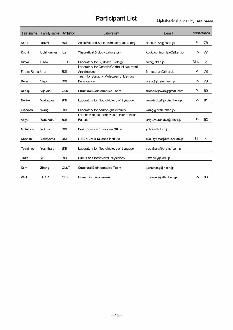
Participant List Alphabetical order by last name
First name Family name Affiliation Laboratory E-mail presentation
Atsushi Miyawaki BSI Cell Function Dynamics/BiotechnologicalOptics Research [email protected] SIII- 3
Ryota Mizushima QBiC Comprehensive Bioimaging Team [email protected] P- 39
Atsushi Mochizuki ILs Theoretical Biology Laboratory [email protected]
Fumito Mori Ils Theoretical Biology Laboratory [email protected] P- 40
Ritsuko Morita CDB Laboratory for Tissue Microenvironment [email protected] P- 41
DivyaMundackalSivaraman CLST Cell Conversion Technology Unit [email protected] P- 42
Yasuhiro Murakawa RInC RIKEN-HMC Clinical Omics Unit [email protected]
Masanori Murayama BSI Laboratory for Behavioral Neurophysiology [email protected] P- 43
Nobuhiro Nakai BSI Laboratory for Mental Biology [email protected] P- 44
Haruna Nakajo BSI Developmental Gene Regulation [email protected] P- 45
Akihiko Nakano RAPLive Cell Super-Resolution Imaging ResearchTeam [email protected]
Kimiya Narikiyo BSI Laboratory for Neurobiology of Synapse [email protected] P- 46
Takehiro Noguchi CDB Developmental Biology Planning Office [email protected]
Noriaki Ogawa RNC Hatsuda Quantum Hadron Physics Laboratory [email protected] P- 47
Kiyomi Ohno BSI Brain Science Promotion Office [email protected]
Mariko Okada IMS Lab for integrated cellular systems [email protected]
Ayumi Onuki ILsInstitute Laboratories' Research PromotionOffice [email protected]
Aditya Padhi CLST Structural Bioinformatics Team [email protected] P- 48
Li-Kun Phng CDB Laboratory for Vascular Morphogenesis [email protected] SI- 3
Charles Plessy CLST Genomics Miniaturization Technology UNit [email protected] P- 49
Rawin Poonperm CDB Laboratory for Developmental Epigenetics [email protected] P- 50
Stephane Poulain CLST Genomic Miniaturization Technology Unit [email protected] P- 51
David Priest QBiC Laboratory for Single Cell Gene Dynamics [email protected] P- 52
Pavel Prosselkov BSI Laboratory for Behavioral Genetics [email protected]
SII-534
Morgane Rosendale BSI Laboratory for Memory Mechanisms [email protected] P- 54
Yulia Rozanova QBiC Laboratory for Comprehensive Bioimaging [email protected] P- 55
Osamu Sadakane BSILaboratory for Molecular Analysis of HigherBrain Function [email protected] P- 56
Emi Saito BSI Brain Science Promotion Office [email protected]
-48-
Participant List Alphabetical order by last name
First name Family name Affiliation Laboratory E-mail presentation
Richi Sakaguchi CDB Laboratory for Sensory Circuit Formation [email protected] P- 57
Yuji Sakai RNC Theoretical Biology Laboratory [email protected] P- 58
Lidia Sasaki ILsInstitute Laboratories Research PromotionOffice [email protected]
Abhilash Sawant BSI Lab for synaptic plasticity and connectivity [email protected] P- 59
Jessica Severin CLST Division of Genomic Technologies [email protected]
SII-603
Youtaro Shibayama CLST Cell Conversion Technology Unit [email protected] P- 61
Hideki Shigematsu CLST Protein Functional and Structural Biology Team [email protected] SI- 2
Yoshihiro Shimizu QBiC Laboratory for Cell-Free Protein Synthesis [email protected] P- 62
Tomomi Shimogori BSIMolecular Mechanisms of ThalamusDevelopment [email protected]
Jay Shin IMS Cell Conversion Technology Unit [email protected] P- 63
Yoichi Shinkai ILs Cellular memory laboratory [email protected]
Go Shioi CLST Genetic Engineering Unit [email protected] P- 64
Momoko Shiozaki BSI Neurobiology of Synapse [email protected] P- 4
Atsushi Shiraishi CDB Laboratory for Cell Asymmetry [email protected] P- 65
Yuka Takahashi CDB Developmental Biology Planning Office [email protected]
Minoru Takasato CDB Laboratory for Human Organogenesis [email protected] P- 66
Toru Takumi BSI Mental Biology [email protected]
Masaru Tamura BRCTechnology and Development Team for MousePhenotype Analysis [email protected] P- 67
Yuji Tanaka PMIPreventive Medicine and Applied Genomicsunit [email protected]
Todd Taylor IMS Laboratory for Integrated Bioinformatics [email protected] P- 68
Daiki Terada CLST Structural Bioinformatics Team [email protected] P- 69
Pierre THEVENET CLST Division of Structural and Synthetic Biology [email protected] P- 70
Bo Thomsen ILs Theoretical Molecular Science Laboratory [email protected] P- 71
Takuro Tojima RAPLive Cell Super-Resolution Imaging ReseachTeam [email protected] P- 72
Makio Torigoe BSI Developmental Gene Regulation [email protected] P- 73
Tsuyoshi Toya BSI Laboratory for Mental Biology [email protected] P- 74
Yoko Toyama CDB Developmental Biology Planning Office [email protected]
Kiminori Toyooka CSRS Mass Spectrometry and Microscopy Unit [email protected] P- 75
-49-
Participant List Alphabetical order by last name
First name Family name Affiliation Laboratory E-mail presentation
Anna Truzzi BSI Affiliative and Social Behavior Laboratory [email protected] P- 76
Kouki Uchinomiya ILs Theoretical Biology Laboratory [email protected] P- 77
Hiroki Ueda QBiC Laboratory for Synthetic Biology [email protected] SIII- 2
Fatma Rabia Urun BSILaboratory for Genetic Control of NeuronalArchitecture [email protected] P- 78
Rejan Vigot BSITeam for Synaptic Molecules of MemoryPersistence [email protected] P- 79
Dileep Vijayan CLST Structural Bioinformatics Team [email protected] P- 80
Noriko Wakisaka BSI Laboratory for Neurobiology of Synapse [email protected] P- 81
Xiaowen Wang BSI Laboratory for neuron-glia circuitry [email protected]
Akiya Watakabe BSILab for Molecular analysis of Higher BrainFunction [email protected] P- 82
Motohide Yokota BSI Brain Science Promotion Office [email protected]
Charles Yokoyama BSI RIKEN Brain Science Institute [email protected] SI- 4
Yoshihiro Yoshihara BSI Laboratory for Neurobiology of Synapse [email protected]
Jinze Yu BSI Circuit and Behavioral Physiology [email protected]
Kam Zhang CLST Structural Bioinformatics Team [email protected]
WEI ZHAO CDB Human Organogenesis [email protected] P- 83
-50-