Embed Size (px)
Citation preview
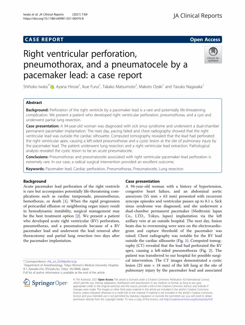
CASE REPORT Open Access
Right ventricular perforation,pneumothorax, and a pneumatocele by apacemaker lead: a case reportShihoko Iwata1* , Ayana Hirose1, Ikue Furui1, Takako Matsumoto2, Makoto Ozaki1 and Yasuko Nagasaka1
Abstract
Background: Perforation of the right ventricle by a pacemaker lead is a rare and potentially life-threateningcomplication. We present a patient who developed right ventricular perforation, pneumothorax, and a cyst andunderwent partial lung resection.
Case presentation: A 94-year-old woman was diagnosed with sick sinus syndrome and underwent a dual-chamberpermanent pacemaker implantation. The next day, pacing failed and chest radiography showed that the rightventricular lead was outside the cardiac silhouette. Computed tomography revealed that the lead had perforatedthe right ventricular apex, causing a left-sided pneumothorax and a cystic lesion at the site of pulmonary injury bythe pacemaker lead. The patient underwent lung resection and a right ventricular lead extraction. Pathologicalanalysis revealed the cystic lesion to be an acute pneumatocele.
Conclusions: Pneumothorax and pneumatocele associated with right ventricular pacemaker lead perforation isextremely rare. In our case, a radical surgical intervention provided an excellent outcome.
Keywords: Pacemaker lead, Cardiac perforation, Pneumothorax, Pneumatocele, Lung resection
BackgroundAcute pacemaker lead perforation of the right ventricleis rare but accompanies potentially life-threatening com-plications such as cardiac tamponade, pneumothorax,hemothorax, or death [1]. When the rapid progressionof pericardial effusion or neighboring organ injury resultin hemodynamic instability, surgical management maybe the best treatment option [2]. We present a patientwho developed acute right ventricular (RV) perforation,pneumothorax, and a pneumatocele because of a RVpacemaker lead and underwent the lead removal afterthoracotomy and partial lung resection two days afterthe pacemaker implantation.
Case presentationA 94-year-old woman with a history of hypertension,congestive heart failure, and an abdominal aorticaneurysm (55 mm × 63 mm) presented with recurrentsyncope episodes and ventricular pauses up to 8.1 s. Sicksinus syndrome was diagnosed, and she underwent adual-chamber permanent pacemaker (Medtronic JapanCo., LTD., Tokyo, Japan) implantation via the leftaxillary vein at an outside hospital. The next day, fusionbeats due to oversensing were seen on the electrocardio-gram and capture threshold of the pacemaker wasraised. Chest radiography was notable for the RV leadoutside the cardiac silhouette (Fig. 1). Computed tomog-raphy (CT) revealed that the lead had perforated the RVapex, causing a left-sided pneumothorax (Fig. 2). Thepatient was transferred to our hospital for possible surgi-cal intervention. The CT images demonstrated a cysticlesion (25 mm × 18 mm) of the left lung at the site ofpulmonary injury by the pacemaker lead and associated
© The Author(s). 2021 Open Access This article is licensed under a Creative Commons Attribution 4.0 International License,which permits use, sharing, adaptation, distribution and reproduction in any medium or format, as long as you giveappropriate credit to the original author(s) and the source, provide a link to the Creative Commons licence, and indicate ifchanges were made. The images or other third party material in this article are included in the article's Creative Commonslicence, unless indicated otherwise in a credit line to the material. If material is not included in the article's Creative Commonslicence and your intended use is not permitted by statutory regulation or exceeds the permitted use, you will need to obtainpermission directly from the copyright holder. To view a copy of this licence, visit http://creativecommons.org/licenses/by/4.0/.
* Correspondence: [email protected] of Anesthesiology, Tokyo Women’s Medical University Hospital,8-1, Kawada-cho, Shinjuku-ku, Tokyo 162-8666, JapanFull list of author information is available at the end of the article
Iwata et al. JA Clinical Reports (2021) 7:69 https://doi.org/10.1186/s40981-021-00470-8

pneumothorax (Fig. 2). The patient did not report anydyspnea and was hemodynamically stable; however, herSpO2 levels gradually decreased to 90% on room air andoxygen supplementation was started. A multidisciplinaryteam decided that immediate surgical intervention wasnecessary.The patient was transferred to the operating room.
Standard anesthesia monitoring was initiated, and arterialand central venous catheters were inserted. After induc-tion of anesthesia, a left-sided double-lumen endotrachealtube (Broncho-Cath®, Mallinckrodt Medical, Inc., St Louis,MO, USA) was used for intubation and maintenance of
anesthesia. One-lung ventilation was immediately startedto prevent tension pneumothorax. Transesophageal echo-cardiography (TEE; Philips iE33 Ultrasound System, Phi-lips Healthcare, Bothell, WA, USA) revealed twopacemaker leads; however, the location of the perforatedmyocardium was not identified. Mild aortic regurgitation,mild tricuspid regurgitation, mild reduction of left ven-tricular systolic motion, and a small pericardial effusionwere noted. The pacemaker (DDD) was reprogrammed toasynchronous mode (AOO) at a heart rate of 80 for theusage of unipolar electrocautery.Thoracotomy was performed at the left anterior 5th
intercostal space. Anatomical location of RV lead perforat-ing the apex, with a neighboring swollen area, was consist-ent with the CT images that presented with the areas ofperforation with pneumatocele (Fig. 3). To remove RVlead from the perforated RV apex, RV lead was detachedfrom the generator, and U shape suture with Prolene™ 4-0sutures (Ethicon Inc., Somerville, NJ, USA) was placedaround the site of perforation. The suture was reinforcedwith Prolene™ 4-0 sutures as the lead was extracted fromthe cardiac wall. Subsequently, the swollen portion of theinjured left upper lingular lung segment was resected toprevent worsening of pneumothorax or infection. A newRV lead placement was avoided because the patient dem-onstrated normal atrioventricular conduction. Pathologyof the resected specimen confirmed an aseptic pneumato-cele. The clinical course was uneventful. The patientreturned to the outside hospital in a stable condition onpostoperative day 9.
DiscussionWe report a rare case of RV perforation, pneumatoceleand pneumothorax after a pacemaker lead placement,requiring partial lung resection.
Fig. 1 A preoperative chest radiograph. Chest radiograph showingthe pacemaker lead perforating the right ventricle (yellow arrow)
Fig. 2 A preoperative chest computed tomography image. Thoraciccomputed tomography showing a left-sided pneumothorax (yellowarrow) and the pacemaker lead (yellow arrowheads) perforating theright ventricle and lung tissue neighboring a pulmonary cyst(red arrow)
Fig. 3 An intraoperative view of the left upper lobe. An operativeimage showing the perforation site (black arrow) in the left upperlingular segment, with swollen lung (yellow arrow) containing apulmonary cyst
Iwata et al. JA Clinical Reports (2021) 7:69 Page 2 of 4

RV perforation from pacemaker lead placement occursrarely (0.3–3%) [1, 3–5]. In contrast to subacuteperforation (24 h–1 month after implantation), acuteperforation (within 24 h after implantation) withhemodynamic compromise warrants immediate atten-tion. Surgery may be best chosen because of neighboringorgan injury, or hemodynamic compromise due to acutecardiac tamponade in the setting of perforated lead re-moval [2, 6]. On the other hand, in stable conditions,simple direct traction can be considered under closeechocardiographic monitoring and with a surgicalbackup [2, 3, 6]. Because the right heart is a low-pressure system, a perforation may be sealed by the leaditself and/or a combination of muscle contraction and fi-brosis over the lead, with minimum sequelae [3, 4].Strategies should depend on the dynamics of symp-
toms, pericardial effusion, hemodynamic status, and in-jured neighboring organs [2, 6].When the patient is pacing-dependent, lead extraction
should be followed by new lead placement in a differentlocation, preferably in the RV outflow tract or the intra-ventricular septum. In the case of open-chest surgery,the implantation of epicardial leads may be considered[2].RV lead replacement was avoided in our patient due
to patent atrioventricular conduction, considering thatthe AAI pacing can achieve a clinical outcome similar tothat of the DDD. Pneumothorax is a potential complica-tion of vascular access during a pacemaker implantation(0.2–3.87%) [1, 5], frequently seen within the first 24 hafter the implantation (1.3–3.87%) [5, 7]. On the otherhand, pacemaker lead penetrating the myocardium andcausing pneumothorax is rare and usually is found over24 h after pacemaker implantation [3].Generally, management of pneumothorax is guided by
the amount of air and patient’s hemodynamic status [8].A chest tube should be considered when the patient hasrespiratory distress, hemopneumothorax, or anypneumothorax larger than 20% of the hemithorax, irre-spective of the symptoms [8]. Our patient presented witha pneumothorax that involved almost 10% of the pleuralcavity.Pathological analysis of the resected lung specimen re-
vealed that the cystic space on CT imaging was a pneu-matocele. Pneumatocele is an air-filled cystic cavity inthe lungs, and frequently caused by severe pneumonia,blunt thoracic trauma, chronic obstructive pulmonarydisease, or hydrocarbon ingestion with aspiration [9–12].Although several mechanisms have been proposed forthe development of pulmonary pneumatocele, the exactreasons are unknown [9–11]. On the other hand, pneu-matoceles may occur when bronchial injury or inflam-mation creates a check-valve mechanism for air entryinto the lung parenchyma [9, 11, 12]. In general,
pneumatocele is a benign, self-limited condition thatrarely requires surgical intervention [13]. However, life-threatening tension pneumatocele with rapidenlargement can result in rupture and pneumothorax.Secondary infection may require surgical interventions[10, 12]. There are no well-established or widely ac-cepted treatment algorithms for pneumatocele [12, 13].Pneumatocele due to RV pacemaker lead perforation
has scarcely been reported, thus, standard of care re-mains to be determined. In the present case, surgery waschosen because of its potential for infection andpneumothorax, and surgery has better outcomes toavoid recurrent pneumothorax, compared with conser-vative treatment [14].
ConclusionsPneumothorax and pneumatocele associated with RVpacemaker lead perforation is extremely rare, therefore,the treatment is not well established. In our patient, aradical surgical intervention provided an excellentoutcome.
AbbreviationsRV: Right ventricular; CT: Computed tomography
AcknowledgementsThe authors thank Dr. Masahide Komagamine for his guidance regarding thecardiac surgical procedures.
Authors’ contributionsSI wrote and reviewed the manuscript. AH and IF helped in writing themanuscript and perioperative management of anesthesia. TM discussed thesurgical implications. MO and YN helped draft and review the manuscript. SIprepared the final draft of the manuscript. The authors read and approvedthe final manuscript.
FundingNone.
Availability of data and materialsNot applicable.
Declarations
Ethics approval and consent to participateNot applicable.
Consent for publicationWritten informed consent for publication of the clinical details was obtainedfrom the patient and her brother.
Competing interestsThe authors declare that they have no competing interests.
Author details1Department of Anesthesiology, Tokyo Women’s Medical University Hospital,8-1, Kawada-cho, Shinjuku-ku, Tokyo 162-8666, Japan. 2Department ofThoracic Surgery, Tokyo Women’s Medical University Medical Center East,2-1-10, Nishiogu, Arakawa-ku, Tokyo 116-8567, Japan.
Iwata et al. JA Clinical Reports (2021) 7:69 Page 3 of 4

Received: 5 July 2021 Revised: 13 August 2021Accepted: 18 August 2021
References1. Leong KMW, Pollard C, Cooke CJ. Cardiology registrars and permanent
pacemaker complication rates in a district general hospital - safety andservice implications. Clin Med (Lond). 2014;14(1):34–7. https://doi.org/10.7861/clinmedicine.14-1-34.
2. Banaszewski M, Stępińska J. Right heart perforation by pacemaker leads.Arch Med Sci. 2012;1(1):11–3. https://doi.org/10.5114/aoms.2012.27273.
3. Mori H, Kato R, Ikeda Y, Tsutsui K, Hasegawa S, Tanaka S, et al. Percutaneoussimple lead traction is a feasible and effective method for right ventricularlead perforations. Int Heart J. 2020;61(1):54–9. https://doi.org/10.1536/ihj.19-326.
4. Hirschl DA, Jain VR, Spindola-Franco H, Gross JN, Haramati LB. Prevalenceand characterization of asymptomatic pacemaker and ICD lead perforationon CT. Pacing Clin Electrophysiol. 2007;30(1):28–32. https://doi.org/10.1111/j.1540-8159.2007.00575.x.
5. Carrión-Camacho MR, Marín-León I, Molina-Doñoro JM, González-López JR.Safety of permanent pacemaker implantation: a prospective study. J ClinMed. 2019;8(1):35. https://doi.org/10.3390/jcm8010035.
6. Polin GM, Zado E, Nayak H, Cooper JM, Russo AM, Dixit S, et al. Propermanagement of pericardial tamponade as a late complication ofimplantable cardiac device placement. Am J Cardiol. 2006;98(2):223–5.https://doi.org/10.1016/j.amjcard.2006.01.078.
7. Ogunbayo GO, Charnigo R, Darrat Y, Morales G, Kotter J, Olorunfemi O, et al.Incidence, predictors, and outcomes associated with pneumothorax duringcardiac electronic device implantation: a 16-year review in over 3.7 millionpatients. Heart Rhythm. 2017;14(12):1764–70. https://doi.org/10.1016/j.hrthm.2017.07.024.
8. Tschopp JM, Bintcliffe O, Astoul P, Canalis E, Driesen P, Janssen J, et al. ERStask force statement: diagnosis and treatment of primary spontaneouspneumothorax. Eur Respir J. 2015;46(2):321–35. https://doi.org/10.1183/09031936.00219214.
9. Quigley MJ, Fraser RS. Pulmonary pneumatocele: pathology andpathogenesis. AJR Am J Roentgenol. 1998;150:1275–7.
10. Sugimura A, Takahashi T, Sekihara K, Nagasaka S. Case of rapid formation ofintraoperative pulmonary pneumatocele after lobectomy. Ann Thorac Surg.2020;110(4):e331–2. https://doi.org/10.1016/j.athoracsur.2020.03.028.
11. Raoof S, Bondalapati P, Vydyula R, Ryu JH, Gupta N, Raoof S, et al. Cysticlung diseases: algorithmic approach. Chest. 2016;150(4):945–65. https://doi.org/10.1016/j.chest.2016.04.026.
12. DiBardino DJ, Espada R, Seu P, Goss JA. Management of complicatedpneumatocele. J Thorac Cardiovasc Surg. 2003;126(3):859–61. https://doi.org/10.1016/S0022-5223(03)00367-2.
13. Ulutas H, Celik MR, Ozgel M, Soysal O, Kuzucu A. Pulmonary pseudocystsecondary to blunt or penetrating chest trauma: clinical course anddiagnostic issues. Eur J Trauma Emerg Surg. 2015;41(2):181–8. https://doi.org/10.1007/s00068-014-0427-2.
14. Olesen WH, Katballe N, Sindby JE, Titlestad IL, Andersen PE, Lindahl-Jacobsen R, et al. Surgical treatment versus conventional chest tubedrainage in primary spontaneous pneumothorax: a randomized controlledtrial. Eur J Cardiothorac Surg. 2018;54(1):113–21. https://doi.org/10.1093/ejcts/ezy003.
Publisher’s NoteSpringer Nature remains neutral with regard to jurisdictional claims inpublished maps and institutional affiliations.
Iwata et al. JA Clinical Reports (2021) 7:69 Page 4 of 4