Embed Size (px)
Citation preview

1
Emergency Radiology, 1st Nordic CourseGothenburg, Sweden, May, 2007
Right Lower Quadrant PainRight Lower Quadrant Pain
Robert A. Novelline, MDProfessor of Radiology, Harvard Medical School
Director of Emergency Radiology,Massachusetts General Hospital
Differential Diagnosis: RLQ Pain
GastrointestinalAppendicitisDiverticulitisEpiploic AppendagitisSegmental Omental InfarctionIleocolitisMesenteric AdenitisCecal CarcinomaCrohn DiseaseMeckel DiverticulitisCholecystitis
GynecologicalHemorrhagic Ovarian CystRuptured Ovarian CystEctopic PregnancyAdnexal TorsionPelvic Inflammatory Disease
with Tubo-Ovarian AbscessDegenerating Uterine
Leiomyoma
UrologicalNephrolithiasisPyelonephritisHydronephrosis
2
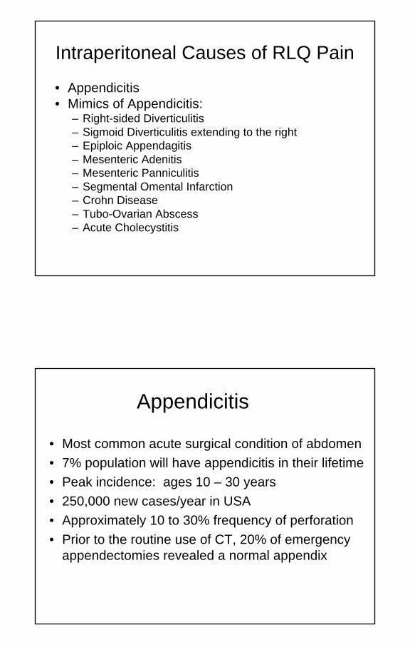
Intraperitoneal Causes of RLQ Pain
• Appendicitis• Mimics of Appendicitis:
– Right-sided Diverticulitis– Sigmoid Diverticulitis extending to the right– Epiploic Appendagitis – Mesenteric Adenitis– Mesenteric Panniculitis– Segmental Omental Infarction– Crohn Disease– Tubo-Ovarian Abscess– Acute Cholecystitis
Appendicitis
• Most common acute surgical condition of abdomen• 7% population will have appendicitis in their lifetime• Peak incidence: ages 10 – 30 years• 250,000 new cases/year in USA• Approximately 10 to 30% frequency of perforation• Prior to the routine use of CT, 20% of emergency
appendectomies revealed a normal appendix

3
Presenting Signs and Symptoms of Acute Appendicitis
• RLQ pain and/or tenderness 96%• Duration of symptoms < 5 days 80%• WBC > 10,000/mm3 66%• Temperature > 37.5°C (99.5°F) 63%• Nausea 62%• Vomiting 32%• Rebound tenderness 26%• Anorexia 24%• RLQ guarding 21%
MGH MDCT Protocol for AppendicitisRectal Contrast; Limited MDCT Scan
• Rectal Contrast: – 40cc of 60% contrast in 1000cc saline
• IV Contrast: – 75-125cc of 370 concentration @ 3.0 cc/sec
• MDCT Scan Protocol– Scan after 150 sec delay– Scan from L3 to acetabular roof (reduce radiation)– View slices at 2.5mm thickness– If reformations needed:
• 16-slice: 1.25mm at 1.00 spacing• 64-slide: Contiguous 0.625mm

4
Colon ContrastAdvantages
• Faster• Normal appendix fills
better• Cecal apical changes
seen better due to cecal distention
• Higher reported accuracy• Greater interpreter
confidence
Avoids
• Delays• Nauseated patients do
not wish to drink large amounts of contrast
• General anesthesia problems after CT
MGH MDCT Protocol for AppendicitisRectal Contrast; Limited MDCT Scan
• Option: if appendix not seen well on axials:– Coronal and saggital reformations– Decubitus scans:
• Left side down if cecum well-opacified• Right side down with more rectal contrast material
if cecum not well-opacified
• Option: if no appendicitis seen and no alternative diagnosis identified– Extend scan to full abdomen

5
Finding the Appendix at CT
• Find ileocecal valve as a landmark (40% of patients reflux into terminal ileum)
• Origin of appendix is 2 - 3 cm caudal to valve, and usually posteromedial
• Location: variable• Follow appendix to its blind-ending tip
Normal Appendix at CT1
Normal filled with contrast Normal filled with air
Normal filled with contrast Appendicitis
6
CT Signs of Appendicitis
• Focal apical thickening• Arrowhead sign• Cecal bar
• Fat stranding• Fluid • Phlegmon• Extraluminal air bubbles• Abscess• Adenopathy
• Diameter >6mm• Fails to completely fill with contrast• Appendoliths• Wall thickening • Wall enhancement with IV contrast
Cecal Apical Changes
Periappendiceal Inflammation
Abnormal Appendix
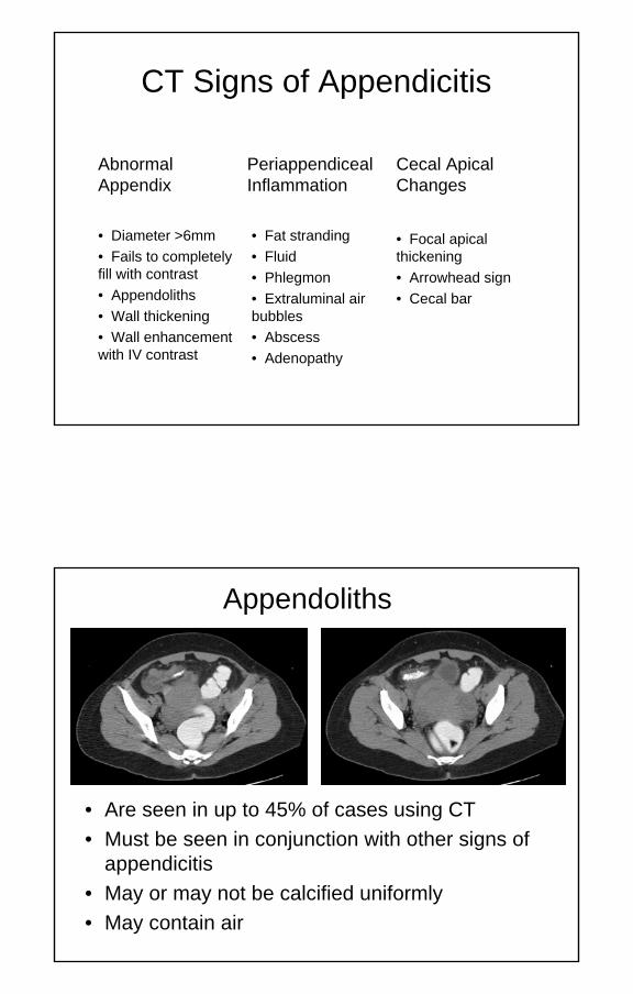
Appendoliths
• Are seen in up to 45% of cases using CT• Must be seen in conjunction with other signs of
appendicitis• May or may not be calcified uniformly• May contain air

7
Peri-Appendiceal Inflammation
• In soft tissues surrounding or adjacent to appendix see fat stranding, fluid, phlegmon, extraluminal air bubbles, abscess or adenopathy
• Adenopathy usually present with appendicitis– located anterior to psoas just cephalad to origin of
appendix; also see with mesenteric adenitis• Phlegmon (inflamed soft tissue mass) may prevent
visualization of abnormal appendix– Value of IV contrast material
• With phlegmon diagnosis appendicitis based on:– Appendoliths– Cecal apical changes specific for appendicitis
Cecal Apical Findings
1. Focal cecal apical thickening
2. Arrowhead sign
3. Cecal bar

8
Cecal Apical Findings1. Focal cecal apical thickening
– Caused by spread of inflammation and edema into wall of cecum
– Complete filling of cecum with contrast is needed to visualize this sign
– The sign is frequent and pathognomonic for appendicitis• Focal wall thickening from diverticulitis is
centered at the diverticulum, not the cecal apex
Cecal Apical Findings2. Arrowhead Sign
– Inflammatory thickening of cecal apex with contrast funneling into center of inflammation
– Visualization depends on CT slice coinciding with position of arrowhead
– Arrowhead points toward appendix Appendolith
9
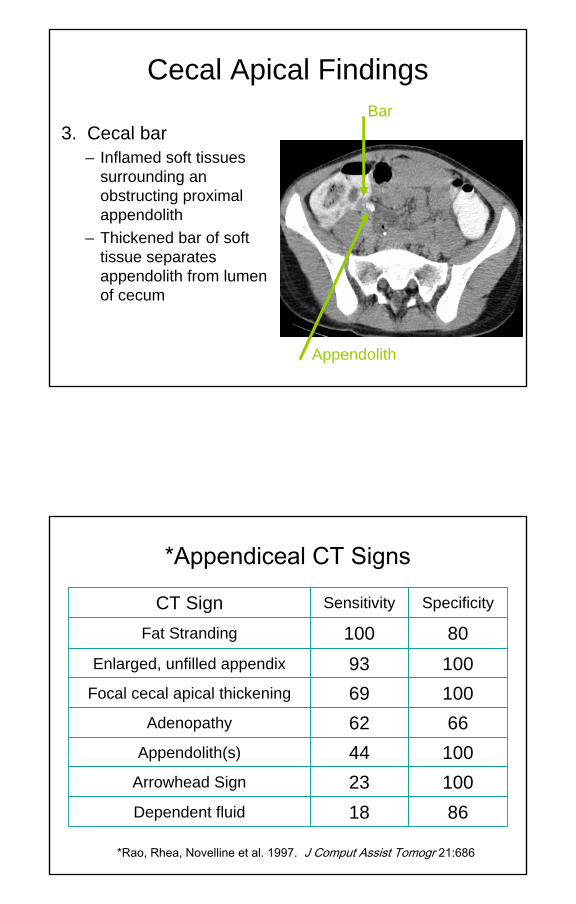
Cecal Apical Findings
3. Cecal bar– Inflamed soft tissues
surrounding an obstructing proximal appendolith
– Thickened bar of soft tissue separates appendolith from lumen of cecum
Bar
Appendolith
*Appendiceal CT Signs
8618Dependent fluid
10023Arrowhead Sign
10044Appendolith(s)
6662Adenopathy
10069Focal cecal apical thickening
10093Enlarged, unfilled appendix
80100Fat Stranding
SpecificitySensitivityCT Sign
*Rao, Rhea, Novelline et al. 1997. J Comput Assist Tomogr 21:686

10
10011Abscess
10010Cecal Bar
910Diffuse cecal wall thickening
980Focal cecal wall thickening953Sigmoid wall thickening863Terminal ileal wall thickening997Phlegmon978Extraluminal air
SpecificitySensitivityCT Sign
*Rao, Rhea, Novelline et al. 1997. J Comput Assist Tomogr 21:686
*Appendiceal CT Signs
Appendicitis in Pregnancy
• Imaging Options– Ultrasound– Limited CT scan– MR scan

11
*Distal (Tip) Appendicitis
• Appendicitis usually results from luminal obstruction at the appendiceal orifice
• When lumen is obstructed distal to orifice the resulting condition is “distal/tip appendicitis”
• Lumen of proximal appendix may opacify and appear normal resulting in a false negative diagnosis of appendicitis
• This condition is readily recognized by CT
*Rao PM, Rhea JT, Novelline RA. Distal appendicitis: CT appearance and diagnosis. Radiology. 204:709-712, 1997.
Right-Sided Diverticulitis
• Diverticulitis may produce RLQ pain when it involves the cecum and right colon, or when sigmoid diverticulitis extends to the right of midline
• Combination of RLQ pain, fever and leukocytosis mimics appendicitis
• Look for the normal appendix at CT

12
Mesenteric AdenitisTwo Types:• Primary Mesenteric Adenitis
– Adenitis without an acute inflammatory process– May see mild thickening of the terminal ileum– Self-limited process that affects lymph nodes in the RLQ– Non-surgical condition, more common under 15 years– Presentation may mimic appendicitis– Etiology: infection viral pathogens, sometimes other organisms– Incidence in patients who have CT for suspected appendicitis– 12% adenitis as isolated finding– 6% adenitis plus ileum wall thickening
• Secondary Mesenteric Adenitis– Adenopathy with Crohn Disease, appendicitis, diverticulitis, or
neoplasms such a lymphoma and carcinoma
CT Findings inMesenteric Adenitis
• Enlarged (>5 mm shortest dimension) and clustered (3 or more) mesenteric nodes
• Location in RLQ– Anterior to psoas– Small bowel mesentery
• Adenitis may be seen with small bowel wall thickening (>3 mm)

13
Mesenteric Panniculitis
• Non-specific inflammatory and fibrotic process affecting fatty tissues of mesentery
• If predominately fibrosis called “retractile mesenteritis”• Rare, most cases idiopathic• Peak incidence sixth and seventh decades• More common in males• Present with abdominal pain, fever, nausea, vomiting• Exam may reveal abdominal tenderness, palpable mass• Symptoms may persist for a year or more
CT Findings inMesenteric Panniculitis
• CT findings may vary:– Well-defined soft tissue mesentery mass– Ill-defined areas of higher attenuation in the
mesenteric fat representing inflammation and fibrosis• Abnormality often surrounds SMA and SMV• No involvement of adjacent bowel• When the process is focal it may mimic a
teratomatous or liposarcomatous tumor

14
Segmental Omental Infarction
• Rare, 300 cases in literature• Etiology of infarction
– Necrosis caused by interruption of blood supply to the omentum from torsion or venous thrombosis, more common on right side
– Primary (idiopathic) or Secondary torsion (adhesions, neoplasm, trauma, surgery, hernia)
– Primary may be precipitated by coughing, straining, overeating• Clinical presentation
– Sudden, severe abdominal pain, RLQ or peri-umbilical tenderness, may have fever
• Differential Diagnosis– Appendicitis, diverticulitis, cholecystitis – Metastases, liposarcoma, secondarily inflamed fat
CT Findings inSegmental Omental Infarction
• CT shows an abdominal mass (ovoid, cake-like) of fat stranding or fat with dense streaks representing folds within the mass
• Location is superficial and paraumbilical – Between rectus abdominis and colon or small bowel– Right-sided more than left-sided (right-sided
segmental omental infarction)• Complications: necrosis and abscess

15
Crohn Disease
• Inflammatory disease of the distal small bowel which may involve cecum and colon
• May be associated with secondary appendicitis
Tubo-Ovarian Abscess (TOA)
• Advanced form pelvic inflammatory disease (PID)• Often caused by Chlamydia trachomatis and
Neisseria gonorrhoeae• Infection of ovary and fallopian tube with
hydrosalpinx and collection of pus• Present with pain, fever, vaginal discharge• Usually diagnosed by ultrasound• CT may show a complex cystic mass representing
dilated tubes and the TOA

16
Acute Cholecystitis
• In older patients, acute cholecystitis may occasionally present with right-sided abdominal pain mimicking appendicitis
• May have fever and elevated white blood cells• CT may show: gallbladder distention, mural
thickening, wall enhancement, wall irregularity, intraluminal membranes, pericholecystic stranding, pericholecystic fluid, gallstones, gas in the gallbladder wall or in the gallbladder lumen
Summary: Intra-Abdominal Causes of RLQ Pain
• Appendicitis• Mimics of Appendicitis:
– Right-sided Diverticulitis– Epiploic Appendagitis – Mesenteric Adenitis– Mesenteric Panniculitis– Segmental Omental Infarction– Tubo-Ovarian Abscess– Acute Cholecystitis

17
Robert A. Novelline, MDProfessor of Radiology, Harvard Medical School
Director of Emergency Radiology,Massachusetts General Hospital