Embed Size (px)
Citation preview
International Wound Journal ISSN 1742-4801
O R I G I N A L A R T I C L E
Rigenera protocol in the treatment of surgical wounddehiscenceMarco Marcarelli1, Letizia Trovato2, Elvio Novarese1, Michele Riccio3 & Antonio Graziano4
1 Santa Croce Hospital, Unit of Orthopedics and Traumatology of Chieri and Moncalieri, Turin, Italy2 Human Brain Wave srl, Turin, Italy3 Plastic and Reconstructive Surgery, AOU “Ospedali Riuniti”, Ancona, Italy4 SHRO Center of Biotechnology, Temple University, Philadelphia, PA, USA
Key words
Autologous; Dehiscence; Micro-grafts;Tissue regeneration; Wound healing
Correspondence to
L Trovato, PhDHuman Brain WaveCorso Galileo Ferraris 6310128 TurinItalyE-mail: [email protected]
Marcarelli M, Trovato L, Novarese E, Riccio M, Graziano A. Rigenera protocol in thetreatment of surgical wound dehiscence. Int Wound J 2016; doi: 10.1111/iwj.12601
Abstract
The effective management of post-operative wounds is important to prevent potentialcomplications such as surgical-site infections and wound dehiscence. The purpose ofthis study was to treat wound dehiscence in elderly patients who were subjected toorthopaedic surgical interventions. The dehisced wounds were treated with autologousmicro-grafts obtained using a promising CE-certified medical device called Rigener-acons. This instrument is a biological disruptor of human tissues able to specificallyselect progenitor cells that, as already reported in previous studies, maintain high cellviability but mainly have a high regenerative potential, allowing the repair of damagedtissues. Autologous micro-grafts obtained by Rigeneracons are ready to use and can beapplied alone or in combination with biological scaffolds directly on the injured area.We observed in our patients a complete remission of dehisced wounds, on average, after30 days from micro-grafts application and a total wound re-epithelialisation after 1 yearfrom the surgical intervention. In conclusion, although we reported only three patients,autologous micro-grafts can be considered a promising approach for the treatment ofdehisced wounds, improving the wound-healing process and in general the patient’squality of life without using other dressings.
Introduction
The effective management of post-operative wounds is impor-tant to prevent potential complications such as surgical-siteinfections and wound dehiscence. Moreover, this aspectremains today a big challenge for surgeons and patientswho often experience high comorbidity correlated to along and exhausting wound-healing process (1). The useof post-operative drains and the type of post-operative dressingis at the discretion of the surgeon, with no available clinicalguidelines. The principal aim of drains is theoretically todecrease the incidence of post-operative haematoma, whileocclusive dressings maintain a sterile barrier for longer timeperiods post-operatively (2).
Post-surgical wound dehiscence can arise as a complicationin different type of interventions, such as the transplantation oflung (3) and kidney (4), colon resection (5), the following ofa laparotomy procedure with an incidence of about 0.5% (6)and gynaecological procedures, such as Caesarean section andtotal abdominal hysterectomy with an incidence of 15% (7).
Orthopaedic surgical complications can lead to longer hospi-talization, increased patient morbidity and high costs for bothpatients and national health services. Several studies reportedthe association between wound closure method and woundcomplications, but the correlation between comorbidities, riskfactors and surgical wound dehiscence is not still well defined(8).
Key Messages
• successful wound healing depends on several factors,including optimal wound closure; in several and differentprimary surgical procedures, wound closure does notoften occur, leading to the onset of wound complicationssuch as wound dehiscence influencing the quality oflife of the patients and increasing both hospitalisation
© 2016 Medicalhelplines.com Inc and John Wiley & Sons Ltd doi: 10.1111/iwj.12601 1
Rigenera protocol and dehisced wounds M. Marcarelli et al.
and economic costs for patients and the National HealthService
• the goal of our study was to display the efficacy of autol-ogous micro-grafts to treat wound dehiscence appearingafter primary orthopaedic interventions in three elderlypatients; autologous micro-grafts provided by Rigenera-cons are enriched with progenitor cells and ready to usein the same patient from whom they were obtained
• after the application of autologous micro-grafts, weobserved a complete remission of wound dehiscence, onaverage, 1 month after the micro-grafts injection, with atotal wound re-epithelialisation 1 year after the primarysurgical intervention
All wounds heal for primary, secondary or tertiary inten-tion, and several factors can delay wound healing, includingchronic disease, vascular insufficiency, diabetes, neurologicaldefects, nutritional deficiencies, advanced age and local factorssuch as pressure, infection and oedema. Effective wound carerequires an accurate and detailed identification of the specificfactors interfering with wound healing in order to auspicate theachievement of an optimal outcome (9). In addition, a preoper-ative assessment can be appropriate to prevent or limit the riskfactors responsible for wound dehiscence incidence (6).
Furthermore, dehisced wounds, similar to chronic wounds,are also becoming a socioeconomic emergency because ofthe high costs of hospitalisation to care for the patients (10).Actually, re-operation is the elective approach for dehiscedwounds mainly in abdominal interventions, but this approachcan lead to a high mortality rate, which was estimated to bebetween 14% and 50% (11).
Several other strategies can improve the management ofthese wound complications, including negative pressure woundtherapy (NPWT), silver ion-impregnated dressing or the useof topical growth factors such as platelet-derived growth fac-tor (PDGF), fibroblast growth factor, transforming growthfactor-beta and epidermal growth factor (12). The benefits ofall these strategies were widely reported in the literature andcertainly have ameliorated the clinical approach to wound com-plications, leading to reduction of both costs and number ofsurgical interventions to close the wound.
Based on these considerations, the aim of this study wasto display a new promising clinical protocol, named Rigen-era protocol, in the management of post-surgical dehiscence inpatients who underwent primary orthopaedic surgical interven-tions. This new approach is based on the application of autol-ogous micro-grafts directly on the site of dehiscence, and theefficacy of this approach was already shown in other clinicalfields such as dentistry (13,14), dermatology (15) and in themanagement of other post-operative wounds (16).
Patients and methods
Patients
We analysed in this study three patients enrolled in the Unitof Ortopedics and Traumatoly of Santa Croce Hospital (Turin,
Italy) in August 2014 and followed-up these patients for about1 year. All patients provided informed consent, and the studyprotocol conformed to the ethical guidelines of the 1975 Dec-laration of Helsinki. The primary diagnosis and initial operativeprocedures leading to wound dehiscence are listed in Table 1.We also reported the correlated diseases affecting the patients,which, from a clinical point of view, are all characterised bydelays in healing greater than 7 weeks.
Methods
All cases reported in this study were treated in ambulatorywithout the need of a recovery or operating room, and we usedthe Rigenera protocol on all the patients, which allows theregeneration of damaged human tissues, as already reported inprevious studies (16,17). The Rigenera protocol is based on theuse of the Rigenera machine and Rigeneracons (Human BrainWave, Turin, Italy), a biological disruptor able to disaggregatesmall pieces of human connective tissues and select a specificcell population that includes progenitor cells, maintaining thecapacity to differentiate into several cell types and then regen-erate a damaged tissue. These progenitor cells, in associationwith growth factors derived by starting tissue, create autologousmicro-grafts that are ready to use, which can be applied on theinjured area alone or in combination with different biologicalscaffolds, such as collagen.
The Rigenera protocol consists of four steps: (i) collectionof a small piece of skin tissue of 1 cm from a distant donor sitewith respect to the recipient site, (ii) disaggregation of tissueby Rigeneracons through the addition of 1 ml of sterile salinesolution, (iii) collection of autologous micro-grafts obtainedafter the disaggregation and (iv) injection of these micro-graftsalone into the site of injury by perilesional infiltrations or incombination with scaffolds embedded with micro-grafts.
In our patients, we collected small pieces of tissue bytrochanteric region after local anaesthesia, and we disag-gregated the tissue for 90–120 seconds and used an equinecollagen sponge as a scaffold to apply the micro-grafts onwound dehiscence. Following the application of micro-grafts,we performed the first dressing after 7 days and subjectedthe patients to weekly controls to evaluate the progression ofwound healing.
Results
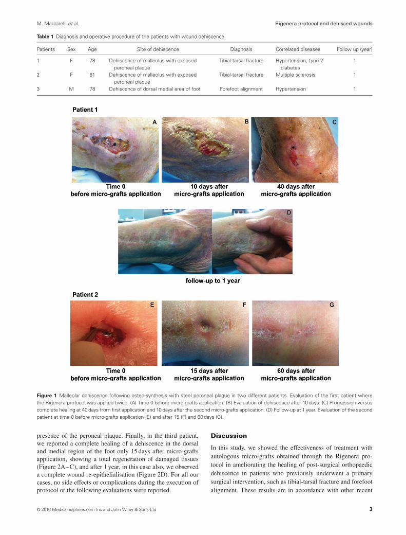
In all three patients, we observed, on average, a good remissionof wound dehiscence 1 month after the micro-grafts applica-tion, with a range variable between 15 and 60 days. In general,the micro-grafts were applied only once, except in patient 1,where the Rigenera protocol was applied twice after 30 daysfrom the first application. In Figure 1, we display the healing ofa malleolar dehiscence after 40 days from the first micro-graftsinjection or 10 days after the second micro-grafts application(Figure 1A–C), and after 1 year, we can observed a completewound re-epithelialisation (Figure 1D). In the second patient,we performed the Rigenera protocol only once, observing thehealing of dehiscence after 60 days (Figure 1E–G). This differ-ence in the time of dehiscence healing can be attributed to the
2 © 2016 Medicalhelplines.com Inc and John Wiley & Sons Ltd
M. Marcarelli et al. Rigenera protocol and dehisced wounds
Table 1 Diagnosis and operative procedure of the patients with wound dehiscence
Patients Sex Age Site of dehiscence Diagnosis Correlated diseases Follow up (year)
1 F 78 Dehiscence of malleolus with exposedperoneal plaque
Tibial-tarsal fracture Hypertension, type 2diabetes
1
2 F 61 Dehiscence of malleolus with exposedperoneal plaque
Tibial-tarsal fracture Multiple sclerosis 1
3 M 78 Dehiscence of dorsal medial area of foot Forefoot alignment Hypertension 1
Figure 1 Malleolar dehiscence following osteo-synthesis with steel peroneal plaque in two different patients. Evaluation of the first patient wherethe Rigenera protocol was applied twice. (A) Time 0 before micro-grafts application. (B) Evaluation of dehiscence after 10 days. (C) Progression versuscomplete healing at 40 days from first application and 10 days after the second micro-grafts application. (D) Follow-up at 1 year. Evaluation of the secondpatient at time 0 before micro-grafts application (E) and after 15 (F) and 60 days (G).
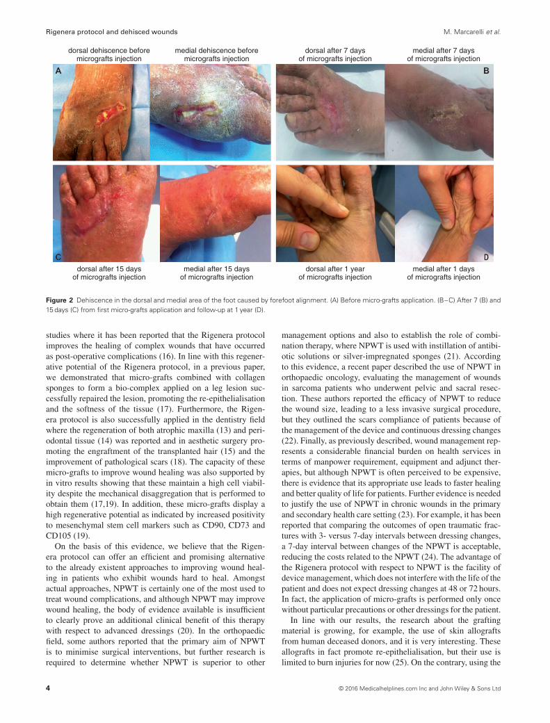
presence of the peroneal plaque. Finally, in the third patient,we reported a complete healing of a dehiscence in the dorsaland medial region of the foot only 15 days after micro-graftsapplication, showing a total regeneration of damaged tissues(Figure 2A–C), and after 1 year, in this case also, we observeda complete wound re-epithelialisation (Figure 2D). For all ourcases, no side effects or complications during the execution ofprotocol or the following evaluations were reported.
Discussion
In this study, we showed the effectiveness of treatment withautologous micro-grafts obtained through the Rigenera pro-tocol in ameliorating the healing of post-surgical orthopaedicdehiscence in patients who previously underwent a primarysurgical intervention, such as tibial-tarsal fracture and forefootalignment. These results are in accordance with other recent
© 2016 Medicalhelplines.com Inc and John Wiley & Sons Ltd 3
Rigenera protocol and dehisced wounds M. Marcarelli et al.
dorsal dehiscence beforemicrografts injection
medial dehiscence beforemicrografts injection
dorsal after 7 daysof micrografts injection
medial after 7 daysof micrografts injection
dorsal after 15 daysof micrografts injection
medial after 15 daysof micrografts injection
dorsal after 1 yearof micrografts injection
medial after 1 daysof micrografts injection
Figure 2 Dehiscence in the dorsal and medial area of the foot caused by forefoot alignment. (A) Before micro-grafts application. (B–C) After 7 (B) and15 days (C) from first micro-grafts application and follow-up at 1 year (D).
studies where it has been reported that the Rigenera protocolimproves the healing of complex wounds that have occurredas post-operative complications (16). In line with this regener-ative potential of the Rigenera protocol, in a previous paper,we demonstrated that micro-grafts combined with collagensponges to form a bio-complex applied on a leg lesion suc-cessfully repaired the lesion, promoting the re-epithelialisationand the softness of the tissue (17). Furthermore, the Rigen-era protocol is also successfully applied in the dentistry fieldwhere the regeneration of both atrophic maxilla (13) and peri-odontal tissue (14) was reported and in aesthetic surgery pro-moting the engraftment of the transplanted hair (15) and theimprovement of pathological scars (18). The capacity of thesemicro-grafts to improve wound healing was also supported byin vitro results showing that these maintain a high cell viabil-ity despite the mechanical disaggregation that is performed toobtain them (17,19). In addition, these micro-grafts display ahigh regenerative potential as indicated by increased positivityto mesenchymal stem cell markers such as CD90, CD73 andCD105 (19).
On the basis of this evidence, we believe that the Rigen-era protocol can offer an efficient and promising alternativeto the already existent approaches to improving wound heal-ing in patients who exhibit wounds hard to heal. Amongstactual approaches, NPWT is certainly one of the most used totreat wound complications, and although NPWT may improvewound healing, the body of evidence available is insufficientto clearly prove an additional clinical benefit of this therapywith respect to advanced dressings (20). In the orthopaedicfield, some authors reported that the primary aim of NPWTis to minimise surgical interventions, but further research isrequired to determine whether NPWT is superior to other
management options and also to establish the role of combi-nation therapy, where NPWT is used with instillation of antibi-otic solutions or silver-impregnated sponges (21). Accordingto this evidence, a recent paper described the use of NPWT inorthopaedic oncology, evaluating the management of woundsin sarcoma patients who underwent pelvic and sacral resec-tion. These authors reported the efficacy of NPWT to reducethe wound size, leading to a less invasive surgical procedure,but they outlined the scars compliance of patients because ofthe management of the device and continuous dressing changes(22). Finally, as previously described, wound management rep-resents a considerable financial burden on health services interms of manpower requirement, equipment and adjunct ther-apies, but although NPWT is often perceived to be expensive,there is evidence that its appropriate use leads to faster healingand better quality of life for patients. Further evidence is neededto justify the use of NPWT in chronic wounds in the primaryand secondary health care setting (23). For example, it has beenreported that comparing the outcomes of open traumatic frac-tures with 3- versus 7-day intervals between dressing changes,a 7-day interval between changes of the NPWT is acceptable,reducing the costs related to the NPWT (24). The advantage ofthe Rigenera protocol with respect to NPWT is the facility ofdevice management, which does not interfere with the life of thepatient and does not expect dressing changes at 48 or 72 hours.In fact, the application of micro-grafts is performed only oncewithout particular precautions or other dressings for the patient.
In line with our results, the research about the graftingmaterial is growing, for example, the use of skin allograftsfrom human deceased donors, and it is very interesting. Theseallografts in fact promote re-epithelialisation, but their use islimited to burn injuries for now (25). On the contrary, using the
4 © 2016 Medicalhelplines.com Inc and John Wiley & Sons Ltd
M. Marcarelli et al. Rigenera protocol and dehisced wounds
Rigenera protocol, we can create autologous micro-grafts thatare ready to use from the same patient, who, therefore, is boththe donor and acceptor of these micro-grafts.
Usually, post-operative dehiscences with underlying instru-mentations are treated by removing them, but in our case, thepremature removal of instrumentations could have caused fur-ther interventions accompanied by a longer time of hospital-isation and wound healing. The use of the Rigenera protocolin the malleolar dehiscence with exposure of peroneal plaqueallow us to promote tissue regeneration, accelerating the woundhealing and allowing the removal of the plaque 1 year after thefirst intervention following a same incision, without meetingany inconvenience or complication for the patients.
In conclusion, our results, although not yet significantbecause of the number of treated patients, showed a goodand safe clinical effectiveness of the Rigenera protocol in themanagement of orthopaedic wound dehiscence, but furtherstudies with larger clinical records will be needed to validatethe use of this method as an alternative or in combination withtraditional wound care therapies.
References
1. Yao K, Bae L, Yew WP. Post-operative wound management. Aust FamPhysician 2013;42:867–70.
2. Andrew Glennie R, Dea N, Street JT. Dressings and drains in posteriorspine surgery and their effect on wound complications. J Clin Neurosci2015;22:1081–7. DOI: 10.1016/j.jocn.2015.01.009.
3. Santacruz JF, Mehta AC. Airway complications and management afterlung transplantation: ischemia, dehiscence, and stenosis. Proc AmThorac Soc 2009;6:79–93. DOI: 10.1513/pats.200808-094GO.
4. Karam G, Maillet F, Braud G, Battisti S, Hétet JF, Glémain P, LeNormand L, Bouchot O, Rigaud J. Surgical complications in kidneytransplantation. Ann Urol (Paris) 2007;41:261–75.
5. Ruggiero R, Sparavigna L, Docimo G, Gubitosi A, Agresti M, Pro-caccini E, Docimo L. Post-operative peritonitis due to anastomoticdehiscence after colonic resection. Multicentric experience, retrospec-tive analysis of risk factors and review of the literature. Ann Ital Chir2011;82:369–75.
6. Spiliotis J, Tsiveriotis K, Datsis AD, Vaxevanidou A, Zacharis G,Giafis K, Kekelos S, Rogdakis A. Wound dehiscence: is still a prob-lem in the 21th century: a retrospective study. World J Emerg Surg2009;4:12. DOI: 10.1186/1749-7922-4-12.
7. Mahana S, Biswas S. An analytical study on wound dehiscenceand related factors. Int J Reprod Contracept Obstet Gynecol2013;2:506–8.
8. Sandy-Hodgetts K, Carville K, Leslie GD. Determining risk fac-tors for surgical wound dehiscence: a literature review. Int Wound J2015;12:265–75. DOI: 10.1111/iwj.12088.
9. Fonder MA, Lazarus GS, Cowan DA, Aronson-Cook B, Kohli AR,Mamelak AJ. Treating the chronic wound: a practical approach to the
care of nonhealing wounds and wound care dressings. J Am AcadDermatol 2008;58:185–206.
10. Hartoch RS, McManus JG, Knapp S, Buettner MF. Emergencymanagement of chronic wounds. Emerg Med Clin North Am2007;25:203–21.
11. Waqer S, Malik Z, Razzaq A, Abdullah MT, Shaima A, Zahid MA.Frequency and risk factors for wound dehiscence/burst abdomen inmidline laparotomies. J Ayub Med Coll Abbottabad 2005;17:70–3.
12. Stojadinovic A, Carlson JW, Schultz GS, Davis TA, Elster EA. Topicaladvances in wound care. Gynecol Oncol 2008;111(2 Suppl):S70–80.DOI: 10.1016/j.ygyno.2008.07.042.
13. Brunelli G, Motroni A, Graziano A, D’Aquino R, Zollino I, CarinciF. Sinus lift tissue engineering using autologous pulp micro-grafts:a case report of bone density evaluation. J Indian Soc Periodontol2013;17:644–7.
14. Graziano A, Carinci F, Scolaro S, D’Aquino R. Periodontal tissuegeneration using autologous dental ligament micro-grafts: case reportwith 6 months follow-up. AOMFS 2013;1:20.
15. Zanzottera F, Lavezzari E, Trovato L, Icardi A, Graziano A. Adiposederived stem cells and growth factors applied on hair transplantation.Follow-up of clinical outcome. JCDSA 2014;4:268–74.
16. Giaccone M, Brunetti M, Camandona M, Trovato L, Graziano A. Anew medical device, based on Rigenera protocol, in the managementof complex wounds. J Stem Cells Res, Rev & Rep 2014;1:3.
17. Purpura V, Bondioli E, Graziano A, Trovato L, Melandri D, GhettiM, Marchesini A, Cusella de Angelis MG, Benedetti L, Ceccarelli G,Riccio M. Tissue characterization after a new disaggregation methodfor skin micro-grafts generation. J Vis Exp 2016;109:e53579. doi:10.3791/53579.
18. Svolacchia F, De Francesco F, Trovato L, Graziano A, Ferraro GA.An innovative regenerative treatment of scars with dermal micrografts.J Cosmet Dermatol 2016. DOI: 10.1111/jocd.12212.
19. Trovato L, Monti M, Del Fante C, Cervio M, Lampinen M, AmbrosioL, Redi CA, Perotti C, Kankuri E, Ambrosio G, Rodriguez Y, Baena R,Pirozzi G, Graziano A. A new medical device Rigeneracons allows toobtain viable micro-grafts from mechanical disaggregation of humantissues. J Cell Physiol 2015;230:2299–303.
20. Gregor S, Maegele M, Sauerland S, Krahn JF, Peinemann F, Lange S.Negative pressure wound therapy: a vacuum of evidence? Arch Surg2008;143:189–96. DOI: 10.1001/archsurg.2007.54.
21. Streubel PN, Stinner DJ, Obremskey WT. Use of negative-pressurewound therapy in orthopaedic trauma. J Am Acad Orthop Surg2012;20:564–74.
22. Siegel HJ. Management of open wounds: lessons from orthopediconcology. Orthop Clin North Am 2014;45:99–107.
23. Sinha S, Mudge E. The national health-care agenda in relation to neg-ative pressure wound therapy. Br J Community Nurs 2013;18:(Suppl9):S6–S13.
24. Kim YH, Hwang KT, Kim JT, Kim SW. What is the ideal intervalbetween dressing changes during negative pressure wound therapyfor open traumatic fractures? J Wound Care 2015;24:536–42. DOI:10.12968/jowc.2015.24.11.536.
25. Leon-Villapalos J, Eldardiri M, Dziewulski P. The use of humandeceased donor skin allograft in burn care. Cell Tissue Bank2010;11:99–104.
© 2016 Medicalhelplines.com Inc and John Wiley & Sons Ltd 5