Embed Size (px)
Citation preview

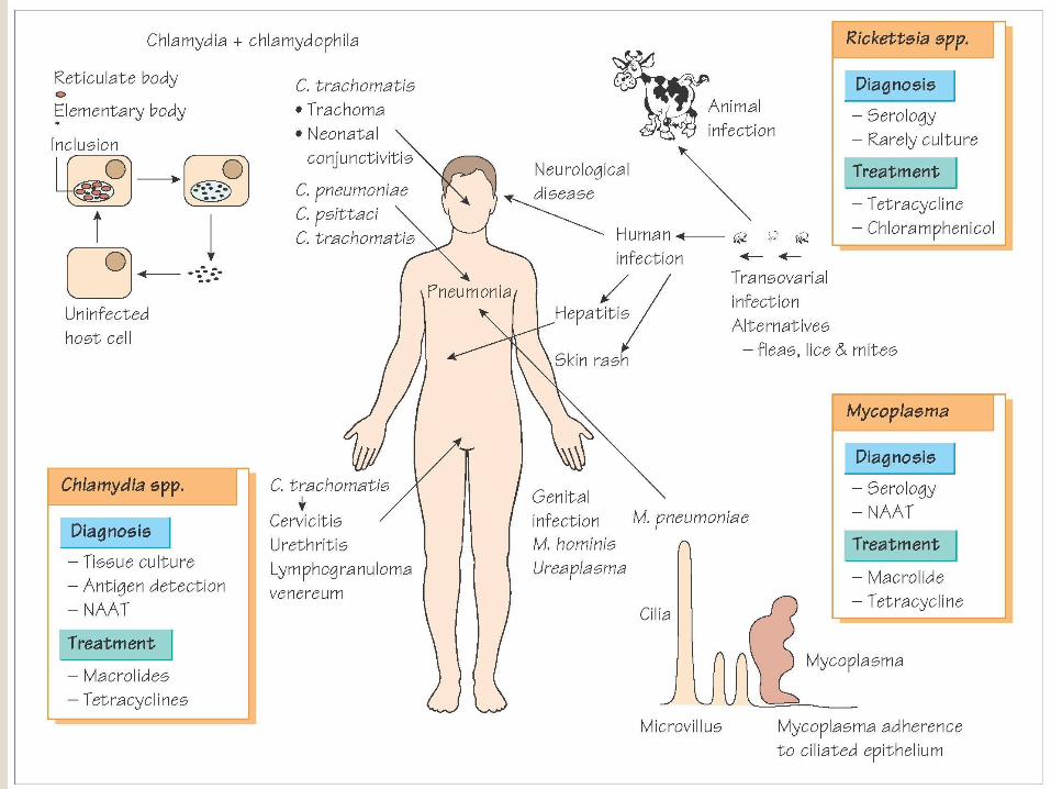
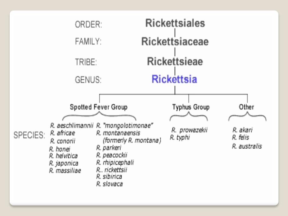
Rickettsiaceae
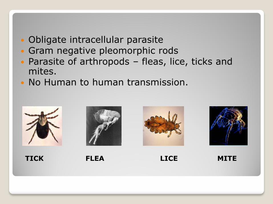
Obligate intracellular parasite Gram negative pleomorphic rods Parasite of arthropods – fleas, lice, ticks and
mites. No Human to human transmission.
TICK FLEA LICE MITE
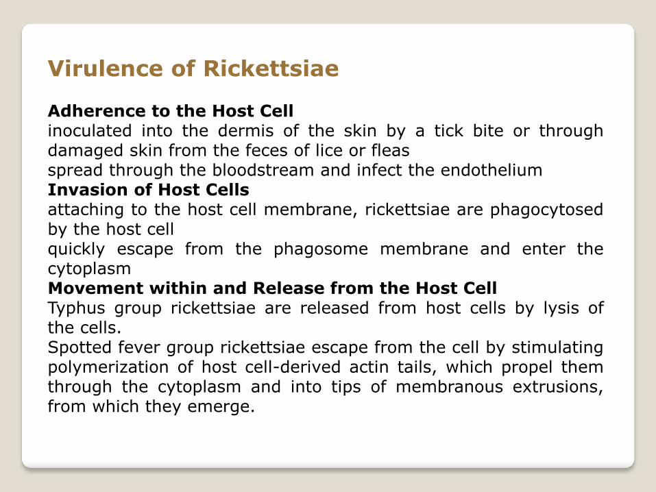
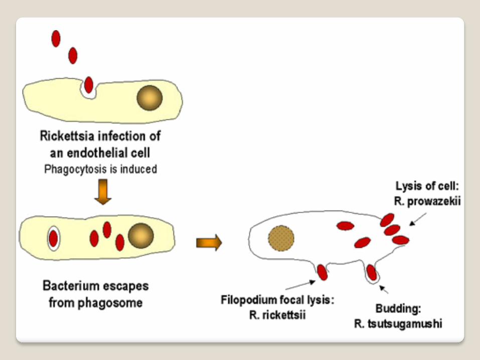
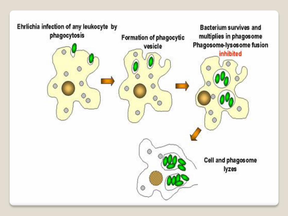
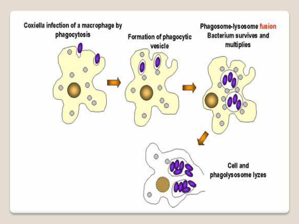
Virulence of Rickettsiae Adherence to the Host Cell inoculated into the dermis of the skin by a tick bite or through damaged skin from the feces of lice or fleas spread through the bloodstream and infect the endothelium Invasion of Host Cells attaching to the host cell membrane, rickettsiae are phagocytosed by the host cell quickly escape from the phagosome membrane and enter the cytoplasm Movement within and Release from the Host Cell Typhus group rickettsiae are released from host cells by lysis of the cells. Spotted fever group rickettsiae escape from the cell by stimulating polymerization of host cell-derived actin tails, which propel them through the cytoplasm and into tips of membranous extrusions, from which they emerge.
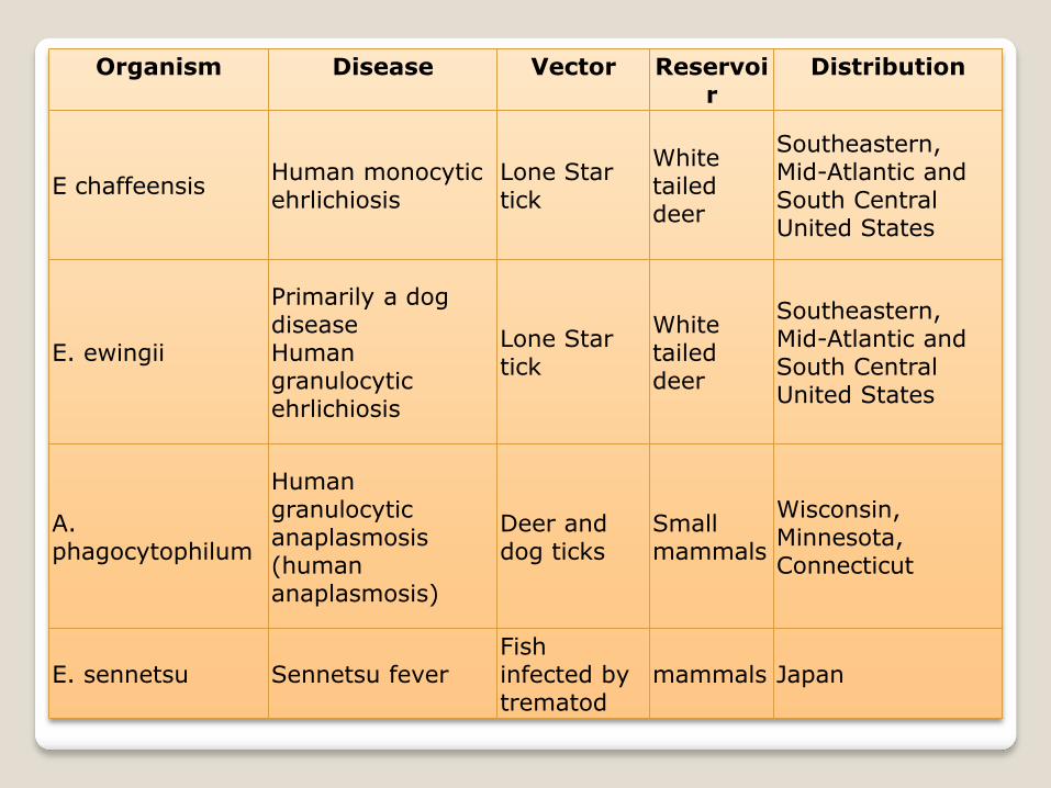
Organism Disease Vector Reservoir
Distribution
E chaffeensis Human monocytic ehrlichiosis
Lone Star tick
White tailed deer
Southeastern, Mid-Atlantic and South Central United States
E. ewingii
Primarily a dog disease Human granulocytic ehrlichiosis
Lone Star tick
White tailed deer
Southeastern, Mid-Atlantic and South Central United States
A. phagocytophilum
Human granulocytic anaplasmosis (human anaplasmosis)
Deer and dog ticks
Small mammals
Wisconsin, Minnesota, Connecticut
E. sennetsu Sennetsu fever Fish infected by trematod
mammals Japan
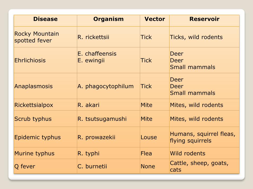
Disease Organism Vector Reservoir
Rocky Mountain spotted fever
R. rickettsii Tick Ticks, wild rodents
Ehrlichiosis E. chaffeensis E. ewingii
Tick Deer Deer Small mammals
Anaplasmosis A. phagocytophilum Tick Deer Deer Small mammals
Rickettsialpox R. akari Mite Mites, wild rodents
Scrub typhus R. tsutsugamushi Mite Mites, wild rodents
Epidemic typhus R. prowazekii Louse Humans, squirrel fleas, flying squirrels
Murine typhus R. typhi Flea Wild rodents
Q fever C. burnetii None Cattle, sheep, goats, cats
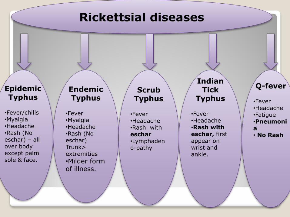
Rickettsial diseases
Epidemic Typhus
•Fever/chills •Myalgia •Headache •Rash (No eschar) – all over body except palm sole & face.
Endemic Typhus
•Fever •Myalgia •Headache •Rash (No eschar) Trunk> extremities
•Milder form of illness.
Scrub Typhus
•Fever •Headache •Rash with eschar •Lymphadeno-pathy
Indian Tick
Typhus
•Fever •Headache •Rash with eschar, first appear on wrist and ankle.
Q-fever
•Fever •Headache •Fatigue •Pneumonia • No Rash

Rickettsial diseases
Rocky Mountain Spotted Fever
•Fever •Headache •Rash (No eschar) – first appear on wrist & ankle •Palms & soles involved •Systemic Complications – R/S, CVS, CNS, Renal, Hepatic
Rickettsial Pox
•Mild Illness •Fever •Headache •Vesicular Rash with eschar •Lymphadenopathy •Resemblance to chicken pox

Weil-Felix Proteus Agglutination Test
Weil-Felix test- Based on the antigenic cross reactions among
rickettsial antigens, mostly LPS, and Proteus vulgaris strains
OX19 and OX2, and Proteus mirabilis OXK .
Procedure: Serum is diluted in three separate series of tubes followed by the addition of equal amount of OX19,OX2,OXK in 3 separate series of tubes. Incubation at 370C for overnight. Observe for agglutination.
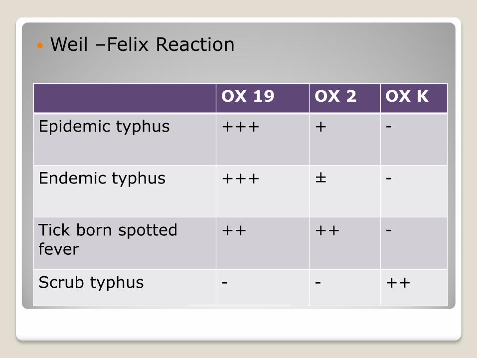
Weil –Felix Reaction
OX 19 OX 2 OX K
Epidemic typhus +++ + -
Endemic typhus +++ ± -
Tick born spotted fever
++ ++ -
Scrub typhus - - ++

Treatment
Treatment should be started early in the first week of illness.
Doxycycline (first choice)
Tetracycline (alternate)