Embed Size (px)
Citation preview
MRI IN NEUROLOGYHOW PHYSICS MAKES MY LIFE EASIER
Richard Simmons, M.D.
Child Neurologist
Schenectady Neurological Consultants

Today’s Talk
Not about physics Why MRI in Neurology About MRI Neuroanatomy Lesson Examples of normal brain Utility of different sequences How MRI helps make diagnoses (the fun
part)

MRI
In 1977, this technology was harnessed to produce the first human MRI scan
Commercial scanning began in the early 1980’s
MRI is now performed thousands of times daily across the world
Neurology is one of the top specialties using MRI
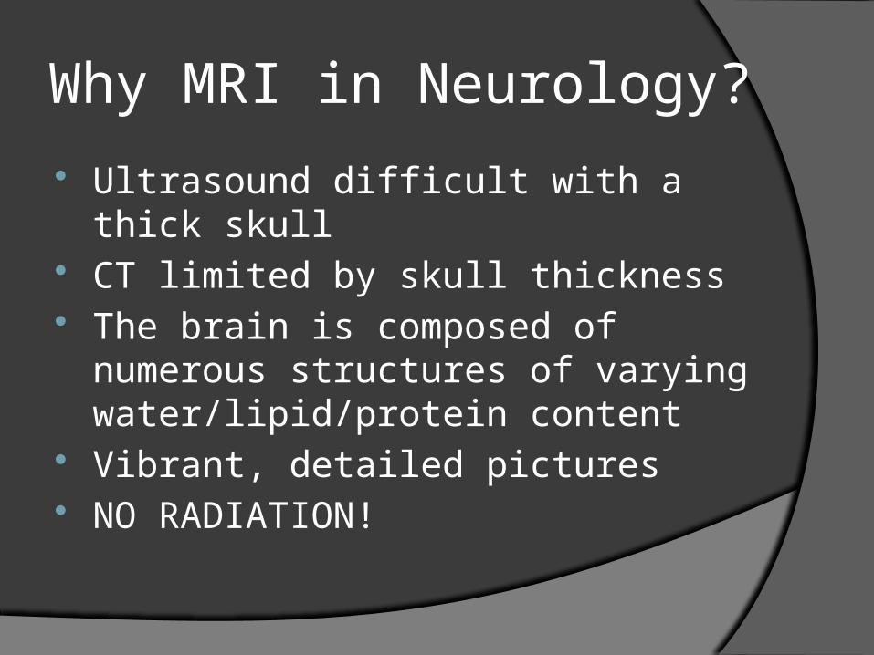
Why MRI in Neurology?
Ultrasound difficult with a thick skull CT limited by skull thickness The brain is composed of numerous
structures of varying water/lipid/protein content
Vibrant, detailed pictures NO RADIATION!
The Brain
Complex structure not easily visualized with surgical explorationDeep structures, brainstem, cranial nerves
Subtle structures not easily visualized with the naked eye
Damage by exploration not easily repaired
Utility of MRI in Neurology Multiple sequences allow for views of:
AnatomyPathologyBlood flow
○ Arterial○ Venous
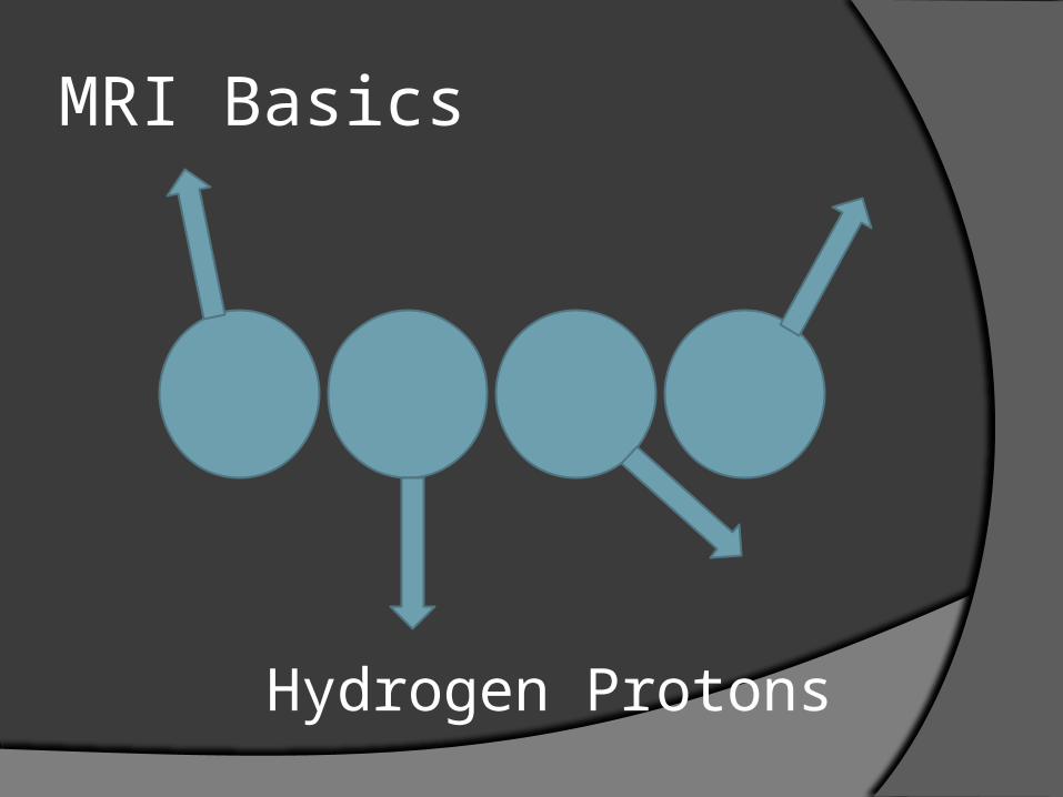
MRI Basics
Hydrogen Protons
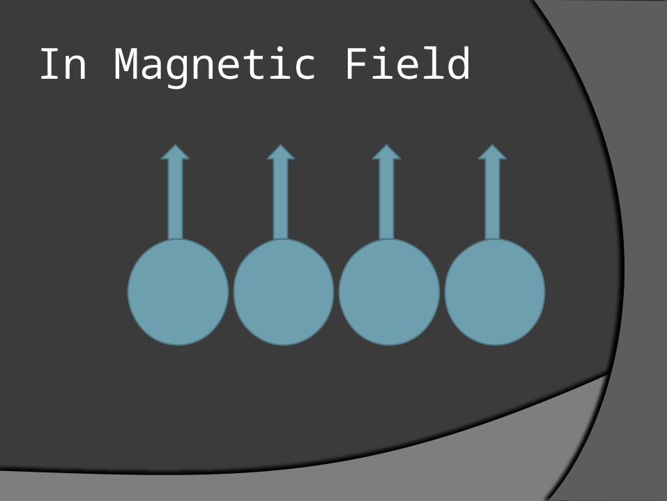
In Magnetic Field

RF Pulse
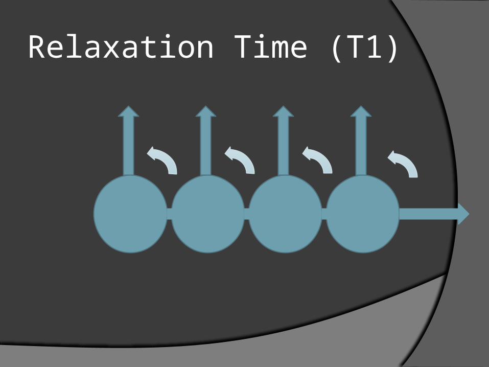
Relaxation Time (T1)

Echo Sequences
Can use 180º RF pulse Perform repeatedly Measure time to 63% decay (T2 time)

So What?
Used to measure hydrogen concentration
Water in different environments processes differently
Data collected turned into grayscale image
NORMAL BRAIN

Coronal
Axial
Sagittal
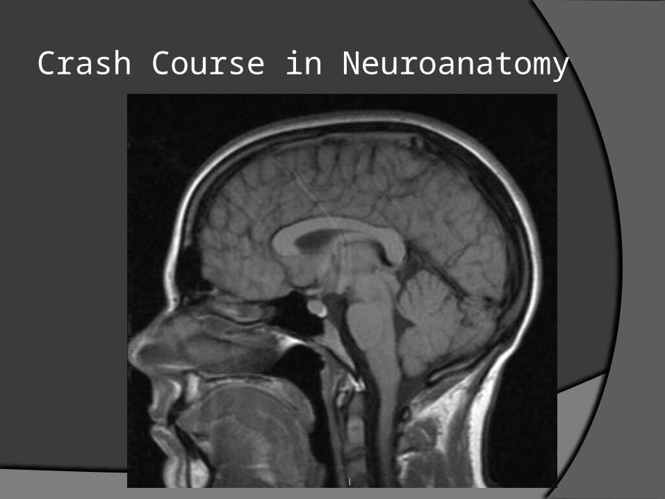
Crash Course in Neuroanatomy
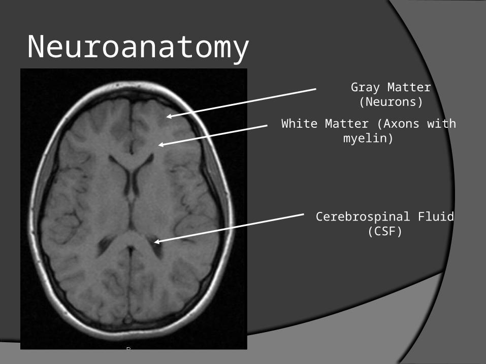
NeuroanatomyGray Matter (Neurons)
White Matter (Axons with myelin)
Cerebrospinal Fluid (CSF)

Neuroanatomy
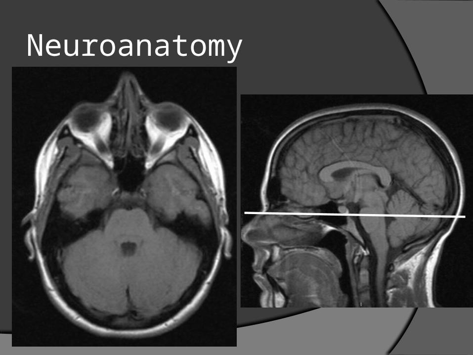
Neuroanatomy

T1 – “Anatomy”
Presents the brain “as is” Gray matter is grayWhite matter is whiteCSF is black
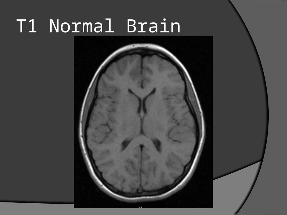
T1 Normal Brain
T2- “Pathology”
Presents the brain “inverted”Gray matter is whiteWhite matter is grayCSF is bright white
Emphasizes water-containing areas with greater clarity than T1Pathologic lesions frequently have
increased water content (edema)
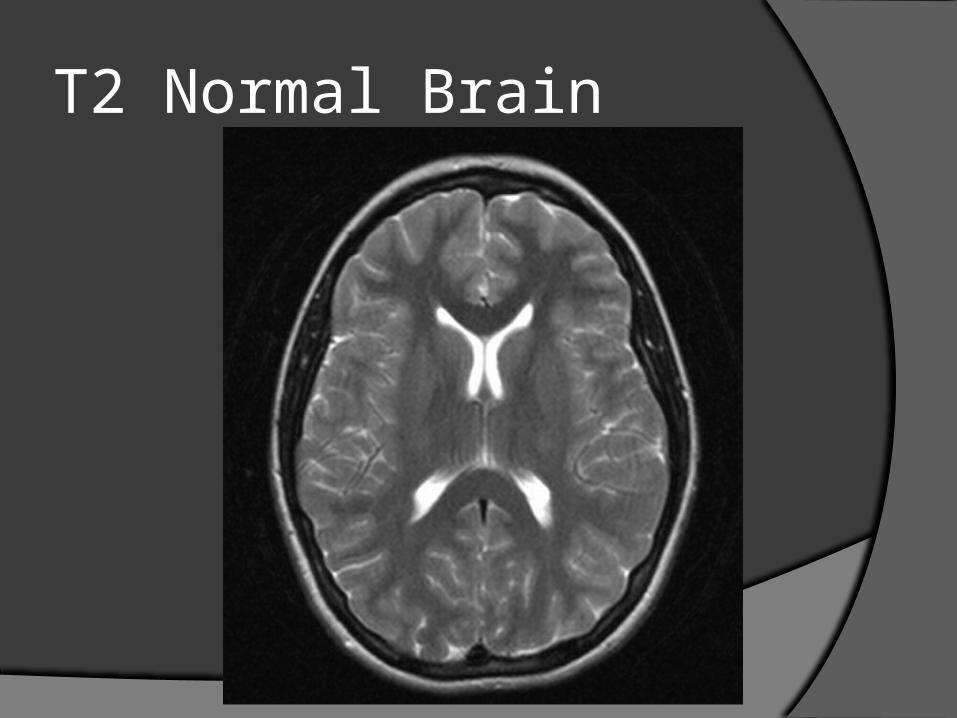
T2 Normal Brain

FLAIR
Fluid Attenuated Inversion Recovery T2 sequence with CSF signal dropped Allows better visualization of
periventricular pathology The sequence of choice for Multiple
Sclerosis

FLAIR Normal Brain
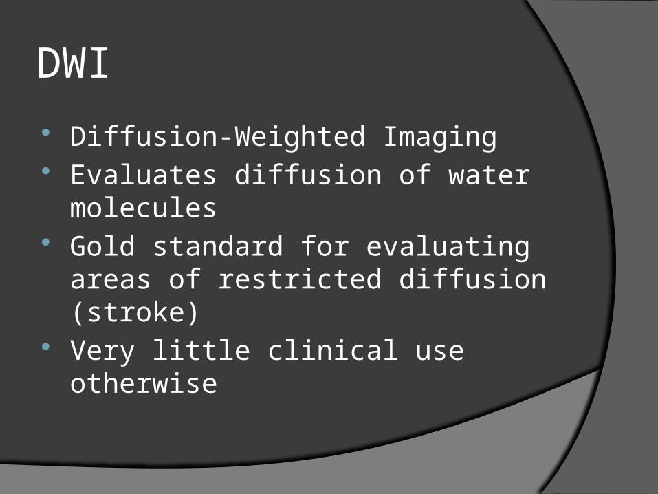
DWI
Diffusion-Weighted Imaging Evaluates diffusion of water molecules Gold standard for evaluating areas of
restricted diffusion (stroke) Very little clinical use otherwise

DWI Normal Brain
Contrast
Gadolinium (Gd) Rare-earth metal Paramagnetic Ideal for visualizing breakdown of the
blood-brain barrier

Clinical Cases
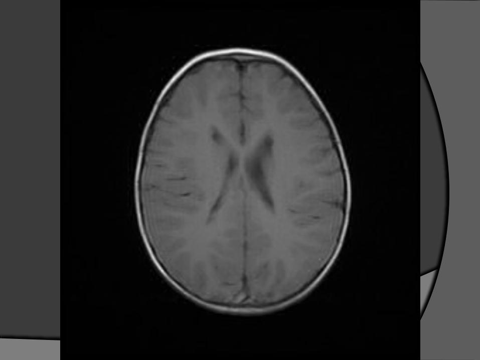
Case 1: Weakness
6 year-old girl Mild cognitive delay Gross motor delays since birth Now with primarily right-sided
“clumsiness”

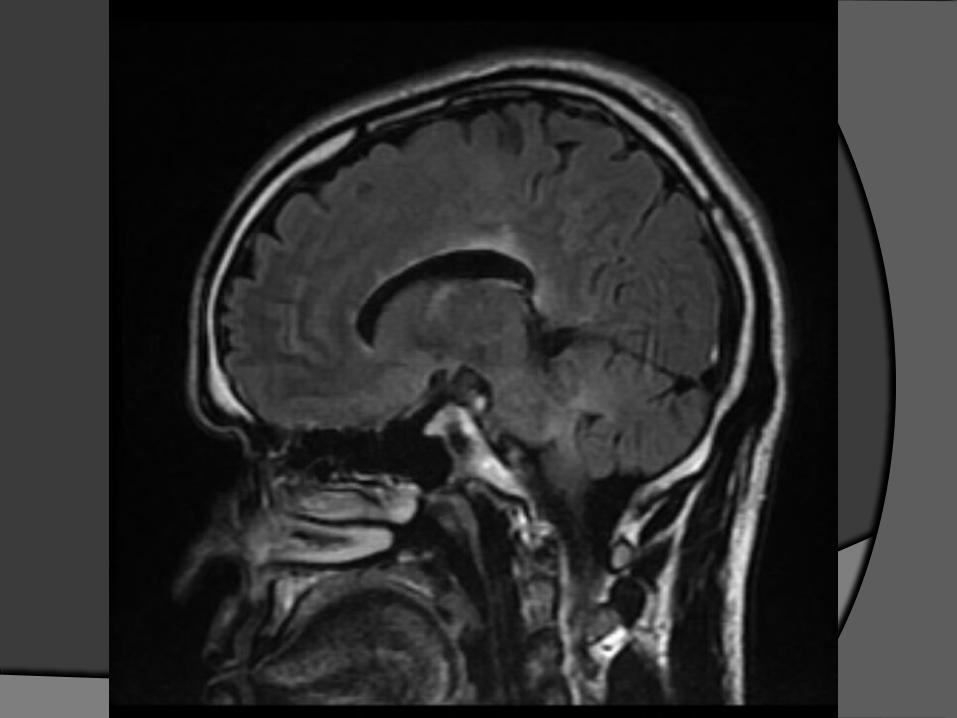
Case 2: Progressive Weakness
27 year old man Intermittent loss of strength/sensation
that nearly recovers each time Over time has had progressive
weakness of all extremities Mild cognitive decline


Case 3: Fall with Headache 5 year-old boy Fell 5 feet from deck hitting head CT in the ER was abnormal Child was physically OK without
neurological deficit MRI to confirm CT findings

Case 4: Partial Seizure
17 year-old male Had shaking of right arm for 3 minutes Mild cognitive disability (IQ 85) Mild weakness of right side face, arm,
and leg Mild asymmetry of limbs (circumference
of right calf less than left calf)

Case 5: New Headaches
11 year-old girl 1 month of daily headaches in the right
frontal region Moderate headaches that never really
go away Some episodes of staring off on a daily
basis EEG showed seizure activity in right
frontal region

FLAIR Image T1 Image after Gd
Case 6: Right-sided weakness 72 year-old man Smoker and frequent alcohol use High blood pressure Sudden onset right-sided weakness and
difficulty speaking
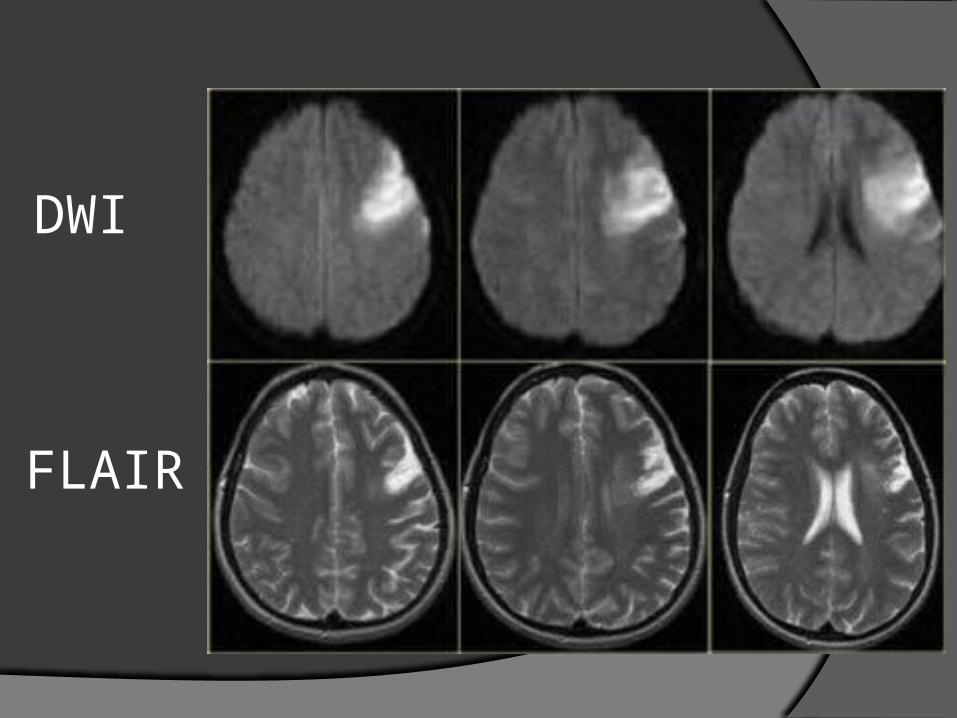
DWI
FLAIR

The Future
Diffusion Tensor ImagingWater diffuses faster down axons than
across axonsCan calculate this speed with DWIThen create 3D images with directionality to
show tracts

Questions?