Embed Size (px)
Citation preview

lable at ScienceDirect
Nutrition 30 (2014) 466–472
Contents lists avai
Nutrition
journal homepage: www.nutr i t ionjrnl .com
Basic nutritional investigation
Rice bran enzymatic extract–supplemented diets modulateadipose tissue inflammation markers in Zucker rats
Manila Candiracci Ph.D. a, Maria Luisa Justo M.D. b, Angelica Casta~no Ph.D. a,Rosalia Rodriguez-Rodriguez Ph.D. b, Maria Dolores Herrera Ph.D. b,*aDepartment of Biochemistry and Molecular Biology, School of Pharmacy, University of Seville, Seville, SpainbDepartment of Pharmacology, School of Pharmacy, University of Seville, Seville, Spain
a r t i c l e i n f o
Article history:Received 8 April 2013Accepted 26 September 2013
Keywords:ObesityRice branInflammation markersAdipose tissueZucker rat
MC and MLJ contributed equally to this work. RR-Rdesigned the study. MLJ and MC carried out the expeexperiments and analyzed the data. All authors werpaper and had final approval of the submitted andauthors declared no conflicts of interest.* Corresponding author: Tel.: þ34 95 455 9878; fa
E-mail address: [email protected] (M. D. Herrera).
0899-9007/$ - see front matter � 2014 Elsevier Inc. Ahttp://dx.doi.org/10.1016/j.nut.2013.09.016
a b s t r a c t
Objective: Chronic low-grade inflammation in obesity is characterized by macrophage accumula-tion in white adipose tissue and adipokine production deregulation. Obesity also is characterizedby oxidative stress related to inflammatory signaling. The aim of this study was to analyze whetherdietary supplementation with a rice bran enzymatic extract (RBEE), rich in bioactive compoundswith antioxidant and hypocholesterolemic properties, would ameliorate the inflammatory stateexisting in visceral adipose tissue of obese Zucker rats.Methods: Obese Zucker rats and their littermate controls, lean Zucker rats ages 8 wk, were daily fedan enriched diet with either 1% or 5% RBEE supplementation over 20 wk. Measurement ofadipocyte size and mRNA expression of proinflammatory molecules from visceral abdominal/epididymal tissue was performed.Results: An RBEE-supplemented diet decreased the overproduction of tumor necrosis factor-a,interleukin (IL)-6, IL-1 b, and inducible nitric oxide synthase (iNOS), as well as the overproductionof IL-6 and iNOs in visceral abdominal adipose tissue and visceral epididymal adipose tissue,respectively. An RBEE-supplemented diet modified the adipocyte-size distribution pattern in bothabdominal and epididymal adipose tissue, shifting it toward smaller cell sizes.Conclusions: Chronic administration of a novel water-soluble RBEE, rich in polyphenols, toco-trienols and g-oryzanol, could be a suitable treatment to ameliorate the obesity-associatedproinflammatory response.
� 2014 Elsevier Inc. All rights reserved.
Introduction
Obesity is characterized by a chronic and systemic low-gradeinflammation in adipose tissue that is believed to contribute tothe development of insulin resistance (IR) leading to type 2diabetes and is also a risk factor for cardiovascular diseases(CVDs). This hypothesis has gradually replaced the idea ofconsidering adipose tissue as a simple energy store but also as anendocrine organ that secretes a number of bioactive peptidescollectively named adipokines, which are relevant at the inter-face between the immune and the metabolic systems [1]. The
and MDH conceived andriments. AC conceived thee involved in writing thepublished versions. The
x: þ34 95 455 6074.
ll rights reserved.
adipose organ includes numerous discrete anatomical depotsand although subcutaneous adipose tissues store > 80% of totalbody, visceral adipose tissue have been shown to correlate betterwith metabolic syndrome and IR than subcutaneous fat depots[2]. Also, the expression of proinflammatory cytokines is gener-ally higher in visceral than in subcutaneous fat [3,4]. Adiposetissue contains adipocytes as well as fibroblasts, preadipocytes,tissue-resident macrophages, and vascular constituents, beingmacrophages crucial contributors to inflammation. In fact,inflammation in adipose tissue is partially due to an influx ofmacrophages that secrete proinflammatory factors like tumornecrosis factor (TNF)-a, interleukin (IL)-6, IL-1 beta (IL-1 b) andinducible nitric oxide synthase (iNOS) [5–8]. It is known that inobesity there is a remarkable shift in the pool of tissue macro-phages from the alternatively activated M2 type to the classicallyactivated M1 type, changing the secretion of cytokines frompredominantly anti-inflammatory (M2) to proinflammatory(M1) [9]. In addition to macrophages, adipocytes also secrete
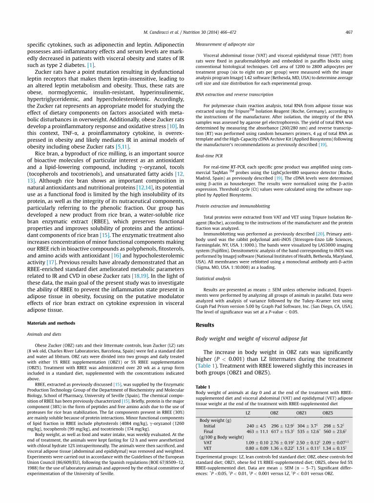
Table 1Body weight of animals at day 0 and at the end of the treatment with RBEE-supplemented diet and visceral abdominal (VAT) and epididymal (VET) adiposetissue weight at the end of the treatment with RBEE-supplemented diet
LZ OBZ OBZ1 OBZ5
Body weight (g)Initial 240 � 4.5 296 � 12.9z 304 � 3.7z 298 � 5.2z
Final 463 � 11.1 617 � 15.3z 535 � 12.6* 560 � 23,6y
(g/100 g Body weight)VAT 1.09 � 0.10 2.76 � 0.19z 2.50 � 0.12z 2.09 � 0.07z,x
VET 0.80 � 0.09 1.36 � 0.22z 1.51 � 0.11z 1.34 � 0.15z
Experimental groups: LZ, lean controls fed standard diet; OBZ, obese controls fedstandard diet; OBZ1, obese fed 1% RBEE-supplemented diet; OBZ5, obese fed 5%RBEE-supplemented diet. Data are mean � SEM (n ¼ 5–7). Significant differ-ences: *P <0.05, yP < 0.01, zP < 0.001 versus LZ, xP < 0.01 versus OBZ.
M. Candiracci et al. / Nutrition 30 (2014) 466–472 467
specific cytokines, such as adiponectin and leptin. Adiponectinpossesses anti-inflammatory effects and serum levels are mark-edly decreased in patients with visceral obesity and states of IRsuch as type 2 diabetes. [1].
Zucker rats have a point mutation resulting in dysfunctionalleptin receptors that makes them leptin-insensitive, leading toan altered leptin metabolism and obesity. Thus, these rats areobese, normoglycemic, insulin-resistant, hyperinsulinemic,hypertriglyceridemic, and hypercholesterolemic. Accordingly,the Zucker rat represents an appropriate model for studying theeffect of dietary components on factors associated with meta-bolic disturbances in overweight. Additionally, obese Zucker ratsdevelop a proinflammatory response and oxidative stress [10]. Inthis context, TNF-a, a proinflammatory cytokine, is overex-pressed in obesity and likely mediates IR in animal models ofobesity including obese Zucker rats [5,11].
Rice bran, a byproduct of rice milling, is an important sourceof bioactive molecules of particular interest as an antioxidantand a lipid-lowering compound, including g-oryzanol, tocols(tocopherols and tocotrienols), and unsaturated fatty acids [12,13]. Although rice bran shows an important composition innatural antioxidants and nutritional proteins [12,14], its potentialuse as a functional food is limited by the high insolubility of itsprotein, as well as the integrity of its nutraceutical components,particularly referring to the phenolic fraction. Our group hasdeveloped a new product from rice bran, a water-soluble ricebran enzymatic extract (RBEE), which preserves functionalproperties and improves solubility of proteins and the antioxi-dant components of rice bran [15]. The enzymatic treatment alsoincreases concentration of minor functional components makingour RBEE rich in bioactive compounds as polyphenols, fitosterols,and amino acids with antioxidant [16] and hypocholesterolemicactivity [17]. Previous results have already demonstrated that anRBEE-enriched standard diet ameliorated metabolic parametersrelated to IR and CVD in obese Zucker rats [18,19]. In the light ofthese data, the main goal of the present study was to investigatethe ability of RBEE to prevent the inflammation state present inadipose tissue in obesity, focusing on the putative modulatoreffects of rice bran extract on cytokine expression in visceraladipose tissue.
Materials and methods
Animals and diets
Obese Zucker (OBZ) rats and their littermate controls, lean Zucker (LZ) rats(8 wk old, Charles River Laboratories, Barcelona, Spain) were fed a standard dietand water ad libitum. OBZ rats were divided into two groups and daily treatedwith either 1% RBEE supplementation (OBZ1) or 5% RBEE supplementation(OBZ5). Treatment with RBEE was administered over 20 wk as a syrup formincluded in a standard diet, supplemented with the concentrations indicatedabove.
RBEE, extracted as previously discussed [15], was supplied by the EnzymaticProduction Technology Group of the Department of Biochemistry and MolecularBiology, School of Pharmacy, University of Seville (Spain). The chemical compo-sition of RBEE has been previously characterized [15]. Briefly, protein is the majorcomponent (38%) in the form of peptides and free amino acids due to the use ofproteases for rice bran stabilization. The fat components present in RBEE (30%)are mainly soluble because of protein interactions. Minor functional componentsof lipid fraction in RBEE include phytosterols (4084 mg/kg), g-oryzanol (1260mg/kg), tocopherols (99 mg/kg), and tocotrienols (174 mg/kg).
Body weight, as well as food and water intake, was weekly evaluated. At theend of treatment, the animals were kept fasting for 12 h and were anesthetizedwith chloral hydrate 12% intraperitoneally. The animals were then sacrificed, andvisceral adipose tissue (abdominal and epididymal) was removed and weighted.Experiments were carried out in accordance with the Guidelines of the EuropeanUnion Council (86/609/EU), following the Spanish regulations (BOE 67/8509–12,1988) for the use of laboratory animals and approved by the ethical committee ofexperimentation of the University of Seville.
Measurement of adipocyte size
Visceral abdominal tissue (VAT) and visceral epididymal tissue (VET) fromrats were fixed in paraformaldehyde and embedded in paraffin blocks usingconventional histological techniques. Cell area of 1200 to 2800 adipocytes pertreatment group (six to eight rats per group) were measured with the imageanalysis program ImageJ 1.42 software (Bethesda, MD, USA) to determine averagecell size and size distribution for each experimental group.
RNA extraction and reverse transcription
For polymerase chain reaction analysis, total RNA from adipose tissue wasextracted using the TripureTM Isolation Reagent (Roche, Germany), according tothe instructions of the manufacturer. After isolation, the integrity of the RNAsamples was assessed by agarose gel electrophoresis. The yield of total RNA wasdetermined by measuring the absorbance (260/280 nm) and reverse transcrip-tion (RT) was performed using random hexamers primers, 4 mg of total RNA astemplate and the High-Capacity cDNA Archive Kit (Applied Biosystems) followingthe manufacturer’s recommendations as previously described [19].
Real-time PCR
For real-time RT-PCR, each specific gene product was amplified using com-mercial TaqMan TM probes using the LighCycler480 sequence detector (Roche,Madrid, Spain) as previously described [19]. The cDNA levels were determinedusing b-actin as housekeeper. The results were normalized using the b-actinexpression. Threshold cycle (Ct) values were calculated using the software sup-plied by Applied Biosystems.
Protein extraction and immunoblotting
Total proteins were extracted from VAT and VET using Tripure Isolation Re-agent (Roche), according to the instructions of the manufacturer and the proteinfraction was analyzed.
Immunoblotting was performed as previously described [20]. Primary anti-body used was the rabbit polyclonal anti-iNOS (Stressgen-Enzo Life Sciences,Farmingdale, NY, USA. 1:1000.). The bands were visualized by LAS3000 imagingsystem (Fujifilm). Densitometric analysis of the band corresponding to iNOS wasperformed by ImageJ software (National Institutes of Health, Bethesda, Maryland,USA). All membranes were reblotted using a monoclonal antibody anti-b-actin(Sigma, MO, USA. 1:10.000) as a loading.
Statistical analysis
Results are presented as means � SEM unless otherwise indicated. Experi-ments were performed by analyzing all groups of animals in parallel. Data wereanalyzed with analysis of variance followed by the Tukey–Kramer test usingGraph Pad Prism version 5.00 by Graph Pad Software, Inc. (San Diego, CA, USA).The level of significance was set at a P-value < 0.05.
Results
Body weight and weight of visceral adipose fat
The increase in body weight in OBZ rats was significantlyhigher (P < 0.001) than LZ littermates during the treatment(Table 1). Treatment with RBEE lowered slightly this increases inboth groups (OBZ1 and OBZ5).
M. Candiracci et al. / Nutrition 30 (2014) 466–472468
The weight of abdominal and epididymal fat tissue are eval-uated for all experimental groups and are summarized in Table 1.Treatment with RBEE did not alter significantly fat weight in OBZrats. Only weight of VAT in rats fed a 5% RBEE diet was signifi-cantly reduced compared with non-treated obese rats.
Adipocyte size distribution
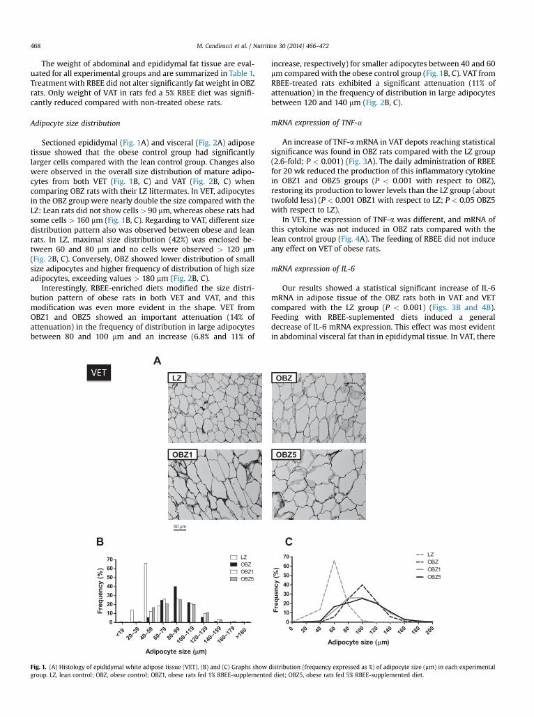
Sectioned epididymal (Fig. 1A) and visceral (Fig. 2A) adiposetissue showed that the obese control group had significantlylarger cells compared with the lean control group. Changes alsowere observed in the overall size distribution of mature adipo-cytes from both VET (Fig. 1B, C) and VAT (Fig. 2B, C) whencomparing OBZ rats with their LZ littermates. In VET, adipocytesin the OBZ group were nearly double the size compared with theLZ: Lean rats did not show cells > 90 mm, whereas obese rats hadsome cells > 160 mm (Fig. 1B, C). Regarding to VAT, different sizedistribution pattern also was observed between obese and leanrats. In LZ, maximal size distribution (42%) was enclosed be-tween 60 and 80 mm and no cells were observed > 120 mm(Fig. 2B, C). Conversely, OBZ showed lower distribution of smallsize adipocytes and higher frequency of distribution of high sizeadipocytes, exceeding values > 180 mm (Fig. 2B, C).
Interestingly, RBEE-enriched diets modified the size distri-bution pattern of obese rats in both VET and VAT, and thismodification was even more evident in the shape. VET fromOBZ1 and OBZ5 showed an important attenuation (14% ofattenuation) in the frequency of distribution in large adipocytesbetween 80 and 100 mm and an increase (6.8% and 11% of
B
A
Fig. 1. (A) Histology of epididymal white adipose tissue (VET). (B) and (C) Graphs show dgroup. LZ, lean control; OBZ, obese control; OBZ1, obese rats fed 1% RBEE-supplemented
increase, respectively) for smaller adipocytes between 40 and 60mm compared with the obese control group (Fig. 1B, C). VAT fromRBEE-treated rats exhibited a significant attenuation (11% ofattenuation) in the frequency of distribution in large adipocytesbetween 120 and 140 mm (Fig. 2B, C).
mRNA expression of TNF-a
An increase of TNF-amRNA in VAT depots reaching statisticalsignificance was found in OBZ rats compared with the LZ group(2.6-fold; P < 0.001) (Fig. 3A). The daily administration of RBEEfor 20 wk reduced the production of this inflammatory cytokinein OBZ1 and OBZ5 groups (P < 0.001 with respect to OBZ),restoring its production to lower levels than the LZ group (abouttwofold less) (P < 0.001 OBZ1 with respect to LZ; P < 0.05 OBZ5with respect to LZ).
In VET, the expression of TNF-a was different, and mRNA ofthis cytokine was not induced in OBZ rats compared with thelean control group (Fig. 4A). The feeding of RBEE did not induceany effect on VET of obese rats.
mRNA expression of IL-6
Our results showed a statistical significant increase of IL-6mRNA in adipose tissue of the OBZ rats both in VAT and VETcompared with the LZ group (P < 0.001) (Figs. 3B and 4B).Feeding with RBEE-suplemented diets induced a generaldecrease of IL-6 mRNA expression. This effect was most evidentin abdominal visceral fat than in epididymal tissue. In VAT, there
C
istribution (frequency expressed as %) of adipocyte size (mm) in each experimentaldiet; OBZ5, obese rats fed 5% RBEE-supplemented diet.

Fig. 2. (A) Histology of abdominal white adipose tissue (VAT). (B) and (C) Graphs show distribution (frecquency expressed as %) of adipocyte size (mm) in each experimentalgroup. LZ, lean control; OBZ, obese control; OBZ1, obese rats fed 1% RBEE-supplemented diet; OBZ5, obese rats fed 5% RBEE-supplemented diet.
M. Candiracci et al. / Nutrition 30 (2014) 466–472 469
was a fourfold decrease of mRNA IL-6 in OBZ1 (P < 0.001) andthreefold decrease in OBZ5 (P < 0.001). These results also werestatistically significant with respect to the lean group (P < 0.01OBZ1; P < 0.05 OBZ5). In VET only, the 1% RBEE diet was shownto attain statistical significance (P < 0.001 compared with OBZ)with a value of IL-6 mRNA similar to the lean group.
mRNA expression of IL-1 b
We did not find significant changes in the expression of IL-1 bmRNA in OBZ rats compared with LZ in both visceral fat tissuesexamined. However, administration of RBEE reduced the pro-duction of the inflammatory cytokine IL-1 b in visceral adiposetissue from obese rats. In VAT, this effect was statisticallysignificant in both RBEE-supplemented diet groups (1% and 5%;P < 0.001 in both LZ and OBZ). In VET the decrease of expressionof IL-1 b was statistically different only in OBZ1 (P < 0.01 OBZ;P < 0.05 LZ) (Figs. 3C and 4C).
Expression of iNOS
Our results showed an up-regulation of iNOS at both mRNA(Figs. 3D and 4D) and protein levels (Fig. 5) in both adipose tis-sues studied compared with LZ rats. RBEE-enriched dietsdecreased iNOS mRNA and protein expression in both VAT andVET (P < 0.001 and P < 0.05 in all cases of OBZ; Figs. 3D, 4D,and 5) reaching values lower than in the LZ group (P < 0.05OBZ5with respect to LZ in VAT; P< 0.01 OBZ1with respect to LZ;P < 0.05 OBZ5 with respect to LZ in VET).
Discussion
The purpose of this study was to investigate the effect of anew water-soluble RBEE on inflammatory markers in OBZ rats.Although no significant differences were observed in the finalbody weight and in the adipose tissue weight between thedifferent groups, RBEE-enriched diets were able to modulate thealtered production of cytokines, characteristic of adipose tissuein OBZ rats, and also modified the adipocyte size distributionpattern in both VAT and VET shifting it toward smaller cell sizes.
Obesity is associated with a chronic inflammatory response,which is characterized by abnormal cytokine production,increased synthesis of acute-phase reactants, such as C-reactiveprotein, and activation of proinflammatory signaling pathways[3]. It has been strongly evidenced that adipokines secreted bythe adipose tissue contribute to obesity-associated systemicinflammation and may constitute potential important targets forthe prevention of inflammation-induced IR or vasculopathy [1,9].As a result of overweight and obesity, adipocytes increase in sizeand release more saturated free fatty acids (FFAs) and chemo-kines, followed by macrophages infiltration into adipose tissuethat leads to an increase in expression of IL-1 b, IL-6, andiNOS [21].
As previously indicated, functional components of the lipidfraction in RBEE include phytosterols, g-oryzanol, tocopherols,and tocotrienols. Tocotrienols, as well as polyphenols, haveproven antioxidant and anti-inflammatory properties andexhibit activity against different chronic diseases, such as cancer,diabetes, CVDs, and neurologic disorders [22,23]. Additionally,g-tocotrienol has been found to improve obesity-related

Fig. 3. RBEE effect on gene expression of TNF- (A), IL-6 (B), IL-1 (C) and iNOS (D) in visceral abdominal adipose tissue (VAT) from lean (LZ) and obese Zucker (OBZ) ratsdetermined using real-time polymerase chain reaction of different inflammatory factors. Experimental groups: LZ, lean control; OBZ, obese control; OBZ1, obese rats fed 1%RBEE-supplemented diet; OBZ5, obese rats fed 5% RBEE-supplemented diet. Data are representative of six to seven rats/treatment group and are expressed as mean � SEM.*P < 0.05; yP < 0.01; zP < 0.001 represent a significant differences respect LZ sample using one-way ANOVA with a Tukey’s Multiple Comparison Test. {P < 0.001 of OBZ1 orOBZ5 with respect to OBZ.
M. Candiracci et al. / Nutrition 30 (2014) 466–472470
functional abnormalities in adipocytes by attenuating the ex-pression of inflammatory adipokinesin 3 T3-L1 [23]. Previousresults have described that RBEE exerts antioxidant [16]and hypocholesterolemic effects [17]. Interestingly, besidestocotrienols, the main bioactive compound of RBEE is g-oryzanol(1260 mg/kg), which has been suggested to possess lipid-low-ering [24], anti-inflammatory [25], anticancer [26], and antioxi-dant effects [27]. In vivo and in vitro studies have reported thatrice bran g-oryzanol induces anti-inflammatory effects bydown-regulating the inflammatory transcription factor, nuclearfactor-kB (NF-kB), which in turn dicreases the expression of in-flammatory enzymes such as COX-2 and iNOS, and proin-flammatory cytokines such as IL-1 b, IL-6, and TNF-a [28].Accordingly, our results show that an RBEE-supplemented dietclearly improved the inflammation state characteristic of visceralfat in Zucker rats, attenuating the increase of proinflammatoryfactors such as TNF-a, IL-6, and iNOS, with this effect being moreevident in VAT than in VET.
According to previous works describing regional heteroge-neity in the mRNA expression of proinflammatory and anti-inflammatory cytokines between different white adipose tissuedepots [29], our results show a difference between VAT and VETin the inflammatory feature. The major discrepancy accounts forTNF-a, a cytokine that is strongly linked to obesity and was notincreased in VET. It is recognized that TNF-a is more abundantlyproduced by stromal-vascular cells (mainly macrophages) thanby adipocytes [3,5,7]. In this regard, depot differences in cellpopulations may contribute to depot differences in adipokineproduction, and can contribute to variations in adipocyte
function via paracrine interactions. Interestingly, this study in-dicates that the effect of RBEE was more evident on theexpression of proinflammation factors in abdominal visceraladipose tissue (VAT) than in epididymal fat (VET) on OBZ rats.Further studies are needed to clarify whether the difference inthe macrophage infiltration rate into different adipose depotsaccounts for the difference we have found in the mRNAexpression of proinflammatory cytokines. In this line, it is worthnoting that macrophages have been identified as the primarysource of many of the circulating inflammatory molecules thatare detected in the obese state [30] and macrophage infiltrationinto visceral adipose tissue is higher than into subcutaneousadipose tissues [31], being the expression of proinflammatorycytokines generally higher in visceral than in subcutaneousfat [3, 4].
IL-6 is a proinflammatory cytokine produced by several celltypes (fibroblasts, endothelial cells, monocytes, adipocytes),which also are linked to obesity. Thus, the production andcirculating level of IL-6 in the obese adipose tissue is increased[3,6]. It is extremely clear from our results, that RBEE has aprofound effect on the production of both cytokines in obese rats,particularly in VAT, and there are no major variations on theeffects of different RBEE-supplemented diets (1% and 5%).
Endotoxins or inflammatory cytokines, such as TNF-a, nor-mally induce the expression of the iNOS in macrophages [32].Many inflammatory diseases are accompanied by an increase inNO production and, in appropriate animal models, a beneficialaction of iNOS inhibitors has been demonstrated [33]. Interest-ingly, we found that RBEE-supplemented diets showed a positive

Fig. 4. RBEE effect on gene expression of TNF- (A), IL-6 (B), IL-1 (C) and iNOS (D) in visceral epididymal adipose tissue (VET), from lean (LZ) and obese Zucker (OBZ) ratsdetermined by real-time polymerase chain reaction of different inflammatory factors. Experimental groups: LZ, lean control; OBZ, obese control; OBZ1, obese rats fed 1%RBEE-supplemented diet; OBZ5, obese rats fed 5% RBEE-supplemented diet. Data are representative of six to seven rats/treatment group and are expressed as mean � SEM. *P< 0.05; yP < 0.01; zP < 0.001 indicates a significant differences respect LZ sample using one-way ANOVA with a Tukey’s Multiple Comparison Test. {P < 0.001 versus OBZ.
M. Candiracci et al. / Nutrition 30 (2014) 466–472 471
effect inhibiting iNOS expression. As stated previously, in obesitythere is a remarkable shift in the pool of tissue macrophagesfrom the alternatively activated M2 type to the classically acti-vated M1 type [9]. M2 macrophages are characterized, amongother features, by the expression of arginase, an enzyme thatblocks iNOS activity, whereas M1 macrophages express not onlyhigh levels of proinflammatory cytokines (e.g., TNF-a, IL-6), butalso iNOS [34].
Present results show that RBEE-enriched diets did not exertsignificant results on body weight and fat weight (except in VET).
A
Fig. 5. Expression of iNOS protein in visceral abdominal (VAT) (A) and epididymal tissucontrol; OBZ, obese Zucker control; OBZ1, 1% RBEE-treated OBZ rats; OBZ5, 5% RBEE-tre
Probably the beneficial effect of RBEE on the inflammatory state inthe adipose tissue is mainly carried out on activatedmacrophage-derived cytokines, more likely than in adipocytes, especially inVAT, in which the anti-inflammatory effect of RBEE was higher.However, we must also consider that the beneficial effect of RBEEon the inflammatory state alsomay be due, at least partially, to theeffect on adipocyte size. Present results show that RBEE induces ageneral decrease in the adipocyte volume, changing the adipocytesize distribution in rats fed RBEE-supplemented diets in com-parison with obese rats fed a standard diet. It has been suggested
B
e (VET) (B) from Zucker rats. Results are the mean � SEM (n ¼ 4). LZ, lean Zuckerated OBZ rats. *P < 0.05 versus LZ. yP < 0.05 versus OBZ.
M. Candiracci et al. / Nutrition 30 (2014) 466–472472
that in OBZ obese rats, the enlargement of white adipose depotsis due to the hypertrophy of adipocytes [35]. Adipocytes enlargeas a consequence of hyperalimentation and large adipocytesreleasemore (saturated) FFAs,which can bind tomacrophage toll-like receptor-4 resulting in NF-kB activation, and ultimatelyleading to augmented TNF-a production [36,37]. In turn,macrophage-derived TNF-a activates human adipocytes, inducingfurther lipolysis and enhancing the expression of various genesthat facilitate the diapedesis of monocytes and consequent dif-ferentiation into macrophages [38,39]. Thus, it has been proposedthat this local paracrine loop involving adipocyte-derived FFAsandmacrophage-derivedTNF-a establishes a gradual vicious cyclethat presumably leads to a proinflammatory state of both mac-rophages and adipocytes [21].
Conclusions
In summary, our present work demonstrates that chronicadministration of a novel water-soluble RBEE could be a suitabletreatment to ameliorate the obesity-associated proinflammatoryresponse. Our results provide evidence of the nutraceuticalproperties of RBEE against the pathogenesis of obesity andreinforce the potential of RBEE as a functional food.
Acknowledgments
This research was supported by The Spanish Ministry of Sci-ence and Innovation (AGL2009-1159). ML Justo has been arecipient of an FPU fellowship from the Spanish Government.The authors acknowledge the Enzymatic Production TechnologyGroup of University of Seville (Spain) for supplying the drug forthis study.
References
[1] Tilg H, Moschen AR. Adipocytokines: Mediators linking adipose tissue,inflammation and immunity. Nat Rev Immunol 2006;6:772–83.
[2] Matsuzawa Y. Metabolic syndromeddefinition and diagnostic criteria inJapan. J Atheroscler Thromb 2005;12:301.
[3] Maury E, Brichard SM. Adipokinedysregulation, adipose tissue inflamma-tion and metabolic syndrome. Mol Cell Endocrinol 2010;314:1–16.
[4] Lee MJ, Wu Y, Fried SK. Adipose tissue heterogeneity: Implication of depotdifferences in adipose tissue for obesity complications. Mol Aspects Med2013;34:1–11.
[5] Hotamisligil GS, Shargill NS, Spiegelman BM. Adipose expression of tumornecrosis factor-alpha: Direct role in obesity-linked insulin resistance.Science 1993;259:87–91.
[6] Bastard JP, Maachi M, Van Nhieu JT, Jardel C, Bruckert E, Grimaldi A, et al.Adipose tissue IL-6 content correlates with resistance to insulin activationof glucose uptake both in vivo and in vitro. J Clin Endocrinol Metab2002;87:2084–9.
[7] Kanda H, Tateya S, Tamori Y, Kotani K, Hiasa K, Kitazawa R, et al. MCP-1contributes to macrophage infiltration into adipose tissue, insulin resis-tance, and hepatic steatosis in obesity. J Clin Invest 2006;116:1494–505.
[8] Todoric J, LöfflerM,Huber J, BilbanM, ReimersM, Kadl A, et al. Adipose tissueinflammation inducedbyhigh-fat diet in obese diabeticmice is prevented byn-3 polyunsaturated fatty acids. Diabetologia 2006;49:2109–19.
[9] Karalis PK, Giannogonas P, Kodela E, Koutmani Y, Zoumakis M, Teli T.Mechanisms of obesity and related pathology: Linking immune responsesto metabolic stress. FEBS Journal 2009;276:5747–54.
[10] Aleixandre de Arti~nano A, Miguel Castro M. Experimental rat models tostudy the metabolic syndrome. Br J Nutr 2009;102:1246–53.
[11] Picchi A, Gao X, Belmadani S, et al. Tumor necrosis factor-alpha inducesendothelial dysfunction in the prediabetic metabolic syndrome. Circ Res2006;99:69–77.
[12] Jariwalla RJ. Rice-bran products: Phytonutrients with potential applicationsin preventive and clinical medicine. Drugs Exp Clin Res 2001;27:17–26.
[13] Ha TY, Han S, Kim SR, Kim IH, Lee HY, Kim HK. Bioactive components in ricebran oil improve lipid profiles in rats fed a high-cholesterol diet. Nutr Res2005;25:597–606.
[14] Fabian C, Ju YH. A review on rice bran protein: Its properties and extractionmethods. Crit Rev Food Sci Nutr 2011;51:816–27.
[15] Parrado J, Miramontes E, Jover M, Gutierrez JF, Collantes de Ter�an L, et al.Preparation of a rice bran enzymatic extract with potential use as func-tional food. Food Chem 2006;98:742–8.
[16] Santa-Mar�ıa C, Revilla E, Miramontes E, Bautista J, Garc�ıa-Mart�ınez A,Romero E, et al. Protection against free radicals (UVB irradiation) of awater-soluble enzymatic extract from rice bran. Study using human kera-tinocyte monolayer and reconstructed human epidermis. Food ChemToxicol 2010;48:83–8.
[17] Revilla E, Santa-Mar�ıa C, Miramontes E, Bautista J, Garc�ıa-Mart�ınez A,Cert R, et al. Nutraceutical composition, antioxidant activity and hypo-cholesterolemic effect of a water soluble enzymatic extract from rice bran.Food Res Int 2009;42:387–93.
[18] Justo ML, Rodriguez-Rodriguez R, Claro CM, Alvarez de Sotomayor M,Parrado J, Herrera MD. Water-soluble rice bran enzymatic extract attenu-ates dyslipidemia, hypertension and insulin resistance in obese Zucker rats.Eur J Nutr 2013;52:789–97.
[19] Justo ML, Candiracci M, Dantas AP, Alvarez de Sotomayor M, Parrado J,Vila E, et al. Rice bran enzymatic extract restores endothelial function andvascular contractility in obese rats by reducing vascular inflammation andoxidative stress. J Nutr Biochem 2013;24:1453–61.
[20] Rodriguez-Rodriguez R, Herrera MD, de Sotomayor MA, Ruiz-Gutierrez V.Pomace olive oil improves endothelial function in spontaneously hyper-tensive rats by increasing endothelial nitric oxide synthase expression. AmJ Hypertens 2007;20:728–34.
[21] Hajer GR, vanHaeften TW, Visseren FLJ. Adipose tissue dysfunction inobesity, diabetes, and vascular diseases. Eur Heart J 2008;29:2959–71.
[22] Siddiqui S, Rashid Khan M, Siddiqui WA. Comparative hypoglycemic andnephroprotective effects of tocotrienol rich fraction (TRF) from palm oiland rice bran oil against hyperglycemia induced nephropathy in type 1diabetic rats. Chem Biol Interact 2010;188:651–8.
[23] Matsunaga T, Shoji A, GuN, Joo E, Li S, Adachi T, et al. g-tocotrienol attenuatesTNF-a-induced changes in secretion and gene expression of MCP-1, IL-6 andadiponectin in 3 T3-L1 adipocytes. Mol Med Report 2012;5:905–9.
[24] Wilson TA, Nicolosi RJ, Woolfrey B, Kritchevsky D. Rice bran oil and ory-zanol reduce plasma lipid and lipoprotein cholesterol concentrations andaortic cholesterol ester accumulation to a greater extent than ferulic acid inhypercholesterolemic hamsters. J Nutr Biochem 2007;18:105–12.
[25] Akihisa T, Yasukawa K, Yamaura M, Ukiya M, Kimura Y, Shimizu N, et al.Triterpene alcohol and sterol ferulates from rice bran and their antiin-flammatory effects. J Agric Food Chem 2000;48:2313–9.
[26] Yasukawa K, Akihisa T, Kimura Y, Tamura T, Takido M. Inhibitory effect ofcycloartenylferulate, a component of rice bran, on tumor promotion in twostage carcinogenesis in mouse skin. Biol Pharm Bull 1998;21:1072–6.
[27] Isram MS, Yoshida H, Matsuki N, Ono K, Nagasaka R, Ushio H, et al. Anti-oxidant, free radical scavenging and NF-kB inhibitory activities of phytos-terylferulates: Structure–activity studies. J Pharmacol Sci 2009;111:328–37.
[28] Islam MS, Nagasaka R, Ohara K, Hosoya T, Ozaki H, Ushio H, et al. BiologicalAbilities of Rice Bran-Derived Antioxidant Phytochemicals for MedicalTherapy. Curr Top Med Chem 2011;11:1847–53.
[29] Barbu A, Hedlund GP, Lind J, Carlsson C. Pref-1 and adipokine expressionin adipose tissues of GK and Zucker rats. Mol Cell Endocrinol 2009;299:163–71.
[30] Rocha VZ, Libby P. Obesity, inflammation, and atherosclerosis. Nat RevCardiol 2009;6:399–409.
[31] Cancello R, Tordjman J, Poitou C, Guilhem G, Bouillot JL, Hugol D, et al.Increased infiltration of macrophages in omental adipose tissue is associ-ated with marked hepatic lesions in morbid human obesity. Diabetes2006;55:1554–61.
[32] Beck KF, Eberhardt W, Frank S, Huwiler A, Messmer UK, Mühl H, et al.Inducible NO synthase: Role in cellular signalling. J Exp Biol 1999;202:645–53.
[33] Kanwar JR, Kanwar RK, Burrow H, Baratchi S. Recent advances on the rolesof NO in cancer and chronic inflammatory disorders. Curr Med Chem2009;16:2373–94.
[34] Gordon S. Macrophage heterogeneity and tissue lipids. J Clin Invest2007;117:89–93.
[35] Johnson PR, Zucker LM, Cruce JA, Hirsch J. Cellularity of adipose depots inthe genetically obese Zucker rat. J Lipid Res 1971;12:706–14.
[36] Suganami T, Nishida J, Ogawa Y. A paracrine loop between adipocytesand macrophages aggravates inflammatory changes: Role of free fatty acidsand tumor necrosis factor alpha. Arterioscler Thromb Vasc Biol 2005;25:2062–8.
[37] Suganami T, Tanimoto-Koyama K, Nishida J, Itoh M, Yuan X, Mizuarai S,et al. Role of the Toll-like receptor 4/NF-kappaB pathway in saturated fattyacid-induced inflammatory changes in the interaction between adipocytesand macrophages. Arterioscler Thromb Vasc Biol 2007;27:84–91.
[38] Ruan H, Hacohen N, Golub TR, Van Parijs L, Lodish HF. Tumor necrosisfactor alpha suppresses adipocyte-specific genes and activates expressionof preadipocyte genes in 3 T3-L1 adipocytes: Nuclear factor-kappaB acti-vation by TNF-alpha is obligatory. Diabetes 2002;51:1319–36.
[39] Permana PA, Menge C, Reaven PD. Macrophage-secreted factors induceadipocyte inflammation and insulin resistance. Biochem Biophys ResCommun 2006;341:507–14.


![[Ross Zucker] Democratic Distributive Justice(BookZZ.org)](https://img.pdfslide.us/doc/110x75/55cf8df7550346703b8d1fcb/ross-zucker-democratic-distributive-justicebookzzorg.jpg)