Embed Size (px)
Citation preview
Cell and molecular biologyAssignment
Ribosomes and protein
synthesis
Prepared by:
SULFEEKKER. AM tech molecular medicine
ACNSMM, AIMS, KOCHI
KHNSP2MLM110
RIBOSOMES AND PROTEIN SYNTHESIS
INTRODUCTION :
CENTRAL DOGMA OF THE LIFE:
The DNA is organized in to genes, the fundamental units of genetic information. The genes control the protein synthesis through the mediation of RNA, as shown below
DNA transcription------> RNA translation-----> PROTEINS
The inter relationship of these three classes of biomolecules (DNA, RNA, Proteins) constitute the central dogma of molecular biology or more commonly the central dogma of life. Transcription is a process in which RNA synthesised from DNA. The genetic information stored in DNA is passed on to RNA (transcription), and ultimately expressed in the language of proteins. The biosynthesis of a protein or a polypeptide in a living cell is referred to as translation.
The functionally active ribosomes are the centres or factories for protein synthesis purely a cytoplasmic process, while transcription is nuclear process. Here detail illustration of the ribosomes and protein synthesis.
Ribosomes
Ribosomes are remarkable organelles of cell, both prokaryotic and eukaryotic cells. They are small, dense, rounded and granular particles of the ribonucleoprotein. They occur either freely in the matrix of mitochondria, chloroplast and cytoplasm (cytoplasmic matrix) or remain attached with the membranes of the endoplasmic reticulum and nucleus. They occur in most prokaryotic and eukaryotic cells and are known to provide a scaffold for the ordered interaction of all the molecules involved in protein synthesis.
In prokaryotic cells the ribosomes often occur freely in the cytoplasm. In eukaryotic cells the ribosomes either occur freely in the cytoplasm or
remain attached to the outer surface of the membrane of endoplasmic reticulum. The cells in which active protein synthesis take place as pancreatic cells, plasma cells, hepatic parenchymal cells, Nissles bodies, osteoblasts, serous cells, chief cells of the glandular stomach, thyroid cells and mammary gland cells, the ribosomes remain attached with the membranes of endoplasmic reticulum. The cells which synthesize specific proteins for the intracellular utilization and storage as erythroblasts, developing muscle cells, skin and hair often contain large number of free ribosomes.
TYPES OF RIBOSOMES:
The ribosomes are usually isolated from the cell by the deferential centrifugation method. The sedimentation coefficient of the ribosomes is determined by the various optical and electronic techniques. The sedimentation coefficient is expressed in the Svedberg unit. That is denoted as S. The S related with the size and molecular weight of the ribosomal particles. Recently according to the size and the sedimentation coefficient (S), two types of ribosomes have been recognised.
1. 70S ribosomes: They occur in the prokaryotic cells of the blue green algae and bacteria and also in mitochondria and chloroplast of eukaryotic cells. These are comparatively smaller in size and have sedimentation coefficient 70S and the MW 2.7*106 Daltons.
2. 80S ribosomes: They occur in eukaryotic cells of the plants and animals. These ribosomes have the sedimentation coefficient of 80S and the MW 40*106 Daltons.
STRUCTURE OF RIBOSOMES:
The ribosomes are oblate spheroid structure of 150 to 250 A° in diameter. Each ribosome is porous, hydrated and composed of two sub units. One ribosomal subunit is large in size and has a dome like shape, while the other ribosomal sub unit is smaller in size and occurring above the large sub unit and forming a cap like structure.
The 70S ribosome consists of two sub units as 50S and 30S. The 50S ribosomal subunit is larger in size and the 30S ribosomal sub unit is smaller in size and occurs above the 50S sub unit like a cap. The 80S ribosomes also consist of two sub units as 60S and 40S. The 60S ribosomal sub unit is dome shaped and larger in size. In the case of bound ribosomes the 60S subunit remains attached with the membrane. The 40S subunit is smaller in size and occurs above the 60S sub unit as a cap like structure. Both the sub units are separated by a narrow cleft.
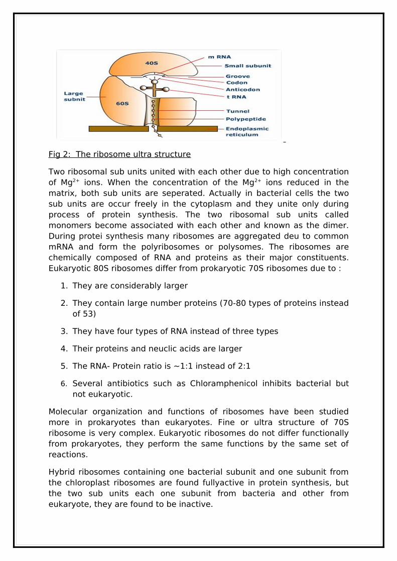
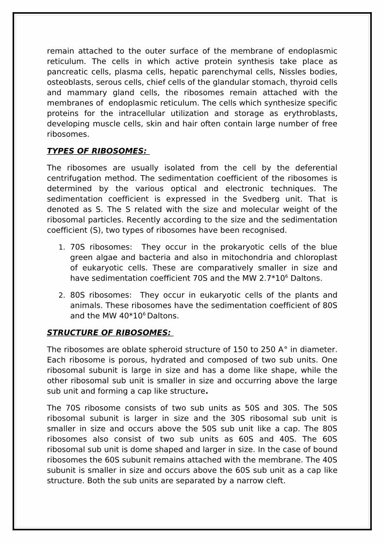
Fig 2: The ribosome ultra structure
Two ribosomal sub units united with each other due to high concentration of Mg2+ ions. When the concentration of the Mg2+ ions reduced in the matrix, both sub units are seperated. Actually in bacterial cells the two sub units are occur freely in the cytoplasm and they unite only during process of protein synthesis. The two ribosomal sub units called monomers become associated with each other and known as the dimer. During protei synthesis many ribosomes are aggregated deu to common mRNA and form the polyribosomes or polysomes. The ribosomes are chemically composed of RNA and proteins as their major constituents. Eukaryotic 80S ribosomes differ from prokaryotic 70S ribosomes due to :
1. They are considerably larger
2. They contain large number proteins (70-80 types of proteins instead of 53)
3. They have four types of RNA instead of three types
4. Their proteins and neuclic acids are larger
5. The RNA- Protein ratio is ~1:1 instead of 2:1
6. Several antibiotics such as Chloramphenicol inhibits bacterial but not eukaryotic.
Molecular organization and functions of ribosomes have been studied more in prokaryotes than eukaryotes. Fine or ultra structure of 70S ribosome is very complex. Eukaryotic ribosomes do not differ functionally from prokaryotes, they perform the same functions by the same set of reactions.
Hybrid ribosomes containing one bacterial subunit and one subunit from the chloroplast ribosomes are found fullyactive in protein synthesis, but the two sub units each one subunit from bacteria and other from eukaryote, they are found to be inactive.
Characteristics of ribosomes of various organisms
SourceIntact
Ribosomes
Ribosome
Subunits
rRNA in Subunits
No: of Proteins
in Subunits
PROKARYOTES
70S 30S50S
16S23S, 5S
2132-34
EUKARYOTES
80S 40S60S
~30~50
Animals 40S60S
18S28S, 5S, 5.8S
Plants 40S60S
18S25-26S, 5S, 5.8S
Fungi 40S60S
Protozoa 40S60S
Table 1: some characteristic of ribosomes of various organisms ((Avers 1976)
Protein synthesisDNA with its own correct mechanism of replication, serves to carry genetic information from cell to cell and from generation to generation. This information is translated in to proteins that determine the phenotype. Proteins are composed of one or more long linear polymer of aminoacid residues - polypeptide chains - that are synthesized almost exclusively in the cytoplasm. In this assignment for how the information present in the sequence of bases – triplet codons – of the mRNA is translated in to sequence of aminoacids in proteins.
There are wide variations in the cells with respect to the quality and quandity of protein synthesised. This largely depends on the need and ability of the cells. Erythrocytes lack the machinary for traslation and cannot synthesize proteins. The normal liver cells are very rich in th eprotein biosynthetic machinary, so the liver regarded as the protein factory in the human body.
TRIPLET CODONS:
The three nucleotide (triplet) base sequence in mRNA that act as code for amino acids in protein constitute the genetic code ar simply codons. The
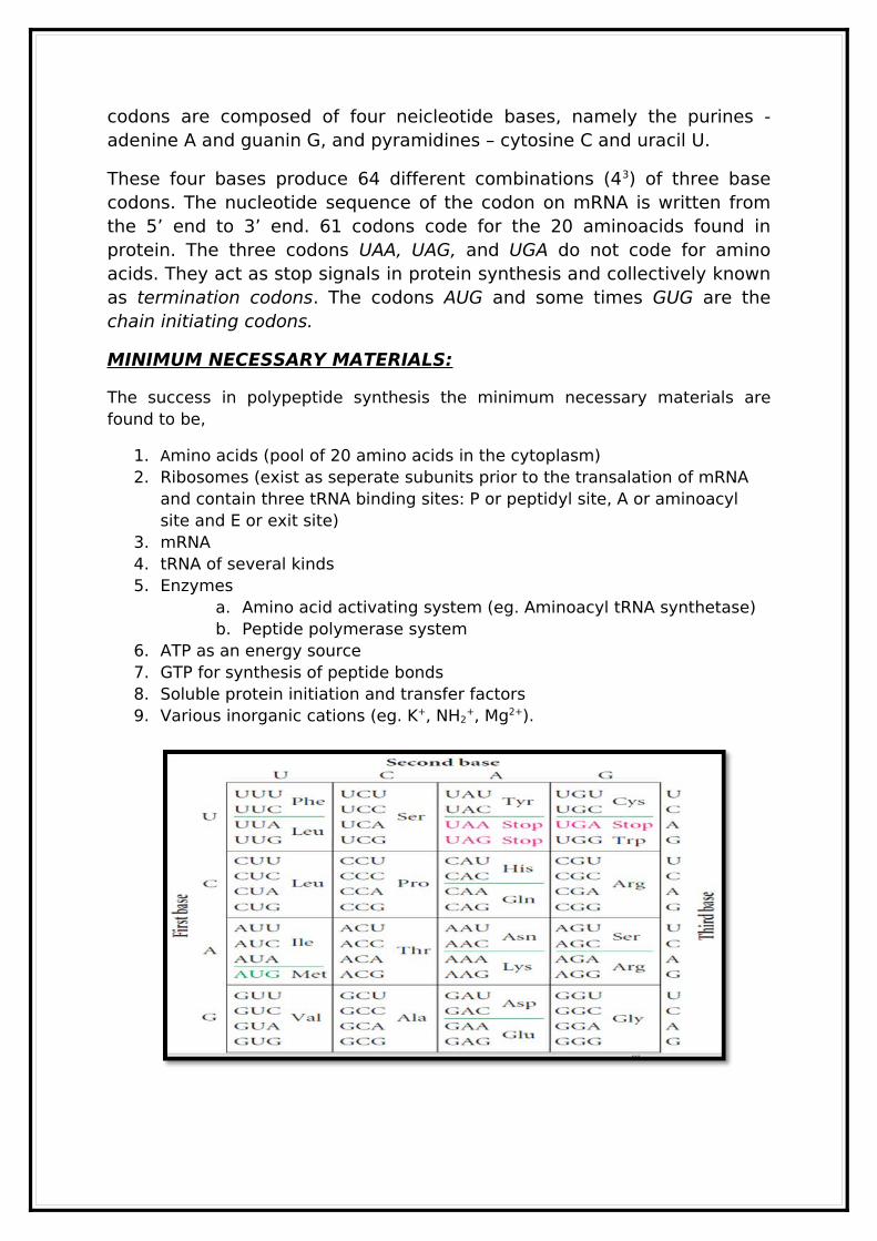
codons are composed of four neicleotide bases, namely the purines - adenine A and guanin G, and pyramidines – cytosine C and uracil U.
These four bases produce 64 different combinations (43) of three base codons. The nucleotide sequence of the codon on mRNA is written from the 5’ end to 3’ end. 61 codons code for the 20 aminoacids found in protein. The three codons UAA, UAG, and UGA do not code for amino acids. They act as stop signals in protein synthesis and collectively known as termination codons. The codons AUG and some times GUG are the chain initiating codons.
MINIMUM NECESSARY MATERIALS:
The success in polypeptide synthesis the minimum necessary materials are found to be,
1. Amino acids (pool of 20 amino acids in the cytoplasm)2. Ribosomes (exist as seperate subunits prior to the transalation of mRNA
and contain three tRNA binding sites: P or peptidyl site, A or aminoacyl site and E or exit site)
3. mRNA4. tRNA of several kinds5. Enzymes
a. Amino acid activating system (eg. Aminoacyl tRNA synthetase)b. Peptide polymerase system
6. ATP as an energy source7. GTP for synthesis of peptide bonds8. Soluble protein initiation and transfer factors9. Various inorganic cations (eg. K+, NH2
+, Mg2+).
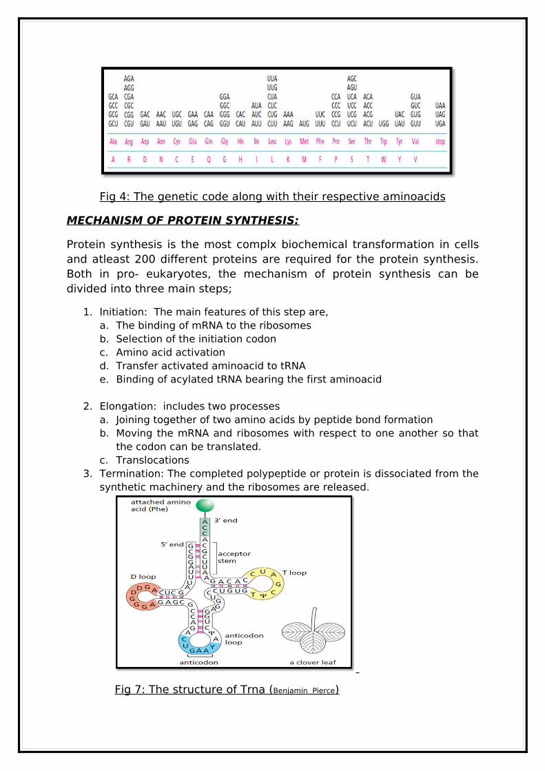
Fig 4: The genetic code along with their respective aminoacids
MECHANISM OF PROTEIN SYNTHESIS:
Protein synthesis is the most complx biochemical transformation in cells and atleast 200 different proteins are required for the protein synthesis. Both in pro- eukaryotes, the mechanism of protein synthesis can be divided into three main steps;
1. Initiation: The main features of this step are,a. The binding of mRNA to the ribosomesb. Selection of the initiation codonc. Amino acid activationd. Transfer activated aminoacid to tRNA e. Binding of acylated tRNA bearing the first aminoacid
2. Elongation: includes two processesa. Joining together of two amino acids by peptide bond formationb. Moving the mRNA and ribosomes with respect to one another so that
the codon can be translated.c. Translocations
3. Termination: The completed polypeptide or protein is dissociated from the synthetic machinery and the ribosomes are released.
Fig 7: The structure of Trna ( Benjamin Pierce )
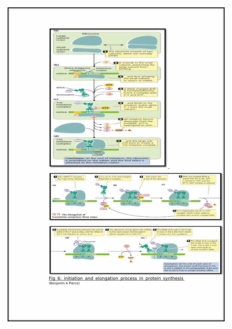
Fig 6: initiation and elongation process in protein synthesis (Benjamin A Pierce)
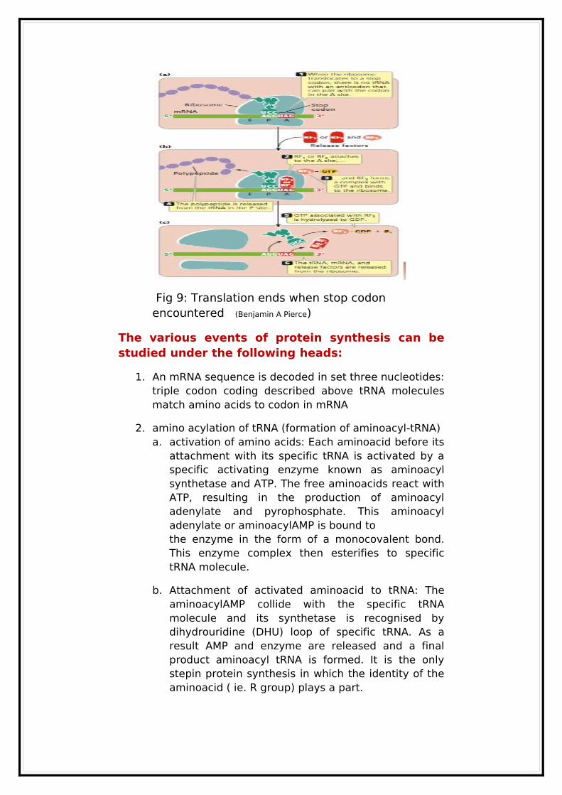
Fig 9: Translation ends when stop codon encountered (Benjamin A Pierce)
The various events of protein synthesis can be studied under the following heads:
1. An mRNA sequence is decoded in set three nucleotides: triple codon coding described above tRNA molecules match amino acids to codon in mRNA
2. amino acylation of tRNA (formation of aminoacyl-tRNA)a. activation of amino acids: Each aminoacid before its
attachment with its specific tRNA is activated by a specific activating enzyme known as aminoacyl synthetase and ATP. The free aminoacids react with ATP, resulting in the production of aminoacyl adenylate and pyrophosphate. This aminoacyl adenylate or aminoacylAMP is bound tothe enzyme in the form of a monocovalent bond. This enzyme complex then esterifies to specific tRNA molecule.
b. Attachment of activated aminoacid to tRNA: The aminoacylAMP collide with the specific tRNA molecule and its synthetase is recognised by dihydrouridine (DHU) loop of specific tRNA. As a result AMP and enzyme are released and a final product aminoacyl tRNA is formed. It is the only stepin protein synthesis in which the identity of the aminoacid ( ie. R group) plays a part.
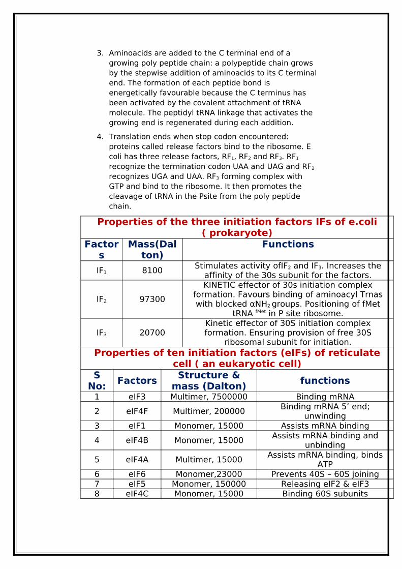
3. Aminoacids are added to the C terminal end of a growing poly peptide chain: a polypeptide chain grows by the stepwise addition of aminoacids to its C terminal end. The formation of each peptide bond is energetically favourable because the C terminus has been activated by the covalent attachment of tRNA molecule. The peptidyl tRNA linkage that activates the growing end is regenerated during each addition.
4. Translation ends when stop codon encountered: proteins called release factors bind to the ribosome. E coli has three release factors, RF1, RF2 and RF3. RF1
recognize the termination codon UAA and UAG and RF2
recognizes UGA and UAA. RF3 forming complex with GTP and bind to the ribosome. It then promotes the cleavage of tRNA in the Psite from the poly peptide chain.
Properties of the three initiation factors IFs of e.coli ( prokaryote)
Factors
Mass(Dalton)
Functions
IF1 8100Stimulates activity ofIF2 and IF3. Increases the
affinity of the 30s subunit for the factors.
IF2 97300
KINETIC effector of 30s initiation complex formation. Favours binding of aminoacyl Trnas with blocked αNH2 groups. Positioning of fMet
tRNA fMet in P site ribosome.
IF3 20700Kinetic effector of 30S initiation complex formation. Ensuring provision of free 30S
ribosomal subunit for initiation.Properties of ten initiation factors (eIFs) of reticulate
cell ( an eukaryotic cell)S
No:Factors Structure &
mass (Dalton)functions
1 eIF3 Multimer, 7500000 Binding mRNA
2 eIF4F Multimer, 200000Binding mRNA 5’ end;
unwinding3 eIF1 Monomer, 15000 Assists mRNA binding
4 eIF4B Monomer, 15000Assists mRNA binding and
unbinding
5 eIF4A Multimer, 15000Assists mRNA binding, binds
ATP6 eIF6 Monomer,23000 Prevents 40S – 60S joining7 eIF5 Monomer, 150000 Releasing eIF2 & eIF38 eIF4C Monomer, 15000 Binding 60S subunits
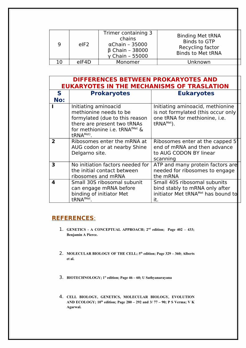
9 eIF2
Trimer containing 3 chains
αChain – 35000β Chain – 38000γ Chain – 55000
Binding Met tRNABinds to GTP
Recycling factorBinds to Met tRNA
10 eIF4D Monomer Unknown
DIFFERENCES BETWEEN PROKARYOTES AND EUKARYOTES IN THE MECHANISMS OF TRASLATION
S No:
Prokaryotes Eukaryotes
I Initiating aminoacid methionine needs to be formylated (due to this reason there are present two tRNAs for methionine i.e. tRNAfMet & tRNAMet).
Initiating aminoacid, methionine is not formylated (this occur only one tRNA for methionine, i.e. tRNAMet).
2 Ribosomes enter the mRNA at AUG codon or at nearby Shine Delgarno site.
Ribosomes enter at the capped 5’ end of mRNA and then advance to AUG CODON BY linear scanning
3 No initiation factors needed for the initial contact between ribosomes and mRNA
ATP and many protein factors are needed for ribosomes to engage the mRNA
4 Small 30S ribosomal subunit can engage mRNA before binding of initiator Met tRNAfMet.
Small 40S ribosomal subunits bind stably to mRNA only after initiator Met tRNAMet has bound to it.
REFERENCES :
1. GENETICS - A CONCEPTUAL APPROACH; 2nd edition; Page 402 – 433;
Benjamin A Pierce.
2. MOLECULAR BIOLOGY OF THE CELL; 5th edition; Page 329 – 360; Alberts
et al.
3. BIOTECHNOLOGY; 1st edition; Page 46 – 60; U Sathyanarayana
4. CELL BIOLOGY, GENETICS, MOLECULAR BIOLOGY, EVOLUTION
AND ECOLOGY; 10th edition; Page 280 – 292 and 3/ 77 – 90; P S Verma; V K Agarwal.
5. GENETICS; 3RD Edition; Page 540 – 562; Monroe W Strickberger.