Embed Size (px)
Citation preview
Proceeding S.Z.P.G.M.I. vol: 18(2): pp. 107-114, 2004.
Rhinocerebral Mucormycosis-An Uncommon but Deadly Disease
Naveed Aslam and Sarfraz Latif Department of ENT and Head & Neck Surgery, Shaikh Zayed Hospital, Lahore
SUMMARY
Muconnycosis is caused by fungi of the order Mucorales and is one of the most rapidly fatal fungal infections known to man. Rhinocerebral mucormycosis is the most common type and its extension to the orbit and brain is quite usual. Location of mucormycosis on the palate is a rare and late occurrence. Rhinocerebral mucormycosis is an invasive fungal infection initiated in the paranasal sinuses that frequently progresses to orbital and brain involvement. If recognized early, involvement is limited to the nasal cavity and paranasal sinuses. Diabetics in poor control are at greatest risk, however, any immunocompromised individual may be infected. We present here two cases of muconnycosis with different presentation and outcome. By presenting these case reports we would like to point out that mucormycosis should be included in the differential diagnosis of hard palate ulcers and orbital cellulitis. They also illustrate the clinical course of this unusual, but potentially fatal, fungal infection. Taxonomy, clinical presentation, diagnosis, and management of mucormycosis of the paranasal sinuses are reviewed in detail.
INTRODUCTION
M Muconnycosis (or zygomycosis) is the term for infection caused by fungi of the order
Mucorales. Mucoraceae may produce severe disease in susceptible individuals, notably patients with diabetes mellitus, leukemia and immunocompromised states. Rhinocerebral mucormycosis most commonly manifests itself in the setting of poorly controlled diabetes, especially with ketoacidosis.1
l
Rhinocerebral mucorycycosis (RCM) is an aggressive fungal infection with a high mortality rate. It frequently develops in patients with uncontrolled diabetes mellitus or immunocompromised patients. RCM typically presents in a rapidly fulminant manner with headache, fever, mucosal necrosis, and ophthalmic symptoms. Although the definitive diagnosis is achieved by histopathological examination, computed tomography (CT) scanning and magnetic resonance (MR) are the best imaging procedures in early diagnosis to assess the extent of the disease.2
Continuous awareness of systemic mycosis in immunocompromised patients is important. Early
diagnosis is based on (direct) histological examination and CT scan. Since treatment should start as early as possible, there is usually no time to await results of tissue cultures. Systemic treatment with amphotericin B and aggressive surgical debridement should be performed as soon as possible, while the place of hyperbaric oxygen and G-CSF remains to be established. In addition to routine preventive measures, prophylactic intranasal application of amphotericin B seems to be of value.3
The mainstays of therapy are reversal of immunosuppression, systemic amphortericin B, and surgical debridement. Survival has improved dramatically, yet deaths still occur if the infection is not recognized and not treated early in its course or if the source of immunocompromise is not reversible.2
> An account of 2 cases of mucormycosis 1s given here with detailed discussion about the subject.
CASE-1
A 45 year old lady, known diabetic for 10 years, presented, with a month's history of left sided
N. Aslam
nasal obstruction with discharge, fever, left orbital pain, swelling and progressive loss of vision on the left side.
On general examination, she was found to be pale with pyrexia of 100 °F. Local examination showed a black necrotic left inferior turbinate with blood stained nasal discharge. She also had left sided proptosis, ophthalmoplegia, visual loss and cellulitis with an area of necrosis of skin of the left cheek.
Investigations showed haemoglobin of I 0.1 gm/di with normal white cell count, serum urea and creatinine and fasting blood sugar of 360 mg/di. CT scan of face showed a destructive lesion filling left nasal cavity, maxillary, ethmoid, & sphenoid sinuses, extending into left orbit and cheek. Provisional diagnosis of mucormycosis was made.
EUA nose & sinuses showed necrotic turbinates and ethmoids on the left side. Necrotic tissue was removed and sent for histology. Biopsy confirmed clinical diagnosis of mucormycosis. Intravenous Amphotericin B was started.
Left total maxillectomy, orbital exenteration was performed with wide excision of necrotic skin of cheek. This was followed few days later with further debridement and reconstruction with Latissimus Dorsi Myocutaneous flap. She recovered well and was discharged home. She remains well and disease free on follow up.
CASE-2
A 13 years old girl, uncontrolled insulin dependent diabetic, presented with 20 days history of, blocked nose, epistaxis, facial swelling and headaches.
Examination showed her to be pale, irritable and drowsy. Local examination showed a necrotic mass in both nostrils with blood stained discharge and ulceration and perforation of hard palate. She had normal vision and ocular movements.
Investigations showed a haemoglobin of 9 .2 gm/ di and normal white cell count. She had +++ sugar in urine with a fasting blood sugar level of 383 mg/di. Provisional diagnosis of fungal rhinocerebral infection was made although possibility of infective rhino-sinusitis was kept in mind.
Management included broad spectrum
intravenous antibiotics, fluids and control of diabetes mellitus. EUA nose showed necrotic tissue filling nasal cavity and biopsies were taken from nose and hard palate. Histology showed it to be mucormycosis.
A CT Scan of face & brain was done to assess the extent of the disease. It showed involvement of nose, maxillary, ethmoid sinuses & cribriform plate with bony erosion of the hard palate. An area of cerebritis in frontal lobe of the brain was also noted.
Treatment was discussed with the family. She was advised intravenous Amphotericin B and radical surgical exenteration of the disease. The family refused any further treatment and took discharge on request.
DISCUSSION
108
Mucormycosis is an infection caused by fungi of the Mucorales order, of which the most relevant family is Mucoraceae, which includes the Rhizopus, Mucor and Absidia genera. It is a severe infection which involves diabetic patients with ketoacidotic decompensation and immunosuppressed patients.4
The clinical infections due to Mucor include rhinocerebral, pulmonary, cutaneous, gastrointestinal and disseminated diseases. The first two are the most common diseases and all entities are associated with a high mortality rate. Hepatic involvement of mucor is rarely reported.5
Mucormycosis refers to the disease caused by a growing number of members of the Mucorales. Typically an airborne infection, primary disease is initiated in the upper or lower airways and is associated with the clinical development of sinusitis, rhinocerebral mucormycosis, or pulmonary infection. Dissemination of infection to skin, brain, and other sites is less common, but direct extension of the infection to contiguous sites is common if patients do not receive aggressive surgical and medical therapy.
Mucormycosis is an opportunistic fungal infection occurring in patients with diabetic ketoacidosis, malignancy, or immunodeficiency, and in those receiving wide-spectrum antibiotics, corticosteroids, or cytotoxic therapy. Mucor most frequently involves the face, and rhinocerebral type
Rhinocerebral Mucormycosis
is the predominating type.9 Risk factors for the development of mucormycosis include diabetic ketoacidosis; neutropenia; protein-calorie malnutrition; and iron overload, with or without the concomitant use of deferoxamine (Table I). This last association has only recently been recognized and has emerged as a major life-threatening complication for patients who are undergoing hemodialysis. Intravenous drug abusers may inject spores of Mucorales with their drugs and may present with space-occupying lesions of the CNS. The underlying immunologic defects that are responsible for predisposing different populations of patients to the development of mucorrnycosis are not well understood, and there is no unifying theory to explain why most individuals have innate immunity to this group of fungi. 8
Table I : Predisposing risk factors for Mucormycosis
Predisposing Factors
• Uncontrolled diabetes mellitus
• Long term steroid therapy
• Immuno-suppression
• Chemotherapy
• Organ transplantation
• Haematological malignancies
• Uraemia
• Severe malnutrition
• Severe bums
• Long term deferoxamine therapy
• AIDS
Rhinocerebral mucormycosis is recognized as a potentially aggressive and commonly fatal fungal infection. The classic presentation is involvement of nasal mucosa with invasion of the paranasal sinuses and orbit. Mucormycosis is most commonly seen in association with diabetic ketoacidosis, but disease demographics have changed with the onset of AIDS and the advent of powerful immunosuppressive drugs.7 Mucorrnycosis should be considered as a differential diagnosis of orbital apex syndrome especially in untreated diabetic patients complaining of diplopia and headache. Neurological examination may demonstrate abducent nerve palsy. MRI may show a mass lesion in the orbital apex. Total ophthalmoplegia and visual Joss rapidly develops. A
109
craniotomy is recommended to decompress the orbital apex and remove the mass.8
The diagnosis of mucorrnycosis is very difficult to make in cases in which the typical presentation and classical signs are not present. It has to be differentiated from some more common conditions (Table 2) and a high index of suspicion, based on identified risk factors, may assist in more rapid diagnosis of this life-threatening mycosis.9
Table 2: Differential diagnosis of rhinocerebral mucormycosis.
Differential Diagnosis • Acute sinusitis ( maxillary, ethmoid) • Orbital cellulitis • Allergic fungal sinusitis ( Aspergillosis ) • Nasal I sinus malignancy • Cavernous sinus thrombosis • Orbital tumours
Rhinocerebral mucormycosis has been reported in patients with membranous glomerulonephritis. 10 Mucormycosis in HIV is rare. However, it can be the presenting opportunistic infection in AIDS. Predisposing factors for Mucor infection in HIV disease include low CD4 count, neutropenia, and active intravenous drug use. Mucormycosis can present in the basal ganglia, the skin, the gastrointestinal tract, the respiratory tract, or may be disseminated. The disease may develop insidiously or may progress rapidly with a fulminant course.11 In patients with burns the rhinocerebral form is rare, and mucormycotic infections more commonly involve the cutaneous bum wound. Both for'lls are associated with a high mortality rate that increases with delays in treatment. 12 Rhinocerebral mucormycosis has followed donor leukocyte infusion in bone marrow transplanted patients. 13
Sphenoidal mucormycosis in an otherwise healthy individual has been reported where headache was the only early symptom. It emphasizes the importance of a high index of suspicion for early diagnosis and prompt management.1
Zygomycosis is a rare but highly invasive fungal infection that occurs in liver transplant recipients. There are reported cases of invasive gastrointestinal zygomycosis that occurred in a
N. Aslam
heavily immunosuppressed liver transplant recipient Knowledge of the diverse clinical manifestations (including gastrointestinal involvement) and predisposing factors in transplant recipients with zygomycosis can aid in early recognition of this disease in this patient population.15
Rhinocerebral mucormycosis has been reported during deferoxamine therapy. 16 In a study
. of 59 cases on dialysis, the presenting forms of mucormycosis included disseminated in 44%, rhinocerebral in 31 %, and other forms in 25%. The diagnosis was made during life in only 39%, while the diagnosis was discovered at autopsy in 61 % of the cases. The fungus, cultured in only 36%, was always Rhizopus. The infection was fatal in 86% of cases. No known risk factors for fungal infections, eg, diabetes mellitus, liver disease, splenectomy, neutropenia, steroid therapy, or other immunosuppressive therapy, were present in 70% of patients, but 78% of patients were being treated with deferoxamine. The role played by this drug and more particularly by its iron chelate, feroxamine, in the pathogenesis of mucormycosis in these patients is underscored. Because of this risk, deferoxamine therapy in dialysis patients should be limited to severe aluminum toxicity, the deferoxamine should be given at the lowest possible dose, and dialytic methods to augment the removal of feroxamine should be studied. 17
'18
Mucormycosis involves arteries causing, dissection of internal elastic lamina from media, extensive endothelial damage and intra-vascular thrombosis. It results in extensive necrosis, propagating thrombo-phlebitis and may involve major blood vessels, bones, nerves, meninges and brain. Infection may extend locally by destruction of bone into orbit, cranial cavity and can cause extensive necrosis of soft tissues. It can have local intra vascular spread causing intra vascular propagating th.rombophlebitis, and may involve major blood vessels like internal carotid artery,21
internal jugular vein or cavernous sinus.22 Mucor cerebral abscess associated with intravenous drug abuse has been reported.23 It can have a distan_t. spread to lungs, GIT etc.
Invasive zygomycosis is rapidly progressive and is associated with angioinvasion and infarction. Invasive disease requires emergent surgical and
110
medical intervention. Invasive disease is characterized by prominent infarcts (94%), angioinvasion ( 100% ), and, surprisingly, prominent perineural invasion (90%) in biopsies that contained nerves for evaluation. Perineural invasion is a common finding in invasive zygomycosis, as are angioinvasion and infarcts. Therefore, prior to excluding the presence of these fungi in biopsies suspected to contain zygomycetes, the perineural space should be carefully examined.19
Nasal scrapings and fine-needle aspiration cytology (FNAC) of the paranasal masses can show fungal hyphae morphologically resembling Mucor. Surgical material shows features of mucormycosis. FNAC and scrape smears can give a conclusive diagnosis of mucormycosis, and the patient can be treated with appropriate antifungal therapy and surgical debridement. Preoperative cytology is an effective technique to establish a diagnosis of mucormycosis and may obviate the need for a preoperative biopsy in some cases.20
CT and MRI scans are extremely useful in assessing extent of the disease. Non-enhancement of the superior ophthalmic vein and ophthalmic artery appears to be a CT sign specific to this disease. CT gives better bony details and is usually the first line investigation while assessing inflammatory changes in the paranasal sinuses and orbit. Extension of the infection into the cavernous sinus, with cavernous sinus thrombosis and internal carotid artery narrowing, is well demonstrated on pre- and postgadolinium MR images. Infarctions in the anterior choroidal artery distribution suggests intracranial invasion of basal arteries. Magnetic resonance can effectively demonstrate the wide array of findings possible in rhinoorbitocerebral mucormycosis.25 On MRI, an extremely low signal is detected from the fungal mass. This appearance is a result of high concentrations of iron and manganese. This low-signal area simulates air in paranasal sinuses; however, CT displays a mildly enhancing soft tissue mass and allows the correct diagnosis to be made.26 Invasion of internal carotid artery, cavernous sinus or ophthalmic artery may be demonstrated on MR angiography.27
~Therapy usually consists of surgical debridemenhist . I exc1s10n accompanied by intravenous amphotericin B.11 Dosage and method
Rhinocerebral M ucormycosis
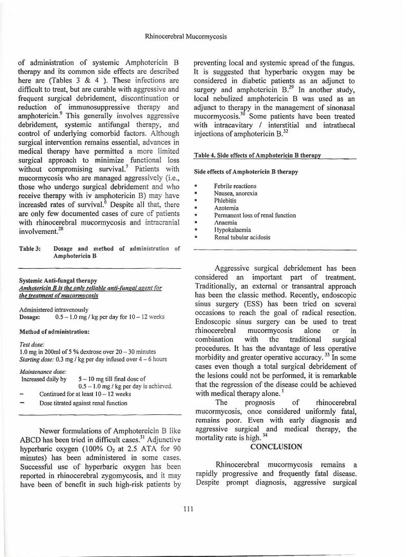
of administration of systemic Amphotericin B therapy and its common side effects are described here are (Tables 3 & 4 ). These infections are difficult to treat, but are curable with aggressive and frequent surgical debridement, discontinuation or reduction of immunosuppressive therapy and amphotericin.9 This generally involves aggressive debridement, systemic antifungal therapy, and control of underlying comorbid factors. Although surgical intervention remains essential, advances in medical therapy have permitted a more limited surgical approach to minimize functional loss without compromising survival.7 Patients with mucormycosis who are managed aggressively (i.e., those who undergo surgical debridement and who receive therapy with iv amphotericin B) may have increased rates of survival.6 Despite all that, there are only few documented cases of cure of patients with rhinocerebral mucormycosis and intracranial involvement.28
Table3: Dosage and method of administration of Amphotericin B
Systemic Anti-fungal therapy Amhotericin Bis the onlv reliable anti-fungal agent (or the treatment o(mucormvcosis
Administered intravenously Dosage: 0.5 - 1.0 mg I kg per day for I 0 - 12 weeks
Method of administration:
Test dose: 1.0 mg in 200ml of 5 % dextrose over 20 - 30 minutes Starting dose: 0.3 mg I kg per day infused over 4 - 6 hours
Maintenance dose: Increased daily by 5 - 10 mg till final dose of
0.5 - 1.0 mg I kg per day is achieved. Continued for at least I 0 - 12 weeks Dose titrated against renal function
Newer formulations of Amphotereicin B like ABCD has been tried in difficult cases.31 Adjunctive hyperbaric oxygen ( l 00% 0 2 at 2.5 AT A for 90 minutes) has been administered in some cases. Successful use of hyperbaric oxygen has been reported in rhinocerebral zygomycosis, and it may have been of benefit in such high-risk patients by
111
preventing local and systemic spread of the fungus. It is suggested that hyperbaric oxygen may be considered in diabetic patients as an adjunct to surgery and amphotericin B.29 In another study, local nebulized amphotericin B was used as an adjunct to therafy in the management of sinonasal mucormycosis.3 Some patients have been treated with intracavitary I interstitial and intrathecal injections of amphotericin B.32
Table 4. Side effects of Amphotericin B therapy
Side effects of Amphotcricin B therapy
• Febrile reactions • Nausea, anorexia
Phlebitis • Azotemia
• •
Permanent loss of renal function Anaemia Hypokalaemia Renal tubular acidosis
Aggressive surgical debridement has been considered an important part of treatment. Traditionally, an external or transantral approach has been the classic method. Recently, endoscopic sinus surgery (ESS) has been tried on several occasions to reach the goal of radical resection. Endoscopic sinus surgery can be used to treat rhinocerebral mucormycosis alone or in combination with the traditional surgical procedures. It has the advantage of less operative m~rbidity and greater operative accuracy. 33 In some cases even though a total surgical debridement of the lesions could not be performed, it is remarkable that the regression of the disease could be achieved with medical therapy alone. 1
The prognosis of rhinocerebral mucormycosis, once considered uniformly fatal, remains poor. Even with early diagnosis and aggressive surgical and medical therapy, the mortality rate is high. 34
CONCLUSION
Rhinocerebral mucormycosis remains a rapidly progressive and frequently fatal disease. Despite prompt diagnosis, aggressive surgical
N. Aslam
debridement, therapy with amphotericin B, and correction of metabolic acidosis, mortality rate is over 80%. In some studies, use of amphotericin B, combined with aggressive surgical debridement, has increased survival rates from approximately 20% to 70%.35
Long-term survival with invasive rhinocerebral mucormycosis is rare, but possible, with aggressive multimodality treatment, including carotid sacrifice for en bloc resection of the pathology, when indicated.34 Combined treatment is mandatory for these patients. It usually results in death, but powerful efforts may save the patient. 36
We conclude that with an infection as morbid as rhinocerebral mucormycosis, it is advisable to use surgical debridement and all available routes for delivering amphotericin B to infected cerebral parenchyma and other tissues, which include intravenous, intracavitary I interstitial, and cerebrospinal fluid perfusion pathways. 32
REFERENCES
1. Cagatay AA, Oncu SS, Calangu SS, Yildirmak TI, Ozsut HH, Eraksoy HH. Rhinocerebral mucormycosis treated with 32 gram liposomal amphotericin B and incomplete surgery: a case report. : BMC Infect Dis 2001; 1(1 ): 22
2. Alobid I, Bernal M, Calvo C, Vilaseca I, Berenguer J, Alos L. Treatment of rhinocerebral mucormycosis by combination of endoscopic sinus debridement and amphotericin B. Am J Rhino! 2001 Sep-Oct; 15(5): 327-31
3. Schmidt JM, Poublon RM. Rhinocerebral mycosis in immunocompromised patients. A case report and review of the literature. Rhinology 1998 Jun; 36(2): 90-3
4. del Rio Perez 0, Santin Cerezales M, Manos M, Rufi Rigau G, Gudiol Munte F. [Mucormycosis: a classical infection with a high mortality rate. Report of 5 cases] Rev Clin Esp 2001Apr;201(4): 184-7
5. Suh IW, Park CS, Lee MS, Lee JH, Chang MS, Woo JH, Lee IC, Ryu JS. Hepatic and small bowel mucormycosis after chemotherapy in a patient with acute
lymphocytic J Korean Med Sci 2000 Jun; 15(3): 351-41.
6. Sugar AM. Mucormycosis. Clin Infect Dis 1992 Mar;14 Suppl l:S126-9
7. Peterson KL, Wang M, Canalis RF, Abemayor E. Rhinocerebral mucormycosis: evolution of the disease and treatment options. Laryngoscope 1997 Jul; I 07(7): 855-62
8. Umemura A, Suzuka T. [A case of rhinocerebral mucormycosis presenting orbital apex syndrome] No Shinkei Geka 1998 May; 26(5): 439-42
9. Webb M, Dowdy L, Bundschu C, Nery J, Schiff E, Tzakis AG. Cerebral mucormycosis after liver transplantation: a case report. Clin Transplant 1998 Dec; 12(6): 596-9
10. Akcay A, Altun B, Usalan C, Gunes D, Ulusoy S, Kiykim AA, Erdem Y, Yasavul U, Turgan C, Caglar S. Rhinocerebral mucormycosis in a patient with membranous glomerulonephritis. Nephron 2000 Nov; 86(3): 352-3
11 . Moraru RA, Grossman ME. Palatal necrosis in an AIDS patient: a case of mucormycosis. Cut is 2000 Jul; 66( 1):15-8
112
12. Stem LE, Kagan RJ. Rhinocerebral mucormycosis in patients with bums: case report and review of the literature. J Bum Care Rehabil 1999 Jul-Aug;20( 4):303-6
13. Penalver FJ, Romero R, Fores R, Cabrera R, Diez-Martin JL, Regidor C, Fernandez MN. Rhinocerebral mucormycosis following donor leukocyte infusion: successful treatment with liposomal amphotericin B and surgical debridement. Bone Marrow Transplant 1998 Oct; 22(8): 817-8
14. Del Valle Zapico A, Rubio Suarez A, Mellado Encinas P, Morales Angulo C, Cabrera Pozuelo E. Mucormycosis of the sphenoid sinus in an otherwise healthy patient. Case report and literature review. J Laryngol Otol 1996 May; 110(5): 471-3
15. Singh N, Gayowski T, Singh J, Yu VL. Invasive gastrointestinal zygomycosis in a liver transplant recipient: case report and review of zygomycosis in solid-organ transplant recipients. Clin Infect Dis 1995
Rhinocerebral Mucormycosis
Mar; 20(3): 617-20 16. Ammon A, Rumpf KW, Hommerich CP,
Behrens-Baumann W, Ruchel R. [Rhinocerebral] mucormycos1s during deferoxamine therapy. Dtsch Med Wochenschr 1992 Sep 18; 117(38): 1434-8
17. Boelaert JR, Fenves AZ, Coburn JW. Deferoxamine therapy and mucormycosis in dialysis patients: report of an international registry. Am J Kidney Dis 1991; 18(6): 660-7
18. Kim J, Fortson JK, Cook HE. A fatal outcome from rhinocerebral mucormycosis after dental extractions: a case report. J Oral Maxillofac Surg 2001 Jun; 59(6): 693-7
19. Frater JL, Hall GS, Procop GW. Histologic features of zygomycos1s: emphasis on perineural invasion and fungal morphology. Arch Pathol Lab Med 2001 Mar; 125(3): 375-8
20. Deshpande AH, Munshi MM. Rhinocerebral mucormycosis diagnosis by aspiration cytology. Diagn Cytopathol 2000 Aug; 23(2): 97-100
21. Galetta SL, Wulc AE, Goldberg HI, Nichols CW, Glaser JS. Rhinocerebral mucormycosis: management and survival after carotid occlusion. Ann Neural 1990 J ul;28( 1):103-7
22. Onerci M, Gursel B, Hosal S, Gulekon N, Gokoz A. Rhinocerebral mucormycosis with extension to the cavernous sinus. A case report. Rhinology 1991 Dec;29( 4):321-4
23. Fong KM, Seneviratne EM, McCormack JG. Mucor cerebral abscess associated with intravenous drug abuse. Aust N Z J Med 1990 Feb; 20( 1 ):74-7
24. Kilpatrick C, Tress B, King J. Computed tomography of rhinocerebral mucormycosis. Neuroradiology 1984; 26(1):71-3
25. Yousem DM, Galetta SL, Gusnard DA, Goldberg HI. MR findings in rhinocerebral mucormycosis. J Comput Assist Tomogr 1989 Sep-Oct; 13(5): 878-82
26. Terk MR, Underwood DJ, Zee CS, Colletti PM. MR imaging m rhinocerebral and intracranial mucormycos1s with CT and pathologic correlation. Magn Reson Imaging 1992; 10(1):81-7
27. O'Brien TJ, McKelvie P. Rhinocerebral
mucormycosis presenting as periorbital cellulitis with blindness: report of 2 cases. Cl in Exp Neural 1994;31 :68-78
28. Strasser MD, Kennedy RJ, Adam RD Rhinocerebral mucormycosis. Therapy with amphotericin ·B lipid complex. Arch Intern Med 1996 Feb 12; 156(3): 337-9
29. Bentur Y, Shupak A, Ramon Y, Abramovich A, Wolfin G, Stein H, Krivoi N. Hyperbaric oxygen therapy for cutaneous/soft-tissue zygomycosis complicating diabetes mellitus. Plast Reconstr Surg 1998 Sep; 102(3): 822-4
30. Raj P, Vella EJ, Bickerton RC. Successful treatment of rhinocerebral mucormycosis by a combination of aggressive surgical debridement and the use of systemic liposomal amphotericin B and local therapy with nebulized amphotericin--a case report. J Laryngol Otol 1998 Apr; 112(4): 367-70
31. Moses AE, Rahav G, Barenholz Y, Elidan J, Azaz B, Gillis S, Brickman M, Polacheck I, Shapiro M. Rhinocerebral mucormycosis treated with amphotericin B colloidal dispersion in three patients. Clin Infect Dis 1998 Jun; 26(6): 1430-3
32. Adler DE, Milhorat TH, Miller JI. Treatment of rhinocerebral mucormycosis with intravenous interstitial, and cerebrospinal fluid administration of amphotericin B: case report. Neurosurgery 1998 Mar; 42(3): 644-8; discussion 648-9
113
33. Jiang RS, Hsu CY. Endoscopic sinus surgery for rhinocerebral mucormycosis. Am J Rhino) 1999 Mar-Apr; 13(2): I 05-9
34. Alleyne CH Jr, Vishteh AG, Spetzler RF, Detwiler PW. Long-term survival of a patient with invasive cranial base rhinocerebral mucormycosis treated with combined endovascular, surgical, and medical therapies: case report. Neurosurgery 1999 Dec; 45(6): 1461-3; discussion 1463-4
35. Ochi JW, Harris JP, Feldman JI, Press GA. Rhinocerebral mucormycosis: results of aggressive surgical debridement and amphotericin B. Laryngoscope 1988 Dec; 98(12): 1339-42
36. Akoz T, Civelek B, Akan M. Rhinocerebral mucormycosis: report of two cases. Ann Plast
Surg 1999 Sep; 43(3): 309-12
The Authors:
Naveed Aslam Associate Professor Department of ENT and Head & Neck Surgery, Shaikh Zayed Hospital, Lahore, Pakistan
Sarfraz Latif Medical Officer,
N. Aslam
114
Department of ENT and Head & Neck Surgery, Shaikh Zayed Hospital, Lahore, Pakistan
Address for Correspondence:
Naveed Aslam Associate Professor Department of ENT and Head & Neck Surgery, Shaikh Zayed Hospital, Lahore, Pakistan

![Diagnosis of an intestinal mucormycosis ‘fungus …...CASE REPORT Open Access Diagnosis of an intestinal mucormycosis ‘fungus ball’ located with PET/CT with [18F]FDG-PET/CT Franklin](https://img.pdfslide.us/doc/110x75/5e6ffc94e53b4860491a2263/diagnosis-of-an-intestinal-mucormycosis-afungus-case-report-open-access-diagnosis.jpg)