Embed Size (px)
Citation preview
1
Rheumatology Tips and Pearls
Andrew J. Gross, MD
Rheumatology Clinic Chief
Associate Clinical Professor
University of California, San Francisco
Disclosures
• None

2
Objectives
• Recognize the key features of polymyalgia rheumatica
• Recognize inflammatory back pain
• Know the differential diagnosis of subacute monoarticular arthritis
Clinical Case #1
• A 66 year old man comes to see you complaining of shoulder pain. The pain came on suddenly about 3 weeks ago, initially affecting his right shoulder and then the left. The pain radiates down into the upper arms and somewhat across his upper back and is exacerbated by shoulder abduction.
• He also complains of new onset lower back and hip discomfort.

3
Clinical Case #1 - Question
You diagnose him with Polymyalgia Rheumatica (PMR). All of the following symptoms tipped you off to the diagnosis of PMR EXCEPT:
a. Morning stiffness lasting >45 minutes
b. Pain & stiffness affects the lower back and pelvic girdle
c. Pain & stiffness improves with activity
d. ESR >40 mm/hr
e. ANA 1:320 speckled pattern
Dasgupta B, et al, Ann Rheum Dis 2012, EULAR/ACR Classification Criteria
Some Tips about PMR
• Typical distribution of PMR symptoms…
• Subdeltoid bursitis & biceps tenosynovitis are common in one or both shoulders
• Patients may develop adhesive capsulitis
Salvarani, C, et al, Nat Rev Rheumatol, 2012, PMID 22825731

4
Some more Tips about PMR
• PMR is uncommon in patients < 60 years old97 cases of PMR identified during a 10 year study from Olmstead County, Minnesota
0-49 years 1 in a million
50-59 years 1 in 5,000
60-69 years 1 in 2,000
70-79 years 1 in 900
Chuang TY, et al, Ann Intern Med 1983, PMID 6982645
Some more Tips about PMR
• PMR is uncommon in patients < 60 years old
• ESR is helpful - but it is <40 mm/hr in 10-20% of patients
– CRP can be helpful when ESR is <40
• ANA test is not associated with PMR (but is more commonly positive in older adults)
Dasgupta B, et al, Ann Rheum Dis 2012, EULAR/ACR Classification Criteria

5
Some more Tips about PMR
• PMR is uncommon in patients < 60 years old
• ESR is helpful - but it is <40 mm/hr in 10-20% of patients
– CRP can be helpful when ESR is <40
• 15% will have Giant Cell Arteritis (new onset head pain)– New onset head pain
– Scalp tenderness
– Jaw claudication when chewing
– Sudden vision loss or diplopia
Dasgupta B, et al, Ann Rheum Dis 2012, EULAR/ACR Classification Criteria
Things patients with PMR often tell me
• “I feel like I am 100 years old!”
• “I need to crawl out of bed in the morning”
• “I feel okay as long as I keep moving, but I stiffen up like the Tin-man as soon as I sit down”
• “That prednisone is a miracle”

6
When To Refer PMR to a rheumatologist:
Rheumatologists are pleased to see cases of PMR
Consider referring when:
Your patient has only a partial response to treatment with prednisone – most patients should have a very good response to 15-20 mg/d of prednisone.
Your patient reflares whenever you try to taper the prednisone dose
Your patient has any symptoms of Giant Cell Arteritis (and send to an ophthalmologist for consideration of temporal artery biopsy).
Clinical Case #2
• A 26 year old man comes to see you complaining of shoulder pain. The pain came on about 3 weeks ago, initially affecting his right shoulder and then the left. The pain does not radiate. Range of motion of motion of both shoulders is limited.
• He also notices pain and stiffness in his neck and lower back. This is worse recently, but has been present on an off for the past couple of years.
• He complains of a hour of morning stiffness in his shoulders and low back.
7
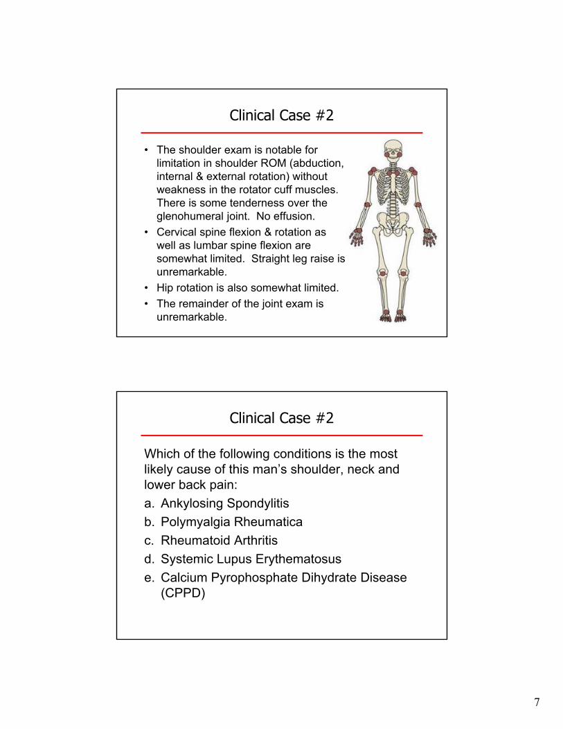
Clinical Case #2
• The shoulder exam is notable for limitation in shoulder ROM (abduction, internal & external rotation) without weakness in the rotator cuff muscles. There is some tenderness over the glenohumeral joint. No effusion.
• Cervical spine flexion & rotation as well as lumbar spine flexion are somewhat limited. Straight leg raise is unremarkable.
• Hip rotation is also somewhat limited.
• The remainder of the joint exam is unremarkable.
Clinical Case #2
Which of the following conditions is the most likely cause of this man’s shoulder, neck and lower back pain:
a. Ankylosing Spondylitis
b. Polymyalgia Rheumatica
c. Rheumatoid Arthritis
d. Systemic Lupus Erythematosus
e. Calcium Pyrophosphate Dihydrate Disease (CPPD)

8
Typical distribution of involved joints in rheumatoid arthritis
(and lupus)
www.studyblue.com
https://dundeemedstudentnotes.wordpress.com/2014/06/16/polyarthritis/
Rheumatoid Arthritis
Psoriatic Arthritis
Ankylosing Spondylitis
Osteoarthritis

9
Ankylosing Spondylitis
Ankylosing Spondylitis - sacroiliitis

10
AS – “bamboo spine”
Back pain
Sacroiliitis on MRI
Back pain
Radiographic sacroiliitis
Back painSyndesmophytes
DIAGNOSIS
Ankylosing Spondylitis
Rudwaliet M, et al. Arthritis Rheum. 2005;52(4):1000-1008.
Non-radiographic stage Radiographic stage
Time (years)

11
Clinical Case #2
All of the following symptoms are associated with Ankylosing Spondylitis EXCEPT:
a. Pain & stiffness improve with exercise.
b. Onset of back pain was insidious
c. Back pain & stiffness gets worse at night
d. Burning pain in the thighs with standing
e. Symptoms began before age 40
Inflammatory Back Pain: Hallmark Features
Feature Odds Ratios
Insidious onset 12.7
Pain at night (with improvement upon getting up) 20.4
Age at onset <40 years 9.9
Improvement with exercise 23.1
No improvement with rest 7.7
Sensitivity 79.6% & Specificity 72.4%Positive LR = 79.6/(100-72.4) = 2.9 ~ Probability = 14%
Sieper J, et al, Ann Rheum Dis 2009, PMID 19147614Rudwaleit M, et al. Ann Rheum Dis. 2009; 68(6):777-83. Ozgocmen S, et al. J Rheumatol. 2010;37(9):1978.
LR=likelihood ratio

12
When to refer a patient with back pain to a rheumatologist
Inflammatory Back Pain Plus:
• HLA-B27+ (present in 85-95% of patients with AS)
• Family history of Ankylosing Spondylitis
• Elevated c-reactive protein (CRP)
• Sacroiliitis on imaging (x-rays or MR)
Poddubnyy D, van Tubergen A, Landewé R, et al. Ann Rheum Dis 2015;74:1483–1487
AS: Treatment
NSAID NSAIDs sulfasalazine TNF inhibitors
Axial disease only
Physical Therapy
Braun J, et al.,, Ann Rheum Dis 2011; 70: 896-904; van der Heijde D, et al, Ann Rheum Dis 2011; 70:905-08

13
Clinical Case #3
• 45 year old man comes to see you with left knee swelling for the past 7 days. He has no other complaints. No recent or prior trauma.
• ROS is unremarkable. No fevers or rashes
• Physical Exam: unremarkable except for swelling and warmth of the left knee with limited ROM.
Clinical Case #3
To identify the cause of the knee swelling, what is the best next test to obtain:
A. Aspirate Knee Fluid for cell count and crystal search
B. MRI of knee
C. X-ray of knee
D. CBC with Differential
E. Rheumatoid factor & CCP antibody
14
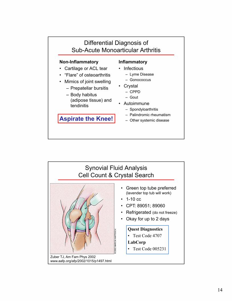
Differential Diagnosis of Sub-Acute Monoarticular Arthritis
Non-Inflammatory
• Cartilage or ACL tear
• “Flare” of osteoarthritis
• Mimics of joint swelling
– Prepatellar bursitis
– Body habitus (adipose tissue) and tendinitis
Inflammatory
• Infectious– Lyme Disease
– Gonococcus
• Crystal– CPPD
– Gout
• Autoimmune– Spondyloarthritis
– Palindromic rheumatism
– Other systemic diseaseAspirate the Knee!
Synovial Fluid AnalysisCell Count & Crystal Search
Quest Diagnostics
• Test Code 4707
LabCorp
• Test Code 005231
• Green top tube preferred (lavender top tub will work)
• 1-10 cc
• CPT: 89051; 89060
• Refrigerated (do not freeze)
• Okay for up to 2 days
Zuber TJ, Am Fam Phys 2002www.aafp.org/afp/2002/1015/p1497.html

15
Synovial Fluid AnalysisCell Count & Crystal Search
Zuber TJ, Am Fam Phys 2002www.aafp.org/afp/2002/1015/p1497.html
Type
Non-Inflammatory
e.g. osteoarthritis
Inflammatorye.g.
rheumatoid arthritis
Infectiouse.g.
crystal or septic
Appear-ance
ClearViscousamber
Turbidyellow
less viscous
Turbidyellow
less viscous
WBC<2000
cells/mm32000 - 50,000
cells/mm3>50,000
cells/mm3
Cell Type
MononuclearPMNs and/or
lymphocytesPMNs
Synovial Fluid AnalysisCell Count & Crystal Search
Zuber TJ, Am Fam Phys 2002www.aafp.org/afp/2002/1015/p1497.html

16
Tips on subacute septic arthritis
Erythema Chronicum Migrans
Tips on subacute septic arthritis
Lyme Disease• Unlikely unless traveled to
Lyme endemic region
• Initial phase with erythema migrans rash & sometimes fever and diffuse arthralgia
• If untreated, later can develop monoarticular arthritis, usually of the knee
• Lyme ELISA & WB will be strongly positive
• No role for testing joint fluid www.findarthritistreatment.com/eight-causes-of-migrating-arthritis/
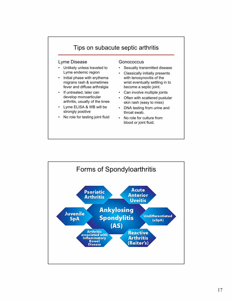
17
Tips on subacute septic arthritis
Lyme Disease• Unlikely unless traveled to
Lyme endemic region
• Initial phase with erythema migrans rash & sometimes fever and diffuse arthralgia
• If untreated, later can develop monoarticular arthritis, usually of the knee
• Lyme ELISA & WB will be strongly positive
• No role for testing joint fluid
Gonococcus• Sexually transmitted disease
• Classically initially presents with tenosynovitis of the wrist eventually settling in to become a septic joint.
• Can involve multiple joints
• Often with scattered pustularskin rash (easy to miss)
• DNA testing from urine and throat swab.
• No role for culture from blood or joint fluid.
Forms of Spondyloarthritis

18
Tips on spondyloarthritis
Reactive arthritis• Sterile oligoarticular arthritis,
usually of lower extremities
• Develops 10-14 daysfollowing an infectious process, usually dysentery or chlamydia urethritis
• Sometimes associated with– Conjunctivitis or uveitis
– Urethritis (independent of Chlamydia)
• More than 50% of cases will resolve in <6 months.
Psoriatic Arthritis• Occurs in 15% of patients
with psoriasis
• More common in people with psoriasis affecting the scalp or diffuse severe disease
Tips on spondyloarthritis
Reactive arthritis• Sterile oligoarticular arthritis,
usually of lower extremities
• Develops 10-14 days following an infectious process, usually dysentery or chlamydia urethritis
• Sometimes associated with– Conjunctivitis or uveitis
– Urethritis (independent of Chlamydia)
• More than 50% of cases will resolve in <6 months.
Psoriatic Arthritis• Occurs in 15% of patients
with psoriasis
• More common in people with psoriasis affecting the scalp or diffuse severe disease

19
Clinical Case #5
A B
D
A
B
C D
T2 MRI
Case 5: A 50 year old healthy active woman with severe exacerbation of chronic right shoulder pain. Which image is most likely associated with her disorder?

20
A
B
C D
A. Rheumatoid arthritis (late disease)
B. Milwaukee Shoulder Syndrome (apatite-associated destructive arthritis)
C. Calcific Tendinitis
D. Rotator cuff tear
T2 MRIroentgenrayreader.blogspot.com
Summary
• Don’t diagnose patients <50 y.o. with PMR• Recognize inflammatory back pain• Aspirate swollen joints• Recognize calcific tendinitis

21
Thanks!
Bonus Slides
22
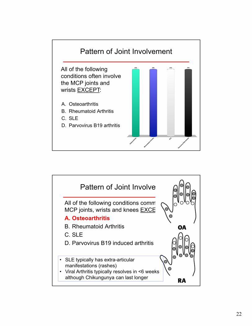
Pattern of Joint Involvement
A. Osteoarthritis
B. Rheumatoid Arthritis
C. SLE
D. Parvovirus B19 arthritis
All of the following conditions often involve the MCP joints and wrists EXCEPT:
Pattern of Joint Involvement
All of the following conditions commonly involve MCP joints, wrists and knees EXCEPT:
A. Osteoarthritis
B. Rheumatoid Arthritis
C. SLE
D. Parvovirus B19 induced arthritis
• SLE typically has extra-articular manifestations (rashes)
• Viral Arthritis typically resolves in <6 weeks although Chikungunya can last longer

23
http://www.mridoc.com/mskatlas/Arthritis/Arthritis_Common_Joints_Involved/
Osteoarthritis
• Osteoarthritis of the hands is common and rheumatology consultation is usually not necessary. It can be managed with:– Acetaminophen 1 gm three times a day
– NSAIDs if normal kidney function and no risk factors for gastritis
– Topical Diclofenac 1% gel
– Hand Therapy
– Paraffin baths
See American College of Rheumatology Guidelines - www.rheumatology.org
![Efficacy of Triamcinolone Acetonide Extended-Release in ...The American College of Rheumatology (ACR) and the Osteoarthritis Research Society International [21, 22] recommend traditional](https://img.pdfslide.us/doc/110x75/5fb68d8752b34d7a6b770325/efficacy-of-triamcinolone-acetonide-extended-release-in-the-american-college.jpg)