Embed Size (px)
Citation preview

Rheumatology in ICU
Dr KL LeeDepartment of Medicine, Pamela
Youde Nethersole Eastern Hospital2010

Rheumatic diseases
More Common • SLE• RA• Scleroderma• Dermatomyositis / polymyositis• Gout
Uncommon• Vasculitis• Wegener’s granulomatosis• Takayasu’s arteritis• Relapsing polychondritis

• Most rheumatic disorders managed at outpatient• 10% to 25% hospitalization • < 10% require intensive care • 20% first diagnosed rheumatic disease at ICU• In singapore study: RA > SLE > scleroderma
(total 75%) • Organ involvement: Resp > renal > GI > nervous
system• Infection > 50%• Mortality 42 – 50%• Poor prognostic factors: renal failure, coma,
acute abdomen
Annals of the Rhem Dis 1992;52:627-631Critical Care Clinics 2002;18(4):729-48

Reasons for ICU admission
Related to underlying rheumatic disease• Flare up • Develop new, life-threatening manifestations • Infections• Adverse effects of drugs esp immunosuppressant • Malignancy due to prolong use of cytotoxic drugs or
association with underlying malignancy
NOT related to underlying rheumatic disease

Diseases pattern in ICU
A) Known rheumatic disease– Disease exacerbation– Treatment complications– Infection
B) Unknown rheumatic disease presented with life threatening manifestation
c) Arthritis in critical patients

Common rheumatic diseases encountered in ICU
1. SLE2. Rheumatoid Arthritis3. Dermatomyositis / polymyositis4. Scleroderma

Systemic lupus erythematosus (SLE)糸統性紅斑狼瘡

What is SLE?
•Systemic disease •Characterized by autoantibodies directed
against self-antigens, immune complex formation, and immune dysregulation
•Can damage to any organ especially kidney, skin, blood cells, and the CNS, .
•Clinical symptoms markedly variable •may present with many years of symptoms or
with acute life-threatening disease

SLE-epidemiology
• F:M 9:1• Peak 15-40• 40% , onset > 60 yrs old• Prevalence rates are higher in Asian, Latin
American, and black patients.

SLE SeizuresCerebral vasculitis
GlomerulonephritisRenal failure
Lymphadenophathy
ArthritisRaynand’s phenomenon
Hemolytic anaemiaLeukopeniaThrombocytopeniaTTP
EndocarditisMyocarditisPericarditisPericardial tamponade
PleuritisPlerual effusionPulmonary haemorrhageLupus pneumonitisPulmonary hypertension
Butterfly rashDiscoid lupusOral ulcerationHair loss

SLE – diagnostic criteria1. Malar rash2. Discoid rash3. Photosensitivity4. Oral ulcers5. Arthritis6. Serositis7. Renal disorder8. Neurological disorder9. Haematologic disorder10. Immunological disorder – DsDNA, anti-SM, anti-
phospholipid antibodies11. ANA +ve
the American College of Rheumatology 4 out of 11 criteria are diagnostic of SLE

SLE -- pathogenesis1) Genetic 2) Environmental
• sunlight, • Stress• infectious agent
3) hormonal factors4) Drug induced
It is likely that a combination of factors work together to cause the disease.

Treatment• Minor symptoms
– Rash – topical steroid, hydroxycholoquine– Arthritis – hydroxycholoquine, NSAID
• Major organ involvment– Prednisolone– Methotrexate– Azathioprine– Cyclophosphamide– Cyclosporine A– MMF (myclophenonate mofetil)– Tacrolimus– IVIg– Rituximab

Common presentations of SLE patients admitted to ICU
• confusion / convulsion• renal failure• infection

Approach to SLE with confusion / convulsion
DDx– CNS infection– Metabolic causes
• Electrolyte disturbance• Uraemia• Posterior reversible encephalopathy syndrome
– Cerebral vascular accident • ischaemia • hemorrhage
– Cerebral vasculitis– Cerebral sinuses thrombosis

History:– Assess SLE disease activities– Drug compliance
Physical examination:– Oral ulcer, malar rash, arthritis, alopecia– Weight loss
Investigation : – Recent investigations– CT brain– LP– EEG– +/- MRI brain
Treatment:– Underlying causes

Case 1 • F/20• SLE with lupus nephritis since 14, given various immunosuppressants
(Azathioprine, cyclosporine A and MMF)• Disseminated TB 1/2005 (left knee, kidney), therefore immunosuppressant
stopped • Right eye CMV retinitis 2/2005
c/o• headache 2/2005 • No vomiting, deny neck pain• GCS 15/15
Ix:• Hb 9.8, plt 314, wcc/N/L 15.2/13.10.9• Cr 95, alb 30• DsDNA 31, C3 0.69 (static)• ACA/LA –ve

CT brain

• On call MO : NAD• Neurologist consulted as persistent headache

CT brain

CT brain


hydrocephalus over lateral, 3rd and 4 th ventricle

WHAT is the cause of hydrocephalus + headache ????
• ? sinus thrombosis• ? TB meningitis• ? SAH
• LP exclude infection


Subacute SAH in left sylvianfissure and medial aspect of left middle cranial fossa

Subacute SAH in left sylvianfissure and medial aspect of left middle cranial fossa
Cause of SAH ??Rupture aneurysmCerebral vasculitis

DSA

DSA>7 fusiform aneurysms in insular and proximal cortical branches of R MCA6 L ACA, parietal branch of L callomarginal artery3 L PCA2 L MCA
=>The angiographic features are extremely unusual showing multiple intracranial aneurysms and likely related to SLE with vasculits
Tx: conservative

(Day 5)• Decrease GCS 9/15• Repeated CT brain

• Neurosurgical Tx : – emergency clot evacuation and excision and of left
frontal aneurysm• Medical Tx :
– pulse methypred 1g daily for 3 days– IV cyclophosphomide

• Neurosurgical Tx : – emergency clot evacuation and excision and of left
frontal aneurysm• Medical Tx :
– pulse methypred 1g daily for 3 days– IV cyclophosphomide
Left frontal artery biopsy– compatible with rupture aneurysm secondary to
lupus vasculitis of intracranial vessels

progress• Post operatively, gradually regain
full consciousness• Extubated on day 1 post op• Monthly CTx for 4 more doses
given, then switch to monthly IVIgbecause of low WCC after CTx
• 2009 ADL indep, work as clerk, no focal neurological deficit

Unanswer question
• Will the 2nd rebleed be prevented ?????

Case 2• F/25• SLE with class IV lupus nephritis treated by private
rheumatologist with Myclophenonate Mofitel at age of 22• Relapse of lupus nephritis 2 yrs later, see herbist for 6
months with no improvement, admitted to PWH because of severe edema and was diagnosis to have ESRF 5/2008 required HD. CAPD started.
• HT poor drug compliance
c/o • confusion at home• Develop convulsion at A&E, abort by valium• In medical ward, convulsion again with decrease GCS
5/15, • BP 190/100• Consult ICU for airway protection
CT brain

• DDx:– Cerebral infarction– Sinus thrombosis– Cerebral vasculitis – Posterior reversible encephalopathy
syndrome (PRES)

MRI brain
T2-weighted image shows hyperintensity in the white matter of the occipital lobes.

progress
• Control BP• Regain conscious afterwards

Posterior reversible encephalopathy syndrome (PRES)Association:
– Uncontrolled hypertension– Acute or chronic renal failre– TTP/ HUS– Eclampsia– CTD : SLE, PAN, WG …– Immunosuppressant / chemotherapy – Cyclosporin A, Methotrexate, Tacrolimus, IVIg

PRES• Clinical features:
– Headache– Alter consciousness– Visual disturbance– Seizures
• Investigation:– CT brain– LP– MRI brain– EEG
• Treatment:– BP control– Dilantin for seizures– Decrease dosage or withdraw of culprit drugs

Approach to SLE with renal failure
• Poor prognostic factors for SLE:– Lupus nephritis usually class IV– Refractory to various immunosuppressant– Poor compliance– Male– Asian, African American
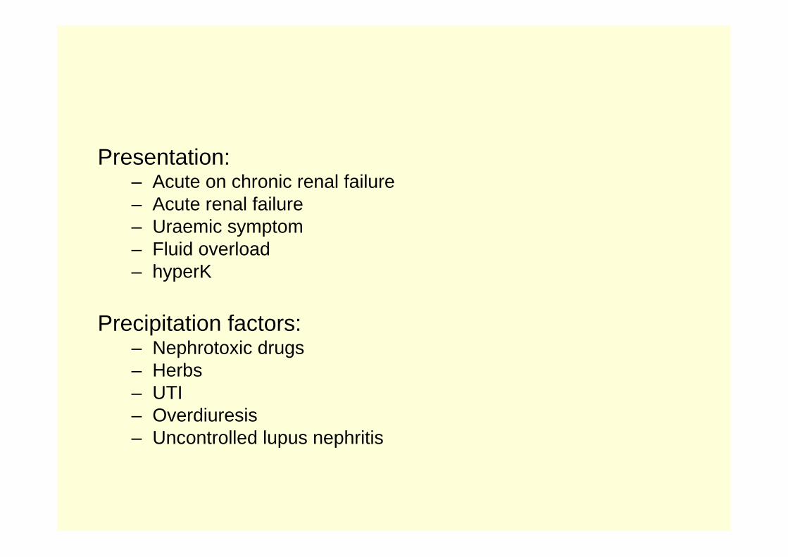
Presentation:– Acute on chronic renal failure– Acute renal failure– Uraemic symptom– Fluid overload– hyperK
Precipitation factors:– Nephrotoxic drugs– Herbs– UTI– Overdiuresis– Uncontrolled lupus nephritis

Investigation:• MSU to exclude UTI• Urine microscopy dysmorphic RBC, casts• 24 hour urine for albumin • Blood test including DsDNA, C3 level• Renal ultrasound
Tx:• Dialysis support

Case 5• M/46 labour worker• Known SLE with class IV lupus nephritis FU RH, poor
compliance to MMF• Admitted PY 8/2008 for lower limb edema, serum alb 28 • Diagnosed as Class IV lupus nephritis, probably
refractory to MMF (or poor compliance)• Switch MMF to intravenous monthly cyclophosphamide• On schedule 4th dose of cyclophosphamide, admitted
clinically for anacasa and deteriorating renal function Cr 250 (baseline130), CXR bilateral pleural effusion
• Pulse methylprednisolone 500 mg given for 3 doses• In view of the poor respond to cyclophosphamide,
rituximab was given

• 3 days after the rituximab, condition further deteriorate with increasing SOB
• CXR congested and bilateral plerualeffusion
• Cr increased to 430• ICU consulted for support• HD offer and NIV supported• Total 11 kg of fluid drawn out through the
HD session

• However, complicated with PTB, further doses of rituximab was not given
• After PTB was treated for 4 weeks• Rechallenge with low dose MMF +
tacrolimus• Gradual improved after 3 months with
serum alb 15 35 though the Cr remained 280+

Approach to patient for suspected autoimmune disease

Rheumatoid arthritis (RA)
•

RA – diagnostic criteria1. Morning stiffness2. Arthritis of 3 or more joints 3. Arthritis of hand joints4. Symmetric arthritis5. Presence of rheumatoid nodule6. Positive rheumatoid factor7. X-ray erosion
1987 ACR diagnostic criteria for RA required 4 out of the 7 criteria above

RA- treatment• Pharmacological treatment
– Pain killer– Simple analegesic– NSAID
– DMARDs– Hydroxycholoquine– Sulphasalazine– Methotrexate– Leflunomide– Cyclosporine– Azathioprine– Cyclophosphamide
– Biologics– Anti-TNF (infliximab, enbrel, humira)– IL6– rituximab

Potential life threatening condition in RA patients
• Severe infection• Intubation • Respiratory failure
– Pneumonitis (e.g MTx induced)• any time at any dosage• nonproductive cough. fever, dyspnea, hypoxemia, or pulmonary
infiltrate
– Interstitial lung disease

Severe Infections in rheumatoid arthritis
Most common infections:• Pneumonia• UTI• Septicaemia • Cellulitis • Septic Arthritis

Tx principle:• Antibiotic – broad spectrum• Prednisolone – stress dose• Withold immunosuppressants
* neutropenic fever* if due to drugs• G-CSF if Neutrophile count < 0.5• MTx – folinic acid rescue, 15 mg Q6H po• Leflunomide – cholestryamine wash out
• Watch out for opportunistic infection esp on biologics

Case 3• F/45• Obesity• DM on metformin + diamicron• Rheumatoid arthritis on leflunomide 20 mg daily +
Methotrexate 20mg weekly
• Admitted for left lower limb cellulitis after toe-nail cutting
• Given IV ampicillin + cloxacillin• Poor response with extension of the cellulitis up to left
thigh, septicaemia shock• Transfer to ICU for further care• Still uncontrolled sepsis with ARDS, ARF, VAP and
required above knee amputation.• Post operatively, prolonged stayed in ICU, then was
taken over to F6 respiratory bed for weaning of tracheotomy

Intubation in rheumatoid arthritis• C1-2 subluxation• Definition: (lateral flexion view) Distance
between the anterior surface of the dens and the posterior surface of the tubercle of C1 >3 mm in adults and >5 mm in children
normal

Intubation in rheumatoid arthritis• Almost all atlantoaxial dislocations involve forward
movement of C1 on C2; posterior dislocation is extremely rare
• 21-44%• 1% neurological impairment• Risk group:
– Severe disease : • active synovitis, • Rapidy progressive erosive peripheral joint disease• Early peripheral joint subluxation• High inflammatory marker
– Late onset RA• Fibro-optic intubation

Dermatomyositis

Clinical features1) Skin rash
– Gottron’s rash– Heliotrope rash– Shawl sign and V sign
2) Muscle weakness– usually insidious onset– typical proximal and symmetrical – myalgia / muscle tenderness (25-50%)
3) Lung involvement– interstitial lung disease (10%)– risk of rapidly progressive pulmonary failure and death
4) Esophageal involvement– dysphagia, nasal regurgitation, aspiration increase risk of aspiration pneumonia
5) Cardiac involvement– myocarditis– heart failure
6) Other systemic manifestation– fever, weight loss, raynand phenomenon, nonerosive inflammatory arthritis
Diagnostic criteria
1. Typical rash 2. Symmetric proximal muscle weakness3. Elevated CK (normal in 4-6%, correlate with severity of
weakness)4. EMG classical triad for myositis
• spontaneous fibrillations• Low amplitude, short-duration polyphasic motor
potentials• Bizarre high freq, repetitive discharges Muscle biopsy
5. Muscle biopsy• 20% -ve because of pathy disease• Site: affected muscle, usual site quadriceps , detold• Biopsying the muscle contralateral to the side having abnormal
EMG finding

SOB in dermatomyositis
• ? Disease progression• ? Infection

Case 4• M/60 good past health• c/o: shortness of breath, generalised
malaise and weakness for 2 months• P/E: multiple raised, scaly, symmetrical
skin lesion over knee, back, buttock, neck, hand and elbow
• Chest bilateral fine crepitation• SaO2 88 RA,• CK 394

CXR on admission
HRCT

• EMG evidence of myositis• Dx: Dermatomyositis with interstitial
pneumonitis• Tx: high dose prednisolone (1mg/kg/day) • Progress: increasing SOB, desaturation
required intubation

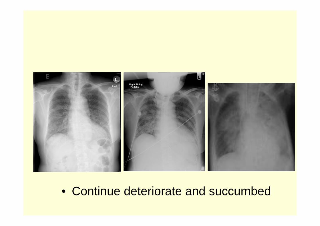
CXROn admission Day 9

• Impression: rapidly progressing interstitial pneumonitis
• Tx: IV pulse prednisolone 1g daily x 3 days cover with broad spectum antibiotic
• Cx: bilateral pneumo-thorax • Progress: continue deteriorated and
succumbed despite maximum support

Case 5
• F/62• Known
dermatomyositis5/2009 Dx on high dose steroid and Azathioprine

• 1 month later, admitted for fever, cough, sputum and increased SOB

• Treated as chest infection with background Dermatomyositis + ILD
• Required high O2 and contacted ICU for support and required intubation
• Tazocin, azithromycin, tamiflu, amantadinegiven

• Continue deteriorate and succumbed

Whenever deteriorate, Had to differentiate flare up of disease vs Infection
High risk infection • High dose immunosuppressant• Diseased lung• Bulbar involvement aspiration pneumonia

Scleroderma

• Major organ involvement• Renal renal crisis• Lung interstitial pneumonitis, pulmonary
fibrosis• Cardiac pulmonary hypertension

Case 6
• F/49 good past health• Admitted orthopaedic ward for digital ulcer• Consulted rheuamtologist for raynand
Phenomenon• C/F: sclerodactyly, tighten skin over face,
chest wall• Dx: scleroderma• Started on aspirin, norvasc

• Admitted for SOB, lower limb edema• Self detected high blood pressure at home
180/110• P/E dyspnoea, LL edeam, chest
bilateral creps• CXR congested• Hb 7+ • Cr 1000

• IMP: scleroderma related renal crisis• Consulted ICU for renal support• HD and ACEI started• However, poor response and succumbed
• Risk factors for renal crisis– Diffuse skin involvment– High dose prednisolone use (>20mg/day) in
preceding 6 months– Cyclosporin use
Life threatening rheumatic disease diagnosed in ICU
• 20% rheumatic disease first Dx in ICU• Rheumatic disease sometimes is life
threatening• High index of suspicious is required• How to interpret the investigation result
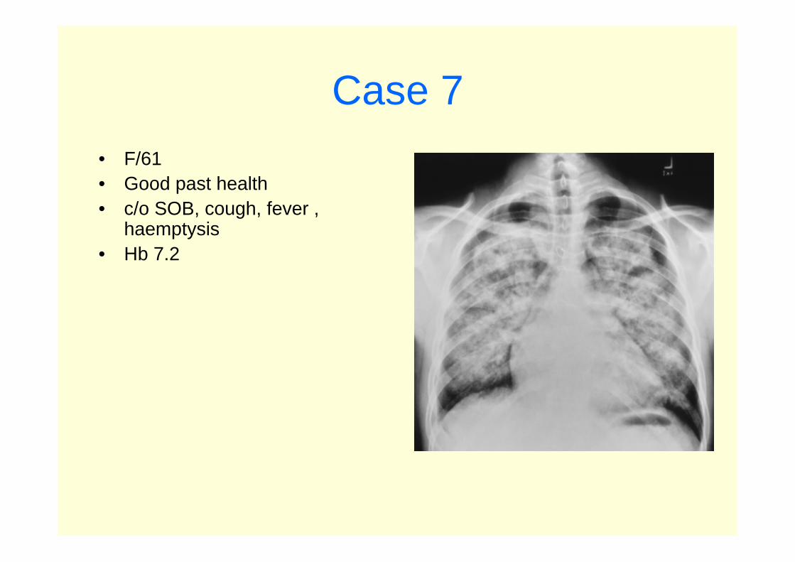
Case 7• F/61• Good past health• c/o SOB, cough, fever ,
haemptysis• Hb 7.2

Case 7• F/61• Good past health• c/o SOB, cough, fever ,
haemptysis• Hb 7.2• Urine RBC +++• Bronchoscopy capillaritis,
bronchiolitis and organisingpneumonitis
• BAL hemosiderin-laden macrophages
• Urgent cANCA +ve• Dx: Wegener’s granulomatosis

Treatment:• pulse methylprednisolone• Oral cyclophosphamide• Dramatic improvement

Case 8• F/26• Admitted A&E for severe abdominal pain • Already gasing in A&E required intubation• PEA (pulseless electrical activity) after intubation• P/E : distended abdomin with guarding• Ix: severe metabolic acidosis, AXR dilated bowels• Emergency laparotomy showed small bowel
strangulation, adhesion and intestinal obstruction, adhesionalysis and ileostomy done
• Post operative condition remined critical• Succumbed 10 hrs after admission

• Postmortum diagnosis Takayasuvasculitis
• Recall : fever and wt loss on and off for 2 months

Common problem don’t forget !!

Common problem don’t forget !!