Embed Size (px)
Citation preview
LECTURE NOTES OFINTERNAL MEDICINE
Rheumatology
Dr. Osama Mahmoud MohamedAssistant Professor of Internal Medicine
Ain Shams University
CONTENTS
PageAnatomy 1
History and Examination 2
Laboratory tests 5
Systemic lupus erythematosus 10
Polymyositis and dermatomyositis 18
Vasculitides 20
Rheumatoid arthritis 29
The spondyloarthropathies 44
Scleroderma 50
Mixed C.T. disease .. 53
Behcet's disease 54
Crystal induced arthropathy 55
Osteoarth ritis ...... ......... .... ...... .. ... 62
Septic arthritis 65
Osteomalacia 66
Osteoporosis 67
Inherited disorders of collagen 69
Fibromyalgia syndrome 69
Classification of joint disease 70
Neck pain, back ache 71
1
RItEUMATOloGY• Rheumatology is a medical science devoted to the study of rheumatic diseases
that include a range of musculoskeletal and systemic disorders that share theclinical involvement of joins and periarticular tissues.
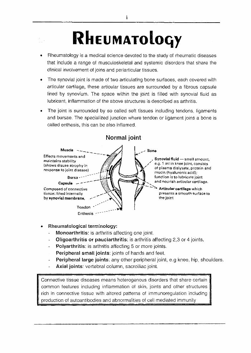
• The synovial joint is made of two articulating bone surfaces, each covered witharticular cartilage, these articular tissues are surrounded by a fibrous capsulelined by synovium. The space within the joint is filled with synovial fluid aslubricant, inflammation of the above structures is described as arthritis.
• The joint is surrounded by so called soft tissues including tendons, ligamentsand bursae. The specialized junction where tendon or ligament joins a bone iscalled enthesis, this can be also inflamed.
Normal joint
Tendon ----Enthesis - --
...'"Bone.-Muscle -_ -- '
Effects movements and - ---~~maintains stability "1;jJ' .J(shows disuse atrophy inf~response to joint disease) /Ji
• SYnovial fluid - small amount,,,/ e.g. 1 ml in knee joint, consists
I' of plasma dialysate, protein andmucin (hyaluronic acid);function is to lubricate joint
_ .11. '..... and nourish articular cartilage.
fA - -~ Articular cartilage wh ichrtf presents a smooth surface to
the joint
Bursa - - - - i
Capsule -------~\\Composed of connective 1,tissue: lined internally .....' .by synovial membrane, ..-'- .••.
• Rheumatological terminology:Monoarthritis: is arthritis affecting one joint.Oligoarthritis or pauciarthritis: is arthritis affecting 2,3 or 4 joints.Polyarthritis: is arthritis affecting 5 or more joints.Peripheral small joints: joints of hands and feet.Peripheral large joints: any other peripheral joint, e.g knee, hip, shoulders.Axial joints: vertebral column, sacroiliac joint.
Connective tissue diseases means heterogenous disorders that share certaincommon features including inflammation of skin, joints and other structuresrich in connective tissue with altered patterns of immunoregulation includingproduction of autoantibodies and abnormalities of cell mediated immunity
2
~iS!QR~ ANd .EX~MiNAti()N.oFMu~cu~oskELETALS~~JEMHistDry:
1- Ask about peripheral joints (UL & LL) for pain, swelling, hotness,redness, limitation of movement (stiffness) or deformity.
2- Ask about axial joints (cervical & lumbar) as regard pain or limitation ofmovement.
• Ask about weakness either a primary or secondary muscle abnormality.• Ask about the duration and severity of early morning stiffness.
3- Ask about extraarticular manifestations for example-CVS -+ chest pain, dyspnea- Chest -+ dyspnea, wheezy chest- L.N -+ swelling (cervical, axilla)-Skin -+ rash- Eye -+ redness-Kidney -+ puffines , hypertension
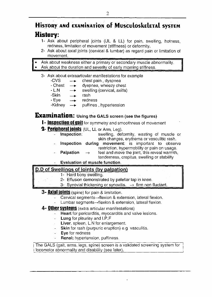
Examination: Using the GALS screen (see the figures)
I- Inspection of gait for symmetry and smoothness of movementi- Peripheral joints (UL, LL or Arm, Leg).
Inspection: swelling, deformity, wasting of muscle orskin changes, erythema or vasculitic rash.
Inspection during movement: is important to observerestriction, hypermobility or pain on usage.
Palpation ~ feel and move the joint, this reveal warmth,tenderness, crepitus, swelling or stability
Evaluation of muscle function.
D.D of Swellings of joints (by palpation>.1- Hard bony swelling.2- Effusion demonstrated by patellar tap in knee.3- Synovial thickening or synovitis. ~ firm non fluctant.
3- Axialjoints (spine) for pain & limitation.Cervical seqrnents-oflexion & extension, lateral flexion.Lumbar seqrnents-eflexion & extension, lateral flexion.
4- Other Systems (extra articular manifestations)Heart for pericarditis, myocarditis and valve lesions.Lung for pleurisy and I.P.FLiver, spleen, L.N for enlargement.Skin for rash (purpuric eruption) e.g vasculitis.Eye for rednessRenal: hypertension, puffiness.
The GALS (gait, arms, legs, spine) screen is a validated screening system forlocomotor abnormalit and disabilit see later.
3
Sequence of GALS screening examinationInspection of gait
Symmetry, smoothnessof movement, see later
Touch your toes
Normal lumbar spine(and hip) flexion
Open jaw, move sideto side
Normal ternoro-mandibularmovement
Place tip of finger ontip of thumb
Fine precisron pinch
Inspection of patientstanding from behind
Straight spineMuscle bulk/symmetry ofparaspinal, shoulder andgluteal musclesLevel iliac crestsNo popliteal swellingNo hindfoot swellingor deformity
Inspection from thefront
Full elbow extensionShoulder and quadricepsmuscle bulk, symmetryNo knee swelling or deformityNo forefoot or midfootdeformity
Hands in front, palmsdown
No swelling or deformity ofhands/wristsAble to extend fingers
Metacarpal squeeze
?Metacarpopllalatlgeallolnttenderness
Press over eachmid-supraspinatus
? Hyperalgesia offibromyalgial
Hands behind head,elbows right back
Full shoulder abduction,external rotationNormalacromioclavicular andsternoclavicularmovementFull elbow flexion
Turn hands over
Normal supination(wrist, distal radio-ulnarJoint)Normal palms
Examination on couch'Put your heel on yourbottom' (flex knee and
hip, holding knee)
Full knee and hlp fleXionNo knee crepitus
Inspection from theside
Normal cervical andlumbar lordosisNormal thoracic kyphosis
Place ear onshoulder
Normal pain-free cervicallateral flexion
Make a fist
Strong power grip
Internal rotation of hipin flexion
No pam or restriction of hipmovement
4
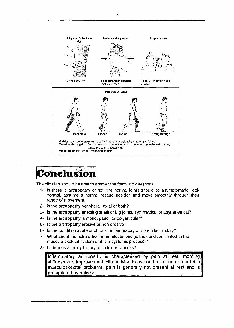
Palpate for balloonsign
No knee effusion
Metatarsal squeeze Inspect soles
No metatarsophalangealjoint tenderness
No callus or adventitiousbursitis
Phases of Gait
(~
~
\~fl_SWing-throughStance Toe-off
Antaigic gait: Jerky asymmetric gait with less time weight-bearing on painful legTrendelenburg gait: Due to weak hip abductors-pelvis drops on opposite side during
stance phase on affected sideWaddling gait: Bilateral Trendelenburg gait.
The clinician should be able to answer the following questions:1- Is there is arthropathy or not, the normal joints should be asymptomatic, look
normal, assume a normal resting position and move smoothly through theirrange of movement.
2- Is the arthropathy peripheral, axial or both?3- Is the arthropathy affecting small or big joints, symmetrical or asymmetrical?4- Is the arthropathy is mono, pauci, or polyarticular?5- Is the arthropathy erosive or non erosive?6- Is the condition acute or chronic, inflammatory or non-inflammatory?7- What about the extra articular manifestations (is the condition limited to the
musculo-skeletal system or it is a systemic process)?8- Is there is a family history of a similar process?
Inflammatory arthropathy is characterized by pain at rest, morningstiffness and improvement with .activity. In osteoarthritis and non arthriticmusculoskeletal problems, pain is generally not present at rest and isprecipitated by activity
5
LAboRATORy IMMUNOLoGicAL TESTSof RItEUMATOloGicAl DisORdERS
Autoantibodies are antibodies directed against self antigens includingimmunoglobulins, cell surfaces, circulating molecules as well as cytoplasmicantigens, nuclear antigens they are frequently observed in the sera of patientswith rheumatological diseases.
Rheumatoid lactor (R.F)• It is an IgM Ab (occasionally IgG, IgA or IgE) which reacts with Fc
portion of human IgG, this occurs usually in patients with rheumatoiddisease (70-80% of cases), but is not diagnostic.
• It exists as an (Rh.F. - Ig.G - C) complex in synovial membrane.So, complement is lowered in synovial fluid and not in the serum.
Methods Ilildetection1- Latex method (agglutination test)
Ab (Rh. F. in the serum) +Ag (latex coated with Ig G)
2- Rose waaler test. (agglutination test)Ab (Rh. F. in the serum) +Ag (sheep's RBCs coated witl1 anti-erythrocyte antibodies)
Latex is more sensitive, but Rose Waaler is more specific.
Conditions in which Rh. F is +ve• Autoimmune rheumatic diseases ..
Rheumatoid arthritis (70 % - 80 %).SLE (15-35 %)Polymyositis, dermatomyositis (5-10 %).Mixed connective tissue disease (50-60%).Sjogren's disease (90 %).Scleroderma (30 %).
• InfectionsT. B. - Infective endocarditis.Syphilis - Kala - azarHCV - HIV
• Normal populationEspecially elderly and relatives of patients with rheumatoidarthritis.
• Miscellaneaous .,Autoimmune hepatitis. - Fibrosing alveolitis.Sarcoidosis. - Waldenstroms macroglobulmamia.Chronic liver disease. - Cryoglobulinemia.
6
ANA (antinuclear antibody)• It is any autroantibody directed against one or more components of
the nucleus.• It is used to detect a disorder but not to rule out C.T disease as it
can be + ve in other unrelated diseases. Immunofluorescencemicroscopy after serum has been applied to a nucleated tissuesubstrate e.g (rodent organs) or human cell lines is the standardmethod of detection giving different patterns of staining.
ANA patterns:• Speckled pattern.• Homogenous pattern.• Nucleolar pattern.
• Causes of + ve ANA1- SLE 95 -100%.2- Scleroderma 80 %.3- Polymyositis 80 %.4- Rheumatoid arthritis 30 %, Sjogren's disease 70 %.5- Mixd connective tissue disease 95%. Sjogren's syndrome
60-70%.6- Primary biliary cirrhosis, autoimmune hepatitis7- Normal elderly people, infective endocarditis.
The results of ANA can be expressed by a titre. AUtre of 1:40 - 1:80 isconsidered positive, but 1:160 is significant.If ANA is positive, it is important to ask about specificaptiP9diS$ (ANA?ptqfile)
Dther specific antibodies or specific antinuclearantibodies (ANA profile) i.e tests that measure ANAs specificfor certain nuclear antigens.
1- Antidouble stranded DNA (antinative DNA).• It is specific for S L E.• It determines the activity of the disease, it is positive in
about 40-75% of cases (negative in mild or inactivedisease). High titre indicates poor prognosis
2- Antihistone antibody.It is positive in drug induced SLE. (>90%)
3- Anti-Smith Antibody. (anti-sm)Directed against non histone nuclear protein, specific for SLEand indicates a poor prognosis
4- Anti - ScI. - 70It is a marker for Scleroderma (20-50%).
5- Ab to Ro and La particles in SLE and Sjogren's syndrome.6- Ab to centromere in CREST $7- Anti-RNP (ribonucloprotien) in cases of mixed connective tissue
disease and in SLE.
7
Other AutoantibodiesA - Antimitochondrial Abs.
• In primary biliary cirrhosis.
B - Antismooth muscle Abs.• Autoimmune hepatitis.• Primary biliary cirrhosis.
C - Organ specific Abs.• Anti thyroid Ab in thyroiditis.• Anti parietal Ab in pernicious anemia.• Anti islet cell Abin 100M.• Anti GBM in Good pasture $• Anti adrenal Ab in Addison disease.• Anti platelet Ab in Idiopathic Thrombocytopenic purpura.
Antineutrophil cytoplasmic antibodies (ANCA), see vasculitis• C-ANCA + ve in wegener's granulomatosis.• P-ANCA is less specific, + ve in:
- Microscopic polyangiitis- Churg-strauss disease- SLE and rheumatoid disease.- Inflammatory bowel disease
• Sero -ve arthropathy = arthropathy with Rh.FSero +ve arthropathy = arthropathy with Rh.F
• HLA - B27 related to sero -ve group of arthropathy.
-ve (see later)!?+vee.g. ankylosingspondylitis.
Other laboratory lests in the evaluation ofthe rheumatic diseases:[1] Erythrocvte sedimentation Rate [ES8l
• Acute phase reactants are proteins e.g fibrinogen are produced in theliver as a response to inflammation i.e acute phase response (APR).
• Increase in the acute phase proteins e.g fibrinogen andgammaglobulins leads to increase of ESR, this is due to change of therepellent electrostatic negative surface charge of RBCs --7 rouleauxformation
• So, ESR is increased in cases of inflammatory diseases e.g connectivetissue diseases, infections or malignancy.
For details see haematology II
8
(2) C-ReacliveJJrotein [CRP]• It is an acute phase reactant produced by the liver as a response to
inflammation.• It starts elevation within 4 hours of tissue injury with peak after 24-
72 hours. It is rapidly decline once the cause is abolished.• It is measured by ELISA technique as follows:
0-1 mg/dl (normal)1-10 mg/dl (moderate level).> 10 mg/dl (high level).
• It is therefore the single most useful direct measure of the APR(acute phase response)
[3~ASOT-7 See C.I.S. [Rheumatic fever][4~Serum uric acid -7 See Gout
\Vgy yO.•..•.•..i dia,gDC:Jsis is one <>oftheI
f(jllowing disea~es'!(E~iD.ples)I. Rheumatoid Arthritis (R.A)
• It is mainly arthropathy.• It affects mainly the peripheral joints (symmetrical involvement) e.g:
- UL. - LL.
• It involves the small joints e.g:- Hands - Feet.
• The arthropathy is erosive (leading to deformity).• The axial joint affection is uncommon (vertebral column - sacroiliac
joint).• The extra-articular manifestations are present in the following sites:
- Eye - Skin - Lung- Kidney - Heart - Blood
II. Ankylosing Spondylitis• It is mainly arthropathy.• It is mainly axial arthropathy.
- Vertebral column. - Sacroiliac joints.• Peripheral joints affection is asymmetrical.• This arthropathy is ankylosing (ankylosis of vertebral column)• The extra-articular manifestations are present in the following sites:
- Eye - Heart- Lung - Blood
9
III. S.L.E• It is mainly an extra - articular problem.• The manifestations present in the following sites:
- Brain. - Kidney. - Heart. - Blood- Blood vessels. - Skin. - Serous membranes - Lung
• The arthropathy is non erosive (i.e. No residual deformity)• The arthropathy affecting mainly the peripheral joints, it is symmetrical and
mainly in the form of arthralgia.
Musculoskeletal disorders are classified into:• Inflammatory joint diseases e.g rheumatoid disease, seronegative spondarthritis,
crystals associated disease, juvenile idiopathic arthritis.• Diseases of bone e.g osteoporosis, osteomalacia.• Osteoarthritis.• Systemic C.T diseases, e.g SLE, scleroderma, mixed C.T disease,
dermatomyositis, vasculitis.
Diagnostic imaging:(1) Plain x rayS
Diagnosis of established rheumatoid arthritis, trauma, osteopenia,osteoarthritis and spondylosis.
[2] UltrasoundIt is useful for periarticular structures, soft tissue swelling and tendons, it isalso used to guide local injections.
[3] Magnetic resonance imaging (MRIlIt can show intraarticular structures and bone changes in detail and veryearly. Also it is of choice for spinal disorders. Gadolinium injection enhancesinflamed tissue. MRI can also detect muscle diseases e.g. myositis.
[4] Computerized axial tomographyIt is useful for spinal disorders.
[5] Bone scintigraoJIV (scan] bv 99mTcIt can detect areas of imflammation, infection or malignancy of bone.
[6] OEM scanning [See Later]It measures bone density, so it is used for screening and monitoring ofosteoporosis.
[7] Positron emission tomography [PEll scanningIt can detect fluorodeoxyglucose uptake that indicates areas of increasedglucose metabolism. It is used to locate tumours, also it can diagnose largevessel vasculitis (Takayasu's arteritis).
[8] Arthroscoml especiallv for knee and shoulder, biopsy can be taken,
surgery can be performed e.g. repair of meniscal tears.
10
SYSTEMic Lupus ERyTItEMATOSUS (S.L.E)• It is a systemic connective tissue disorder affecting mainly females,
female: male ratio (9 : 1) with peak onset in the second and third decade.• It leads to systemic disorders (extra - articular mainfestations), but joint
pain is the presenting feature in 50 % of cases. Skin rash and arthralgiaare common presentations but renal and cerebral diseases are themost serious problems.The prevalence varies from 30/100,000 in Caucasians to 200/100,000in Afro-Caribbeans.
Aetiology (It is an autoimmune disease)(1) Defect in T. suppressor lymphocytes with exaggerated B cell
activity leading to production of auto antibodies to a variety ofantigens (nuclear, cytoplasmic and plasma membrane), this willlead to immune complexes with systemic manifestations• There is hypergammaglobulinaemia and increased level of
IL1, IL2 and IL6(2) Expression of novel or hidden antigens on the cell surface
during apoptosis (during apoptosis these antigens migrate to cellsurfaces). This hypothesis is supported by the fact thatenvironmental factors leading to oxidative stress withsubsequent increased apoptosis e.g exposure to sunlight andartificial ultra violet light, pregnancy and infection. The increasedapoptosis with expression of antigens triggers T cells thatstimulate B cell to produce autoantibodies.
Some contributing factors.1- Sunlight, artificial ultraviolet light, environmental triggers? as above.2- Drugs (drug induced lupus).
• C / P of drug induced SLE:1- Drug history, equal sex ratio. 2- Fever and skin rash.3- No nephritis or cerebral disease. 4- Arthralagia, serositis.5- Resolution on drug withdrawal.
• Causative drugs: - Hydralazine. - Procainamide. - Phenytion.• Investigations:
- ANA + ve.- Anti DNA is - ve , anti histone antibody is +ve
• Treatment is by withdrawal of the causative agent, short course ofsteroid can be given if symptoms are severe.
3- Hormonal factor!?SLE is common in child bearing period, and in those usingcontraceptive pills.Exacerbation in pregnancy. - Exacerbation in the puerperium !?Hormonal (estrogen) replacement therapy may lead to flare-ups.
1 1
Estrogen binds to receptors on T and B lymphocytes increasing activation andsurvival of those cells thus favoring prolonged immune responses
P~t.b9.J9.~1- Joint:- synovitis with little cartilage destruction.2- Heart:- libman sacks endocarditis (affecting mitral and aortic valves).3- Kidney:- thickening of glomerular B.M. due to immune complex
deposition.4- Skin:- immune complex in demo epidermal junction.S- Serositis and vasculitis.6- Lung:- vasculitis - interstitial pulmonary fibrosis.7- Microscopic changes of tissues in cases of SLE:
• Haematoxylin bodies: Amorphous masses of nuclear material found inC.T. lesions that become purple blue with hematoxyline, P.N.L thatingest these bodies called LE cells.
• Onion skin lesions occur in splenic arteries due to disposition ofcollagen around them.
e Silver wire appearance in lupus nephritis.
Clinical picture• Male: female ratio is 1 : 9• The presentation and course are highly variable.
1- Fever of unknown origin.2- Musculoskeletal
Joint involvement is mainly arthralgia with mild morning stiffness.The arthropathy is bilateral and symmetrical. The small joints areusually affected mimic rheumatoid disease.It is non deforming but tendosynovitis may lead to deformity(Jaccoud's arthropathy), it is due to tendon or ligament laxity.A vascular necrosis of the hip may occur with steroid therapy.Myalgia is common
3- The skinButterfly rash: fixed erythema (flat or raised) on the cheeks of theface and across the bridge of the nose, occurs in a photosensitivedistribution that spares the nasolabial folds.Discoid rash: erythematous raised patches with adherent scaling,atrophic scarring may occur. It may lead to scarring alopecia ifpresent on the scalp.Photosensitivity: skin rash as a result of unusual reaction to sunlight.Purpuric lesion due to thrombocytopenia or vasculitis.Leg ulcers.
12
Vasculitic lesions • Nail bed and finger bulb infarcts .• Purpuric rash with elevated edge.
Raynaud's phenomenon.Urticaria.Panniculitis (Lupus profundus)
Alopecia.Lichen plannus like.Livedo reticularis.
4- The EyeRetinal vasculitis can cause infarcts, cytoid bodies whichappear as hard exudatesEpiscleritis, conjunctivitis or optic neuritis may occur.Kerataconjunctivitis sicca with Sjogren's syndrome.
S- The heartPericarditis and pericardial effusion.Myocarditis with heart failure.Libman sacks endocarditis (affecting mitral or aortic valvescausing regurge), It is a sterile endocarditis.Blood pressure is increased with renal hypertension.Coronary heart disease (accelerated atherosclerosis).
6- The KidneyLupus nephritis (WHOclassification)• Type I Minimal pathology (Normal glomeruli)• Type II Mesangial widening with or without hypercellularity.• Type III Focal proliferative G.N.• Type IV Diffuse proliferative G.N.• Type V Membranous G.N.• Type VI Advancing sclerosing G.N.
7- GIT Mesenteric vasculitis with acute abdomen. Liver involvement isunusual, pancreatitis is uncommon. Nausea, vomiting anddiarrhea can occur with an SLE flare.
8- The LungPleurisy and pleural effusionInterstitial pulmonary fibrosis.Shrinking lung syndrome with elevation of the diaphragm
due to recurrent pulmonary infarction.Pulmonary hypertension with antiphospholipid syndrome.Adult respiratory distress syndrome.
9- Neuro psychiatric manifestationsPsychosis, depression, cognitive dysfunction (difficulties withmemory and resoning).Lymphocytic meningitis, transverse myelitis.Chorea.Cerebral vasculitis leading to cerbrovascular stroke.Polyneu ropathy.
13
Lupus headache.Seizures
Psychosis due to lupus must be differentiated from steroid induced psychosiswhich occurs in the first weeks of steroid therapy at doses of 2 40 mg ofprednisone or equivalent, it resolves over several days after steroids aredecrease or stopped.
10-BloodAutoimmune thrombocytopenia and haemolytic anaemia,Lymphopenia (guide to disease activity).Antiphospholipid $ leading to thrombo-embolism ..
11- Polyserositis affecting:- Pleura. - Pericardium. - Peritoneum.
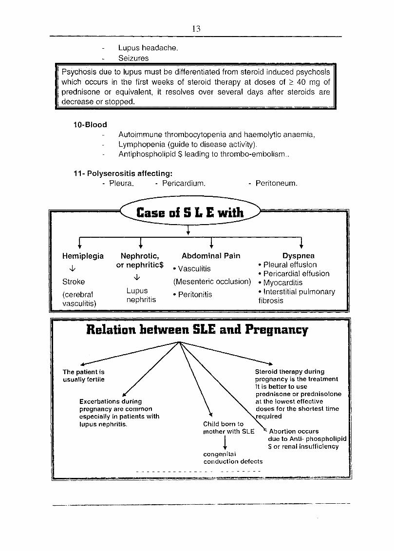
~ l ~ lHemiplegia Nephrotic, Abdominal Pain Dyspnea
-J,- or nephritic$ • Vasculitis • Pleural effusion-J,- • Pericardial effusion
Stroke (Mesenteric occlusion) • Myocarditis(cerebral Lupus • Peritonitis • Interstitial pulmonaryvasculitis) nephritis fibrosis
Relation between SLE and Pregnancy
Excerbations duringpregnancy are commonespecially in patients withlupus nephritis.
Steroid therapy duringpregnancy is the treatmentIt is better to useprednisone or prednisoloneat the lowest effectivedoses for the shortest timerequired
Child born tomother with SLE
!Abortion occursdue to Anti- phospholipid$ or renal insufficiency
congenitalconduction defects
14
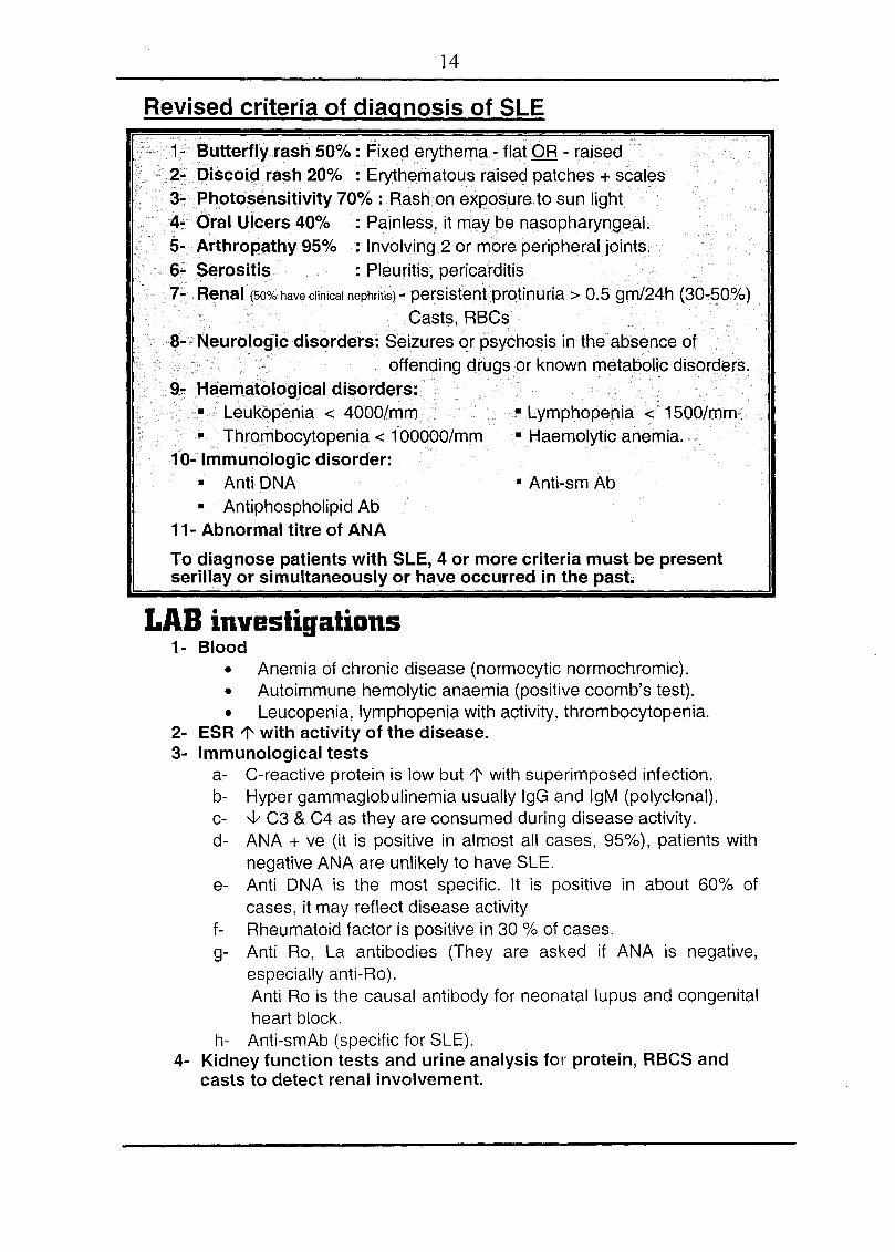
Revised criteria of diagnosis of SLE
1.. ~~tterflyrash 50% : Fixed erythema- flatOR - ralsed2~ Discoi$i rash 20% : ErythE;lrnatous raised patches + scales3~ Photo!)ensitivity 70% : Rashon exposure.to sun light4.. Oral Ulcers 40% : Painless, it may be nasopharyngeaL5- ArthroPfithy 95% : Involving 2 or rp6re peripheraljoints.6~ ~erositis. : Pleuritis, pericarditis7/- Flenal(!?O%haveclinicalnephritis)- persistennprotinuria > 0.5 grn/24h ($0..50%)
Casts, RHCs8.. N¢urologlcdisorders: Seizures or psychosis in theabssnce of
offencHng cixugsor known metabolic disQrd.ers.9... t;tejelllfitological disorders:
• ....•••..Leuk()PE3niCi< 4000/mm-Thr()rnbOCytopenia< 100000/mm
10- Immunologic disorder:• Anti DNA• Antiphospholipid Ab
11- Abnormal titre of ANA
Lymphopenia < 1500/mrn• Haemolytic anemia.
• Anti-sm Ab
To diagnose patients with SLE, 4 or more criteria must be presentserillay or simultaneously or have occurred in the past~
LAB investigations1- Blood
• Anemia of chronic disease (normocytic normochromic).• Autoimmune hemolytic anaemia (positive coomb's test).• Leucopenia, lymphopenia with activity, thrombocytopenia.
2- ESR l' with activity of the disease.3- Immunological tests
a- C-reactive protein is low but l' with superimposed infection.b- Hyper gammaglobulinemia usually IgG and IgM (polyclonal).c- ~ C3 & C4 as they are consumed during disease activity.d- ANA + ve (it is positive in almost all cases, 95%), patients with
negative ANA are unlikely to have SLE.e- Anti DNA is the most specific. It is positive in about 60% of
cases, it may reflect disease activityf- Rheumatoid factor is positive in 30 % of cases.g- Anti Ro, La antibodies (They are asked if ANA is negative,
especiallyanti-Ro).Anti Ro is the causal antibody for neonatal lupus and congenitalheart block.
h- Anti-smAb (specific for SLE).4- Kidney function tests and urine analysis for protein, RBCS and
casts to detect renal involvement.
15
o Symptoms and signs suggesting active SLE?• Weight loss, fever, arthritis, seizures, hair loss, anaemia,
haematuria, rashes, mouth sores and oliguria.
o Laboratory diagnosis of disease activity?• -J; C3 , C4 • +ve Anti-DNA (high titre)
o Patient with SLE+ fever?• Disease activity (see above) • Infection (+ ve C- reactive protein)
o Patients with SLE+ leucocytosis• Steroid therapy.
- PNL l' -Eosinophils -J; - Lymphocytes -J;• Infection:
Toxic granulations within WBCs, presence of staff cells andpositive CRP
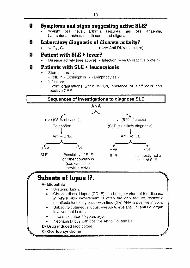
Sequences of investigations to diagnose SLE
ANA___ A.-.__ ~r "'\+ ve (95 % of cases) - ve (5 % of cases)
To confirm (SLE is unlikely diagnosis)
~ ~Anti - DNA Anti Ro, La
___ A___ _ __ A _+\,e \e ( ,
+ ve - veSLE Possibility of SLE
or other conditions(see causes ofpositive ANA)
SLE It is mostly not acase of SLE.
Subsets ullupus !?A- Idiopathic
• Systemic lupus.• Chronic discoid lupus (COLE) is a benign variant of the disease
in which skin involvement is often the only feature, systemicmanifestations may occur with time (5%) ANA is positive in 30%.
• Subacute cutaneous lupus, -ve ANA, +ve anti Ro, anti La, organinvolvement is rare.
• Late onset after 50 years age.e Neonatal Lupus with positive Ab to Ro. and La.
B- Drug induced (see before)C- Over lop syndrome
16
TreatmentAll patients require education and general prophylactic measures to preventdisease flares:
• Sunscreens and protective clothing are effective in avoidingphotosensitivity reactions.
• The use of estrogen containing oral contraceptives is controversialin SLE, but many centers avoid these medications because theymay increase the disease activity.
• Avoidance of vasoconstrictive drugs are helpful in treatingRaynaud's phenomenon, also patients with SLE may benefit fromvasodilator therapy.
• Low dose aspirin for patients with positive antiphospholipidantibodies to prevents thrombotic events.
• Psychological support is essential because SLE may causedepression and anxiety.
• Routine immunizations for influenza and pneumococci arerecommended.
1- Arthralgia, mild arthritis, fever and serositis respond to NSAIDs2- Skin manifestations respond to hydroxychloroquine 400mg/d + topical
steroid (hydroxychloroquine is also indicated in arthralgia resistant toNSAIDs)
3- Corticosteroid therapy is the main subject of treatment.
Steroids are used for almost all manifestations of lupus in doses ranging fromextremely small alternate day doses to huge pulsed intravenous doses.Prolonged steroid therapy usually lead to OM, accelerated atherosclerosis,osteoporosis, glucoma, cataract, avascular necrosis and increased risk ofinfections. To avoid such toxicities, different cytotoxic drugs can be used toprovide steroid sparing effect (see below).
Role of steroidsTo control the inflammatory reaction ~ ~. end organ damage.
MethodGive full dose of steroid 60-80 mg predinsolone / day till activity of thedisease disappears i.e. + Resolution of symptoms and signs.
+ - ve Anti-DNA+ Normal C3 and C4
Then gradual withdrawal followed by low dose steroid as maintenance.10-15 mg / day to: + prevent relapse.
+ prevent end organ damage.
I Pulse steroid therapy can be used in severe cases (see later). I
17
4- Immunosuppressive drugsUsed in severe disease activity e.g severe lupus nephritis or cerebraldisease.
• Azathioprine (Immuran): - 2mg/kg/d orally. It is used when steroidalone is not fully effective. It is also a steroid sparing drug. i.e it allowsa reduction of steroid dose. The side effects are leucopenia, anaemia,infections.
• Cyclophosphamide (Endoxan):
1-3 mg/kg/d orally, also it can given as pulse therapy 0.5-1 gm/m Iv. Itis important to monitor the side effects e.g Infections, bone marrowdepression and infertility. This drug is extremely toxic so, it reserved forthe most severe disease manifestations.
• Cyclosporine (Sandimmun) and mycophenolate (myfortic) can also beused with severe disease activity and to avoid the side effects of otherimmunosuppressive drugs.
Prolonged use of azathioprine may increase the risk of haematologicalmalignancy5- Plasmapharesis can be used in cases with severe exacerbations
refractory to steroid.6- Immunoglobulin therapy is effective for thrombocytopenia of SLE
o Pulse steroid therapy?~ose:1
500 - 1000 mg methyl prednisolone/ day I.V. 3 - 5 days.To be followed by full dose steroid until improvement (laboratory andclinical).Then low dose steroid as maintenance 10 - 15 mg.l day
Pulse I.V cyclophosphamide in combination with pulse steroid is moreeffective.
Pulse steroid therapy can be used in SLE with severe activity. e.g.Vasculitis, cresentic GN, severe cerebral or haematological disease.
IOtherindications of pulse steroid theram1- Cresentic glomerulonephritis (rapidly progressive G.N.)
2- Multiple sclerosis
3- Optic neuritis.
18
[p~~cautionsl• Prophylaxis for peptic ulceration by proton pump inhibitors.
• Control blood pressure.• Control blood sugar.• Isolation to guard against infection.
[Course and prognosis of SLE]• The course is characterized by remission and exacerbation.• Chronic course occasionally seen.• 5 years survival rate is about 90 %.• Severe renal or neurological disease have the worst prognosis.
POLYMyosiTis (PM) ANd DERMATOMyosiTis (DM)These are connective tissue disorders with inflammatory reaction in
skeletal muscles and / OR skin of autoimmune pathogenesis !?
Pathalagy (Lymphocystic Infiltration of skeletal muscles)
Classificatian1- Primary idiopathic polymyositis .
.2- Primary idiopathic dermatomyositis (polymyositis + skin lesion).3- Dermatomyositis or polymyositis associated with malignancy.4- Childhood dermatomyositis.5- Polymyositis or dermatomyositis associated with other connective
tissue disorders e.g. (mixed connective tissue disease).Clinical picture ... It is an inflammatory myopathy, occurs at age
of 30-60 years, female to male ratio 3 : 1 with insidious onset.1- Muscle There is bilateral proximal weakness (shoulder and pelvic girdle
muscles). The muscles may be wasted but are not usuallytender. Face and distal limb muscles are not usually affected.Tendon jerks are preserved.Heliotope rash ~ It is a violaceous discoloration of the uppereyelids, forehead and nasolabial folds.Gottron's sign ~ erythematous eruption over the extensorsurfaces of P.I.P joints and M.P joints.Arthralgia.Myocarditis causing heart failure, arrythrmias.
2- Skin
3- Joint4- Heart
19
5- Malignancy this occurs particularly in males with dermatomyositis withage of 50 years or more so, you must to search for malignancy:
• Lung • Prostate, ovary • Colon • Cancer breast6- Raynaud's phenomenon7- Lung: Interstitial pulmonary disease.8- GIT: Dysphagia due to esophageal dysmotility (myositis of striated muscle
in the upper one third of esophagus
iola.ceousdiscolonreationsirni.larto heliotrope rash may occur on theer back, chest and shoulders Shawl distribution.
Investigations• Electromyography -7 showing myopathic changes.• Muscle biopsy from quadriceps or deltoid? features of fibre necrosis,
reQeneration and inflammatory cell infiltrate.
MRI is a useful means of identifying areas of abnormal muscle that aresuitable to biopsy as the myositis may be patchy. The ideal muscle tosample for biopsy is one that is involved but not atrophic
• Creatine kinase (CK) is usually raised and is a guide to diseaseactivity, however a normal CK does not exclude the diagnosis.
• ANA positive in 50-80 of cases, rheumatoid factor positive in 50% ofcases,
• Myositis specific antibody e.g. Anti-Jo-1 (antibody to histidyl tRNAsynthetase) 30 %.
• ESR is high during disease activity in about 50%.
Antisynthetase syndrome:About 20-30% of patients with PM. or OM have antibodies to t RNAsynthetase enzymes, these patients. are more .liable to .interstitialpulmonary fibrosis, arthritis, Haynauld's phenomenon and fissuringof skin over the pulp of the finqers (mechanic's hand).
Treatment1- Search for neoplasm any where.2- Steroids ? 60 mg / day Prednisolone
till improvement i.e, Sand S ~, CK ~Then? maintenance 10-15 mg / day.
3- Immunosuppressive drugs can be used with small dose steroid e.g.azathioprine. or methotrexate. If these drugs are ineffective we can usecyclosporin, cyclophosphamide or tacrolimus
Pulse steroid therapy may be used in cases with severe weakness orwith respiratory or pharyngeal weakness
20
This term refers to an inflammation and necrosis of the vessel wall often withassociated organ involvement. Vasculitides include arteritis or venulitis orboth. Some vasculopathies involve only skin and surface areas, whereasothers involve deep tissues with systemic manifestations. The tissue andorgan damage is due to ischaemia (vascular occlusion).
Classificatian af systemic vasculitis1- Large vessels (Aorta and its major branches).
Giant cell arteritis.Takayasu's disease.
2- Medium sized vessels (medium and small sized arteries and arterioles).Polyarteritis nodosaKawasaki disease.
3- Small vessels (small arteries, arterioles, venules and capillaries).Wegener's granuloma.Churg strauss vasculitis.Hypersensitivity vasculitis and its types e.g. Henoch-Schonlein purpura.Cryoqlobulinernia.
PathologyDeposition of Ag - Ab and complement (immune complex) can be identifiedand associated with the blood vessel wall? vasculitis leading to.
!- __ I__ -lVascular occlusion Increased fragility with
and ischemia bleeding tendency(The main problem)
21
DefinitionPolyarteritis nodosa can occur at any age but usually occur in the40s and 50s with 0 : ¥ ratio 2 : 1, it is a necrotizing arteritis, withtransmural inflammation affecting medium sized arteries.
Etiology1- Circulating immune complex with low C3, C4
2- Hepatitis HBsAg +ve in 25 % (serum sickness like syndrome), alsothere is an association with HCV.
3- Other viruses are implicated e.g. hepatitis A, CMV, parvovirus.
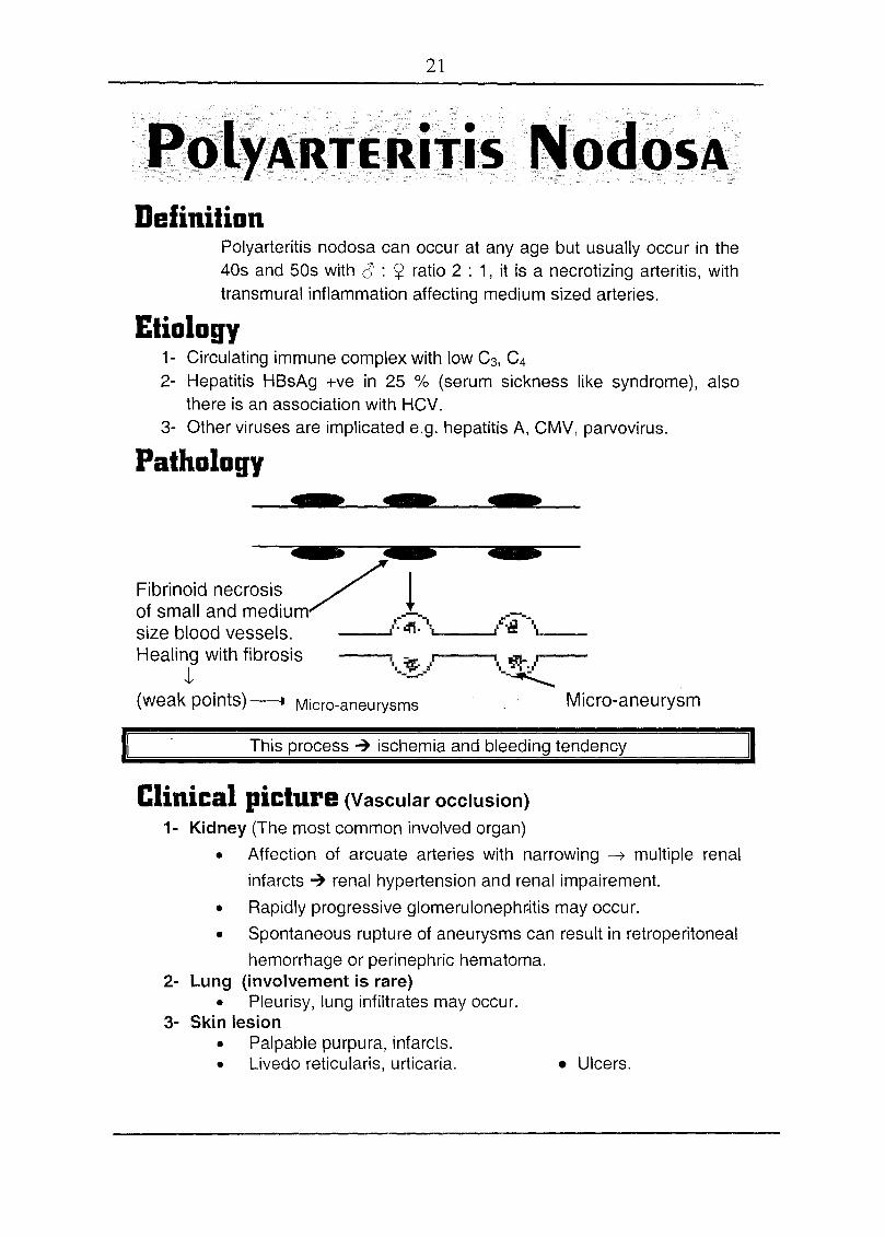
Pathology
Fibrinoid necrosis / !of small and medium • ..• _size blood vessels. ," 4'1. '_, /'-/1. --"-' --Healing with fibrosis
-1(weak points) ----J Micro-aneurysms
i :;.po iII., ,.,.- •.••--- \.~_"i
~Micro-aneurysm
This process 7 ischemia and bleeding tendency
Clinical picture (Vascular occlusion)1- Kidney (The most common involved organ)
• Affection of arcuate arteries with narrowing ---7 multiple renalinfarcts 7 renal hypertension and renal impairement.
• Rapidly progressive glomerulonephr:itis may occur.• Spontaneous rupture of aneurysms can result in retroperitoneal
hemorrhage or perinephric hematoma.2- Lung (involvement is rare)
• Pleurisy, lung infiltrates may occur.3- Skin lesion
• Palpable purpura, infarcts.• Livedo reticularis, urticaria. • Ulcers.
22
4- CVS• Coronary heart disease (angina pectoris or infarction).• Pericarditis may occur.
5- Nervous system.• Stroke.• Mononeuritis multiplex (arteritis in vasa nervorum)
6- Musculoskeletal:• Arthralgia, myalgia.
7- Abdomen• Mesenteric occlusion? acute abdomen.• Intestinal bleeding.• Pancreatitis.
LAB.• ESR 1', + ve CRP• Hb ~, platelets l'• ANA -ve, if it is positive it is mostly lupus vasculitis !?• Rh.F -ve, if it is positive it is mostly RA complicated with
vasculitis.• HB s Ag +ve 25 %• Cs ~ , C4 ~ (Hypocomplementemia), ANCA is rarely positive.• Tissue biopsy from the kidney, muscle or sural nerve.• Eosinophilia.• Angiography of renal, hepatic or mesenteric arteries showing
multiple micro aneurysms.
Treatment• The recommended initial therapy is prednisone 1-2 mg / kg / d
and cyclophosphamide 2- mg. / kg / d, gradual tapering withimprovement, then maintenance therapy to maintain remission.
• Steroid plus interferon if there is positive hepatitis B or C.
23
~i~NT GEU ~R]Eltrfj;s(CGl\J(Temporal Arteritis)
• It is a large vessel vasculitis, predominantely affecting the cranialvessels especially branches of temporal & ophthalmic arteries.
• There is inflammatory infiltrate of lymphocytes, plasma cells andgiant macrophages.
Clinical pictureIt usually affects individuals with mean age around 70 years with female: maleratio 4 : 1.
Headache (usually the first symptom)• Unilateral (temporal or occipital).• Temporal or occipital arteries are thick and tender.
Visual disturbanceThe optic nerve is supplied by the posterior ciliary artery, vasculitis ofwhich leads to occlusion with acute anterior ischaemic optic neuropathy,leading to loss of visual acuity and field in one eye, blindness usuallyoccur rapidly. Fundus examination showing pale optic disc withhaemorrhages, but these changes may take 24-36 hours to develop(fundi may initially appear normal). Once blindness has occurred steroidtherapy of no value other than preventing blindness in the other eye.Arthralgia.Jaw claudication brought on by chewing or talking due to ischaemia ofthe masseters.Tenderness of the scalp (combing the hair may be painful).Transient ischaemic attacks, brain stem infarcts and hemiparesis mayoccur.
Diagnosis• l' TLC, ESR 1',+ ve CRP (ESR may be normal so CRP is helpful in
this situation).• Temporal artery biopsy showing, necrosis of media with inflammatory
cells e.g Iympocytes and plasma cells, negative biopsy does notexclude the diagnosis.
24
91d age with severe headache on one side + tender temporal orogcipital arteries.
*'Ask for ESR....... If .1'. (GCA must be suspected)
*'Give steroid prednisolone 60 mg/d before the result of
investigations to prevent visual loss.• So new onset of headache in old persons should raise the suspicion
of giant cell arteritis.• The elevated ESR remains a sine qua non for the diagnosis
of giant cell arteritis.
Treatment• Good prognosis with treatment.• Prednisolon 60 mg / day, it produces dramatic response within 24-48
hours.It must be started early for fear of blindness.
G!l The steroid dose can be tapered with clinical and laboratoryimprovement, maintenance therapy may be required for at least oneyear.
•MATOSIS
• The peak incidence occurs in 30s and 40s.• This form of systemic vasculitis presents with upper and lower
respiratory tract lesions in association with a focal G.N.III It may be preceded by months or years with recurrent rhinitis, epistaxis
(nasal crusting), sinusitis, otitis media (serous) or history of pulmonarysymptoms such as cough, haemoptysis, chest pain or dyspnea. Themost common ocular abnormality is proptosis due to inflammation ofthe retro-orbital tissue, diplopia may occur due to entrapment of theextraocular muscles.
e Limited wegener's granulomatosis, there is minimal systemicnecrotising vasculitis and local granuloma predominates with chronicsinusitis, nasal and orbital destruction with or c.ivitatinq lunglesions ..
----~-~-~-------------------------
25
Investigations1- ESR 1\ normochromic normocytic anaemia of chronic disease + PNL l'
+ thrombocytosis.2- Eosinophilia is a characteristic in patients with churg - strauss vasculitis
and Wegner's granulomatosis with pulmonary lesions.3- Anti-neutophil cytoplasmic antibodies (ANCA), two patterns of immuno-
fluorescence are distinguished:a- Cytoplasmic granular staining of neutophil (C-ANCA) -ve in 80
% of Wegner's granuloma.b- Perinuclear staining (P-ANCA) in microscopic polyangiitis.
PositiveANCA occur in many other diseases ..including malignancy, infection,inflarrmatory bowel disease, rheumatoid arthritis, systemic .Iupusandpulmonary fibrosis so, the diagnosis of these conditions cannot be made onthe ANCA test alone
Treatment • Steroid and cyclophosphamide• Plasmapharesis• Mycophenolate recently can be used.
TAI<AyASU's DisEASE• ·It is called pulseless disease or aortic arch. $• It is a chronic granulomatous panarteritis affecting the aorta and its
branches and the carotid, ulnar, brachial and radial arteries.• It occurs in young females. It is rare except in Japan.
C / P (female: male ratio 8 : 1), age (25 - 30 yrs)• Fever, weight loss, anaemia, and arthralgia.• Angina, arm claudication.• Hypertension - Aortic incompetence.• Absent peripheral pulses.
Investingations1- Hb ~ due to chronic disease, TLC l' , ESR 1'.2- Angiography ? narrowing of the vessels (four types):
• Type I in aortic arch. • Type II in descending aorta .• Type III mixed (I + II). • Type IV involves the pulmonary artery.
Treatment: • Steroid and cyclophosphamide .• Reconstructive vascular surgery should be avoided
during periods of active inflammation.
Prognosis: Good in more than 90% of cases.
26
I<AWASAk~Systemic disease affecting children ;) years. It is also calledmucocutaneous lymph node $. It predorninatelv occurs in Japan. It may beassociated with mycoplasma and HIV infection In some cases.
C/P• Fever than bilateral conjunctival congestion, dryness and redness of
Lips and oral cavity, redness of the palms and soles.• Vasculitis in the coronary arteries with coronary aneurysm or
myocardial infarction. Myocarditis or pecicarditis may occur.• Cervical lymph node enlargement.
Investigations:• t TLC, l' ESR, t CRP, thrombocytosis.• Positive anti-endothelial cell antibodies ..• Two-dimensional echo or anqioqraphv to detect coronary aneurysms.
Treatment• Intravenous gamrna globulins 400 mg/kg/d 4 consecutive days,
followed after the acute phase by aspirin 200<300 mg/d• Steroids should be avoided because worserunq tho coronary artery
dilatation.
• It 1S a clinical syndrome characterized .by muscle pain with stiffnessthe neck, back, shoulders, upper amiclassically there is increased R
• Polyarthralgias or true synovitis can• It is not a true vasculitis but there is a association with giant
arteritis.
C/P (It is a disease of Hlderly, iillfJan onset IS
• The cardinal features are muscle stiffness and pain attectinq mainlythe proximal muscles of the upper arms and commonly thebuttocks and thighs,
• Early morning stiffness with pain.• Weight loss, depression, swears• On examination there may oainfu] rostrictton
movement but passive movements art:tender.
27
Conditions that may mimic polymyalgia rheumatica• Fibromyalgia. • Inflammatory myopathy.• Cervical spondylosis. • Rheumatoid arthritis.• Malignancy.
Investigations High ESR, very occasionally the ESR is low (early).CRP may be elevated prior to ESR.
Treatment• Dramatic response within 72 hours to steroid 15 mg prednisolone /day
and then tapering after 4-8 weeks, low dose maintenance may berequired for a time 5-10 mg/day. Osteoporosis prophylaxis withbisphosphonates should be considered.
• Some patients require steroid sparing drugs e.g methotrexate orazathioprine.
HypERSENSiTiviTY VAscuLiTis• It is the most common form of vasculitis.• The characteristic histopathological picture is a leukocytoclastic
vasculitis (Ieukocytoclasis refers to inflammation and fibrinoid necrosis ofvessel walls and deposition of cellular debris in the surrounding tissue ofthe skin).
• There are variety of cutaneous lesions appear first on the lower extremities.~.g. palpable purpura.
• ESR can be either normal or elevated, also complement can be normal ordepressed.
• Hypersensitivity vasculitidies are seen in the following conditions:• Henoch schonlein purpura (see haematology).• Serum sickness.• Vasculitis associated with infectious diseases, neoplasms or
connective tissue diseases.• Henoch schonlein purpura showing high levels of IgA, complement levels
are usually normal, IgA deposits can be demonstrated in the vessel wall.• Hypersensitivity Vasculitis is also seen in cases of essential
cryoglobulinemia which is characterized by arthralgia, purpura,hepatosplenomegally, lymphadenopathy and G.N.
• Treatment of hypersensitivity vasculitis includes management of theassociated conditions e.g. drug reactions, bacterial infections. This form ofvasculitis is usually self limited but sometimes patients require NSAID,steroids or immunosuppressive agents.
28
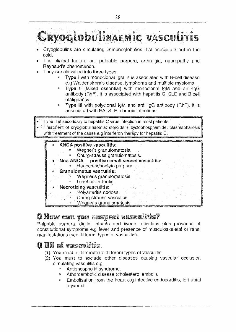
III Cryoglobulins are circulating immunoglobulins that precipitate out in thecold.
e The clinical feature are palpable purpura, arthralgia, neuropathy andRaynaud's phenomenon.
III They are classified into three types." Type i with monoclonal igM, it is associated with B-cell disease
e.g Waldenstrom's disease, lymphoma and multiple myeloma.ill Type II (Mixed essential) with monoclonal IgM and anti-lgG
antibody (RhF), it is associated with hepatitis C, SLE and B cellmalignancy.
•• Type III with poiyclonal IgM and anti IgG antibody (RhF), it isassociated with RA, SLE, chronic infections.
• Type Ili~ secondary to hepatitis C virus infection in most patients.• Treatment of cryoglobulinaemia: steroids + cyclophosphamide, plasmapharesis
with treatment of the cause e.g interferon therapy for hepatitis C.
€I ANCA positive vasculitis:G Wegner's granulomatosis.iii Churg-strauss granulomatosis.
• Non ANCA positive small vessel vasculitis:•• Henoch-schonlein purpura.
Granulornatus vasculltls:ill Wegner's granulomatosis." Giant cell arteritis.
III Necrotizing vasculitis:III Polyarteritis nodosa,ill Churq-strauss vasculitis.I1l We ner'_~granulomatosis.
"i~"~',C>.
o Horwcan yau suspect vasculitiS?Palpable purpura, digital infarcts and livedo reticularis plus presence ofconstitutional symptoms e.g fever and presence of musculoskeletal or renalmanifestations (see different types of vasculitis).
o DB 01 vasculitis ..(1) You must to differentiate different types of vasculitis.(2) You must to exclude other diseases causing vascular occlusion
simulating vasculitis e.gIII Antiphospholid syndrome.ill Atheroembolic disease (cholesterol emboli).'" Embolisation from the heart e.g infective endocarditis, left atrial
myxoma.
29
• Rheumatoid arthritis (RA) is the most common inflammatory arthritis andhence an important cause of potentially preventable disability.
• It is a chronic systemic disease leading to symmetrical inflammatorypolyarthritis affecting mainly peripheral joints, with progressive jointdamage, also it is associated with extra-articular manifestations.
Aetiology- Unknown (triggered by T lymphocyte activation)- Possible factors I?:
• Autoimmune (infiltration of synovial membrane with plasmacells and lymphocytes).
• HLA association e.g. in HLA-DR4.
• Infection, bacterial or slow virus infections have beenimplicated (no definite evidence).
• Smoking also is a risk factor for RA and for positivity ofrheumatoid factor in non RA subjects!?
Pathogenesis• Production of autoantibodies or rheumatoid factors (lg M)in the
blood which react with Fc altered portion of Ig Gwith activation ofthe complement, causing immune complex in synovial membraneswith release of intlarnrnatory.rnediators, cytokines e.g TNF,. IL1, IL6.Rheumatoid factors are positive (sera positive cases) in 80% of
cases of rheumatoid disease. The triggering, antigen remain unclear,. it issuqqestedthat the glycosylation pattern of immunoglobulins maybe abnormal in RA rendering them potentially antigenic
• T lymphocyte activation and macrophages in genetically predisposedpersons e.g. HLA-DR4.
• The presence of activated· T cells and macrophagesand productionof rheumatoid factor autoantibodies in RA suggests that immunedysregulation plays a major role in pathogenesis.
PathologyThe earliest change is swelling and congestion of the synovial membraneand the underlying connective tissue .
• Synovial membrane is infiltrated with lymphocytes, plasma cellsand macrophages. Effusion of synovial fluid intothe joint space takes place during activephases of the disease.
• The inflamed synovial membrane becomes thick oedematousand proliferating forming villi filling the joint space.
30
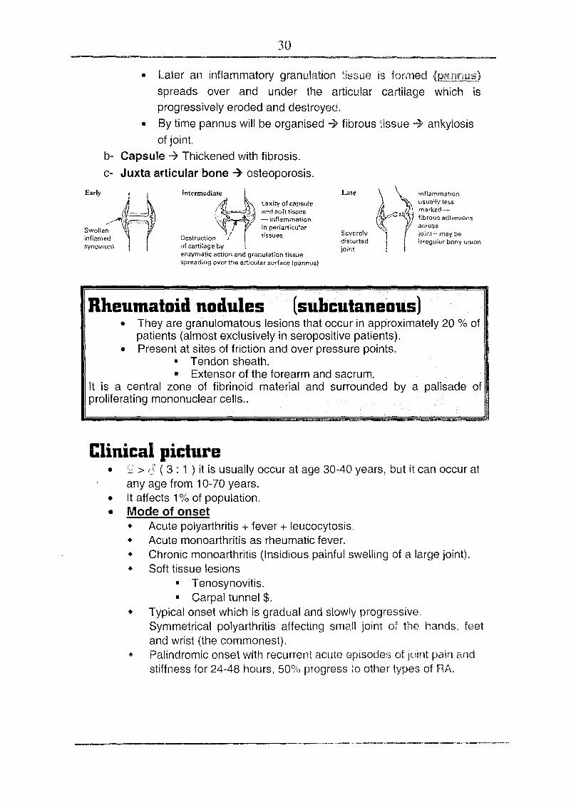
• Later an inflammatory granulation tissue is formed (P81illi"UlUiS)
spreads over and under the articular cartilage which isprogressively eroded and destroyed.
• By time pannus will be organised ~ fibrous tissue ~ ankylosisof joint.
b- Capsule -7 Thickened with fibrosis.c- Juxta articular bone 7 osteoporosis.
Early Intermediate Late Inflammationusually lessmarked--fibrous adhesionsacrossjoint- may beirregular bony union
Swolleninflamedsvnovium
laxiN of capsuleand soft tissue- inflammationin periarticulartissues Severely
distortedjoint
Destructionof cartilsqe byenzymatic action and granulation tissuespreading over the articular surface (pannus)
Rheumatoid nodules (subcutaneous)• They are granulomatous lesions that occur in approximately 20 % of
patients (almost exclusively in seropositive patients).• Present at sites of friction and over pressure points.
• Tendon sheath.• Extensor of the forearm and sacrum.
It is a central zone of fibrinoid material and surrounded by a palisade ofproliferating mononuclear cells ..
Clinical picture• ¥' > c~ ( 3 : 1 ) it is usually occur at age 30-40 years, but it can occur at
any age from 10-70 years.• It affects 1% of population.• Mode of onset
• Acute polyarthritis + fever + leucocytosis.• Acute monoarthritis as rheumatic fever.• Chronic monoarthritis (Insidious painful swelling of a large joint).• Soft tissue lesions
• Tenosynovitis.• Carpal tunnel $.
• Typical onset which is gradual and slowly progressive,Symmetrical polyarthritis affecting srnalt joint of the hands, feetand wrist (the commonest).
• Palindromic onset with recurrent acute episodes of joint pain andstiffness for 24-48 hours, 50% progress to other types of RA.
31
Clinical course: It is usually life-long with intermittentremissions and exacerbations
UScultlskeleta);;manifestatitlD.s
• Painful, swollen, stiff joints.(mainly of the hands and feet)
• Effusion in large joints may occur.• Morning stiffness (duration is an index of activity, it ~ towards end of
the day).
The affected Joints (symmetrical arthropathy)1- Hands ----7 - Proximal interphalangeal joint.
- Metacarpophalangeal joint.(The distal interphalangeal joints are usually spared)
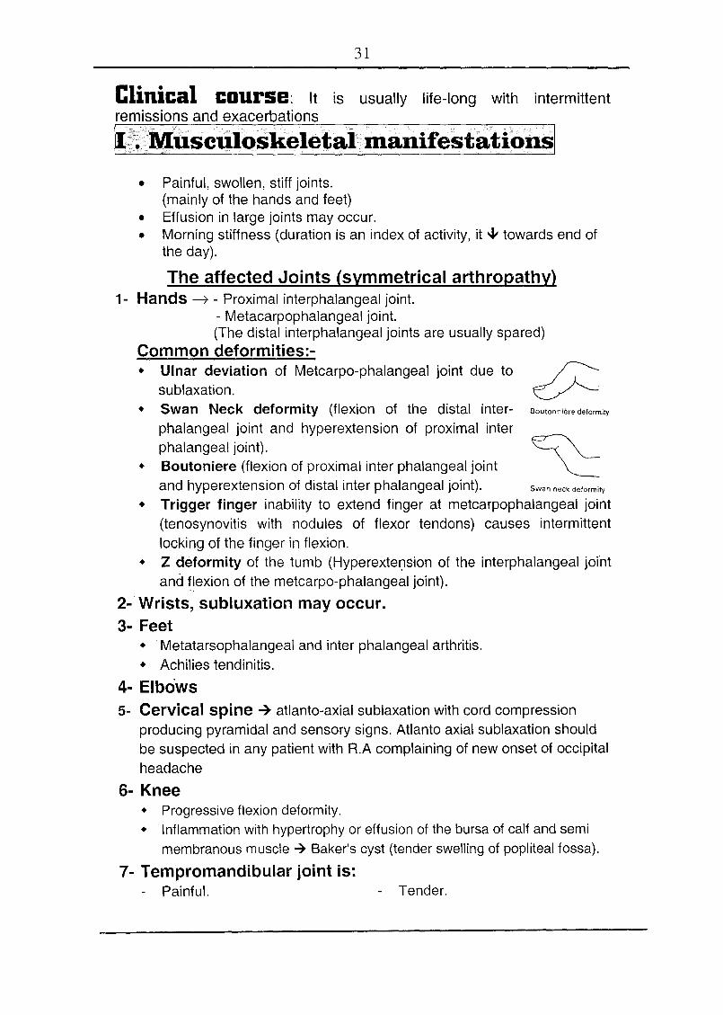
Common deformities:-• Ulnar deviation of Metcarpo-phalangeal joint due to r---
sublaxation. G/"-• Swan Neck deformity (flexion of the distal inter- Boutonniere deformity
phalangeal joint and hyperextension of proximal interphalangeal joint). ~
• Boutoniere (flexion of proximal inter phalangeal joint Cand hyperextension of distal inter phalangeal joint). Swan neck deformity
• Trigger finger inability to extend finger at metcarpophalangeal joint(tenosynovitis with nodules of flexor tendons) causes intermittentlocking of the finger in flexion.
• Z deformity of the tumb (Hyperextension of the interphalangeal jointand Ilexion of the metcarpo-phalangeal joint).
2- Wrists, subluxation may occur.3- Feet
• Metatarsophalangeal and inter phalangeal arthritis.• Achilies tendinitis.
4- Elbows5- Cervical spine ~ atlanto-axial sublaxation with cord compression
producing pyramidal and sensory signs. Atlanto axial sublaxation shouldbe suspected in any patient with R.A complaining of new onset of occipitalheadache
6- Knee• Progressive flexion deformity.• Inflammation with hypertrophy or effusion of the bursa of calf and semi
membranous muscle ~ Baker's cyst (tender swelling of popliteal fossa).
7- Tempromandibular joint is:- Painful. - Tender.
32
8- Other joints e.g. acromioclavicular, sternoclavicular, cricoarytenoied canbe affected.
9- Other musculoskeletal manifestations e.g Bursitis, periarticularosteoporosis, disuse muscle wasting especially the small muscle of thehand.
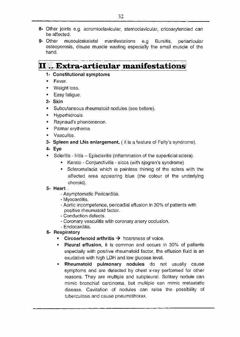
~1);~:)·.·~xtra.artic1l1Q,r···.manifestationsl1- Constitutional symptoms• Fever.• Weight loss.• Easy fatigue.2- Skin• Subcutaneous rheumatoid nodules (see before).• Hyperhidrosis.• Raynaud's phenomenon.• Palmar erythema.• Vasculitis.3- Spleen and LNs enlargement. ( it is a feature of Felty's syndrome).4- Eye• Scleritis - Iritis - Episcleritis (inflammation of the superficial sclera).
• Kerato - Conjunctivitis - sicca (with sjogren's syndrome)• Scleromalacia which is painless thining of the sclera with the
affected area appearing blue (the colour of the underlyingchoroid).
5- Heart.- Asymptomatic Pericarditis.- Myocarditis.- Aortic incompetence, pericadial effusion in 30% of patients with
positive rheumatoid factor.- Conduction defects.- Coronary vasculitis with coronary artery occlusion.- Endocarditis.
6- Respiratory• Circoartenoid arthritis? hoarsness of voice.• Pleural effusion, it is common and occurs in 30% of patients
especially with positive rheumatoid factor, the effusion fluid is anexudative with high LDH and low glucose level.
• Rheumatoid pulmonary nodules do not usually causesymptoms and are detected by chest x-ray performed for otherreasons. They are multiple and subpleural. Solitary nodule canmimic bronchial carcinoma, but multiple can mimic metastaticdisease. Cavitation of nodules can raise the possibility oftuberculosis and cause pneumothorax.
33
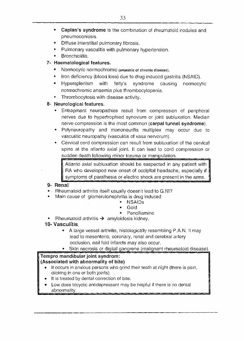
• Caplan's syndrome is the combination of rheumatoid nodules andpneu moconiosis.
• Diffuse interstitial pulmonary fibrosis.• Pulmonary vasculitis with pulmonary hypertension.• Bronchiolitis.
7- Haematological features.• Normocytic normochromic (anaemia of chronic disease).
• Iron deficiency (blood loss) due to drug induced gastritis (NSAID).• Hypersplenism with felty's syndrome causing normocytic
normochromic anaemia plus thrombocytopenia.• Thrombocytosis with disease activity.
8- Neurological features.• Entrapment neuropathies result from compression of peripheral
nerves due to hypertrophied synovium or joint subluxation. Mediannerve compression is the most common (carpal tunnel syndrome).
• Polyneuropathy and mononeuritis multiplex may occur due tovasculitic neuropathy (vasculitis of vasa nervorum).
• Cervical cord compression can result from subluxation of the cervicalspine at the atlanto axial joint. It can lead to cord compression orsudden death following minor trauma or manipulation.
Atlanto axial subluxation should be suspected in any patient withRA who developed new onset of occipital headache, especially ifsymptoms of parathesia or electric shock are present in the arms.
9- Renal• . Rheumatoid arthritis itself usu.ally doesn't lead to G.N!?• Main cause of glomerulonephritis is drug induced
II NSAIDs•• GoldII Pencillamine
• Rheumatoid arthritis ~ amyloidosis kidney.10- Vasculitis.
• A large vessel arthritis, histologically resembling P.A. N. It maylead to mesenteric, coronary, renal and cerebral arteryocclusion, nail fold infarcts may also occur.
• Skin necrosis or digital gangrene (malignant rheumatoid disease).
Tempro mandibular joint syndrom:(Associated with abnormality of bite)
• It occurs in anxious persons who grind their teeth at night (there is pain,clicking in one or both joints).
• It is treated by dental correction of bite.• Low dose tricyclic antidepressant may be helpful if there is no dental
abnormality.
34
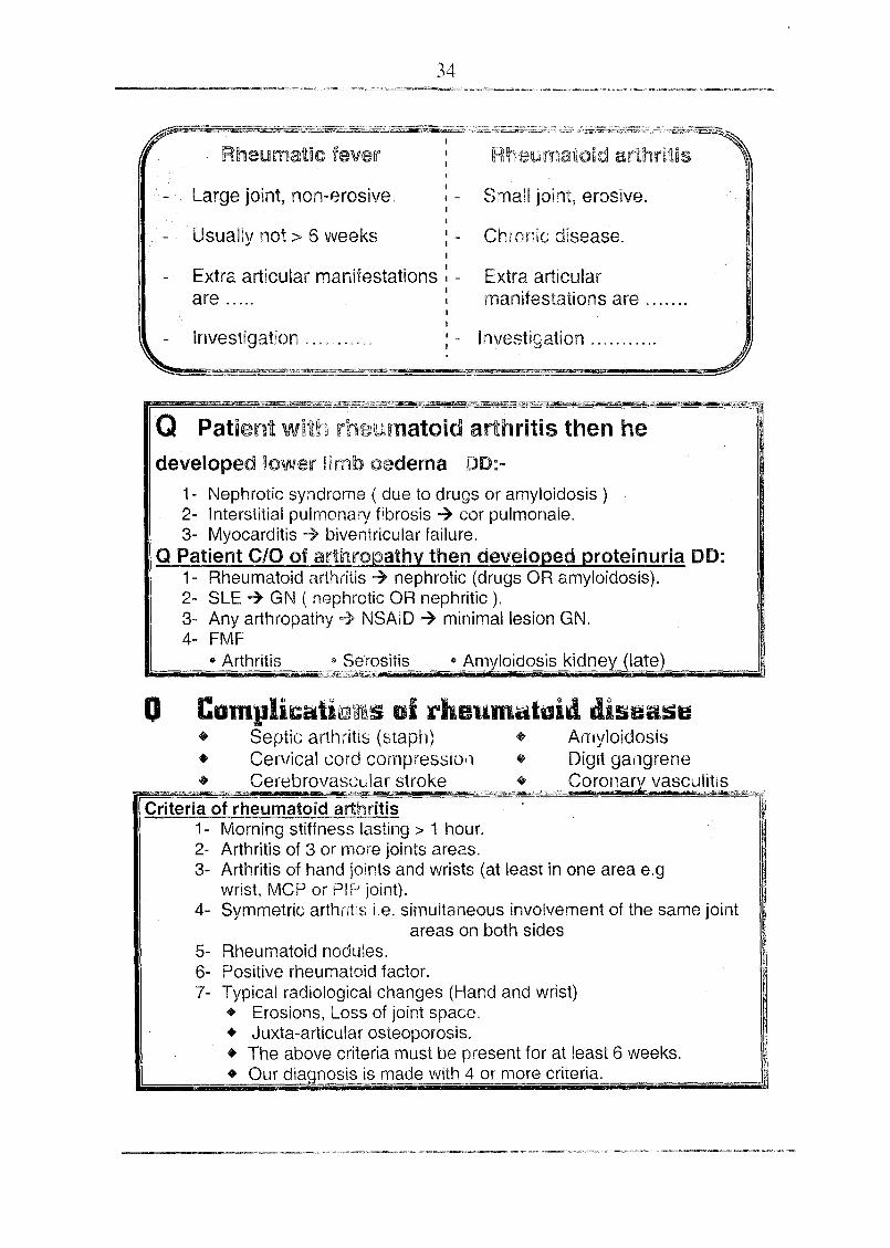
Rheumatic fever
Large joint, non-erosive.
Usually not> 6 weeks
I
II -II
III
Chronic disease.
Small joint, erosive.
Extra articular manifestations: -I
are ..... IIII
I
Extra articularmanifestations are .
Investigation . Investigation .",....",----
Q Patient wllth rheumatoid arthritis then hedeveloped lower limb oedema DD:-
1- Nephrotic syndrome ( due to drugs or amyloidosis)2- Interstitial pulmonary fibrosis ~ cor pulmonale.3- Myocarditis ~ biventricular failure.
Q Patient C/O at arthropathy then developed proteinuria DO:1- Rheumatoid arthritis ~ nephrotic (drugs OR amyloidosis).2- SLE ~ GN ( nephrotic OR nephritic ).3- Any arthropathy =} NSAID ~ minimal lesion GN.4- FMF
••Arthritis " Serositis e_f.myloidosis kidn,eyJ§te)
o Complicaliwn:ms01 rheumaluid diseas~+ Septic arthritis (staph) + Amyloidosis• Cervical cord compression + Digit gangrene
n~~rz Cereb~v_~~~;~}~.s1roke ",_._.=~~" .. ,_Cor2~lary vasc~~""""'~JCriteria of rheumatoid arthritis )
1- Morning stiffness lasting> 1 hour.2- Arthritis of 3 or more joints areas.3- Arthritis of hand joints and wrists (at least in one area e.g
wrist, Mep or PIP joint).4- Symmetric arthntis i.e. simultaneous invoivement of the same joint
areas on both sides5- Rheumatoid nodules.6- Positive rheumatoid factor.7- Typical radiological changes (Hand and wrist)
• Erosions, Loss of joint space,• Juxta-articular osteoporosis.• The above criteria must be present for at least 6 weeks.• Our diagnosis is made with 4 o~ more criteria:-,
35
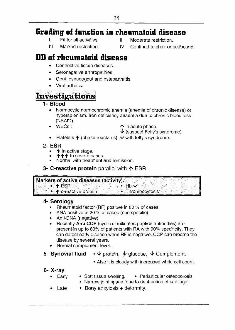
Grading allunetian in rheumataid diseaseI Fit for all activities. II Moderate restriction.III Marked restriction. IV Confined to chair or bedbound.
DD al rheumataid disease• Connective tissue diseases.• Seronegative arthropathies.• Gout, pseudogout and osteoarthritis.• Viral arthritis.
~P~~~~igj!;~iq#sl1- Blood
• Normocytic normochromic anemia (anemia of chronic disease) orhypersplenism. Iron deficiency anaemia due to chronic blood loss(NSAID).
• WBCs: l' in acute phase.~ (suspect Felty's syndrome)
• Platelets l' (phase reactants), ~ with felty's syndrome.
2- ESR• l' In active stage.• 1'1'1' In severe cases.• Normal with treatment and remission.
3- C-reactive protein parallel with l' ESR
Markers of active-diseases (activity).-•.l' ESR' • Hb• l' c-reactive rotein Thromboc tosls:
4- Serology• Rheumatoid factor (RF) positive in 80 % of cases.• ANA positive in 20 % of cases (non specific).• Anti-DNA (negative)• Recently Anti CCP (cyclic citrullinated peptide antibodies) are
present in up to 80% of patients with RA with 90% specificity. Theycan detect early disease when RF is negative. CCP can predate thedisease by several years.
• Normal complement level.
5- Synovial fluid • ~ protein, ~ glucose, ~ Complement.
• Also it is cloudy with increased white cell count.
6- X-ray• Early
• Late
• Soft tissue swelling. • Periarticular osteoporosis.• Narrow joint space (due to destruction of cartilage)• Bony ankylosis + deformity.
36
7· Other investigations• Synovial biopsy.• U/S - C.T scan - MRI for joint involvement.
Investigations aestablish diagnosis
• Clinica criteria • Serological tests. • X-ray • Acute phase reac
To monitor disease activity and drug efficacy:• Pain • Joint tenderness• Morning stiffness. Acute phase
To monitor disease damage:• X-ray • Functional
To monitor side effects of drugs (drug safety):• Urine analysis • Blood picture.• Blood chemistr e. kidne functions.
ITreatmentlThe goals of treatment are:
• Relief of symptoms.
• .Suppression of inflammation.
• Conservation and restoration of function.
• Reduction of mortality.
1- Rest in bed ~ It is valuable in early cases and duringexacerbation.
2- Splinting • to decrease pain and muscle spasm.• to prevent deformity.
3- Physiotheray: It can be started when the phase ofexacerbation disappears.
37
I)RUG<ii-r~·~Up)'.••·•··•··••ofiB 1i..~IJ~A-r·~··.i4\\•••••.~..5i~~5~(1) NSAIDs (for relieve of pain and stiffness), no disease modifying effect.
• e.g. - Phenylbutazone 100mg/8 hrs- Diclofenac 50 mg/8 hrs- Piroxicam 20 mg/8 hrs- Fenoprufen 60mg/8hr.- Ketoprufen 100mg/8hr.- Indomethacin 25-5018 hrs.- Aspirin 600-900 mg/4hr.
+ Side effects- Antiplatelt effect ~ haemorrhage.- Nephrotoxicity, bronchospasm.- Hepatoxicity.- GIT irritation.- Salt, water retension.
Mode of action antiprostaglandins through inhibiton of cyclooxygenaseenzyme.
COX·2 selective NSAID• Celecoxib (celebrex) 100-200 mg twice daily.
. • Meloxicam mobic or melocam 7 .5~15 m Ida .
(2) Glucocorticoids• Low dose of prednisolone can be given 5-10 mg (average 7.5mg)
daily for symptomatic relieve.• The addition of 7.5 mg prednisolone daily to NSAID with disease
modifying antirheumatic drug, may slow the rate of radiologicalprogression over 2 years in patients with early R.A.
• Prophylaxis against osteoporosis is important in patients under longterm steroid therapy.We can use hormone replacement therapy and/or calcium andvitamin 0 or bisphosphonate.
Side effects of steroids:- Hypertension - DM - osteoporosis- Cataract - Weight gain
- Myopathy- Peptic ulcer
I.M. depot injections (40-120mg) methyl prednisolone help to control severedisease flares, but should be used infrequently.
38
Intra-articular steroidsWe use long acting steroids. .Indications • Used for joints that remain painful des
general measures.• It is the treatment of choice in:
~Bursitis.~Tenosynovitis.~Carpal tunnal syndrome.
Side effects 1~Septic arthritis.2- Arthropathy.3~ Rebound ain.
(3) Disease Modifying Anti-Rheumatic Drugs (DMARDs)• The introduction of DMARDs is central to the modern management of
RA.
• These drugs decrease progression of erosive changes and decreaseactivity of the disease. These drugs can be used either singly or incombinations.
• These drugs do not have immediate anti-inflammatory or analgesiceffects but will improve symptoms and acute phase response andreduce radiographic progression as later effects so, it is better to beused in early cases.
(A)IAntimalarial (Hydroxychloroquine) IMechanism: ~e PG
- ~. phagocytic activity of PNL- 200 mg / 12 hr~within 3-6 months.
Dose:Response:Side effects:
- Retinopathy~GIT disturbance
Monitoring: Fundus examination every 6 months.
(B)lSulphasalazine JMechanism: - Anti-inflammatory.
Monitoring:
- 1000 mg /12 hr.. within 3-6 rn- Rashes, BM depression. Megloblastic anemia.- Blood picture and transaminases
Dose:Response:Side effects:
39
(C)IPeniciliamine I (less commonly used)
Does: 250 mg / day.Response: within 3 - 6 months.Side effects:
• Nephrotic syndrome.• Pancytopenia (B.M depression).• Skin rash.
Monitoring: Urine analysis, kidney function tests and blood picture.
(D)IGold 1M I (less commonly used)
It alters the function of macrophages and complement.Dose: after does of 10 mg (for idiosyncrasy)
Give 50 mg /week.Response: within 4-6 months, stopped if there is no response after 6months.Side effects:
• Skin rash, thrombocytopenia, leucopenia.• Nephrotic syndrome.
Monitoring: Blood picture, urine analysis and kidney function tests.
(E)IOral Gold 1 (less commonly used)
Dose: 3 mg/12 hr.Side effects: - Leucopenia. Diarrhea.Monotoring: Blood picture, urine analysis.
(F)IMethotrexate: ]dose: 7.5-15 mg/week.
It can be given oral or s.c. injection, oral folic acid should be given.Response: 1-3 months.Side effects:
Hepatotoxicity. - Leucopenia, thrombocytopenia, anaemia.Alopecia, nausea, diarrhea.
Monitoring: blood picture, liver enzymes.
(G)ILeflunomide 1(Avara)It prevent pyrimidine production in proliferating, lymphocytes, it iseffective as methotrexate but is less likely to suppress bone marrow.
Dose: 1OOmg/day for 3 daysThen 20 mg/day
Side effects:- Alopecia, Diarrhea, Skin rash
Leucopenia, thrombocytopenia. - Hypertension.Disturbed liver biochemistry. e.g increased transaminases.
(H)IAzathioprinii]Dose: 1-2 mg/kg/d orallySide effects: BM depression - Nausea - Infection
40
• Methotrexate is current first choice DMARDs for RA, many Cliniciansselect methotrexate as a first line therapy.
• Leflunomide (Avara) can be used as alternative if methotrexate can notbe tolerated due to side effects.
• Hydroxychloroquine and sulphasalazine can be used in mild cases or ifthere is contraindication to methotrexate or leflunomide.
• Gold, penicillamin, cyclosporine and azathioprine have less favourabletoxicity/efficacy ratio.
• Combination therapy of DMARDs can be used if the use of single drug isnot effective.
(4) Biological TherapyAnticytokine therapy is now being used:
(a) Blockade of IL-1 and IL-6 with receptor antagonists showing rapid anti-inflammatory effects e.g Anakinra (Kineret) 100mg S.C once daily.
(b) Anti TNF monoclonal antibody.
gep l1iof jQint • (;t!'nqofDMARDs.
(5) Surgical treatment• Tendon repair.• Nerve decompression• Correction of deformity.
• Synovectomy.• Arthrodesis• 'Joint replacement.
41
JUVENilE CItRONic OR idiopathicARYltRhis fJCA oRJIA)
• Juvenile chronic arthritis is chronic inflammatory arthritis before age 16years for at least 6 weeks.
• It can be divided into the following types:
(1)Systemic onset arthritis (Still's disease)It affects boys and girls, adult onset still's disease is rare.Prominent systemic complaints and extra articular involvement.Fever, rash (non pruritic fleeting maculopapular rash).Lymphadenopathy, hepatosplenomegally.Pericarditis, pleurisy.
- Arthritis or arthralgia and mylagia.Rheumatoid factor usually is negative.
- High ESR and CRP, neutrophilia and thrombocytosis.
(2) Polyarticular arthritis (5 joints or mare)Bilateral symmetrical polyarthritis specially hands, wrists, PIP and DIP.Rheumatoid factor +ve in 10-20% of cases.
- ANA is positive in 20-40% of cases.
(3)Paueiartieular arthritis.(a) Oligoarthritis and anterior uveitis:- Affects up to 4 joints (four or fewer joints) especially wrists, knees, ankles.- -ve rheumatoid factor.
Uveitis, this requires regular screening by slit-lamp, blindness may occur.Positive ANA in 60% of patients, which identifies those at higher riskfactor for chronic uveitis.Negative rheumatoid factor.
(b) Axial skeletone oligoarthritis:- Asymmetrical knee, ankle arthritis, followed by sacroiliac joints, uveitis
can occur, 50% of patients have HLA-B27 but few have positiverheumatoid factor.
(4) PSDriatic arthritis:- This affects fingers and toes, also polyarthritis involving large and small
joints may occur, psoriasis may be present in the child or a first degreerelative.
42
Treatment al juvenill chronic arthritis• Salicylates are no longer the primary drugs used in the treatment of
juvenile arthritis due to the potential precipitation of Reye's syndrome.• Other NSAIDs with low doses and paracetamol are helpful as regard pain
and stiffness.• Methotrexate, the most commonly used second line agent. Leflunomide is
also effective. Sulfasalazine is also used.• Corticosteroids are used with systemic complaints e.g pericarditis, also in
the treatment of chronic uveitis (Local or systemic).• Intravenous infusion of gamma globulin to control severe systemic onset
or polyarticular disease.• Physical and occupational therapy.
• It is a variant of rheumatoid disease.• It is a rheumatoid disease + splenomegally + leucopenia.• C/P. Picture of rheumatoid disease.
• LN++ (they are palpable in the distribution of affected joints)and splenomegally with hypersplenism.
• Liver enlargement (due to. lymphocytic infiltration of the liver) ..• Anaemia of chronic disease usually occur or due to iron
deficiency (blood loss) due to NSAIDs or rarely haemolyticanaemia (coomb's positive).
• Investigations• As rheumatoid disease (sero -ve) i.e -ve rheumatoid factor.• Blood picture showing pancytopenia (hypersplenism).
• Treatment .. directed to joint disease + splenectomy if needed.
Definition: It is a kerotoconjunctivitis sicca with xerostomia, the diseasemay be primary or secondary in association with other autoimmune disorder often rheumatoid disease, SLE orIymphoproliferative diseases.
Primary Sjogren syndrome . .siccarheumatoid arthritis or any other a oimmSecondary Sjogren syndrome Le. th is associated anautoimmune or C.T. disease e.g. rheumatoid disease, SLE, PM,primary biliary cirrhosis or thyroditis.
43
J\.ssQciatedautpimmune disorders in cases.otsjoqren's syndrome:SLE :.. Chronic active hepatitis. Primary biliary cirrhosis.
~Progressive systemic sclerosis. Myastheniaqravis,
• C/P • Occular dryness confirmed by schirmer tear test usinga standard strip of filter paper placed under eachlower eye lid. Wetting of < 5 mm in 5 minutesindicates defective teat production (A young personnormally moistens 15 mm).
• Xerostomia.• Salivary and parotid enlargement is seen.
• Non erosive arthritis, Raynaud's phenomenon.• Cryoglobulinaemia.• Peripheral neuropathy.• Lymphadenopathy, there is increased incidence of lymphoma.• Renal tubular acidosis, glomerulonephritis.
• Investigations • Rheumatoid factor +ve.• +ve ANA in 80% of cases.• Antisalivary duct Ab (Siogren's Ab).• Anti Ro.• Antiparietal cell antibodies.• Rose Bengal staining of the eye StlOWS punctate
or filamentary keratitis.• Treatment..• Xerophthalmia can be treated with artificial tears e.g (hypromellose)
and ,lubricating ointment at night. Soft contact lenses can be useful forcorneal protection.
• Sugar free chewing gum or lozenges can stimulate saliva flow. Using asaliva substitute containing carboxy methylcellulose as mouth wash,drugs causing disease salivary secretion should be avoided(anticholinergics) .
• Vaginal dryness is treated with lubricants e.g K-Y jelly.• Extra glandular and musculoskeletal manifestations may respond to
steroids, azathioprine can be added.• If there is massive lymphadenopathy, biopsy should be performed to
exclude malignancy.
44
These are group of sero negative arthritis (negative rheumatoid factor) thatshare certain epidemiologic, clinical and pathologic features. They all showconsiderable overlap and similarity of articular and extra - articular clinicalfeatures.
fil1ey inclu4el• Ankylosing spondylitis.• Reiter's disease.• Psoriatic arthritis.• Post infectious reactive arthropathies.• Enteropathic arthropathies associated with:.
• Ulcerative colitis.• Chron's disease.
1~~p.~rai/fe~tq~e~1• Sacroiliitis and / or spondylitis.• Asymmetrical peripheral arthritis.• Familial association.• High prevalence with HLA - 827•
• Negative rheumatoid factor.• Uveitis.• Aortic root fibrosis -» aortic incompetence.• Erythema nodosum.• Enthesitis i.e 'inflammation at the enthesis (the site of insertion of
ligaments or tendons into bone).
Why possession of HLA - 827 predispose towards seronegativearthropathies !?
• There is a cross reactivity between HLA - 827 and an antigen carried bysome invading organisms e.g. yersinea, Chlamydia, klebsiella?
• The invading organism in HLA - 827 initiates an autoimmune reactionor render the cells more susceptible to cytotoxic lymphocytes?
45
It is an inflammatory arthropathy starting in the sacroiliac and spinal joints withtendency to ankylosis of the axial skeleton.
[Aetiolo~• The prevalence is about 0.2% of the general population.• Age of onset: 15 - 40 years.• Sex: (j' : ~ = 3 : 1" HLA - 827 association (95% of patients have HLA-827).
• High incidence of prostatic infection by klebsiella?So, there may be an association between this disease and klebsiellainfection !?
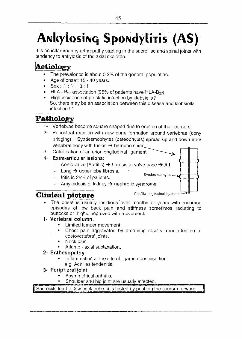
ratholo~1- Vertebrae become square shaped due to erosion of their corners.2- Periosteal reaction with new bone formation around vertebrae (bony
bridging) = Syndesmophytes (osteophytes) spread up and down fromvertebral body with fusion ~ bamboo spin~
3- Calcification of anterior longitudinal ligament. ~4- Extra-articular lesions:
- Aortic valve (Aortitis) ~ fibrosis at valve base ~ A.1.- Lung? upper lobe fibrosis.
Iritis in 25% of patients.- Amyloidosis of kidney ~ nephrotic syndrome.
Syndesmophytes
jClinical picturel Calcific longitudinalligament~
• The onset is usually insidious over months or. years with recurringepisodes of low back pain and stiffness sometimes radiating tobuttocks or thighs, improved with movement.
1- Vertebral column.• Limited lumber movement.• Chest pain aggravated by breathing results from affection of
costovertebral joints.• Neck pain.• Atlanto - axial sublaxation.
2- Enthesopathy• Inflammation at the site of ligamentous insertion.
e.g. Achilles tendenitis.3- Peripheral joint
• Asymmetrical arthritis.• Shoulder and hi joint are usuall affected.
I Sacroiliits lead tolqw back ache. it is tested by pushing the sacrum forward.
464- Extra-articular manifestations
• Amyloidosis kidney.• Aortic incompetence, cardiac conduction defects, pericarditis.• Apical lung fibrosis.• Prolonged fever.• Uveitis.
How can you differentiate between mechanical andinflammatory back ache e.g AS?
1- Character of pain.Inflammatory. 7 1'1' at night, it is improved by movement.Mechanical 1'1' with movement and improved with rest.
2- Laboratory findings.- ESR, CRP 1'1' in inflammatory back ache.
etectmade
m above the lum osacral junctiond by rizontalline betw n the posterior superior iliac spines). The
ient then bends forward maximally, and the distance between the twoarks is measured. The distance increases 5cm or more in normal lumbarobility and less than 4cm in case of decreased lumbar mobility.
~DofASI• The inflammatory back pain of AS is usually distinguished by the
following features.- Age.of onset < 40 years - Insidious onset- Duration> 3 m - Improvement with exercise.
• AS must be differentiated from other causes of bachache (see later).• Diffuse idiopathic skeletal hyperstosis (DISH) must be differentiated
from AS .as it leads to marked calcification and ossification ofparaspinous ligaments.
~g~~~~i,gB~igrt~j1- X ray Sarco iliac joint 7 erosion, sclerosis. It is often the first abnormality.2- X ray Spine (The disc is preserved, unlike in spondylosis).
• Square shaped vertebrae.• Calcification of longitudinal ligament.• Bony bridging between vertebral bodies = syndesmophytes -7
bamboo spine.
3- Laboratory finding (there is no specific laboratory test).• Normochromic anemia.• ESR l' ,l' CRP during activity.• Rheumatoid factor -ve.
47• HLA Testing is rarely of value because of the high frequency of
HLA-B27 in the population.• Alkaline Phosphatase l' with activity!?
ITreatmentl (The principles are to relieve pain and spinal stiffness).
• The key of treatment is early diagnosis to start preventive exerciseprogram before syndesmophytes have formed. Excercises aim tomaintain spinal mobility, posture and chest expansion (swimming isrecommended).
• NSAIDs -7 relieve symptoms, but don't alter the course of the disease• Local steroid -7 for enthesopathy and planter fasciitis.• Genetic counselling.• Systemic steroid sometimes required for treatment of uveitis.• Sulfasalazine and methotrexate may be of value for peripheral
arth ropathy.• TNF blockers are effective• Surgery: plaster jackets to correct kyphosis, hip arthroplasty.
Prognosis.With exercise and pain relief, the prognosis is excellent and over 80%of patients are employed.
REACTivE ARTItRiTis• It is a sterile arthritis which occurs following an infection.• The intection is either sexually transmitted or gastrointestinal which
occurs before the onset of arthritis by 1-4 weeks.• The arthritoqenic bacteria causing reactive arthritis are salmonella,
yersinia, shigella, helicobacter or klebsiella pneumoniae, alsoChlamydia can cause arthritis.
• Reactive arthritis may be just arthritis, but sometimes the full picture ofReiter's disease may occur.
REiTER' sSYNd ROME
kletioloii1It is an inflammatory arthritis which is usually associated with sexuallyacquired infection e.g non specific urethritis in males, non specific cervicitisin females. Also it may follow an acute attack of gastrointestinal infection. Avariety of organisms can be the trigger (see below) so Reiter's syndromemay follows sexually acquired infection or gastrointestinal infection).
48
ICJlgt~~(piciipr~1(predominantly a disease of young men).• The onset is typically acute with development of urtheritis,
conjunctivitis and arthritis.• Sex ~ (; > ¥ with ratio of 15-1• Age ~ 20 - 40 years• Asymmetric arthritis ~ big joint of L.L. (knee, ankles, and feet).• Planter faciitis and achilles tendenitis ~ painful heel syndrome.• Sacroilitis and spondylitis may occur• Eye ~ conjunctivitis, uveitis.• Skin - keratoderma ~ yellow brown papules with desquamating
margins on the soles and plasms. Circinate balanitis (ulcers on glanspenis), present in 20-50% of patients.
• Heart ~ A.1. - pericarditis.• Neurological ~ meningoencephalitis and neuropathy.• Buccal ulceration (tongue, palate, buccal mucosa and lips), they are painless.
~~"y"~$tiptionsl• ESR i, iCRP • Rheumatoid factor -ve.• HLA B27 is present in 80% of patients• Anemia. (normocytic, normochromic) • Sterile pyuria.• X-ray showing soft tissue swelling, narrowing of joint spaces with or
without sacroiliitis.
fireatmentl• Rest..• NSAIDs ~ for arthritis.• Steroid (not for arthritis), it is used for:
• Achilles tendenitis (local injection.)• Eye (topical)
• Systemic corticosteroids are rarely required.• Methotrexate, azathioprine or sulphasalazin may be used.• The non-specific urethritis or any infection should be treated with short
course of antibiotics e.g tetracycline.• Anterior uveitis is a medical emergency requiring topical, subconjunctival
or systemic steroids.
• ArthritA~elljc b(.\.ft~ri~.c~UJ.sing• Salmonella,. Yerslnlaand •• Chlamydia causing urethritis.
Spondylolisthesis:It is subluxation of lumbar vertebrae usually occurring in adolescence, itoccur due to congenital pars interarticularis defects -7 instability thatpermit the vertebrae to slip. It may lead to cauda equina syndrome, itmay require orthopedic assessment.
49
• It is an inflammatory arthritis that occurs in about 10 % of patients withpsoriasis.
• It shares symptoms, signs and x-ray finding of spondyloarthritis.
IClinical picture (thel"e>are five clinical. subse.tsl!1-Asymmetric oligoarthropathy:-
• There is asymmetric involvemment of both large and small joints• There is a sausage finger or toe (dactylitis) due to
interphalangeal joint synovitis, tensosynovitis and inflammation ofthe intervening tissues.
2- Symmetric polyarthropathy like rheumatoid disease.3- Arthritis mutilans:-
• This is a severe destructive arthropathy of fingers and toes withperiarticular osteolysis and bone shortening causing telescoping ofdigits i.e the encasing skin appears invaginated and telescoped.
4- Psoriatic spondylitis with sacroiliitis.5- Psoriatic nail disease with distal interphalangeal joint involvement.
~estigationsl• Rheumatoid factor is negative.• Anaemia• Hyperuricemia.• Polyclonal hypergamma globulinemia.• There is osteolysis of the metatarsal heads and central erosion of the
proximal phalanges causing (pencil cup) appearance.• Bone resorption giving opera-glass appearance. Also sacroiliitis and
spondylitis may present.
ITreatmentj• Rest, NSAIDs, local synovitis responds to intraarticular steroid
injection.
• Gold and methotrexate are tried.• Photochemotherapy and long wave U.V light for skin lesions.
• TNF blockers can be used in resistant cases.
50
~~thritisa~sociatedwitljinf.la.mlJlatorY bowel disease(Enteropc:ithicsynovitis)
• It occurs in 10-15% of patients with ulcerative colitis and Crohn's disease,50% of patients have HLA-B27
• Acute, often migratory.• Non erosive.• Affects mainly knees and ankles.• There is sacroiliitis or spondylitis.• It is treated by NSAIDs.• Intra articular steroids for monoarthritis• Sulphasalazine is frequently prescribed as this may help both bowel and
joint disease.
SCLEROdERMA(SYSTEMic SCLEROsis)
• Systemic sclerosis is a connective tissue disease characterized bydegenerative changes and fibrosis of the integument (skin) associated withvessel oblitrative disease and internal organ Lesions in:
. • Heart. • Lung.• Kidney. • GIT.
So it. is a multi system disease to be differentiated from localizedscleroderma synd~omes e.g. '!1orphoea that do not involve internal organ.
!Pathology and pathogenesisl1- Vascular lesions:-
• Endothelial damage with release of endothelin causing vasoconstriction.• Also there is release of cytokines (IL 1,4,6,8 ), transforming growth
factor (TGF), and platelets derived growth factor (PDGF) withactivation of fibrblasts, this leads to damage of blood vessels andwid spread obliterative arterial lesions with subsequent chronic
ischaemia2- Fibrotic features:
• The activated fibroblasts synthesize increased quantities ofcollagen types I and III and fibronectin producing fibrosis of the
dermis of the skin and internal organs.
51
IClllticft,lp.icture!• Mainly ¥ ( 4 : 1 )• 4th and 5th decades.• Rarely visceral scleroderma occurs in absence of the skin changes.• Skin changes:
• Initially the skin is oedematous (non pitting edema) and thenbecomes tight.The skin tightening begins on the fingers and hands then theproximal parts of the extremities, in addition to the thorax andabdomen with restriction of movement of the fingers.
• Later skin becomes shiny with atrophy and ulceration, areas ofhyperpigmentation or hypopigmentation are common, calcinosismay occur.
• Raynaud's phenomenon: It is vasospasm of peripheral arteriescausing color changes (pallor, cyanosis and erythema). It occurs inmost patients with systemic sclerosis.
• Musculoskeletal• Non erosive arthritis or arthralgia.• Myopathy ( = sclerodermatomyostitis )
• GIT1. Dysphagia (esophageal hypomotility)2. Reflux, (due to loss of lower esophageal tone).3. Malabsorption syndrome (due to over bacterial growth).4. Colonic involvement with constipation, intestinal pseudo
obstruction may occur.5. Primary biliary cirrhosis.
• Heart• Cardiomyopathy with heart failure.• Pericarditis, pericardial effusion.• Coronary heart disease.
• Lung• Interstitial pulmonary fibrosis.• Pulmonary hypertension.• Aspiration pneumonia (due to oesophageal dysfunction).
• Kidney• Scleroderma kidney (thickness of intima of the interlobular
arteries)leading to:-• Malignant hypertension. • Renal failure.
• Scleroderma renal crisis i.e abrut onset of acceleratedhypertension, oliguria and microangiopathic hemolysis.
Diffuse skin disease, pulmonary hypertension and renalinvolvement are associated with poor prognosis.
52
1- ANA +ve in 90% of patients.2- Auto Ab against sclero nuclear protein called anti-topoisomerase
(Anti-Sci 70) in 20-30% of patients (it is specific).3- Rheumatoid factor +ve in 30 %.4- Microangiopathic haemolytic anemia.5- Skin biopsy 7 collagen in dermis.6- Sa swallow for impaired oesophageal motility.7- X-ray hand showing calcification around fingers, late erosion and
resorption of the tuft of the distal phalanges
Treatment (no specific statisfactory drug therapy)• Penicillimine (antifibrotic drug) causing minimal skin softening.• Raynaud's may be treated by hand warmers, vasodilators e.g calcium
channel blockers e.g verapamil, deltiazem and nifedipine as well asnitroglycerin ointment.
• Steroids are indicated for treatment of inflammatory myositis,pericarditis or pulmonary fibrosis.
• Oesophageal symptoms can be treated by prokinetic drugs e.g domperidone.
• ACE inhibitors for treatment of hypertension and prevention of renalfailure. Also they are used in renal crisis ..
• Bacterial over growth due to intestina,l stasis respond to broadspectrum antibiotic.
• Steroids and cyclophosphamide may be of value in treating alveolitisand pulmonary fibrosis.
• Endothelin receptor antagonist (bosentan) can be used for severepulmonary hypertension and digital ulceration.
• Sympathectomy to prevent auto amputation.• Other novel agents currently under study include mycophenolate and
tacrolimus.
53
~ub.~etsof.§ystemicscierosisaod scleroderma syndromesSystemic sclercsls
.(.Diffuse cutaneous scleroderma.~.•. Limited cutaneous (CR~ST syndrome), -ve.anticentromere .Ab.
l.:ocalized scleroderma• Morphoea and linear scleroderrna-lirnlted'tc well demarcated
lesion of theskin and subcutaneous C.T, serology like systemicselerosls.occaslonally systemicfeatu res develop, Raynaud'sphenomenon is rare.
Overlap syndrome (scleroderma associated with other autoimmune disease)~cleroderma like syndromes i.e other conditions in which indurations or
brawny oedema of skin occur and considered as D.O. of sclerodermainctude scleredema, scleromyxoederna, amyloidosis,acromegally andeosinophilic fasciitis.
It CREST Syndrome- C? Calcinosis.- E ~. Esophageal dysfunction.- T ? Telangectasia.There is minimal visceral involvement, positive anticentromere antibodies.• Systemic sclerosis may occur in association with features of otherconnective tissue diseases. The term overlap syndrome has been to describesuch patients.
- R ? Raynaud's phenomenon.-S? Sclerodactly.
MiXEd CONNECTivE TiSSUE diSEASE• This syndrome involving mixed features of :-
- Myositis. - SLE.- Scleroderma.
C/P- It is of gradual onset. - Renal affection is rare.
Features (according to the frequency)1- Arthritis or arthralgia.2- Haynaud's phenomenon.3- Oesophageal dysmotility.4- Lymphadenopathy.5- Serositis.
6- Myositis.7- Skin rash.8- Fever.9- Renal disorder.
Investigations • ANA +ve.• Positive anti-ribonucleoprotein (RNP) antibodies.
Treatment Good response to steroid.
54
• It is a systemic vasculitis of unknown etiology- Viral.!? - Autoimmune.!?
• Diagnostic criteria- Recurrent oral ulcers, at least three times in a 12 month period.
• Plus two of:- Uveitis or retinal vasculitis- Skin lesions e.g erythema nodosum.- Recurrent genital ulcers.- Positive pathergy test
Oligoarthritis, thrombophlebitis and neurobehcet (multiple sclerosis like) mayalso occur.
fl'reatlDentl• Oral ulcers can be treated by topical steroids.• Colchicine is effective for erythema nodosum and arthralgia.• Systemic steroids in combination with other immunosuppressive drugs
for systemic manifestations e.g ocular or neurological disease.
The pathergy test or reacti~n is highly specific to Behcet's d!seasinflammatory' reactivity to any scratches o.r intraderma:1 saline in'leads to papule or pustule formation .\'\(ithin24-48 hours -.
55
~ItY~TJ\ll NduGid ARTItOPA:r:kyIGout and Hyperuriceriii81
• The term gout refers to a disorder characterized by defect in purinemetabolism, and manifested by:1- Hyperuricemia.2- Crystalline deposition of monosodium urate in tissues (tophi).3- Gouty arthritis.4- Uric acid stones and gouty nephropathy.
/
AMP ) AdenineHGPRT
~ xo xoPurines ~ I.M~ Hypoxanthine ~ Xanthine ~ Uric acid.
\ HGPRTv--- .GMP ) Guanine
• AMP = Adenosine monophosphate.• IMP = Inosine monophosphate.• GMP = Guanosine monophosphate.• XO = Xanthine oxidase.• HGPRT· = Hypoxanthine-guanir'1e~p'hosphoribosyl-transferase
!Classificationof hyperuricemi8JPrimary(Genetic)
1- Metabolic over production• Idiopathic, 10-20% of patients with primary gout.• Enzymatic defects. e.g:
(Hypoxanthine - guanine - phosphoribosyl -transferase) which is essential for reconversion ofguanine, hypoxanthine into their mother substanceso this enzymatic defect -7 accumulation of uricacid.
Lesh-Nyhan syndromeX linked recessive.
Uric acid 1',Choreoathetosis, mental
retardation and selfmutilation
• Glucose 6 phosphatase deficiency.11-Renal
• Idiopathic under excretion (Isolated tubular defect), more than twothirds of individuals with primary gout.
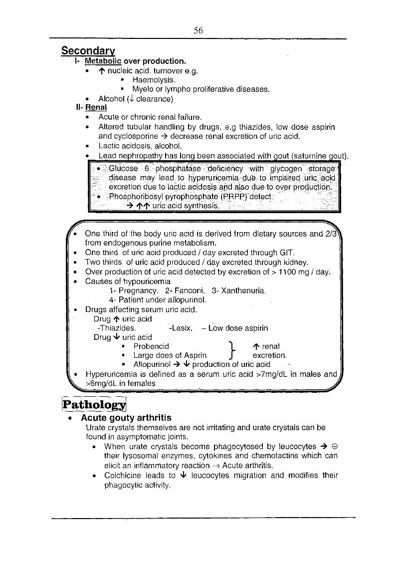
56
Secondary1- Metabolic over production.
• l' nucleic acid. turnover e.g.• Haemolysis.• Myelo or Iympho proliferative diseases.
• Alcohol (t clearance)11-Renal
• Acute or chronic renal failure.• Altered tubular handling by drugs. e.g thiazides, low dose aspirin
and cyclosporine -7 decrease renal excretion of uric acid.• Lactic acidosis, alcohol.• Lead ne hro ath has Ion been associated with out saturnine out).
• G lucpsei6 .,.phosphat~?epeficiehGy~ithi9Iy~ogrn .r?to.t:l~g·~g.ise?Sr ",may lead. to ....q~~erl,lfi.9~Ti§l,l·;du~rtoim~aiJ~.gl.lrjS.i~Si?excr~tion d.ueto laqtic~sipo?iSi?pd§l,J.so.gue to oy~r prqpYGUqQ,Phosphoripo?yl pyroph.<>spqatr(!3.8P!3) def~ct.
71'1' uric acid s nthesis ..
• One third of the body uric acid is derived from dietary sources and 2/3from endogenous purine metabolism.
• One third of uric acid produced / day excreted through GIT.• Two thirds of uric acid produced / day excreted through kidney.• Over production of uric acid detected by excretion of> 1100 mg 1 day.• Causes of hypouricemia
1- Pregnancy. 2- Fanconi. 3- Xanthenuria.4- Patient under allopurinol.
• Drugs affecting serum uric acid.Drug l' uric acid-Thiazides. -Lasix. - Low dose aspirin
Drug ~ uric acid• Probencid } l' renal• Large does of Asprin excretion.• Allopurinol 7 ~production of uric acid
• Hyperuricemia is defined as a serum uric acid >7mg/dL in males and>6mg/dL in females
IPatholon• Acute gouty arthritis
Urate crystals themselves are not irritating and urate crystals can befound in asymptomatic joints.
• When urate crystals become phagocytosed by leucocytes 7 EBtheir lysosomal enzymes, cytokines and chemotactins which canelicit an inflammatory reaction -7 Acute arthritis.
• Colchicine leads to ~ leucocytes migration and modifies theirphagocytic activity.
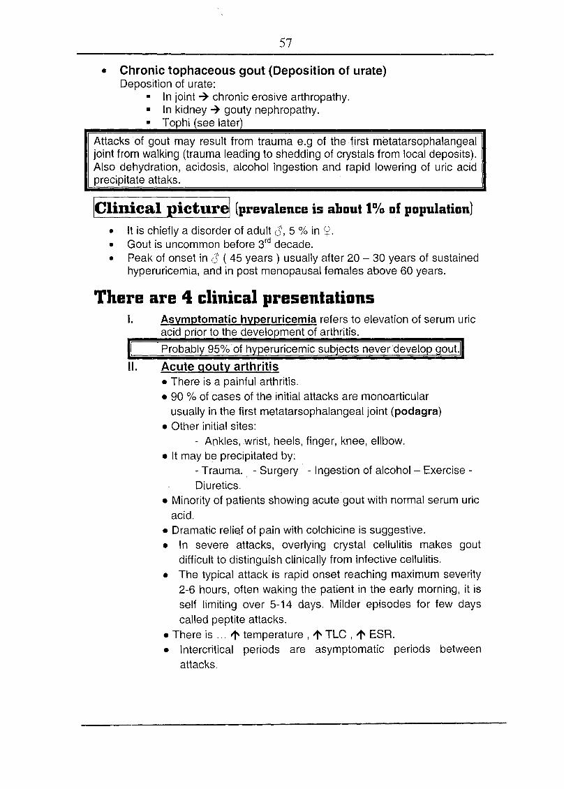
57
• Chronic tophaceous gout (Deposition of urate)Deposition of urate:
• In joint ~ chronic erosive arthropathy.• In kidney ~ gouty nephropathy.• Tophi (see later)
Attacks of gout may result from trauma e.g of the first metatarsophalangealjoint from walking (trauma leading to shedding of crystals from local deposits).Also dehydration, acidosis, alcohol ingestion and rapid lowering of uric acidprecipitate attaks.
IClinical picturel (prevalence is about 10/0 of population)• It is chiefly a disorder of adult 0,5 % in ¥.• Gout is uncommon before 3rd decade.• Peak of onset in ° (45 years) usually after 20 - 30 years of sustained
hyperuricemia, and in post menopausal females above 60 years.
There are 4 clinical presentationsI. Asymptomatic hyperuricemia refers to elevation of serum uric
acid rior to the develo ment of arthritis.I Probably 95% of hyperuricemic subjects never develop gout.1
II. Acute gouty arthritis• There is a painful arthritis.• 90 % of cases of the initial attacks are monoarticular
usually in the first metatarsophalangeal joint (podagra)• Other initial sites:
- Ankles, wrist, heels, finger, knee, ellbow.• It may be precipitated by:
- Trauma .. - Surgery - Ingestion of alcohol- Exercise -Diuretics.
• Minority of patients showing acute gout with normal serum uricacid.
• Dramatic relief of pain with colchicine is suggestive.• In severe attacks, overlying crystal cellulitis makes gout
difficult to distinguish clinically from infective cellulitis.• The typical attack is rapid onset reaching maximum severity
2-6 hours, often waking the patient in the early morning, it isself limiting over 5-14 days. Milder episodes for few dayscalled peptite attacks.
• There is ... l' temperature, l' TLC , l' ESR.• Intercritical periods are asymptomatic periods between
attacks.
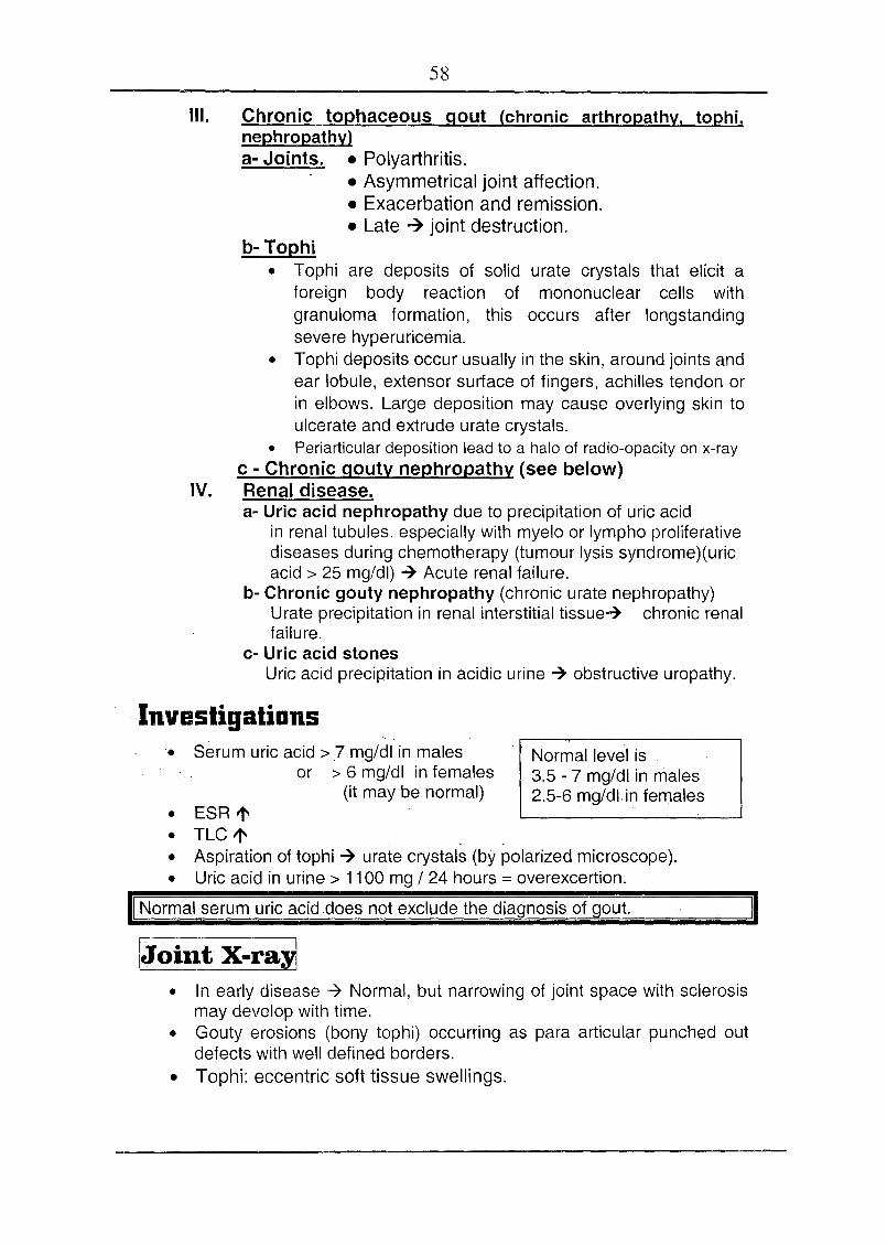
58
III. Chronic tophaceous gout (chronic arthropathy, tophi,nephropathy)a- Joints. • Polyarthritis.
• Asymmetrical joint affection.• Exacerbation and remission.• Late ~ joint destruction.
b- Tophi• Tophi are deposits of solid urate crystals that elicit a
foreign body reaction of mononuclear cells withgranuloma formation, this occurs after longstandingsevere hyperuricemia.
• Tophi deposits occur usually in the skin, around joints andear lobule, extensor surface of fingers, achilles tendon orin elbows. Large deposition may cause overlying skin toulcerate and extrude urate crystals.
• Periarticular deposition lead to a halo of radio-opacity on x-rayC - Chronic gouty nephropathy (see below)
IV. Renal disease.a- Uric acid nephropathy due to precipitation of uric acid
in renal tubules. especially with myelo or Iympho proliferativediseases during chemotherapy (tumour lysis syndrome)(uricacid> 25 mg/dl) ~ Acute renal failure.
b- Chronic gouty nephropathy (chronic urate nephropathy)Urate precipitation in renal interstitial tissue-s chronic renalfailure.
c- Uric acid stonesUric acid precipitation in acidic urine ~ obstructive uropathy.
Investigations.• Serum uric acid> ? mg/dl in males
or > 6 mg/dl in females(it may be normal)
Normal level is3.5 - 7 mg/dl in males2.5-6 mg/dl in females
• ESR l'• TLC l' . .• Aspiration of tophi ~ urate crystals (by polarized microscope).• Uric acid in urine> 1100 mg / 24 hours = overexcertion.
I Normal serum .uric acid does not exclude the diagnosis of gout.
IJointX.ra~• In early disease -7 Normal, but narrowing of joint space with sclerosis
may develop with time.• Gouty erosions (bony tophi) occurring as para articular punched out
defects with well defined borders.• Tophi: eccentric soft tissue swellings.
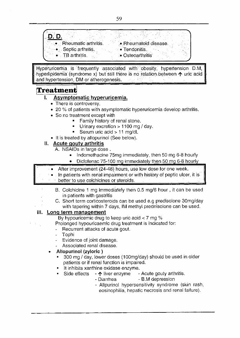
59
0:<0.• Rheumatic arthritis.
Septicarfhriti$.• TB arthritis.
..Hheumatoid disease.• Tendonitis.••Osteoarthritis
~yperyriceITlI~. is frequently associat§d with/obesity, ..hypertension D.M,hyperlipidemia (syndrome x) but still there is no relation between l' uric addand hypertension, DM or atherogenesis.
ITreatmentlI. Asymptomatic hyperuricemia.
• There is controversy.• 20 % of patients with asymptomatic hyperuricemia develop arthritis.• So no treatment except with
• Family history of renal stone.• Urinary excretion> 1100 mg / day.• Serum uric acid> 11 mg/dl.
• It is treated by allopurinol (See below).II. Acute gouty arthritis
A. NSAIDs in large dose.• Indomethacine 75mg immediately, then 50 mg 6-8 hourly• Diclofenac 75-100 mg immediately then 50 mq 6-8 hourly
• After improvement (24-48) hours, use low dose for one week.• In patients with renal impairment orwith history of peptic ulcer, it is
better to use colchicines or steroids.
B. Colchicine 1 mg immediately then 0.5 mg/6 hour, it can be usedin patients with gastritis
C. Short term corticosterolds can be used e.g predisolone 30mg/daywith tapering within 7 days, 1Mmethyl prednisolone can be used.
III. Long term managementBy hypouricemic drug to keep uric acid < 7 mg %
Prolonged hypouricaemic drug treatment is indicated for:Recurrent attacks of acute gout.
- TophiEvidence of joint damage.
- Associated renal disease.• Allopurinol (zyloric )
• 300 mg / day, lower doses (1OOmg/day) should be used in olderpatients or if renal function is impaired.
• It inhibits xanthine oxidase enzyme.• Side effects - l' liver enzyme - Acute gouty arthritis.
- Diarrhea - B.M depression- Allpurinol hypersensitivity syndrome (skin rash,
eosinophilia, hepatic necrosis and renal failure).
60
•
110 .gg e
an be initiated 1-2 weeks after an acute attack and while patientIS on prophylactic cholchicine . .The sharp reduction in tissue uric ac vels that follows initiation
treatment can partially dissolve the urate crystals and triggerte attack, this can be minimized by using a lower starting dose
(100mg/d) or by concurrent use of oral colchicine (O.5mg/1the first few weeks.
IV. Gouty kidney• Plenty fluids.• Alkaline urine by sodium citrate.• Allopurinol.
Diet in Gout (this can reduce serum urate by 15%)• There is no need for severe dietary restrictions but
excessive purine intake and alcohol should be avoided.• Weight reduction, Severe caloric restriction must be
avoided as ~ i lactic acid ~ i uric acid.Excessive meat intake ~ i risk of gout!?, but vegetable protein ~ Jrisk of gout.
PtR!lP;~~SP'~:.~I:~~T,~~,~:~t\:t!,It~(~I:t0N4~o~J,\lciNO$i,~"}
IDefinitioDJIt is a crystal deposition of calcium pyrophosphate dehydrate (CPPD) incartilage (chondrocalcinosis). It is a common age associatedphenomenon (>55) that particularly targets the knee.
!Etiology:jIt is idiopathic, it can occur with certain metabolic diseases e.ghemochromatosis, Wilson's disease, hyperparathyroidism and chronicrenal failure patients under regular hemodialysis.
61
Ic/~1-
There are different clinical patterns• Mono arthritis. } Attacks of• Especially in the knee. pseudogout• 0 = ~ I old age.
2- Pseudo-rheumatoid arthritis with polyarthritis for months.3- Pseudo-osteoarthritis.4- Pseudo-neuropathic with severe destruction (charcot joint like)5- A symptomatic (common).
!Diap.osis\• X ray ~ calcification of articular cartilage.• ESR l' -l' CRP• TLC l'• Examination of joint fluid crystals by polarizing microscopy.
f1'reatmentl• NSAID• Intra articular corticosteroids if indicated.
Pseudo-Pseudo Gout (Hydroxy appatlte.arthropathy),.' ~ >0- Shoulderjoint is the oornrnonst-slte, but it may
occur at knee, hip or elbow.• Treatment with NSAID or intraarticular corticosteroids.
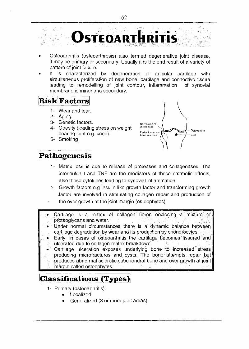
62
.05T.EO·I\R1"1t RiTis• Osteoarthritis (osteoarthrosis) also termed degenerative joint disease,
it may be primary or secondary. Usually it is the end result of a variety ofpattern of joint failure.
• It is characterized by degeneration of articular cartilage withsimultaneous proliferation of new bone, cartilage and connective tissueleading to remodelling of joint contour, inflammation of synovialmembrane is minor and secondary.
Illis)(Factorsl1- Wear and tear.2- Aging.3- Genetic factors.4- Obesity (loading stress on weight
bearing joint e.g. knee).5- Smoking
Narrowing ofjoint space ~
Periarticularbone sclerosis
- Osteophyte
Cyst
IPathogenesisj1- Matrix loss is due to release of proteases and collagenases. The
interleukin I and TNF are the mediators of these catabolic effects,also these cytokines leading to synovial inflammation.
2- Growth factors e.g insulin like growth factor and transforming growth.factor are involved in stimulating collagen repair and production ofthe over growth at the joint margin (osteophytes).
• Cartilage is a matrix of collagen fibr~senclo§ing amixfure.>pfproteoqlycans and.water:
• Under normal circumstances there is a> dynanlIpeBlan.ce be!wElEl.Pcartilage deqradation.by wear and its prod4ctionby.cbondrocytes.
• Early, in cases of osteoarthritis the cartilagebecprnesfissuredtandulcerated due to collagen matrix breakdown.
• Cartilage ulceration exposes underlying b()netoi9creased· str~ssproducing rnicrotractures and cysts. The bone~ttempts repairJ:)4tproduces abnormalsclerotic .subchondral bone and over growth atjoihtrnarqin called osteophytes.
\CIlJ,ssifications (Types )\1- Primary (osteoarthritis):
• Localized.• Generalized (3 or more joint areas)
63
~~,~t1l?~~6'~17o,~rthrili$iti~~<~ff~Rt~P@~1,..~ip.§aQQ aritlsi.edistaland, roximalinter· halan eal 'oints:
2- Secondary osteoarthritis:-• Mechanical e.g. occupational, hypermobility, cruciate tears.• Metabolic: Haemochromatosis, Wilson's disease.• Inflammatory: Rheumatoid arthritis, Gout, sero-ve arthropathy.• Developmental: Epiphyseal dysplasia.• Miscellaneous: Haemophilia, neuropathies.
Ie / ..., (patient over age 45 and often over age of 60)
Osteoarthritis affects many joints with insidious onset. Hip and kneeosteoarthritis is the major cause of disability.
1- Pain (progressive i.e. months to years) which worsens with activity andis relived by rest.
2- Morning stiffness (brief < 15 minutes).3- Gelling phenomenon refers to the sense of renewed stiffness in
diseased joint after prolonged inactivity «1 minute)
Is_tfusl1- Tenderness with limitation of range of movement (capsular
thickening, blocking by osteophyte).2- Crepitus on movement due to rough articular surfaces.3-· Joint enlargement due to soft tissue swellin.9 or osteophytes.4- Deformity e.g - (varus) ? medial angulation .
.; (valgus) ? lateral angulation5·:" Joint effusion, wasting of muscles.6- Osteoarthritis of the hands leading to:
• Heberden's nodes: Bony swelling of distal inter-phalyngeal joint.• Bouchard's nodes: Bony swelling of proximal inter-phalyngeal joint.
~n1r~!;tikJltiQnsl• Blood picture ESR, and eRP are normal.• Rheumatoid factor and ANA are nagative• X ray - Joint space narrowing (loss of cartilage).
- Marginal osteophytes.- Subchondral sclerosis.
• MRI Showing early cartilage changes• Arthroscopy can reveal surface erosion and fissuring of the
cartilage.
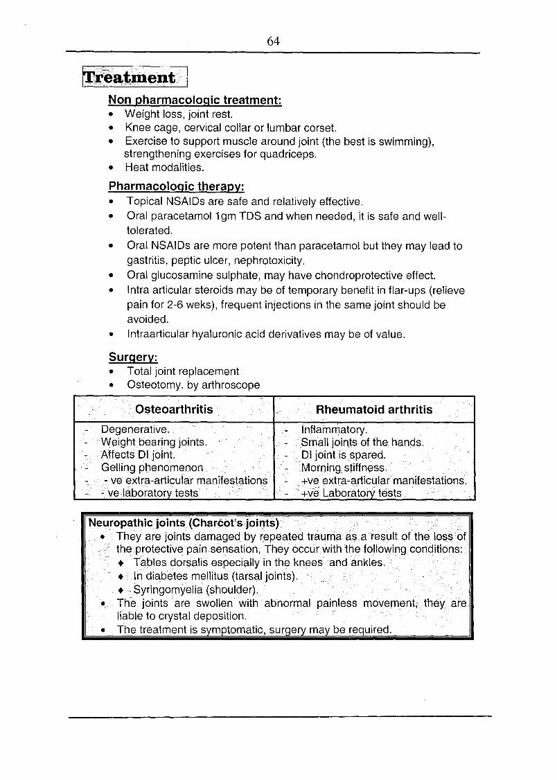
64
OO~,~~llteg.t<1Non pharmacologic treatment:• Weight loss, joint rest.• Knee cage, cervical collar or lumbar corset.• Exercise to support muscle around joint (the best is swimming),
strengthening exercises for quadriceps.• Heat modalities.
Pharmacologic therapy:• Topical NSAIDs are safe and relatively effective.• Oral paracetamol 1gm TDS and when needed, it is safe and well-
tolerated.• Oral NSAIDs are more potent than paracetamol but they may lead to
gastritis, peptic ulcer, nephrotoxicity.• Oral glucosamine sulphate, may have chondroprotective effect.• Intra articular steroids may be of temporary benefit in flar-ups (relieve
pain for 2-6 weks), frequent injections in the same joint should beavoided.
• Intraarticular hyaluronic acid derivatives may be of value.
Surgery:• Total joint replacement• Osteotomy. by arthroscope
Osteoarthrttls
Degenerative.V\((3ightbearing joints.Affects DI joint.Gelling phenomenon'"eveextra-articular manifestations
;,;ve·Iaborator .tests
Rheumatoid arthritisInf-Iamnjatory..Small [oints of the hands.01joint is spared.Morning, stiffness.+ve extra-articular manifestations.
'+ve Laborato tests
N~uroPt:lthicjo ints (Charoot' sjoi nts)• They are joints damaged by r~peated trauma as al'e$oltofthe loss.ot
the. protective pain sensation, They occurwiththe f()lIowing conditions:• Ta.b1esdorsalis especially in the knees and ankles .• <1n diabetes mellitus (tarsal joints).• Syringomyelia (shoulder).
• The joints are swollen with abnormal painless movement; they areliable to crystal deposition.
• The treatment is s m tomatic, sur e rna be re ulred.
65
This can accompany bacteraemia or septicemia, the most common organismis staphylococcus aureus. It is a medical emergency as it leads to severe jointdestruction within short time.
ICaus~sl• Staph aureus.• Streptococci, gonococci.• Haemophilus influenza and other Gram negative bacilli.
IRi~k.factorsl• Increasing age.• Pre-existing joint disease e.g. rheumatoid disease.• DM and immunosuppression.• Arificial joints (prostheses).
rn1- Fever, malaise. 2- Painful swelling usually in one large joint.3- Signs of inflammation (redness, hotness, tenderness ± effusion).
IInvestigation~• i TLC i -i ESR-blood culture.• X ray showing soft tissue swelling then articular erosions.• Synovial fluid examination showing T PNL, positive culture.
ITreatmentl• Rest of joint, analgesics.• Drainage of the joint and arthroscopic joint washouts are helpful in
relievil!g pain, initially the joint should be aspirated daily to keep theeffusion at a minimum.
• Intravenous antibiotic for 3 week e.g I.V f1ucloxacillin 2 gm/6hrsfollowed by oral treatment for 6 weeks in total.
• The joint should be immobilized initially and then physiotherapyshould be started to prevent stiffness and muscle wasting.
Viral arthritis.Viral. arthritis are usually self limiting.The usual presentation is with acute polyarthritis, fever or viralprodrome and rash.Parvovirus is the most common.Polyarthritis may also rarely occur with hepatitis B, C and HIV.Diagnosis is confirmed by viral serology.
66
• Osteomalacia is characterized by defective bone mineralization, bonepain, muscle weakness and pathological fractures.
• There is failure to replace the turn over of Ca and P in bone matrix?bone become demineralised and the bony substance becomes replacedby soft osteoid tissue so it is mainly a qualitative bone defect.
iil~j}Qg~.Ii~~islThe most common cause is vitamin D deficiency, the low levels of vitamin Dcauses a reduction of calcium absorption from the intestine. The low calciumabsorption stimulates parathyroid hormone secretion which restores serumcalcium levels towards normal by increasing bone resorption and renaltubular calcium reabsorption. The level of parathyroid hormone also promotesphosphaturia and causes phosphate depletion. It is the combination ofcalcium loss from bone and phosphate depletion that leads to impaired bonemineralization.
Causes of osteomalacia• Vito D deficiency
- Dietary ~ - Lack of synthesis in skin. - ~ absorption.-Defective metabolism - Anticonvulsants - CRF
• Low P with normal vitoD• Familial hypophosphatemic rickets.• Renal tubular disease.
• ·Oste~malacia with normal Ca, P and vitoD• Hypophosphatasia. • Fibrogenesis imperfecta.
.• Aluminium bone disease.C I P (Osteomalacia is the adult counterpart of rickets).
• Skel.etal discomfort (from bone and muscle pain).• Bone tenderness.Tetany may be manifested.• Muscular weakness with marked proximal myopathy with waddling gait.
Investigations• S. Ca~, S. P ~ but l' in CRF• Alkaline phosphatase 1',parathyroid hormone l' , ~ vitamin D level.• X ray ? bone rarefaction with translucent band (pseudofraction or
looser's zones) i.e linear areas of low density surrounded by scleroticborders.
Treatment• Treatment of the cause.• Vit. D, Ca supplements.• Diet e.g milk, cheese or yoghurt• Alfacalcidol especially in cases of renal failure.
67
• It is defined as a decrease in the absolute amount of bone mass leadingto enhanced bone fragility with increased risk of pathological fractures.
• Unlike osteomalacia, the defect in osteoporosis is that the bone that ispresent is normally mineralized but is deficient in guantity, quality andstructural integrity.
lfathogenesisl• Bone mass increases rapidly up to the age of puberty and rises slightly
more in the twenties and thirties and then begins to decline around ageof 40 years.
• In males there is a gradual decline but females show an accelerated lossin the 10 years following the menopause.
• Osteoporosis occurs as the end result of many years mismatch betweenthe rate of bone resorption and bone formation
~iSk factorsl• ~,Family history, early menopause.• J- Ca intake, reduced activity, smoking, alcohol, AI antacid, excessive
caffeine, corticosteroids therapy and genetic factors.
[Causes and·t~• Type l.-Post menopausal osteoporosis.• Type ll.-Senile osteoporosis.• Secondery osteoporosls.-
- Cushing's syndrome. . - Hyperthyroidism.- Acromegally, D.M. - Rheumotoid disease.- Chronic renal failure - Chronic liver disease.- Rheumatoid arthritis. - Immobilization.- Drugs e.g corticosteroids.
• Boney aches, back pain, Pathological fractures• Loss of height due to thoracic kyphosis and collapsed vertebrae.
The most common sites of pathological fractures are the forearm (collesfracture), spine (vertebral fracture) and femur (hip fracture).
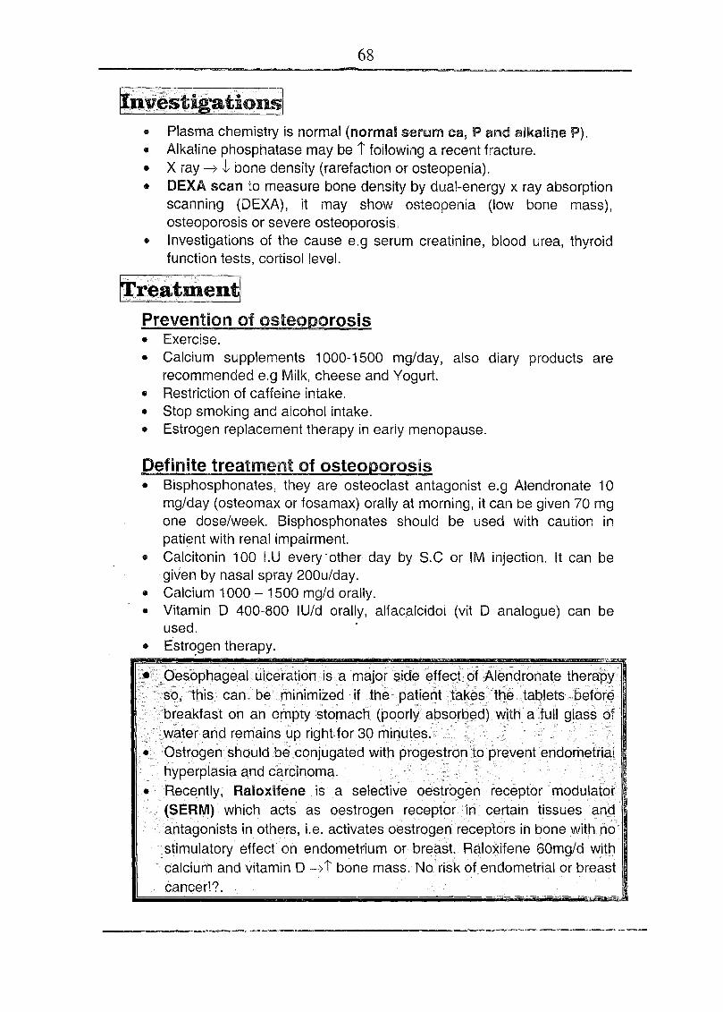
68
~nvestigationsl• Plasma chemistry is normal (normal serum ca, P and alkaline P).• Alkaline phosphatase may be t following a recent fracture.• X ray ~ t bone density (rarefaction or osteopenia).• DEXA scan to measure bone density by dual-energy x ray absorption
scanning (DEXA), it may show osteopenia (low bone mass),osteoporosis or severe osteoporosis.
• Investigations of the cause e.g serum creatinine, blood urea, thyroidfunction tests, cortisol level.
fir~a.tmentlPrevention of osteoporosis• Exercise.• Calcium supplements 1000-1500 mg/day, also diary products are
recommended e.g Milk, cheese and Yogurt.• Restriction of caffeine intake.• Stop smoking and alcohol intake.• Estrogen replacement therapy in early menopause.
Definite treatment of osteoporosis• Bisphosphonates, they are osteoclast antagonist e.g Alendronate 10
mg/day (osteomax or fosamax) orally at morning, it can be given 70 mgone dose/week. Bisphosphonates should be used with caution inpatient with renal impairment.
• Calcitonin 100 I.U every' other day by S.C or IM injection. It can begiven by nasal spray 200u/day.
• Calcium 1000- 1500 mg/d orally.• Vitamin 0 400-800 IU/d orally, altacalcidol (vit 0 analogue) can be
used.• Estr~gen therapy.
• ..8es?phagealulceration •.isa majof9id¥etfe~t+.8!.~le.~dronate t~era.p~s8,lhis can .:berninimized ifth~+ patieQtta~.¥sii tqeta~l.ets ..before
i(t~I:1~~:~;n:~~~i;~i~~~~~~~~::sJ~S;[;~J~!,har!!::;:ss:• Ostrogenshould be conjugated with progestrQnt() prevent endometria,l
hyperplasia and carcinoma.Recently, Raloxltene is a selective oe$trogen reeeptorrnodulatct(SERM) which acts as oestrogen receptor in certain tissues angarttaqonlsts in others, i.e. activates oestrogen receptors in bone with nostimulatory effect on endometrium or breast.. Raloxifene 60mg/dWithcalcium and vitamin 0 ~t bone mass. No risk of endometrial or breastcancer!?
69
INkERiYEd DisORdERS of COLLAGENOsteogenesis imperfectaIt is a rare inherited disorder of collagen (autosomal dominant),characterized by brittle bone and abnormalities of the skin, tendon,teeth and sclera.
II Marfan syndromeIt is an inherited condition (autosomal dominant) charasterized by:-
- Span> height - Lens dislocation - High arched palate.- Arachnodactly - Sternal depression- CVS
- Mitral valve prolapse - Aortic incompetence.- Aortic dissecting aneurysm.
III Ehlers - Danlos syndromeTen different types have been recognizedCharacteristic features:
- Skin laxity.- Short stature.
- Hypermobility of joints.- Skin bruising.
FibROMYAlGiA SYN~ROM .:E• Fibrornyalqia (fibrositis) is a controversial diagrrosis that is not universally
accepted. It is a useful diagnosis of exclusion.
• It is characterized by chronic diffuse pain with characteristic tenderpoints. It is considered as a n.onarticu.lar rheumatism.
• It affe~ts about 2 % of all patients seen in general practice and 20 % ofpatients referred to rheumatologists!?
• All investigations are normal and the value is for exclusion.Aetiology: The condition is poorly understood, two abnormalities have beenreported: sleep abnormality and reduced threshold to pain at certain sites.
C/P
• Diffuse muscle pain, stiffness and fatigue.
• Focal tender points:• Occiput ~ bilateral at suboccipital muscles insertions.• Low cervical ~ bilateral at C 5 - 7 (interspinous ligaments).• Trapezius ~ bilateral at mid point of the upper border.• Supraspinatus ~ bilateral above the medial border of the
scapular spine.• Second rib ~ bilateral at 2nd costochondral junctions.• Lateral epicondyle ~ bilateral 2 cm distal to the epicondyles.
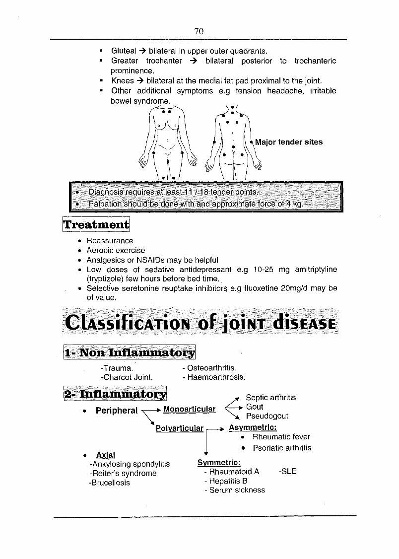
70• Gluteal -7 bilateral in upper outer quadrants.• Greater trochanter -7 bilateral posterior to trochanteric
prominence.• Knees -7 bilateral at the medial fat pad proximal to the joint.• Other additional symptoms e.g tension headache, irritable
bowel syndrome.
• Reassurance• Aerobic exercise• Analgesics or NSAIDs may be helpful• Low doses of sedative antidepressant e.g 10-25 mg amitriptyline
(tryptizole) few hours before bed time.• Selective seretonine reuptake inhibitora e.g fluoxetine 20mg/d may be
of value.
-Trauma.-Charcot Joint.
- Osteoarthritis.- Haemoarthrosis.
• Axial-Ankylosing spondylitis-Reiter's syndrome-Brucellosis
~
Septic arthritisPeripheral ~ Monoarticular Gout
\t _ PseudogoutPOlyarticularr Asymmetric:
• Rheumatic fever• Psoriatic arthritis
Symmetric:- Rheumatoid A- Hepatitis B- Serum sickness
•
-SLE

71
11- Inflammatory- Sero -ve arthropathies- Rheumatoid disease
'ff~reJlYi~Iqi~~.l1osi~()fbacKache
1- Mechanical- Disc prolapse.- Lumber spondylosis.- Muscle spasm due to fracture
2- Inflammatory- Sero -ve arthropathy- Brucellosis
1- Mechanical- Cervical spondylosis.- Muscle spasm.
111- Metabolic- Osteomalacia- Osteoporosis
3- Metabolic-Osteoporosis-Osteomalacia
IV- Neoplam-Metastases- M.myeloma
4- Neoplasm-Metastasis-M.myeloma-Lymphoma
V- Referred pain- Angina pectoris- Pancost tumor- Aortic aneurysm
5- Referred pain-Cancer pancreas-Renal colic-Retroperitonial haematoma-Diseases of cervix or prostate
[Q.D.of arthritis in children'll1- Rheumatic arthritis.4- Viral arthritis.7- Still's disease.
2- Septic arthritis.5- Henoch-schonlein purpura
3- FMF6- Haemophilia.
D.D. of recurrent arthritis or transientarthritis?1- Rheumatic arthritis 2- Gout or pseudogout.4- Sero -ve arthropathies. 5- Rheumatoid disease.
~.J) .of mon.oarthritis?11- Rheumatic arthritis 2- T.B.4- Haemophilia. 5- Gout.7- Traumatic.8- Monoarticular presentation of oligo or polyarthritis. e.g. rheumatoid disease
or seronegative arthropathies
ID.D. of oligoarthropathy?\1- Seronegative arthropathy. 2- Juvenile idiopathic arthritis.3- Infective endocarditis.
3- FMF
3- Septic arthritis.6- Pseudogout
- Harrison text book (Principles of Internall1edicine).- (ecil Textbook (TeXfbookoll1edicine).- Kumar «(linicall1edicine).- Davidson's (Principles and Practice 01 Medicine).- Henry/fhompson «(Unical Surgery).- Robbins (pathologic basis 01 disease).- (ecil Essentials oll1edicinc.- Ihe Nalionall1edical Series for Independent Study (l1edicine).- OXfordHandbook of Endocrinology and Diabetes.- Basic and (linical Endocrinology.
1· Hepatology.2· Gastroenterology.3· Endocrinology.4· Rheumatology.5· (ardiology.6· Nephrology.1· Hematology.8· Neurologyand psychiatry.g. Infectious diseases, tropical diseases, immunology, nutrition, lIenefics,
geriatric, toxicology and therapeutics.to· Respiratory diseases.11· (Unical medicine (symptoms and examination).
II (ardiology.II! {hest.!I l\.bdomen.ill Neurology.l1li General.
~jJJ JJ:,# ~\ J~
fl~\ J\ t:--J\ ty.l