Embed Size (px)
Citation preview
Fakultät für Medizin
I. Medizinische Klinik des Klinikums rechts der Isar
Rheumatoid arthritis: the influence of disease activity on clinical and serological parameters
Christoph Böhler
Vollständiger Abdruck der von der Fakultät für Medizin der Technischen Universität München zur Erlangung des akademischen Grades eines Doktors der Medizin genehmigten Dissertation.
Vorsitzender: Univ.-Prof. Dr. E. J. Rummeny Prüfer der Dissertation: 1. Univ.-Prof. Dr. K.-L. Laugwitz 2. Univ.-Prof. Dr. U. Heemann
Die Dissertation wurde am 26.01.2015 bei der Fakultät für Medizin der Technischen
Universität München eingereicht und durch die Fakultät für Medizin am 18.11.2015
angenommen.
2
Contents
Abbreviation…………………………………………………………………………………4
Abstract ……………………………………………………………………………………...6
1. Introduction……………………………………………………………………………....7
2. Rheumatoid arthritis…………………………………………………………………….9
2.1. Aetiology………………………………………………………………………..9
2.2. Pathogenesis…………………………………………………………………..9
2.3. Clinical manifestations……………………………………………………...10
2.4. Diagnosis……………………………………………………………………...13
2.5. Therapy………………………………………………………………………...16
3. Disease activity in rheumatoid arthritis……………………………………………18
3.1. Core sets of disease activity variables…………………………………..18
3.2. Composite indices…………………………………………………………...19
3.3. Assessments for physical functioning…………………………………..21
3.4. Response criteria…………………………………………………………….22
4. Study variables…………………………………………………………………………23
4.1. Rheumatoid factor…………………………………………………………...23
4.2. Antibodies against citrullinated peptides……………………………….24
4.3. Clinical parameters: Tests of the fall assessment……………………..25
5. Aims of the study………………………………………………………………………27
6. Discussion………………………………………………………………………………28
7. References………………………………………………………………………………33
8. Acknowledgements……………………………………………………………………43
3
9. Article summaries……………………………………………………………………...44
9.1. Rheumatoid arthritis and falls: the influence of disease activity.......44
Christoph Böhler, Helga Radner, Michaela Ernst, Alexa Binder, Tanja Stamm, Daniel Aletaha,
Josef S Smolen and Marcus Köller;
Rheumatology 2012;51:2051-2057
9.2. Serological changes in the course of traditional and biological disease modifying therapy of rheumatoid arthritis....................................45
Christoph Böhler, Helga Radner, Josef S Smolen and Daniel Aletaha;
Ann Rheum Dis 2013 72: 241-244
10. Articles ………………………………………………………………………………...46
4
Abbreviation AAB autoantibody
ACPA antibodies against cyclic citrullinated peptides
ACR American College of Rheumatology
CCP cyclic citrullinated peptides
CDAI clinical disease activity index
CRP c-reactive protein
CRT chair-rising test
DAS-28 disease activity score 28
DIP distal interphalangeal
DMARDS disease modifying anti-rheumatic drugs
EGA evaluator global assessment of disease activity
ELISA enzyme linked immunosorbent assay
ESR erythrocyte sedimentation rate
EULAR European League Against Rheumatism
HAQ health assessment questionnaire
HAQ DI health assessment questionnaire disability index
IG immunoglobulin
IL interleukin
MCP metacarpophalangeal
MRI magnetic resonance imaging
MTP metatarsophalangeal
MTX methotrexate
NSAIDs non-steroidal anti-inflammatory drugs
PGA patient global assessment of disease activity
PIP proximal interphalangeal
RA rheumatoid arthritis
RF rheumatoid factor
SDAI simplified disease activity index
SJC swollen joint count
5
TIT Tinetti test
TJC tender joint count
TNF tumour necrosis factor
TNF-i tumour necrosis factor inhibitor
TS tandem stand test
TUG timed get up and go test
TW tandem walking test
US ultrasound
VAS visual analogue scale
6
Abstract (English)
Control of disease activity is the crucial factor in treatment of rheumatoid arthritis
(RA). Alleviation of acute symptoms as wells as prevention of long-term damages
are highly dependent on suppression of inflammatory activity. The two studies of this
cumulative thesis investigated relations between disease activity and certain clinical
(fall-assessment) and serological (autoantibodies) parameters. The first work could
show that RA patients with an increased inflammatory activity have a higher risk to
fall. The second study could demonstrate that anti-rheumatic therapy and the
consecutive reduction of disease activity is linked with titre changes of the
autoantibodies rheumatoid factors and antibodies against cyclic citrullinated peptides.
Good treatment response leads to a significant decrease of both antibodies, which
have a high diagnostic and also prognostic value in RA. Both studies should
contribute current aims of identifying individual risk factors in RA and therefore
treating patients to a treatment target.
Abstrakt (German)
Die Kontrolle der Krankheitsaktivität spielt bei der Behandlung der Rheumatoiden
Arthritis (RA) die entscheidende Rolle. Sowohl die Milderung akuter Beschwerden,
als auch die Vermeidung von Langzeitschäden, hängen in erster Linie von einer
Unterdrückung der entzündlichen Aktivität ab. In den beiden hier zu einer
kumulativen Dissertation zusammengefassten Arbeiten wurde untersucht, inwieweit
die Krankheitsaktivität sich auf konkrete klinische und serologische Parameter
auswirkt. In der ersten Arbeit konnten wir zeigen, dass bei an RA Erkrankten ein
Zusammenhang zwischen einer erhöhten entzündlichen Aktivität und dem Risiko zu
stürzen, besteht. Die zweite Studie demonstrierte die Auswirkungen einer
antirheumtatischen Therapie und einer damit verbundenen Senkung der
Krankheitsaktivität auf die Autoantikörper Rheumafaktor und Antikörper gegen
zyklisch citrullinierte Peptide. Ein gutes Ansprechen auf eine medikamentöse
Therapie war mit einer signifikanten Titerreduktion dieser beiden Antikörper, welche
sowohl eine diagnostische, als auch eine prognostische Bedeutung haben,
verbunden. Beide Studien sind im Kontext aktueller Bestrebungen nach einer
personalisierten Medizin mit Identifikation individueller Risikofaktoren und der
Behandlung dieser, zu klar definierten Zielen, (“treat to target”) zu sehen.
7
1. Introduction
Rheumatoid arthritis (RA) is the most common inflammatory joint disorder in adults
and is characterized by chronic synovitis, systemic inflammation and autoantibody
production 1. Functional disability, cartilage and bone destruction are the major
negative outcomes in RA and are related to loss of life quality and to premature
death 2-4.
Within the last years the management of RA improved tremendously: Insights into
the pathogenesis made the development of a number of new, highly effective drugs
possible. These biological agents are targeted against single components of the
immune system, which are involved in the inflammatory process of RA. Also
traditional disease modifying anti-rheumatic drugs (DMARDs) like methotrexate,
leflunomide and sulfasalazine, which were established in the treatment of RA for
decades, have been re-examined to improve efficacy. Moreover therapeutic
approaches have changed with gain of knowledge: current treatment strategies
require an early start of DMARD therapy, a tight control of disease activity to survey
treatment response and, if necessary, a rapid switching of therapeutic regimes 5.
Suppressing disease activity is the major goal in RA therapy as uncontrolled activity
causes acute symptoms, but also seems to be the main reason for the adverse long-
term effects 6.
The aim of the presented papers was to examine the impact of disease activity on
clinical and serological parameters. In the first study we investigated how states of
disease activity are related to the risk of falling in RA patients. Former works have
shown that patients who suffer from RA have an increased risk of falling 7. In normal
populations age and related comorbidities are considered to be major risk factor for
falls 8. Interestingly, in RA the fall incidence seems to be independent from age as
well as from disease duration 7, 9. These findings indicate that different risk factors for
falls are relevant in RA, which we tried to identify.
The second study looked at associations between changes of disease activity and
changes of rheumatoid factor (RF) and antibodies against cyclic citrullinated peptides
(ACPA) levels during a treatment course. RF and ACPA are commonly found in the
serum of RA patients and are therefore established diagnostic markers 10.
Furthermore the presence of these auto-antibodies is related to a more aggressive
and destructive disease 11. For this reason changes of RF and ACPA levels can be

8
highly relevant for the long-term outcome of RA. Both studies can be seen in the
context of current efforts of treating RA patients to a treatment target 3 and the
intentions of personalized medicine considering individual risk factors 12.
In the following section a brief overlook of the etiopathology, clinical manifestations,
diagnosis and treatment options of RA shall be given. Moreover important methods
of evaluating disease activity in RA and clinical and serological parameters that have
been used in the presented works will be introduced.
9
2. Rheumatoid Arthritis
Rheumatoid arthritis (RA) is the most frequent inflammatory arthritis and has a
chronic, disabling and aggressive nature. It is related with a twice as high mortality
rate 13. In the industrialized world the prevalence lies between 0.5 – 1% with 5 to 50
per 100.000 new cases each year 1. Two thirds of the affected patients are women.
The disease can appear at any age, with an increase of incidence in elderly people,
so that the prevalence in over 65 year olds is between 2-3% 14.
2.1. Aetiology Aetiology of RA is still unknown. It is suspected that the disease results from a
complex interaction between genes and exogenous influences like infections,
environmental and hormonal factors 15. Several genes have been identified to be risk
factors for the development of RA and there is a significant overlap with genes
associated with other autoimmune diseases, like systemic lupus erythymatodes,
ankylosing spondylitis, inflammatory bowel disease and multiple scleroisis 16. Up to
now the strongest genetic associations are found with the human leukocyte antigen
major histocompatability genes (HLA). In total it is estimated that genetic factors
contribute 50 to 60 percent to the risk of developing RA 17.
The possible role of a viral infection, like Epstein Barr virus, as a trigger of RA in
patients with a genetic susceptibility has been discussed for years and remains an
active area of investigation 18. From the known environmental stimuli smoking seems
to have the strongest impact on developing RA. Smokers have a 3 times higher risk
than non-smokers 19, heavy smoking (41-50 pack years) increases the risk for
developing RA 13-fold 20.
2.2. Pathogenesis Like the aetiology, the pathogenesis of RA remains unclear. In latest opinion RA is
considered as a clinical syndrome, which is caused by numerous disease subsets.
These subsets lead to different inflammatory cascades, which all end in a common
pathway that entail to synovitis and bone and cartilage damage 1, 21. An unknown
environmental trigger causes an activation of T-lymphocytes in genetically vulnerable
10
individuals. The activated T-lymphocytes interact with B-lymphocytes, which lead to
an autoantibody production. Furthermore interactions between activated T-
lymphocytes and macrophages cause an overproduction and over-expression of
several cytokines including tumour necrosis factor (TNF) alpha, interleukin (IL) 6, IL
1. These cytokines initiate a proliferation of fibroblasts and lead to an invasion of
inflammatory cells into the synovial membrane. Fibroblasts are thought to play an
important role for the joint destruction through the production of metalloproteinases.
Activation of osteoclasts seems to be responsible for the bone erosion 22. 1, 23
2.3. Clinical manifestation of RA RA is characteristically a chronic polyarthritis. At the beginning of the disease the
majority of the patients complain about unspecific symptoms like fatigue, weakness,
nebulous musculoskeletal pain and loss of weight. This prodrome can last for weeks
or even months. Later more specific symptoms like morning stiffness, tenderness
and swelling of joints are predominant 23, 24.
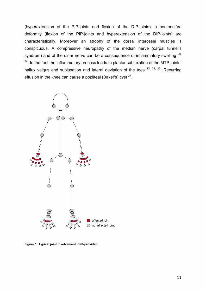
Joint manifestations The clinical manifestation of RA with regard to gravity and pattern of joint involvement
is very variable. In almost every patient the joints of the hand are affected. Typically
alterations occur in the wrists, metacarpophalangeal (MCP) and proximal
interphalangeal (PIP) joints of the fingers and interphalangeal joints of the thumbs
and the metatarsophalangeal (MTP) joints of the toes, whereas the distal
interphalangeal joints (DIP) are usually spared out. Figure 1 shows a typical pattern
of joint involvement. Other joints like elbows, shoulders, ankles, and knees are also
frequently involved. Typically the arthritis is symmetrical 23, 24.
In the axial skeleton, the cervical spine joints (in contrast to the thoracic and the
lumbar vertebrae) are often affected. Initially inflammation may cause stiffness of the
neck and pain. Ongoing disease could lead to destruction and instability in the
segment C1-C2, which could cause a spinal cord compression and paraplegia 25.
Several joint changes can develop with continuing inflammation and are typical for
chronic and established RA. These changes are the result of pathologic processes
like weakening and destruction of tendons, tendon sheaths, ligaments; looseness of
supporting soft tissue structures, loss of cartilage and imbalance of muscles. In the
hand an ulnar deviation of the MCP-joints (“ulnar-drift”), a swan neck deformity
11
(hyperextension of the PIP-joints and flexion of the DIP-joints), a boutonnière
deformity (flexion of the PIP-joints and hyperextension of the DIP-joints) are
characteristically. Moreover an atrophy of the dorsal interossei muscles is
conspicuous. A compressive neuropathy of the median nerve (carpal tunnel’s
syndrom) and of the ulnar nerve can be a consequence of inflammatory swelling 23,
24. In the feet the inflammatory process leads to plantar subluxation of the MTP-joints,
hallux valgus and subluxation and lateral deviation of the toes 23, 24, 26. Recurring
effusion in the knee can cause a popliteal (Baker's) cyst 27.
Figure 1: Typical joint involvement. Self-provided.
12
Extra-artricular manifestations Extra-artricular manifestations are seen in approximately 40 % of RA patients and in
13 % of the cases these manifestations are considered to be severe 28. Studies show
that smoking and high titres of rheumatoid factor (RF) are risk factors for extra-
artricular involvement 29. Furthermore extra-artricular manifestations are usually
associated with a more aggressive disease and premature mortality 30.
Skin involvement: About 20% of RA patients suffer from rheumatoid nodules, which
are mostly located in the subcutaneous fatty tissue on pressure points, but can
develop in all regions of the body (also in inner organs) 23.
The eyes can be affected in the form of keratoconjunctivitis sicca (common),
episcleritis and scleritis. Also uveitis can be seen in RA patients.
Pleuropulmonary manifestations are frequently found in individuals suffering from
RA, but seldom cause clinical symptoms. Involvement of the lung includes pleuritis,
interstitial fibrosis, pleuropulmonary rheumatoid nodules and pneumonitis 23, 24.
Clinical manifest cardial disorders like pericarditis or endo- and myocarditis are
infrequently seen. But several studies have described a significantly increased
incidence of cardiovascular disease in patients with RA and it has been shown that
premature death in RA patients is frequently related to cardiovascular events 31.
Moreover it seems that the elevated risk for cardiovascular disease is independent of
typical risk factors like hypertension, diabetes mellitus, body mass index or
hypercholesterinemia 32 33. It is suggested that there is an association between the
chronic inflammation and atherosclerosis 34, 35.
Generalized osteoporosis is very frequently found in patients suffering from RA. The
reasons are immobility, steady treatment effects with glucocorticoids, in the presence
of a chronic inflammatory process 23.
RA is also associated with a higher incidence of lymphoma 23, 24, 36.
13
2.4. Diagnosis Diagnosis of RA is based on anamnesis, clinical symptoms, laboratory testing and
imaging. Classification criteria Until recently for clinical studies the 1987 American College of Rheumatology (ACR)
classification criteria were used (see Table 1) 37 .
1987 ACR classification criteria for rheumatoid arthritis At least 4/7 criteria must be satisfied. Criteria 1 through 4 must be present ≥ 6 weeks.
1. Morning stiffness lasting for at least 1 hour
2. Arthritis of 3 or more joint areas
3. Arthritis of hand joints (wrist, MCP, or PIP joint)
4. Symmetric arthritis (same joint areas affected on both sides of the body)
5. Presence of rheumatoid nodules
6. Serumpositivity (Rheumatoid factor)
7. Typical radiographic changes (erosions or unequivocal bony decalcification)
Table 1: 1987 ACR classification criteria 37
The 1987 classification criteria included criteria points like rheumatoid nodules and
typical RA radiographic alterations, which are characteristics of established RA. So
the old criteria were not designed for an early diagnosis of RA. But during the last
years knowledge of the importance of an early therapeutic intervention for a better
clinical long-term outcome grew 38, 39. In order to identify and study patients in an
early disease phase new classification criteria were developed. In 2010 the new
ACR/ European League Against Rheumatism (EULAR) classification criteria were
introduced 40 (see Table 2).
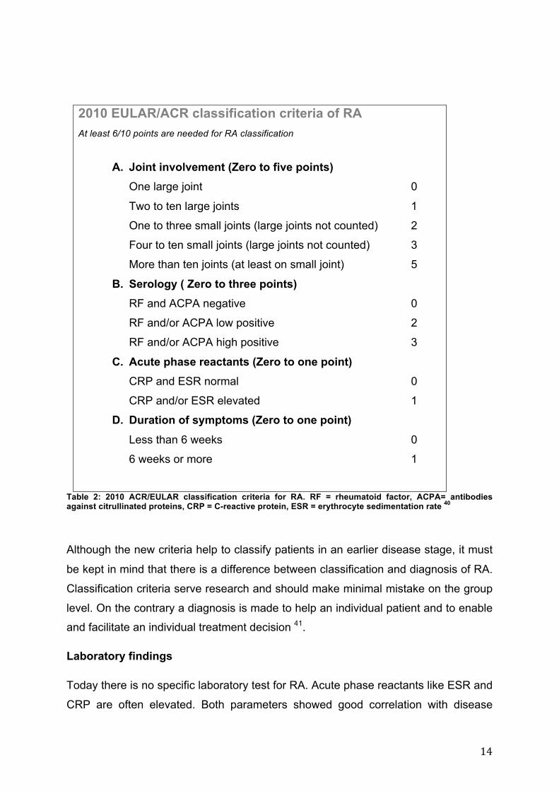
14
2010 EULAR/ACR classification criteria of RA At least 6/10 points are needed for RA classification
A. Joint involvement (Zero to five points) One large joint 0
Two to ten large joints 1
One to three small joints (large joints not counted) 2
Four to ten small joints (large joints not counted) 3
More than ten joints (at least on small joint) 5
B. Serology ( Zero to three points) RF and ACPA negative 0
RF and/or ACPA low positive 2
RF and/or ACPA high positive 3
C. Acute phase reactants (Zero to one point) CRP and ESR normal 0
CRP and/or ESR elevated 1
D. Duration of symptoms (Zero to one point) Less than 6 weeks 0
6 weeks or more 1
Table 2: 2010 ACR/EULAR classification criteria for RA. RF = rheumatoid factor, ACPA= antibodies against citrullinated proteins, CRP = C-reactive protein, ESR = erythrocyte sedimentation rate 40
Although the new criteria help to classify patients in an earlier disease stage, it must
be kept in mind that there is a difference between classification and diagnosis of RA.
Classification criteria serve research and should make minimal mistake on the group
level. On the contrary a diagnosis is made to help an individual patient and to enable
and facilitate an individual treatment decision 41.
Laboratory findings
Today there is no specific laboratory test for RA. Acute phase reactants like ESR and
CRP are often elevated. Both parameters showed good correlation with disease
15
activity. Furthermore increased values of both acute phase reactants are associated
with long-term effects like radiographic erosions 42, 43.
Other typical findings in blood testing are signs of systemic inflammation like a
decreased serum iron, thrombocytosis and normocytic anemia.
Several auto-antibodies can be found in RA patients. Between those RF and ACPA
are the probably most important. RF is detected in 60 – 80% of patients. But
specificity of these antibodies is only modest as they can be also found in other
autoimmune-diseases, like systemic lupus erythematosus and primary Sjögren’s
syndrome. Furthermore increased titres can be found during chronic infections and in
older population 10. ACPA may be more specific (90 to 95%) than RF44, 45 and have a
sensitivity comparable to RF46 although this is still controversial. Both antibodies are
present in the sera of RA up to several years before clinical disease onset 47 48, 49.
Moreover both antibodies also have a prognostic value, since they are associated
with more aggressive and destructive disease 11, 50, 51. RF and ACPA will be
discussed more detailed in section 4.
Anti-RA33 is also an antibody with a potential diagnostic value. It is found in
approximately 30 % of RA patients 52.
Imaging The conventional radiography is the most commonly used imaging type in the
diagnostics and follow up of RA. Structural alterations like bony erosion, cartilage
damage, as well as soft tissue swelling can be detected with conventional
radiography 53. Different scores, like for example the van der Heijde-modified Sharp
score, have been developed to quantify radiographic changes 54.
Magnetic resonance imaging (MRI) is more sensitive in detecting bone erosions than
plain radiography. Moreover with MRI it is possible to assess disease activity by
detecting such as synovitis, tendosynovitis and bone marrow oedema 53.
Ultrasound (US) is able to evaluate soft tissue structures and is useful in detection of
synovitis in inflamed joints, (Baker) cysts and appraisal of tendons 53.
16
2.5. Therapy
The aim of the management of RA is to supress the systemic inflammation and to
prevent bad long-term outcome, which leads to impairment of quality of life and
invalidity. An early, effective and consequent therapeutic approach is necessary.
The treatment of RA is based upon several pillars: On the one hand upon drug
treatment, which includes 1. analgesics and non-steroidal anti-inflammatory drugs
(NSAIDs), 2. traditional disease modifying anti-rheumatic drugs (DMARDs), 3.
biologicals and 4. glucocorticoids and on the other hand upon non-medicamentous
therapy like physiotherapy, occupational therapy and psychological support and
surgical procedures 5.
NSAIDs and analgesics These medications are the basis for treating symptoms like pain of RA, and are
widely used. NSAIDs additionally have a positive effect on inflammation. It must be
pointed out that both medications have no positive influence on the long-term
outcome of the disease 23. Moreover gastrointestinal and cardiac adverse effects
must be kept in mind 55, 56.
DMARDs DMARDs are a heterogeneous group of medicaments, which are able to reduce
disease activity and have a positive influence on the disease course in regard to
preserving joint function and preventing joint destruction. Although the mechanisms
of the immune modulation are still not fully understood, they are the mainstay of the
treatment of RA. Methotrexate (MTX) is the first line DMARD in the absence of
contraindications. If there are contraindications against MTX, leflunomide or
sulfasalazin can be prescribed 5. Gold, azathioprin and ciclosporin are nowadays
only rarely used because of their toxic side effects 23.
Biologicals Research on inflammatory and pathological processes in RA during the last couple of
years allowed the identification and blockade of pro-inflammatory cytokines and so
enabled new therapeutic approaches. Up to four different highly effective classes of
biological agents are used in the treatment of RA. Inhibition of the tumour necrosis
factor (TNF α) was the first licensed biological mode of action. Currently five TNF-
inhibitors (TNF-i) are approved: infliximab, adalimumab, etanercept, certolizumab
and golimumab. Other modes of action are co-stimulation of T-cells inhibition
(abatacept), blockade of interleukin- (IL-) 6 (tocilizumab) and B-cell depletion
17
(rituximab) 1, 57. IL-1 inhibition (anakinra), another treatment strategy, could not match
the efficiency with the previously mentioned biologicals 58. Usually biologicals are
combined with traditional DMARDs, typically MTX 5.
Glucocorticoids
Glucocorticoids have been used in the therapy of RA for decades. They are able to
rapidly reduce disease activity and have the potential to improve the long-term
outcome of RA, but also cause adverse effects like infections and osteoporosis. In
current treatment strategies they are used for bridging the gap between the start of a
new DMARD course and the time point of onset of its clinical effectiveness, and
during flare-ups of disease activity 59. Furthermore intra-articular injections of
corticosteroids in active joints showed high efficacy 60.
18
3. Disease activity in RA
The standardised evaluation of disease activity is of central importance for the
management of RA. As heightened disease activity over prolonged time leads to joint
destruction and functional impairment, the surveillance of activity is an important goal
in the therapy of RA 61, 62. Several instruments and clinical indicators for disease
activity and clinical response criteria have been developed for the assessment of RA.
They are used in the context of clinical studies, but more and more also in clinical
practice 63. A number of important individual variables as well as composite indices
will be described in the following section.
3.1. Core sets of disease activity variables
In the early 1990s different groups of researchers published core sets of disease
activity variables, which should be used in the assessment of RA in clinical trials 64-66.
These variables included swollen and tender joint count (SJC, TJC), pain evaluation
through the patient, patient global assessment of disease activity (PGA), evaluator
global assessment of disease activity (EGA) and evaluation of acute phase reaction
and function. The selection of these variables was based on clinical data and rested
upon various aspects of validity 62.
Pain Pain is the leading symptom in RA. Usually it is measured on a 100 mm visual
analogue scale (VAS) 67, 68. On this scale patients mark their extent of pain during the
last week between 0 mm (no pain) and 100 mm (worst imaginable pain). Other
instruments to measure pain are also available and reliable 69. 70
Tender Joint Count (TJC) and Swollen Joint Count (SJC)
In RA patients, joints are usually evaluated for tenderness on pressure (TJC) and
swelling and effusion (SJC). The first joint count was introduced in 1950s and
included 86 joints 71. Until the late 1980s the number of evaluated joints was
gradually reduced and actually the assessment of 28 joints is mainly used in clinical
studies as well as in daily practice 72, 73 74. The 28 joint count includes the following
joints: 10 PIP joints of the fingers, 10 MCP joints, the wrists, elbows, shoulders, and
knees. Although the measurement with the reduced number of joints does not
include the assessment of feet and ankles, it showed a good validity and reliability in
19
clinical studies 75, 76 70. Moreover the 28 joint count is part of the composite indices
disease activity score 28 (DAS-28) 77, simplified disease activity index (SDAI) 78 and
clinical disease activity index (CDAI)79.
Patient global assessment (PGA) and evaluator global assessment (EGA) For the rating of global disease activity through the patient (PGA) and the physician
(EGA) are also mainly 100 mm visual analogue scales used. The PGA is regarded as
a subjective parameter, while the EGA is considered as a combination of subjective
and objective. Usually the PGA is rated higher than the EGA 62. A recent study
showed that rating of the PGA is mainly pain driven, while the EGA is closely linked
to SJC 80.
Acute phase reactants The most commonly used acute phase reactants are CRP and ESR. They are
employed in clinical practice as well as in clinical trials. Both parameters showed
good correlations with disease activity and also with radiologic progression 42, 81. CRP
may be superior to ESR in regard of measuring disease activity 82.
3.2. Composite indices
RA is a very heterogeneous disease, which makes assessment of disease activity
more complex than in other diseases. A single parameter can hardly reflect the
different aspects and characteristics that can indicate disease activity. In order to
homogenize the evaluation of disease activity several composite indices have been
developed 83. Primarily they were meant for report of disease activity and to measure
therapy response in clinical studies, but nowadays they are also recommended for
daily clinical practice 62. In the following section three commonly used composite
indices, which were also used in the here presented papers, are going to be
presented: disease activity score 28 (DAS-28), the simplified disease activity index
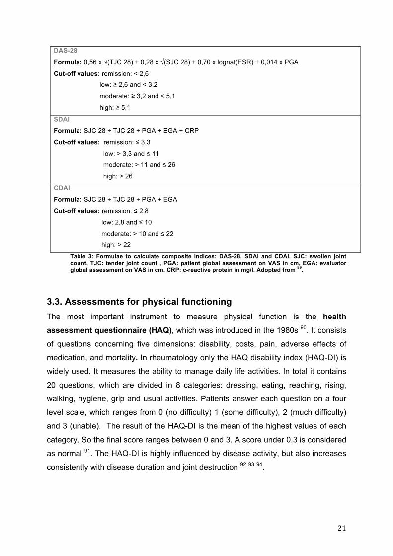
(SDAI) and the clinical disease activity index (CDAI). The formulas to calculate the
three indices are given in table 3.
Disease activity score 28 (DAS-28) The original DAS was introduced in the early 1990s by van der Heijde et al. and
employed the Ritchie articular index and a 44 joint count 84. A few years later the
DAS was successfully modified and the DAS-28 was introduced, which is based on a
more practicable 28 joint count (TJC and SJC). Other included variables are PGA
20
and ESR 77. There also exists a variant using CRP instead of ESR (DAS-28 CRP) 85.
The formula to calculate the DAS-28 remains very complex and requires a calculator.
Four cut-points have been established which divide disease activity into remission (<
2,6), low disease activity ( ≥ 2,6 and < 3,2), moderate disease activity (≥ 3,2 and <
5,1) and high disease activity (≥ 5,1) (See also table 3). Given the complexity of
DAS-28 calculation, the idea to generate a more simple disease activity score came
up and in 2003 the SDAI was introduced 78.
SDAI and CDAI The SDAI is calculated by adding the values of the core set variables SJC, TJC,
PGA, EGA and CRP (table 3). So the used variables were neither weighted nor
transformed. Despite the simplicity the SDAI showed very good correlations with the
DAS-28 and with measures of functional impairment (health assessment
questionnaire). Furthermore it was well associated with radiographic progression. Cut
off levels for remission and other states of disease activity have been postulated and
the SDAI was validated in several trials 86-88.
In 2005 Aletaha et al. could show that acute phase reactants (CRP) in composite
indices only provide little additional information to the clinical core set parameters.
The CDAI was developed, which is only based on SJC, TJC, PGA and EGA and
calculated by adding these parameters. It was the first composite index requiring no
blood testing, thus allowing immediate evaluation, making it very practicable for
clinical use. The CDAI has also been validated and showed good correlations with
other disease activity indices (DAS-28 and SDAI), as well as with function and
radiographic progression over time 79.
21
DAS-28
Formula: 0,56 x √(TJC 28) + 0,28 x √(SJC 28) + 0,70 x lognat(ESR) + 0,014 x PGA
Cut-off values: remission: < 2,6
low: ≥ 2,6 and < 3,2
moderate: ≥ 3,2 and < 5,1
high: ≥ 5,1
SDAI Formula: SJC 28 + TJC 28 + PGA + EGA + CRP
Cut-off values: remission: ≤ 3,3
low: > 3,3 and ≤ 11
moderate: > 11 and ≤ 26
high: > 26
CDAI
Formula: SJC 28 + TJC 28 + PGA + EGA
Cut-off values: remission: ≤ 2,8
low: 2,8 and ≤ 10
moderate: > 10 and ≤ 22
high: > 22
Table 3: Formulae to calculate composite indices: DAS-28, SDAI and CDAI. SJC: swollen joint count, TJC: tender joint count , PGA: patient global assessment on VAS in cm, EGA: evaluator global assessment on VAS in cm. CRP: c-reactive protein in mg/l. Adopted from 89.
3.3. Assessments for physical functioning The most important instrument to measure physical function is the health assessment questionnaire (HAQ), which was introduced in the 1980s 90. It consists
of questions concerning five dimensions: disability, costs, pain, adverse effects of
medication, and mortality. In rheumatology only the HAQ disability index (HAQ-DI) is
widely used. It measures the ability to manage daily life activities. In total it contains
20 questions, which are divided in 8 categories: dressing, eating, reaching, rising,
walking, hygiene, grip and usual activities. Patients answer each question on a four
level scale, which ranges from 0 (no difficulty) 1 (some difficulty), 2 (much difficulty)
and 3 (unable). The result of the HAQ-DI is the mean of the highest values of each
category. So the final score ranges between 0 and 3. A score under 0.3 is considered
as normal 91. The HAQ-DI is highly influenced by disease activity, but also increases
consistently with disease duration and joint destruction 92 93 94.
22
3.4. Response criteria Response criteria have been developed to evaluate drug effects in clinical studies.
The first response criteria were the Paulus response criteria, which have been
published in 1990 95. Based on these criteria the ACR response criteria were
introduced a few years later, which since then have been used widely in clinical RA
trials since then 96. The ACR criteria are defined as a 20% improvement in SJC and
TJC as well as a 20% improvement in three of the five ACR core set variables: PGA,
EGA, pain, disability and an acute phase reactant. The ACR 20% allow certain
discrimination between active drug therapy and placebo 62. As therapy options in RA
improved, a bettering of 20% was considered as low and so the ACR response
criteria were extended by ACR 50% and ACR 70%, to mark more substantial
improvement 97.
In contrast to the dichotomous ACR 20, 50 and 70 criteria, the ACR numeric percentage criteria (ACR-n) allow a relative evaluation of treatment response. ACR-
n is defined as the smallest relative response of SJC, TJC and the median of the
other five ACR core set variables 98. This response measurement is controversial 99,
100. 70
The EULAR response criteria, which were introduced in 1996, are based on the
DAS-28 and distinguish between non-responders, moderate responders and good
responders. Moderate response is achieved when DAS-28 decreases by more than
1.2, but does not reach low disease activity, or by a DAS-28 decrease between 0.6
and 1.2 points and reaching at least moderate disease activity. Good response is
reached when DAS-28 declines by ≥ 1.2 and low disease activity is achieved. So in
the EULAR response criteria not only the change in disease activity is important, but
also the reached state of disease activity 101 102.
In 2012 treatment response criteria based on the SDAI and CDAI were published.
These criteria are graded into mild, moderate and major treatment response. Cut-off
levels have been chosen in a way that they correspond to the ACR response criteria.
A relative change of 50% of the SDAI (SDAI 50%) corresponds to ACR 20%, SDAI
70% to ACR 50% and SDAI 85% to ACR 70% 103.
23
4. Study variables In the following section the parameters, which have been examined in the presented
papers shall be introduced. On the one hand the serological parameters rheumatoid
factor (RF) and antibodies against citrullinated peptides (ACPA), which are
established markers in the diagnostic approach to RA and whose characteristics
during anti-rheumatic therapy and changes of disease activity have been studied. On
the other side the effects of disease activity on clinical parameters/tests, which
predict the risk of fall, like Chair-Rising Test (CRT), Timed Get Up And Go Test
(TUG), Tinetti Test (TIT), Tandem Stand (TS) and Tandem Walking (TW) Test.
4.1. Rheumatoid factor RF are antibodies directed against the Fc fragment of immunoglobulin G (IgG) and
were first described 1940 by Waaler. In RA these auto-antibodies are produced by B-
cells that are located in lymphoid follicles and in germinal center-like structures,
which develop in inflamed joints 10. IgM is the predominant isotype. Determination of
RF levels determination is possible with nephelometry, enzyme linked
immunosorbent assay (ELISA) and also older methods like Waaler-Rose
haemagglutination and latex agglutination. ELISA also allows a detection of subtypes
like RF IgG and IgA, while nephelometry mainly and Waaler-Rose and late
agglutination only measure IgM titres 104.
RF is detected in 60 – 80% of RA patients. But the diagnostic usability is limited,
because, as mentioned before, RF can also be found in a number of other diseases,
especially during infections and other autoimmune-diseases 10. Moreover RF can be
detected in 5 % of general population and up to 10 % in elderly 24. Furthermore about
30 % of RA patients are seronegative for RF. Although RF levels can be increased
years before disease onset 48, RF might only be measured in 50% of patients in early
phase of RA 105. Though several studies could show that RF levels > 50 IU/ml and
the presence of RF IgA subtypes are relatively specific for RA 106 10, 107.
Beside the diagnostic value, RF also has a prognostic relevance. Several studies
could show that seropositivity for RF is associated with a more aggressive disease,
with stronger radiographic progression and the occurrence of extra-articular
manifestations 11, 28, 108-110. For example are elevated RF levels the strongest
24
predictor for the development of rheumatoid vasculitis 111. Rheumatoid nodules are in
almost every case associated with RF seropositivity 23.
4.2. Antibodies against citrullinated peptides
The second types of autoantibodies, which are commonly used in clinical practice,
are antibodies against citrullinated peptides (ACPA). This group of antibodies reacts
with epitopes in which arginine is converted by the enzyme peptidylarginine
deiminase into citrulline 11. Up to now keratin, vimentin, fibrin and alpha enolase have
been identified as citrullinated antigenes 112. Like RF, ACPA are locally produced by
B-lymphocytes in inflamed joints, where citrullination of proteins during inflammatory
processes takes place 10, 113. For detection of ACPA, assays using cyclic citrullinated
peptides (CCP) were developed, which are easier to produce and to standardize than
linear stretches of citrullinated peptides 44, 51. These anti-CCP assays were the first
commercial available tests for ACPA. Advancements of the original anti-CCP assays
are now in use (second generation anti-CCP), which showed an increased sensitivity
and specificity 114.
ACPA are detected in 64 to 89% of RA patients, depending on the study population,
and are highly specific (88 to 99%) for RA 51, 115. It is often seen that RA patients are
seropositive for both RF and ACPA. If RF and ACPA are present, the specificity to
predict RA is almost 100% 51.
Increased ACPA levels can also be detected in other autoimmune diseases like
systemic lupus erythematosous (SLE), Sjögren’s syndrome and myositis, usually
associated with erosive joint involvement 112. Moreover a study showed that ACPA
can be present in the serum of patients with active tuberculosis 116. Interestingly, also
an association between previous or ongoing cigarette smoking and elevated ACPA
levels was described 51.
ACPA can be found in RA patients several years before disease onset. In a study
testing ACPA and RF serum levels of RA patients who had been blood donors before
disease onset, auto-antibodies could have be found on average 4.5 years before RA
symptoms occurred 48.
Like RF the presence of ACPA is also associated with an aggressive course of
disease. Studies described that increased ACPA levels in early RA lead to more joint
erosions 117-119. Moreover ACPA seropositivity and elevated levels are related with
higher CRP and DAS-28 values 120. Although ACPA and RF are associated with
radiographic progression, it has been shown that they are independent risk factors 11.
25
4.3. Clinical parameters: Tests of the fall assessment In the presented paper “Rheumatoid arthritis and falls: the influence of disease
activity” five tests were used to evaluate the risk of falling: the Chair-Rising Test,
Tinetti Test, Timed Get Up and Go-Test, Tandem Walk and Tandem Stand-Test 121.
In the Chair-Rising Test (CRT) 122 a patient is asked to stand up and sit from chair
down five times in a row as quickly as possible. The participant is not allowed to use
his arms. Time is measured. This test checks in first line muscle strength and the risk
of falling is elevated if a patient needs more than 10 seconds to complete the test or
is not able to accomplish the exercise 123.
The Tinetti Test (TIT) or also named Performance Oriented Mobility Assessment
(POMA) is a widely used tool to assess the risk of falling in elderly 124, but also in
patient collectives with neurological diseases like Huntigton’s and Parkinson’s
disease or individuals who have suffered a stroke 125-127. It was developed in the
1986 and consists of two parts: a balance test and gait test. A maximum of 28 points
can be achieved. Values of 23 or lower are associated with an increased risk of
falling.
The Timed Get Up and Go Test (TUG), which was introduced in the early 1990s
measures muscle strength, gait speed and balance 128. At the beginning of the test
the participant is seated on a normal height armchair and has his hands on the
armrests. Time is measured while the person rises, walks 3 meters at his normal gait
speed, turns around, returns back to the chair and sits down again. The more time is
needed to complete the test, the more restricted is his mobility. The TUG is an simple
and effective method to assess functional mobility by simulating an every day life
setting 129. Several geriatric societies recommend this test to identify patients with an
increased risk to fall 130.
The Tandem Stand (TS) and the Tandem Walk (TW) are both tests, which evaluate
balance capacity 122, 131. In the TS it is tested how long a participant can hold a given
position, without losing his balance. First the patient is asked to stand in side-by-side
position for ten seconds. If he is able to do so, the next checked position is the semi-
tandem stand for ten seconds (the heel of one foot is placed on the side of the first
toe of the other foot). At last the full-tandem stand (both feet are directly in line) is
examined for 10 seconds. The longer the given position can be hold the better is the
test performance. The TS has been used in several studies 129.

26
At the TW test an individual is requested to walk heel on toe on a 2 meter long and 5
cm broad line. The false steps are counted. Like the TS, the TW was related with
recurrent falls 132. 121
27
5. Aims of the studies The aims of the presented studies were to analyse the impact of disease activity on
clinical and serological parameters. Both studies should be seen in the context of the
current efforts of treating RA patients to a treatment target 3 and the intentions of
personalized medicine considering individual risk factors 12.
One still underestimated but highly relevant risk in patients with RA is the risk of
falling. It was our objective to examine the relation between the risk of falling in RA
patients and disease activity. Therefore we recruited 78 patients in the outpatient
clinic of the Department for Rheumatology at the General Hospital of Vienna. First
the patients were asked to complete a questionnaire about falls during the last 12
months; fear of falling; disease duration and possible risk factors that might be
associated with an increased danger of falling. Later disease activity was assed. The
following parameters were measured: the core set variables SJC, TJC, PGA, EGA
CRP, ESR. Moreover we determined ACPA, RF, pain (on a VAS), HAQ-DI and the
composite indices SDAI, CDAI and DAS-28. The risk of falling was evaluated with a
fall assessment consisting of the five tests mentioned above: TIT, CRT, TUG, TS and
TW. For statistic analysis we correlated each disease activity parameter with the
tests of the fall assessment. Furthermore we compared the fall assessment results
across the disease activity states (remission, low activity and higher activity).
High titres of RF and the presence of ACPA are associated with a more severe and
destructive disease. Therefore changes in the titres levels can be therapeutically
highly relevant. In the second study it was our objective to examine changes of
ACPA and RF levels during the course of anti-rheumatic treatments. Hence we
obtained data of patients who were seen at our outpatient clinic and which started a
traditional or biological DMARD therapy course. We calculated changes of ACPA and
RF levels as well as disease activity parameters between the baseline visit and after
3, 6, 12 and 18 months. Next we looked at differences in ACPA/RF levels between
treatment responders and non-responders due to SDAI 50% response criteria.
Moreover we correlated changes of disease activity parameters and changes of
ACPA/RF levels. Furthermore we examined effects of disease duration, type of
therapy and the trend over 18 months.
28
6. Discussion In 2010 an international expert group published a guideline for treating RA to a
target. This committee presented four overarching principles and ten
recommendations for the management of RA. Beside recommendations concerning
treatment goals, intervals of clinical visits and measurements of disease activity one
recommendation also included taking individual patient-related factors into account 3.
Moreover in the last years the concept of personalized medicine got more and more
established in RA and the identification of individual risk factors has gained
importance in treatment strategies. In the presented studies we examined two
individual or patient related risk factors and their relation to the state and changes of
disease activity, respectively.
The first study dealt with the risk of falling in RA patients and how the risk to fall is
associated with disease activity. Falls have a tremendous impact on public health
and are one of the main health care concerns in elderly society. Beside mild injuries
like abrasions and lacerations, in 10% of the cases fractures occur 133. This results in
an increased morbidity and mortality 134, 135. Furthermore falls and the consequences
of falls have high economic relevance: only in the USA the direct and indirect annual
costs of falls range between 75 and 100 billion dollars 136. 89
Former studies on the topic falls and RA in first line investigated the prevalence of
falls. They found an increased prevalence of falls, with rates ranging from 33% to
54% per year 7, 9, 137-141. Some risk factors have been described in the quoted
studies, but we are not aware of a work, which could demonstrate a systematic
relation between disease activity and risk of falling. All three of the composite disease
activity indices (SDAI, CDAI and DAS-28) showed significant correlations to tests of
the fall assessment and we could show that patients with higher levels of disease
activity are in greater risk to fall. The strongest association of the collected
parameters was found with the HAQ-DI. The HAQ-DI has also been identified in
former studies as a risk factor for falls. These results are insofar not surprising as the
HAQ-DI is a parameter for physical function. We ascribed the strong correlation to
the fact that the HAQ-DI is influenced by reversible as well as irreversible
components 94. The reversible component is the stage of disease activity: Studies
demonstrated that the HAQ-DI rises with higher disease activity and falls in remission
or lower activity 142. Furthermore disease activity was identified to be the strongest
29
influencing factor for the HAQ-DI at all stages of disease duration 92. Contrariwise
with on going disease duration the impact of RA activity on the HAQ-DI decreases
and the reversibility of HAQ-DI in lower stages of disease activity diminishes. On
going joint destruction, musculoskeletal consequential damages and coexisting
conditions, which limit physical function, are discussed to be responsible. These are
the irreversible components, which influence the HAQ-DI. So it could be possible that
the risk of falling in RA patients is also influenced by reversible components (disease
activity) as well as irreversible components (long-term damages) 94. 89
Other activity parameters, which highly correlated to the fall assessment results,
were TJC, PGA and pain. These parameters have in common that they are patient
reported outcomes, whereas more objective disease activity parameters like SJC,
EGA, acute phase reactants (ESR and CRP) and the specific auto-antibodies (RF
and ACPA) showed no significant relation to the risk of falling. This underlines the
importance of patient reported outcomes and is an useful expansion of their
application in measuring disease activity of RA. Moreover the fact, that mainly
patient-derived disease activity parameters reflect the risk of falling, confirms the
intentions of a “shared decision making” between physician and patient 3, 143.
Of the core set parameters, which were related to the fall risk, pain seems to be the
crucial factor 89: A recent study showed that pain is the sole paramount determinant
for patient’s valuation of disease activity 80. Furthermore former works could
demonstrate that chronic musculoskeletal pain is associated with an increased risk of
falling 144 and that pain is most important factor for physical impairment in early stage
of disease 145.
Beside the patient reported outcomes all used composite disease activity scores
(DAS-28, SDAI and CDAI), which include patient as well as evaluator and laboratory
based RA activity parameters, showed strong correlations to the results of the fall
assessment. This confirms the relevance of composite indices in the complex
measurement of disease activity in RA and their value in assessing outcomes 63.
Nevertheless we found the strongest association to the CDAI, which in contrast to
DAS-28 and SDAI does not include an acute phase reactant component.
Our results imply further research. A study design where the risk to fall is assessed in
patients several times during a treatment course and so at different stages of disease
activity, could help to gain deeper insights into relations between RA activity and risk
of falling and possibly also information about the responsible pathological
30
mechanisms. Moreover radiological changes and their influence on the risk of falling
should be examined.
The parameters, which are highly related to the risk of falling, can all be easily
obtained and do not afford laborious examination. Furthermore they are mainly part
of an international committee recommended measurement of disease activity 3. It
also takes only a few minutes to accomplish the here used tests of the fall
assessment. So in order to prevent falls in RA, in patients with pain or increased
disease activity risk to fall should be evaluated and patients should consult additional
physiotherapeutic and occupational support if needed.
In the second study we examined how ACPA and RF levels react in RA patients
during an anti-rheumatic treatment course and analysed how far changes of the AAB
titres are linked with changes in disease activity. Furthermore we assessed
differences between changes in these antibody systems and also investigated
potential influential factors like treatment response, disease chronicity, or type of
treatment.
ACPA and RF are both linked with radiologic progression 117, 146 as well as with a
more aggressive disease course 50, 51 and RF with the extra-articular disease
manifestations 11. Therefore decreases in their levels may be highly relevant to
improve the long-term outcome of RA.
In the presented study we could demonstrate that RF and ACPA levels decrease
significantly after 6 months of treatment. These findings are in line with smaller
previous studies, which mainly examined changes under individual TNFi 147-149. In our
analyses RF appears to be more reactive than ACPA, as we found significantly
higher declines (absolute and relative) and a larger number of patients who improved
with RF levels compared to ACPA levels. The smaller decreases of ACPA levels
might explain discrepant views on changes of ACPA, which have been reported, and
why some authors even described no decrease of ACPA levels during therapy150-152.
A reduction of both types of AAB went alongside a fall in disease activity, but
treatment responders, in the current study defined as patients with a SDAI50
response, had significantly higher ACPA and RF declines than non-responders.
Previous studies using the ACR response criteria found similar results 147, 148.
Furthermore we could show that chances to have a reduction of AAB levels grow
with achieving better treatment response (ACR and SDAI criteria).
31
An interesting result was that in non-responders ACPA levels did not decline
significantly, whereas RF levels still did. This might be a consequence of the different
isotypes of these two AABs: RF, as measured here, belong to the IgM
immunoglobulin class and may be mostly produced by B-1 B-cells, whereas ACPA,
as measured by ELISA, are of the IgG class which may show less rapid
responsiveness to therapeutic interventions. Other explanations could be that anti-
rheumatic drugs specifically influence RF production or that RF level decreases are a
sign of a subclinical therapeutic effect.
When we correlated changes of single disease activity parameters with changes of
ACPA and RF levels, we found significant relations between both AAB and the acute
phase reactants CRP and ESR. RF changes also correlated with SJC, pain and
PGA. We could only speculate about theses relations: RF may be more strongly
involved in activating cytokine production within the joints and these local events are
linked with swelling and pain.
In our cohort RF level declines were significantly higher in patients with a disease
onset of less than 12 months; in 94% of the study participants with early RA RF
levels improved. In accordance with the bad prognostic impact of these particular
antibodies, this could indicate the long-term outcome can be influenced only at an
early stage of the disease in a large number of patients, consistent with the “window
of opportunity” theory in RA39, 153, 154. In contrast, in our cohort changes of ACPA
were not influenced by disease duration.
We did not find any treatment type that had a specific impact on the changes
observed in the AAB levels; indeed, after adjustment for disease activity all
differences between the treatment groups disappeared.
When we looked at the trend over 18 months we found that SDAI decreased fastest
and remained stable after 6 months of therapy. RF and ACPA declined consistently
and both reached the lowest level at the endpoint of this defined period, but RF levels
decreased considerably faster than ACPA levels. Again, differences in the
immunoglobuline (Ig) class might be an explanation for these differences 10.
Additional research is required to examine the impact of ACPA and RF level declines
for the long-term outcome of RA patients. A study, which compares functional and
structural outcomes of two cohorts: One cohort includes patients, whose RF and/or
ACPA levels could be significantly reduced during a treatment course and the second
32
includes patients, whose AAB levels remained unaffected. This design could help to
understand the prognostic benefit of an AAB levels reduction.
In the two presented studies we could show that the risk to fall in RA patients as well
as changes of RF and ACPA levels are related with state or changes of disease
activity. Furthermore we could identify single parameters, which are related with a
higher risk to fall and which correlated to changes of the AAB levels.
In the last two decades the therapeutic opportunities in individuals with RA made
tremendous progress. This progress is not only based on the development of new,
highly effective drugs, but also on the introduction of structured guidelines for the
management of RA 5, the definition of strict treatment goals and the development of
validate outcome measures 3, 155. In addition, like in other fields of medicine, the
intentions for personalized medicine and individualized treatment strategies grew.
One way to support these aims is to focus research on markers, which can predict
treatment response. In the last year several studies were conducted with the aim to
identify predictors of response 156. Another concept to further individualized medicine
is the identification of individual risk factors, which have influence on the long-term
outcome of RA or which are able to predict adverse events. The current studies can
be seen in the latter context.
At the same time the results of both works confirm the importance of controlling
disease activity, as a reduction of RA activity is also linked with a reduction of the risk
to fall and a decrease RF and ACPA levels and so adverse events can be avoided
and the long-term outcome can be improved.
33
7. References 1. Scott DL, Wolfe F, Huizinga TW. Rheumatoid arthritis. Lancet 2010; 376(9746): 1094-108. 2. Wolfe F, Michaud K, Gefeller O, Choi HK. Predicting mortality in patients with rheumatoid arthritis. Arthritis Rheum 2003; 48(6): 1530-42. 3. Smolen JS, Aletaha D, Bijlsma JW, et al. Treating rheumatoid arthritis to target: recommendations of an international task force. Ann Rheum Dis 2010; 69(4): 631-7. 4. Pincus T, Callahan LF, Sale WG, Brooks AL, Payne LE, Vaughn WK. Severe functional declines, work disability, and increased mortality in seventy-five rheumatoid arthritis patients studied over nine years. Arthritis Rheum 1984; 27(8): 864-72. 5. Smolen JS, Landewe R, Breedveld FC, et al. EULAR recommendations for the management of rheumatoid arthritis with synthetic and biological disease-modifying antirheumatic drugs. Ann Rheum Dis 2010; 69(6): 964-75. 6. Aletaha D, Smolen JS. The Simplified Disease Activity Index (SDAI) and Clinical Disease Activity Index (CDAI) to monitor patients in standard clinical care. Best Pract Res Clin Rheumatol 2007; 21(4): 663-75. 7. Armstrong C, Swarbrick CM, Pye SR, O'Neill TW. Occurrence and risk factors for falls in rheumatoid arthritis. Ann Rheum Dis 2005; 64(11): 1602-4. 8. McKay C, Anderson KE. How to manage falls in community dwelling older adults: a review of the evidence. Postgraduate medical journal 2010; 86(1015): 299-306. 9. Hayashibara M, Hagino H, Katagiri H, Okano T, Okada J, Teshima R. Incidence and risk factors of falling in ambulatory patients with rheumatoid arthritis: a prospective 1-year study. Osteoporos Int 2010. 10. Song YW, Kang EH. Autoantibodies in rheumatoid arthritis: rheumatoid factors and anticitrullinated protein antibodies. QJM 2010; 103(3): 139-46. 11. De Rycke L, Peene I, Hoffman IE, et al. Rheumatoid factor and anticitrullinated protein antibodies in rheumatoid arthritis: diagnostic value, associations with radiological progression rate, and extra-articular manifestations. Ann Rheum Dis 2004; 63(12): 1587-93. 12. Hamburg MA, Collins FS. The path to personalized medicine. N Engl J Med 2010; 363(4): 301-4. 13. Turesson C, O'Fallon WM, Crowson CS, Gabriel SE, Matteson EL. Occurrence of extraarticular disease manifestations is associated with excess mortality in a community based cohort of patients with rheumatoid arthritis. J Rheumatol 2002; 29(1): 62-7. 14. Huscher D, Sengler C, Ziegler S, Gromnica-Ihle E. [Rheumatoid arthritis in the elderly]. Dtsch Med Wochenschr 2009; 134(36): 1766-70. 15. Firestein GS. Evolving concepts of rheumatoid arthritis. Nature 2003; 423(6937): 356-61. 16. Jawaheer D, Seldin MF, Amos CI, et al. A genomewide screen in multiplex rheumatoid arthritis families suggests genetic overlap with other autoimmune diseases. Am J Hum Genet 2001; 68(4): 927-36. 17. MacGregor AJ, Snieder H, Rigby AS, et al. Characterizing the quantitative genetic contribution to rheumatoid arthritis using data from twins. Arthritis Rheum 2000; 43(1): 30-7. 18. Balandraud N, Roudier J, Roudier C. Epstein-Barr virus and rheumatoid arthritis. Autoimmun Rev 2004; 3(5): 362-7.
34
19. Uhlig T, Hagen KB, Kvien TK. Current tobacco smoking, formal education, and the risk of rheumatoid arthritis. J Rheumatol 1999; 26(1): 47-54. 20. Hutchinson D, Shepstone L, Moots R, Lear JT, Lynch MP. Heavy cigarette smoking is strongly associated with rheumatoid arthritis (RA), particularly in patients without a family history of RA. Ann Rheum Dis 2001; 60(3): 223-7. 21. van der Helm-van Mil AH, Huizinga TW. Advances in the genetics of rheumatoid arthritis point to subclassification into distinct disease subsets. Arthritis Res Ther 2008; 10(2): 205. 22. Redlich K, Hayer S, Ricci R, et al. Osteoclasts are essential for TNF-alpha-mediated joint destruction. J Clin Invest 2002; 110(10): 1419-27. 23. Kiener HP RK. 3.2.1 Chronische Polyarthritis. In: Dunky A GW, Herold M, Smolen JS, Wanivenhaus A:, ed. Praktische Rheumatologie: Springer Wien New York; 2011: 210 - 21. 24. Lipsky PE. Chapter 314. Rheumatoid Arthritis. Harrison's Principles of Internal Medicine, 17e: Fauci AS, Braunwald E, Kasper DL, Hauser SL, Longo DL, Jameson JL, Loscalzo J:; 2008. 25. Nguyen HV, Ludwig SC, Silber J, et al. Rheumatoid arthritis of the cervical spine. Spine J 2004; 4(3): 329-34. 26. van der Leeden M, Steultjens MP, Ursum J, et al. Prevalence and course of forefoot impairments and walking disability in the first eight years of rheumatoid arthritis. Arthritis Rheum 2008; 59(11): 1596-602. 27. Liao ST, Chiou CS, Chang CC. Pathology associated to the Baker's cysts: a musculoskeletal ultrasound study. Clin Rheumatol 2010; 29(9): 1043-7. 28. Turesson C, O'Fallon WM, Crowson CS, Gabriel SE, Matteson EL. Extra-articular disease manifestations in rheumatoid arthritis: incidence trends and risk factors over 46 years. Ann Rheum Dis 2003; 62(8): 722-7. 29. Nyhall-Wahlin BM, Petersson IF, Nilsson JA, Jacobsson LT, Turesson C, group Bs. High disease activity disability burden and smoking predict severe extra-articular manifestations in early rheumatoid arthritis. Rheumatology (Oxford) 2009; 48(4): 416-20. 30. Gabriel SE, Crowson CS, Kremers HM, et al. Survival in rheumatoid arthritis: a population-based analysis of trends over 40 years. Arthritis Rheum 2003; 48(1): 54-8. 31. Avina-Zubieta JA, Choi HK, Sadatsafavi M, Etminan M, Esdaile JM, Lacaille D. Risk of cardiovascular mortality in patients with rheumatoid arthritis: a meta-analysis of observational studies. Arthritis Rheum 2008; 59(12): 1690-7. 32. del Rincon ID, Williams K, Stern MP, Freeman GL, Escalante A. High incidence of cardiovascular events in a rheumatoid arthritis cohort not explained by traditional cardiac risk factors. Arthritis Rheum 2001; 44(12): 2737-45. 33. Kitas GD, Gabriel SE. Cardiovascular disease in rheumatoid arthritis: state of the art and future perspectives. Ann Rheum Dis 2011; 70(1): 8-14. 34. Szekanecz Z, Kerekes G, Der H, et al. Accelerated atherosclerosis in rheumatoid arthritis. Ann N Y Acad Sci 2007; 1108: 349-58. 35. Del Rincon I, Williams K, Stern MP, Freeman GL, O'Leary DH, Escalante A. Association between carotid atherosclerosis and markers of inflammation in rheumatoid arthritis patients and healthy subjects. Arthritis Rheum 2003; 48(7): 1833-40. 36. Kronbichler A, Mayer G. Renal involvement in autoimmune connective tissue diseases. BMC Med 2013; 11: 95. 37. Arnett FC, Edworthy SM, Bloch DA, et al. The American Rheumatism Association 1987 revised criteria for the classification of rheumatoid arthritis. Arthritis Rheum 1988; 31(3): 315-24.
35
38. van der Heide A, Jacobs JW, Bijlsma JW, et al. The effectiveness of early treatment with "second-line" antirheumatic drugs. A randomized, controlled trial. Ann Intern Med 1996; 124(8): 699-707. 39. Nell VP, Machold KP, Eberl G, Stamm TA, Uffmann M, Smolen JS. Benefit of very early referral and very early therapy with disease-modifying anti-rheumatic drugs in patients with early rheumatoid arthritis. Rheumatology (Oxford) 2004; 43(7): 906-14. 40. Aletaha D, Neogi T, Silman AJ, et al. 2010 rheumatoid arthritis classification criteria: an American College of Rheumatology/European League Against Rheumatism collaborative initiative. Ann Rheum Dis 2010; 69(9): 1580-8. 41. van der Helm-van Mil AH, Huizinga TW. The 2010 ACR/EULAR criteria for rheumatoid arthritis: do they affect the classification or diagnosis of rheumatoid arthritis? Ann Rheum Dis 2012; 71(10): 1596-8. 42. Plant MJ, Williams AL, O'Sullivan MM, Lewis PA, Coles EC, Jessop JD. Relationship between time-integrated C-reactive protein levels and radiologic progression in patients with rheumatoid arthritis. Arthritis Rheum 2000; 43(7): 1473-7. 43. Aletaha D, Stamm T, Smolen J. [Measuring disease activity for rheumatoid arthritis]. Z Rheumatol 2006; 65(2): 93-6, 8-102. 44. Schellekens GA, Visser H, de Jong BA, et al. The diagnostic properties of rheumatoid arthritis antibodies recognizing a cyclic citrullinated peptide. Arthritis Rheum 2000; 43(1): 155-63. 45. Suzuki K, Sawada T, Murakami A, et al. High diagnostic performance of ELISA detection of antibodies to citrullinated antigens in rheumatoid arthritis. Scand J Rheumatol 2003; 32(4): 197-204. 46. da Mota LM, dos Santos Neto LL, de Carvalho JF. Autoantibodies and other serological markers in rheumatoid arthritis: predictors of disease activity? Clin Rheumatol 2009; 28(10): 1127-34. 47. Rantapaa-Dahlqvist S, de Jong BA, Berglin E, et al. Antibodies against cyclic citrullinated peptide and IgA rheumatoid factor predict the development of rheumatoid arthritis. Arthritis Rheum 2003; 48(10): 2741-9. 48. Nielen MM, van Schaardenburg D, Reesink HW, et al. Specific autoantibodies precede the symptoms of rheumatoid arthritis: a study of serial measurements in blood donors. Arthritis Rheum 2004; 50(2): 380-6. 49. Aho K, Palusuo T, Kurki P. Marker antibodies of rheumatoid arthritis: diagnostic and pathogenetic implications. Semin Arthritis Rheum 1994; 23(6): 379-87. 50. Berglin E, Johansson T, Sundin U, et al. Radiological outcome in rheumatoid arthritis is predicted by presence of antibodies against cyclic citrullinated peptide before and at disease onset, and by IgA-RF at disease onset. Ann Rheum Dis 2006; 65(4): 453-8. 51. Niewold TB, Harrison MJ, Paget SA. Anti-CCP antibody testing as a diagnostic and prognostic tool in rheumatoid arthritis. QJM 2007; 100(4): 193-201. 52. Hassfeld W, Steiner G, Graninger W, Witzmann G, Schweitzer H, Smolen JS. Autoantibody to the nuclear antigen RA33: a marker for early rheumatoid arthritis. Br J Rheumatol 1993; 32(3): 199-203. 53. Tan YK, Conaghan PG. Imaging in rheumatoid arthritis. Best Pract Res Clin Rheumatol 2011; 25(4): 569-84. 54. van der Heijde D. How to read radiographs according to the Sharp/van der Heijde method. J Rheumatol 2000; 27(1): 261-3. 55. Scott PA, Kingsley GH, Smith CM, Choy EH, Scott DL. Non-steroidal anti-inflammatory drugs and myocardial infarctions: comparative systematic review of
36
evidence from observational studies and randomised controlled trials. Ann Rheum Dis 2007; 66(10): 1296-304. 56. Schaffer D, Florin T, Eagle C, et al. Risk of serious NSAID-related gastrointestinal events during long-term exposure: a systematic review. Med J Aust 2006; 185(9): 501-6. 57. Smolen JS, Aletaha D, Koeller M, Weisman MH, Emery P. New therapies for treatment of rheumatoid arthritis. Lancet 2007; 370(9602): 1861-74. 58. Mertens M, Singh JA. Anakinra for rheumatoid arthritis. Cochrane Database Syst Rev 2009; (1): CD005121. 59. Gorter SL, Bijlsma JW, Cutolo M, et al. Current evidence for the management of rheumatoid arthritis with glucocorticoids: a systematic literature review informing the EULAR recommendations for the management of rheumatoid arthritis. Ann Rheum Dis 2010; 69(6): 1010-4. 60. Goossens PH, Heemskerk B, van Tongeren J, Zwinderman AH, Vliet Vlieland TP, Huizinga TW. Reliability and sensitivity to change of various measures of hand function in relation to treatment of synovitis of the metacarpophalangeal joint in rheumatoid arthritis. Rheumatology (Oxford) 2000; 39(8): 909-13. 61. Welsing PM, van Gestel AM, Swinkels HL, Kiemeney LA, van Riel PL. The relationship between disease activity, joint destruction, and functional capacity over the course of rheumatoid arthritis. Arthritis Rheum 2001; 44(9): 2009-17. 62. Aletaha D, Smolen JS. The definition and measurement of disease modification in inflammatory rheumatic diseases. Rheum Dis Clin North Am 2006; 32(1): 9-44, vii. 63. Aletaha D, Smolen JS. The Simplified Disease Activity Index and Clinical Disease Activity Index to monitor patients in standard clinical care. Rheum Dis Clin North Am 2009; 35(4): 759-72, viii. 64. Felson DT, Anderson JJ, Boers M, et al. The American College of Rheumatology preliminary core set of disease activity measures for rheumatoid arthritis clinical trials. The Committee on Outcome Measures in Rheumatoid Arthritis Clinical Trials. Arthritis Rheum 1993; 36(6): 729-40. 65. Scott DL, Panayi GS, van Riel PL, Smolen J, van de Putte LB. Disease activity in rheumatoid arthritis: preliminary report of the Consensus Study Group of the European Workshop for Rheumatology Research. Clin Exp Rheumatol 1992; 10(5): 521-5. 66. Boers M, Tugwell P, Felson DT, et al. World Health Organization and International League of Associations for Rheumatology core endpoints for symptom modifying antirheumatic drugs in rheumatoid arthritis clinical trials. J Rheumatol Suppl 1994; 41: 86-9. 67. Carlsson AM. Assessment of chronic pain. I. Aspects of the reliability and validity of the visual analogue scale. Pain 1983; 16(1): 87-101. 68. Huskisson EC. Measurement of pain. Lancet 1974; 2(7889): 1127-31. 69. Bradley LA. Pain measurement in arthritis. Arthritis Care Res 1993; 6(4): 178-86. 70. Smolen JS AD, Romain PL. Assessment of rheumatoid arthritis activity in clinical trials and clinical practice. 18.04.2012 2012. http://www.uptodate.com/contents/assessment-of-rheumatoid-arthritis-activity-in-clinical-trials-and-clinical-practice (accessed 04.02.2013 2013). 71. Lansbury J, Haut DD. Quantitation of the manifestations of rheumatoid arthritis. 4. Area of joint surfaces as an index to total joint inflammation and deformity. Am J Med Sci 1956; 232(2): 150-5.
37
72. Deandrade JR, Casagrande PA. A Seven-Day Variability Study of 499 Patients with Peripheral Rheumatoid Arthritis. Arthritis Rheum 1965; 8: 302-34. 73. Egger MJ, Huth DA, Ward JR, Reading JC, Williams HJ. Reduced joint count indices in the evaluation of rheumatoid arthritis. Arthritis Rheum 1985; 28(6): 613-9. 74. Fuchs HA, Brooks RH, Callahan LF, Pincus T. A simplified twenty-eight-joint quantitative articular index in rheumatoid arthritis. Arthritis Rheum 1989; 32(5): 531-7. 75. Smolen JS, Breedveld FC, Eberl G, et al. Validity and reliability of the twenty-eight-joint count for the assessment of rheumatoid arthritis activity. Arthritis Rheum 1995; 38(1): 38-43. 76. Prevoo ML, van Riel PL, van 't Hof MA, et al. Validity and reliability of joint indices. A longitudinal study in patients with recent onset rheumatoid arthritis. Br J Rheumatol 1993; 32(7): 589-94. 77. Prevoo ML, van 't Hof MA, Kuper HH, van Leeuwen MA, van de Putte LB, van Riel PL. Modified disease activity scores that include twenty-eight-joint counts. Development and validation in a prospective longitudinal study of patients with rheumatoid arthritis. Arthritis Rheum 1995; 38(1): 44-8. 78. Smolen JS, Breedveld FC, Schiff MH, et al. A simplified disease activity index for rheumatoid arthritis for use in clinical practice. Rheumatology (Oxford) 2003; 42(2): 244-57. 79. Aletaha D, Nell VP, Stamm T, et al. Acute phase reactants add little to composite disease activity indices for rheumatoid arthritis: validation of a clinical activity score. Arthritis Res Ther 2005; 7(4): R796-806. 80. Studenic P, Radner H, Smolen JS, Aletaha D. Discrepancies between patients and physicians in their perceptions of rheumatoid arthritis disease activity. Arthritis Rheum 2012; 64(9): 2814-23. 81. Dawes PT, Fowler PD, Clarke S, Fisher J, Lawton A, Shadforth MF. Rheumatoid arthritis: treatment which controls the C-reactive protein and erythrocyte sedimentation rate reduces radiological progression. Br J Rheumatol 1986; 25(1): 44-9. 82. Emery P, Gabay C, Kraan M, Gomez-Reino J. Evidence-based review of biologic markers as indicators of disease progression and remission in rheumatoid arthritis. Rheumatol Int 2007; 27(9): 793-806. 83. Karonitsch T, Aletaha D, Boers M, et al. Methods of deriving EULAR/ACR recommendations on reporting disease activity in clinical trials of patients with rheumatoid arthritis. Ann Rheum Dis 2008; 67(10): 1365-73. 84. van der Heijde DM, van't Hof MA, van Riel PL, van Leeuwen MA, van Rijswijk MH, van de Putte LB. Validity of single variables and composite indices for measuring disease activity in rheumatoid arthritis. Ann Rheum Dis 1992; 51(2): 177-81. 85. Matsui T, Kuga Y, Nishino J, Kaneko A, Eto Y, Tohma S. Comparison of composite disease activity indices for rheumatoid arthritis. Mod Rheumatol 2011; 21(2): 134-43. 86. Dejaco C, Duftner C, Wipfler-Freissmuth E, Weiss H, Graninger WB, Schirmer M. Similar performance of DAS-28, CDAI, and SDAI in rheumatoid arthritis patients with and without sonographic signs of active inflammation in routine clinical practice. Scand J Rheumatol 2011; 40(3): 234-6. 87. Aletaha D, Smolen J. The Simplified Disease Activity Index (SDAI) and the Clinical Disease Activity Index (CDAI): a review of their usefulness and validity in rheumatoid arthritis. Clin Exp Rheumatol 2005; 23(5 Suppl 39): S100-8.
38
88. Soubrier M, Zerkak D, Gossec L, Ayral X, Roux C, Dougados M. Which variables best predict change in rheumatoid arthritis therapy in daily clinical practice? J Rheumatol 2006; 33(7): 1243-6. 89. Böhler C. Rheumatoide Arthritis und Sturzrisiko [Diplomarbeit]. Vienna: Medical University of Vienna; 2011. 90. Bruce B, Fries JF. The Stanford Health Assessment Questionnaire: a review of its history, issues, progress, and documentation. J Rheumatol 2003; 30(1): 167-78. 91. Lillegraven S, Kvien TK. Measuring disability and quality of life in established rheumatoid arthritis. Best Pract Res Clin Rheumatol 2007; 21(5): 827-40. 92. Drossaers-Bakker KW, de Buck M, van Zeben D, Zwinderman AH, Breedveld FC, Hazes JM. Long-term course and outcome of functional capacity in rheumatoid arthritis: the effect of disease activity and radiologic damage over time. Arthritis Rheum 1999; 42(9): 1854-60. 93. Scott DL, Smith C, Kingsley G. Joint damage and disability in rheumatoid arthritis: an updated systematic review. Clin Exp Rheumatol 2003; 21(5 Suppl 31): S20-7. 94. Aletaha D, Smolen J, Ward MM. Measuring function in rheumatoid arthritis: Identifying reversible and irreversible components. Arthritis Rheum 2006; 54(9): 2784-92. 95. Paulus HE, Egger MJ, Ward JR, Williams HJ. Analysis of improvement in individual rheumatoid arthritis patients treated with disease-modifying antirheumatic drugs, based on the findings in patients treated with placebo. The Cooperative Systematic Studies of Rheumatic Diseases Group. Arthritis Rheum 1990; 33(4): 477-84. 96. Felson DT, Anderson JJ, Boers M, et al. American College of Rheumatology. Preliminary definition of improvement in rheumatoid arthritis. Arthritis Rheum 1995; 38(6): 727-35. 97. Felson DT, Anderson JJ, Lange ML, Wells G, LaValley MP. Should improvement in rheumatoid arthritis clinical trials be defined as fifty percent or seventy percent improvement in core set measures, rather than twenty percent? Arthritis Rheum 1998; 41(9): 1564-70. 98. American College of Rheumatology Committee to Reevaluate Improvement C. A proposed revision to the ACR20: the hybrid measure of American College of Rheumatology response. Arthritis Rheum 2007; 57(2): 193-202. 99. Siegel JN, Zhen BG. Use of the American College of Rheumatology N (ACR-N) index of improvement in rheumatoid arthritis: argument in favor. Arthritis Rheum 2005; 52(6): 1637-41. 100. Boers M. Use of the American College of Rheumatology N (ACR-N) index of improvement in rheumatoid arthritis: argument in opposition. Arthritis Rheum 2005; 52(6): 1642-5. 101. van Gestel AM, Prevoo ML, van 't Hof MA, van Rijswijk MH, van de Putte LB, van Riel PL. Development and validation of the European League Against Rheumatism response criteria for rheumatoid arthritis. Comparison with the preliminary American College of Rheumatology and the World Health Organization/International League Against Rheumatism Criteria. Arthritis Rheum 1996; 39(1): 34-40. 102. Jerram S, Butt S, Gadsby K, Deighton C. Discrepancies between the EULAR response criteria and the NICE guidelines for continuation of anti-TNF therapy in RA: a cause for concern? Rheumatology (Oxford) 2008; 47(2): 180-2.
39
103. Aletaha D, Martinez-Avila J, Kvien TK, Smolen JS. Definition of treatment response in rheumatoid arthritis based on the simplified and the clinical disease activity index. Ann Rheum Dis 2012. 104. Feist E, Egerer K, Burmester GR. [Autoantibody profile in rheumatoid arthritis]. Z Rheumatol 2007; 66(3): 212-4, 6-8. 105. Visser H. Early diagnosis of rheumatoid arthritis. Best Pract Res Clin Rheumatol 2005; 19(1): 55-72. 106. Nell VP, Machold KP, Stamm TA, et al. Autoantibody profiling as early diagnostic and prognostic tool for rheumatoid arthritis. Ann Rheum Dis 2005; 64(12): 1731-6. 107. Jonsson T, Steinsson K, Jonsson H, Geirsson AJ, Thorsteinsson J, Valdimarsson H. Combined elevation of IgM and IgA rheumatoid factor has high diagnostic specificity for rheumatoid arthritis. Rheumatol Int 1998; 18(3): 119-22. 108. van der Heijde DM, van Riel PL, van Leeuwen MA, van 't Hof MA, van Rijswijk MH, van de Putte LB. Prognostic factors for radiographic damage and physical disability in early rheumatoid arthritis. A prospective follow-up study of 147 patients. Br J Rheumatol 1992; 31(8): 519-25. 109. Lindqvist E, Eberhardt K, Bendtzen K, Heinegard D, Saxne T. Prognostic laboratory markers of joint damage in rheumatoid arthritis. Ann Rheum Dis 2005; 64(2): 196-201. 110. Vittecoq O, Pouplin S, Krzanowska K, et al. Rheumatoid factor is the strongest predictor of radiological progression of rheumatoid arthritis in a three-year prospective study in community-recruited patients. Rheumatology (Oxford) 2003; 42(8): 939-46. 111. Voskuyl AE, Zwinderman AH, Westedt ML, Vandenbroucke JP, Breedveld FC, Hazes JM. Factors associated with the development of vasculitis in rheumatoid arthritis: results of a case-control study. Ann Rheum Dis 1996; 55(3): 190-2. 112. Alessandri C, Priori R, Modesti M, Mancini R, Valesini G. The role of anti-cyclic cytrullinate antibodies testing in rheumatoid arthritis. Clin Rev Allergy Immunol 2008; 34(1): 45-9. 113. Reparon-Schuijt CC, van Esch WJ, van Kooten C, et al. Secretion of anti-citrulline-containing peptide antibody by B lymphocytes in rheumatoid arthritis. Arthritis Rheum 2001; 44(1): 41-7. 114. van Gaalen FA, Visser H, Huizinga TW. A comparison of the diagnostic accuracy and prognostic value of the first and second anti-cyclic citrullinated peptides (CCP1 and CCP2) autoantibody tests for rheumatoid arthritis. Ann Rheum Dis 2005; 64(10): 1510-2. 115. Avouac J, Gossec L, Dougados M. Diagnostic and predictive value of anti-cyclic citrullinated protein antibodies in rheumatoid arthritis: a systematic literature review. Ann Rheum Dis 2006; 65(7): 845-51. 116. Elkayam O, Segal R, Lidgi M, Caspi D. Positive anti-cyclic citrullinated proteins and rheumatoid factor during active lung tuberculosis. Ann Rheum Dis 2006; 65(8): 1110-2. 117. van der Helm-van Mil AH, Verpoort KN, Breedveld FC, Toes RE, Huizinga TW. Antibodies to citrullinated proteins and differences in clinical progression of rheumatoid arthritis. Arthritis Res Ther 2005; 7(5): R949-58. 118. Meyer O, Labarre C, Dougados M, et al. Anticitrullinated protein/peptide antibody assays in early rheumatoid arthritis for predicting five year radiographic damage. Ann Rheum Dis 2003; 62(2): 120-6.
40
119. Kroot EJ, de Jong BA, van Leeuwen MA, et al. The prognostic value of anti-cyclic citrullinated peptide antibody in patients with recent-onset rheumatoid arthritis. Arthritis Rheum 2000; 43(8): 1831-5. 120. del Val del Amo N, Ibanez Bosch R, Fito Manteca C, Gutierrez Polo R, Loza Cortina E. Anti-cyclic citrullinated peptide antibody in rheumatoid arthritis: relation with disease aggressiveness. Clin Exp Rheumatol 2006; 24(3): 281-6. 121. Bohler C, Radner H, Ernst M, et al. Rheumatoid arthritis and falls: the influence of disease activity. Rheumatology (Oxford) 2012; 51(11): 2051-7. 122. Guralnik JM, Simonsick EM, Ferrucci L, et al. A short physical performance battery assessing lower extremity function: association with self-reported disability and prediction of mortality and nursing home admission. J Gerontol 1994; 49(2): M85-94. 123. Nevitt MC, Cummings SR, Kidd S, Black D. Risk factors for recurrent nonsyncopal falls. A prospective study. JAMA 1989; 261(18): 2663-8. 124. Tinetti ME. Performance-oriented assessment of mobility problems in elderly patients. J Am Geriatr Soc 1986; 34(2): 119-26. 125. Kegelmeyer DA, Kloos AD, Thomas KM, Kostyk SK. Reliability and validity of the Tinetti Mobility Test for individuals with Parkinson disease. Phys Ther 2007; 87(10): 1369-78. 126. Kloos AD, Kegelmeyer DA, Young GS, Kostyk SK. Fall risk assessment using the Tinetti mobility test in individuals with Huntington's disease. Mov Disord 2010; 25(16): 2838-44. 127. Canbek J, Fulk G, Nof L, Echternach J. Test-retest reliability and construct validity of the tinetti performance-oriented mobility assessment in people with stroke. J Neurol Phys Ther 2013; 37(1): 14-9. 128. Podsiadlo D, Richardson S. The timed "Up & Go": a test of basic functional mobility for frail elderly persons. J Am Geriatr Soc 1991; 39(2): 142-8. 129. Dukas L, Schacht E, Runge M. Independent from muscle power and balance performance, a creatinine clearance below 65 ml/min is a significant and independent risk factor for falls and fall-related fractures in elderly men and women diagnosed with osteoporosis. Osteoporos Int 2010; 21(7): 1237-45. 130. Nordin E, Lindelof N, Rosendahl E, Jensen J, Lundin-Olsson L. Prognostic validity of the Timed Up-and-Go test, a modified Get-Up-and-Go test, staff's global judgement and fall history in evaluating fall risk in residential care facilities. Age Ageing 2008; 37(4): 442-8. 131. Cho BL, Scarpace D, Alexander NB. Tests of stepping as indicators of mobility, balance, and fall risk in balance-impaired older adults. J Am Geriatr Soc 2004; 52(7): 1168-73. 132. Stel VS, Smit JH, Pluijm SM, Lips P. Balance and mobility performance as treatable risk factors for recurrent falling in older persons. J Clin Epidemiol 2003; 56(7): 659-68. 133. Gillespie L, Handoll H. Prevention of falls and fall-related injuries in older people. Inj Prev 2009; 15(5): 354-5. 134. Sattin RW. Falls among older persons: a public health perspective. Annu Rev Public Health 1992; 13: 489-508. 135. Keene GS, Parker MJ, Pryor GA. Mortality and morbidity after hip fractures. BMJ 1993; 307(6914): 1248-50. 136. Hayashibara M, Hagino H, Katagiri H, Okano T, Okada J, Teshima R. Incidence and risk factors of falling in ambulatory patients with rheumatoid arthritis: a prospective 1-year study. Osteoporos Int 2010; 21(11): 1825-33.
41
137. Fessel KD, Nevitt MC. Correlates of fear of falling and activity limitation among persons with rheumatoid arthritis. Arthritis Care Res 1997; 10(4): 222-8. 138. Jamison M, Neuberger GB, Miller PA. Correlates of falls and fear of falling among adults with rheumatoid arthritis. Arthritis Rheum 2003; 49(5): 673-80. 139. Kaz Kaz H, Johnson D, Kerry S, Chinappen U, Tweed K, Patel S. Fall-related risk factors and osteoporosis in women with rheumatoid arthritis. Rheumatology (Oxford) 2004; 43(10): 1267-71. 140. Oswald AE, Pye SR, O'Neill TW, et al. Prevalence and associated factors for falls in women with established inflammatory polyarthritis. J Rheumatol 2006; 33(4): 690-4. 141. Smulders E, Schreven C, Weerdesteyn V, van den Hoogen FH, Laan R, Van Lankveld W. Fall incidence and fall risk factors in people with rheumatoid arthritis. Ann Rheum Dis 2009; 68(11): 1795-6. 142. Smolen JS, Aletaha D. Patients with rheumatoid arthritis in clinical care. Ann Rheum Dis 2004; 63(3): 221-5. 143. Ortendahl M. Models based on value and probability in health improve shared decision making. J Eval Clin Pract 2008; 14(5): 714-7. 144. Leveille SG, Jones RN, Kiely DK, et al. Chronic musculoskeletal pain and the occurrence of falls in an older population. JAMA 2009; 302(20): 2214-21. 145. Lipsky PE, Buckland J. Pain by name, pain by nature. Nat Rev Rheumatol 2010; 6(4): 179-80. 146. Mouterde G, Lukas C, Logeart I, et al. Predictors of radiographic progression in the ESPOIR cohort: the season of first symptoms may influence the short-term outcome in early arthritis. Ann Rheum Dis 2011; 70(7): 1251-6. 147. Atzeni F, Sarzi-Puttini P, Dell' Acqua D, et al. Adalimumab clinical efficacy is associated with rheumatoid factor and anti-cyclic citrullinated peptide antibody titer reduction: a one-year prospective study. Arthritis Res Ther 2006; 8(1): R3. 148. Alessandri C, Bombardieri M, Papa N, et al. Decrease of anti-cyclic citrullinated peptide antibodies and rheumatoid factor following anti-TNFalpha therapy (infliximab) in rheumatoid arthritis is associated with clinical improvement. Ann Rheum Dis 2004; 63(10): 1218-21. 149. Chen HA, Lin KC, Chen CH, et al. The effect of etanercept on anti-cyclic citrullinated peptide antibodies and rheumatoid factor in patients with rheumatoid arthritis. Ann Rheum Dis 2006; 65(1): 35-9. 150. Caramaschi P, Biasi D, Tonolli E, et al. Antibodies against cyclic citrullinated peptides in patients affected by rheumatoid arthritis before and after infliximab treatment. Rheumatol Int 2005; 26(1): 58-62. 151. Bobbio-Pallavicini F, Caporali R, Alpini C, et al. High IgA rheumatoid factor levels are associated with poor clinical response to tumour necrosis factor alpha inhibitors in rheumatoid arthritis. Ann Rheum Dis 2007; 66(3): 302-7. 152. De Rycke L, Verhelst X, Kruithof E, et al. Rheumatoid factor, but not anti-cyclic citrullinated peptide antibodies, is modulated by infliximab treatment in rheumatoid arthritis. Ann Rheum Dis 2005; 64(2): 299-302. 153. Quinn MA, Emery P. Window of opportunity in early rheumatoid arthritis: possibility of altering the disease process with early intervention. Clin Exp Rheumatol 2003; 21(5 Suppl 31): S154-7. 154. O'Dell JR. Treating rheumatoid arthritis early: a window of opportunity? Arthritis Rheum 2002; 46(2): 283-5. 155. Buch MH, Pavitt S, Parmar M, Emery P. Creative trial design in RA: optimizing patient outcomes. Nat Rev Rheumatol 2013; 9(3): 183-94.
42
156. Simsek I. Predictors of response to TNF inhibitors in rheumatoid arthritis - do we have new tools for personalized medicine? Bull NYU Hosp Jt Dis 2012; 70(3): 187-90.

43
8. Acknowledgements
Frist of all I would like to thank Prof. Dr. Karl-Ludwig Laugwitz for giving me the
opportunity to write my dissertation under his supervision and for his great help.
Moreover my special thanks go to my mentors Priv. Doz. Dr. Daniel Aletaha, Prof. Dr.
Marcus Köller and Prof. Dr. Josef Smolen who supported me during the scientific
work with ideas and interesting discussions.
44
9. Article summaries
9.1. Rheumatoid arthritis and falls: the influence of disease activity
Christoph Böhler, Helga Radner, Michaela Ernst, Alexa Binder, Tanja Stamm, Daniel Aletaha, Josef S
Smolen and Marcus Köller
Rheumatology 2012;51:2051-2057
Objectives: To examine the correlation between the disease activity of rheumatoid
arthritis (RA) and the risk of falling. Methods: 78 patients were tested. Disease activity was measured with acute phase
reactants, autoantibodies, swollen and tender joint count (SJC28, TJC28), pain on a
visual analogue scale (VAS-pain), patient and evaluator global assessment of
disease activity (PGA, EGA), Health Assessment Questionnaire Disease Index
(HAQ-DI), Disease Activity Score - 28 (DAS-28), Clinical and Simple Disease Activity
Index (CDAI, SDAI). The risk of falling was evaluated by a fall assessment consisting
of Tinetti Test (TIT), Timed Get Up & Go-Test (TUG), Chair-Rising Test (CRT),
Tandem Walk (TW) and Tandem Stand-Test (TS).
Results: 26.9% of the participants reported a fall during the last 12 months, 46.2%
mentioned the fear of falling. The most evident link (Spearman Correlation [rs]) with
the results of the fall assessment was found in HAQ-DI (CRT: rs=0.523, TUG:
rs=0.620, TIT: rs=-0.676), CDAI (CRT: rs=0.460, TUG: rs=0.504, TIT: rs=-0.472), VAS-
pain (CRT: rs=0.441, TUG: rs=0.616, TIT: rs=-0.548), PGA (CRT: rs=0.473, TUG:
rs=0.577, TIT: rs=-0.520) and TJC (CRT: rs=0.488, TUG: rs=0.394, TIT: rs=-0.385).
Patients with higher disease activity achieved poorer results in the fall assessment.
Conclusion: The strongest relation with falls was evident for patient reported
outcomes. Pain seems to be the common ground of these parameters. At the same
time the disease activity influences pain.
The results suggest an increased attention towards the risk of falling with patients of
higher levels of disease activity or pain, and physio- or ergotherapeutical
interventions as needed.
The author was leading patient examination, data analysis and drafting of the
manuscript.
45
9.2. Serological changes in the course of traditional and biological disease modifying therapy of rheumatoid arthritis
Christoph Böhler, Helga Radner, Josef S Smolen and Daniel Aletaha
Ann Rheum Dis 2013 72: 241-244
Objective: To investigate changes of rheumatoid factor (RF) and antibodies against
citrullinated peptides (ACPA) during therapy with disease modifying anti-rheumatic
drugs (DMARDs).
Methods: We obtained clinical and serological data of patients treated with
traditional and biological DMARDs from the treatment start and after 6 months of
therapy. With non-parametric tests we analysed changes of ACPA and RF levels
between the two visits and the influence of treatment response due to SDAI criteria.
Furthermore we analysed potential influential factors as disease chronicity, different
therapeutics and the trend over 18 months.
Results: 143 ACPA and RF positive patients were included. The median (25th /75th
percentile) relative changes after six months were -35.6% (-63.3; -8.3) for RF, and -
15.2% (-40.0; 10.0) for ACPA (p<0.001 for both). Changes of RF levels were
significantly greater than those seen for ACPA (p<0.001). The decrease of ACPA and
RF was significantly higher in patients with treatment response than in those without
(p=0.034 and p=0.01, respectively). After adjusting for disease activity only a short
disease duration showed an influence on changes of RF levels (p=0.087). After 3
months ACPA declined about 4.6% in relation to baseline, RF about 13.2% and SDAI
about 23.5%; after 12 months these values were 16.9%, 31.4% 40.5%; and after 18
months 23.8%, 35.2% and 44.3%, respectively.
Conclusion: ACPA and RF levels decreased significantly after 6 months of therapy.
Reductions of both AAB were closely linked to a reduction of disease activity. RF
declined faster, to a larger extent and in greater numbers of patients than ACPA.
The author was leading study design, data acquisition, data analysis and manuscript
drafting.
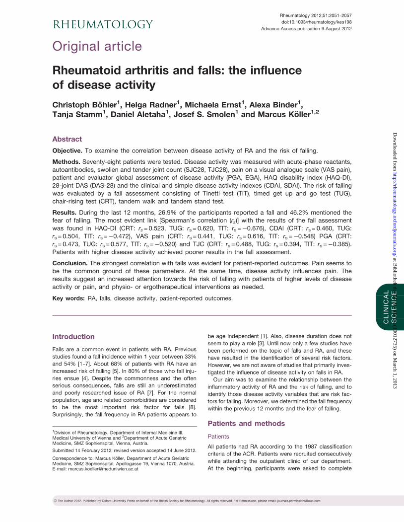
Original article
Rheumatoid arthritis and falls: the influenceof disease activity
Christoph Bohler1, Helga Radner1, Michaela Ernst1, Alexa Binder1,Tanja Stamm1, Daniel Aletaha1, Josef S. Smolen1 and Marcus Koller1,2
Abstract
Objective. To examine the correlation between disease activity of RA and the risk of falling.
Methods. Seventy-eight patients were tested. Disease activity was measured with acute-phase reactants,
autoantibodies, swollen and tender joint count (SJC28, TJC28), pain on a visual analogue scale (VAS pain),
patient and evaluator global assessment of disease activity (PGA, EGA), HAQ disability index (HAQ-DI),
28-joint DAS (DAS-28) and the clinical and simple disease activity indexes (CDAI, SDAI). The risk of falling
was evaluated by a fall assessment consisting of Tinetti test (TIT), timed get up and go test (TUG),
chair-rising test (CRT), tandem walk and tandem stand test.
Results. During the last 12 months, 26.9% of the participants reported a fall and 46.2% mentioned the
fear of falling. The most evident link [Spearman’s correlation (rs)] with the results of the fall assessment
was found in HAQ-DI (CRT: rs = 0.523, TUG: rs = 0.620, TIT: rs =�0.676), CDAI (CRT: rs = 0.460, TUG:
rs = 0.504, TIT: rs =�0.472), VAS pain (CRT: rs = 0.441, TUG: rs = 0.616, TIT: rs =�0.548) PGA (CRT:
rs = 0.473, TUG: rs = 0.577, TIT: rs =�0.520) and TJC (CRT: rs = 0.488, TUG: rs = 0.394, TIT: rs =�0.385).
Patients with higher disease activity achieved poorer results in the fall assessment.
Conclusion. The strongest correlation with falls was evident for patient-reported outcomes. Pain seems to
be the common ground of these parameters. At the same time, disease activity influences pain. The
results suggest an increased attention towards the risk of falling with patients of higher levels of disease
activity or pain, and physio- or ergotherapeutical interventions as needed.
Key words: RA, falls, disease activity, patient-reported outcomes.
Introduction
Falls are a common event in patients with RA. Previous
studies found a fall incidence within 1 year between 33%
and 54% [1�7]. About 68% of patients with RA have an
increased risk of falling [5]. In 80% of those who fall inju-
ries ensue [4]. Despite the commonness and the often
serious consequences, falls are still an underestimated
and poorly researched issue of RA [7]. For the normal
population, age and related comorbidities are considered
to be the most important risk factor for falls [8].
Surprisingly, the fall frequency in RA patients appears to
be age independent [1]. Also, disease duration does not
seem to play a role [3]. Until now only a few studies have
been performed on the topic of falls and RA, and these
have resulted in the identification of several risk factors.
However, we are not aware of studies that primarily inves-
tigated the influence of disease activity on falls in RA.
Our aim was to examine the relationship between the
inflammatory activity of RA and the risk of falling, and to
identify those disease activity variables that are risk fac-
tors for falling. Moreover, we determined the fall frequency
within the previous 12 months and the fear of falling.
Patients and methods
Patients
All patients had RA according to the 1987 classification
criteria of the ACR. Patients were recruited consecutively
while attending the outpatient clinic of our department.
At the beginning, participants were asked to complete
1Division of Rheumatology, Department of Internal Medicine III,Medical University of Vienna and 2Department of Acute GeriatricMedicine, SMZ Sophienspital, Vienna, Austria.
Correspondence to: Marcus Koller, Department of Acute GeriatricMedicine, SMZ Sophienspital, Apollogasse 19, Vienna 1070, Austria.E-mail: [email protected]
Submitted 14 February 2012; revised version accepted 14 June 2012.
! The Author 2012. Published by Oxford University Press on behalf of the British Society for Rheumatology. All rights reserved. For Permissions, please email: [email protected]
RHEUMATOLOGY
Rheumatology 2012;51:2051�2057
doi:10.1093/rheumatology/kes198
Advance Access publication 9 August 2012
CL
INIC
AL
SC
IEN
CE
at Bibliothek der M
edUniW
ien (10012735) on March 1, 2013
http://rheumatology.oxfordjournals.org/
Dow
nloaded from
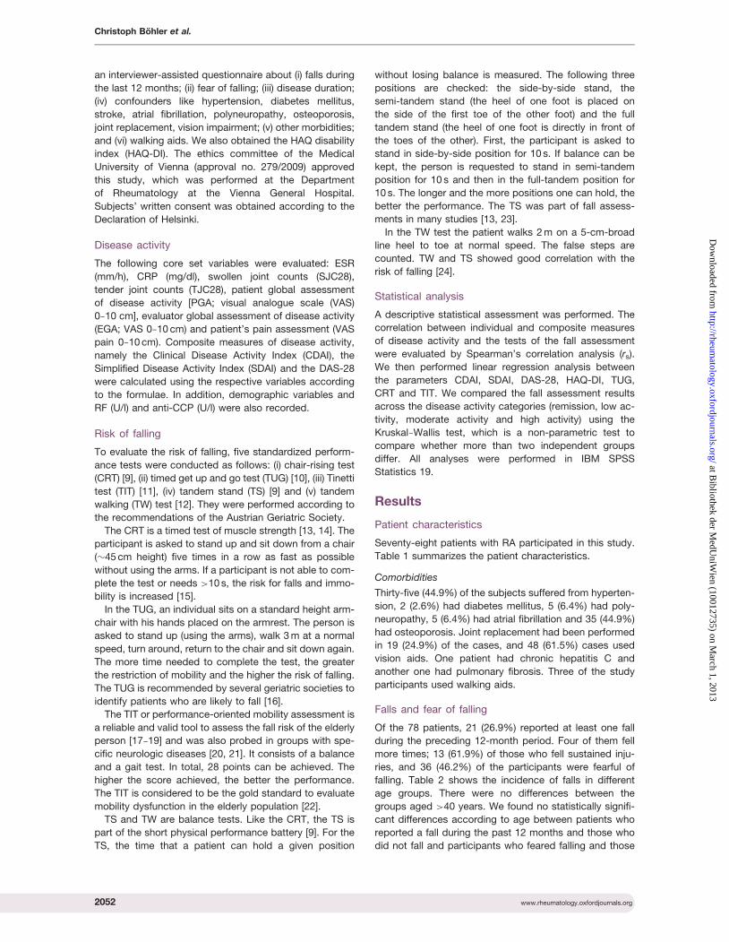
an interviewer-assisted questionnaire about (i) falls during
the last 12 months; (ii) fear of falling; (iii) disease duration;
(iv) confounders like hypertension, diabetes mellitus,
stroke, atrial fibrillation, polyneuropathy, osteoporosis,
joint replacement, vision impairment; (v) other morbidities;
and (vi) walking aids. We also obtained the HAQ disability
index (HAQ-DI). The ethics committee of the Medical
University of Vienna (approval no. 279/2009) approved
this study, which was performed at the Department
of Rheumatology at the Vienna General Hospital.
Subjects’ written consent was obtained according to the
Declaration of Helsinki.
Disease activity
The following core set variables were evaluated: ESR
(mm/h), CRP (mg/dl), swollen joint counts (SJC28),
tender joint counts (TJC28), patient global assessment
of disease activity [PGA; visual analogue scale (VAS)
0�10 cm], evaluator global assessment of disease activity
(EGA; VAS 0�10 cm) and patient’s pain assessment (VAS
pain 0�10 cm). Composite measures of disease activity,
namely the Clinical Disease Activity Index (CDAI), the
Simplified Disease Activity Index (SDAI) and the DAS-28
were calculated using the respective variables according
to the formulae. In addition, demographic variables and
RF (U/l) and anti-CCP (U/l) were also recorded.
Risk of falling
To evaluate the risk of falling, five standardized perform-
ance tests were conducted as follows: (i) chair-rising test
(CRT) [9], (ii) timed get up and go test (TUG) [10], (iii) Tinetti
test (TIT) [11], (iv) tandem stand (TS) [9] and (v) tandem
walking (TW) test [12]. They were performed according to
the recommendations of the Austrian Geriatric Society.
The CRT is a timed test of muscle strength [13, 14]. The
participant is asked to stand up and sit down from a chair
(�45 cm height) five times in a row as fast as possible
without using the arms. If a participant is not able to com-
plete the test or needs >10 s, the risk for falls and immo-
bility is increased [15].
In the TUG, an individual sits on a standard height arm-
chair with his hands placed on the armrest. The person is
asked to stand up (using the arms), walk 3 m at a normal
speed, turn around, return to the chair and sit down again.
The more time needed to complete the test, the greater
the restriction of mobility and the higher the risk of falling.
The TUG is recommended by several geriatric societies to
identify patients who are likely to fall [16].
The TIT or performance-oriented mobility assessment is
a reliable and valid tool to assess the fall risk of the elderly
person [17�19] and was also probed in groups with spe-
cific neurologic diseases [20, 21]. It consists of a balance
and a gait test. In total, 28 points can be achieved. The
higher the score achieved, the better the performance.
The TIT is considered to be the gold standard to evaluate
mobility dysfunction in the elderly population [22].
TS and TW are balance tests. Like the CRT, the TS is
part of the short physical performance battery [9]. For the
TS, the time that a patient can hold a given position
without losing balance is measured. The following three
positions are checked: the side-by-side stand, the
semi-tandem stand (the heel of one foot is placed on
the side of the first toe of the other foot) and the full
tandem stand (the heel of one foot is directly in front of
the toes of the other). First, the participant is asked to
stand in side-by-side position for 10 s. If balance can be
kept, the person is requested to stand in semi-tandem
position for 10 s and then in the full-tandem position for
10 s. The longer and the more positions one can hold, the
better the performance. The TS was part of fall assess-
ments in many studies [13, 23].
In the TW test the patient walks 2 m on a 5-cm-broad
line heel to toe at normal speed. The false steps are
counted. TW and TS showed good correlation with the
risk of falling [24].
Statistical analysis
A descriptive statistical assessment was performed. The
correlation between individual and composite measures
of disease activity and the tests of the fall assessment
were evaluated by Spearman’s correlation analysis (rs).
We then performed linear regression analysis between
the parameters CDAI, SDAI, DAS-28, HAQ-DI, TUG,
CRT and TIT. We compared the fall assessment results
across the disease activity categories (remission, low ac-
tivity, moderate activity and high activity) using the
Kruskal�Wallis test, which is a non-parametric test to
compare whether more than two independent groups
differ. All analyses were performed in IBM SPSS
Statistics 19.
Results
Patient characteristics
Seventy-eight patients with RA participated in this study.
Table 1 summarizes the patient characteristics.
Comorbidities
Thirty-five (44.9%) of the subjects suffered from hyperten-
sion, 2 (2.6%) had diabetes mellitus, 5 (6.4%) had poly-
neuropathy, 5 (6.4%) had atrial fibrillation and 35 (44.9%)
had osteoporosis. Joint replacement had been performed
in 19 (24.9%) of the cases, and 48 (61.5%) cases used
vision aids. One patient had chronic hepatitis C and
another one had pulmonary fibrosis. Three of the study
participants used walking aids.
Falls and fear of falling
Of the 78 patients, 21 (26.9%) reported at least one fall
during the preceding 12-month period. Four of them fell
more times; 13 (61.9%) of those who fell sustained inju-
ries, and 36 (46.2%) of the participants were fearful of
falling. Table 2 shows the incidence of falls in different
age groups. There were no differences between the
groups aged >40 years. We found no statistically signifi-
cant differences according to age between patients who
reported a fall during the past 12 months and those who
did not fall and participants who feared falling and those
2052 www.rheumatology.oxfordjournals.org
Christoph Bohler et al.
at Bibliothek der M
edUniW
ien (10012735) on March 1, 2013
http://rheumatology.oxfordjournals.org/
Dow
nloaded from
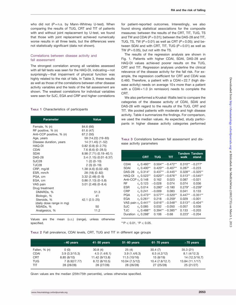
who did not (P = n.s. by Mann�Whitney U-test). When
comparing the results of TUG, CRT and TIT of patients
with and without joint replacement by U-test, we found
that those with joint replacement achieved numerically
worse results in all three tests, but the differences were
not statistically significant (data not shown).
Correlations between disease activity andfall assessment
The strongest correlation among all variables assessed
with all fall tests was seen for the HAQ-DI, indicating—not
surprisingly—that impairment of physical function was
highly related to the risk of falls. In Table 3, these results
as well as those of the correlations between other disease
activity variables and the tests of the fall assessment are
shown. The weakest correlations for individual variables
were seen for SJC, EGA and CRP and higher correlations
for patient-reported outcomes. Interestingly, we also
found strong statistical associations for the composite
measures: between the results of the CRT, TIT, TUG, TS
and TW and CDAI (P = 0.01); between the DAS-28 and TIT,
TUG, TS, TW (P = 0.01) as well as CRT (P = 0.05); and be-
tween SDAI and with CRT, TIT, TUG (P = 0.01) as well as
TW (P = 0.05), but not with the TS.
The results of the regression analysis are shown in
Fig. 1. Patients with higher CDAI, SDAI, DAS-28 and
HAQ-DI values achieved poorer results on the TUG,
CRT and TIT. Regression analyses underline the clinical
relevance of the disease activity for the fall risk. For ex-
ample, the regression coefficient for CRT and CDAI was
0.460. Therefore, a patient with a CDAI = 22.7 (high dis-
ease activity) needs on average 10 s more than a patient
with a CDAI = 1.0 (in remission) needs to complete the
CRT.
We also performed a Kruskal�Wallis test to compare the
categories of the disease activity of CDAI, SDAI and
DAS-28 with regard to the results of the TUG, CRT and
TIT. We pooled patients with moderate and high disease
activity. Table 4 summarizes the findings. For comparison,
we used the median values. As expected, study partici-
pants in higher disease activity categories performed
TABLE 1 Characteristics of participants
Parameter Value
Female, % (n) 84.6 (66)
RF positive, % (n) 61.6 (47)
Anti-CCP positive, % (n) 67.2 (50)Age, years 59 (14.22) (19�83)
Disease duration, years 14 (11.45) (1�52)
HAQ-DI 0.82 (0.8) (0�2.75)
CDAI 7.6 (6.6) (0�28.5)SDAI 8.98 (7.71) (0.19�40.1)
DAS-28 3.4 (1.15) (0.01�6.37)
SJC28 1 (2) (0�10)
TJC28 2 (3) (0�19)CRP, mg/dl 1.38 (4.6) (0.02�40)
ESR, mm/h 28 (18) (0�82)
PGA, cm 3.32 (2.48) (0�9)EGA, cm 0.86 (1.13) (0�5.8)
VAS pain 3.01 (2.49) (0�8.4)
Drug treatment
DMARDs, % 51.3Biologic, % 41
Steroids, %(daily dose range in mg)
42.1 (2.5�25)
NSAIDs, % 50
Analgesics, % 11.2
Values are the mean (S.D.) (range), unless otherwisespecified.
TABLE 2 Fall prevalence, CDAI levels, CRT, TUG and TIT in different age groups
<40 years 41�50 years 51�60 years 61�70 years >70 years
Fallen, % (n) 0 (0) 30.8 (4) 25 (4) 30.4 (7) 35.3 (21)CDAI 3.9 (3.3/15.3) 4.5 (1.4/6.1) 3.9 (1.4/6.3) 6.5 (4.2/12) 8.1 (4/12.3)
CRT 8.85 (8/10) 11.42 (9/13.8) 11.5 (10/16) 15 (8/19) 14 (12.3/16.7)
TUG 7 (6.92/7.77) 8.72 (8/10.5) 10.04 (7.5/12) 10.4 (7.8/12.7) 13.84 (11.1/17)TIT 28 (28/28) 28 (27/28) 28 (26/28) 27 (25/28) 25 (21/27)
Given values are the median (25th/75th percentile), unless otherwise specified.
TABLE 3 Correlations between fall assessment and dis-
ease activity parameters
CRT TUG TITTandem
walkTandem
stand
CDAI rs 0.460** 0.504** �0.472** 0.310** �0.277*
SDAI rs 0.400** 0.420** �0.407** 0.287* �0.223
DAS-28 rs 0.314* 0.437** �0.445** 0.328** �0.325**
HAQ-DI rs 0.523** 0.620** �0.676** 0.513** �0.545**Anti-CCP rs 0.148 0.162 0.023 0.087 �0.021
RF rs 0.123 �0.028 0.074 0.074 0.036
ESR rs 0.014 0.280* �0.180 0.279* �0.239*CRP rs 0.241 �0.009 0.083 0.041 0.133
PGA rs 0.473** 0.577** �0.520** 0.447** �0.351**
EGA rs 0.261* 0.218 �0.259* 0.029 �0.001
VAS pain rs 0.441** 0.616** �0.548** 0.513** �0.404**SJC rs 0.085 0.032 �0.050 �0.057 0.036
TJC rs 0.488** 0.394** �0.385** 0.133 �0.203
Duration rs 0.288* 0.106 �0.68 0.223* �0.204
**P4 0.01, *P4 0.05.
www.rheumatology.oxfordjournals.org 2053
RA and the risk of falling
at Bibliothek der M
edUniW
ien (10012735) on March 1, 2013
http://rheumatology.oxfordjournals.org/
Dow
nloaded from
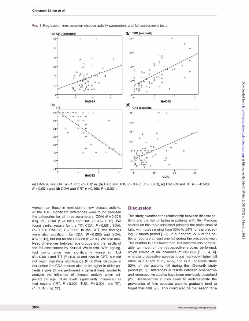
worse than those in remission or low disease activity.
At the TUG, significant differences were found between
the categories for all three parameters: CDAI (P = 0.001)
(Fig. 2a), SDAI (P = 0.001) and DAS-28 (P = 0.012). We
found similar results for the TIT: CDAI, P = 0.001; SDAI,
P = 0.001; DAS-28, P = 0.002. In the CRT, the findings
were also significant for CDAI (P = 0.002) and SDAI,
(P = 0.015), but not for the DAS-28 (P = n.s.). We also ana-
lysed differences between age groups and the results of
the fall assessment by Kruskal�Wallis test. With ageing,
test performance was significantly worse in TUG
(P< 0.001) and TIT (P = 0.016) and also in CRT, but did
not reach statistical significance (P = 0.054). Because in
our cohort the CDAI tended also to be higher in older pa-
tients (Table 2), we performed a general linear model to
analyse the influence of disease activity when ad-
justed for age. CDAI levels significantly influenced all
test results: CRT, P< 0.001; TUG, P = 0.001; and TIT,
P = 0.016 (Fig. 2b).
Discussion
This study examined the relationship between disease ac-
tivity and the risk of falling in patients with RA. Previous
studies on this topic assessed primarily the prevalence of
falls, with rates ranging from 33% to 54% for the preced-
ing 12-month period [1�7]. In our cohort, 27% of the pa-
tients reported at least one fall during the preceding year.
This number is a bit lower than, but nevertheless compar-
able to, most of the retrospective studies performed,
which arrived at an incidence of 33�38% [1, 2, 4, 6],
whereas prospective surveys found markedly higher fall
rates; in a Dutch study 42%, and in a Japanese study
50%, of the patients fell during the 12-month study
period [3, 7]. Differences in results between prospective
and retrospective studies have been previously described
[25]. Retrospective studies seem to underestimate the
prevalence of falls because patients gradually tend to
forget their falls [26]. This could also be the reason for a
FIG. 1 Regression lines between disease activity parameters and fall assessment tests.
(a) DAS-28 and CRT (r = 1.737; P< 0.014), (b) HAQ and TUG (r = 0.435; P< 0.001), (c) HAQ-DI and TIT (r =�0.528;
P< 0.001) and (d) CDAI and CRT (r = 0.460; P< 0.001).
2054 www.rheumatology.oxfordjournals.org
Christoph Bohler et al.
at Bibliothek der M
edUniW
ien (10012735) on March 1, 2013
http://rheumatology.oxfordjournals.org/
Dow
nloaded from
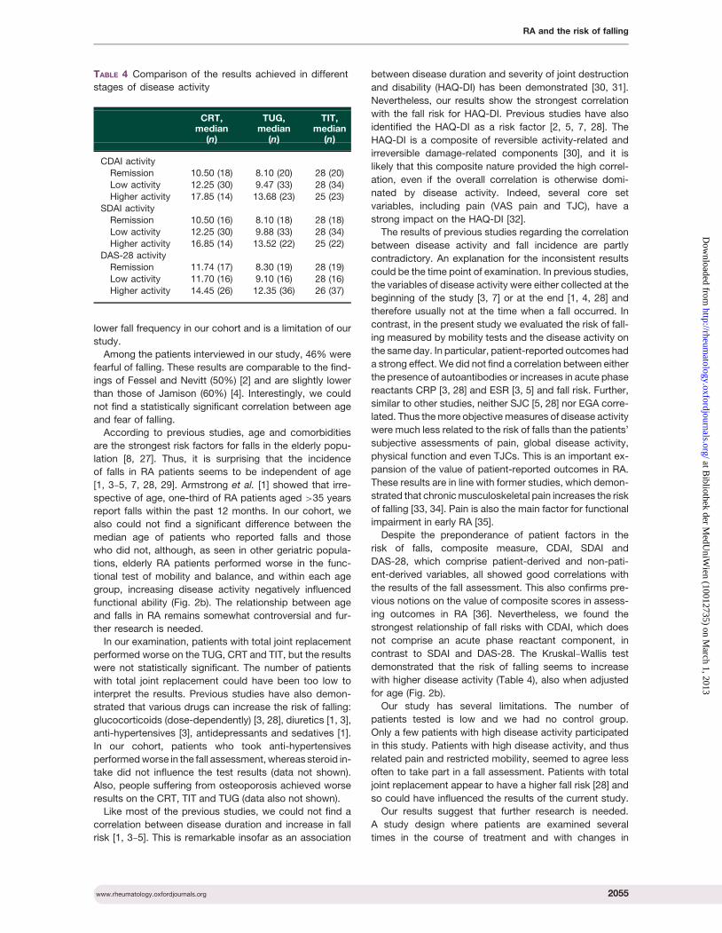
lower fall frequency in our cohort and is a limitation of our
study.
Among the patients interviewed in our study, 46% were
fearful of falling. These results are comparable to the find-
ings of Fessel and Nevitt (50%) [2] and are slightly lower
than those of Jamison (60%) [4]. Interestingly, we could
not find a statistically significant correlation between age
and fear of falling.
According to previous studies, age and comorbidities
are the strongest risk factors for falls in the elderly popu-
lation [8, 27]. Thus, it is surprising that the incidence
of falls in RA patients seems to be independent of age
[1, 3�5, 7, 28, 29]. Armstrong et al. [1] showed that irre-
spective of age, one-third of RA patients aged >35 years
report falls within the past 12 months. In our cohort, we
also could not find a significant difference between the
median age of patients who reported falls and those
who did not, although, as seen in other geriatric popula-
tions, elderly RA patients performed worse in the func-
tional test of mobility and balance, and within each age
group, increasing disease activity negatively influenced
functional ability (Fig. 2b). The relationship between age
and falls in RA remains somewhat controversial and fur-
ther research is needed.
In our examination, patients with total joint replacement
performed worse on the TUG, CRT and TIT, but the results
were not statistically significant. The number of patients
with total joint replacement could have been too low to
interpret the results. Previous studies have also demon-
strated that various drugs can increase the risk of falling:
glucocorticoids (dose-dependently) [3, 28], diuretics [1, 3],
anti-hypertensives [3], antidepressants and sedatives [1].
In our cohort, patients who took anti-hypertensives
performed worse in the fall assessment, whereas steroid in-
take did not influence the test results (data not shown).
Also, people suffering from osteoporosis achieved worse
results on the CRT, TIT and TUG (data also not shown).
Like most of the previous studies, we could not find a
correlation between disease duration and increase in fall
risk [1, 3�5]. This is remarkable insofar as an association
between disease duration and severity of joint destruction
and disability (HAQ-DI) has been demonstrated [30, 31].
Nevertheless, our results show the strongest correlation
with the fall risk for HAQ-DI. Previous studies have also
identified the HAQ-DI as a risk factor [2, 5, 7, 28]. The
HAQ-DI is a composite of reversible activity-related and
irreversible damage-related components [30], and it is
likely that this composite nature provided the high correl-
ation, even if the overall correlation is otherwise domi-
nated by disease activity. Indeed, several core set
variables, including pain (VAS pain and TJC), have a
strong impact on the HAQ-DI [32].
The results of previous studies regarding the correlation
between disease activity and fall incidence are partly
contradictory. An explanation for the inconsistent results
could be the time point of examination. In previous studies,
the variables of disease activity were either collected at the
beginning of the study [3, 7] or at the end [1, 4, 28] and
therefore usually not at the time when a fall occurred. In
contrast, in the present study we evaluated the risk of fall-
ing measured by mobility tests and the disease activity on
the same day. In particular, patient-reported outcomes had
a strong effect. We did not find a correlation between either
the presence of autoantibodies or increases in acute phase
reactants CRP [3, 28] and ESR [3, 5] and fall risk. Further,
similar to other studies, neither SJC [5, 28] nor EGA corre-
lated. Thus the more objective measures of disease activity
were much less related to the risk of falls than the patients’
subjective assessments of pain, global disease activity,
physical function and even TJCs. This is an important ex-
pansion of the value of patient-reported outcomes in RA.
These results are in line with former studies, which demon-
strated that chronic musculoskeletal pain increases the risk
of falling [33, 34]. Pain is also the main factor for functional
impairment in early RA [35].
Despite the preponderance of patient factors in the
risk of falls, composite measure, CDAI, SDAI and
DAS-28, which comprise patient-derived and non-pati-
ent-derived variables, all showed good correlations with
the results of the fall assessment. This also confirms pre-
vious notions on the value of composite scores in assess-
ing outcomes in RA [36]. Nevertheless, we found the
strongest relationship of fall risks with CDAI, which does
not comprise an acute phase reactant component, in
contrast to SDAI and DAS-28. The Kruskal�Wallis test
demonstrated that the risk of falling seems to increase
with higher disease activity (Table 4), also when adjusted
for age (Fig. 2b).
Our study has several limitations. The number of
patients tested is low and we had no control group.
Only a few patients with high disease activity participated
in this study. Patients with high disease activity, and thus
related pain and restricted mobility, seemed to agree less
often to take part in a fall assessment. Patients with total
joint replacement appear to have a higher fall risk [28] and
so could have influenced the results of the current study.
Our results suggest that further research is needed.
A study design where patients are examined several
times in the course of treatment and with changes in
TABLE 4 Comparison of the results achieved in different
stages of disease activity
CRT,median
(n)
TUG,median
(n)
TIT,median
(n)
CDAI activityRemission 10.50 (18) 8.10 (20) 28 (20)
Low activity 12.25 (30) 9.47 (33) 28 (34)
Higher activity 17.85 (14) 13.68 (23) 25 (23)
SDAI activityRemission 10.50 (16) 8.10 (18) 28 (18)
Low activity 12.25 (30) 9.88 (33) 28 (34)
Higher activity 16.85 (14) 13.52 (22) 25 (22)
DAS-28 activityRemission 11.74 (17) 8.30 (19) 28 (19)
Low activity 11.70 (16) 9.10 (16) 28 (16)
Higher activity 14.45 (26) 12.35 (36) 26 (37)
www.rheumatology.oxfordjournals.org 2055
RA and the risk of falling
at Bibliothek der M
edUniW
ien (10012735) on March 1, 2013
http://rheumatology.oxfordjournals.org/
Dow
nloaded from
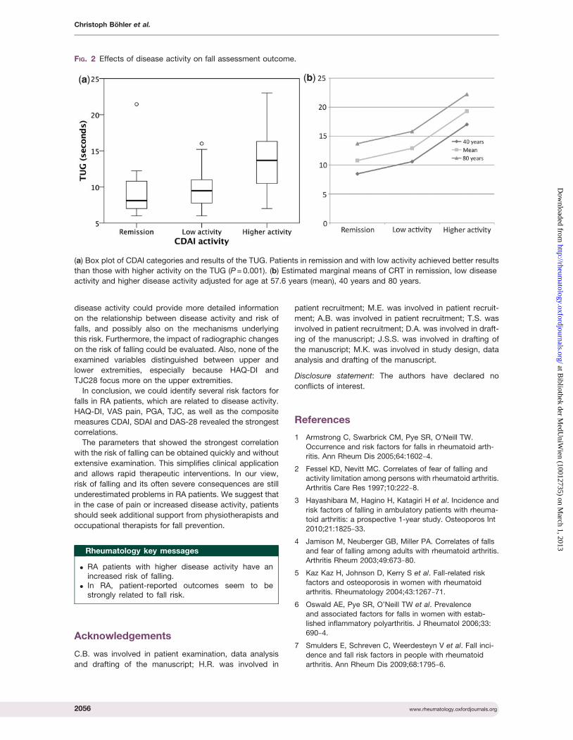
disease activity could provide more detailed information
on the relationship between disease activity and risk of
falls, and possibly also on the mechanisms underlying
this risk. Furthermore, the impact of radiographic changes
on the risk of falling could be evaluated. Also, none of the
examined variables distinguished between upper and
lower extremities, especially because HAQ-DI and
TJC28 focus more on the upper extremities.
In conclusion, we could identify several risk factors for
falls in RA patients, which are related to disease activity.
HAQ-DI, VAS pain, PGA, TJC, as well as the composite
measures CDAI, SDAI and DAS-28 revealed the strongest
correlations.
The parameters that showed the strongest correlation
with the risk of falling can be obtained quickly and without
extensive examination. This simplifies clinical application
and allows rapid therapeutic interventions. In our view,
risk of falling and its often severe consequences are still
underestimated problems in RA patients. We suggest that
in the case of pain or increased disease activity, patients
should seek additional support from physiotherapists and
occupational therapists for fall prevention.
Rheumatology key messages
. RA patients with higher disease activity have anincreased risk of falling.
. In RA, patient-reported outcomes seem to bestrongly related to fall risk.
Acknowledgements
C.B. was involved in patient examination, data analysis
and drafting of the manuscript; H.R. was involved in
patient recruitment; M.E. was involved in patient recruit-
ment; A.B. was involved in patient recruitment; T.S. was
involved in patient recruitment; D.A. was involved in draft-
ing of the manuscript; J.S.S. was involved in drafting of
the manuscript; M.K. was involved in study design, data
analysis and drafting of the manuscript.
Disclosure statement: The authors have declared no
conflicts of interest.
References
1 Armstrong C, Swarbrick CM, Pye SR, O’Neill TW.
Occurrence and risk factors for falls in rheumatoid arth-
ritis. Ann Rheum Dis 2005;64:1602�4.
2 Fessel KD, Nevitt MC. Correlates of fear of falling and
activity limitation among persons with rheumatoid arthritis.
Arthritis Care Res 1997;10:222�8.
3 Hayashibara M, Hagino H, Katagiri H et al. Incidence and
risk factors of falling in ambulatory patients with rheuma-
toid arthritis: a prospective 1-year study. Osteoporos Int
2010;21:1825�33.
4 Jamison M, Neuberger GB, Miller PA. Correlates of falls
and fear of falling among adults with rheumatoid arthritis.
Arthritis Rheum 2003;49:673�80.
5 Kaz Kaz H, Johnson D, Kerry S et al. Fall-related risk
factors and osteoporosis in women with rheumatoid
arthritis. Rheumatology 2004;43:1267�71.
6 Oswald AE, Pye SR, O’Neill TW et al. Prevalence
and associated factors for falls in women with estab-
lished inflammatory polyarthritis. J Rheumatol 2006;33:
690�4.
7 Smulders E, Schreven C, Weerdesteyn V et al. Fall inci-
dence and fall risk factors in people with rheumatoid
arthritis. Ann Rheum Dis 2009;68:1795�6.
FIG. 2 Effects of disease activity on fall assessment outcome.
(a) Box plot of CDAI categories and results of the TUG. Patients in remission and with low activity achieved better results
than those with higher activity on the TUG (P = 0.001). (b) Estimated marginal means of CRT in remission, low disease
activity and higher disease activity adjusted for age at 57.6 years (mean), 40 years and 80 years.
2056 www.rheumatology.oxfordjournals.org
Christoph Bohler et al.
at Bibliothek der M
edUniW
ien (10012735) on March 1, 2013
http://rheumatology.oxfordjournals.org/
Dow
nloaded from
8 McKay C, Anderson KE. How to manage falls in commu-nity dwelling older adults: a review of the evidence.
Postgrad Med J 2010;86:299�306.
9 Guralnik JM, Simonsick EM, Ferrucci L et al. A short
physical performance battery assessing lower extremity
function: association with self-reported disability and
prediction of mortality and nursing home admission. JGerontol 1994;49:M85�94.
10 Podsiadlo D, Richardson S. The timed ‘Up & Go’: a test ofbasic functional mobility for frail elderly persons. J Am
Geriatr Soc 1991;39:142�8.
11 Tinetti ME. Performance-oriented assessment of mobility
problems in elderly patients. J Am Geriatr Soc 1986;34:
119�26.
12 Cho BL, Scarpace D, Alexander NB. Tests of stepping as
indicators of mobility, balance, and fall risk inbalance-impaired older adults. J Am Geriatr Soc 2004;52:
1168�73.
13 Dukas L, Schacht E, Runge M. Independent from muscle
power and balance performance, a creatinine clearance
below 65 ml/min is a significant and independent risk
factor for falls and fall-related fractures in elderly men andwomen diagnosed with osteoporosis. Osteoporos Int
2010;21:1237�45.
14 Guralnik JM, Ferrucci L, Simonsick EM, Salive ME,
Wallace RB. Lower-extremity function in persons over the
age of 70 years as a predictor of subsequent disability. NEngl J Med 1995;332:556�61.
15 Nevitt MC, Cummings SR, Kidd S, Black D. Risk factorsfor recurrent nonsyncopal falls. A prospective study.
JAMA 1989;261:2663�8.
16 Nordin E, Lindelof N, Rosendahl E, Jensen J, Lundin-
Olsson L. Prognostic validity of the Timed Up-and-Go test,
a modified Get-Up-and-Go test, staff’s global judgement
and fall history in evaluating fall risk in residential carefacilities. Age Ageing 2008;37:442�8.
17 Tinetti ME, Speechley M, Ginter SF. Risk factors for falls
among elderly persons living in the community. N Engl J
Med 1988;319:1701�7.
18 Harada N, Chiu V, Damron-Rodriguez J et al. Screening
for balance and mobility impairment in elderly individuals
living in residential care facilities. Phys Ther 1995;75:462�9.
19 Lin MR, Hwang HF, Hu MH et al. Psychometriccomparisons of the timed up and go, one-leg stand,
functional reach, and Tinetti balance measures in
community-dwelling older people. J Am Geriatr Soc
2004;52:1343�8.
20 Kegelmeyer DA, Kloos AD, Thomas KM, Kostyk SK.
Reliability and validity of the Tinetti Mobility Test for indi-viduals with Parkinson disease. Phys Ther 2007;87:
1369�78.
21 Kloos AD, Kegelmeyer DA, Young GS, Kostyk SK. Fall
risk assessment using the Tinetti mobility test in individ-
uals with Huntington’s disease. Mov Disord 2010;25:
2838�44.
22 Kopke S, Meyer G. The Tinetti test: Babylon in geriatric
assessment. Z Gerontol Geriatr 2006;39:288�91.
23 Dukas L, Schacht E, Runge M, Ringe JD. Effect of a
six-month therapy with alfacalcidol on muscle power and
balance and the number of fallers and falls.
Arzneimittelforschung 2010;60:519�25.
24 Stel VS, Smit JH, Pluijm SM, Lips P. Balance and
mobility performance as treatable risk factors for
recurrent falling in older persons. J Clin Epidemiol 2003;
56:659�68.
25 Ganz DA, Higashi T, Rubenstein LZ. Monitoring falls in
cohort studies of community-dwelling older people: effect
of the recall interval. J Am Geriatr Soc 2005;53:2190�4.
26 Cummings SR, Nevitt MC, Kidd S. Forgetting falls. The
limited accuracy of recall of falls in the elderly. J Am
Geriatr Soc 1988;36:613�6.
27 Rubenstein LZ. Falls in older people: epidemiology, risk
factors and strategies for prevention. Age Ageing 2006;
35(Suppl. 2):ii37�41.
28 Furuya T, Yamagiwa K, Ikai T et al. Associated factors for
falls and fear of falling in Japanese patients with rheuma-
toid arthritis. Clin Rheumatol 2009;28:1325�30.
29 Yamagiwa K, Iijima S, Furuya T et al. Incidence of falls and
fear of falling in Japanese patients with rheumatoid arth-
ritis. Mod Rheumatol 2010;21:51�6.
30 Aletaha D, Smolen J, Ward MM. Measuring function in
rheumatoid arthritis: Identifying reversible and irreversible
components. Arthritis Rheum 2006;54:2784�92.
31 Scott DL, Pugner K, Kaarela K et al. The links between
joint damage and disability in rheumatoid arthritis.
Rheumatology 2000;39:122�32.
32 Aletaha D, Ward MM. Duration of rheumatoid arthritis in-
fluences the degree of functional improvement in clinical
trials. Ann Rheum Dis 2006;65:227�33.
33 Leveille SG, Bean J, Bandeen-Roche K et al. Musculos-
keletal pain and risk for falls in older disabled women living
in the community. J Am Geriatr Soc 2002;50:671�8.
34 Leveille SG, Jones RN, Kiely DK et al. Chronic musculo-
skeletal pain and the occurrence of falls in an older
population. JAMA 2009;302:2214�21.
35 Lipsky PE, Buckland J. Pain by name, pain by nature. Nat
Rev Rheumatol 2010;6:179�80.
36 Aletaha D, Smolen JS. The Simplified Disease Activity
Index and Clinical Disease Activity Index to monitor pa-
tients in standard clinical care. Rheum Dis Clin North Am
2009;35:759�72, viii.
www.rheumatology.oxfordjournals.org 2057
RA and the risk of falling
at Bibliothek der M
edUniW
ien (10012735) on March 1, 2013
http://rheumatology.oxfordjournals.org/
Dow
nloaded from
CONCISE REPORT
Serological changes in the course of traditional andbiological disease modifying therapy of rheumatoidarthritisChristoph Böhler,1 Helga Radner,1 Josef S Smolen,1,2 Daniel Aletaha1
▸ Additional supplementarydata are published online only.To view these files please visitthe journal online (http://dx.doi.org/10.1136/annrheumdis-2012-202297).1Division of Rheumatology,Department of Medicine 3,Medical University of Vienna,Vienna, Austria22nd Department of Medicine,Hietzing Hospital, Vienna,Austria
Correspondence toProfessor Daniel Aletaha,Division of Rheumatology,Department of Medicine 3,Medical University of Vienna,Waehringer Guertel 18-20,Vienna 1090, Austria; [email protected]
Accepted 11 September 2012Published Online First19 October 2012
ABSTRACTObjective To investigate changes of rheumatoid factor(RF) and antibodies against citrullinated peptides (ACPA)during therapy with disease modifying antirheumaticdrugs.Methods We obtained clinical and serological data ofpatients from the treatment start and after 6 months oftherapy. With non-parametric tests, we analysed changesof ACPA and RF levels between the two visits and theinfluence of treatment response. Furthermore, weanalysed potential influential factors as disease chronicity,different therapeutics and the trend over 18 months.Results 143 ACPA and RF positive patients wereincluded. The median (25th/75th percentile) relativechanges after 6 months were −35.6% (−63.3; −8.3) forRF and −15.2% (−40.0; 10.0) for ACPA (p<0.001 forboth). Changes of RF levels were significantly greaterthan those seen for ACPA (p<0.001). The decrease ofACPA and RF was significantly higher in treatmentresponders (p=0.034 and p=0.01, respectively). Asidefrom changes in disease activity, only a short diseaseduration showed an independent effect on changes of RFlevels (p=0.087).Conclusions ACPA and RF levels decreasedsignificantly after 6 months of therapy. Reductions of bothautoantibodies were closely linked to a reduction ofdisease activity. RF declined faster, to a larger extent andin greater numbers of patients than ACPA.
Rheumatoid factor (RF) and antibodies againstcitrullinated peptides (ACPA) are establishedmarkers in the diagnostic approach to rheumatoidarthritis (RA).1 Besides their diagnostic relevance,RF and ACPA also have a prognostic value, sinceboth are associated with more aggressive, destruc-tive disease.2–4 Moreover, high RF levels are relatedto extra-articular manifestations.1 For thesereasons, RF and ACPA are part of the 2010 RA clas-sification criteria,5 and considered bad prognosticmarkers in the European League AgainstRheumatism (EULAR) recommendations for themanagement of RA.6 Several studies examinedchanges of these autoantibodies (AAB) in thecourse of therapy, but especially for ACPA, findingswere inconsistent.7–9
Given the current efforts to treat RA to a treat-ment target considering individual risk factors,10 thepresence and levels of these AAB are therapeuticallyhighly relevant.6 The aim of the present study wasto evaluate the responsiveness of ACPA and RFlevels during therapy of RA; in this context, we also
aimed to consider the influence of individual thera-peutic agents and treatment response with a specialfocus on the impact of disease duration and trendover time.
METHODSPatientsWe obtained data on RA outpatients who wereseen at our clinic where data on every visit are pro-spectively documented in an observational data-base.11 All participants fulfilled the 1987 AmericanCollege of Rheumatology (ACR),12 or morerecently, the 2010 ACR/EULAR classification cri-teria for RA5 and were recruited between February2006 and October 2011. We selected patients withat least low disease activity, in whom a traditionalor biological disease modifying antirheumatic drug(DMARD) was initiated, and identified those whowere seropositive for ACPA and RF. For eachpatient, we selected only the treatment coursewith the longest follow-up period in order tomaintain data independence in our analyses. Allpatients consented to an anonymous data analysis,and the local ethics committee had approved thedata collection.
Study variablesAt baseline and after 3, 6, 12 and 18 months oftherapy, levels of RF (U/ml; positive >14 U/ml)and ACPA (U/ml; positive ≥5 U/ml) wereobtained; ACPA levels >340 U/ml had not beenfurther diluted, and since we were interested inchanges in AAB levels, these patients wereexcluded. Furthermore, we collected patient andevaluator global assessments of disease activity;pain; swollen and tender joint count (SJC28 andTJC28); the Health Assessment QuestionnaireDisability Index; C reactive protein (in mg/dl) anderythrocyte sedimentation rate (in mm/h). Wemainly used the Simplified Disease Activity Index(SDAI) to measure disease activity.13
Statistical analysisIn most of our analyses, to account for the startingpoint (baseline level) of RF or ACPA, we evaluatedrelative rather than absolute changes within the first6 months. All data were analysed by non-parametricstatistics. To facilitate graphical illustration, we useda fractional rank depiction (‘probability plots’) witha 100% cap for worsening. All analyses were per-formed with SPSS.
Ann Rheum Dis 2013;72:241–244. doi:10.1136/annrheumdis-2012-202297 241
Clinical and epidemiological research
group.bmj.com on March 1, 2013 - Published by ard.bmj.comDownloaded from
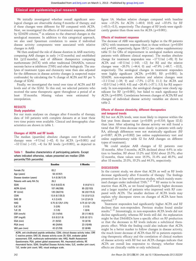
We initially investigated whether overall significant sero-logical changes are observable during 6 months of therapy, andif these changes were different for RF compared with ACPA.Next, we investigated the effects of treatment response, definedby SDAI50 criteria,14 in relation to the observed changes in theserological measures. In addition to this categorical approach,we also used Spearman correlation to assess if individualdisease activity components were associated with relativechanges in AAB.
We then analysed the role of disease duration in AAB reactivity,comparing AAB changes in early (<12 months) and establishedRA (≥12 months), and of different therapeutics comparingmethotrexate (MTX) with other traditional DMARDs, tumournecrosis factor α inhibitors (TNFi) and other biologicals. The ana-lyses of treatment types and chronicity were then also adjustedfor the differences in disease activity changes (a suspected majorconfounder) by calculating the % change of ACPA and RF per %change of SDAI.
Finally, we investigated the trend over time of ACPA and RFlevels and of the SDAI. To this end, we selected patients whoreceived the same therapeutic agent throughout a period of atleast 18 months. Missing values were estimated byinterpolation.
RESULTSStudy populationFor our main analyses on changes after 6 months of therapy,data of 143 patients with complete datasets at at least thesetwo time points were available. Clinical and demographic char-acteristics are shown in table 1.
Changes of ACPA and RF levelsThe median (quartiles) absolute changes over 6 months oftherapy were −9 U/ml (−42; 6) for ACPA (p<0.001) and−32 U/ml (−115; −4) for RF levels (p<0.001), as depicted in
figure 1A. Median relative changes compared with baselinewere −15.2% for ACPA (−40.0; 10.0) and −35.6% for RF(−63.3;−8.3), respectively. The changes of RF levels were signifi-cantly greater than those seen for ACPA (p<0.001).
Effects of treatment responseThe decrease of ABB was significantly higher in the 60 patients(42%) with treatment response than in those without (p=0.034and p=0.01, respectively, figure 1B/C) (see online supplementarytable S1 for ORs of improvement in serological markers amongpatients with different clinical response). The median absolutechange for treatment responders was −17 U/ml (−68; 0) forACPA and −43 U/ml (−143; −12) for RF, and the relativechanges were −26.5% (−44.4; 0) and −47.9% (−66.7; −18.8),respectively. The serological changes in treatment responderswere highly significant (ACPA: p=0.001; RF: p<0.001). InSDAI50, non-responders absolute and relative changes were−3.1 U/ml (−36; 9) and −7.5% (−27.0; 11.1) for ACPA and−18.0 U/ml (−96.0; 4.0) and −26.4% (−57.1; 3.3) for RF, respect-ively. In non-responders, the serological changes were clearly sig-nificant for RF (p<0.001), but failed to reach significance forACPA (p=0.059). Correlations between changes of ACPA/RF andchanges of individual disease activity variables are shown intable 2.
Effects of disease chronicity, different therapeuticsand temporal trendsRF, but not ACPA levels, were more likely to improve within thefirst year from disease onset (p=0.039; p=0.316; figure 1D,E)than later. After adjusting for disease activity we still found atrend that RF titres decrease more clearly in patients with earlyRA, although differences were not statistically significant (RFp=0.087; ACPA p=0.802) (see online supplementary text andonline supplementary table S2 for AAB changes on differenttypes of treatment).
We could analyse AAB changes of 82 patients over18 months. After 3 months, ACPA declined about 4.6% in rela-tion to baseline, RF about 13.2% and SDAI about 23.5%; after12 months, these values were 16.9%, 31.4% and 40.5%; andafter 18 months, 23.8%, 35.2% and 44.3%, respectively.
DISCUSSIONIn the current study, we show that ACPA as well as RF levelsdecrease significantly after 6 months of therapy. The findingspresented are in line with previous studies, which mainly exam-ined changes under individual TNFi.9 15 16 RF seems to be morereactive than ACPA, as we found significantly higher decreasesand a larger number of patients who improved with RF com-pared with ACPA. The smaller declines of ACPA levels mayexplain why discrepant views on changes of ACPA have beenreported.8 17
Treatment responders had significantly higher ACPA and RFdeclines than non-responders. Previous studies found similarresults.9 15 Interestingly, in non-responders ACPA levels did notdecline significantly, whereas RF levels still did. An explanationmight be that DMARDs have a specific effect on RF productionor that the decreases in RF levels indicate a subclinical thera-peutic effect. While the finding could also indicate that ACPAmight be a better marker to follow changes in disease activity,the much lower decrease of ACPA than RF in patients experien-cing therapeutic efficacy and the correlation of clinical markersof disease activity with RF but not ACPA changes indicate thatACPA are overall less responsive to therapy, whether theseeffects are clinically visible or only subclinical.
Table 1 Baseline characteristics of participating patients. Exceptwhere indicated otherwise, values presented are median (25thpercentile/75th percentile)
Baseline 6 Months
Female (%) 83.2Age (years) 58 (47/67)Disease duration (years) 5.4 (0.36/13.9)Patients with early RA (%) 26.3SDAI 15.8 (9.8/22.4) 8 (4.6/14.1)ACPA (U/ml) 107 (46/208) 85 (32/153)RF (U/ml) 120 (38/274) 56 (23/175.5)CDAI 13.8 (8.7/20.2) 7.6 (3.5/13.2)DAS 28 4.3 (3.4/5) 3.4 (2.5/4.3)HAQ 0.75 (0.125/1.375) 0.75 (0.13/1.25)SJC 28 3 (2/6) 2 (1/3)TJC 28 3 (1/6) 1 (0/4)ESR (mm/h) 22 (14/54) 20 (11/40.5)CRP (mg/dl) 0.8 (0.3/1.9) 0.35 (0.12/1)EGA (mm) 21 (9/32) 10 (4/18)PGA (mm) 46 (24/64) 23 (10/49)VAS pain (mm) 43 (21/59) 22 (8/48)
ACPA, anti-citrullinated peptide antibodies; CDAI, clinical disease activity index; CRP,C reactive protein; DAS 28, disease activity score 28; EGA, evaluator globalassessment; ESR, erythrocyte sedimentation rate; HAQ, Health AssessmentQuestionnaire; PGA, patient global assessment; RA, rheumatoid arthritis; RF,rheumatoid factor; SDAI, Simplified Disease Activity Index; SJC, swollen joint count;TJC, tender joint count; VAS, visual analogue scale.
242 Ann Rheum Dis 2013;72:241–244. doi:10.1136/annrheumdis-2012-202297
Clinical and epidemiological research
group.bmj.com on March 1, 2013 - Published by ard.bmj.comDownloaded from
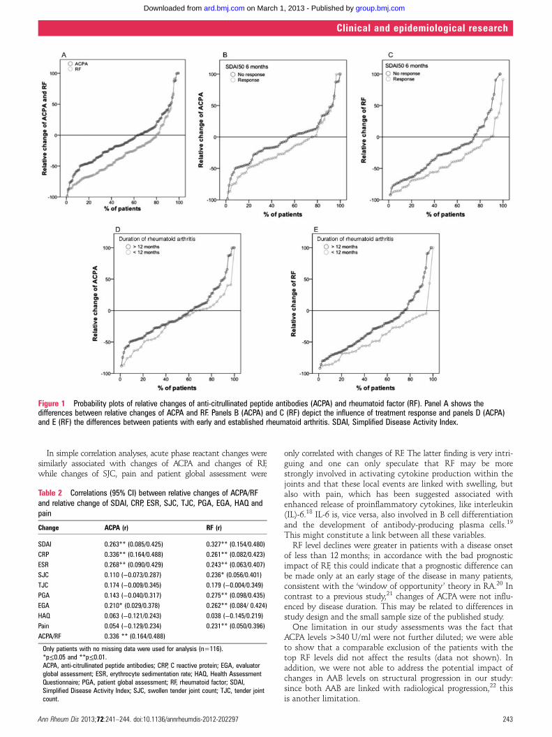
In simple correlation analyses, acute phase reactant changes weresimilarly associated with changes of ACPA and changes of RF,while changes of SJC, pain and patient global assessment were
only correlated with changes of RF. The latter finding is very intri-guing and one can only speculate that RF may be morestrongly involved in activating cytokine production within thejoints and that these local events are linked with swelling, butalso with pain, which has been suggested associated withenhanced release of proinflammatory cytokines, like interleukin(IL)-6.18 IL-6 is, vice versa, also involved in B cell differentiationand the development of antibody-producing plasma cells.19
This might constitute a link between all these variables.RF level declines were greater in patients with a disease onset
of less than 12 months; in accordance with the bad prognosticimpact of RF, this could indicate that a prognostic difference canbe made only at an early stage of the disease in many patients,consistent with the ‘window of opportunity’ theory in RA.20 Incontrast to a previous study,21 changes of ACPA were not influ-enced by disease duration. This may be related to differences instudy design and the small sample size of the published study.
One limitation in our study assessments was the fact thatACPA levels >340 U/ml were not further diluted; we were ableto show that a comparable exclusion of the patients with thetop RF levels did not affect the results (data not shown). Inaddition, we were not able to address the potential impact ofchanges in AAB levels on structural progression in our study:since both AAB are linked with radiological progression,22 thisis another limitation.
Figure 1 Probability plots of relative changes of anti-citrullinated peptide antibodies (ACPA) and rheumatoid factor (RF). Panel A shows thedifferences between relative changes of ACPA and RF. Panels B (ACPA) and C (RF) depict the influence of treatment response and panels D (ACPA)and E (RF) the differences between patients with early and established rheumatoid arthritis. SDAI, Simplified Disease Activity Index.
Table 2 Correlations (95% CI) between relative changes of ACPA/RFand relative change of SDAI, CRP, ESR, SJC, TJC, PGA, EGA, HAQ andpain
Change ACPA (r) RF (r)
SDAI 0.263** (0.085/0.425) 0.327** (0.154/0.480)CRP 0.336** (0.164/0.488) 0.261** (0.082/0.423)ESR 0.268** (0.090/0.429) 0.243** (0.063/0.407)SJC 0.110 (−0.073/0.287) 0.236* (0.056/0.401)TJC 0.174 (−0.009/0.345) 0.179 (−0.004/0.349)PGA 0.143 (−0.040/0.317) 0.275** (0.098/0.435)EGA 0.210* (0.029/0.378) 0.262** (0.084/ 0.424)HAQ 0.063 (−0.121/0.243) 0.038 (−0.145/0.219)Pain 0.054 (−0.129/0.234) 0.231** (0.050/0.396)ACPA/RF 0.336 ** (0.164/0.488)
Only patients with no missing data were used for analysis (n=116).*p≤0.05 and **p≤0.01.ACPA, anti-citrullinated peptide antibodies; CRP, C reactive protein; EGA, evaluatorglobal assessment; ESR, erythrocyte sedimentation rate; HAQ, Health AssessmentQuestionnaire; PGA, patient global assessment; RF, rheumatoid factor; SDAI,Simplified Disease Activity Index; SJC, swollen tender joint count; TJC, tender jointcount.
Ann Rheum Dis 2013;72:241–244. doi:10.1136/annrheumdis-2012-202297 243
Clinical and epidemiological research
group.bmj.com on March 1, 2013 - Published by ard.bmj.comDownloaded from
In conclusion, we were able to show that ACPA and RFlevels decreased significantly after 6 months of therapy as wellas subsequently and that decreases were closely linked to animprovement of disease activity. RF declined faster, to a largerextent and in greater numbers of patients than ACPA. Furtherresearch is needed to investigate whether reductions of ACPAand RF levels are associated with better structural and func-tional outcomes of RA in the longer term.
Acknowledgements This study was partly supported through Coordination Theme 1(Health) of the European Community’s FP7; Grant Agreement numberHEALTH-F2-2008-223404 (Masterswitch), and partly through an unrestricted grant fromMSD Austria. This is a publication of the Joint and Bone Center for Diagnosis, Researchand Therapy of Musculoskeletal Disorders of the Medical University of Vienna.
Contributors CB: Study design, data acquisition, data analysis, manuscript drafting.HR: Data acquisition, data analysis. JS and DA: Study design, data analysis,manuscript drafting.
Funding European Community’s FP7 (partly); MSD Austria (partly).
Competing interests None.
Ethics approval Ethics Committee of the Medical University of Vienna.
Patient consent Obtained.
Provenance and peer review Commissioned; externally peer reviewed.
REFERENCES1. Song YW, Kang EH. Autoantibodies in rheumatoid arthritis: rheumatoid factors and
anticitrullinated protein antibodies. QJM 2010;103:139–46.2. De Rycke L, Peene I, Hoffman IE, et al. Rheumatoid factor and anticitrullinated
protein antibodies in rheumatoid arthritis: diagnostic value, associations withradiological progression rate, and extra-articular manifestations. Ann Rheum Dis2004;63:1587–93.
3. Berglin E, Johansson T, Sundin U, et al. Radiological outcome in rheumatoidarthritis is predicted by presence of antibodies against cyclic citrullinated peptidebefore and at disease onset, and by IgA-RF at disease onset. Ann Rheum Dis2006;65:453–8.
4. Niewold TB, Harrison MJ, Paget SA. Anti-CCP antibody testing as a diagnostic andprognostic tool in rheumatoid arthritis. QJM 2007;100:193–201.
5. Aletaha D, Neogi T, Silman AJ, et al. 2010 rheumatoid arthritis classificationcriteria: an American College of Rheumatology/European League AgainstRheumatism collaborative initiative. Ann Rheum Dis 2010;69:1580–8.
6. Smolen JS, Landewé R, Breedveld FC, et al. EULAR recommendations for themanagement of rheumatoid arthritis with synthetic and biological disease-modifyingantirheumatic drugs. Ann Rheum Dis 2010;69:964–75.
7. Bobbio-Pallavicini F, Caporali R, Alpini C, et al. Predictive value of antibodies tocitrullinated peptides and rheumatoid factors in anti-TNF-alpha treated patients. AnnN Y Acad Sci 2007;1109:287–95.
8. Bobbio-Pallavicini F, Caporali R, Alpini C, et al. High IgA rheumatoid factor levelsare associated with poor clinical response to tumour necrosis factor alpha inhibitorsin rheumatoid arthritis. Ann Rheum Dis 2007;66:302–7.
9. Alessandri C, Bombardieri M, Papa N, et al. Decrease of anti-cyclic citrullinatedpeptide antibodies and rheumatoid factor following anti-TNFalpha therapy (infliximab)in rheumatoid arthritis is associated with clinical improvement. Ann Rheum Dis2004;63:1218–21.
10. Smolen JS, Aletaha D, Bijlsma JW, et al. Treating rheumatoid arthritis to target:recommendations of an international task force. Ann Rheum Dis 2010;69:631–7.
11. Stamm TA, Aletaha D, Pflugbeil S, et al. The use of databases for qualityassessment in rheumatoid arthritis. Clin Exp Rheumatol 2007;25:82–5.
12. Arnett FC, Edworthy SM, Bloch DA, et al. The American Rheumatism Association1987 revised criteria for the classification of rheumatoid arthritis. Arthritis Rheum1988;31:315–24.
13. Smolen JS, Breedveld FC, Schiff MH, et al. A simplified disease activity index forrheumatoid arthritis for use in clinical practice. Rheumatology (Oxford)2003;42:244–57.
14. Aletaha D, Martinez-Avila J, Kvien TK, et al. Definition of treatment response inrheumatoid arthritis based on the simplified and the clinical disease activity index.Ann Rheum Dis 2012.
15. Atzeni F, Sarzi-Puttini P, Dell’ Acqua D, et al. Adalimumab clinical efficacy isassociated with rheumatoid factor and anti-cyclic citrullinated peptide antibody titerreduction: a one-year prospective study. Arthritis Res Ther 2006;8:R30.
16. Chen HA, Lin KC, Chen CH, et al. The effect of etanercept on anti-cycliccitrullinated peptide antibodies and rheumatoid factor in patients with rheumatoidarthritis. Ann Rheum Dis 2006;65:35–9.
17. De Rycke L, Verhelst X, Kruithof E, et al. Rheumatoid factor, but not anti-cycliccitrullinated peptide antibodies, is modulated by infliximab treatment in rheumatoidarthritis. Ann Rheum Dis 2005;64:299–302.
18. Edwards RR, Wasan AD, Bingham CO 3rd, et al. Enhanced reactivity to pain inpatients with rheumatoid arthritis. Arthritis Res Ther 2009;11:R61.
19. Hashizume M, Mihara M. The roles of interleukin-6 in the pathogenesis ofrheumatoid arthritis. Arthritis 2011;2011:765624.
20. O’Dell JR. Treating rheumatoid arthritis early: a window of opportunity? ArthritisRheum 2002;46:283–5.
21. Mikuls TR, O’Dell JR, Stoner JA, et al. Association of rheumatoid arthritistreatment response and disease duration with declines in serum levels of IgMrheumatoid factor and anti-cyclic citrullinated peptide antibody. Arthritis Rheum2004;50:3776–82.
22. Mouterde G, Lukas C, Logeart I, et al. Predictors of radiographic progression in theESPOIR cohort: the season of first symptoms may influence the short-term outcomein early arthritis. Ann Rheum Dis 2011;70:1251–6.
244 Ann Rheum Dis 2013;72:241–244. doi:10.1136/annrheumdis-2012-202297
Clinical and epidemiological research
group.bmj.com on March 1, 2013 - Published by ard.bmj.comDownloaded from
doi: 10.1136/annrheumdis-2012-20229719, 2012
2013 72: 241-244 originally published online OctoberAnn Rheum Dis Christoph Böhler, Helga Radner, Josef S Smolen, et al. therapy of rheumatoid arthritistraditional and biological disease modifying Serological changes in the course of
http://ard.bmj.com/content/72/2/241.full.htmlUpdated information and services can be found at:
These include:
Data Supplement http://ard.bmj.com/content/suppl/2012/10/18/annrheumdis-2012-202297.DC1.html
"Web Only Data"
References http://ard.bmj.com/content/72/2/241.full.html#ref-list-1
This article cites 21 articles, 13 of which can be accessed free at:
serviceEmail alerting
the box at the top right corner of the online article.Receive free email alerts when new articles cite this article. Sign up in
CollectionsTopic
(2204 articles)Rheumatoid arthritis � (3583 articles)Musculoskeletal syndromes �
(3650 articles)Immunology (including allergy) � (3337 articles)Degenerative joint disease � (3084 articles)Connective tissue disease �
Articles on similar topics can be found in the following collections
Notes
http://group.bmj.com/group/rights-licensing/permissionsTo request permissions go to:
http://journals.bmj.com/cgi/reprintformTo order reprints go to:
http://group.bmj.com/subscribe/To subscribe to BMJ go to:
group.bmj.com on March 1, 2013 - Published by ard.bmj.comDownloaded from