Embed Size (px)
Citation preview

1
DOCTORAL THESIS
PATHOLOGICAL CORRELATIONS, SYSTEMIC INFLAMMATORY SYNDROME AND CLINICAL
STATUS IN BRONCHIECTASES SCIENTIFIC SUPERVISOR: DR. Proffesor TRAIAN MIH ĂESCU
DOCTORAL CANDIDATE:
DR. ADINA MAGDALENA ȚURCANU
IAȘI 2014

2
TABLE OF CONTENTS GENERAL INFORMATION CHAPTER 1. BRONCHIECTASES- DEFINITION, EPIDEMIOLOGY, ETIOPATHOGENY, PREDISPODING FACTORS AND CAUSES, MORPHOPATHOLOGY, COMPLICATIONS, DIAGNOSIS, COMPLICA TIONS, PRINCIPLES OF TREATMENT 1.1 Definition of Bronchiectases 5
1.2 Epidemiology of Bronchiectases 5 1.3 Etiopathogeny. Predisposing causes and factors in Bronchiectases 5 1.4 Classification of Bronchiectases 6 1.5 Complications of Bronchiectases 6 1.6 Diagnosis of Bronchiectases 7 1.7 Inflammation and pathology in Bronchiectases 8 1.8 News and principles of medical treatment in Bronchiectases 9 1.9 News and principles of surgical treatment in Bronchiectases 13 CHAPTER 2. CHRONIC OBSTRUCTIVE BRONCHOPNEUMONIA - D EFINITION, EPIDEMIOLOGY, DIAGNOSIS, PATHOLOGY PRINCIPLES OF TR EATMENT 2.1 Definition of COPD 16 2.2 Epidemiology of COPD 16 2.3 Etiopathogeny. Causes and risk factors in COPD 16 2.4 Diagnosis and classification in COPD 17 2.5 Inflammation and morphopathology in COPD 19 2.6 Principles of treatment in COPD 22 CHAPTER 3 SYSTEMIC INFLAMMATORY SYNDROME IN CHRONIC OBSTRUCTIVE BRONCHOPNEUMONIA 3.1 Inflammation markers 25
C-reactive protein 25 Erythrocyte sedimentation rate 27 Other markers used to assess inflammation 28
3.2 Inflammation markers in chronic obstructive lung diseases 29 CHAPTER 4 THE BODY COMPOSITION IN CHRONIC OBSTRUCTI VE LUNG DISEASE 4.1 Methods of measuring the body composition 32 4.2 Body fat and body composition 33

3
4.3 Water and body composition 34 4.4 Lean body mass and body composition 34 4.5 Body composition in chronic obstructive lung diseases 34 CHAPTER 5 LEPTIN IN CHRONIC OBSTRUCTIVE LUNG DISEAS E 5.1 Definition. Genetics 37 5.2 The role and mode of action of leptin 37 5.3 Leptin in Bronchiectases and COPD 39 PERSONAL CONTRIBUTION CHAPTER 6. PATHOLOGICAL CORRELATIONS BETWEEN THE PA RAMETERS OF THE BODY COMPOSITION, THE CHRONIC SYSTEMIC INFLA MMATORY SYNDROME, THE RESPIRATORY FUNCTION IN PATIENTS WITH BRONCHIECTASES Introduction 42 Study protocol 42 Purpose and objectives of the study 42 Working hypothesis 43 Methodology 43 Results and interpretation Characterization of the group 45 Statistical analysis 63 Discussions 72 Conclusions 77 CHAPTER 7 QUALITY OF LIFE IN PATIENTS WITH BRONCHIE CTASES Introduction 78 Study protocol 78 Purpose and objectives of the study 78 Working hypothesis 79 Methodology 79 Results and interpretation Interpretation of St. George's Respiratory Questionnaire for the Bronchiectases group and the control group 81 Interpretation of the Pittsburgh questionnaire on sleep quality for the Bronchiectases group and the control group 96 Correlations between the respiratory function, systemic inflammatory syndrome and St. George’s Questionnaire in patients with Bronchiectases and the control group 103 Correlations between the respiratory function, systemic inflammatory syndrome and the Pittsburgh questionnaire in Bronchiectases patients and the control group 107 Correlations between body composition parameters and St. George’s Questionnaire in patients with Bronchiectases and the control group 108 Correlations between body composition parameters and the Pittsburgh questionnaire in patients with Bronchiectases and the control group 110

4
Correlations between St.George and Pittsburgh questionnaire parameters in patients with Bronchiectases and the control group 111 Discussions 120 Conclusions 125 CHAPTER 8 LEPTIN IN PATIENTS WITH BRONCHIECTASES Introduction 126 Study protocol 126 Purpose and objectives of the study 126 Working hypothesis 127 Methodology 127 Results and interpretation Characterization of the group 129 Statistical analysis 134 Discussions 139 Conclusions 142 GENERAL CONCLUSIONS 143 PERSPECTIVES FOR FURTHER RESEARCH 144 BIBLIOGRAPHY 146 LIST OF ALL THE WORKS THAT HAVE RESULTED FROM PERSO NAL RESEARCH 152 ACKNOWLEDGEMENTS 154 APPENDIX

5
GENERAL INFORMATION
CHAPTER 1. BRONCHIECTASES- DEFINITION, EPIDEMIOLOGY, ETIOPATHOGENY, PREDISPOSING FACTORS AND CAUSES,
MORPHOPATHOLOGY, COMPLICATIONS, DIAGNOSIS, COMPLICA TIONS, PRINCIPLES OF TREATMENT
1.1. Definition of Bronchiectases
Bronchiectases have been described for the first time in 1819 by Laenec and before the
antibiotics they were considered to be a condition with a high degree of mortality due to respiratory failure and related complications.
Bronchiectases are defined from an anatomic point of view as abnormal and definitive dilatations of the bronchial lumens of various sizes (especially the medium ones) that can be localized only to the level of a lung lobe/segment, but also diffusely. 1.3. Etiopathogeny. Predisposing causes and factors in Bronchiectases
The predisposing factors and causes of bronchiectases can be varied. Infectious causes are taken into account given a number of sequelae due to secondary infections that were not treated properly. A number of pathogens have been incriminated most frequently, among these we mention Klebsiella spp, Staphylococcus aureus, Mycobacterium tuberculosis, Mycoplasma pneumonia, non tubercular mycobacteria, Bortadella pertussis, respiratory syncytial virus, adenoviruses, herpes simplex virus, etc. Post tuberculosis Bronchiectases should be mentioned, as they are a common complication, especially in people who have had a form of tuberculosis with significant lung injury. 1.4. Classification of Bronchiectases
Given the predisposing factors and causes, Bronchiectases are as follows: localized (isolated pathology at the level of the lung or bronchus) and generalized (in the lung, congenital, systemic diseases). 1.5. Complications of Bronchiectases
Complications associated with bronchiectasis are multiple. The most common are bacterial pneumonia, pleurisy, pneumothorax, chronic pulmonary heart (in advanced stages), sinusitis, etc.
In general, complications arise when treatment is not initiated early, there is no compliance from the patient, antibiotic therapy is not covering the germ responsible or there is a pulmonary / extrapulmonary pathology adjacent to Bronchiectases. 1.6. Diagnosis of Bronchiectases
The diagnosis of bronchiectasis is complex and integrates a number of specialized
investigations. Computer tomography is the method that confirms the high diagnostic

6
suspicion based on the posterior-anterior thoracic radiography, describes the type of Bronchiectases, measures the internal lumen of the bronchus, and helps to assess the severity of the disease and in the development of differential diagnoses. CHAPTER 2. CHRONIC OBSTRUCTIVE BRONCHOPNEUMONIA - D EFINITION,
EPIDEMIOLOGY, DIAGNOSIS, PATHOLOGY PRINCIPLES OF TR EATMENT 2.1. Definition of COPD
Chronic obstructive bronchopneumonia is defined as a chronic inflammatory lung
disease characterized by obstructive ventilatory dysfunction due to exposure to inhaled pollutants (smoking). 2.3. Etiopathogeny. Causes and risk factors in COPD
Risk factors for COPD may act at any time in life, including before birth. Depending on the factor, they are classified into several types as follows: - external factors-smoking (it is the most important factor shown to be responsible for over 85% of the cases diagnosed with COPD; significantly influences the rate of decline of FEV1, peaking to 100 ml/year); substances used in agriculture; atmospheric pollutants not associated with smoking (affect people with occupational exposure - SO2 , NO2 , coal mines, irritating vapours, dusts, passive prolonged exposure to cigarette smoke, etc.). In underdeveloped countries the exposure to smoke caused by burning the wood for cooking or heating has been considered a factor for COPD. (22-24) - individual factors- deficiency of alpha-1-antitrypsin associated or not with smoking (it is an enzymatic genetic deficiency manifested by juvenile emphysema , the rate of decline in FEV1 reaching up to 150 ml/year); the genetic factor; bronchial hyperreactivity to methacholine.(1.19) 2.4. Diagnosis and classification in COPD
The clinical diagnosis of stable COPD summarizes a series of signs represented by
chronic cough and sputum for at least 3 months per year for at least 2 consecutive years, progressive and persistent effort dyspnea, a history of exposure to risk factors. Exacerbated COPD is associated with worsening of the existing symptomatology and an increased risk of death in the severe stages of the disease.
CHAPTER 3 SYSTEMIC INFLAMMATORY SYNDROME IN CHRONIC OBSTRUCTIVE BRONCHOPNEUMONIA
3.2. Inflammation markers in chronic obstructive lung diseases The link between inflammation and chronic obstructive lung diseases is a subject of interest due to the multiple pathogenic effects associated with it and to potentially predictive values for the clinical and paraclinical status of the patients. Thus, in recent years, a special attention has been paid to the assessment of inflammatory markers, lung function and quality of life.

7
Recent evaluations have shown that in addition to localized inflammatory response in the lung there is a systemic response that is responsible for the decline in lung function and, secondary, of the body composition and quality of life.
CHAPTER 4 THE BODY COMPOSITION IN CHRONIC OBSTRUCTI VE LUNG DISEASES
4.5 Body composition in chronic obstructive lung diseases
The body composition influences the clinical status and evolution of people diagnosed
with chronic lung diseases. Given the fact that a chronic, invalidated pathology is considered to affect the metabolism and therefore the body composition, the importance of maintaining optimal parameters when a chronic lung condition is accompanied by a chronic inflammation that plays a role in modifying the body composition has been noticed.
There are currently relatively limited data on the role of the body composition in chronic obstructive lung diseases.
CHAPTER 5 LEPTIN IN CHRONIC OBSTRUCTIVE LUNG DISEAS ES
5.3 Leptin in bronchiectases and COPD
In a study published in 2011, which included 50 patients (34 women, 16 men) with newly diagnosed Bronchiectases, confirmed by HRCT, the level of multiple inflammatory markers was studied, and also the level of leptin. Leptin was correlated with BMI significantly, its level was higher in women (as was expected), but it could not be correlated with any other parameter. (99) These data are limited due to the low number of patients included and because of the study of woman that can falsely influence the results through measurements if the protocol is not designed to identify changes related to hormonal metabolism.
Olveira G et al has shown in a study published in 2012 that in patients with bronchiectases leptin is correlated significantly the with body mass index, body fat and body fat index have no connection with the lean mass or its index. In bronchiectases, a higher percentage of lean mass deficit unrelated to the etiology of the disease was observed.(100)
The results of a study published in 2011 by Breyer et al show that serum levels of leptin may predict COPD prevalence in women and the severity of the disease, showing a correlation between the disease and the C-reactive protein. (102)
In another study published by Takabatake et al it has been shown that cachectic men with COPD had higher leptin levels than those without associated pulmonary pathology. (103) The immunologic role of leptin, as compared to other pathological conditions, is a current issue. (104) Bruno et al, in a 2005 study published in the European Respiratory Journal, shows that both leptin and CD8 lymphocytes show higher levels in the bronchial submucosa in smoker patients with COPD compared to healthy smokers or nonsmokers, suggesting that leptin may play a role in maintaining inflammation in the respiratory tract. Leptin levels were associated with severity of COPD using the GOLD classification guide. (105)
The role of leptin in chronic obstructive lung diseases is not fully known, thus requiring extensive future studies. There is insufficient data currently known about how leptin values can be interpreted in order to formulate a prognosis for patients with non-cystic Bronchiectases. Also, additional information is needed about leptin and how systemic inflammation varies.

8
PERSONAL CONTRIBUTION
CHAPTER 6. CLINICAL AND PARACLINICAL CORRELATIONS I N PATIENTS WITH BRONCHIECTASES
Introduction
The incidence of bronchiectasis is not exactly known, it is estimated that it is decreasing although there is no evidence in this regard. Modern guides bring into attention this disease as a public health and health management problem. Such a guide, published in the Medical journal of Australia, states that "to provide quality healthcare in poor social and economic conditions is difficult, but an efficient health system must overcome barriers such as poverty, lack of education, dysfunctional communities and comorbidities".
Because patients who are diagnosed with bronchiectasis require long periods of time with antibiotherapy or hospitalization, in many cases long-term physical therapy, this condition causes multiple consequences in what concerns family, the material aspect and the social aspect. Thus, this study is important because it highlights the main problems associated with this pathology and the general implications arising from this disease and how they could be controlled. Study protocol
The study was designed to reveal the correlations between the parameters of the body composition, the inflammatory syndrome and lung function in patients with bronchiectases compared to a selected control group of patients with COPD.
Patients included in the study were hospitalized in the Pneumology Hospital from Iaşi between January 2011-April 2012. A total of 70 patients were evaluated, including 35 patients with Bronchiectases and 35 with COPD.
Purpose and objectives of the study - Establishing a therapeutic relationship between the main parameters studied. - Evaluation of clinical and paraclinical parameters in patients with bronchiectases confirmed by computerized tomography examination
Objectives
The main objective of the study refers to the emphasis of some characteristics of patients
with Bronchiectases that may be influenced and modified by treatment through: - differential analysis in terms of pathology and establishing intra-group relationships of
the body composition parameters, systemic inflammatory syndrome and respiratory function of patients with bronchiectases and of the control group
- differential analysis in terms of pathology, establishing inter-group correlations of the body composition parameters, systemic inflammatory syndrome and respiratory function of patients from the bronchiectases group and from the control group.

9
- evaluation and interpretation of statistically significant differences between patients with Bronchiectases taken in the study and the control group.
Secondary objectives:
- identification of parameters that may characterize obstructive pulmonary diseases - preventing the onset of complications of the pathology associated with patients with
bronchiectases - assessing the possibility to improve the general health status of the patient
Working hypothesis
The study consisted of enrolment, clinical and paraclinical evaluation of 70 patients consecutively admitted to the Pneumology Hospital Iaşi, of which 35 from the bronchiectasis group and 35 from a control group.
Inclusion criteria for the group of patients with bronchiectases:
- existence of a computerized tomography examination or of a bronchography with contrast agent in order to confirm suspected diagnosis of bronchiectasis through chest radiography.
- Patients that have been included must have adult age and must sign an informed consent after being explained the purpose and procedures required in the protocol
- the patient should not have an infectious episode (patient that has come to a follow-up, hospitalized patient but which has exceeded the acute infectious flare). Exclusion criteria for the group of patients with bronchiectases:
- lack of confirmation of the diagnosis of bronchiectases - pregnant women or women that are breastfeeding - the presence of an unbalanced / decompensated condition - refusal to sign the informed consent / refusal to participate
The inclusion criteria for the control group:
- a diagnosis of COPD supported by the results of a spirometry - adult patients who sign an informed consent - the patient shall not have an episode of acute infective exacerbation
Exclusion criteria for the control group:
- refusal to sign the informed consent - presence of a decompensated disease
Methodology
70 patients with obstructive lung diseases, including 35 patients with bronchiectases and a
control group of 35 patients with COPD, were included in the study. Clinical and paraclinical evaluation of both groups was based on the following
parameters: - lung function - which was assessed by reference to FEV1 value - systemic inflammatory syndrome-defined by the study of ESR and C-reactive protein
values - pacients’ age

10
- ponderal status (weight and body mass index) of patients - body composition measured by bioimpedance- the studied parameters are represented
by the body fat expressed in percentages and kilograms, lean body mass expressed in percentages and kilograms, dry lean mass.
The lung function was assessed by performing a spirometry in the laboratory of functional explorations of the Clinic I of the Pneumology Hospital Iaşi. Statistical analysis
Data were entered and processed using the statistical functions of Microsoft Excel and the SPSS 17 statistical analysis program , an alternative statistical processing option which allows the use of groups of 20 patients (without taking into account the errors). The results are expressed as the mean +/- standard deviation. The threshold of statistical significance was considered to be p <0.05.
Data were analyzed form a statistic and descriptive point of view, by calculating the following parameters: • error/standard deviation of the mean: the measure of the closeness of the obtained data compared to the mean, respectively the degree of dispersion of values from the study; • confidence interval of the mean 95%;
To highlight the statistically significant differences between the values of a parameter in the study groups t test was used when the analyzed variables had a numeric type character.
Independent t-test is a special type of ANOVA test involving only two groups. Using the t tests is a method for determining the differences between the means of two independent samples (samples come from populations with equal means). Results and interpretation
Characterization of the groups A histogram of the patients’ age from the bronchiectases group shows an
approximately normal distribution with a maximum of incidence around 55-65 years. We also observe that 17.14% of patients were about 75 years old. We calculated a mean age of 60.80 years with a standard deviation of 12.525.
Regarding the patients’ age in the COPD group, the fact that 34.8% of patients (12 patients) were aged between 60-70 years old should be noted, with a concentration above 65 years old. The calculated mean was 66.54 years with a standard deviation of 9.587.
There is a statistically significant difference between the median age of the two groups of patients from the study. Patients with COPD have a higher median age.
In the histogram of the FEV1 values in patients with bronchiectases two incidence peaks at about 30% and 70% are observed. Most values are in the range of 20-60%. We calculated a mean of FEV1 of 55.25% with a standard deviation of 20.044.
The graphic that describes the FEV1 values of the group of patients with COPD reveals the presence of two incidence peaks, the first around 25% (28.57% of patients) and the second around 45% (17% of patients).
The graphic of values of ESR in the group of patients with bronchiectases shows that 23 of the patients (65.71% of the group) have values in the range of 20-60mm/h. The calculated mean of the ESR values was 55.29mm/h, with a standard deviation of 34.994.
The histogram of C-reactive protein values in the group of patients with Bronchiectases shows three incidence peaks ranging within 20-60 for 25 patients (71.42% of the group). The calculated mean of the C-reactive protein values was 32.69, with a standard deviation of 19,475.

11
In the group of patients with COPD the fact that there is an incidence peak of ESR of about 15mm/1 h. it is noted, i.e. 15 patients (42.85% of the group). In one patient there was an extreme value of 140mm/h. The calculated mean was 34.75 mm/h, with a standard deviation of 35.83.
The histogram performed for C-reactive protein in patients with COPD shows that there is an incidence peak around the value of 5(18 patients, respectively 51.42% of the group). There are also maximal values in four patients (CRP=50). The calculated mean was 11.31, with a standard deviation of 15.471.
Patients with bronchiectases have higher medium values for both CRP and ESR. We can state that the systemic inflammatory syndrome was more pronounced in the group of patients with bronchiectases compared to the control group of those with COPD.
Regarding the weight of patients with bronchiectases that have been studied, an approximately normal distribution with an incidence peak around the value of 65 kg can be observed. According to the histogram showing the distribution of the body weight of COPD patients, it can be observed that there is an incidence peak of around 55 kg (7 of the patients). There is no statistically significant difference between the two groups of patients from the study in terms of weight or body mass index, which indicates homogeneity of the groups.
Regarding the body fat expressed in percentages, a normal distribution on the graphic that has been created in the group of patients with bronchiectases is noticed. There is an incidence peak in 7 of the patients around the value of 35%. The calculated mean was 35.64, with a standard deviation of 10.19.
The histogram performed for the body fat expressed in kilograms in patients with COPD shows an approximately normal distribution of the values with an incidence peak around 15 kilograms (17 patients). There are also extreme values of about 80 kg (1 patient). The calculated mean was 17.44, with a standard deviation of 12.397.
There is a statistically significant difference between the two groups of patients from the study. Patients with bronchiectases have higher medium values for both body fat expressed in percentages and for the one expressed in kilograms.
The histogram of the lean body mass expressed in percentages in the group of patients with bronchiectasis shows us an approximately normal distribution, with an incidence peak at 65% for a number of eight patients. The calculated mean was 64.50%, with a standard deviation of 9.94.
Regarding the lean body mass expressed in kilograms, it can be seen from the graphic that there are two incidence peaks; the first is around 45 kilograms (14 patients, and 40% respectively) and the second around 65 kilograms (10 patients, 28.57% respectively). The calculated mean is 47.61 kg, with a standard deviation of 14.931.
The chart developed for the values of the lean body mass expressed in percentages recorded in patients with COPD shows us a normal distribution of the values with an incidence peak around the value of 78% for a number of 6 patients. There is also a pole of maximum values 95-100% (3 patients). The calculated mean was 77.65%, with a standard deviation of 8.951.
The histogram of the lean body mass in kilograms of the group with COPD shows an incidence peak around the value of 42 kilograms (9 patients).
One can observe a concentration of values in the range of 55-70 kg (16 patients, respectively 45.71% of the group). At the level of this group a mean of the values equal to 54 kg, with a standard deviation of 11.57, was calculated.
The histogram of the dry lean mass from the group of patients with Bronchiectases shows that 13 patients (37.14%) are concentrated around the value of 12 and 16, within the range 15-20. The calculated mean is 13.05, with a standard deviation of 4.723.

12
Regarding the dry lean mass of patients with COPD that have been studied, we noted that most patients are in the range 1-20, with an incidence peak around the value of 12 for 7 patients. We calculated a mean of 10.56, with a standard deviation of 6.933.
Patients with bronchiectases have higher median values for both the lean body mass in percentages and lean body mass in kilograms, but not for the dry lean mass.
The histogram presenting the distribution of the water percentages for patients with bronchiectasis shows that a total of 30 patients (85.71% of the group) fall in the range of 40-56% water, with a maximum incidence peak at 48%. The calculated mean was 44.22, with a standard deviation of 8.472.
According to chart 23, the majority of patients with bronchiectases (57.14% of patients) are in the range of 30-38 liters of water. One can also observe two incidence peaks. 5 patients are concentrated around the value of 23 liters and 4 patients around the value of 43 liters. The mean of the values was 33.79 liters of water, with a standard deviation of 7.356.
As it can be seen, the majority of patients with COPD belong to the range of 50-70% in terms of water distribution in percentages. There is an incidence peak of 65% for 11 of the patients that have been studied (31.42% of the group). It can be seen that there is an extreme value for one of the patients, which represents more than 90% water, which can be regarded as an error of measurement. The mean of the percentages of water that was calculated for this group is 63.85, with a standard deviation of 8.757. Regarding the distribution of water in liters in the group with COPD, it can be seen that on the histogram there is a ''sawtooth'' distribution, with a percentage of 7 patients with COPD around the value of 35 liters of water. The calculated mean of liters of water is 44.89, with a standard deviation of 6.907.
There is a statistically significant difference between the two groups of patients from the study. Patients from the group of Bronchiectases have median values of water, expressed as percentages and in liters, lower than those of the control group with COPD. Statistical analysis
From the above correlations the fact that there is a strong correlation (r=0.549) between the age of patients with bronchiectasis and the value of the C-reactive protein, with a significance threshold of p=0.001 can be observed.
There is also a medium correlation (r=-0.459) between the ESR value and the patients’ age, for a significance threshold of p=0.006. The fact that as patients’ age decreases the ESR values are higher was also observed.

13
Another strong correlation (r=-0.500) is obvious, that is the one between the FEV1 value and the age of patients that have been included in the study, with a significance threshold of p=0.002.
The decrease in FEV1 values correlates with the increase in age of patients with Bronchiectases. The values of ESR and of the C-reactive protein vary as alternation, so a significant correlation between the two (r=-0.284, p=0.098) could not be observed.
From Table 16 one notices that in patients with COPD, compared to the group of
patients with Bronchiectases, there is no correlation between the values of the FEV1-age (r=0.026, p=0.882), ESR-age (r=0.100, p=0.568) and C-reactive protein-age (r=0.132, p=0.449) parameters. Also, from the analysis of the table, the fact that there is a correlation between ESR-CRP ( r=0.347, p=0.041) could be observed, which indicates the presence of inflammatory syndrome in patients with COPD.
With regard to patients age and body fat in the group of patients with bronchiectases, a
medium correlation (r=0.435) could be observed, with a significance threshold p=0.009 for the body fat in percentages and the patients’ age. A correlation between the mass in kilograms and age could not be calculated.
Image 26 ESR-CRP correlation in pacients with CPOD
Image 27 Age-body fat mass correlation in percentages in patients with bronchiectases

14
In what concerns the group of patients with COPD we have found that there is no correlation between patients’ age and the body fat expressed in percentages (r=-0.262, p=0.128) and between age and body fat expressed in kilograms (r=-0.280, p=0.103).
Thus, if we compare the two groups, we notice that in patients with Bronchiectases these parameters can correlate comparatively with the ones with COPD, where this is impossible.
From the correlation analysis (Table 18) we can notice that a medium correlation (r=-
0.401) between the dry lean mass and the age of patients with bronchiectases could be calculated, for a significance threshold of p=0.017. Also, we notice that with the decrease in dry lean mass, the patients' age increases. There is a medium correlation (r=-0.398) between the lean mass and age for a significance threshold p=0.018. In this case also it is noted that the decrease in lean mass is correlated with the increase of patients' age.
In the group of patients with COPD no statistically significant correlation between age and lean mass expressed in kilograms and percentages or dry mass was emphasized.
From Table 19 it can be observed that there is a series of strong correlations between
the age the patients with bronchiectases and water expressed in percentages as well as in liters. Thus, there is a strong correlation (r =-0.639) between patient age and water in liters for a significance threshold of p 0.001.
Image 28 Correlations dry lean mass- age and age- lean mass in percentages in patients with bronchiectases
Image 29 Correlation between age-water in liters in patients with bronchiectases
15
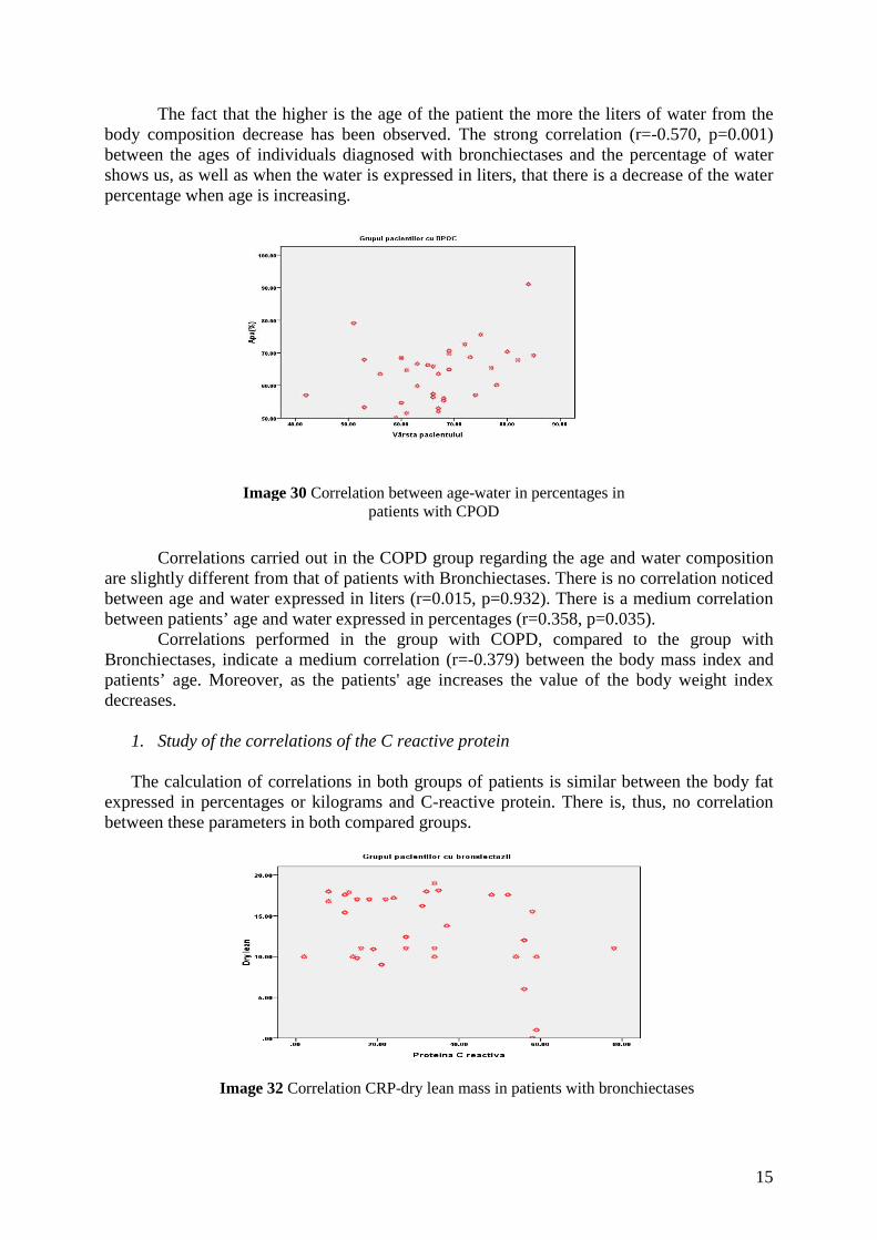
The fact that the higher is the age of the patient the more the liters of water from the body composition decrease has been observed. The strong correlation (r=-0.570, p=0.001) between the ages of individuals diagnosed with bronchiectases and the percentage of water shows us, as well as when the water is expressed in liters, that there is a decrease of the water percentage when age is increasing.
Correlations carried out in the COPD group regarding the age and water composition
are slightly different from that of patients with Bronchiectases. There is no correlation noticed between age and water expressed in liters (r=0.015, p=0.932). There is a medium correlation between patients’ age and water expressed in percentages (r=0.358, p=0.035).
Correlations performed in the group with COPD, compared to the group with Bronchiectases, indicate a medium correlation (r=-0.379) between the body mass index and patients’ age. Moreover, as the patients' age increases the value of the body weight index decreases.
1. Study of the correlations of the C reactive protein
The calculation of correlations in both groups of patients is similar between the body fat
expressed in percentages or kilograms and C-reactive protein. There is, thus, no correlation between these parameters in both compared groups.
Image 30 Correlation between age-water in percentages in patients with CPOD
Image 32 Correlation CRP-dry lean mass in patients with bronchiectases

16
In the group of patients with bronchiectases a medium correlation (r=-0.383) between
the dry lean mass and C-reactive protein could be emphasized, for a significance threshold p=0.023. The increase of the CRP value is accompanied by the decrease of the dry lean mass. Another medium correlation was observed between the dry lean mass and the lean body mass expressed in kilograms (r=0.440, p=0.008), which is normal. No significant correlation between the lean mass expressed in kilograms and C-reactive protein (r=-0.155, p=0.374) or between the lean body mass expressed in percentages and C-reactive protein (r=-0.277, p=0.108), lean body mass expressed in kilograms and CRP (r=0.81, p=0.64), the lean body mass in percentages and CRP (r=0.24, p=0.15) was observed.
The results of the COPD group of patients differ from those with bronchiectases, so we could not reveal a significant correlation between the dry lean mass and C-reactive protein (r=-0.011, p=0.949).
There is a medium correlation between the dry lean mass and the lean mass in percentages (r=-0349, p=0.40). As in the group with bronchiectases, a correlation, but in this case a strong one, was noticed between the dry the lean mass and the lean body mass
expressed in kilograms (r=0.566, p=0.001).
The analysis of the group of patients with bronchiectases suggests that there is a strong
correlation (r=-0.540) between the C-reactive protein and the water expressed in liters for a significance threshold p=0.001. Thus, we can notice that when C-reactive protein values increase, water (in liters) decreases. Similarly, a medium correlation was also found between the CRP values and water expressed in percentages (r=-0.384, p=0.023). Now, we may observe that when CRP increases the water (in percentages) decreases.
In the case of the COPD group we could not correlate the CRP value with the water expressed in liters (r=0.092, p=0.188) and neither CRP with the water expressed in percentages (r=0.092, p=0.598). The results are totally different from the group with Bronchiectases.
2. Study of the correlations of ESR
From the performed analyzes we could not reveal a significant correlation in both compared groups of the type ESR value and body fat expressed in percentages and ESR and body fat expressed in kilograms.
Image 33 Correlation CRP-water in liters and CRP- water in percentages in patients with bronchiectases

17
We have observed from the analysis of the correlations that there are no important correlations in patients with bronchoiectases with regard to their ESR values and the dry lean mass (r=0.329, p=0.054), ESR and lean body mass expressed in kilograms (r=-0.050, p=0.775) ESR and the lean body mass expressed in percentages (r=0.208, p=0.229). A similar situation is noticed in the COPD group, where a significant correlation of the parameters r=-0.289 and p=0.090, r=-0.820 and p=0.640, r=-0.100 and p=0.530 does not exist.
With regard to the correlations in patients with bronchiectases there is a significant
medium correlation (r=0.406) between the ESR value and the water from the body composition expressed in liters for a significance threshold p=0.016.
Also, a significant medium correlation (r=0.379) exists between the ESR and water expressed in percentages for p = 0.025.
Compared to the group with bronchiectases, the calculation of similar correlations from the COPD group showed no significant correlation between ESR and water percentage (r=0.291, p=0.089) and ESR and water in liters (r =0.170, p=0.329). Discussions The pathology of bronchiectases is underdiagnosed, and when the affected lung region shows the presence of a symptomatic obstructive ventilatory dysfunction or of a microbial superinfection, the patient is admitted late, requiring periodic evaluations and treatment because of the secondary complications and chronic colonization.
Some of the secondary pathological modifications that have been reported refer to the appearance of the chronic inflammatory syndrome and impairment of the body composition (58-63). The starting point of the study was the highlight of the correlations between the body composition parameters, inflammatory syndrome and respiratory function in patients diagnosed with bronchiectases compared to a selected control group of patients with COPD (both groups being diagnosed with obstructive/mixed ventilatory dysfunction).
In literature there are few data on the role of the body composition and its relationship to systemic inflammation (90,91,100). Available data mainly refer to groups of patients with COPD and study the modifications of the body composition in relation to various inflammation markers (64,71,79,85). The association between the presence of bronchiectases
Image 34 Correlation ESR- water in percentages and ESR-water in liters in patients with bronchoiectases

18
and obstructive ventilatory dysfunction related to the changes in the body composition and to the chronic inflammation is not subject of many studies in the literature.
A study conducted in 2012 attempted to determine the relationship between the parameters of the respiratory function and nutritional status, serum levels of adipocytokines and of inflammatory cytokines in patients with bronchiectases. Anthropometric indeces, a questionnaire related to diet, the leptin level, adiponectin, IL6, TNF alpha, CRP, respiratory parameters obtained through spirometry, clinical and radiological examination were analyzed.(100) Unlike the group studied in this PhD thesis, some of the patients were diagnosed with cystic fibrosis.
Thus, the results obtained by Olveira G et al show a body fat depletion in 31% of patients (no differences in the etiology of bronchiectases), a correlation between inflammatory cytokines and exacerbations, bronchorrhea, FEV1, Bhalla score. Patients with diabetes, cachexia and poor respiratory function had high levels of IL-6; a positive correlation was found between the body fat and its index, as well as higher levels of adiponectin in the patients with depletion of the body fat.(100)
In the group studied in this chapter of the PhD thesis results showed that body composition is a parameter that can be influenced and the inflammatory syndrome is associated with significant changes in body composition. With reference to the body fat expressed in percentages we noticed a normal distribution thereof and a maximum incidence peak at 7 of the patients, around 35%, with its depletion. Inflammatory syndrome in patients with bronchiectases was pronounced, with elevated values of the studied parameters. Compared to the study of Olivier G et al, patients with diabetes were not included, which could significantly change results, perhaps in the sense of affecting the body composition and mainly the body fat (by lipodystrophy associated with diabetes).
Compared results, based on the different parameters that we have followed (of systemic inflammation), are somewhat similar, identifying a systemic inflammatory syndrome in relation to changes in the body composition (increased CRP is accompanied by decrease of the dry lean mass and of the water, a correlation between the ESR value and the water from the body composition was also identified).
It should be noted that with the increase in age there was a decrease in lean body mass and lung function in these patients. Thus, taking into account the data as a whole, the inflammatory syndrome can be considered a follow-up marker in relation to the occurrence of changes in body composition.
A number of obtained personal results identify strong correlations between the ages of patients with bronchiectases and water expressed in percentages as well as in liters (r=-0.639, p=0.001). It has been observed that as patient’s age increases the liters of water from the lower body composition decrease. This analysis is important because it explains why some patients with bronchiectases have difficulties when expectorating and, secondary, it favors recurrent episodes.
It is possible that when correcting this body composition parameter to obtain the optimum clearance and to improve the clinical status and the long term evolution. Age can be considered a factor indicating the possibility of appearance of body composition imbalances. Any associated pathology, unbalanced or uncompensated, that could influence both the results as well as the way of intervention, must be taken into account.
The data obtained from the study of the control group are not identical, in this case requiring a larger group of patients in order to confirm the results.

19
Conclusions � The homogeneity of the chosen groups, expressed by the similar mean values of
weight and body mass index, reflects the importance of changes in body composition analysis in patients with bronchiectases.
� In patients diagnosed with bronchiectases the mean values of body fat expressed in percentages and kilograms are significantly higher than in the control group. � The better lung function in the group with bronchiectases being influenced by the mean values of the lean body mass expressed in percentages and kilograms, that are higher in these patients compared to the control group. � Episodes of exacerbations and poor contribution of muco-ciliary clearance in
eliminating secretions may be due to poor body composition in water in patients with bronchiectases, compared to the control group.
� Age is a factor that is associated with the presence of more important inflammatory syndrome in the bronchiectases group compared to the control group.
� The older age of the of patients with bronchiectases was associated with elevated values of the body fat expressed in percentages, of the dry lean mass and of the lean body mass.
� It has been established that there is a series of strong correlations between the age of patients with bronchiectasis and water from the body composition expressed in liters and in percentages.
� Chronic systemic inflammatory syndrome is more important in patients with bronchiectases than in those with COPD.
� Inflammation plays an important role in the determinism of body composition, by influencing the values of the water from the body composition in patients with bronchiectases.
� The dosage of ESR and CRP lead to the identification of imbalances in the body composition that could be prevented.
� Chronic systemic inflammation markers may be used for the evaluation of patients with bronchiectases.
� Chronic systemic inflammation and body composition are parameters that can be influenced in order to improve the clinical status of patients with bronchiectasis.
CHAPTER 7. QUALITY OF LIFE IN PATIENTS WITH BRONCHI ECTASIS
Introduction
Patients with bronchiectases sometimes require longer hospitalisations periods, associated to long-term physiotherapy, which influences the patient’s qualify of life, both during an infectious episode, as well as afterwards.
This study is important as it highlights the main problems of these patients concerning quality of life related to the subjective assessment of the respiratory function and of the sleep quality - parameters that influence the daily condition of the patient. Moreover, it has to be underlined that this self-assessment of the health condition was correlated to the paraclinical results obtained from the evaluation of the studied group.

20
The study protocol
The study was designed to highlight the correlations between the results obtained by filling out St. George’s Respiratory Questionnaire and the Pittsburgh Questionnaire on the quality of sleep and the parameters of the body composition, inflammatory syndrome and the respiratory function (obtained in the previous study) on patients with bronchiectases in relation to a selected control group of COPD patients.
The patients included in the study were admitted at the Pneumology Hospital of Iasi during January 2011 - April 2012. There were assessed 70 patients out of which 35 suffering from Bronchiectases and 35 from COPD. Purpose and objective of the study
- Assess quality of life on patients with bronchiectases by evaluating the respiratory
function and sleep quality - Establish a potential therapeutical relationship between studied parameters
Objectives
The main objective of the study concerns highlighting features of patients with
Bronchiectases that might be influenced and changed by treatment through: - differential analysis from the point of view of pathology and establishment of intra-
group relations of the results obtained from filling out the St. George and Pittsburg Questionnaires and highlighting potential correlations to the parameters of the chronic systemic inflammatory syndrome, lung function and the body composition for patients with bronchiectases and the control group
- differential analysis from the point of view of pathology for establishing inter-group correlations of the results obtained from filling out the St. George and Pittsburg Questionnaires and highlighting potential correlations to the parameters of the chronic systemic inflammatory syndrome, the lung function and the body structure for patients with bronchiectases and the control group
- evaluation and interpretation of the statistically significant differences between patients with bronchiectases selected for the study and the control group.
Secondary objectives:
- identify parameters that can characterise obstructive lung diseases - prevent the occurrence of pathological complications associated to patients with
Bronchiectases - assess the possibility of improving quality of life to patients with Bronchiectases
Working hypothesis
The study consisted of the selection, clinical and paraclinical assessment of 70 consecutive patients admitted in the Pneumology Hospital of Iasi, out of which 35 with bronchiectases and 35 patients included in the control group.
Inclusion criteria for the group of patients with bronchiectases:

21
- the existence of a CT scan or bronchography with contrast that would confirm the suspicion of diagnostic through thorax X-ray of bronchiectases
- the patients included need to be adults and sign the informed consent after they have been explained the purpose and procedures included in the protocol
- the patient needs to be outside an infectious episode (follow-up patient, patient admitted in the hospital but who overcame the acute infectious episode) Exclusion criteria for the group of patients with bronchiectases:
- lack of certainty confirmation for the bronchiectasis diagnostic - pregnant or nursing women - the presence of an imbalanced/decompensated disorder - refusal to sign the informed consent/refusal to participate
Inclusion criteria for the control group:
- diagnostic of COPD supported by spirometry results - adult patient that signs the informed consent - the patient need not be under an acute infectious exacerbation episode
Exclusion criteria for the control group:
- refusal to sign the informed consent - presence of decompensated disorder
Methodology In the study there were included 70 patients with obstructive lung diseases, out of which
35 patients with Bronchiectases and a control group of 35 patients with COPD. The clinical and paraclinical assessment of both groups was based on the following
parameters: - filling out the Pittsburg Questionnaire related to sleep quality (Romanian version) - filling out the St. George’s Respiratory Questionnaire (Romanian version) - lung function - evaluated according to the FEV1 value - the systemic inflammatory syndrome - defined through the study of VSH value and C-
reactive protein - the body composition measured through bioimpedance - the parameters studied are
represented by the fat mass expressed in kilograms, the lean mass expressed in kilograms, the fat dry mass, the body mass index
Filling out the St. George and Pittsburg Questionnaires and using the results were possible
only after having obtained the consent of the delegated persons. The parameters of the lung function, chronic systemic inflammatory syndrome and the
body composition were used from the previous study, as the groups used in this paper are the same. Therefore, the above-mentioned parameters were established as follows:
- the lung function was assessed by carrying out spirometry within the functional
examination laboratory of the 1st Clinic within the Pneumology Hospital of Iasi. - the instrument used to measure body composition was the Bodystat 1500 device
available in the hospital’s inventory, set at a frequency of 50 kHZ.

22
- the dosage of inflammatory markers, VSH and CRP, was made within the accredited analysis lab, the haematology department of the Pneumology Hospital
Results and interpretation
The interpretation of St. George’s Respiratory Questionnaire for the group with bronchiectases and control group (with COPD)
The first item of this questionnaire relates to the perception of patients with respiratory
disorders on the current health condition. It was noticed in the 1st chart that the patients with Bronchiectases perceive their health condition as moderate, namely 34.29%, and 48.57% perceive it as poor, whereas 54.29% of the patients with COPD have a moderate health perception and 37.14% of them perceive it as poor. The group with bronchiectases declares a very poor health level, i.e. 14.29%, as compared to the lower number of COPD (5.71% of the control group).
According to the 2nd chart, in the case of the group with bronchiectases there are two situations only: the former, when they cough only at the moment of respiratory infections (68.57%) and the latter only several days a month (31.43%).
The patients in the control group, with COPD, declare the presence of cough only several days a month, 51.43%, 34.29% cough several days a week, and 11.43% most of the days of the week (chart 2).
As for the next item, which aims the presence of smear, there can be noticed the presence of slightly equal percentages on patients with sputum only several days a month (51.43% vs. 60%). The patients with COPD expectorate more frequently than those with
Fig. 1. Current health condition according to St. George’s Questionnaire for the group with bronchiectases and the CPOD group

23
Fig. 6 Severe or unpleasant respiratory problems, according to St. George’s Questionnaire for the group with bronchiectases and the CPOD group
bronchiectases over a week’s time (37.14%) and those who expectorate frequently have equal percentages on both groups (8.57%, chart 3).
The patients with Bronchiectases have encountered breathing difficulties most of the days of the week, namely 8.57%, as compared to those with COPD, who have a percentage of 22.86%. The percentages of those who encountered breathing difficulties more days a week differ on the 2 groups: 40% for the group with bronchiectases and 51.43% for the group with COPD (Fig. 4).
The application of the test t to check for significant differences between the answers to
the item on cough of the two groups of patients revealed the following: - Levene Test F=10.8 p=0.002 - t(68)=-8.2 p≤0.001 There is a statistically significant difference between the answers to the item on cough of the two groups of patients of the study. The patients with COPD particularly revealed the presence of cough.
The Figure 5 shows that 50% of the patients with bronchiectases presented whistling
only when they had respiratory infections, as compared to the COPD patients (8.57% of the control group).
The percentages of those who showed this symptom for more days a week were remarkably close (31.43% vs. 34.29%) while the percentages of those who had whistling during most of the days were 48.57% for the group of patients with bronchiectases and 37.14% for the control group.
Fig 5 Whistling according to St. George’s Questionnaire for the group with bronchiectases and the CPOD group

24
Fig. 13 Cough and breathing difficulties over the last days, according St. Georges’s Questionnaired for the group with bronchiectases and the CPOD group
The patients of the control group with COPD manifested respiratory problems more than 3 times over the last 3 months, which is less frequent (5.71%) than to those with groups with bronchiectases (20%). In both groups there can be noticed that patients presented respiratory disorders 2 times over the last 3 months, in similar percentages (34.29% vs. 31.43%) (fig. 6).
The patients with bronchiectases declared the presence of whistling in the morning more often. In the COPD group there were significantly more patients who had days influenced by breathing disorders.
We noticed that 51.43% of the subjects of the control group declared to have breathing disorders with a duration of more than 3 days as compared to 31.43% corresponding to the group with bronchiectases (fig.7).
The percentages of patients who declare that breathing causes more problems are equal, i.e. 17.14%. Breathing causes fewer problems in 48.57% of the patients with COPD of the control group and in 11.43% of those with bronchiectases. The working ability is not affected in 60% of those in the COPD group and only 34.29% of those with bronchiectases. 54.29% of the patients with bronchiectases are affected in terms of limiting the work capacity in both groups, as compared to the COPD percentage, which is a lot lower (25.71%) (Chart 11).
There are no significant differences on the breathing condition and the working ability of both groups.
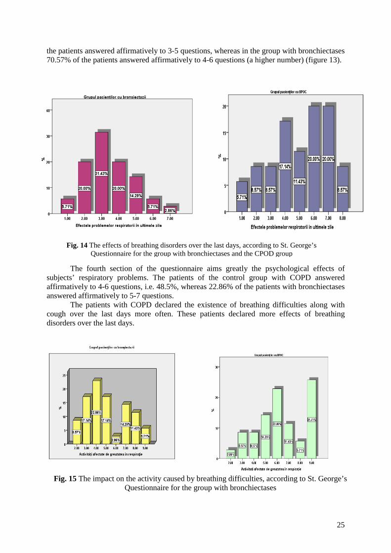
This section of the questionnaire aims the activities that make the patient feel breathing difficulties, 7 questions being asked for this purpose. It was noted that in the group of patients with bronchiectases 68.57% of the patients answered affirmatively to 3-5 questions, whereas in the control group, a lower percentage of 57.41% of the patients included in the study answered affirmatively (fig.12).
The third section of the questionnaire aims at additional questions on cough and
breathing difficulties. It was noted that in the control group of the COPD patients, 65.29% of
25
Fig. 14 The effects of breathing disorders over the last days, according to St. George’s Questionnaire for the group with bronchiectases and the CPOD group
Fig. 15 The impact on the activity caused by breathing difficulties, according to St. George’s Questionnaire for the group with bronchiectases
the patients answered affirmatively to 3-5 questions, whereas in the group with bronchiectases 70.57% of the patients answered affirmatively to 4-6 questions (a higher number) (figure 13).
The fourth section of the questionnaire aims greatly the psychological effects of
subjects’ respiratory problems. The patients of the control group with COPD answered affirmatively to 4-6 questions, i.e. 48.5%, whereas 22.86% of the patients with bronchiectases answered affirmatively to 5-7 questions.
The patients with COPD declared the existence of breathing difficulties along with cough over the last days more often. These patients declared more effects of breathing disorders over the last days.

26
Fig. 16 The impact on everyday life of respiratory problems, according to St. George’s Questionnaire for the group with bronchiectases and the CPOD group
Fig. 17 Sleep latency according to Pittsburg Questionnaire for the group with bronchiectases and the CPOD group
This section of the St. George’s Questionnaire includes questions concerning the activities that might be hindered by basic breathing disorder. It was noted that in the group with bronchiectases, 65.77% of the patients declared between 6 and 9 activities impaired by breathing.
In the case of the control group with COPD, it can be observed that 31.47% of the patients included in the study declared between 6 and 9 activities impaired by breathing (figure 15).
The last section represented under chart 16 concerns the everyday effects of respiratory problems. In addition, it is noted that patients with bronchiectases are more affected, as 62.89% answered positively to 4-5 questions, as compared to those with COPD, who answered positively to 3-5, i.e. 34.29%, of the studied group.
There is a significant statistical difference between the two groups of patients of the
study. The patients with Bronchiectases declared the presence of whistling in the morning more often. In the group with COPD there were more patients who had days impacted by breathing disorders.
The interpretation of the Pittsburg Questionnaire concerning sleep quality for the group
with Bronchiectases and the control group (with COPD)

27
Fig. 18 Sleep efficiency according to Pittsburg Questionnaire for the group with bronchiectases and the CPOD group
Fig. 19 Sleep duration according to the Pittsburg Questionnaire for the group with bronchiectases and the CPOD group
This sub-scale quantifies the time necessary for the patient to fall asleep. Therefore,
Figure 17 reveals that 42.86% of the group with patients with bronchiectases need more than 60 minutes to fall asleep, whereas the patients of the BOPC control group, i.e. 11.43%, need that much time to fall asleep. Thus, there can be noticed a much higher latency in patients with Bronchiectases as compared to the control group considered for the study.
As far as the sleep efficiency is concerned, there can be noticed a value higher than
85% in both groups (71.43%). The patients with bronchiectases need a value of this sub-scale under 65% to the value of 25.17%, whereas those with COPD need 14.23% (fig. 18).
Concerning the sleep duration, the patients with bronchiectases declare that they sleep 6-7 hours, i.e. 32.3%, whereas subjects of the control group with COPD provide for 54.29%. It was noted that 41.18% of the patients with bronchiectases have a poor sleep quality, declaring 5-6 hours of sleep per night (fig. 19).
28
Fig. 22 Subjective sleep quality according to the Pittsburg Questionnaire for the group with bronchiectases and the CPOD group
Fig. 23 Somnolence according to Pittsburg Questionnaire for the group with bronchiectases and the BOPOC group
There is a significant statistic difference between the two groups of patients within the study. The patients with bronchiectases need more time to fall asleep.
The use of sleep medication is denied in significant percentages by both groups, but it was noted that 37.47% of the subjects with bronchiectases declared to have used it. In the COPD group, this is stated by 25.71%, which represents a significant lower number of patients (chart 21).
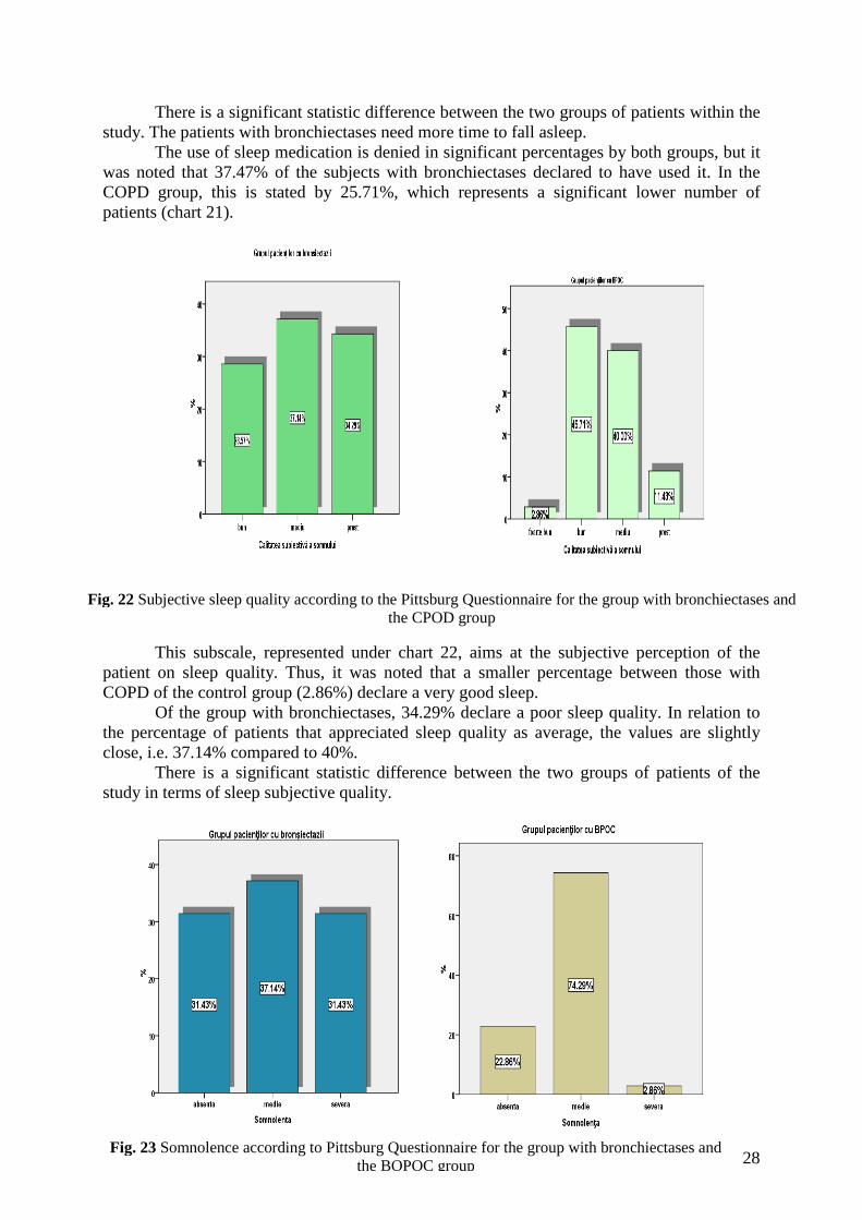
This subscale, represented under chart 22, aims at the subjective perception of the
patient on sleep quality. Thus, it was noted that a smaller percentage between those with COPD of the control group (2.86%) declare a very good sleep.
Of the group with bronchiectases, 34.29% declare a poor sleep quality. In relation to the percentage of patients that appreciated sleep quality as average, the values are slightly close, i.e. 37.14% compared to 40%.
There is a significant statistic difference between the two groups of patients of the study in terms of sleep subjective quality.

29
Fig. 24 PSQI score according to Pittsburg Questionnaire for the group with bronchiectases and the CPOD group
Somnolence was present as severe in 31.43% of the group with bronchiectases, and in a percentage significantly lower for the other group (2.86%). An average form of somnolence was stated by 74.29% of the COPD patients and by 37.14% of the patients with Bronchiectases (chart 23).
The patients with bronchiectases constantly registered high scores on this
questionnaire correlated to the values obtained for each subscale calculated previously. 71.44% of the patients have values between 13 and 20. The more the values are closer to 21, the poorer the quality of the sleep.
Patients with bronchiectases constantly register higher scores on this questionnaire, hence, it can be said the sleep quality is deficient.
Table 19 refers to correlations between the lung function quantified through the FEV1 value, the systemic inflammatory syndrome represented by VSH and CRP and the first nine items of the St. George’s Questionnaire for the group of patients with bronchiectases. Therefore, there can be noticed strong correlations between: - FEV1- the current health subjective condition (r=0.722, p≤0.001), which reflects the fact that the respiratory function quantified paraclinically by spirometry is correlated to the subjective description of the patient (the lower the FEV1 the poorer the condition of health and viceversa). - FEV1-cough (r=0.656, p≤0.001), it can be noted the decrease of the respiratory function correlated to the decrease of the cough episodes - FEV1- smear (r=0.785, p≤0.001), once with the lower smear, a lower value of FEV1 parameter can be noticed. - FEV1- whistling (r= 0.746, p≤0.001) - FEV1- respiratory problems (r=0.694, p≤0.001) - FEV1- days without respiratory problems (r=-0.348, p=0.041), this is a moderate reverse correlation, which reflects the fact that patients appreciated that the number of days of respiratory disorders are decreasing when FEV1 increases.
Table 19 reveals that out of the first 9 parameters of the St. Georg Questionnaire applied to patients with bronchiectases, only 2 parameters could not correlate to the FEV1

30
value: breathing difficulties (r=0.198, p=0.255) and duration of the respiratory problem r=0.694, p≤0.1).
As far as the parameters of the systemic inflammatory system quantified through the value of C-reactive protein and VSH are concerned, correlated to the first eight items of the St. George’s Questionnaire, it was noted a single moderate reverse correlation between CRP and cough (r=-0.370, p=0.028). Therefore, it can be said that a more pronounced systemic inflammatory system is associated to a decrease of cough episodes. However, by looking at the bigger picture and analysing the correlations with FEV1-cough, FEV1-smar, it can be said that an infectious episode is associated to the change of the lung function and decrease of the clearance ability of lung secretions on patients with bronchiectases.
It was noted the lack of any correlation between the parameters of the systemic inflammatory syndrome, FEV1 and the first eight items of the St. George’s Questionnaire on patients of the control group with COPD.
The correlations between the parameters of the systemic inflammatory syndrome, the FEV1 value and the items of the St. George Questoinnaire revealed higher and moderate correlations, out of which the following can be reminded (Table 20):
- FEV1- the described respiratory condition, strong correlation (r= 0.794, p≤0.001) - FEV1- the affectation of the ability to over the last days, strong reverse correlation
(r=-0.791, p≤0.001), which reflects a decrease of the respiratory function correlated to more breathing difficulties
- FEV1- cough and breathing difficulties over the last days, strong reverse correlation (r=-0.671, p≤0.001). The FEV1 decrease, associated to the decrease of cough episodes and breathing difficulties.
- FEV1- the effects of the respiratory disorders over the last days, strong reverse correlation (r=-0.705, p≤0.001). The FEV1 decrease associated to more current problems determined by breathing
- FEV1- activities affected by breathing difficulties, strong reverse correlation (r=-0.785, p≤0.001). The more affected the respiratory function, the more affected the activities carried out by breathing difficulties.
- FEV1- respiratory disorders and impact on everyday life, strong reverse correlation (r=-0.729, p≤0.001).
As far as the parameters of the systemic inflammatory syndrome are concerned, there
could be noted the following correlations to the items of St. George’s Questionnaire, applied to the patients from the group with bronchiectases:
- a single reverse significant moderate correlation between the VSH value and cough and breathing difficulties over the last days (r=-0.362, p=0.033).
- C-reactive protein - the described breathing condition, reverse moderate correlation (r=-0.363, p=0.032). Therefore, the poorer the breathing condition, the higher the CRP value
- C-reactive protein - the impact on the working ability, reverse moderate correlation (r=-0.340, p=0.045). It can be noted that when the CRP value increases, the working ability decreases.
- C-reactive protein - breathing difficulties over the last days, strong correlation (r=-0.517, p=0.001).
- C-reactive protein - cough and breathing difficulties over the last days, moderate correlation (r=-0.468, p=0.005).
- C-reactive protein - the impact on the breathing disorders over the last days, strong correlation (r=0.527, p=0.001).

31
- C-reactive protein - activities impacted by respiratory problems moderate correlation (r=-0.484, p=0.003).
- C-reactive protein - respiratory problems and impact on everyday life, moderate correlations (r=-0.471, p=0.004). As compared to the group with patients with bronchiecstases in the control group with COPD significantly less existing correlations were observed (Table 21).
Therefore, two correlations can be noted: a moderate reverse one between VSH and the breathing condition (r=-0.364, p= 0.032) and a moderate one between VSH and the effects of the respiratory problems over the last days (r=0.350, p=0.039).
There were not noticed significant correlations concerning the systemic inflammatory syndrome on patients with bronchiectases, namely: a reverse moderate correlation between VSH and sleep efficiency (r=-0.352, p=0.038) and a moderate correlation between CRP and somnolence (r=0.387, p=0.022).
Concerning the quantified lung function through the FEV1 value it could be observed that there are four correlations between these parameters and the items of the Pittsburgh Questionnaire on the group with bronchiestases:
- FEV1- sleep latency, strong reverse correlation (r=-0.710, p≤0.001). The lower the FEV1 value, the higher the sleeping period. - FEV1- the subjective quality of sleep, strong reverse correlation (r=-0.788, p≤0.001). - FEV1- sleep medication, strong reverse correlation (r=-0.552, p=0.001). The higher the FEV1 the lower the use of medication that causes sleep - FEV1- somnolence, strong reverse correlation (r=-0.834, p≤0.001). It can be noted that the decrease of the lung function is associated to the increase of somnolence.
Concerning the control group, no significant correlation could be highlighted between the systemic inflammatory syndrome, lung function and parameters of the Pittsburgh Questionnaire.
Table 23 reveals that in the case of patients with bronchiectases there are the following correlations between the parameters of the body composition and the first nine items of the St. George’s Questionnaire, as follows: - fat mass (kg) - whistling in the morning, strong reverse correlation (r=-0.466, p=0.005). Therefore, it can be noted that when the fat mass lowers there is an increase of whistling episodes during morning time. - dry lean mass - cough, moderate correlation (r=0.381, p=0.024) - water (l)- respiratory problems, moderate correlation (r=0.355, p=0.049)
Table 24 reveals that significant correlations can be identified only between water (l) and four of the parameters of the St. George’s Questionnaire, analysed as follows: - water(l) – breathing difficulties over the last days, strong reverse correlation (r=-0.511, p=0.002). Therefore, there can be noted that once with the decrease of the water the breathing difficulties increase (secondary to dehydration and clearance). - water(l)– cough and breathing difficulties over the last days, reverse correlation (r=-0.586, p≤0.001) - water(l) – the effects of the breathing difficulties over the last days, strong reverse correlation (r=-0.510, p=0.002). The lower the water the higher the respiratory problems. - water(l) – respiratory problems and impact on everyday life, moderate reverse correlation (r=-0.472, p=0.004). It can be noted that when the water of the body mass decreases, the respiratory problems increase and the quality of life is affected (secondary to dehydration).
In the case of the control group with COPD it could not be noted any significant correlation between the parameters of the body composition and the first eight items of the St. George’s Questionnaire.

32
Table 25 reveals two significant moderate correlations between the parameters of the body composition and those of the Pittsburg Questionnaire on sleep quality: dry mass - sleep efficiency (r=0.473, p=0.004) and BMI- sleep efficiency (r=0.492, p=0.003).
There is no significant correlation for the control group of the patients with COPD for the parameters of the body mass and the parameters of the Pittsburg Questionnaire.
- sleep latency (Pittsburg Questionnaire) and the first nine parameters of the St. George’s Questionnaire, both on the group with bronchiectases and on the control group.
- sleep duration (Pittsburg Questionnaire) and the first nine parameters of the St. George’s Questionnaire, both on the bronchiectases group and on the control group.
- sleep disorders (Pittsburg Questionnaire) and the first nine parameters of the St. George’s Questionnaire, both on the group with bronchiectases and on the control group.
- subjective sleep quality (Pittsburgh Questionnaire) and the first nine parameters of the St. George’s Questionnaire, both on the group with bronchiectases and on the control group.
- the PSQI score and the first nine parameters of the St. George’s Questionnaire, both on the group with bronchiectases and on the control group.
It can be noted in Table 27 that there are two moderate reverse correlations between: - the current health condition and the medication used to induce sleep (r= -0.430,
p=0.010). Therefore, the poorer the health condition the higher the use of medicinal products within the group with bronchiestases.
- smear - the medication used to induce sleep (r= -0.354, p=0.050). It can be noted that once with the increase of the expectoration, the use of medicinal products decreases, probably by improving clearance.
Table 28 reveals that in the group with bronchiectases significant correlations were identified in terms of somnolence (Pittsburg Questionnaire) and some of the St. George parameters, as follows:
- somnolence - current health condition, moderate reverse correlation (r=-0.493, p=0.003). The higher the somnolence, the lower the self-appreciation of the health condition.
- somnolence - cough, strong reverse correlation (r=-0.621, p≤0.001). - somnolence - expectoration, strong reverse correlation (r=-0.521, p=0.001) - somnolence - whistling, strong reverse correlation (r=-0.650, p≤0.001) - somnolence - respiratory problems, strong correlations (r=-0.609, p≤0.001)
In the case of control group with COPD it could not be highlighted any significant correlation of these parameters. Based on Table 29, it can be observed that in the case of patients with bronchiectases there is a series of significant correlations between sleep latency and parameters of St. George’s Questionnaire, as follows: - sleep latency - breathing condition, reverse moderate correlation (r= -0.468, p=0.005). - sleep latency - impact on the working capacity, strong reverse correlation (r=-0.535, p=0.001) - sleep latency - cough and breathing difficulties over the last days, strong correlation (r=0.537, p=0.001) - sleep latency - the impact of breathing disorders over the last days, strong correlations (r=0.521, p=0.001) - sleep latency - activities impacted by breathing difficulties, strong correlation (r=0.595, p≤0.001) - sleep latency - respiratory problems and impact on everyday life, moderate correlation (r=0.493, p=0.003)
In the case of the control group, a single moderate correlation could be noted between sleep latency and the medication used that induces sleep (r=0.358, p=0.035).

33
According to the analyses performed, no significant correlation could be noted in the following cases:
- sleep efficiency (Pittsburg Questionnaire) and St. George’s Questionnaire (the last 8 items) on the group of patients with bronchiestases, but also on the control group.
- sleep duration (Pittsburg Questionnaire) and St. George’s Questionnaire (the last 8 items) on the group of patients with bronchiestases.
- sleep disorders (Pittsburg Questionnaire) and St. George’s Questionnaire (the last 8 items) on the group of patients with bronchiestases, but also on the following group. Exception: reverse moderate correlation between sleep disorder and the use of medication that induces sleep (r=-0.361, p=0.033). Concerning the group of patients with bronchiestases, Table 30 reveals a series of significant correlations between the subjective quality of sleep (Pittsburg Questionnaire) and some of the last eight items of the St. George’s Questionnaire, as follows:
- subjective sleep quality - breathing status, strong reverse correlation (r=-0.611, p≤0.001)
- subjective sleep quality - impact on everyday life, strong reverse correlation (r=-0.599, p≤0.001)
- subjective sleep quality - cough and breathing difficulties over the last days, strong correlation (r=0.556, p=0.001)
- subjective sleep quality - effects of respiratory problems over the last days, strong correlation (r=0.621, p≤0.001)
- subjective sleep quality - activities impacted by breathing difficulties, strong correlation (r=0.648, p≤0.001)
- subjective sleep quality - respiratory problems and impact on everyday life, strong reverse correlation (r=0.552, p=0.001)
In the case of COPD control group, no significant correlation could be identified. Concerning the group of patients with bronchiestases, Table 31 reveals a series of
significant correlations between sleep medication (Pittsburg Questionnaire) and some of the last eight items of St. George’s Questionnaire, as follows:
- sleep medication - breathing status, moderate reverse correlation (r=-0.346, p=0.042) - sleep medication - impact on the work ability, moderate reverse correlation (r=-
0.402, p=0.017) - sleep medication - breathing difficulties over the last days, moderate correlation
(r=0.362, p=0.033) - sleep medication - effects of respiratory problems over the last days, moderate
correlation (r=0.421, p=0.012) - sleep medication - activities impacted by breathing difficulties, moderate correlation
(r=0.424, p=0.011) In the case of control group with COPD, a single significant moderate reverse
correlation stood out between sleep medication and breathing condition (r=-0.349, p=0.040). Concerning the group of patients with bronchiestases, Table 32 reveals a series of
significant correlations between somnolence (Pittsburgh Questionnaire) and some of the last eight items of the St. George’s Questionnaire, as follows:
- somnolence - breathing status, strong reverse correlation (r=-0.653, p≤0.001) - somnolence - impact on the work ability over the last days, strong reverse correlation
(r=-0.680, p≤0.001) - somnolence - cough and breathing difficulties over the last days, strong correlation
(r=0.659, p≤0.001 - somnolence - activities impacted by respiratory problems, strong correlation
(r=0.736, p≤0.001)

34
- somnolence - respiratory problems and impact on everyday life, strong correlation (r=0.650, p≤0.001).
In the case of patients of the COPD control group, no significant correlation could be noted.
No significant correlation could be noted between the PSQI score and the last eight parameters of the St. George’s Questionnaire, neither on the group with bronchiectases nor on the control group. Discussions The evolution of bronchiectases leads to a chronic systemic inflammatory syndrome and to obstructive ventilation dysfunction that becomes symptomatic in time. The changes of the respiratory function leads to an impact of the patients’ quality of life, which translates into an impact on the effort ability, occurrence of sleep disorders and various forms of depression (86,87,89). The purpose of the study is to highlight correlations between the parameters of the Staing George, Pittsburg Questionnaires and the parameters of the body mass, inflammatory syndrome and the breathing function of patients diagnosed with bronchiestases, as compared to a selected control group of the COPD patients (all patients are diagnosed with obstructive/mixed ventilatory dysfunction). Identically to the previous study, the COPD control group was considered, as both studied disorders are part of the group of chronic obstructive disorders.
All the patients included in the study did not have any acute pathology that might have influenced chronic inflammation. However, the data concerning other chronic disorders (heart, digestive, etc.) were not included, which might have been inflammatory.
There is a series of parameters that significantly influences patients’ quality of life diagnosed with bronchiectases as compared to the COPD control group. The data concerning the patients is limited in the literature. (63, 84,91)
The study highlighted the fact that on most of the items analysed, from a statistical point of view, it was noted a significant difference in the seriousness of the symptomatology in the case of bronchiestases, which was highlighted in the scores obtained for filling out the Pittsburg and Saint George’s Questionnaires, and also from paraclinical point of view.
Conclusions
� On the patients with bronchiestases, the objective sleep quality, established by using the Pittsburg Questionnaire is impacted, thus proving a higher degree of seriousness as compared to those of COPD. � Quality of life on patients with objective bronchiectases through SGRQ is impacted, thus proving a higher degree of seriousness as compared to those of COPD. � Cough, problems in spitting out secretions and the secondary dyspnoea are the main factors that establish quality of life among patients with bronchiestases. � The chronic systemic inflammatory syndrome is an important factor that influences quality of life on patients with bronchiestases. � A more pronounced systemic inflammatory syndrome is associated to a decrease of cough episodes, although with a lower clearance. � Once the parameters of body composition are affected, the decrease of sleep quality occurs, as well as that of life on patients with bronchiestases.

35
� The affected water of the body composition leads to a more pronounced symptomatology. � The objective quality of life, due to the symptomatology importance, is directly related to sleep quality and use of medicinal products meant to induce it. Considering the obstructive ventilatory constituent present on both groups and high degree of impact on life and quality of life among patients with bronchiestases, the inflammatory markers could be considered for tracking and making prognoses for the patients concerned.
CHAPTER 8. LEPTIN IN PATIENTS WITH BRONCHIECTASIS
Introduction
Due to the results obtained in the previous chapters it was observed the change of the parameters of the body mass on patients with bronchiestases, which correlated significantly to the breathing function, systemic inflammatory syndrome, self-assessment of the health condition. Letpin, among other factors, aims at maintain the optimal weigh and is connected to systemic inflammation. The purpose of this study is to discover if there is an influence of leptin on patients with bronchiectases on the systemic inflammatory syndrome and on the change of body weight that is specific to this diseases, the way in which leptin influences the evolution of the basis disorder and whether there is the possibility of improving the overall condition of the patient. The study protocol
The study was set up to highlight correlations between leptin’s values, inflammatory syndrome and breathing function, the age of patients with bronchiectases in relation to a selected control group of COPD patients.
The patients included in the study were admitted in the Pneumology Hospital of Iasi between January 2011 - April 2012. 50 patients were assessed, out of which 25 suffering from bronchiectases and 25 from COPD. Purpose and objectives of the study
- Assess the paraclinical parameters on patients with bronchiectases confirmed by CT
scan Objectives
The main objective of the study relates to highlighting certain features of patients with
bronchiectases that might be influenced and changed through treatment by: - differential analysis from point of view of the pathology and establish intra-group
relations of leptin’s serum values, systemic inflammatory syndrome and breathing function of the patients with bronchiectases and of the control group.
- differential analysis from point of view of the pathology, establish inter-group correlations of leptin’s serum values, systemic inflammatory syndrome and breathing function of patients included in the group with bronchiectases and the control group.

36
- assess and interpret statistically significant differences between patients with bronchiestases, considered for the study, and the control group.
Secondary objectives:
- identify parameters that can characterise the obstructive lung diseases - prevent the installation of pathology complications associated to patients with
bronchiestases - assess the possibility of improving the overall status of the patient.
The working hypothesis
The study consisted of selecting, clinically and paraclinically assessing 50 consecutive patients admitted in the Pneumology Hospital of Iasi, out of which 25 with bronchiectases and 25 as control group.
Inclusion criteria for the group of patients with bronchiestases:
- existence of CT scan or bronchography with contrast substance that would confirm the suspicion of diagnostic by thorax X-ray of bronchiestases
- the patients need to be adults, male and sign the informed consent, after having been explained the purpose and procedures provided under the protocol
- the patient needs to be outside the infectious episode (patient for follow-up, patient admitted in hospital, but overcame the acute infectious episode). Exclusion criteria for the group of patients with bronchiestases:
- lack of certainty confirmation of the diagnostic of bronchiectasis - female - presence of imbalanced/decompensated disorder - refusal to sign the informed consent/refusal to participate
Inclusion criteria for the control group:
- COPD diagnostic, supported by results of spirometry - adult male patient who signs the informed consent - the patient needs not be under an acute infectious exacerbation episode
Exclusion criteria for the control group:
- refusal to sign the informed consent - presence of decompensated disorder - female
Methodology
In the study there were included 50 patients with obstructive pulmonary disorders, out of
which 25 of patients with bronchiectases and a control group of 25 patients with COPD. The clinical and paraclinical assessment of both groups was based on the following
parameters: - value of serum leptin measured in ng/ml - lung function - assessed depending on the FEV1 value

37
- systemic inflammatory syndrome - defined through studying the VSH value and the C-reactive protein
- patients’ age - weight (weight and BMI) of the patients
The lung function was assessed through conducting spirometry within the laboratory of
function examinations of the 1st Clinic within the Pneumology Hospital of Iasi.
Results
Characterization of the group with bronchiectases and of the COPD control group
The average age was 63.72 years old (DS±7.3) for the group of patients with COPD
and of 60.56 years (DS±10.9) for the group of patients with bronchiestases. The average value for COPD normoponderal patients was 63.82 years old (DS±7.2)
and of 63.50 years old (DS±8.1) for obese people. In the group of patients with bronchiestases, the average age was 57.45 (DS±7.1) for normoponderal patients and 63 (DS±12.8) for obese.
The average BMI value was 24.4 kg/m2 (DS±2.6) years for the group of COPD patients and of 25.9 kg/m2(DS±4.37) for the group of patients with bronchiestases.
Fig. 1 Averages of the age parameter for the two groups
Fig. 2 BMI values for both groups

38
Fig. 4 Ponderal status for both groups
The BMI average for COPD normoponderal patients was 23.09 kg/m2 (DS±1.5) and 27.4 kg/m2 (DS±2.05) for obese patients. In the group of patients with bronchiestases, the BMI value was 22.04 kg/m2 (DS±1.6) for normoponderal patients and 29 kg/m2 (DS±3.2) for obese patients.
In order to make statistical processing easier, depending on the BMI value, the patients of the two groups were subdivided into two normoponderal sub-groups with BMI ≤ 25 kg/m2 and obese patients with BMI>25kg/m2.
For the group of COPD patients, there are 68% (17 patients) with normal weight and 32% (8 patients) with different degrees of obesity. For the group of patients with bronchiestases, there are 44% (11 patients) with normal weight and 56% (14 patients) with different degrees of obesity (fig. 4).
The average of the leptin’s values was 7.5ng/ml (DS±6.8) for COPD patients, and of 12.73 ng/ml (DS±11.76) for the group of patients with bronchiestases.
Fig. 3 Ponderal status for both groups

39
The average VSH value was 26.32 mm/h(DS±15.6) for COPD patient group and of
32.88mm/h (DS±29.9) for the group of patients with bronchiectases (Table 6). The average VSH value for COPD normoponderal patients was 26.47mm/h
(DS±16.03) and of 26 (DS±15.9) for obese patients. For patients with bronchiestatsis, the leptin average was 32.73 mm/h (DS±33.6) for normoponderal patients and of 33 mm/h (DS±28) for obese patients (fig.6).
The average CRP value was 5.36 mg/dl (DS±5.7) for COPD patients and of 11.08 mg/dl (DS±12.9) for the group of patients with bronchiectasis (Table 7).
The average CRP value for COPD normoponderal patients was 5.29 mg/dl (DS±16.03) and of 5.5 mg/dl (DS±15.9) for obese patients. For patients with bronchiectasis, the CRP average was 10.36 mg/dl (DS±16.14) for normoponderal patients and of 11.64mg/dl (DS±28)for obese patients (fig.7).
The average FEV1 value was 50.39 (DS±19.3) for the group of COPD patients and of
62.32(DS±16.64) for the group of patients with bronchiactasis (Table 8). The comparative analysis of the averages of the main analysed parameters for the two
groups highlighted the following, according to Table 9: - CRP - the average of the values for patients with bronchiectases significantly higher
than that of COPD patients (p=0.27, F=5.2) - LEPTIN- the average of the values for patients with bronchiectases significantly
higher than that of COPD patients (p= 0.008, F=7.5). - FEV1- the average of the values for patients with bronchiectases significantly higher
than that of COPD patients (p= 0.024, F=0.12)
Fig. 5 Leptin’s values on both groups
Fig. 8 FEV1 values for both groups

40
Fig. 10 Correlation matrix on normoponderal and overweight and obese patients with bronchiectases
The comparative analysis of the averages of the main parameters analysed within the two groups depending on the weight highlighted:
LEPTIN - the average of the values of obese COPD patients is significantly higher than that of COPD normoponderal patients (U=32, p=0.037)
The correlation analysis on the group of COPD patients depending on the weight highlighted the following (Table 11):
- the leptin values in the case of normoponderal patients were not significantly correlated to any of the analysed parameters
- the leptin values in the case of overweight and obese patients correlated significantly to VSH (rleptin-VSH=0.819, p=0.013) and PCR (rleptin-PCR=0.72, p=0.04)
The leptin values for COPD normoponderal patients were not significantly correlated to any of the parameters studied. The leptin values in the case of overweight and obese COPD patients correlated significantly with VSH and CRP (fig 8.II.1) COPD significantly correlated to VSH and CRP (Fig.9). The correlation analysis for the group of patients with bronchiectasis depending on the weight highlighted the following (Table 12): - the leptin values in the case of normoponderal patients correlated significantly to BMI (rleptin-
BMI=0.69, p=0.018) - the leptin values in the case of overweight and obese patients correlated significantly to PCR (rleptin-PCR=0.48, p=0.043).
The leptin’s values in the case of normoponderal patients with bronchiectases
correlated significantly to BMI. The leptin’s values in the case of overweight and obese patients with bronchiectases
correlated significantly to PCR (rleptin-PCR=0.48, p=0.043). The increase values of leptin was associated to high values of CRP and to the weight
of overweight and obese COPD patients (table 14) The variance analysis for each regression model shows that the prediction of the
suggested model is efficient (F=4.2, p=0.02) by knowing all the independent variables, compared to the situation where the prediction would take place randomly.

41
Discussions The purpose of the leptin’s changed values on patients with bronchiectases is a
deficient topic in literature. There is currently data related to systemic inflammation, leptin and evolution of COPD patients (101-105). Changes at the level of the parameters of body’s composition have been reported as related to the systemic inflammatory syndrome and the leptin’s values in the obstructive chronic lung disorders - stable COPD, in the absence of infectious exacerbations (100,106,109,110,112).
The purpose of the study was to observe relations between leptin and systemic inflammatory syndrome associated to bronchiestases, body weight of the patients and lung function. Starting from the results obtained in the previous studies, due to which there could be noticed the impact on body composition, systemic inflammatory syndrome and quality of life on patients with bronchiectases in relation to a control group with another pathology classified in the class of chronic lung diseases, this chapter attempts to establish an objective relation between leptin and the parameters studied previously.
It needs to be pointed out that for the study only male patients were considered, because of the leptin’s variability that exists on female patients in relation to the variation of other hormones, which might have changed the results obtained, especially on a group with a limited number of participants. The average age of the control group patients was slightly higher as compared to the COPD group. In order to make the processing of statistical data depending on BMI easier, the patients were divided into groups of normoponderal patients and overweight, obese patients, finally obtaining results for each individual subgroup.
In the study it was noted that the average of leptin’s values was 7.5ng/ml for COPD patients and 12.73 ng/ml for patients with Bronchiectases, which shows a difference almost double between the two subgroups. The highest values were recorded for the obstructive ventilatory dysfunction associated to Bronchiectases, and the value of the spirometric FEV1 parameters was the best on the COPD group.
The leptin’s values are in relation to the body mass index. Therefore, the relevant data for statistical processing can be obtained on overweight patients and normoponderal patients. This was also confirmed by Wahab et al, who investigated a group of children diagnosed with bronchial asthma and noticed that higher leptin levels and lower adiponectin levels were recorded on obese children, without any relation to the level of asthma (131). Taking into consideration the results obtain, the patient classification of our groups is correct.
The comparative study of leptin’s values on COPD patients, patients with Bronchiectases and asthma was conducted recently by Kurtipek et al.(132) He obtained results similar to those of this study. Higher serum levels were identified among the group with Bronchiectases in comparison to the COPD one. In addition, due to the linear regression, similar to the personal analysis, it was confirmed that the leptin’s values are significantly correlated statistically to the body mass index, the average of the values of the obese COPD patients being significantly higher than that of normoponderal patients (U=32, p=0.037). In the case of normoponderal patients with bronchiestases, the leptin’s values correlated significantly to BMI (rleptin-BMI=0.69, p=0.018). This data supports the hypothesis based on which leptin is related to the body mass index.
During our study, the control group selected with COPD confirms the results existing in the literature about leptin association to these patients with BMI and certain inflammation markers. Zhou et al, suggests using leptin’s serum values as markers for tracking the systemic inflammatory syndrome in exacerbations, due to the observation of a correlation with the TNF alpha factor. In addition, his results support the idea of a correlation between the body mass index and leptin, thus confirming a series of the study’s conclusions. (133)

42
Conclusions
� The leptin’s serum values do not depend on the degree of the obstructive ventilatory dysfunction.
� The pathological changes of the body composition influence leptin’s values. � The leptin’s serum levels correlate to the body mass index. � Leptin can be considered an evolution marker for the chronic inflammatory
syndrome because of its correlation to the parameters. � Leptin is suggested as prognostic indicator of the parameters’ variability, due to
which patients with bronchiectases can be assessed, but additional studies are also required.
GENERAL CONCLUSIONS
• The study identified major changes of the body composition on patients with
bronchiestases, which may be corrected by proper and individual interventions. • The systemic inflammatory syndrome is an important factor associated to the quality
of life of patients with bronchiectases reflected on the decrease of the lung function and on the self-assessment through SGRQ and Pittsburg Questionnaires.
• Sleep quality is deficient on patients with bronchiectases compared to the control group that has obstructive chronic bronchopneumopathy.
• Although patients with bronchiectases have better lung function than the control group, the systemic inflammatory syndrome is more acute.
• The lung function is influenced by more parameters that might be corrected. • The parameters of the body composition that recorded abnormal values on each
patient were correlated significantly and influenced the overall condition of those diagnosed with bronchiectases as compared to the control group with chronic obstructive lung disease.
• In the case of bronchiestases, the leptin’s average values are higher than on other patients with obstructive pathology.
• The leptin’s values correlate to the marker of inflammations of the studied obstructive lung diseases on overweight and obese patients.
• The analysis of all three studies conducted reveals that the systemic inflammation is strongly connected to all parameters analysed (body composition, lung function, quality of life, leptin’s values) on patients with bronchiestases.
• Taking into account the type of correlation between the parameters analysed, specific interventions can be made for correcting each of it, but also for managing the systemic inflammatory syndrome.
• Certain inflammatory markers could be used in monitoring and assessing patients with bronchiestases.
PERSPECTIVES FOR FURTHER RESEARCH
Bronchiectases are an under-diagnosed pathology. For the time being there is no national register for patients with bronchiestases, except for patients with cystic fibrosis. Considering this aspect, it is necessary to implement a database with all these patients. The importance of doing so resides in the lack of knowledge of epidemiology.

43
The topic analysed under this doctoral thesis is of interest for contemporary society as there is little data on the evolution of patients diagnosed with bronchiestases. Therefore, the information obtained adds up to literature by bringing new information on inflammatory pathogenesis.
As far as the COPD control group is concerned, there were results and evaluations that obtained recent data from literature, thus ensuring a proper control group for the group of analysed bronchiestases.
The body composition is one of the factors that could be influenced to obtain an increase of the quality of life on patients with bronchiestases. If for COPD a series of studies that regulate the purpose of body mass and its relation with the systemic inflammatory syndrome already exists, in the case of bronchiectases there are far less works with published results that would explain the interaction between the two.
It is necessary to define the relation between the lung function, systemic inflammation and changes of body composition, so that they influence the quality of patients’ life, which in this stage was rendered objectively by filling out the SGRQ and Pittsburg Questionnaires. As compared to current data that describes certain parameters of COPD that can be influenced in order to optimise paraclinical assessments, in the case of bronchiectases the information is limited.
Throughout this thesis it was discovered that on homogenous patient groups there are significant statistical differences concerning the quality of life and the systemic inflammation in the case of bronchiestases, which translates into the need to implement prognostic markers for the disorder’s progress. The results of the statistical processing support these statements and fill in the gaps existing in literature. As far as the leptin study on patients with bronchiectases concerned, the data obtained shows that it could be a prognostic marker whose interpretation should be associated to CRP and VSH values on overweight and obese patients for accuracy. These obtained results are valid on male patients and studies for female patients are needed, in the light of hormones that could interfere with the interpretation of leptin’s values. The study limitations are connected to the number of patients, which requires data validation on bigger groups. From the point of view of perspectives for further research, the multi-factor approach can be noted. The three chapters aim at researching the way in which all the elements related to the paraclinical evaluation of a patient with bronchiestases, clinically stable, translate into having an impact on the quality of life and the possibility of monitoring and setting a prognostic on the disease.
The interventions suggested for the management of bronchiestases, in addition to the results obtained by the doctoral thesis, are:
- prevent regression of the disorder by monitoring inflammation markers and leptin’s values
- the reduction of symptomatology analysed objectively, due to the SGRP and Pittsburg Questionnaires, and confirmed by the inflammatory markers for monitoring the disorder and the leptin’s values
- prevent and reduce complications and exacerbations by directly influencing the body composition, according to the pathological changes proven in the thesis
- the increase of tolerance to effort and quality of life by directly influencing the body mass and analysing them objectively through the evaluation of the lung function and SGRQ and Pittsburg scores. Other suggested interventions aim at improving patients’ management, the decrease
of number and periods of hospitalization by correcting certain parameters within primary medical care. The involvement of General Practitioners, as well as that of Specialised

44
Physicians and recovery centres is essential to increase quality of life. At the same time, the purpose of regular check-ups aiming for monitoring and prevention is essential.
Selective bibliography 1. Bogdan M, Mihaescu T, Bumbacea D et al. Pneumologia. Editura Carol Davila.2008. 2. British Thoracic Society guideline for non-CF Bronchiectasis. Thorax. 2010;65. 3. Flume PA, O’Sullivan BP, Robinson KA et al. Cystic Fibrosis Pulmonary Guidelines Chronic Medications for Maintenance of Lung Health. Am. J. Respir. Crit. Care Med.2007;176:957-969. 4. Butcovan D. Morfopatologie generala si speciala. Editura Universitas XXI.2005. 6. Tsang KW, Ho PI, Chan KN, et al. A pilot study of low-dose erythromycin in bronchiectasis. Eur Respir J.1999;13(2):361-364. 7. Yalcin E, Kiper N, Ozcelik U, et al.Effects of clarithromycin on inflammatory parameters and clinical conditions in children with bronchiectasis. J Clin Pharm Ther.2006;31(1):49-55. 8. Koh YY, Lee MH, Sun YH, Sung KW, Chae JH. Effect of roxithromycin on airway responsiveness in children with bronchiectasis: a double-blind, placebo-controlled strudy. Eur Respir J.1997;10(5);994-999. 9. Menezes AM et al. Chronic obstructive pulmonary disease in five Latin American cities (the PLATINO study): a prevalence study. Lancet 2005, 366:1875-1881 10. GOLD-Global Strategy for the Diagnosis, Management and Prevention of Chronic obstructivePulmonary Disease - updated version 2008; www.goldcopd.com 11. Garcia-Aymerich J, Serra Pons I, Mannino DM, Maas AK, Miller DP, Davis KJ. Lung function impairment, COPD hospitalisations and subsequent mortality. Thorax. 2011;66(7):585-590. 12. Joppa P, Petrasova D, Stancak B, et al. Systemic Inflammation in Patients With COPD and Pulmonary Hypertension. Chest. 2006;130(2):326-333. 13. S. Santos, V.I. Peinado, J. Ramírez, et al. Characterization of pulmonary vascular remodelling in smokers and patients with mild COPD. Eur Respir J. 2002;19(4):632-638. 14. McIvor RA, et al. Systemic Therapy for Chronic Obstructive Pulmonary Disease: Pathophysiology of COPD & Implications for Treatment. Expert Rev Resp Med. 2008;2(5):539-549. 15. Suzuki T et al. Erithromycin and common cold in COPD. Chest. 2001;120(3):730-733. 59. Martínez-García A, Soler-Cataluña JJ, Perpiñá-Tordera M et al. Factors Associated With Lung Function Decline in Adult Patients With Stable Non-Cystic Fibrosis Bronchiectasis. Chest 2007;132(5). 60. Ergan Arsava B, Coplu L. Does airway colonization cause systemic inflammation in bronchiectasis?. Tuberk Toraks. 2011;59(4):340-7. 61. Martínez-García M, Perpiñá-Tordera M, Román-Sánchez P et al. Association Between Bronchiectasis, Systemic Inflammation, and Tumor Necrosis Factor α. Arch Bronconeumol. 2008;44(1):8-14. 79. Faisy C, Rabbat A, Kouchakji B et al..Bioelectrical impedance analysis in estimating nutritional status and outcome of patients with chronic obstructive pulmonary disease and acute respiratory failure. Intensive Care Med. 2000;26(5):518-525. 103. Takabatake N, Nakamura H, Abe S et al. Circulating leptin in patients with chronic obstructive pulmonary disease. Am J Respir Crit Care Med. 1999;159:1215–1219. 135. Cirić Z, Stanković I, Pejčić T et al. Nutrition disorder and systemic inflammation in patients with chronic obstructive pulmonary disease. Med Glas 2013;10(2):266-271.