Embed Size (px)
Citation preview

R
Ni
J
SC
a
A
R
A
A
K
N
M
A
P
n
df
2
Document downloaded from http://www.elsevier.es, day 29/01/2013. This copy is for personal use. Any transmission of this document by any media or format is strictly prohibited.
r e v c o l o m b a n e s t e s i o l . 2 0 1 2;40(4):293–303
Revista Colombiana de AnestesiologíaColombian Journal of Anesthesiology
www.revcolanest .com.co
eview
euromuscular monitoring and its importancen neuromuscular blockade�
oaquín Fabregat López ∗, César Augusto Candia Arana, Caridad Greta Castillo Monzón
pecialist of Anesthesiology and Reanimation, Departament of Anesthesia and Reanimation, Complejo Hospitalario Universitario deartagena, Murcia, Spain
r t i c l e i n f o
rticle history:
eceived 14 January 2012
ccepted 11 April 2012
vailable online 10 July 2012
eywords:
euromuscular blocking agents
yography
nesthesia
aralysis
a b s t r a c t
Introduction: The incorporation of new guidelines or strategies as part of good practices in
the use of muscle relaxants is not a requirement at present in the practice of anesthesia.
There are only action recommendations designed to persuade clinicians of the fact that
neuromuscular monitoring is a very useful tool for the rational use of muscle relaxants.
Methodology: Complications occur, and residual paralysis is a significant event. For this rea-
son, the authors advocate that monitoring neuromuscular block may be a determining factor
in improving patient care and reducing morbidity and mortality. This review and its method-
ology based on the experience of the authors is designed to present, in a simple format,
the knowledge that is considered fundamental for the systematic use of neuromuscular
monitoring in every day practice.
Results and conclusions: This update describes the fundamental principles of the methods
available at present, emphasizing quantitative recording measurements. It then describes
the different ways in which muscles respond to the effect of neuromuscular blockade,
as these are critical fundamental principles that have to be known. Neuromuscular monitor-
ing is a practice that should be implemented every time a neuromuscular block is required.
We are aware of the difficulty of generating an explicit recommendation, but our enthusiasm
is derived from the benefits we have personally experienced when applying these methods
that have been known for a long time. Due to the potential morbidity associated with residual
muscle relaxation, perioperative monitoring of neuromuscular function is essential.
© 2012 Published by Elsevier España, S.L. on behalf of Sociedad Colombiana de
Anestesiología y Reanimación.
� Please cite this article as: Fabregat López J, et al. La monitorización neuromuscular y su importancia en el uso de los bloqueanteseuromusculares. Rev Colomb Anestesiol. 2012;40:293–303.∗ Corresponding author at: Department of Anesthesia and Perioperative Care. Complejo Hospitalario Universitario Santa Lucia/Santa Mariael Rosell Cartagena, Murcia, Spain. C/ Mezquita, s/n, Paraje Los Arcos, 30202, Santa Lucía, Cartagena, Spain. Tel.: +968128600x951569;ax: +968128633.
E-mail addresses: [email protected] (J. Fabregat López), [email protected] (C. Candia Arana), [email protected](C.G. Castillo Monzón).
256-2087/$ – see front matter © 2012 Published by Elsevier España, S.L. on behalf of Sociedad Colombiana de Anestesiología y Reanimación.

294 r e v c o l o m b a n e s t e s i o l . 2 0 1 2;40(4):293–303
La monitorización neuromuscular y su importancia en el uso de losbloqueantes neuromusculares
Palabras clave:
Bloqueantes neuromusculares
Miografía
Anestesia
Parálisis
r e s u m e n
Introducción: La necesidad por incorporar nuevas guías o estrategias en la buena práctica de
uso de los bloqueantes neuromusculares no es un hecho de obligado cumplimento en la
actualidad dentro de la anestesiología. Solo existen recomendaciones de actuación con el
propósito de convencer que la monitorización neuromuscular es una herramienta muy útil
para el buen uso racional de los bloqueantes neuromusculares.
Metodología: Las complicaciones surgen, y la parálisis residual es un evento destacado. Por
esta razón, los autores propugnamos que la monitorización del bloqueo neuromuscular
puede ser un factor determinante en la mejora del cuidado de nuestros pacientes, dismin-
uyendo tanto la morbilidad como la mortalidad. Esta revisión y su metodología en base a
la experiencia de los autores solo pretende exponer de forma sencilla conocimientos que
consideramos básicos para su utilización sistemática en nuestra práctica rutinaria.
Resultados y conclusiones: Esta actualización describe los principios fundamentales de los
métodos que disponemos en la actualidad, priorizando las medidas cuantitativas de reg-
istro. Y también demuestra el diferente comportamiento de la musculatura al efecto de los
bloqueantes neuromusculares, fundamentos relevantes que es preciso conocer. La mon-
itorización neuromuscular es una práctica que debe utilizarse siempre que un bloqueo
neuromuscular sea necesario. Somos conscientes que generar una recomendación explícita
es difícil. Pero nuestro entusiasmo parte del beneficio de una experiencia personal con
estos métodos que son conocidos desde antiguo. Debido a la potencial morbilidad asociada
con bloqueos neuromusculares residuales, la monitorización perioperatoria de la función
neuromuscular es esencial.© 2012 Publicado por Elsevier España, S.L. en nombre de Sociedad Colombiana de
Document downloaded from http://www.elsevier.es, day 29/01/2013. This copy is for personal use. Any transmission of this document by any media or format is strictly prohibited.
Introduction
It was as a result of the work published by Beecher andTodd in 19541 about the toxicity of d-tubocurarine (dTc) andthe mortality associated with its use when compared topatients who did not receive it, that a group of authors likeChristie and Churchill-Davidson in 1958 suggested the useof the nerve stimulator as a diagnostic tool for prolongedapnea after the use of a muscle relaxant. These researcherspopularized the observation of the adductor pollicis (AP)response, stimulated on the ulnar nerve at the wrist.2 Thesepractices, just like “old editorials”, need to be remembered.“The only satisfactory means for determining the degreeof neuromuscular blockade is to stimulate a motor nervewith electrical current and observe the degree of contrac-tion of the muscles innervated by that nerve”.3 The reasonwhy neuromuscular monitoring (NMM) has not been intro-duced into clinical practice reflects the discrepancy betweenwhat the literature recommends and what we clinicians areable to measure. Many anesthetists do not monitor neuro-muscular function, or do not know how to interpret resultscorrectly. Clinicians are not really convinced of the benefitsprovided by NMM.4 If we add to this that we are still look-ing for a monitor that is easy to use, inexpensive and safe,it is not surprising that the use of the nerve stimulator ismore of an exception than the rule in anesthesia services.
We may assert that “residual paralysis” is a difficult les-son still to be learned and to which we deny the value itdeserves.5Anestesiología y Reanimación.
Literature review methodology
Based on the personal experience of the authors. APubMed search using the key phrases: “neuromuscularmonitoring/neuromuscular block/degree o muscle relax-ation/residual paralysis/adductor pollicis muscle/corrugatorsupercilii muscle/mechanomyography/Acceleromyography/electromyography/fade/twitch stimulation/train of fourTOF/train of four ratio/Post-Tetanic Count (PTC)/tetanus/normalization of the TOF/TOF- Watch/intubation/guidelines”.
Consensus guidelines must prove their usefulness in thosesituations in which they are necessary, and the fundamen-tal goal is to improve outcomes. Today, even after more than50 years since the introduction of a peripheral nerve stimula-tor, its usefulness is still under debate. According to the “ASATask Force on Post-anesthetic Care” the assessment of neu-romuscular function includes NMM only occasionally.6 Thescientific community tries to persuade, but we are only onthe road towards a simple recommendation, and there is nomandatory standard. NMM requires training and a learningcurve, and there are inherent technical factors that need to beknown. Anesthetists working in this area must communicatethe true meaning of this method from a critical perspective,and it is important to recognize that evidence in this field islimited. Meta-analyses of randomized controlled trials are cru-cial in order to create scientific evidence, but expert opinions
are also important as an alternate road to decision-making.7This review attempts to communicate the personal experi-ence of the authors. First we describe the stimulation patterns

o l . 2 0 1 2;40(4):293–303 295
(rwWkttab
W
Nritdemmn
ibmmfagmtA
M
Nr
P
Ikctmclssdtnid
Fig. 1 – Quantitative monitoring of the adductor pollicis (AP)muscle, with the electrodes stimulating the ulnar nerve.Negative distal black electrode. Acceleration transducerfixed with tape on the internal distal aspect of the thumb.The model is a TOF-Watch®-SX monitor, Organon IrelandLtd., a division of MSD, Swords, Co., Dublin, Ireland. A 100%
Document downloaded from http://www.elsevier.es, day 29/01/2013. This copy is for personal use. Any transmission of this document by any media or format is strictly prohibited.
r e v c o l o m b a n e s t e s i
supramaximal stimulus), but with greater emphasis onecording methods such as acceleromyography, the mostidely used at the present time because it is easy to set up.e then refer to the practical aspects of visualization, where a
ey factor is to stabilize the signal in order for the muscle con-raction to be constant and easy to interpret. Finally, we refero the clinical relevance of residual paralysis (RP), its diagnosisnd treatment. NMM is an evidence-based practice that muste used whenever muscle relaxation is required.8
hy use muscle function monitoring
MM is good guidance whenever there is a need to use neu-omuscular blockade to significantly improve the quality ofntubation and reduce airway injury.9 NMM is useful for main-aining adequate neuromuscular blockade, but also for theiagnosis of residual paralysis (RP). Residual paralysis occursven with the use of intermediate-acting non-depolarizinguscle relaxants (NDP).10 Only through the use of objectiveonitoring is it possible to avoid RP.11 Evoked responses do
ot require patient cooperation.NMM reports the degree of neuromuscular blockade only
n the paralyzed muscle, and there are substantial differencesetween different muscle groups. The adductor pollicis (AP)uscle does not reflect the neuromuscular block of laryngealuscles. In cases of thoracic or abdominal surgery, where pro-
ound relaxation is required, a second option is to monitormuscle that behaves similarly to diaphragmatic and laryn-
eal muscles, as is the case of the corrugator supercilii (CSC)uscle.12 In contrast, it is better to monitor the AP for extuba-
ion, considering that it is a more sensitive muscle. CompleteP recovery rules out any issue of residual paralysis.13
ethods for assessing neuromuscular function
MM is a quick and simple maneuver, but in order for it to beeliable, it is important to bear in mind certain aspects.
rinciples of nerve stimulation (supramaximal current)
n assessing muscle activity, nerve stimulation amplitude isey. The reaction of the neuromuscular junction to an electri-al stimulus is an “all-or-nothing” kind of thing. This meanshat it may or may not contract but, when it does, there is
aximum contraction. During nerve stimulation, the muscleontraction force increases as the intensity of the stimu-us grows, until a plateau is reached where the stimulus isufficiently intense as to activate all axons. That is whenupramaximal intensity is achieved, and muscle responseoes not increase beyond that point even if the intensity of
he stimulus increases (this intensity varies depending on theerves and the individual patient). Supramaximal stimulations a pre-requisite to ensure that the recorded muscle responseepends exclusively on the degree of neuromuscular blockade.
TOFR is seen on the monitor screen.Source: Authors.
Sites for neuromuscular monitoring
The ideal site for stimulation is the one most readily accessibleduring surgery and where muscle response may be identi-fied clearly and unmistakably. The best-studied muscle is theadductor pollicis, which serves as a useful marker of the mostimportant aspects of neuromuscular function, as is the caseof recovery from relaxation.
The degree of paralysis may be quantified appropriatelyby assessing the AP response (Fig. 1). In order to monitor deAP muscle, the ulnar nerve has to be stimulated. The elec-trodes are attached on the internal aspect of the wrist on thesurface of the skin, along the course of the ulnar nerve. Thecontact area of the stimulating electrodes must not be greaterthan 7–11 mm. Most of the knowledge pertaining to the phar-macology of NDP neuromuscular blockers comes from this“nerve-muscle group”. When there is no access to the upperlimbs, muscle response may be monitored by means of facialnerve stimulation.
Tactile and visual assessment
In daily practice, many anesthetists use tactile and visualassessment to determine the degree of neuromuscular block-ade by means of peripheral nerve stimulation. Although it isa simple procedure, responses are observed with the nakedeye and counted, making it an imprecise method, given thesubjective interpretation of the response. Even though we cancount and feel muscle weakening, there is some inability toestimate with certainty the difference in the force of the con-traction between successive responses. Even when the patientis conscious, the accuracy of the clinical tests is limited andthey do not rule out RP with certainty.14
Muscle response recording, quantitative monitoring
Responses may be measured using quantitative recordingmethods such as mechanomyography (MMG), which measures

296 r e v c o l o m b a n e s t e s i o l
Fig. 2 – Electromyograph. Relaxograph NMT Monitor (DatexInstrumentarium Corp., Helsinki, Finland). Recording stripwith real time TOF.
Document downloaded from http://www.elsevier.es, day 29/01/2013. This copy is for personal use. Any transmission of this document by any media or format is strictly prohibited.
Source: Authors.
the isometric contraction of the AP in response to ulnar nervestimulation.15 Electromyography (EMG) records muscle actionpotentials generated by the electrical stimulation of a periph-eral motor nerve. An example of EMG is the Relaxograph NMTMonitor (Datex Instrumentarium Corp., Helsinki, Finland),which measures T1, TOF and TOFR (Figs. 2 and 3).
Acceleromyography (AMG)
Acceleromyography (Fig. 1) is a good solution for the techni-cal and commercial difficulties of the traditional methods: itrecords the isotonic acceleration of a muscle (e.g., the thumb),in response to peripheral nerve stimulation. It was describedby Viby-Mogensen et al.,16 and it may be applied on all muscleswhose movement or acceleration may be evoked by electri-cal stimulation. AMG is based on Newton’s second law thatforce is equal to mass multiplied by acceleration (F: M × A).If the mass of the thumb remains constant, acceleration willbe directly proportional to force. When the thumb respondsto stimulation with a twitch, the electrical signal produced isproportional to the acceleration generated. The advantages of
this method are many, including a real-time, objective mea-surement of the neuromuscular function, rapid calibration,low cost, and no need for preloading or special immobiliza-tion of the hand. The acceleration sensor is fixed with tapesupramaxim
al stimulus 45
Gain =
2
-1
00:15
00:04
00:02120100
500
Fig. 3 – Electromyographic recording of a rocuronium-induced nowith the administration of an intubation dose (0.6 mg/kg). SpontSource: Authors.
. 2 0 1 2;40(4):293–303
on the internal distal aspect of the thumb, which needs to befree to move unimpeded. The TOF-Watch®-SX (TOF-Watch®-SX monitoring software (Organon Ireland Ltd., a division ofMerck and Co., Inc., Swords, Co., Dublin, Ireland) allows tocapture evoked responses with the use of a computer, a fiberoptic cable and an excellent software package (TOF-Watch®-SX Monitor, version 2.2 INT, Organon) that assess data in realtime.
“Monitoring Practice”: interpretation of anon-depolarizing neuromuscular block(NDPNMB)
The fundamental characteristic of NDP neuromuscular block-ade is “fade”. As indicated below, the graphic representation isthe TOFR, which consists of a gradual fading of the responsewith repetitive stimulation in the curarized patient. Visually, itis seen as a reduction of the muscle contraction force. From thephysiological point of view, it is important to know the mech-anism of action at the level of the motor plate receptors, sincethe two phenomena are the representation of different mech-anisms of action depending on the site. Inhibition or blockadeof the first TOF twitch depends on a competitive antagonistaction on the post-synaptic nicotinic receptor (����). Weak-ening or fade reflects the blockade mediated mainly by thepre-synaptic nicotinic receptor (�3�2).17
Degrees of neuromuscular blockade18
Intense blockNeuromuscular block induced immediately after the admin-istration of a non-depolarizing muscle relaxant (intubationdose) where there is no response evoked after tetanic stim-ulation (Fig. 4).
Deep blockThe fundamental characterictic of (NDPNMB). This is the
phase following intense blockade where there is no TOFresponse. It begins with responses to single stimuli aftertetanic stimulation (post-tetanic count) and ends with the firstTOF twitch.-2
01:15
01:00
00:4800:47
00:45
00:30
n-depolarizing neuromuscular blockade. Point 1 coincidesaneous recovery is seen with TOFR 1 values close to one.

r e v c o l o m b a n e s t e s i o l . 2 0 1 2;40(4):293–303 297
Injection of NLM
Degree of blocking
Response to the TOFResponse to the CPT
Count of TOF ≥1 Count of TOF 0CPT 0
Count of TOF 0CPT≥1
Count of TOF 01-3PTC≥1
Quotient TOF
Start Intenseblockade
Deepblockade
Moderateblockade
Recovery
PTC stimulation during the deep blockade
Fig. 4 – Degrees of non-depolarizing neuromuscular blockade depending on the responses to different stimuli (tetanus, CPTand TOF).S
MIT
RTd
T
S
Ip((casstmmarniccruotli
T
Testf
Document downloaded from http://www.elsevier.es, day 29/01/2013. This copy is for personal use. Any transmission of this document by any media or format is strictly prohibited.
ource: Authors.
oderate blockt is defined as the time period between the first and the fourthOF twitch.
ecovery phasehis is when the fourth TOF twitch occurs and the TOFR isetermined.
ypes of stimuli
ingle stimulus
t consists of the application of supramaximal stimuli on aeripheral motor nerve at a frequency ranging between 1 Hz
one stimulus per second) and 0.1 Hz (one stimulus every 10 s)Fig. 5). A single stimulus is a good tool to study the pharma-odynamics of muscle relaxants. If after giving 0.3 mg/kg ofnon-depolarizing neuromuscular blocker the height of the
ingle stimulus is reduced by 90% over a control value in apecific patient, at a frequency of 0.10 Hz, it can be said thathe administered dose is ED90 (the effective dose to inhibit
uscle contraction by 90%). It is important to make a measure-ent before administering a muscle relaxant in order to makecorrect calibration of the response so that any changes with
egard to the control value determine the onset of action of theeuromuscular block. On occasions, the baseline control level
s not available and it is not possible to compare or assess mus-le weakness in the post-operative period. With superficialurarization, or when assessing residual effects, the muscleesponse to the single stimulus is hardly significant, beca-se it may produce contractions of similar amplitude as thosebserved during the control period. Ideally, we should be ableo make a quantitative estimate of the degree of neuromuscu-ar blockade without the need for a control twitch, in particularf RP is suspected.
rain of four (TOF)
OF is the standard method for NMM (Fig. 6). In 1971, Ali19
t al. published that when four stimuli were produced at 0.5-intervals there was progressive weakening of the subsequentwitches in curarized patients, and that the magnitude of theade was dependent on the degree of curarization. The TOF
technique has remained the most useful method for assess-ing neuromuscular function for more than 40 years becauseit is simple and easy to assess. It is based on the observationthat increased stimulation frequency produces muscle fatigueor fade. TOF frequency is sufficiently slow to distinguish indi-vidual contractions, and sufficiently fast to show fade. Theproportion that results from dividing the fourth by the firstevoked twitch (T4/T1) is the train-of-four ratio (TOFR). TOFhas been recommended in clinical practice because it is thetest that measures neuromuscular function exclusively and iscapable of providing information even when no prior valuehas been obtained. Moreover, it is easy to use and may beused repeatedly.20 The following is the TOF rule for the AP:the onset of twitches 1, 2, 3, and 4 is approximately consis-tent with the height over the control value of 5, 15, 25 and35%, respectively.21 Consequently, the TOF count is an excel-lent guide considering that it reports not only the degree ofneuromuscular block but also the state of recovery from it.It predicts recovery from neuromuscular blockade (balancebetween anticholinesterase activity and spontaneous recov-ery from neuromuscular blockade).22
Train-of-four ratio (TOFR) assessment: new knowledge,learning curveThe TOFR is the graphic quantitative representation of thetypical fade phenomenon of a NDPNMB.23 The ratio reflectsthe effects of a NDPNMB at a pre-synaptic level. Duringspontaneous recovery from a NDPNMB, as well as during rever-sal with a anticholinesterases, when the first TOF responsereaches the control value (100%) the TOFR reaches valuesranging between 64% and 80%24 (Figs. 7 and 8). This can beconsidered as a typical pattern in both circumstances wherethe first TOF twitch always precedes the TOFR because TOFRfade phenomenon persists for a longer period of time (red dot-ted line in Figs. 7 and 8). The time relationship between the firsttwitch and the TOFR after reversal with sugammadex follow-ing rocuronium-induced muscle relaxation is different. Staalset al. were the first authors to demonstrate this finding. TOFR
recovery of 0.9 precedes the recovery of the height of the firsttwitch (Fig. 9). The real meaning of this finding in unknownand probably does not have significant clinical repercussions.However, these researchers concluded that the TOFR, if it is
298 r e v c o l o m b a n e s t e s i o l . 2 0 1 2;40(4):293–303
Effective dose (ED90) = 90% of reduction of the contraction force
10090
75
50
% o
f the
con
trac
tion
heig
ht
25
NMB
StartDuration 25
Duration 90
IR25-75
Time
0
Fig. 5 – Graphic representation of the course of a non-depolarizing neuromuscular blockade. In this case, the singlestimulus defines the ED90 as the dose of an X muscle relaxant required to achieve 90% inhibition of muscle contraction.Source: Authors.
11:17:56 21% 34.1ºC
100%
50%
11:15:05 11:20:56 11:26:56 11:32:56 11:38:56 11:44:54 11:50:54 11:56:54 12:02:54 12:08:54 12:14:54 12:20:54 12:26:54
37ºC
30ºC
∗ ∗ ∗ ∗ ∗
Fig. 6 – TOF recording (AMG). Application of train of four stimuli at a frequency of 2 Hz every 15 s. The loss of successiveresponses is observed in relation to the degree of curarization.Source: Authors.
Time ?!* Mode
TOF
Tw1%
104
Tw2%85
Tw3%66
Tw4%62
TOF%60
-[mA]2 [55]
TOF alarm Comments
13:50:48 104% 34.0ºC
124
Sens. CAL [Curr.]Tμs200
CNT TempºC34.0
StimmA50.00
15/12/2011
13:50:48
100%
50%
12:27:07 12:35:15 12:43:33 12:51:48 13:00:03 13:08:18 13:24:48 13:33:03 13:41:18 13:49:33 13:57:48 14:06:03 14:14:18
37ºC
30ºC
13:16:33
∗ ∗ ∗ ∗
Fig. 7 – Spontaneous recovery from a rocuronium-induced non-depolarizing neuromuscular blockade. When the first twitchof the TOF reaches its baseline value (104%) the TOFR reaches a value of 60% (red dotted line).Source: Authors.
Document downloaded from http://www.elsevier.es, day 29/01/2013. This copy is for personal use. Any transmission of this document by any media or format is strictly prohibited.

r e v c o l o m b a n e s t e s i o l . 2 0 1 2;40(4):293–303 299
11:14:18 93%34,5ºC
10:37:09 10:41:05 10:45:14 10:49:15 10:53:15 10:57:14 11:05:22 11:09:33 11:13:33 11:17:33 11:21:33 11:25:3311:01:15
Time ?!* Mode
TOF
Tw1%92
Tw2%84
Tw3%71
Tw4%68 74
TOF% - [mA]
2 [55]
TOF alarm Comments
121
Sens. CAL [Curr.]Tμs
Temp StimCNTºC34.6
mA60.00200
20/01/2012
11:13:33
37ºC
30ºC100%
50%
Fig. 8 – Reversal with neostigmine after rocuronium-induced non-depolarizing neuromuscular blockade. When the firstTOF twitch reaches 92%, the TORF reaches a value of 75% (red dotted line).S
ta
NIatcb(htsiepvt(trt
FS
Document downloaded from http://www.elsevier.es, day 29/01/2013. This copy is for personal use. Any transmission of this document by any media or format is strictly prohibited.
ource: Authors.
o be used as an adequate measure of reversal, needs to beccompanied by complete recovery of the first twitch.25
ormalization of the TOF responsen a recent publication, AMG was shown to have some vari-bility in terms of prior calibration; this might challengehe accuracy of recovery from neuromuscular blockade whenompared with MMG and EMG.26 Unlike MMG and EMG, whereefore giving a muscle relaxant the baseline TORF value is 1
T4 is 100% of T1), the TORF control value of AMG tends to beigher than 100%. In order to avoid this finding, the signal haso be stabilized (baseline calibration). This effect, commonlyeen with AMG, suggests that the TORF has to be normal-zed or corrected. Correction implies comparing values at thend of monitoring with prior or baseline values. For exam-le, if the control TORF is as high as 111%, with a recordedalue of 102% at the end of monitoring, it would be consis-ent with a recording of 0.91 (102/111) over the baseline value
Fig. 10). AMG is a technique for neuromuscular monitoringhat has shown to be useful in daily practice as well as inesearch programs. However, initial calibration before givinghe muscle relaxant in order to obtain a T4/T1 value as close asTime ?!*
*
Mode Tw1%
10038383738 35.0
StimmA
60.00200 8
STμs% % % ºC%
TempCNTTOFTw4Tw3Tw2
TOF26/01/2006
11:14:57
50%
08:55:33 09:06:13 09:16:58 09:27:43 09:38:28 09:49:13 09:59:58 10:10:4
100%
∗ ∗∗∗ ∗∗ ∗∗ ∗∗∗
ig. 9 – Time relationship between T1 and TORF after reversal wiource: Authors.
possible to 100%, modulates stimulation amplitude, whichfavors interpretation and information to avoid the potentialdanger of an incomplete neuromuscular blockade. Normal-ization of a TOFR of 1.027 in relation to the baseline value isconsidered to ensure adequate recovery of the neuromuscularblockade and to improve detection of RP.28
Post-tetanic count (PTC): deep curarization
TOF is of limited use during deep blockade. The clinician isunable to assess the degree of muscle paralysis with certainty.The answer to this problem was found by Viby-Mogensenet al.,29 who showed that post-tetanic potentiation is a use-ful tool for the accurate assessment of the degree of NDPNMB.This means that the clinician may observe, after tetanic stim-ulation, responses to single stimuli where they did not existpreviously. They suggested the following sequence: tetanicstimulus at 50 Hz for 5 s, stop for 3 s and follow with 20 single
stimuli at 1-s intervals. In patients with a deep block, only onepost-tetanic contraction is detectable at first (PTC: 1). As recov-ery of the NDP muscle blockade progresses, the PTC increases.These authors were able to show that the PTC is a very-[mA]
2 [55]
CommentsTOF alarm
TOF 0.9
11:14:57 38% 35.0ºC
37ºC
24ºC
1
ens. CAL [Curr.]
3 10:32:13 10:42:58 10:53:43 11:04:28 11:15:1210:21:28
∗ ∗ ∗ ∗∗
th sugammadex. TORF recovery (90%) precedes T1 recovery.
300 r e v c o l o m b a n e s t e s i o l . 2 0 1 2;40(4):293–303
Time06/04/2011
Mode
TOF
Tw1%
10133.910710810796 55.00 200111% % % ºC% mA μs
TOF alarmT Comments
2 [50]
-[mA]TempCNTTOFTw4Tw3Tw2 Slim Sens.
11:16:11
Time
06/04/201112:28:24 TOF
Mode TempCNTTOFTw4Tw3Tw2Tw1% % % % %
90919489
ºC33.6 55.00102
TOF alarm
101
Sens.CAL [Curr.]Slim
200
TμsmA
2 [50]
-[mA]CAL [Curr.]?!* ?!*
Fig. 10 – Acceleromyography recording. A baseline control value, previously calibrated, is displayed first on the left togetherwith a control TORF of 111% before the administration of a NDPNMB. To the right, after recovery from neuromuscularblockade, a TORF recorded value of 102% at the end of monitoring must be normalized. It corresponds with a recorded valueof 0.91 (102/111) over the baseline value.
Document downloaded from http://www.elsevier.es, day 29/01/2013. This copy is for personal use. Any transmission of this document by any media or format is strictly prohibited.
Source: Authors.
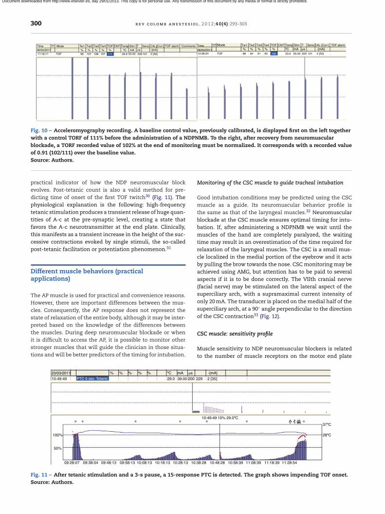
practical indicator of how the NDP neuromuscular blockevolves. Post-tetanic count is also a valid method for pre-dicting time of onset of the first TOF twitch30 (Fig. 11). Thephysiological explanation is the following: high-frequencytetanic stimulation produces a transient release of huge quan-tities of A-c at the pre-synaptic level, creating a state thatfavors the A-c neurotransmitter at the end plate. Clinically,this manifests as a transient increase in the height of the suc-cessive contractions evoked by single stimuli, the so-calledpost-tetanic facilitation or potentiation phenomenon.31
Different muscle behaviors (practicalapplications)
The AP muscle is used for practical and convenience reasons.However, there are important differences between the mus-cles. Consequently, the AP response does not represent thestate of relaxation of the entire body, although it may be inter-preted based on the knowledge of the differences betweenthe muscles. During deep neuromuscular blockade or when
it is difficult to access the AP, it is possible to monitor otherstronger muscles that will guide the clinician in those situa-tions and will be better predictors of the timing for intubation.23/03/2011 % % % % μsmAºC%
29.0 39.00 200 210:49:49 PTC 5 sec. Tetanic
100%
50%
09:28:07 09:38:04 09:48:13 09:58:13 10:08:13 10:18:13 10:28:13 10:3
∗ ∗ ∗ ∗
Fig. 11 – After tetanic stimulation and a 3-s pause, a 15-responseSource: Authors.
Monitoring of the CSC muscle to guide tracheal intubation
Good intubation conditions may be predicted using the CSCmuscle as a guide. Its neuromuscular behavior profile isthe same as that of the laryngeal muscles.32 Neuromuscularblockade at the CSC muscle ensures optimal timing for intu-bation. If, after administering a NDPNMB we wait until themuscles of the hand are completely paralyzed, the waitingtime may result in an overestimation of the time required forrelaxation of the laryngeal muscles. The CSC is a small mus-cle localized in the medial portion of the eyebrow and it actsby pulling the brow towards the nose. CSC monitoring may beachieved using AMG, but attention has to be paid to severalaspects if it is to be done correctly. The VIIth cranial nerve(facial nerve) may be stimulated on the lateral aspect of thesuperciliary arch, with a supramaximal current intensity ofonly 20 mA. The transducer is placed on the medial half of thesuperciliary arch, at a 90◦ angle perpendicular to the directionof the CSC contraction33 (Fig. 12).
CSC muscle: sensitivity profile
Muscle sensitivity to NDP neuromuscular blockers is relatedto the number of muscle receptors on the motor end plate
-[mA]
29 2 [35]
8:28 10:48:28 10:58:39 11:08:39 11:18:39 11:28:54
37ºC
28ºC
10:49:49 10% 29.0ºC∗ ∗ ∗
PTC is detected. The graph shows impending TOF onset.

r e v c o l o m b a n e s t e s i o l . 2
Fig. 12 – Monitoring of the corrugator supercilii (CSC)muscle. The arrow points to the correct placement of thetransducer on the medial half of the superciliary arch at a90◦ angle perpendicular to the direction of the musclecontraction.S
asoisomsdtscrmm
R
Tmfbo
D
TTsihTcn
twb
Prevention of residual paralysis
Document downloaded from http://www.elsevier.es, day 29/01/2013. This copy is for personal use. Any transmission of this document by any media or format is strictly prohibited.
ource: Authors.
nd the size of the muscle fiber. The morphological compo-ition of the muscle fibers has been determined on the basisf histological studies.34 Receptor density in central muscles
s higher than in peripheral muscles. Therefore, the relation-hip between the number of A-c receptors and the thicknessf the muscle fiber is a morphological predictor of the varioususcle responses (sensitivity) to NDP muscle relaxants. Sen-
itivity increases with the size and diameter of the fiber androps in relation to the number of A-c receptors. This impor-ant anatomical factor explains the similarity between theensitivity profiles of the laryngeal muscles and facial mus-les (higher resistance). For this reason, monitoring the CSCesponse is a guide to a more adequate prediction of the opti-
al timing for intubation35 when compared to the peripheraluscles (AP).
esidual paralysis: clinical relevance
he adverse effects of RP are a cause of morbidity andortality during the immediate post-operative period.36 The
ollowing are the main factors that help reduce the problem,ased on the systematic use of NMM and the administrationf a reversal agent.
iagnosis and incidence of residual paralysis
OF ratio (T4/T1)he absence of RP means that neuromuscular transmission isufficiently recovered, but it is important to be able to measuret effectively. In the absence of neuromuscular blockade, theeight of the four twitches is the same, and the TOF equals 1.he TOFR is a very sensitive test that correlates well with thelinical tests used to assess the degree of recovery from theeuromuscular blockade.
With the introduction of TOF in 1970, it was determined
hat the TOFR37 in the adductor pollicis (AP) correlated wellith the clinical signs of recovery, and it was considered aasic parameter in neuromuscular monitoring. Values of <0.60 1 2;40(4):293–303 301
were associated with significant muscle weakness. In 1979,Viby-Mogensen et al. were the first to show that, despite appar-ent clinical recovery, 42% of the patients had a TOFR < 0.7.38 Atpresent, after numerous studies, researchers have agreed ona definition of TOFR that represents a safe level of recovery.Using AMG, it has been accepted that the safest TOFR must be0.9. This endpoint ensures complete control of the pharyngealmuscles and normal ventilator response to hypoxia.39,40 Whenassessing all the studies that refer to the wide incidence of RPin the post-anesthetic care units, it is important to considera series of details pertaining to the intra-operative manage-ment, which are not always available. Was a nerve stimulatorused? Was reversal used for residual blockade? We may sug-gest that RP is infrequent when NMM is used. Considering thatthe AP is one of the last muscles to recover, it is better to assessrecovery by means of AP monitoring.41
Mandatory strategies for the reduction of residualparalysis
The following are the principles that should be followed inorder to avoid RP42–44:
1. Intra-operative neuromuscular monitoring. AMG, as aquantitative method, is better than visual assessment fordiagnosing RP.
2. Avoiding total TOF inhibition.3. Use of intermediate-acting NDP muscle relaxant.4. The administration of anticholinesterases with a certain
degree of spontaneous recovery of neuromuscular trans-mission is a critical step in reducing or eliminating RP.
5. Delaying extubation until a TOF ratio of 0.9 is achieved.6. A proposal developed recently is to use the new molecule
sugammadex (abbreviation of sugar and �-cyclodextrin),specifically designed to bind to rocuronium selectively. Atthe present time, it is approved and marketed in Europe andAustralia, but not in North America. Its main advantageis rapid action with minimal variation between individu-als, after an adequate dose that can achieve a TOFR of 0.9(3–5 min).45
Extubation and residual paralysis
Research carried out during the immediate post-operativeperiod at the time of extubation has shown RP with ahigher possibility of morbidity. Murphy et al.46 conducted astudy of TOFR quantified using AMG, immediately before tra-cheal extubation. It was concluded that RP was present inmost patients at the time of extubation. Despite a protocoldesigned for monitoring and reversal, and despite the use of anintermediate-acting NDPNMB, there was some degree of RP atthe end of surgery, while the patient was still in the operatingroom. These authors recommend NMM to ensure that recov-ery is complete and that respiratory and pharyngeal functionis normal.
Perhaps one of the most convincing pieces of evidence regard-ing RP assessment came from the studies by Baillard et al.

s i o l
r
1
1
1
1
1
1
1
1
1
1
2
2
Document downloaded from http://www.elsevier.es, day 29/01/2013. This copy is for personal use. Any transmission of this document by any media or format is strictly prohibited.
302 r e v c o l o m b a n e s t e
These researchers showed that intra-operative NMM usingAMG as an objective method, together with an effort at edu-cating the clinicians, led to a reduction of RP from 62% down tovery low levels.47,48 This finding contributed to the provisionof quantitative neuromuscular function assessment devicesin all the operating rooms.
Conclusions: what is the final lesson?
Despite the landmark introduction in 1942 of curare in clinicalpractice and the important advances in the understanding ofthe physiology of neuromuscular transmission, it is surprisingto see that NMM continues to be considered only as an option.The purpose of this review is to convey practical and academicreasons to convince clinicians that NMM should be used indaily practice. Published surveys show a low rate of use ofNMM, as revealed by a survey conducted recently in the UnitedKingdom among 715 anesthetists. Of them, 28% used a mon-itor occasionally, 10% used it routinely, and up to 62% neverused it.49 We are aware that NMM is no absolute guarantee ofperfect intubation conditions, optimal intra-operative musclerelaxation, or complete recovery. AP monitoring may be usedfor the duration of anesthesia, alternating if needed, with CPT.The information provided by NMM must be interpreted on thebasis of the individual patient, the drugs used, and the clinicalsigns. NMM is very useful provided it is interpreted correctly.AMG and a TOFR of 1 at the end of surgery help rule out RP. Inan editorial,50 Eriksson concluded that, “it is time to act andgive priority to the introduction of neuromuscular transmis-sion monitors in all operating rooms and use them every timewe give NDP muscle relaxants, instead of using them exclu-sively for research. It is our duty as researchers interested inthis matter to call the attention on the advances of the art andthe science, and to highlight that muscles relaxants must beused with good judgment in order to maintain an adequatedegree of relaxation according based on timing and need”.
Funding
Own resources.
Conflicts of interest
None declared in relation with the article.
Acknowledgements
We thank Belén Mingorance Diaz, the nurse at our RecoveryUnit, for her help with CSC muscle monitoring image.
e f e r e n c e s
1. Beecher HK, Todd DP. A study of the deaths associated withanesthesia and surgery. Ann Surg. 1954;140:2–35.
2. Churchill-Davidson HC, Christie TH. The diagnosis ofneuromuscular block in man. Br J Anaesth. 1959;31:290–301.
2
. 2 0 1 2;40(4):293–303
3. Churchill-Davidson HC. The d-tubocurarine dilemma.Anesthesiology. 1965;26:132–3.
4. Naguib M, Kopman A, Cynthia AL, Hunter J, Lopez A, Brull SJ.A survey of current management of neuromuscular block inthe United States and Europe. Anesth Analg. 2010;111:110–9.
5. Murphy GS, Brull SJ. Residual neuromuscular block: lessonsunlearned. Part I. Definitions, incidence, and adversephysiologic effects of residual neuromuscular block. AnesthAnalg. 2010;111:120–8.
6. The American Society of Anesthesiologists Task for onPost-anesthesia Care. Practice Guidelines for PostanestheticCare. Anesthesiology. 2002;96:742–52.
7. Crosby ET. An evidence-based approach to airwaymanagement: is there a role for clinical practice guidelines?Anaesthesia. 2011;66 Suppl. 2:112–8.
8. Murphy GS, Szokol JW, Avram MJ, Greenberg SB,Marymont JH, Vender JS, et al. Intraoperativeacceleromyography monitoring reduces symptoms of muscleweakness and improves quality of recovery in the earlypostoperative period. Anesthesiology. 2011;115:946–54.
9. Mencke T, Fuchs-Buder T. Does the timing of trachealintubation based on neuromuscular monitoring decreaselaryngeal injury? A randomized, prospective, controlled trial.Anesth Analg. 2006;102:306–12.
0. Hayes AH, Mirakhur RK, Breslin DS, Reid JE, McCourt KC.Postoperative residual block after intermediate actingneuromuscular blocking drugs. Anaesthesia. 2001;56:312–8.
1. Viby-Mogensen J. Postoperative residual curarization andevidence based anaesthesia. Br J Anaesth. 2000;84:301–4.
2. Hemmerling TM, Donati F, Beaulieu P, Babin D.Phonomyography of the corrugator supercilii muscle: signalcharacteristics, best recording site and comparison withacceleromyography. Br J Anaesth. 2002;88:389–93.
3. Fabregat López J. Monitorización de la relajación muscular,revisión actual. Diferencias entre músculos. Act AnestReanim (Madrid). 2002;12:59–62.
4. Viby-Mogensen J, Jensen NH, Engbaek J, Ording H, SkovgaardLT. Tactile and visual evaluation of the response totrain-of-four nerve stimulation. Anesthesiology.1985;63:440–3.
5. Rowaan CJ, Vandenbrom RHG, Wierda JMKH. TheRelaxometer: a complete and comprehensive computercontrolled neuromuscular transmission measurementsystem. J Clin Monit. 1993;9:38–44.
6. Viby-Mogensen J, Jensen E, Werner M, Kirkegaard Neilsen H.Measurement of acceleration a new method of monitoringneuromuscular function. Acta Anaesthesiol Scand.1988;32:45–8.
7. Martyn JA, Fagerlund MJ, Eriksson LI. Basic principles ofneuromuscular transmission. Anaesthesia. 2009;64 Suppl.1:11–9.
8. Fuchs Buder T, Claudius C, Skovgaard LT, Eriksson LI,Mirakhur RK, Viby-Mogensen J. Good clinical researchpractice in pharmacodynamic studies of neuromuscularblocking agents. II. The Stockholm revision. ActaAnaesthesiol Scand. 2007;51:789–808.
9. Ali HH, Utting JE, Gray TC. Stimulus frequency in thedetection of neuromuscular block in humans. Br J Anaesth.1970;42:967–78.
0. Kopman F, Klewicka MM, Neuman GG. The relationshipbetween acceleromyographic train-of-four fade and singletwitch depression. Anesthesiology. 2002;96:583–7.
1. Lee CM, Katz RL. Fade of neurally evoked compound EMGduring neuromuscular block by d-tubocurarine. Anesth
Analg. 1977;56:271–5.2. Beemer GH, Bjorksten AR, Dawson PJ, Dawson RJ, Heenan PJ,Robertson BA. Determinants of the reversal time of

o l . 2
2
2
2
2
2
2
2
3
3
3
3
3
3
3
3
3
3
4
4
4
4
4
4
4
4
4
4
Document downloaded from http://www.elsevier.es, day 29/01/2013. This copy is for personal use. Any transmission of this document by any media or format is strictly prohibited.
r e v c o l o m b a n e s t e s i
competitive neuromuscular block by anticholinesterases. Br JAnaesth. 1991;66:469–75.
3. Glavinovic MI, Law Min JC, Kapural L, Donati F, Bevan DR.Speed of action of various muscle relaxants at theneuromuscular junction binding vs. buffering hypothesis. JPharmacol Exp Ther. 1993;265:1181–6.
4. Ali HH, Savarese JJ, Lebowitz PW, Ramsay FM. Twitch, tetanusand train-of-four as indices of recovery fromnon-depolarizing neuromuscular blockade. Anesthesiology.1981;54:294–7.
5. Staals LM, Driessen JJ, Van Egmond J, De Boer HD, Klimek M,Flockton EA, et al. Train-of-four ratio recovery often precedestwitch recovery when neuromuscular block is reversed bysugammadex. Acta Anaesthesiol Scand. 2011;55:700–7.
6. Kopman AF, Chin WA. A dose–response study of rocuronium.Do acceleromyographic and electromyographic monitorsproduce the same results? Acta Anaesthesiol Scand.2005;49:323–7.
7. Capron F, Alla F, Hottier C, Meistelman C, Fuchs-Buder T. Canacceleromyography detect low levels of residual paralysis?Anesthesiology. 2004;100:1119–24.
8. Suzuki T, Fukano N, Kitajima O, Saeki S, Ogawa S.Normalization of acceleromyographic train-of-four ratio bybaseline value for detecting residual neuromuscular block. BrJ Anaesth. 2006;96:44–7.
9. Viby-Mogensen J, Howardy-Hansen P, Chræmmer-JorgensenB, Ørding H, Engbæk J, Nielsen A. Posttetanic count (PTC): anew method of evaluating an intense nondepolarizing block.Anesthesiology. 1981;55:458–61.
0. Aitoh Y, Fujii Y, Takahashi K, Makita K, Tanaka H, Amaha K.Recovery of posttetanic count and train-of-four responses atthe great toe and thumb. Anaesthesia. 1998;53:44–8.
1. Bowman WC. In: Fukushima K, Ochiai R, editors. Mechanismsof action of reversal agents, muscle relaxants: physiologic andpharmacologic aspects. Tokyo: Springer Verlag; 1995. p. 19–30.
2. Plaud B, Debaene B, Donati F. The corrugator supercilii, notthe orbicularis oculi reflects rocuronium neuromuscularblockade at the laryngeal adductor muscles. Anesthesiology.2001;95:96–101.
3. Ortiz Gómez JR, Fabregat López J, Palacio-Abizanda FJ, FornetRuiz I, Perez Cajaraville J, Arino Irujo JJ, et al. Monitorizacióndel bloqueo neuromuscular. 2a parte. Rev Esp AnestesiolReanim. 2010;57:161–72.
4. Ibebunjo C, Srikant C, Donati F. Morphological correlates ofthe differential responses of muscles to vecuronium. Br J
Anaesth. 1999;83:284–91.5. Hemmerling TM, Le N. Brief review: neuromuscularmonitoring: an update for the clinician. Can J Anaesth.2007;54:58–72.
5
0 1 2;40(4):293–303 303
6. Bevan DR. Recovery from neuromuscular block and itsassessment. Anesth Analg. 2000;90:S7–13.
7. Ali HH, Utting JE, Gray TC. Quantitative assessment ofresidual antidepolarizing block (Part II). Br J Anaesth.1971;43:478–85.
8. Viby-Mogensen J, Chraemmer Jorgensen B, Ordin H. Residualcurarization in the recovery room. Anesthesiology.1979;50:539–41.
9. Eriksson LI, Lennmarken C, Wyon N, Johnson A. Attenuatedventilatory response to hypoxaemia after vecuroniuminduced partial neuromuscular block. Acta AnaesthesiolScand. 1992;36:710–5.
0. Eriksson LI, Sundman E, Olsson R, Nilsson L, Witt H, Ekberg O.Functional assessment of the pharynx at rest and duringswallowing in partially paralyzed humans: simultaneousvideomanometry and mechanomyography of awake humanvolunteers. Anesthesiology. 1997;87:1035–43.
1. Baillard C. Monitorage de la curarisation. EMC (ElsevierMasson SAS, Paris). Anesthésie-Réanimation. 2011,36-390-A-10.
2. Murphy GS. Residual neuromuscular blockade: incidence,assessment, and relevance in the postoperative period.Minerva Anestesiol. 2006;72:97–109.
3. Plaud B, Debaene B, Donati F, Marty J. Residual paralysis afteremergence from anesthesia. Clinical concepts andcommentary. Anesthesiology. 2010;112:1013–22.
4. Donati F. Neuromuscular monitoring: what evidence do weneed to be convinced? Anesth Analg. 2010;111:1–2 [Editorial].
5. Sorgenfrei IF, Norril DK, Larsen PB, Stensballe J, Ostergaard D,Prins ME, et al. Reversal of rocuronium-inducedneuromuscular block by these elective relaxant binding agentsugammadex: a dose-finding and safety study.Anesthesiology. 2006;104:667–74.
6. Murphy GS, Szokol JW, Marymont JH, Franklin M, Avram ML,Vender JS. Residual paralysis at the time of trachealextubation. Anesth Analg. 2005;100:1840–5.
7. Baillard C, Gehan G, Reboul Marty J, Larmignat P, Samana CM,Cupa M. Residual curarization in the recovery room aftervecuronium. Br J Anaesth. 2000;84:301–3.
8. Baillard C, Clec’h C, Catineau J, Catineau J, Salhi F, Gehan G,et al. Postoperative residual neuromuscular block: a survey ofmanagement. Br J Anaesth. 2005;95:622–6.
9. Grayling M, Sweeney BP. Recovery from neuromuscular
blockade: a survey of practice. Anaesthesia. 2007;62:806–9.0. Eriksson LI. Evidence-based practice and neuromuscularmonitoring: it’s time for routine quantitative assessment.Anesthesiology. 2003;98:1037–9.