Embed Size (px)
Citation preview
Annals of Academy of Romanian Scientists Series: Medical Sciences
ISSN 2067-7766 Volume 2, Number 2/2011 39
RREEVVIISSIIOONN -- TTRREEAATTMMEENNTT LLOOOOSSEENNIINNGG IINN TTOOTTAALL HHIIPP AARRTTHHRROOPPLLAASSTTYY
Nicolae GORUN1, Emil MAREŞ2,
1) Member of Academy of Romanian Scientists, professor, Ph.D. “Sfântul Ioan” Clinical Emergency Hospital - Bucharest, Romania
2) Orthopedist, “Elias” Clinical Emergency Hospital - Bucharest, Romania
Abstract
The most important achievement of modern orthopedics, total hip arthroplasty, provides a high level of activity, but act and some risks and complications, most important being loosening of prosthetic components. This loosening require one or two replays, framed in the wider notion: review. This is a difficult operation with loss of blood and implies a significant risk of suppuration and even life threatening. Revision requires complex equipment (instruments, a wide range of revision prosthesis), the surgical team trained in the field, adequate resuscitation and prolonged recovery, but not always with good enough results. Along with some general remarks on loosening, there are presented causes of revision, clinical and radiological examination, and revisions objectives.
The paper is based on a personal statistical study of 25 cases (11 M and 14 F), recorded and subject to revision, in a period of 10 years (2001-2010). They are presented in a summary table and divided by age and gender, side of interest. The conclusions lead to the belief and recommendation that the review be performed in specialized centers, other than those in the primary arthroplasty was performed.
Keywords: total hip arthroplasty, loosening, revision
Rezumat
Cea mai importantă realizare a ortopediei moderne, artroplastia totală de şold, asigură un nivel ridicat de activitate, dar comportă şi unele riscuri şi complicaŃii, dintre care cea mai importanta şi mai gravă este decimentarea pieselor protetice. Aceste decimentări impun una-două reluări, încadrate în noŃiunea mai largă, de revizie. Este vorba de intervenŃii laborioase şi dificile, care antrenează pierderi importante de sânge şi comportă un risc crescut de supuraŃie şi chiar un risc vital. Revizia impune o dotare complexă (instrumentar, o gamă largă de proteze de revizie), o echipă chirurgicală antrenată în domeniu, reanimare adecvată şi recuperare prelungită, dar nu totdeauna
1 Address for correspondence: Professor Nicolae Gorun, E-mail: [email protected]
Nicolae GORUN, Emil MAREŞ
40
cu rezultate suficient de bune. Alături de câteva precizari de ordin general asupra decimentărilor, sunt prezentate cauzele reviziilor, examenul clinic şi radiologic, precum şi obiectivele reviziilor.
Lucrarea are la bază un studiu statistic personal pe 25 de cazuri (11 B şi 14 F), înregistrate şi supuse reviziei, într-o perioadă de 10 ani (2001-2010). Ele sunt prezentate într-un tabel sintetic şi repartizate pe grupe de varstă şi sexe, şoldul interesat. Concluziile formulate conduc la convingerea şi recomandarea ca aceste revizii să fie efectuate în centre specializate, altele decât acelea în care s-a efectuat artroplastia primară.
Cuvinte-cheie: artroplastia totală de şold, decimentare, revizie
1. General information
One of the most important achievements of modern orthopedics is obviously total
hip arthroplasty. Hip osteoarthritis has today many surgical techniques, which aim at
restoring hip mobility and increased average lifespan. Total hip arthroplasty provides a
high level of activity, both professionally and sports. However, even with a very high
success rate in primary arthroplasty, the number of cases that require one or even multiple
revision is growing. This increased frequency of revision of primary hip arthroplasty is a
practical reality, although some authors imply, first, increasing the number of patients
undergoing this surgery (statistically) (Figure 1-2, Figure 3, Figure 4 ).
Figure 1. Primitive bilateral coxarthrosis, right
global decompensated. Radiological aspect -
front view
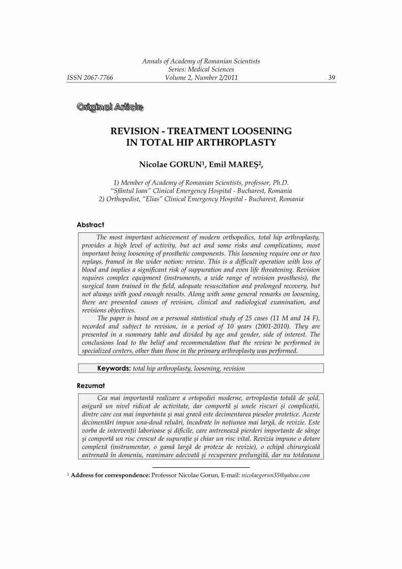
Figure 2. Previous case. Postoperative
radiological appearance (right) - total
arthroplasty with cemented prosthesis type
Charnley-Müller
RReevviissiioonn -- ttrreeaattmmeenntt lloooosseenniinngg iinn ttoottaall hhiipp aarrtthhrrooppllaassttyy
41
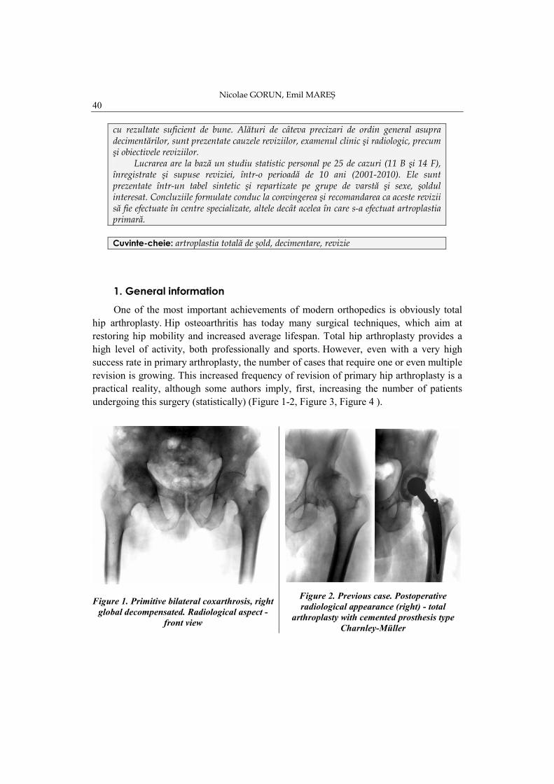
Figure 3. Left coxarthrosis secondary to
dysplasia. Front view preoperative radiologic
appearance (left) and front view
postoperative radiological appearance
(right): total arthroplasty with Charnley-
Müller cemented prosthesis
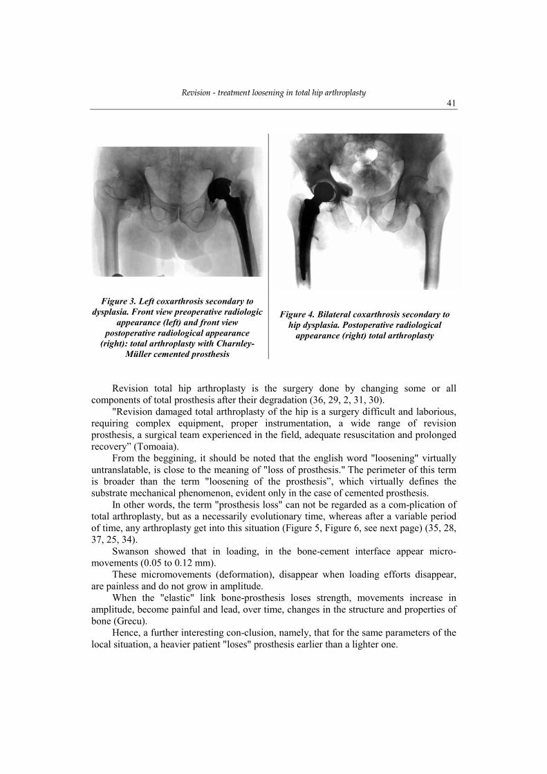
Figure 4. Bilateral coxarthrosis secondary to
hip dysplasia. Postoperative radiological
appearance (right) total arthroplasty
Revision total hip arthroplasty is the surgery done by changing some or all
components of total prosthesis after their degradation (36, 29, 2, 31, 30).
"Revision damaged total arthroplasty of the hip is a surgery difficult and laborious,
requiring complex equipment, proper instrumentation, a wide range of revision
prosthesis, a surgical team experienced in the field, adequate resuscitation and prolonged
recovery” (Tomoaia).
From the beggining, it should be noted that the english word "loosening" virtually
untranslatable, is close to the meaning of "loss of prosthesis." The perimeter of this term
is broader than the term "loosening of the prosthesis”, which virtually defines the
substrate mechanical phenomenon, evident only in the case of cemented prosthesis.
In other words, the term "prosthesis loss" can not be regarded as a com-plication of
total arthroplasty, but as a necessarily evolutionary time, whereas after a variable period
of time, any arthroplasty get into this situation (Figure 5, Figure 6, see next page) (35, 28,
37, 25, 34).
Swanson showed that in loading, in the bone-cement interface appear micro-
movements (0.05 to 0.12 mm).
These micromovements (deformation), disappear when loading efforts disappear,
are painless and do not grow in amplitude.
When the "elastic" link bone-prosthesis loses strength, movements increase in
amplitude, become painful and lead, over time, changes in the structure and properties of
bone (Grecu).
Hence, a further interesting con-clusion, namely, that for the same parameters of the
local situation, a heavier patient "loses" prosthesis earlier than a lighter one.
Nicolae GORUN, Emil MAREŞ
42
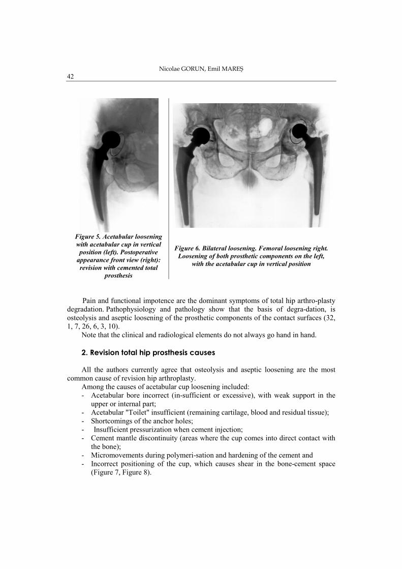
Figure 5. Acetabular loosening
with acetabular cup in vertical
position (left). Postoperative
appearance front view (right):
revision with cemented total
prosthesis
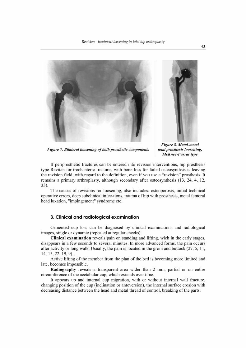
Figure 6. Bilateral loosening. Femoral loosening right.
Loosening of both prosthetic components on the left,
with the acetabular cup in vertical position
Pain and functional impotence are the dominant symptoms of total hip arthro-plasty
degradation. Pathophysiology and pathology show that the basis of degra-dation, is
osteolysis and aseptic loosening of the prosthetic components of the contact surfaces (32,
1, 7, 26, 6, 3, 10).
Note that the clinical and radiological elements do not always go hand in hand.
2. Revision total hip prosthesis causes
All the authors currently agree that osteolysis and aseptic loosening are the most
common cause of revision hip arthroplasty.
Among the causes of acetabular cup loosening included:
- Acetabular bore incorrect (in-sufficient or excessive), with weak support in the
upper or internal part;
- Acetabular "Toilet" insufficient (remaining cartilage, blood and residual tissue);
- Shortcomings of the anchor holes;
- Insufficient pressurization when cement injection;
- Cement mantle discontinuity (areas where the cup comes into direct contact with
the bone);
- Micromovements during polymeri-sation and hardening of the cement and
- Incorrect positioning of the cup, which causes shear in the bone-cement space
(Figure 7, Figure 8).
RReevviissiioonn -- ttrreeaattmmeenntt lloooosseenniinngg iinn ttoottaall hhiipp aarrtthhrrooppllaassttyy
43
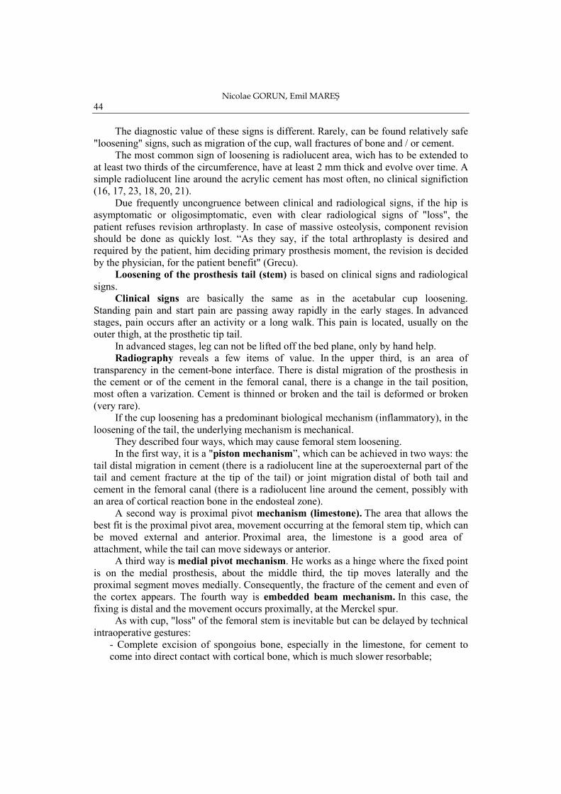
Figure 7. Bilateral loosening of both prosthetic components
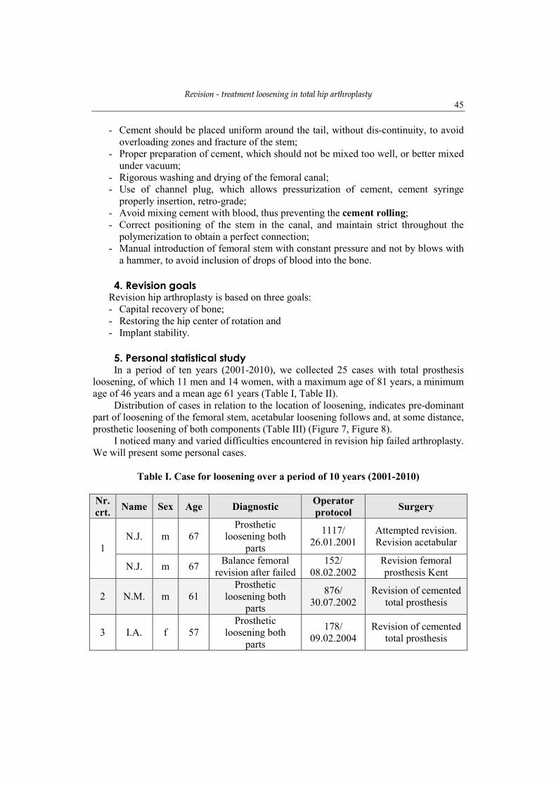
Figure 8. Metal-metal
total prosthesis loosening,
McKnee-Farrar type
If periprosthetic fractures can be entered into revision interventions, hip prosthesis
type Revitan for trochanteric fractures with bone loss for failed osteosynthsis is leaving
the revision field, with regard to the definition, even if you use a “revision” prosthesis. It
remains a primary arthroplasty, although secondary after osteosynthesis (13, 24, 4, 12,
33).
The causes of revisions for loosening, also includes: osteoporosis, initial technical
operative errors, deep subclinical infec-tions, trauma of hip with prosthesis, metal femoral
head luxation, "impingement" syndrome etc.
3. Clinical and radiological examination
Cemented cup loss can be diagnosed by clinical examinations and radiological
images, single or dynamic (repeated at regular checks).
Clinical examination reveals pain on standing and lifting, wich in the early stages,
disappears in a few seconds to several minutes. In more advanced forms, the pain occurs
after activity or long walk. Usually, the pain is located in the groin and buttock (27, 5, 11,
14, 15, 22, 19, 9).
Active lifting of the member from the plan of the bed is becoming more limited and
late, becomes impossible.
Radiography reveals a transparent area wider than 2 mm, partial or on entire
circumference of the acetabular cup, which extends over time.
It appears up and internal cup migration, with or without internal wall fracture,
changing position of the cup (inclination or anteversion), the internal surface erosion with
decreasing distance between the head and metal thread of control, breaking of the parts.
Nicolae GORUN, Emil MAREŞ
44
The diagnostic value of these signs is different. Rarely, can be found relatively safe
"loosening" signs, such as migration of the cup, wall fractures of bone and / or cement.
The most common sign of loosening is radiolucent area, wich has to be extended to
at least two thirds of the circumference, have at least 2 mm thick and evolve over time. A
simple radiolucent line around the acrylic cement has most often, no clinical significtion
(16, 17, 23, 18, 20, 21).
Due frequently uncongruence between clinical and radiological signs, if the hip is
asymptomatic or oligosimptomatic, even with clear radiological signs of "loss", the
patient refuses revision arthroplasty. In case of massive osteolysis, component revision
should be done as quickly lost. “As they say, if the total arthroplasty is desired and
required by the patient, him deciding primary prosthesis moment, the revision is decided
by the physician, for the patient benefit" (Grecu).
Loosening of the prosthesis tail (stem) is based on clinical signs and radiological
signs.
Clinical signs are basically the same as in the acetabular cup loosening.
Standing pain and start pain are passing away rapidly in the early stages. In advanced
stages, pain occurs after an activity or a long walk. This pain is located, usually on the
outer thigh, at the prosthetic tip tail.
In advanced stages, leg can not be lifted off the bed plane, only by hand help.
Radiography reveals a few items of value. In the upper third, is an area of
transparency in the cement-bone interface. There is distal migration of the prosthesis in
the cement or of the cement in the femoral canal, there is a change in the tail position,
most often a varization. Cement is thinned or broken and the tail is deformed or broken
(very rare).
If the cup loosening has a predominant biological mechanism (inflammatory), in the
loosening of the tail, the underlying mechanism is mechanical.
They described four ways, which may cause femoral stem loosening.
In the first way, it is a "piston mechanism”, which can be achieved in two ways: the
tail distal migration in cement (there is a radiolucent line at the superoexternal part of the
tail and cement fracture at the tip of the tail) or joint migration distal of both tail and
cement in the femoral canal (there is a radiolucent line around the cement, possibly with
an area of cortical reaction bone in the endosteal zone).
A second way is proximal pivot mechanism (limestone). The area that allows the
best fit is the proximal pivot area, movement occurring at the femoral stem tip, which can
be moved external and anterior. Proximal area, the limestone is a good area of
attachment, while the tail can move sideways or anterior.
A third way is medial pivot mechanism. He works as a hinge where the fixed point
is on the medial prosthesis, about the middle third, the tip moves laterally and the
proximal segment moves medially. Consequently, the fracture of the cement and even of
the cortex appears. The fourth way is embedded beam mechanism. In this case, the
fixing is distal and the movement occurs proximally, at the Merckel spur.
As with cup, "loss" of the femoral stem is inevitable but can be delayed by technical
intraoperative gestures:
- Complete excision of spongoius bone, especially in the limestone, for cement to
come into direct contact with cortical bone, which is much slower resorbable;
RReevviissiioonn -- ttrreeaattmmeenntt lloooosseenniinngg iinn ttoottaall hhiipp aarrtthhrrooppllaassttyy
45
- Cement should be placed uniform around the tail, without dis-continuity, to avoid
overloading zones and fracture of the stem;
- Proper preparation of cement, which should not be mixed too well, or better mixed
under vacuum;
- Rigorous washing and drying of the femoral canal;
- Use of channel plug, which allows pressurization of cement, cement syringe
properly insertion, retro-grade;
- Avoid mixing cement with blood, thus preventing the cement rolling;
- Correct positioning of the stem in the canal, and maintain strict throughout the
polymerization to obtain a perfect connection;
- Manual introduction of femoral stem with constant pressure and not by blows with
a hammer, to avoid inclusion of drops of blood into the bone.
4. Revision goals
Revision hip arthroplasty is based on three goals:
- Capital recovery of bone;
- Restoring the hip center of rotation and
- Implant stability.
5. Personal statistical study In a period of ten years (2001-2010), we collected 25 cases with total prosthesis
loosening, of which 11 men and 14 women, with a maximum age of 81 years, a minimum
age of 46 years and a mean age 61 years (Table I, Table II).
Distribution of cases in relation to the location of loosening, indicates pre-dominant
part of loosening of the femoral stem, acetabular loosening follows and, at some distance,
prosthetic loosening of both components (Table III) (Figure 7, Figure 8).
I noticed many and varied difficulties encountered in revision hip failed arthroplasty.
We will present some personal cases.
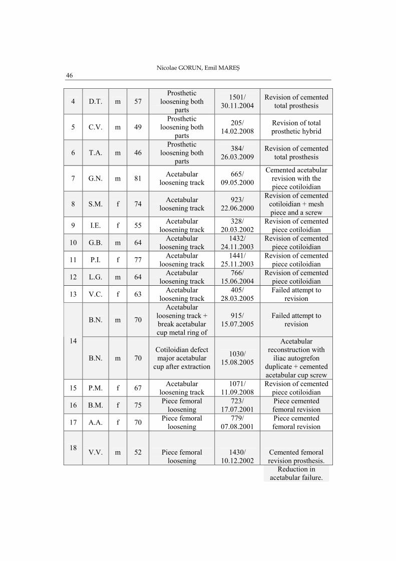
Table I. Case for loosening over a period of 10 years (2001-2010)
Nr. crt.
Name Sex Age Diagnostic Operator protocol
Surgery
N.J. m 67
Prosthetic
loosening both
parts
1117/
26.01.2001
Attempted revision.
Revision acetabular 1
N.J. m 67 Balance femoral
revision after failed
152/
08.02.2002
Revision femoral
prosthesis Kent
2 N.M. m 61
Prosthetic
loosening both
parts
876/
30.07.2002
Revision of cemented
total prosthesis
3 I.A. f 57
Prosthetic
loosening both
parts
178/
09.02.2004
Revision of cemented
total prosthesis
Nicolae GORUN, Emil MAREŞ
46
4 D.T. m 57
Prosthetic
loosening both
parts
1501/
30.11.2004
Revision of cemented
total prosthesis
5 C.V. m 49
Prosthetic
loosening both
parts
205/
14.02.2008
Revision of total
prosthetic hybrid
6 T.A. m 46
Prosthetic
loosening both
parts
384/
26.03.2009
Revision of cemented
total prosthesis
7 G.N. m 81 Acetabular
loosening track
665/
09.05.2000
Cemented acetabular
revision with the
piece cotiloidian
8 S.M. f 74 Acetabular
loosening track
923/
22.06.2000
Revision of cemented
cotiloidian + mesh
piece and a screw
9 I.E. f 55 Acetabular
loosening track
328/
20.03.2002
Revision of cemented
piece cotiloidian
10 G.B. m 64 Acetabular
loosening track
1432/
24.11.2003
Revision of cemented
piece cotiloidian
11 P.I. f 77 Acetabular
loosening track
1441/
25.11.2003
Revision of cemented
piece cotiloidian
12 L.G. m 64 Acetabular
loosening track
766/
15.06.2004
Revision of cemented
piece cotiloidian
13 V.C. f 63 Acetabular
loosening track
405/
28.03.2005
Failed attempt to
revision
B.N. m 70
Acetabular
loosening track +
break acetabular
cup metal ring of
915/
15.07.2005
Failed attempt to
revision
14
B.N. m 70
Cotiloidian defect
major acetabular
cup after extraction
1030/
15.08.2005
Acetabular
reconstruction with
iliac autogrefon
duplicate + cemented
acetabular cup screw
15 P.M. f 67 Acetabular
loosening track
1071/
11.09.2008
Revision of cemented
piece cotiloidian
16 B.M. f 75 Piece femoral
loosening
723/
17.07.2001
Piece cemented
femoral revision
17 A.A. f 70 Piece femoral
loosening
779/
07.08.2001
Piece cemented
femoral revision
18 V.V. m 52 Piece femoral
loosening
1430/
10.12.2002
Cemented femoral
revision prosthesis.
Reduction in
acetabular failure.
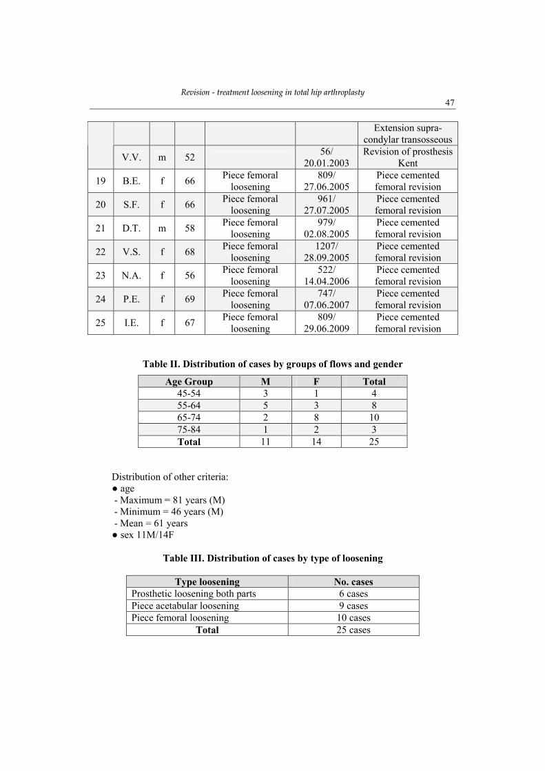
RReevviissiioonn -- ttrreeaattmmeenntt lloooosseenniinngg iinn ttoottaall hhiipp aarrtthhrrooppllaassttyy
47
Extension supra-
condylar transosseous
V.V. m 52 56/
20.01.2003
Revision of prosthesis
Kent
19 B.E. f 66 Piece femoral
loosening
809/
27.06.2005
Piece cemented
femoral revision
20 S.F. f 66 Piece femoral
loosening
961/
27.07.2005
Piece cemented
femoral revision
21 D.T. m 58 Piece femoral
loosening
979/
02.08.2005
Piece cemented
femoral revision
22 V.S. f 68 Piece femoral
loosening
1207/
28.09.2005
Piece cemented
femoral revision
23 N.A. f 56 Piece femoral
loosening
522/
14.04.2006
Piece cemented
femoral revision
24 P.E. f 69 Piece femoral
loosening
747/
07.06.2007
Piece cemented
femoral revision
25 I.E. f 67 Piece femoral
loosening
809/
29.06.2009
Piece cemented
femoral revision
Table II. Distribution of cases by groups of flows and gender
Age Group M F Total
45-54 3 1 4
55-64 5 3 8
65-74 2 8 10
75-84 1 2 3
Total 11 14 25
Distribution of other criteria:
● age
- Maximum = 81 years (M)
- Minimum = 46 years (M)
- Mean = 61 years
● sex 11M/14F
Table III. Distribution of cases by type of loosening
Type loosening No. cases
Prosthetic loosening both parts 6 cases
Piece acetabular loosening 9 cases
Piece femoral loosening 10 cases
Total 25 cases
Nicolae GORUN, Emil MAREŞ
48
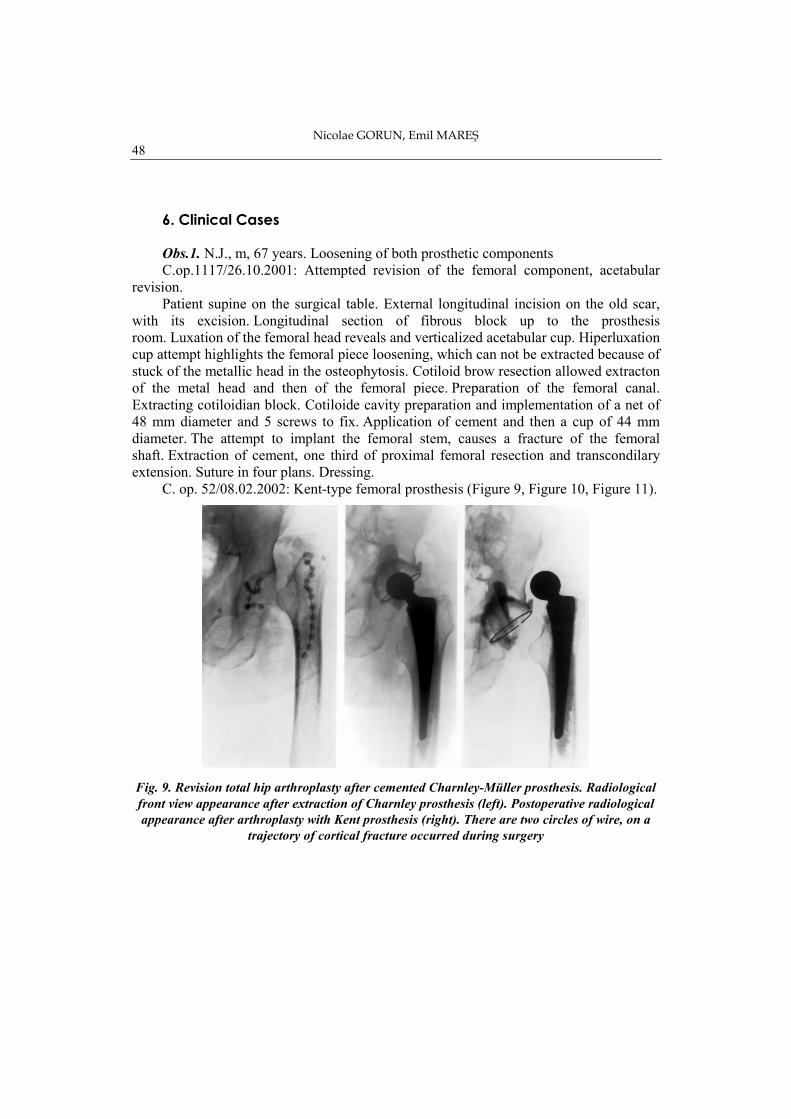
6. Clinical Cases Obs.1. N.J., m, 67 years. Loosening of both prosthetic components
C.op.1117/26.10.2001: Attempted revision of the femoral component, acetabular
revision.
Patient supine on the surgical table. External longitudinal incision on the old scar,
with its excision. Longitudinal section of fibrous block up to the prosthesis
room. Luxation of the femoral head reveals and verticalized acetabular cup. Hiperluxation
cup attempt highlights the femoral piece loosening, which can not be extracted because of
stuck of the metallic head in the osteophytosis. Cotiloid brow resection allowed extracton
of the metal head and then of the femoral piece. Preparation of the femoral canal.
Extracting cotiloidian block. Cotiloide cavity preparation and implementation of a net of
48 mm diameter and 5 screws to fix. Application of cement and then a cup of 44 mm
diameter. The attempt to implant the femoral stem, causes a fracture of the femoral
shaft. Extraction of cement, one third of proximal femoral resection and transcondilary
extension. Suture in four plans. Dressing.
C. op. 52/08.02.2002: Kent-type femoral prosthesis (Figure 9, Figure 10, Figure 11).
Fig. 9. Revision total hip arthroplasty after cemented Charnley-Müller prosthesis. Radiological
front view appearance after extraction of Charnley prosthesis (left). Postoperative radiological
appearance after arthroplasty with Kent prosthesis (right). There are two circles of wire, on a
trajectory of cortical fracture occurred during surgery
RReevviissiioonn -- ttrreeaattmmeenntt lloooosseenniinngg iinn ttoottaall hhiipp aarrtthhrrooppllaassttyy
49
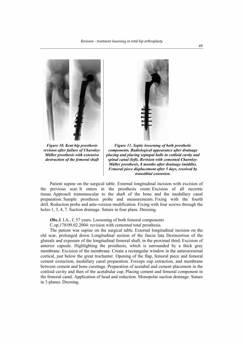
Figure 10. Kent hip prosthesis
revision after failure of Charnley-
Müller prosthesis with extensive
destruction of the femoral shaft
Figure 11. Septic loosening of both prosthetic
components. Radiological appearance after drainage
placing and placing septopal balls in cotiloid cavity and
spinal canal (left). Revision with cemented Charnley-
Müller prosthesis, 8 months after drainage (middle).
Femoral piece displacement after 5 days, resolved by
transtibial extension.
Patient supine on the surgical table. External longitudinal incision with excision of
the previous scar. It enters in the prosthesis room. Excision of all necrotic
tissue. Approach transmuscular to the shaft of the bone and the medullary canal
preparation. Sample prosthesis probe and measurements. Fixing with the fourth
drill. Reduction probe and ante-version modification. Fixing with four screws through the
holes 1, 3, 4, 7. Suction drainage. Suture in four plans. Dressing.
Obs.3. I.A., f, 57 years. Loosening of both femoral components
C.op.178/09.02.2004: revision with cemented total prosthesis.
The patient was supine on the surgical table. External longitudinal incision on the
old scar, prolonged down. Longitudinal section of the fascia lata. Dezinsertion of the
gluteals and exposure of the longitudinal femoral shaft, in the proximal third. Excision of
anterior capsule. Highlighting the prosthesis, which is surrounded by a thick gray
membrane. Excision of the membrane. Create a rectangular window in the anteroexternal
cortical, just below the great trochanter. Opening of the flap, femoral piece and femoral
cement extraction, medullary canal preparation. Forceps cup extraction, and membrane
between cement and bone curettage. Preparation of acetabul and cement placement in the
cotiloid cavity and then of the acetabular cup. Placing cement and femoral component in
the femoral canal. Application of head and reduction. Monopolar suction drainage. Suture
in 3 planes. Dressing.

Nicolae GORUN, Emil MAREŞ
50
Obs.4. D.T., m, 57 years. Prosthetic loosening both pieces.
C. op. 1501/30.11.2004: revision with cemented total prosthesis.
Patient supine on the surgical table. External longitudinal incision on the old scar.
Section of fibrosclerous block layer to the prosthesis room. It highlights the metal head
and acetabular cup. Femoral piece is luxated and we discover a rich granulo-matous
tissue in the form of gray membrane, which is rigorously excised together with cement
debris in the channel. Relatively easy extraction of the acetabular cup and cement.
Acetabular preparation, pre-paration of the femoral canal. Introduction of the ciment in
the canal, then the femoral stem. Application of the head and reduction. Monopolar
suction drainage. Suture in four planes. Dressing.
Obs.8. S.M., f, 74 years. Acetabular loosening.
C. op. 923/22.06.2000: revision with cemented cup + metal net and a screw.
The patient was supine on the surgical table. External longitudinal incision on the
old scar, with its excision. Longitudinal section of fascia lata and oblique of middle
gluteal. Excision of anterior capsule. Acetabular piece highlighting and femoral piece.
Extraction of them, with great difficulty. Cotiloide cavity pre-paration, placing cement,
and metal mesh. Upper leg is fixed to the acetabulum with a screw. Preparation of the
femoral canal and placing cement in the canal, then inserting the femoral stem, placing
the head on the stem and reduction in cotiloid cavity. Monopolar suction drainage. Suture
in four planes. Dressing.
Obs.12. L.G., m, 64. Acetabular piece loosening.
C. op. 766/15.06.2004 Cemented cup revision.
Patient supine on the surgical table. External longitudinal incision on the old scar.
Longitudinal section of the block to the bone and prosthetis. Excision of fibrous tissue
around the prosthesis and ossification. Femoral luxation piece is difficult. Very difficult
loosening of the acetabular cup, which can only happen through the back of the femoral
dislocated head. Cotyle preparation with chisel and curette. Placing cement in cotiloid
cavity and then a cup of 32 mm diameter. Reduction. Monopolar suction drainage.
Sutured in three planes. Dressing.
Obs.13. V.C., f., 63. Acetabular loosening.
C. op. 405/28.03.2005: failed attempt to revision.
The patient was supine on the surgical table. External longitudinal incision on the
old scar. Approach transmuscular to the prosthesis. Extracting the cup with cement layer.
Graft harvesting and fixing her with a pin on cotiloid cavity and four screws. Application
of cement and acetabular part. Thereafter, the cup with cement and bone graft is
mobilized, so that is extracted and femoral stem left in place. Monopolar suction
drainage. Suture in four planes. Dressing. Transcondilarry extension.
Obs.14. B.N., m, 70. Cup loosening + rupture of metal ring of acetabular cup.
C.op.915/15.07.2005: failed attempt to revision.
RReevviissiioonn -- ttrreeaattmmeenntt lloooosseenniinngg iinn ttoottaall hhiipp aarrtthhrrooppllaassttyy
51
Patient supine on the surgical table. External longitudinal incision, with scar
excision. Approach transmuscular to the prosthesis. Extraction of metal head. Femoral
piece is very stable. Extraction of the cup and cement. Cotiloid cavity is practically
destroyed. Attempting to place a cup on cement which does not succeed. Bipolar suction
drainage. Suture in two planes. Dressing. Transcondillary ex-tension.
Obs.14. B.N., m, 70.
C. op. 1030/18.08.2005 (acetabular major bone defect after removal of acetabular
cup): cotiloid cavity re-construction with iliac duplicate autograft and screws and
cemented acetabular cup.
Patient supine on the surgical table. Curved incision on the opposite iliac crest,
which collection of a total graft of 5 / 4 cm, which is duplicate. Suture in three planes.
Monopolar suction drainage. Smith-Petersen incision on the right hip. It is highlighted the
iliac crest, external and internal up to acetabular defect. On the internal face a screwed
graft is applied. At the bottom of the cotyle a second graft is applied and secured with two
screws. Application of cement and then 44 mm diameter acetabular cup. Application of
the femoral head and reduction in cotiloid piece. Monopolar suction drainage. Suture in
four planes. Dressing.
Obs.16. B.M., f, 75 years. Femoral piece loosening.
C. op. 723/17.07.2001: Femoral re-vision with cemented piece.
The patient was supine on the surgical table. External longitudinal incision on the
old scar. Incision in the tissue block of the fibroscleroase tissues to the prosthesis.
Luxation and extraction of head prosthesis. Create a rectangle of cortical in the third
upper shaft, and incomplete removal of the cement, difficult femoral stem and cement
extraction. Application of cortical flap and fixation with two circles of wire. Introduction
of cement into the canal, then a new rod (like the first one). Applying a new head on the
rod and reduction in cotiloid cavity. Monopolar suction drainage. Suture in four
planes. Dressing.
Obs.18. V.V., m, 52. Femoral piece loosening.
C. op. 1430/10.12.2002: Femoral revision with cemented prosthesis. Failure to
reduce in the cotiloid cavity. Transcondilarry extension.
Patient supine on the surgical table. External longitudinal incision on the old scar.
Fibromuscular longitudinal section of the block to the bone. Highlighting the
prosthesis. Extracting femoral piece and cement. Preparation of the medularry canal.
Introducing a new cemented component. Failed attempt to reduce in the cotiloid cavity.
intervention scale is forcing us to interrupt surgery. Trans-condilarry pin for continued
extension. Monopolar suction drainage. Loose sutures in four planes. Dressing.
C. op. 56/20.01.2003: revision with Kent prosthesis.
Patient supine on the surgical table. External longitudinal incision on the old scar.
Approaches transmuscularry to the trohanterian massive and dislocated prosthesis.
Extraction and removal of the femoral piece and femoral cement out of the canal, with
great difficulty. There are done trials with different femoral com-ponents and dihherent
head sizes.
Nicolae GORUN, Emil MAREŞ
52
Placement of a femoral piece with nine holes, the uperior hole being located in the
upper tranche of the osteotomy. Fixing with five screws. Putting the head in cotiloid
cavity. Monopolar suction drainage. Sutures in five plans. Dressing.
Obs.21. D.T., m, 58 years. Femoral piece loosening.
C. op. 979/02.08.2005: Femoral revision with cemented stem.
Patient supine on the surgical table. External longitudinal incision on the old scar,
with its excision. Section of the fibrous tissue block to the prosthesis room. Femoral piece
luxation. Easy extraction of the femoral stem with his head. The cement is extracted from
the canal after it was done a cortical antero-external clap. Preparation and placement of
cement into the canal. Inserting the rod in the canal and the application of metallic head,
then reduction in cotiloid cavity. Monopolar suction drainage. Suture in four
planes. Dressing.
7. Conclusions
a) The most important achievement of modern orthopedics is total hip arthroplasty. It
eliminates pain, restores mobility of the hip and provide a high level of activity, both
professionally and sports.
b) Between the complications of total hip arthroplasty, first is loosening of prosthetic
components.
c) Revision of hip arthroplasty, meaning replacement of prosthetic com-ponents, is a
laborious and delicate procedure, which involves significant blood loss and carries
an increased risk of infection or even life-threatening.
d) Experience gained on a personal series of 25 cases of hip prosthesis loosening,
demonstrated the extreme difficulties of revision total hip prosthesis, and that
technical the solutions are often atypical.
e) Like many other authors, we believe that the prosthetic hip revision must be
performed in specialized centers, equipped with necessary equipment and complex
surgical team, fully trained in this type of intervention
References 1. ASPENBERG P., VIS H. – Fluid pressure may cause periprosthetic osteolysis: Particles are
not the only thing, Acta Orthop.Scand, 1998, vol. 69, no.1,. 1-4. 2. ASTION A.J., SALVAN P. et all. – The porous coated anatomic total proshesis: failure of
the metal backed acetabular component, J.B.Jt.Surg., 1996, vol. 78-A, no. 5,. 755-766. 3. BACIU C.CL. – Chirurgia şi protezarea aparatului locomotor, Ed.Medicală, Bucureşti,
1986,.330-332. 4. BERRY D.J., HARMSEN W.S., ILSTRUP D.M. – The Natural History of Debonding of
the Femoral Component from the Cement and its Effect on Long-Term Survival of Charnley Total Hip Replacement, J. B. Jt. Surg., 1998, vol. 80-A, nr.5,.715-721.
RReevviissiioonn -- ttrreeaattmmeenntt lloooosseenniinngg iinn ttoottaall hhiipp aarrtthhrrooppllaassttyy
53
5. CAMPBELL P.A., MARK WANG H.C, AMSTUTZ H.C. – Positive cytokine production in failed metal-on-metal total hip replacements, Acta Orthop.Scand. 2002, vol.73, no.5,.506-512.
6. CASE C.P., LANGKAMER V.G., JAMES C.et al. – Systemic distribution of car debris after hip replacement. A cause for concern?, J.B.Jt.Surg., 1992, vol.78-B,.831-839.
7. CHILDS L.M. – Effect of anti-tumor factor-alpha gene therapy on wear debris-induced osteolysis, J.B.Jt.Surg., 2001, vol.83-A,.1789-1797.
8. CHITRANJAN C.R., DESHMUKH R.G., LANCE E.P., UMLAS M.E. – Prediction of the Long-Term Durability of All-Polyethylene Cemented Sockets, Clin.Orthop., 1995, vol.317, no.1,. 89-105.
9. CRISTEA ŞT., POPESCU M., ANTONESCU D. – Revizia artroplastiei de şold degradate neinfectate. ExperienŃa Spitalului „Foişor”, Revista de Ortopedie şi Traumatologie, (Bucureşti), 1997, vol. 7, nr. 4,. 195-199.
10. CROWE J.F., MANI V.J., RANAWAT C.S. – Total hip replacement in a congenital dislocation and dysplasia of the hip, J.B.Jt.Surg., 1979, vol.61-A, no.1,.15-23.
11. DARCIUC M. – Decimentări aseptice ale endoprotezelor de şold, Arta Medica (Chişinău), 2009, nr. 1 (34), 48-49
12. DINULESCU I., STĂNCULESCU D., NICOLESCU M., ORBAN H., IONCU A, BĂDILĂ A. – Decimentarea aseptică cu pierderi mari de masă osoasă după artroplastia totală de şold, Revista de Ortopedie şi traumatologie (Bucureşti), 1997, vol.7, nr. 4,. 219-222.
13. DINULESCU I., STĂNCULESCU D., NICOLESCU M., DINU G. – ParticularităŃile reviziei în decimentarea asepticã, Revista de Ortopedie şi Traumatologie, (Bucureşti), 1996, vol. 6, nr. 1-2, 3-7.
14. DINULESCU I., STĂNCULESCU D., PĂTRU A., IONCU A., PÂRVULESCU V., FEGHIU B., ORBAN H. – ParticularităŃile reviziei în decimentarea aseptică, Revista de Ortopedie şi Traumatologie (Bucureşti), 1996, vol. 6, nr. 1-2,. 3-7.
15. DUMBLETON J.H., MANLEY M.T., EDIDIN A.A. – A literature review of the association between wear rate and osteolysis in total hip arthroplasty, J.Arthroplasty, 2002, vol. 17,. 649-661
16. GEORGEANU V.A. – Cauzele mobilizprii componentelor protezei totale de şold, Revista de Ortopedie şi Traumatologie, Bucureşti, 1999, vol.9, nr.3-4,.139-148.
17. GEORGESCU N., STRATAN L.– Polietilena, uzura şi teoria particulelor, Revista de Ortopedie şi Traumatologie (Bucureşti), 1998, vol.8, nr.3-4,.115-121.
18. GROSS A.E., DUNCAN C.P., GARBUZ D., MOHAMED E.M.Z. – Revision Arthroplasty of the Acetabulum in association with Loss of Bone Stock. Instructional course lecture, The american Academy of Orthopaedic Surgeons, J.B.Jt.Surg., 1998, vol.80-A, no.3,.440-460.
19. GRUEN T.A., McNEICE G.M., AMSTUTZ H.C. – „Modes of failure” of cemented stem-type femoral components: a radiological analysis of loosening, Clin.Orthop., 1979, vol.141, no.1, p.17-27.
20. HARRIS W.H., CROTHER O. – The hip replacement and femural head bone grafting for severe acetabular deficiency in adults, J.B.Jt.Surg., 1977, vol.59-A, p.752-759.
21. HARRIS W.H., McGANN W.A. – Loosening of the femoral component after use of the medullary-plug cementing tehnique. Follow-up note with a minimum five-year follow-up, J.B.Jt.Surg., 1986, vol.68-A, p.1064-1066.

Nicolae GORUN, Emil MAREŞ
54
22. IORIO R., EFTEKHAR N.S., KOBAYASHI S., GRELSAMER R.P. – Cemented revision of failed total hip arthroplasty, Clin. Orthop, 1995, vol. 316, p.121-130.
23. JASTY M., ANDERSON M.J., HARRIS W.H. – Total Hip Replacement for Developmental Dysplasia of the Hip, Clin. Orthop., 1995, vol.311, no.1,.40-45.
24. KANG J.S., PARK S.R. – Measurement of polyethylene wear of total hip arthroplasty – Accuracy vs.case of use, Yonsei Med.J., 2003, vol.44,.473-478.
25. McCALDEN R.W., NAUDIE D. – Radiographic methods for the assesment of polyethylene wear after THA, J.B.Jt.Surg., 2005, vol.87-A,.2323-2334.
26. PAAVILEINEN T. – Total hip replacement for developmental dysplasia of the hip, Acta Orthop.Scand., 1997, vol. 69, no.1,. 77-85.
27. PAPROSKY W.G., PERONA P.G., LAWRENCE J.M. – Acetabular defect classification and surgical reconstruction in revision arthroplasty. A 6-year folow-up evaluation, J.Arthroplasty, 1994, vol.9, no.1,.33-44.
28. POP T.S., NAGY O. et all. – ReconstrucŃia acetabulară cu grefe osoase morselate şi inele de ranforsare în artroplastia de revizie a şoldului, Revista de Ortopedie şi Traumatologie, 2004, vol.14, nr.1-2,.95-100.
29. RIEGELS-NILSEN P., SØRENSEN L., MORGEN ANDERSEN H., LINDEQUIST S. – Boneloc cemented total hip prostheses: loosening in 24/43 cases after 3-38 month, Acta Orthop.Scand, 1995, vol.66, no.3,.215-217.
30. SCHÜLLER H.M., SCHOLTEN P.E., LETTINGA K., MORTI R.K., NOORDEN C.J.F.van – High cathepsin B activity in arthroplasty interface membranes, Acta Orthop. Scand., 1993, vol.64, no.12,.613-618.
31. STĂNCULESCU D., DINULESCU I., NICOLESCU M., PRUTEANU A., ORBAN H., RĂDULESCU R., BĂDILĂ A. – Artroplastia totală de şold în coxartroza secundară displaziei luxante de şold. Obiective şi dificultăŃi. Revista de Ortopedie şi traumatologie, Bucureşti, 1997, vol. 7, no. 4,. 209-212.
32. STOCHS G.W., FREEMAN M.A.R., EVANS S.J.W. – Acetabular cup migration of aseptic loosening, J.B.Jt.Surg., 1995, vol.76-B,.853-861.
33. TAILOR M., TANNER K.E. – Fatigue failure of cancelous bone: a possible cause of implant migration and loosening, J.B.Jt.Surg, 1997, vol.79-B,.181-182.
34. TOMOAIA GH., BACIU B., MACOVEI M., GRIN M., HARABAGIU E., JULA F. – Artroplastia şoldului în coxartroza după displazia congenitală de şold, Revista de Ortopedie şi Traumatologie (Bucureşti), 2007, vol.17, nr.4,.255-263.
35. TOMOAIA GH., BENEA H., MACOVEI M., GRIN M. – Revizia artroplastiei totale de şold, Arta medica (Chişinău), 2009, nr.1 (34),.112-113.
36. TOMOAIA GH., BENEA H., STAN I., GRIN M., MACOVEI M. – ComplicaŃiile artroplastiei totale de şold, Revista de Ortopedie şi Traumatologie (Bucureşti), 2009, vol. 19, nr.1, p. 3-25.
37. ZAHARIA C. – Elemente de patologie a aparatului locomotor, Ed. Paideia, Bucureşti, 1994, p. 303-310.