Embed Size (px)
Citation preview
404 Revista Facultad de Odontología Universidad de Antioquia - Vol. 27 N.o 2 - Primer semestre, 2016
REVISIÓN DE TEMAREVIEW ARTICLE
ABSTRACT. Hemifacial microsomia is the second congenital malformation in prevalence, after cleft lip and palate, and is described as a congenital alteration of the first and second branchial arches. As a condition of wide spectrum, its characteristics are expressed in many different ways and therefore treatments are usually individualized. This topic review discusses its etiology, classification, characteristics, and treatment with mandibular surgery.
Key words: facial asymmetry, craniofacial anomalies, hemifacial microsomia.
Veliz S, Agurto P, Leiva N. Hemifacial microsomia. A literature review. Rev Fac Odontol Univ Antioq 2016; 27(2): 404-424. DOI: http://dx.doi.org/10.17533/udea.rfo.v27n2a9
RECIBIDO: DICIEMBRE 3/2013 - ACEPTADO: SEPTIEMBRE 15/2015
1 Cirujano dentista, Estadía en la Unidad de Malformación Craneofacial, Facultad de Odontología Universidad de Chile.
2 Ortodoncista. Hospital Luis Calvo Mackenna, Chile.3 Profesor Asistente. Facultad de Odontología Universidad de Chile.
Ortodoncista. Hospital Luis Calvo Mackenna, Chile.
MICROSOMÍA HEMIFACIAL. REVISIÓN DE LA LITERATURA
HEMIFACIAL MICROSOMIA. A LITERATURE REVIEW
SEBASTIÁN VÉLIZ M1, PAMELA AGURTO V.2, NOEMÍ LEIVA V.3
RESUMEN. La microsomía hemifacial corresponde a la segunda malformación congénita en prevalencia, luego de la fisura labiopalatina, y se describe como una alteración congénita del primer y el segundo arcos branquiales. Al ser una entidad en espectro, presenta características de expresión variable y por tanto los tratamientos son acordes a su individualidad. En esta revisión de tema se analizan su etiología, clasificaciones, características y tratamiento quirúrgico mandibular.
Palabras Claves: asimetría facial, anomalías craneofaciales, microsomía hemifacial.
Véliz S, Agurto P, Leiva N. Microsomía hemifacial. Revisión de la literatura. Rev Fac Odontol Univ Antioq 2016; 27(2): 404-424. DOI: http://dx.doi.org/10.17533/udea.rfo.v27n2a9
1 DDS, Intern in Craniofacial Malformation, Universidad de Chile School of Dentistry.
2 Orthodontist. Hospital Luis Calvo Mackenna, Chile.3 Assistant Professor. Universidad de Chile School of Dentistry.
Orthodontist. Hospital Luis Calvo Mackenna, Chile.
SUBMITTED: DECEMBER 3/2013 - ACCEPTED: SEPTEMBER 15/2015
405
HEMIFACIAL MICROSOMIA. A LITERATURE REVIEW
Revista Facultad de Odontología Universidad de Antioquia - Vol. 27 N.o 2 - Primer semestre, 2016
INTRODUCCIÓN
La microsomía hemifacial (MHF) corresponde a un es-pectro de malformaciones congénitas craneofaciales caracterizadas por la hipoplasia de los tejidos derivados embriológicamente del primer y el segundo arcos bran-quiales. Su manifestación es altamente variable, presen-tando defectos incluso a nivel cardiaco, vertebral y del sistema nervioso central.
Es la segunda malformación craneofacial más común después de la fisura de labio y paladar, con una inci-dencia estimada de 1/5.600 nacidos vivos.1 Se presenta unilateral en un 70% de los casos, y cuando aparece en forma bilateral, se presenta en forma asimétrica, afec-tando más a un lado que otro.2
Al ser una alteración compleja y heterogénea, los pa-cientes con alteraciones pertenecientes a este espectro han recibido distintos diagnósticos, como Síndrome de Goldenhar, espectro óculo-aurículo-vertebral, síndrome del primer y el segundo arcos branquiales, microsomía craneofacial, entre otros. Aún no se ha establecido un criterio diagnóstico común para dicha entidad.3
Su patogénesis obedece a un carácter heterogéneo explicado por diferentes teorías.4 Una de ellas es la propuesta por Poswillo, para quien la causa sería una disrupción vascular que produce una hemorragia du-rante la formación embriológica de la arteria estapedial, lo que se asocia con alteraciones en el desarrollo del primer y el segundo arcos branquiales. El tamaño del hematoma y la lesión del tejido resultante explica-rían la morfología y las diferentes variaciones de MHF en los modelos experimentales, ya que, a mayor tama-ño, mayores alteraciones en el desarrollo de estos ar-cos branquiales.5 Otra de las teorías es la postulada por Johnston,6 para quien el factor causal sería una altera-ción en la migración de las células de la cresta neural hacia la formación del ganglio trigeminal. Esta falta en la migración, y por ende la ausencia de interacción entre las células de la cresta neural y el mesénquima celu-lar, también se ha asociado a otros problemas obser-vados en pacientes con MHF, como la microdoncia y la hipodoncia,7 la fisura palatina y problemas cardiacos.8
INTRODUCTION
Hemifacial microsomia (HFM) corresponds to a spectrum of congenital craniofacial malformations characterized by hypoplasia of tissues embryologically originating from the first and second branchial arches. Its expression is highly variable, even with defects of heart, spine, and central nervous system.
It is the second most common craniofacial malformation after cleft lip and palate, with an estimated incidence of 1/5,600 births.1 It appears one-sided in 70% of cases, and its bilateral form is usually asymmetrical, affecting one side more than the other.2
As a complex heterogeneous alteration, patients showing its wide manifestations have received various diagnoses, such as Goldenhar Syndrome, oculo-auriculo-vertebral spectrum, syndrome of the first and second branchial arches, craniofacial microsomia, and among others. There are no common diagnostic criteria for this disorder yet.3
Its pathogenesis responds to a heterogeneous character explained by different theories.4 One of them has been suggested by Poswillo, who claims that the cause would be a vascular disruption causing bleeding during embryologic formation of the stapedial artery, accompanied with alterations in the development of the first and the second branchial arches. The size of hematoma and the resulting tissue injury would explain the morphology and different variations of HFM in experimental models, since the larger in size the larger the alterations in the development of branchial arches.5 Another theory has been postulated by Johnston,6 who claims that the causal factor would be an alteration in the migration of cells of the neural crest towards the formation of the trigeminal ganglion. This lack in migration, and therefore the absence of interaction between mesenchyme and neural crest cells, has been associated with other problems observed in HFM patients, such as microdontia and hypodontia,7 cleft palate and heart problems.8
406
MICROSOMÍA HEMIFACIAL. REVISIÓN DE LA LITERATURA
Revista Facultad de Odontología Universidad de Antioquia - Vol. 27 N.o 2 - Primer semestre, 2016
Otros autores sugieren esta relación entre falta de migra-ción de las células de la cresta neural y MHF debido a que en ausencia de estas células existe menor factor de crecimiento vascular endotelial (VEGF, por sus iniciales en inglés). Este factor de crecimiento promueve la proli-feración del cartílago de Meckel y, en ausencia de VEGF, existe una correlación con hipoplasia mandibular.9
Si bien aún no hay claridad sobre su etiología, se han reconocido distintos factores ambientales y genéticos. Dentro de las causas ambientales, se han observado diversos factores de riesgo asociados a su presencia durante el embarazo, como el consumo de medicamen-tos vasoactivos,10 sangrado vaginal durante el segundo trimestre, gestaciones múltiples,11 el uso materno de tecnología reproductiva asistida12 y diabetes preexistente o gestacional.13, 14
M. Werler ha publicado distintos estudios sobre la re-lación entre MHF y el consumo de drogas vasoactivas por la madre durante el embarazo. Uno de ellos es un estudio retrospectivo de casos y controles, en el que se analizaron diversos factores a los que estaba expuesta la madre durante el embarazo, entre ellos drogas vasoac-tivas como la pseudoefedrina, la fenilpropanolamina, le aspirina y el ibuprofeno. Se encontró que el uso de medicación vasoactiva en el primer trimestre, particu-larmente en combinación con el consumo de cigarrillos, se asociaba con un aumento del riesgo de MHF; también se asociaron otros eventos como gestaciones múltiples, diabetes, sangrado durante el segundo trimestre y fuerte consumo de alcohol.15
Dentro de las causas genéticas, si bien la mayoría de los casos son esporádicos, en algunos casos se ha obser-vado un componente genético autosómico dominante (asociado al cromosoma 14),16, 17 un componente au-tosómico recesivo18 y alteraciones cromosómicas, prin-cipalmente en los cromosomas 5 (deleción de 5p), 18 (trisomía) y 22 (deleción de 22q11.2), entre otros.19, 20
Other authors suggest this relationship between lack of migration of cells of the neural crest and HFM because in the absence of these cells there is less vascular endothelial growth factor (VEGF). This growth factor promotes the proliferation of Meckel cartilage, and the absence of VEGF creates a correlation with mandibular hypoplasia.9
While its etiology is still unknown, various environmental and genetic factors have been recognized. Within the environmental causes, there are various risk factors associated with its presence during pregnancy, such as vasoactive medications,10 vaginal bleeding during the second quarter, multiple gestations,11 use of assisted reproductive technology by the mother12 and pre-existing or gestational diabetes.13, 14
M. Werler has published several studies on the relationship between HFM and the consumption of vasoactive drugs by the mother during pregnancy. One of them is a retrospective study of cases and controls discussing several factors to which the mother is exposed to during pregnancy, including vasoactive drugs such as pseudoephedrine, phenylpropanolamine, aspirin, and ibuprofen. The use of vasoactive medication in the first quarter, particularly in combination with smoking, was found to be associated with an increase in the risk of HFM; other factors were also associated, such as multiple gestations, diabetes, bleeding during the second quarter, and intensive alcohol consumption.15
Within the genetic causes, even though most cases are sporadic, in some cases there has been an autosomal dominant genetic component (associated with chromosome 14),16, 17 an autosomal recessive component,18 and chromosomal alterations, mainly in chromosomes 5 (5p deletion), 18 (trisomy) and 22 (22q11.2 deletion), among others.19, 20
407
HEMIFACIAL MICROSOMIA. A LITERATURE REVIEW
Revista Facultad de Odontología Universidad de Antioquia - Vol. 27 N.o 2 - Primer semestre, 2016
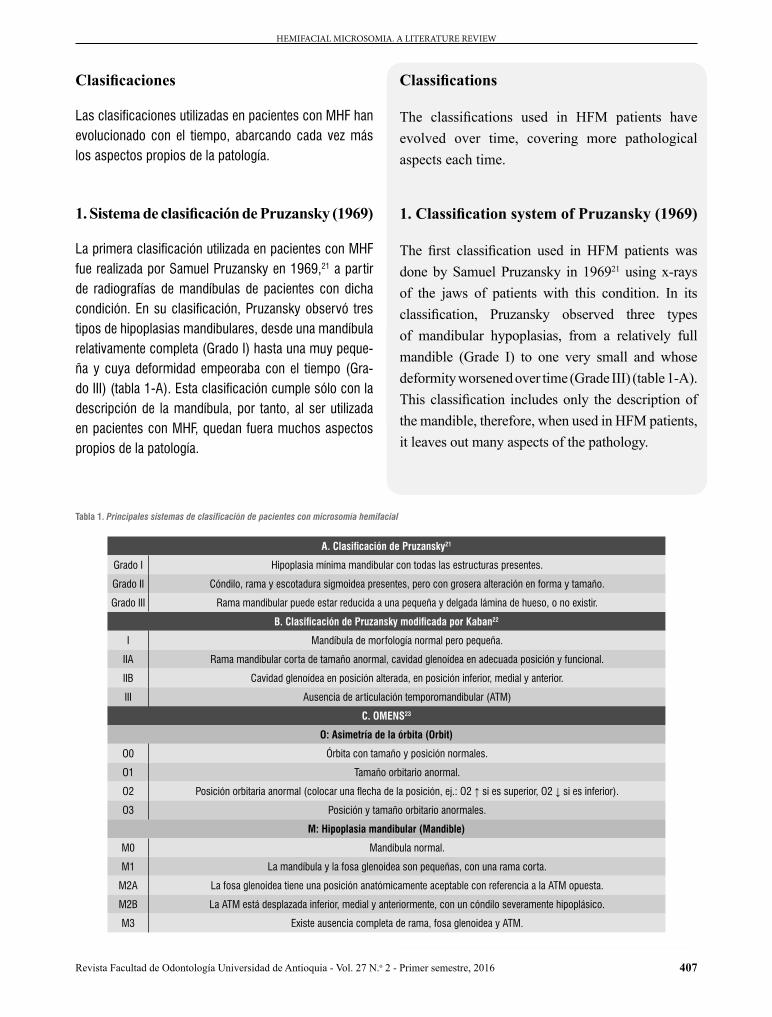
Clasificaciones
Las clasificaciones utilizadas en pacientes con MHF han evolucionado con el tiempo, abarcando cada vez más los aspectos propios de la patología.
1. Sistema de clasificación de Pruzansky (1969)
La primera clasificación utilizada en pacientes con MHF fue realizada por Samuel Pruzansky en 1969,21 a partir de radiografías de mandíbulas de pacientes con dicha condición. En su clasificación, Pruzansky observó tres tipos de hipoplasias mandibulares, desde una mandíbula relativamente completa (Grado I) hasta una muy peque-ña y cuya deformidad empeoraba con el tiempo (Gra-do III) (tabla 1-A). Esta clasificación cumple sólo con la descripción de la mandíbula, por tanto, al ser utilizada en pacientes con MHF, quedan fuera muchos aspectos propios de la patología.
Classifications
The classifications used in HFM patients have evolved over time, covering more pathological aspects each time.
1. Classification system of Pruzansky (1969)
The first classification used in HFM patients was done by Samuel Pruzansky in 196921 using x-rays of the jaws of patients with this condition. In its classification, Pruzansky observed three types of mandibular hypoplasias, from a relatively full mandible (Grade I) to one very small and whose deformity worsened over time (Grade III) (table 1-A). This classification includes only the description of the mandible, therefore, when used in HFM patients, it leaves out many aspects of the pathology.
Tabla 1. Principales sistemas de clasificación de pacientes con microsomía hemifacial
A. Clasificación de Pruzansky21
Grado I Hipoplasia mínima mandibular con todas las estructuras presentes.
Grado II Cóndilo, rama y escotadura sigmoidea presentes, pero con grosera alteración en forma y tamaño.
Grado III Rama mandibular puede estar reducida a una pequeña y delgada lámina de hueso, o no existir.
B. Clasificación de Pruzansky modificada por Kaban22
I Mandíbula de morfología normal pero pequeña.
IIA Rama mandibular corta de tamaño anormal, cavidad glenoídea en adecuada posición y funcional.
IIB Cavidad glenoídea en posición alterada, en posición inferior, medial y anterior.
III Ausencia de articulación temporomandibular (ATM)
C. OMENS23
O: Asimetría de la órbita (Orbit)
O0 Órbita con tamaño y posición normales.
O1 Tamaño orbitario anormal.
O2 Posición orbitaria anormal (colocar una flecha de la posición, ej.: O2 ↑ si es superior, O2 ↓ si es inferior).
O3 Posición y tamaño orbitario anormales.
M: Hipoplasia mandibular (Mandible)
M0 Mandíbula normal.
M1 La mandíbula y la fosa glenoidea son pequeñas, con una rama corta.
M2A La fosa glenoidea tiene una posición anatómicamente aceptable con referencia a la ATM opuesta.
M2B La ATM está desplazada inferior, medial y anteriormente, con un cóndilo severamente hipoplásico.
M3 Existe ausencia completa de rama, fosa glenoidea y ATM.
408
MICROSOMÍA HEMIFACIAL. REVISIÓN DE LA LITERATURA
Revista Facultad de Odontología Universidad de Antioquia - Vol. 27 N.o 2 - Primer semestre, 2016
Table 1. Main classification systems of hemifacial microsomia patients
A. Classification of Pruzansky21
Grade I Minimum mandibular hypoplasia with all structures present.
Grade II Condyle, ramus and sigmoid notch are present, but with serious alteration in shape and size.
Grade III Mandibular ramus can be reduced to a small thin layer of bone, or does not exist.
B. classification of Pruzansky modified by Kaban22
I Mandibular morphology is normal but small.
IIA Mandibular ramus is short but of abnormal size; glenoid cavity in right position and functional.
IIB Glenoid cavity is in an altered position, in inferior, medial, and anterior position.
III Absence of temporomandibular joint (TMJ)
C. OMENS23
O: Asymmetry of the orbit (Orbit)
O0 Orbit with normal size and position.
O1 Abnormal orbital size.
O2 Abnormal orbital position (place a position arrow, e.g.: O2 ↑ for upper, O2 ↓ for lower).
O3 Abnormal orbital size and position.
M: Mandibular hypoplasia (Mandible)
M0 Normal mandible.
M1 The mandible and glenoid fossa are small, with a short ramus.
M2A The glenoid fossa has an anatomically acceptable position with reference to the opposite TMJ.
M2B The TMJ is displaced in a lower, medial and anterior way, with a severely hypoplastic condyle.
M3 There is a complete absence of ramus, glenoid fossa and TMJ.
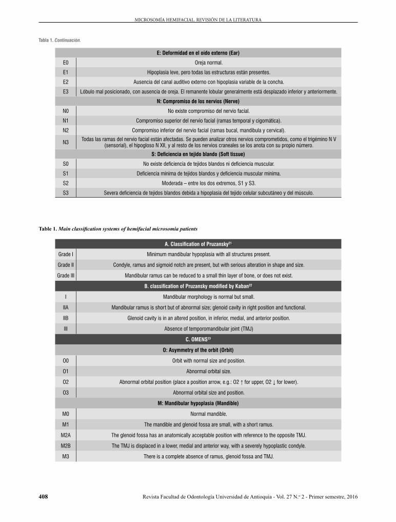
E: Deformidad en el oído externo (Ear)
E0 Oreja normal.
E1 Hipoplasia leve, pero todas las estructuras están presentes.
E2 Ausencia del canal auditivo externo con hipoplasia variable de la concha.
E3 Lóbulo mal posicionado, con ausencia de oreja. El remanente lobular generalmente está desplazado inferior y anteriormente.
N: Compromiso de los nervios (Nerve)
N0 No existe compromiso del nervio facial.
N1 Compromiso superior del nervio facial (ramas temporal y cigomática).
N2 Compromiso inferior del nervio facial (ramas bucal, mandíbula y cervical).
N3 Todas las ramas del nervio facial están afectadas. Se pueden analizar otros nervios comprometidos, como el trigémino N V (sensorial), el hipogloso N XII, y al resto de los nervios craneales se los anota con su propio número.
S: Deficiencia en tejido blando (Soft tissue)
S0 No existe deficiencia de tejidos blandos ni deficiencia muscular.
S1 Deficiencia mínima de tejidos blandos y deficiencia muscular mínima.
S2 Moderada – entre los dos extremos, S1 y S3.
S3 Severa deficiencia de tejidos blandos debida a hipoplasia del tejido celular subcutáneo y del músculo.
Tabla 1. Continuación.
409
HEMIFACIAL MICROSOMIA. A LITERATURE REVIEW
Revista Facultad de Odontología Universidad de Antioquia - Vol. 27 N.o 2 - Primer semestre, 2016
2. Clasificación de Pruzansky modificada
por Kaban et al (1988)
En 1988, Kaban et al22 modificaron la clasificación de Pruzansky, añadiendo la descripción de las deforma-ciones vistas en telerradiografías de la articulación temporomandibular. La gran diferencia se observa en el Grado II, el cual presenta dos subclasificaciones depen-diendo de la posición de la cavidad glenoídea, la cual puede estar normal (IIA) o alterada (IIB) (tabla 1-B).
3. OMENS (1991)
Dada la gran variabilidad en la expresión fenotípica de esta malformación, en 1991 se postuló la clasificación OMENS,23 hecha con base en tres criterios, pensando en que una clasificación para MHF debía ser inclusiva y versátil, que los componentes anatómicos se analizaran por separado y que los datos se expresaran en escala numérica para que fuesen útiles clínicamente. La sigla OMENS corresponde a un acrónimo de las siglas en inglés para las cinco principales manifestaciones de la
Table 1. Continue
2 Pruzansky’s classification modified by Kaban et al (1988)
In 1988, Kaban et al22 changed Pruzansky’s classification by adding the description of deformations seen in temporomandibular joint teleradiography. The greatest difference appears in Grade II, which presents two sub-classifications depending on position of the glenoid cavity, which may be normal (IIA) or altered (IIB) (table 1-B).
3 OMENS (1991)
Given the abundant phenotypic variability of this malformation, in 1991 the OMENS classification was proposed23 based on three criteria, considering that an HFM classification should be inclusive and versatile, that the anatomic components should be separately analyzed, and that data should be expressed in a numerical scale to make them clinically useful. The OMENS abbreviation corresponds to its acronym for the five main manifestations of
E: Deformity in the outer ear (Ear)
E0 Normal ear.
E1 Mild hypoplasia, but all structures are present.
E2 Absence of the external auditory canal with variable hypoplasia of the shell.
E3 Lobe poorly positioned, with absence of ear. Lobar remnant is generally moved towards a lower anterior position.
N: Nerve involvement (Nerve)
N0 There is no facial nerve involvement.
N1 Upper involvement of facial nerve (temporal and zygomatic rami).
N2 Lower involvement of facial nerve (buccal, mandibular and cervical rami).
N3 All rami of facial nerve are affected. Other nerves can be involved, such as trigeminal NV (sensory), hypoglossal N XII, and the rest of cranial nerves with their own numbers.
S: Deficiency in soft tissue (Soft tissue)
S0 There is no deficiency of soft tissue nor muscle deficiency.
S1 Minimal soft tissue and minimal muscle deficiency.
S2 Moderate – between both extremes, S1 and S3.
S3 Severe soft tissue deficiency due to hypoplasia of subcutaneous tissue and muscle.

410
MICROSOMÍA HEMIFACIAL. REVISIÓN DE LA LITERATURA
Revista Facultad de Odontología Universidad de Antioquia - Vol. 27 N.o 2 - Primer semestre, 2016
MHF: O: Asimetría de la órbita (Orbit); M: hipoplasia mandibular (Mandible); E: Deformidad en el oído externo (Ear); N: Compromiso de los nervios (Nerve); S: Defi-ciencia en tejido blando (Soft Tissue) (tabla 1-C).
3.1 Modificaciones a la OMENS
• OMENS+ (1995): Postulada con el fin de ampliar la expresión fenotípica de los pacientes con MHF hacia las alteraciones extracraneales.24
• Representación pictográfica de la OMENS+: En 2007 se introdujo una representación pictográfica de la OMENS+,25 la cual fue luego modificada en 2011,26 con el fin de facilitar su comprensión en la práctica clínica, en la docencia y en la estandarización de la clasificación de pacientes con MHF.
4. CFDS (Craniofacial Deformity Scoring)27
(2001)
Corresponde a la suma de MDS-Mandibular Deformi-ty Scoring (valoración de la deformidad mandibular) + CDS-Cranial Deformity Scoring (Valoración de la de-formidad craneal). A través del uso de tomografía com-putarizada, se analizan diferentes estructuras óseas y se les asignan valores de acuerdo con el grado en que es-tán comprometidas; los máximos valores son 16 puntos para MDS y 19 para CDS.
Características clínicas
Al ser esta una anomalía del primer y el segundo arcos branquiales, la mayoría de las alteraciones están aso-ciadas a estructuras desarrolladas a partir de dichos arcos,28 presentando una gran variación fenotípica. Se puede observar compromiso de:
• Mandíbula y ATM: Desarrollo asimétrico man-dibular por hipoplasia, ausencia de estructuras mandibulares (cóndilo y rama), ausencia o an-quilosis de la articulación temporomandibular
HFM: O: asymmetry of the orbit (Orbit); M: mandibular hypoplasia (Mandible); E: Deformity in the outer ear (Ear); N: Nerve involvement (Nerve); S: Deficiency in soft tissue (Soft Tissue) (table 1-C).
3.1 Amendments to the OMENS
• OMENS+ (1995): It was postulated in order to expand the phenotypic expression of HFM patients to extracranial alterations.24
• Graphic representation of the OMENS+: a graphic representation of the OMENS+ was introduced in 200725 and later modified in 2011,26 in order to facilitate its understanding in clinical practice, teaching and the standardization of classification of HFM patients.
4. CFDS (Craniofacial Deformity Scoring)27
(2001)
This corresponds to the sum of MDS (Mandibular Deformity Scoring) + CDS (Cranial Deformity Scoring). Through the use of computed tomography, different bone structures are analyzed and assigned values according to the extent to which they are involved; the maximum values are 16 points for MDS and 19 for CDS.
Clinical features
As this is an anomaly of the first and second branchial arches, most alterations are associated with structures developed from these arches,28 presenting wide phenotypic variation. The following structures are involved:
• Jaw and TMJ: asymmetrical mandibular development for hypoplasia, absence of mandibular structures (condyle and ramus), absence or ankylosis of temporomandibular
411
HEMIFACIAL MICROSOMIA. A LITERATURE REVIEW
Revista Facultad de Odontología Universidad de Antioquia - Vol. 27 N.o 2 - Primer semestre, 2016
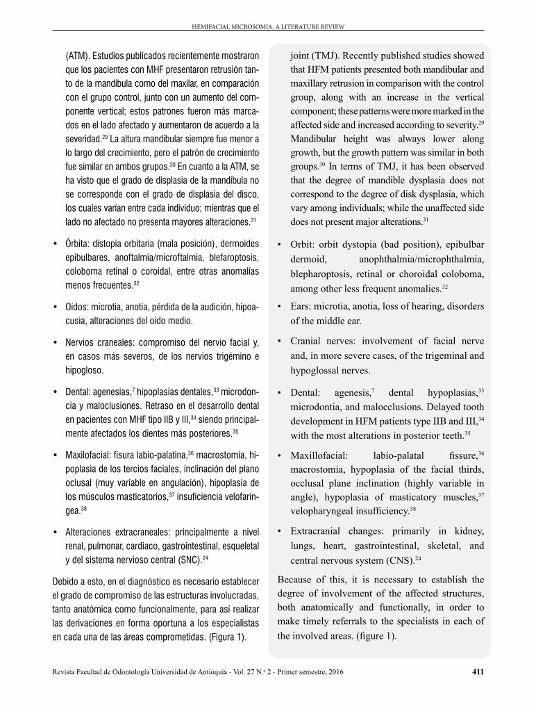
(ATM). Estudios publicados recientemente mostraron que los pacientes con MHF presentaron retrusión tan-to de la mandíbula como del maxilar, en comparación con el grupo control, junto con un aumento del com-ponente vertical; estos patrones fueron más marca-dos en el lado afectado y aumentaron de acuerdo a la severidad.29 La altura mandibular siempre fue menor a lo largo del crecimiento, pero el patrón de crecimiento fue similar en ambos grupos.30 En cuanto a la ATM, se ha visto que el grado de displasia de la mandíbula no se corresponde con el grado de displasia del disco, los cuales varían entre cada individuo; mientras que el lado no afectado no presenta mayores alteraciones.31
• Órbita: distopía orbitaria (mala posición), dermoides epibulbares, anoftalmia/microftalmia, blefaroptosis, coloboma retinal o coroidal, entre otras anomalías menos frecuentes.32
• Oídos: microtia, anotia, pérdida de la audición, hipoa-cusia, alteraciones del oído medio.
• Nervios craneales: compromiso del nervio facial y, en casos más severos, de los nervios trigémino e hipogloso.
• Dental: agenesias,7 hipoplasias dentales,33 microdon-cia y maloclusiones. Retraso en el desarrollo dental en pacientes con MHF tipo IIB y III,34 siendo principal-mente afectados los dientes más posteriores.35
• Maxilofacial: fisura labio-palatina,36 macrostomía, hi-poplasia de los tercios faciales, inclinación del plano oclusal (muy variable en angulación), hipoplasia de los músculos masticatorios,37 insuficiencia velofarín-gea.38
• Alteraciones extracraneales: principalmente a nivel renal, pulmonar, cardiaco, gastrointestinal, esqueletal y del sistema nervioso central (SNC).24
Debido a esto, en el diagnóstico es necesario establecer el grado de compromiso de las estructuras involucradas, tanto anatómica como funcionalmente, para así realizar las derivaciones en forma oportuna a los especialistas en cada una de las áreas comprometidas. (Figura 1).
joint (TMJ). Recently published studies showed that HFM patients presented both mandibular and maxillary retrusion in comparison with the control group, along with an increase in the vertical component; these patterns were more marked in the affected side and increased according to severity.29 Mandibular height was always lower along growth, but the growth pattern was similar in both groups.30 In terms of TMJ, it has been observed that the degree of mandible dysplasia does not correspond to the degree of disk dysplasia, which vary among individuals; while the unaffected side does not present major alterations.31
• Orbit: orbit dystopia (bad position), epibulbar dermoid, anophthalmia/microphthalmia, blepharoptosis, retinal or choroidal coloboma, among other less frequent anomalies.32
• Ears: microtia, anotia, loss of hearing, disorders of the middle ear.
• Cranial nerves: involvement of facial nerve and, in more severe cases, of the trigeminal and hypoglossal nerves.
• Dental: agenesis,7 dental hypoplasias,33 microdontia, and malocclusions. Delayed tooth development in HFM patients type IIB and III,34 with the most alterations in posterior teeth.35
• Maxillofacial: labio-palatal fissure,36 macrostomia, hypoplasia of the facial thirds, occlusal plane inclination (highly variable in angle), hypoplasia of masticatory muscles,37 velopharyngeal insufficiency.38
• Extracranial changes: primarily in kidney, lungs, heart, gastrointestinal, skeletal, and central nervous system (CNS).24
Because of this, it is necessary to establish the degree of involvement of the affected structures, both anatomically and functionally, in order to make timely referrals to the specialists in each of the involved areas. (figure 1).
412
MICROSOMÍA HEMIFACIAL. REVISIÓN DE LA LITERATURA
Revista Facultad de Odontología Universidad de Antioquia - Vol. 27 N.o 2 - Primer semestre, 2016
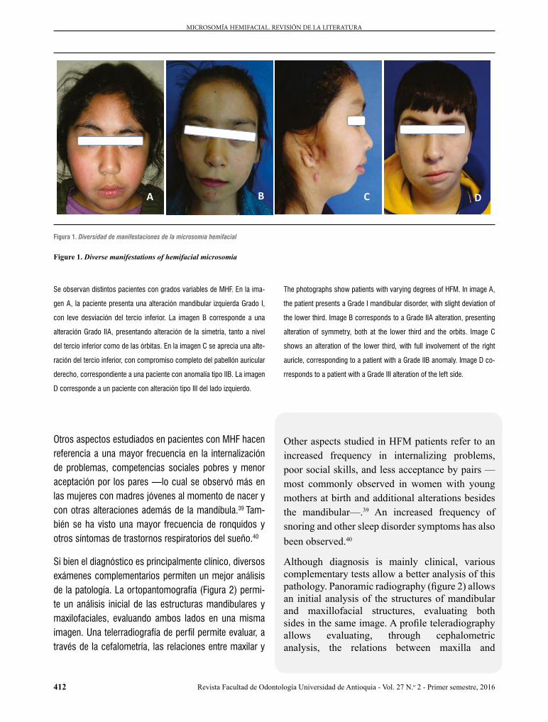
Se observan distintos pacientes con grados variables de MHF. En la ima-
gen A, la paciente presenta una alteración mandibular izquierda Grado I,
con leve desviación del tercio inferior. La imagen B corresponde a una
alteración Grado IIA, presentando alteración de la simetría, tanto a nivel
del tercio inferior como de las órbitas. En la imagen C se aprecia una alte-
ración del tercio inferior, con compromiso completo del pabellón auricular
derecho, correspondiente a una paciente con anomalía tipo IIB. La imagen
D corresponde a un paciente con alteración tipo III del lado izquierdo.
Otros aspectos estudiados en pacientes con MHF hacen referencia a una mayor frecuencia en la internalización de problemas, competencias sociales pobres y menor aceptación por los pares —lo cual se observó más en las mujeres con madres jóvenes al momento de nacer y con otras alteraciones además de la mandíbula.39 Tam-bién se ha visto una mayor frecuencia de ronquidos y otros síntomas de trastornos respiratorios del sueño.40
Si bien el diagnóstico es principalmente clínico, diversos exámenes complementarios permiten un mejor análisis de la patología. La ortopantomografía (Figura 2) permi-te un análisis inicial de las estructuras mandibulares y maxilofaciales, evaluando ambos lados en una misma imagen. Una telerradiografía de perfil permite evaluar, a través de la cefalometría, las relaciones entre maxilar y
The photographs show patients with varying degrees of HFM. In image A,
the patient presents a Grade I mandibular disorder, with slight deviation of
the lower third. Image B corresponds to a Grade IIA alteration, presenting
alteration of symmetry, both at the lower third and the orbits. Image C
shows an alteration of the lower third, with full involvement of the right
auricle, corresponding to a patient with a Grade IIB anomaly. Image D co-
rresponds to a patient with a Grade III alteration of the left side.
Other aspects studied in HFM patients refer to an increased frequency in internalizing problems, poor social skills, and less acceptance by pairs —most commonly observed in women with young mothers at birth and additional alterations besides the mandibular—.39 An increased frequency of snoring and other sleep disorder symptoms has also been observed.40
Although diagnosis is mainly clinical, various complementary tests allow a better analysis of this pathology. Panoramic radiography (figure 2) allows an initial analysis of the structures of mandibular and maxillofacial structures, evaluating both sides in the same image. A profile teleradiography allows evaluating, through cephalometric analysis, the relations between maxilla and
D C B A
Figura 1. Diversidad de manifestaciones de la microsomía hemifacial
Figure 1. Diverse manifestations of hemifacial microsomia
413
HEMIFACIAL MICROSOMIA. A LITERATURE REVIEW
Revista Facultad de Odontología Universidad de Antioquia - Vol. 27 N.o 2 - Primer semestre, 2016
mandíbula, mientras que una telerradiografía frontal per-mite observar el grado de asimetría y desviación mandi-bular. Una radiografía oclusal otorga una visión apropia-da de la bóveda palatina en caso de fisura labio-palatina.
mandible, while front teleradiography allows observing the degree of asymmetry and mandibular deviation. An occlusal x-ray offers a clear view of the palatal vault in case of labio-palatal fissure.
A B
C
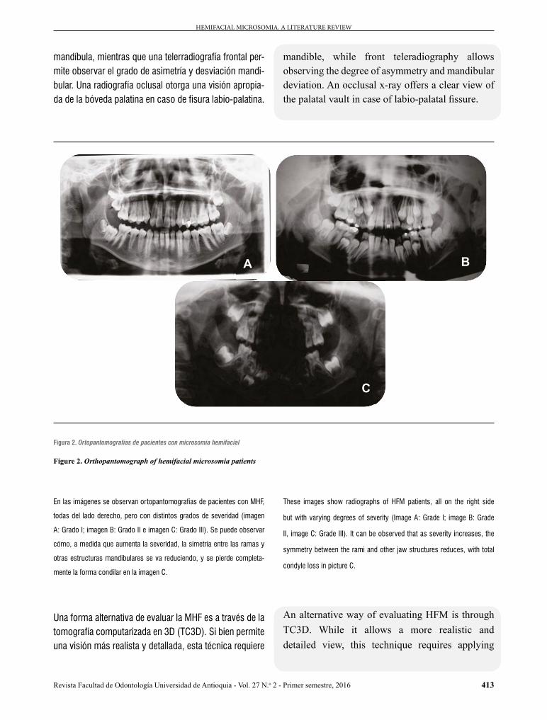
Figura 2. Ortopantomografías de pacientes con microsomía hemifacial
Figure 2. Orthopantomograph of hemifacial microsomia patients
These images show radiographs of HFM patients, all on the right side
but with varying degrees of severity (Image A: Grade I; image B: Grade
II, image C: Grade III). It can be observed that as severity increases, the
symmetry between the rami and other jaw structures reduces, with total
condyle loss in picture C.
An alternative way of evaluating HFM is through TC3D. While it allows a more realistic and detailed view, this technique requires applying
En las imágenes se observan ortopantomografías de pacientes con MHF,
todas del lado derecho, pero con distintos grados de severidad (imagen
A: Grado I; imagen B: Grado II e imagen C: Grado III). Se puede observar
cómo, a medida que aumenta la severidad, la simetría entre las ramas y
otras estructuras mandibulares se va reduciendo, y se pierde completa-
mente la forma condilar en la imagen C.
Una forma alternativa de evaluar la MHF es a través de la tomografía computarizada en 3D (TC3D). Si bien permite una visión más realista y detallada, esta técnica requiere

414
MICROSOMÍA HEMIFACIAL. REVISIÓN DE LA LITERATURA
Revista Facultad de Odontología Universidad de Antioquia - Vol. 27 N.o 2 - Primer semestre, 2016
aplicar una gran cantidad de radiación sobre el paciente. Un estudio de Takahashi y colaboradores en 2013 su-giere el uso cuidadoso de ambos métodos (radiografía panorámica y TC3D), debido a que la primera tenía una buena fiabilidad en casos de pacientes con MHF Grado I de Pruzansky, mientras que en pacientes Grado II la fiabi-lidad era baja, por lo que el uso de TC3D era preferible.41 Otra herramienta utilizada en pacientes con MHF es la fotogrametría, que permite una aproximación no invasiva en la planificación quirúrgica.42
Los protocolos fotográficos también son útiles durante el diagnóstico del paciente y en la evaluación de los avan-ces del resultado postratamiento. Un estudio publicado en 2013 por Birgfeld y colaboradores43 arrojó que era factible establecer el fenotipo de pacientes con MHF tanto con imagenología 2D como con 3D, las cuales presenta-ban diversas ventajas y desventajas. La fortaleza de las técnicas 2D consistía en que para los clínicos era más fácil trabajar debido a su familiaridad con ellas; mientras que su debilidad era que dependía mucho de la coope-ración del paciente y de las habilidades del fotógrafo; además, no se podía girar y se necesitaba estandarizar correctamente las fotografías, debido a que las leves inclinaciones dificultaban determinar correctamente el grado de asimetría. La ventaja de las imágenes en 3D era que permitía girar las imágenes y ver el aspecto fa-cial desde distintos ángulos; además era más rápido que seguir un protocolo fotográfico. Su principal desventaja era la falta de definición a nivel del oído y los párpados, y la interferencia del pelo, lo que se traducía en pérdida de información.
Diagnóstico diferencial
• Hipoplasia hemimandibular con colapso condilar-coronoídeo: cuadro generalmente no diagnosticado al nacimiento, sin alteraciones de tejidos blandos y caracterizado principalmente por desviación del mentón por hipoplasia del cóndilo, proceso coronoides y rama mandibular, siempre en presencia de fosa temporal (cavidad glenoídea).44, 45
a large amount of radiation on patients. A study by Takahashi et al in 2013 suggests careful use of both methods (panoramic radiography and TC3D), since the first had good reliability in cases of HFM patients in Grade I of Pruzansky, while reliability was low in Grade II patients, so the use of TC3D was preferable.41 Another tool used in HFM patients is photogrammetry, enabling a non-invasive approach in surgical planning.42
Photographic protocols are also useful in diagnosis and in evaluation of post-treatment advances. A study published in 2013 by Birgfeld et al43 showed that it was feasible to establish the phenotype of HFM patients with both 2D and 3D imaging, which had several advantages and disadvantages. The strength of the 2D techniques was that clinicians find them easier to handle due to familiarity with them, while their weakness was that they were highly dependable on patient cooperation and photographer skills; in addition, they could not be rotated and needed proper photograph standardization, since slight inclinations made it difficult to correctly determine the degree of asymmetry. The advantage of 3D images was that they allowed rotating images and seeing the facial appearance from different angles; it was also faster to follow than a photographic protocol. Their main disadvantage was the lack of definition at the level of ear and eyelids, and hair interference, which resulted in loss of information.
Differential diagnosis
• Hemimandibular hypoplasia with condylar-coronoid collapse: a condition not usually diagnosed at birth, with no soft tissue alterations and mainly characterized by chin deviation for hypoplasia of condyle, coronoid process, and mandibular ramus, always in the presence of temporal fossa (glenoid cavity).44, 45
415
HEMIFACIAL MICROSOMIA. A LITERATURE REVIEW
Revista Facultad de Odontología Universidad de Antioquia - Vol. 27 N.o 2 - Primer semestre, 2016
• Síndromes: La MHF, al ser clínicamente variable, debe ser descar tada por un genetista de síndromes que comprometen también el macizo maxilofacial. Síndromes como Treacher-Collins, Miller-Dieker, Townes-Brocks, CHARGE, branquio-oto-renal, Parry-Romberg, entre otros, presentan caracterís-ticas similares a la MHF, como alteraciones man-dibulares, colobomas o alteraciones palpebrales y malformaciones del oído externo.3
Tratamiento
En general, se acepta que la mejor forma de tratar a pa-cientes con MHF es a través de equipos interdisciplina-rios,46 cuya diversidad de especialidades permita, a tra-vés de un trabajo conjunto, el tratamiento apropiado para cada paciente acorde a sus necesidades.
La planificación del tratamiento se hará dependiendo del tipo de malformación y la severidad en la expresión de ésta, siempre tomando en cuenta las expectativas del paciente y la familia en cuanto a los resultados.
Con el tratamiento, principalmente de cirugía ortognáti-ca/plástica y ortodoncia, se debe buscar mejorar la fun-cionalidad, junto con una simetría facial óptima, con el fin de:47, 48
1. Aumentar el tamaño del lado mandibular afectado y su tejido blando asociado.
2. Crear una articulación que simule la ATM en los ca-sos en los que ésta esté ausente.
3. Corregir deformidades secundarias en el maxilar.
4. Establecer una oclusión funcional, así como una apa-riencia estética facial y dentaria.
5. Mejorar y horizontalizar el plano de oclusión.
6. Obtener apertura bucal cuando ésta se presenta limi-tada.
El tratamiento ortodóntico convencional puede com-prender inicialmente aparatología funcional con el uso
• Syndromes: being a clinically variable condition, HFM must be discarded by a geneticist of syndromes involving the maxillofacial complex. Syndromes such as Treacher-Collins, Miller-Dieker, Townes-Brocks, CHARGE, branquio-oto-renal, Parry-Romberg, among others, have similar characteristics to HFM, such as mandibular disorders, colobomas or eyelid malformations and abnormalities of the outer ear.3
Treatment
In general, it is accepted that the best way to treat HFM patients is through interdisciplinary teams46
whose diversity of specialties allows, through a joint effort, the appropriate treatment for each patient according to individual needs.
Treatment planning depends on type of malformation and severity in its expression, always taking into account patient and family expectations in terms of the results.
Mainly through plastic/orthognathic surgery and orthodontics, the treatment seeks to improve functionality, along with optimum facial symmetry, in order to:47, 48
1. Increase the size of the affected mandibular side and its associated soft tissue.
2. Create a joint simulating the TMJ in cases where it is absent.
3. Correct secondary deformities in maxilla.
4. Achieve functional occlusion, as well as aesthetic facial and dental appearance.
5. Improve and horizontalize the occlusal plane.
6. Achieve mouth opening if it is limited.
Conventional orthodontic treatment may initially include functional appliances with the use of
416
MICROSOMÍA HEMIFACIAL. REVISIÓN DE LA LITERATURA
Revista Facultad de Odontología Universidad de Antioquia - Vol. 27 N.o 2 - Primer semestre, 2016
de activadores rígidos de acrílico, individualizados se-gún el caso. Estos aparatos permiten la expansión del tejido afectado, aprovechando el crecimiento fisiológico del paciente. En ocasiones pueden tener planos de altura en el lado sano, permitiendo la compensación vertical de la zona afectada, siempre teniendo presente mante-ner centrada la línea media facial con la dentaria. Pos-teriormente se puede complementar con ortodoncia fija convencional.
Tratamiento quirúrgico mandibular
Si bien la literatura es amplia en cuanto a técnicas y tiem-pos quirúrgicos, el tratamiento de los pacientes con MHF se puede dividir en dos grupos, según la clasificación de Kaban-Prusansky: los pacientes Grado I y Grado IIA se tratan de la misma forma, mientras que los pacientes Grado IIB y Grado III se tratan de manera similar entre ellos, pero distinta al primer grupo.49 (tabla 2).
Tabla 2. Procedimiento quirúrgico de acuerdo al tipo de microsomía hemifacial
según la clasificación de Kaban, según Liu y colaboradores, 201249
Grado de MHF (Kaban) Procedimiento quirúrgico
Grado I y IIA
Alargamiento vertical mandibular (osteotomía e injerto) (SSMO)
Distracción ósea mandibular (vector vertical). Rama corta verticalmente.
Grado IIB
Injerto óseo de la rama y el cóndilo, con reconstruc-ción de cavidad glenoídea.
Distracción ósea mandibular (DMO) (vector oblicuo). Rama corta vertical y horizontalmente.
Grado III
Injerto óseo de la rama y el cóndilo, con reconstruc-ción de cavidad glenoídea para crear ATM funcional
Distracción ósea mandibular (vector oblicuo) e injerto óseo.
El manejo quirúrgico mandibular es esencial, y la dis-tracción ósea mandibular tiene diversas ventajas so-bre el injer to costocondral. Entre ellas está aumentar el largo ver tical de la mandíbula, mejorar la asimetría del tejido blando, producir menor pérdida de sangre, tener un mejor control del vector de avance y obtener una mejora sustancial en la biomecánica de la man-díbula.50
rigid acrylic activators, which are individualized according to each case. These devices allow for expansion of affected tissue, taking advantage of patient’s physiological growth. Sometimes they can have height planes on the healthy side, allowing for vertical compensation of the affected area, always bearing in mind that facial midline should be centered with tooth midline. This can later be complemented with conventional fixed orthodontics.
Mandibular surgery
While the literature is broad in terms of techniques and surgical times, the treatment of HFM patients can be divided into two groups, according to the classification by Kaban-Prusansky: Grade I and Grade IIA patients are treated in the same way, while Grade IIB and Grade III patients are treated similarly among them but different to the first group.49 (table 2).
Table 2. Surgical procedure according to hemifacial microsomia type
following the classification by Kaban, according to Liu et al, 201249
Degree of HFM (Kaban) Surgical procedure
Grade I and IIA
Vertical mandible lengthening (osteotomy and graft) (SSMO)
Mandible bone distraction (vertical vector). Ramus vertically short.
Grade IIB
Bone graft of ramus and condyle, with reconstruction of glenoid cavity.
Mandible bone distraction (MBD) (oblique vector). Ramus vertically and horizontally short.
Grade III
Bone graft of ramus and condyle, with reconstruction of glenoid cavity to create functional TMJ.
Mandible bone distraction (oblique vector) and bone graft.
Surgical management of the mandible is essential, and mandible bone distraction has several advantages over costochondral graft. These include: increase mandible vertical length, improve asymmetry of soft tissue, produce less blood loss, have better control of progress vector, and obtain a substantial improvement in the biomechanics of the lower mandible.50
417
HEMIFACIAL MICROSOMIA. A LITERATURE REVIEW
Revista Facultad de Odontología Universidad de Antioquia - Vol. 27 N.o 2 - Primer semestre, 2016
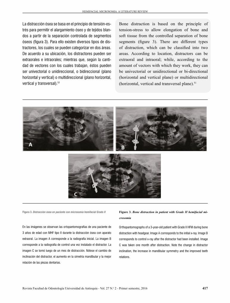
La distracción ósea se basa en el principio de tensión-es-trés para permitir el alargamiento óseo y de tejidos blan-dos a partir de la separación controlada de segmentos óseos (figura 3). Para ello existen diversos tipos de dis-tractores, los cuales se pueden categorizar en dos áreas. De acuerdo a su ubicación, los distractores pueden ser extraorales e intraorales; mientras que, según la canti-dad de vectores con los cuales trabajan, éstos pueden ser univectorial o unidireccional, o bidireccional (plano horizontal y vertical) o multidireccional (plano horizontal, vertical y transversal).51
Bone distraction is based on the principle of tension-stress to allow elongation of bone and soft tissue from the controlled separation of bone segments (figure 3). There are different types of distraction, which can be classified into two areas. According to location, distractors can be extraoral and intraoral; while, according to the amount of vectors with which they work, they can be univectorial or unidirectional or bi-directional (horizontal and vertical plane) or multidirectional (horizontal, vertical and transversal plane).51
A
C
B
A
C
B
A
C
B
Figura 3. Distracción ósea en paciente con microsomía hemifacial Grado II
En las imágenes se observan las ortopantomografías de una paciente de
3 años de edad con MHF tipo II durante la distracción ósea con aparato
extraoral. La imagen A corresponde a la radiografía inicial. La imagen B
corresponde a la radiografía de control una vez instalado el distractor. La
imagen C se tomó luego de un mes de distracción. Nótese el cambio de
inclinación del distractor, el aumento en la simetría mandibular y la mejor
relación de las piezas dentarias.
Figure 3. Bone distraction in patient with Grade II hemifacial mi-
crosomia
Orthopantomographs of a 3-year-old patient with Grade II HFM during bone
distraction with headgear. Image A corresponds to the initial x-ray. Image B
corresponds to control x-ray after the distractor had been installed. Image
C was taken one month after distraction. Note the change in distractor
inclination, the increase in mandibular symmetry and the improved teeth
relations.
418
MICROSOMÍA HEMIFACIAL. REVISIÓN DE LA LITERATURA
Revista Facultad de Odontología Universidad de Antioquia - Vol. 27 N.o 2 - Primer semestre, 2016
La elección del aparto de distracción dependerá de di-versos factores, entre ellos la edad del paciente, el grado de severidad de la malformación y la necesidad de mo-vilidad de los segmentos. En general, los aparatos ex-traorales requieren una cirugía menos compleja, pueden lograr distancias de distracción más largas y permiten manejar la distracción en los tres sentidos del espacio, pero dejan cicatrices en la piel, son más visibles y pue-den llegar a ser más pesados, presentando molestias para el paciente. Los aparatos intraorales son menos visibles y no dejan cicatriz facial, pero tienen un limitado rango de movilidad en el espacio, y su cirugía es más compleja tanto para su inserción como para su remo-ción. Recientemente se han desarrollado dispositivos reabsorbibles que permiten la distracción mandibular en una etapa, logrando avances de entre 15 y 30 mm.52 De todas formas, la simulación quirúrgica en un modelo sólido puede ayudar a lograr mejor simetría mandibular, especialmente en distracción ósea con multivector.53
Es importante recalcar la necesidad de tomar en cuen-ta, más allá del estadio del crecimiento del paciente, las necesidades de tratamiento que éste tiene, ya que varían entre los pacientes, y por tanto cada caso debe ser ana-lizado como único.
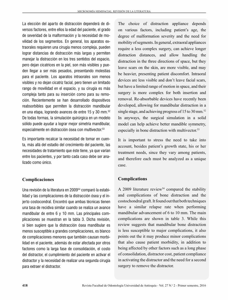
Complicaciones
Una revisión de la literatura en 200954 comparó la estabi-lidad y las complicaciones de la distracción ósea y el in-jerto costocondral. Encontró que ambas técnicas tienen una tasa de recidiva similar cuando se realiza un avance mandibular de entre 6 y 10 mm. Las principales com-plicaciones se muestran en la tabla 3. Dicha revisión, si bien sugiere que la distracción ósea mandibular es menos susceptible a grandes complicaciones, es blanco de complicaciones menores que también causan morbi-lidad en el paciente, además de estar afectada por otros factores como la larga fase de consolidación, el costo del distractor, el cumplimiento del paciente en activar el distractor y la necesidad de realizar una segunda cirugía para extraer el distractor.
The choice of distraction appliance depends on various factors, including patient’s age, the degree of malformation severity and the need for mobility of segments. In general, extraoral appliances require a less complex surgery, can achieve longer distraction distances, and allow handling the distraction in the three directions of space, but they leave scars on the skin, are more visible, and may be heavier, presenting patient discomfort. Intraoral devices are less visible and don’t leave facial scars, but have a limited range of motion in space, and their surgery is more complex for both insertion and removal. Re-absorbable devices have recently been developed, allowing for mandibular distraction in a single stage, and achieving progress of 15 to 30 mm.52 In anyways, the surgical simulation in a solid model can help achieve better mandible symmetry, especially in bone distraction with multivector.53
It is important to stress the need to take into account, besides patient’s growth state, his or her treatment needs, since they vary among patients, and therefore each must be analyzed as a unique case.
Complications
A 2009 literature review54 compared the stability and complications of bone distraction and the costochondral graft. It found out that both techniques have a similar relapse rate when performing mandibular advancement of 6 to 10 mm. The main complications are shown in table 3. While this review suggests that mandibular bone distraction is less susceptible to major complications, it also points out the it may produce minor complications that also cause patient morbidity, in addition to being affected by other factors such as a long phase of consolidation, distractor cost, patient compliance in activating the distractor and the need for a second surgery to remove the distractor.
419
HEMIFACIAL MICROSOMIA. A LITERATURE REVIEW
Revista Facultad de Odontología Universidad de Antioquia - Vol. 27 N.o 2 - Primer semestre, 2016
Tabla 3. Posibles complicaciones de la osteotomía bilateral sagital de rama y de la distracción ósea mandibular
Procedimiento Complicaciones
BSSO
RecaídaTemprana
Deslizamiento de la osteotomía.Hundimiento condilar sin contacto.
Tardía Remodelado o reabsorción del cóndilo.Alteraciones en el nervio alveolar inferior.Alteraciones temporomandibulares.Crecimiento y reabsorción impredecible.Falta de tejido blando y de irrigación vascular en la zona.
DMO57
Mordida abierta anterior.Quiebre o mal función del distractor.Incumplimiento del paciente en la activación y en los cuidados.Infección.Úlceras en labio inferior por presión del distractor.En el caso de DMO temprana, solo se indica en casos de obstrucción severa de la vía aérea, debido a la limitación mostrada en el tiempo de mantenerse estable.58, 59
Table 3. Possible complications of bilateral sagittal osteotomy of ramus and mandible bone distraction
Procedure Complications
BSSO
RelapseEarly
Sliding of osteotomy.Sinking of condyle without contact.
Late Remodeling or resorption of condyle.Alterations in the inferior alveolar nerve.Temporomandibular disorders.Growth and unpredictable reabsorption.Lack of soft tissue and vascular irrigation in the area.
MDO57
Anterior open bite.Breakage or malfunction of distractor.Patient failure to activate and provide care.Infection.Ulcers in lower lip by pressure of distractor.In the case of early DMO, it is only indicated in cases of severe obstruction of airway, due to limitations to remain stable over time.58, 59
Otras cirugías
Al ser una malformación con gran variación fenotípica, los pacientes con MHF pueden necesitar otras cirugías, dependiendo de las estructuras comprometidas. Una revisión de la microsomía craneofacial realizada por Birgfeld y Heike en 201255 muestra una línea de tiempo como guía en el manejo médico-quirúrgico de los pa-cientes con MHF, propuesta por los miembros del Centro Craneofacial en el Hospital de Niños de Seattle, contem-plando su avance desde el nacimiento hasta la adultez. Dependiendo del paciente, puede ser necesario realizar:
Other surgeries
Being a malformation with abundant phenotypic variation, HFM patients may need other surgeries, depending on the involved structures. A review of craniofacial microsomia performed by Birgfeld and Heike in 201255 shows a timeline as a guide in the medical and surgical management of HFM patients, proposed by members of the Seattle Children’s Craniofacial Center, monitoring its progress from birth to adulthood. Depending on patient, it may be necessary:
420
MICROSOMÍA HEMIFACIAL. REVISIÓN DE LA LITERATURA
Revista Facultad de Odontología Universidad de Antioquia - Vol. 27 N.o 2 - Primer semestre, 2016
Órbita: Puede involucrar cirugías de dermoides epibulba-res y coloboma del párpado. En casos de alteraciones en tamaño o posición de la órbita, la reposición se realiza alrededor de los 3 o 4 años de edad.
Oídos: Tubos de ventilación timpánica (colleras). En caso de alteración del oído externo, la cirugía varía de acuerdo al compromiso del pabellón auricular, desde la remodelación del cartílago en caso de hipoplasias leves, hasta la reconstrucción completa a través de injertos autólogos o aloplásticos, como el polietileno poroso de alta densidad. En pacientes con pérdida auditiva, se debe evaluar la posibilidad de reparación de atresia del canal auditivo.
Nervio facial: En casos de parálisis del nervio, se debe evaluar la zona y el grado de compromiso, que puede ser superior (afecta a la zona temporal y cigomática), inferior (zona bucal, mandibular y cervical) o total. En caso de compromiso bucal, se debe evaluar la necesi-dad de realizar reanimación facial. En caso de alteración del movimiento palpebral, se debe considerar algún tipo de tratamiento con el fin de evitar queratitis corneal por exposición.
Tejidos blandos: Cierre de fisuras orofaciales, corrección de vía aérea, cirugía de aumento de tejidos blandos e injertos de grasa.
En Chile no existen protocolos de tratamiento publicados para la microsomía hemifacial, patología que tampoco está cubierta por el sistema público de salud, por lo que la mayoría de los pacientes realiza sus tratamientos en forma privada, siendo condicionados por diversos facto-res como el acceso a especialistas con conocimiento en MHF, costo de los procedimientos, previsión de salud, entre otros.
En Latinoamérica sólo existe un protocolo de seguimien-to,56 publicado en Argentina, pero no existe ningún pro-tocolo de tratamiento.
Orbit: It may involve surgeries of epibulbar dermoid and eyelid coloboma. In cases of changes in size or position of the orbit, the replacement is done around the age of 3 to 4 years.
Ears: Tympanic ventilation tubes (clamps). In case of alteration of the outer ear, surgery varies according to the involvement of pinna, from remodeling cartilage in the event of slight hypoplasias to the complete reconstruction through autologous or alloplastic grafts, such as porous high-density polyethylene. In patients with hearing loss, the possibility of repairing atresia of the auditory canal should be evaluated.
Facial nerve: in cases of nerve palsy, the area and degree of involvement should be evaluated; this involvement may be upper (affects the temporal and zygomatic area), lower (buccal, mandibular and cervical area), or total. In the case of oral involvement, the need for facial reanimation should be evaluated. In case of alteration of eyelid movement, some type of treatment should be considered in order to prevent corneal keratitis by exposure.
Soft tissue: Closure of orofacial fissures, correction of airway, surgery of soft tissue augmentation, and fat grafts.
In Chile there are no published treatment protocols for hemifacial microsomia, a pathology that is not covered by the public health system, so most patients have their treatments in privet offices and therefore are conditioned by various factors such as access to specialists with knowledge in HFM, cost of procedures, or health prevision, among others.
In Latin America there is only one follow-up protocol56 published in Argentina, but there is no treatment protocol.
421
HEMIFACIAL MICROSOMIA. A LITERATURE REVIEW
Revista Facultad de Odontología Universidad de Antioquia - Vol. 27 N.o 2 - Primer semestre, 2016
CONCLUSIONES
La microsomía hemifacial es una enfermedad hetero-génea, variable y de expresividad única en cada sujeto, tanto en su etiología, como en su severidad y por ende en su tratamiento. Al ser una alteración en espectro, afecta diversas estructuras del individuo de acuerdo a su gravedad. Es por ello que en estos pacientes es de vital importancia el trabajo interdisciplinario muy bien coordi-nado, ya que pueden presentar incluso alteraciones psi-cosociales y extracraneales, que deben ser exploradas y tratadas a tiempo.
CONFLICTOS DE INTERÉS
Los autores declaran no tener ningún conflicto de inte-rés.
CORRESPONDENCIA
Sebastián Véliz Méndez(+5698) 166 [email protected] La Paz N.° 750, 2.° piso, Oficina de Unidad de Malformación CraneofacialSantiago, Chile
CONCLUSIONS
Hemifacial microsomia is a heterogeneous, variable disease of unique expression in each subject, both in its etiology and severity and therefore in its treatment. Being an alteration of wide spectrum, it affects various structures of the individual according to its severity. This is why a very well-coordinated interdisciplinary work is vital in these patients, since they may even have psychosocial and extracranial alterations which should be timely explored and treated.
CONFLICTS OF INTEREST
The authors declare not having any conflict of interest.
CORRESPONDING AUTHOR
Sebastián Véliz Méndez(+5698) 166 [email protected] La Paz N.° 750, 2.° piso, Oficina de Unidad de Malformación CraneofacialSantiago, Chile
REFERENCIAS / REFERENCES
1. Cohen MM Jr, Rollnick BR, Kaye CI. Oculoauriculovertebral spectrum: an updated critique. Cleft Palate J 1989; 26(4): 276-286.
2. Mielnik-Blaszczak M. Hemifacial microsomia. Review of the literature. Dent Med Probl 2011; 40(1): 80-85.
3. Heike CL, Luquetti DV, Hing AV. Craniofacial microsomia overview. In: Pagon RA, Adam MP, Bird TD editors. GeneReviews® [Internet]. Seattle: University of Washington, Seattle; 1993-2013. Disponible en: http://www.ncbi.nlm.nih.gov/books/NBK5199/.
4. Hartsfield JK. Review of the etiologic heterogeneity of the oculo-auriculo-vertebral spectrum (Hemifacial Microsomia). Orthod Craniofac Res 2007; 10(3): 121-128.
5. Poswillo D. The pathogenesis of the first and second branchial arch syndrome. Oral Surg Oral Med Oral Pathol 1973; 35(3): 302-328.
6. Johnston MC, Bronsky PT. Animal models for human craniofacial malformations. J Craniofac Genet Dev Biol 1991; 11(4): 277-291.
422
MICROSOMÍA HEMIFACIAL. REVISIÓN DE LA LITERATURA
Revista Facultad de Odontología Universidad de Antioquia - Vol. 27 N.o 2 - Primer semestre, 2016
7. Maruko E, Hayes C, Evans CA, Padwa B, Mulliken JB. Hypodontia in hemifacial microsomia. Cleft Palate Craniofac J 2001; 38(1): 15-19.
8. Sulik KK, Cook CS, Webster WS. Teratogens and craniofacial malformations: relationships to cell death. Development 1988;103 (Suppl): 213-231.
9. Wiszniak S, Mackenzie FE, Anderson P, Kabbara S, Ruhrberg C, Schwarz Q. Neural crest cell-derived VEGF promotes embryonic jaw extension. Proc Natl Acad Sci U S A. 2015; 112(19): 6086-6091.
10. Werler MM, Sheehan JE, Hayes C, Mitchell AA, Mulliken JB. Vasoactive exposures, vascular events, and hemifacial microsomia. Birth Defects Res A Clin Mol Teratol 2004; 70(6): 389-395.
11. Werler MM, Sheehan JE, Hayes C, Padwa BL, Mitchell AA, Mulliken JB. Demographic and reproductive factors associated with hemifacial microsomia. Cleft Palate Craniofac J 2004; 41(5): 494-450.
12. Wieczorek D, Ludwig M, Boehringer S, Jongbloet PH, Gillessen-Kaesbach G, Horsthemke B. Reproduction abnormalities and twin pregnancies in parents of sporadic patients with oculo-auriculo-vertebral spectrum/Goldenhar syndrome. Hum Genet 2007; 121(3-4): 369-376.
13. Ewart-Toland A, Yankowitz J, Winder A, Imagire R, Cox VA, Aylsworth AS et al. Oculoauriculovertebral abnormalities in children of diabetic mothers. Am J Med Genet 2000; 90(4): 303-309.
14. Wang R, Martinez-Frias ML, Graham JM Jr. Infants of diabetic mothers are at increased risk for the oculo-auriculo-vertebral sequence: A case-based and case-control approach. J Pediatr 2002; 141(5): 611-617.
15. Werler MM, Starr JR, Cloonan YK, Speltz ML. Hemifacial microsomia: from gestation to childhood. J Craniofac Surg 2009; 20 (Suppl 1): 664-669.
16. Tasse C, Majewski F, Bohringer S, Fischer S, Ludecke HJ, Gillessen-Kaesbach G et al. A family with autosomal dominant oculo-auriculo-vertebral spectrum. Clin Dysmorphol 2007; 16(1): 1-7.
17. Kelberman D, Tyson J, Chandler DC, McInerney AM, Slee J, Albert D et al. Hemifacial microsomia: progress in understanding the genetic basis of a complex malformation syndrome. Hum Genet 2001; 109(6): 638-645.
18. Brady AF, Winter RM, Wilson LC, Tatnall FM, Sheridan RJ, Garrett C. Hemifacial microsomia, external auditory canal atresia, deafness and Mullerian anomalies
associated with acro-osteolysis: a new autosomal recessive syndrome? Clin Dysmorphol 2002; 11(3): 155-161.
19. Choong YF, Watts P, Little E, Beck L. Goldenhar and cri-du-chat syndromes: a contiguous gene deletion syndrome? J AAPOS 2003; 7(3): 226-227.
20. Derbent M, Yilmaz Z, Baltaci V, Saygili A, Varan B, Tokel K. Chromosome 22q11.2 deletion and phenotypic features in 30 patients with conotruncal heart defects. Am J Med Genet A 2003; 116A(2): 129-135.
21. Pruzansky S. Not all dwarfed mandibles are alike. Birth Defects 1969; 1: 120-9.
22. Kaban LB, Moses MH, Mulliken JB. Surgical correction of hemifacial microsomia in the growing child. Plast Reconstr Surg 1988; 82(1): 9-19.
23. Vento AR, LaBrie RA, Mulliken JB. The O.M.E.N.S. classification of hemifacial microsomia. Cleft Palate Craniofac J 1991; 28(1): 68-76.
24. Horgan JE, Padwa BL, LaBrie RA, Mulliken JB. OMENS-Plus: analysis of craniofacial and extracraniofacial anomalies in hemifacial microsomia. Cleft Palate Craniofac J 1995; 32(5): 405-412.
25. Gougoutas AJ, Singh DJ, Low DW, Bartlett SP. Hemifacial microsomia: clinical features and pictographic representations of the OMENS classification system. Plast Reconstr Surg 2007; 120(7): 112e-120e.
26. Birgfeld CB, Luquetti DV, Gougoutas AJ, Bartlett SP, Low DW, Sie KC et al. A phenotypic assessment tool for craniofacial microsomia. Plast Reconstr Surg 2011; 127(1): 313-320.
27. Huisinga-Fischer CE, Zonneveld FW, Vaandrager JM, Prahl-Andersen B. CT-based size and shape determination of the craniofacial skeleton: a new scoring system to assess bony deformities in hemifacial microsomia. J Craniofac Surg 2001; 12(1): 87-94.
28. Sze RW, Paladin AM, Lee S, Cunningham ML. Hemifacial microsomia in pediatric patients: asymmetric abnormal development of the first and second branchial arches. AJR Am J Roentgenol 2002; 178(6): 1523-1530.
29. Ongkosuwito EM, van Neck JW, Wattel E, van Adrichem LN, Kuijpers-Jagtman AM. Craniofacial morphology in unilateral hemifacial microsomia. Br J Oral Maxillofac Surg 2012; 51(8): 902-907
30. Ongkosuwito EM, van Vooren J, van Neck JW, Wattel E, Wolvius EB, van Adrichem LN et al. Changes of mandibular ramal height, during growth in unilateral
423
HEMIFACIAL MICROSOMIA. A LITERATURE REVIEW
Revista Facultad de Odontología Universidad de Antioquia - Vol. 27 N.o 2 - Primer semestre, 2016
hemifacial microsomia patients and unaffected controls. J Craniomaxillofac Surg 2013; 41(2): 92-97.
31. Kitai N, Murakami S, Takashima M, Furukawa S, Kreiborg S, Takada K. Evaluation of temporomandibular joint in patients with hemifacial microsomia. Cleft Palate Craniofac J 2004; 41(2): 157-162.
32. Gorlin RJ, Cohen MM Jr, Hennekam RCM. Syndromes of the head and neck. 4 ed. New York: Oxford University Press; 2001.
33. Farias M, Vargervik K. Tooth size and morphology in hemifacial microsomia. Int J Pediatr Dent 1998; 8(3): 197-201.
34. Ongkosuwito EM, de Gijt P, Wattel E, Carels CE, Kuijpers-Jagtman AM. Dental development in hemifacial microsomia. J Dent Res 2010; 89(12): 1368-1372.
35. Seow WK, Urban S, Vafaie N, Shusterman S. Morphometric analysis of the primary and permanent dentitions in hemifacial microsomia: a controlled study. J Dent Res 1998; 77(1): 27-38.
36. Fan WS, Mulliken JB, Padwa BL. An association between hemifacial microsomia and facial clefting. J Oral Maxillofac Surg 2005; 63(3): 330-334.
37. Heude E, Rivals I, Couly G, Levi G. Masticatory muscle defects in hemifacial microsomia: a new embryological concept. Am J Med Genet A 2011; 155A(8): 1991-1995.
38. Funayama E, Igawa HH, Nishizawa N, Oyama A, Yamamoto Y. Velopharyngeal insufficiency in hemifacial microsomia: analysis of correlated factors. Otolaryngol Head Neck Surg 2007; 136(1): 33-37.
39. Dufton LM, Speltz ML, Kelly JP, Leroux B, Collett BR, Werler MM. Psychosocial outcomes in children with hemifacial microsomia. J Pediatr Psychol 2011; 36(7): 794-805.
40. Cloonan YK, Kifle Y, Davis S, Speltz ML, Werler MM, Starr JR. Sleep outcomes in children with hemifacial microsomia and controls: a follow-up study. Pediatrics 2009; 124(2): e313-321.
41. Takahashi-Ichikawa N, Susami T, Nagahama K, Ohkubo K, Okayasu M, Uchino N et al. Evaluation of mandibular hypoplasia in patients with hemifacial microsomia: a comparison between panoramic radiography and three-dimensional computed tomography. Cleft Palate Craniofac J 2013; 50(4): 381-387.
42. Jayaratne YS, Lo J, Zwahlen RA, Cheung LK. Three-dimensional photogrammetry for surgical planning of
tissue expansion in hemifacial microsomia. Head Neck 2010; 32(12): 1728-1735.
43. Birgfeld CB, Saltzman BS, Luquetti DV, Latham K, Starr JR, Heike CL. Comparison of two-dimensional and three-dimensional images for phenotypic assessment of craniofacial microsomia. Cleft Palate Craniofac J 2013; 50(3): 305-314.
44. Meazzini MC, Brusati R, Diner P, Gianni E, Lalatta F, Magri AS et al. The importance of a differential diagnosis between true hemifacial microsomia and pseudo-hemifacial microsomia in the post-surgical long-term prognosis. J Craniomaxillofac Surg 2011; 39(1): 10-16.
45. Meazzini MC, Brusati R, Caprioglio A, Diner P, Garattini G, Gianni E et al. True hemifacial microsomia and hemimandibular hypoplasia with condylar-coronoid collapse: diagnostic and prognostic differences. Am J Orthod Dentofacial Orthop 2011; 139(5): e435-447.
46. Association AC-P-C. Parameters for the Evaluation and treatment of patients with cleft lip/palate or other craniofacial anomalies. Revised Edition 2009. Original Publication Cleft Palate-Craniofacial Journal 1993; 30 (Suppl 1).
47. Ohtani J, Hoffman WY, Vargervik K, Oberoi S. Team management and treatment outcomes for patients with hemifacial microsomia. Am J Orthod Dentofacial Orthop 2012; 141 (Suppl 4): S74-81.
48. Vargervik K, Oberoi S, Hoffman WY. Team care for the patient with cleft: UCSF protocols and outcomes. J Craniofac Surg 2009; 20 (Suppl 2): 1668-1671.
49. Liu SYC, Good P, Lee JS. Chapter 94 - Surgical care of the hemifacial microsomia patient. Current therapy in oral and maxillofacial surgery. Saint Louis: W.B. Saunders; 2012, p. 828-834.
50. Reina-Romo E, Sampietro-Fuentes A, Gomez-Benito MJ, Dominguez J, Doblare M, Garcia-Aznar JM. Biomechanical response of a mandible in a patient affected with hemifacial microsomia before and after distraction osteogenesis. Med Eng Phys 2010; 32(8): 860-866.
51. Lo J, Cheung LK. Distraction osteogenesis for the craniomaxillofacial region. Part 2: a compendium of devices for the mandible. Asian J Oral Maxillofac Surg. 2007; 19(1): 6-18.
52. Burstein FD. Resorbable distraction of the mandible: technical evolution and clinical experience. J Craniof Surg 2008; 19(3): 637-643.
424
MICROSOMÍA HEMIFACIAL. REVISIÓN DE LA LITERATURA
Revista Facultad de Odontología Universidad de Antioquia - Vol. 27 N.o 2 - Primer semestre, 2016
53. Sakamoto Y, Ogata H, Nakajima H, Kishi K, Sakamoto T, Ishii T. Role of solid model simulation surgery for hemifacial microsomia. Cleft Palate Craniofac J 2013; 50(5): 623-626.
54. Ow A, Cheung LK. Skeletal stability and complications of bilateral sagittal split osteotomies and mandibular distraction osteogenesis: an evidence-based review. J Oral Maxillofac Surg 2009; 67(11): 2344-2353.
55. Birgfeld CB, Heike C. Craniofacial microsomia. Semin Plast Surg 2012; 26(2): 91-104.
56. Lara-García TU, Ulfe I, Rodriguez JC, Dogliotti PLV. Protocolo de seguimiento de pacientes con microsomía hemifacial. Prensa Med Argent 2002; 89(5): 414-423.
57. Van Strijen PJ, Breuning KH, Becking AG, Perdijk FBT, Tuinzing DB. Complications in bilateral mandibular distraction osteogenesis using internal devices. Oral Surg Oral Med Oral Pathol Oral Radiol Endod 2003; 96(4): 392-397.
58. Meazzini MC, Mazzoleni F, Bozzetti A, Brusati R. Comparison of mandibular vertical growth in hemifacial microsomia patients treated with early distraction or not treated: follow up till the completion of growth. J Craniomaxillofac Surg 2012; 40(2): 105-111.
59. Gursoy S, Hukki J, Hurmerinta K. Five year follow-up of mandibular distraction osteogenesis on the dentofacial structures of syndromic children. Orthod Craniofac Res 2008; 11(1): 57-64.


![Revisión de Tema Pag.[ 101 - 130 ] ISSN 2071-081X · 2019-04-24 · una función superior y una zona identificable del córtex cerebral en el hemisferio izquierdo” (Manning, 1992,](https://img.pdfslide.us/doc/110x75/5f08cb177e708231d423bfc7/revisin-de-tema-pag-101-130-issn-2071-2019-04-24-una-funcin-superior.jpg)


![Revisión de Tema Pag.[ 151 - ]169 ISSN 2071-081X](https://img.pdfslide.us/doc/110x75/6217b67c50b77c15f55641ce/revisin-de-tema-pag-151-169-issn-2071-081x.jpg)