Embed Size (px)
Citation preview
1
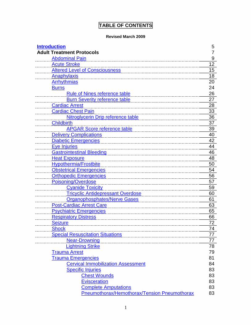
TABLE OF CONTENTS
Revised March 2009
Introduction 5
Adult Treatment Protocols 7
Abdominal Pain 9
Acute Stroke 12
Altered Level of Consciousness 15
Anaphylaxis 18
Arrhythmias 20
Burns 24
Rule of Nines reference table 26
Burn Severity reference table 27
Cardiac Arrest 28
Cardiac Chest Pain 33
Nitroglycerin Drip reference table 36
Childbirth 37
APGAR Score reference table 39
Delivery Complications 40
Diabetic Emergencies 42
Eye Injuries 44
Gastrointestinal Bleeding 46
Heat Exposure 48
Hypothermia/Frostbite 50
Obstetrical Emergencies 54
Orthopedic Emergencies 56
Poisoning/Overdose 57
Cyanide Toxicity 59
Tricyclic Antidepressant Overdose 60
Organophosphates/Nerve Gases 61
Post-Cardiac Arrest Care 63
Psychiatric Emergencies 65
Respiratory Distress 66
Seizure 72
Shock 74
Special Resuscitation Situations 77
Near-Drowning 77
Lightning Strike 78
Trauma Arrest 79
Trauma Emergencies 81
Cervical Immobilization Assessment 84
Specific Injuries 83
Chest Wounds 83
Evisceration 83
Complete Amputations 83
Pneumothorax/Hemothorax/Tension Pneumothorax 83
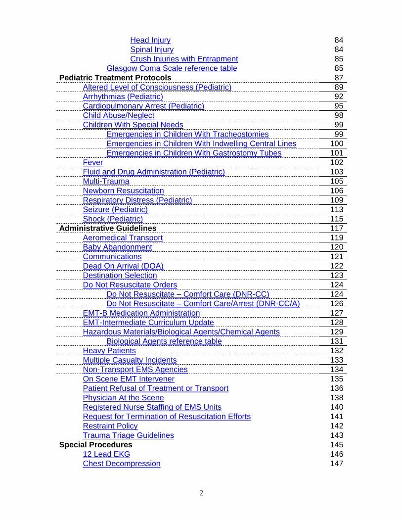
2
Head Injury 84
Spinal Injury 84
Crush Injuries with Entrapment 85
Glasgow Coma Scale reference table 85
Pediatric Treatment Protocols 87
Altered Level of Consciousness (Pediatric) 89
Arrhythmias (Pediatric) 92
Cardiopulmonary Arrest (Pediatric) 95
Child Abuse/Neglect 98
Children With Special Needs 99
Emergencies in Children With Tracheostomies 99
Emergencies in Children With Indwelling Central Lines 100
Emergencies in Children With Gastrostomy Tubes 101
Fever 102
Fluid and Drug Administration (Pediatric) 103
Multi-Trauma 105
Newborn Resuscitation 106
Respiratory Distress (Pediatric) 109
Seizure (Pediatric) 113
Shock (Pediatric) 115
Administrative Guidelines 117
Aeromedical Transport 119
Baby Abandonment 120
Communications 121
Dead On Arrival (DOA) 122
Destination Selection 123
Do Not Resuscitate Orders 124
Do Not Resuscitate – Comfort Care (DNR-CC) 124
Do Not Resuscitate – Comfort Care/Arrest (DNR-CC/A) 126
EMT-B Medication Administration 127
EMT-Intermediate Curriculum Update 128
Hazardous Materials/Biological Agents/Chemical Agents 129
Biological Agents reference table 131
Heavy Patients 132
Multiple Casualty Incidents 133
Non-Transport EMS Agencies 134
On Scene EMT Intervener 135
Patient Refusal of Treatment or Transport 136
Physician At the Scene 138
Registered Nurse Staffing of EMS Units 140
Request for Termination of Resuscitation Efforts 141
Restraint Policy 142
Trauma Triage Guidelines 143
Special Procedures 145
12 Lead EKG 146
Chest Decompression 147
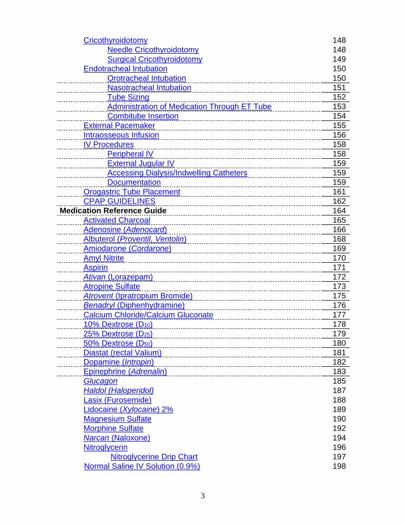
3
Cricothyroidotomy 148
Needle Cricothyroidotomy 148
Surgical Cricothyroidotomy 149
Endotracheal Intubation 150
Orotracheal Intubation 150
Nasotracheal Intubation 151
Tube Sizing 152
Administration of Medication Through ET Tube 153
Combitube Insertion 154
External Pacemaker 155
Intraosseous Infusion 156
IV Procedures 158
Peripheral IV 158
External Jugular IV 159
Accessing Dialysis/Indwelling Catheters 159
Documentation 159
Orogastric Tube Placement 161
CPAP GUIDELINES 162
Medication Reference Guide 164
Activated Charcoal 165
Adenosine (Adenocard) 166
Albuterol (Proventil, Ventolin) 168
Amiodarone (Cordarone) 169
Amyl Nitrite 170
Aspirin 171
Ativan (Lorazepam) 172
Atropine Sulfate 173
Atrovent (Ipratropium Bromide) 175
Benadryl (Diphenhydramine) 176
Calcium Chloride/Calcium Gluconate 177
10% Dextrose (D10) 178
25% Dextrose (D25) 179
50% Dextrose (D50) 180
Diastat (rectal Valium) 181
Dopamine (Intropin) 182
Epinephrine (Adrenalin) 183
Glucagon 185
Haldol (Haloperidol) 187
Lasix (Furosemide) 188
Lidocaine (Xylocaine) 2% 189
Magnesium Sulfate 190
Morphine Sulfate 192
Narcan (Naloxone) 194
Nitroglycerin 196
Nitroglycerine Drip Chart 197
Normal Saline IV Solution (0.9%) 198
4
Nubain (Nalbuphine) 200
Oxygen (O2) 201
Phenergan (Promethazine) 202
Sodium Bicarbonate (8.4%) 203
Tetracaine (Pontocaine) Ophthalmic Drops 205
Vasopressin (Pitressin) 206
Verapamil (Isoptin, Calan) 207
Versed (Midasolam) 208
Zofran (Ondansertron) 210
5
INTRODUCTION
This Protocol Guidebook was written to serve as an outline of the protocols written and approved by the Emergency Medical Services Advisory Council (EMSAC). These protocols are based on a set of templates written by the Ohio Regional Physician Advisory Board and have been modified to best fit the needs of the providers in our area. The scope of these protocols is wide, but it is understood that not every prehospital situation will be covered completely by these protocols. It is for this reason, among others, that on-line Medical Control has been made available at all times in the Emergency Departments at both Good Samaritan Medical Center and Bethesda Hospital. It is strongly encouraged that all prehospital providers make use of this resource whenever any questions, problems, or unusual situations arise in the course of patient care. These protocols were written for the daily use of EMS services operating under EMSAC Medical Control. We encourage other services to function under these protocols, but only with the approval of their individual Medical Director (s). We are thankful for your support of EMSAC and your tireless efforts to provide quality prehospital emergency medical care to the residents of this region. Date: __/ __/ __ Dr. Charles Feicht, DO. EMSAC Medical Director.

6
7
Adult
Treatment
Protocols

8
9
ABDOMINAL PAIN
GENERAL CONSIDERATIONS
It is important to remember that abdominal pain can be caused by a large number of different disease processes, some of which represent serious problems and some of which do not. The organ systems that may be involved in abdominal pain include esophagus, stomach, intestines, liver, pancreas, spleen, kidneys, genital organs, bladder, as well as referred pain that can involve the heart, lungs, or pleura. Abdominal pain may also be caused by musculoskeletal problems. There are a number of problems that present with abdominal pain that are immediately life-threatening or may progress to become life-threatening, such as:
A. Myocardial infarction B. Perforated stomach, gall bladder, or bowel C. Gastrointestinal bleeding from an ulcer D. Pancreatitis E. Appendicitis F. Diabetic ketoacidosis G. Ruptured esophagus H. Dissecting or ruptured abdominal aortic aneurysm I. Toxic ingestions J. Ectopic pregnancy K. Ischemic bowel
Morbidity and mortality from acute abdominal emergencies can result from blood/fluid loss, infection, and/or severe electrolyte abnormalities. Myocardial infarction (particularly inferior wall infarctions) can present as abdominal pain, particularly in diabetic and elderly patients. The use of prehospital analgesic medications is frowned upon, except in severe cases, due to the diagnostic difficulties that masking the pain can present in the ED.
EMT-B
A. Secure airway
1. administer oxygen as needed 2. apply pulse oximeter
10
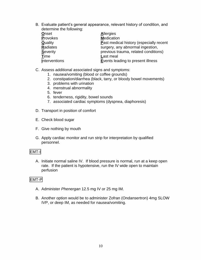
B. Evaluate patient’s general appearance, relevant history of condition, and
determine the following: Onset Allergies Provokes Medication Quality Past medical history (especially recent Radiates surgery, any abnormal ingestion, Severity previous trauma, related conditions)
Time Last meal Interventions Events leading to present illness
C. Assess additional associated signs and symptoms:
1. nausea/vomiting (blood or coffee grounds) 2. constipation/diarrhea (black, tarry, or bloody bowel movements) 3. problems with urination 4. menstrual abnormality 5. fever 6. tenderness, rigidity, bowel sounds 7. associated cardiac symptoms (dyspnea, diaphoresis)
D. Transport in position of comfort
E. Check blood sugar
F. Give nothing by mouth
G. Apply cardiac monitor and run strip for interpretation by qualified
personnel.
EMT-I
A. Initiate normal saline IV. If blood pressure is normal, run at a keep open
rate. If the patient is hypotensive, run the IV wide open to maintain perfusion
EMT-P
A. Administer Phenergan 12.5 mg IV or 25 mg IM. B. Another option would be to administer Zofran (Ondansertron) 4mg SLOW
IVP, or deep IM, as needed for nausea/vomiting.
11
MEDICAL CONTROL (EMT-I or EMT-P)
A. If the patient is in severe pain, contact Medical Control to request
morphine sulfate 2-4 mg IV or IM, or Nubain 10 mg IV or IM (most physicians prefer not to give these in the prehospital setting for abdominal pain)
12
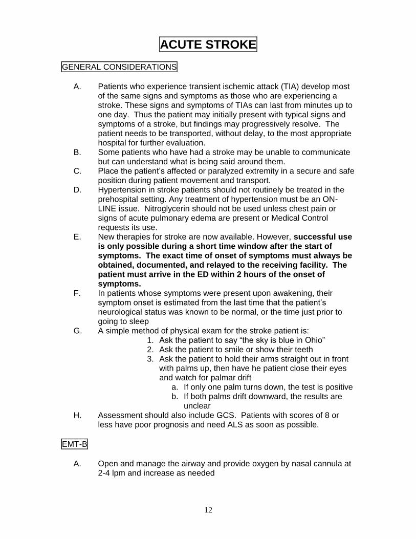
ACUTE STROKE
GENERAL CONSIDERATIONS
A. Patients who experience transient ischemic attack (TIA) develop most
of the same signs and symptoms as those who are experiencing a stroke. These signs and symptoms of TIAs can last from minutes up to one day. Thus the patient may initially present with typical signs and symptoms of a stroke, but findings may progressively resolve. The patient needs to be transported, without delay, to the most appropriate hospital for further evaluation.
B. Some patients who have had a stroke may be unable to communicate but can understand what is being said around them.
C. Place the patient’s affected or paralyzed extremity in a secure and safe position during patient movement and transport.
D. Hypertension in stroke patients should not routinely be treated in the prehospital setting. Any treatment of hypertension must be an ON-LINE issue. Nitroglycerin should not be used unless chest pain or signs of acute pulmonary edema are present or Medical Control requests its use.
E. New therapies for stroke are now available. However, successful use is only possible during a short time window after the start of symptoms. The exact time of onset of symptoms must always be obtained, documented, and relayed to the receiving facility. The patient must arrive in the ED within 2 hours of the onset of symptoms.
F. In patients whose symptoms were present upon awakening, their symptom onset is estimated from the last time that the patient’s neurological status was known to be normal, or the time just prior to going to sleep
G. A simple method of physical exam for the stroke patient is: 1. Ask the patient to say ―the sky is blue in Ohio‖ 2. Ask the patient to smile or show their teeth 3. Ask the patient to hold their arms straight out in front
with palms up, then have he patient close their eyes and watch for palmar drift
a. If only one palm turns down, the test is positive b. If both palms drift downward, the results are
unclear H. Assessment should also include GCS. Patients with scores of 8 or
less have poor prognosis and need ALS as soon as possible.
EMT-B
A. Open and manage the airway and provide oxygen by nasal cannula at
2-4 lpm and increase as needed
13
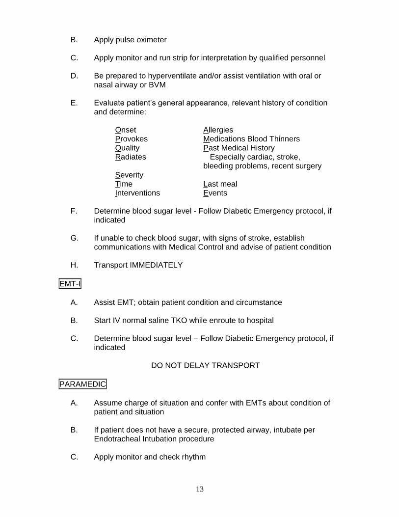
B. Apply pulse oximeter
C. Apply monitor and run strip for interpretation by qualified personnel
D. Be prepared to hyperventilate and/or assist ventilation with oral or nasal airway or BVM
E. Evaluate patient’s general appearance, relevant history of condition
and determine:
Onset Allergies Provokes Medications Blood Thinners Quality Past Medical History Radiates Especially cardiac, stroke,
bleeding problems, recent surgery Severity Time Last meal Interventions Events
F. Determine blood sugar level - Follow Diabetic Emergency protocol, if
indicated
G. If unable to check blood sugar, with signs of stroke, establish communications with Medical Control and advise of patient condition
H. Transport IMMEDIATELY
EMT-I
A. Assist EMT; obtain patient condition and circumstance
B. Start IV normal saline TKO while enroute to hospital
C. Determine blood sugar level – Follow Diabetic Emergency protocol, if
indicated
DO NOT DELAY TRANSPORT
PARAMEDIC
A. Assume charge of situation and confer with EMTs about condition of
patient and situation
B. If patient does not have a secure, protected airway, intubate per Endotracheal Intubation procedure
C. Apply monitor and check rhythm
14
D. Establish IV normal saline TKO
E. Determine blood sugar level – Follow Diabetic Emergency protocol, if indicated
F. Re-evaluate patient condition, contact Medical Control, and transport
to hospital

15
ALTERED LEVEL OF CONSCIOUSNESS
EMT-B
A. Secure airway and consider cervical spine injury
1. administer 100% oxygen by nonrebreather mask 2. immobilize cervical spine, if necessary 3. apply pulse oximeter 4. be prepared to assist ventilations with oral/nasal airway and/or
BVM
B. Evaluate patient’s general appearance, relevant history of condition, and determine the following: Onset Allergies Provokes Medication Quality Past medical history (especially recent Radiates surgery, any abnormal ingestion, Severity previous trauma, related conditions)
Time Last meal Interventions Events leading to present illness Assess the unresponsive patient using the Glasgow Coma Scale.
Patients with scores of 8 or less have a poorer prognosis and need ALS as soon as possible.
In possible stroke patients who are alert and cooperative, assessments of
language, motor responses, and sensation should be completed to establish baselines for future changes.
C. Check blood sugar
D. Administer 15g oral glucose to conscious and alert patients with a blood
sugar less than 70
E. Transport IMMEDIATELY
F. Apply cardiac monitor and run strip for interpretation by qualified personnel
EMT-I
A. Assist EMT-B, obtain patient condition and circumstance
B. Start IV normal saline TKO (provide 500 cc fluid bolus if signs of
hypoperfusion are present)
16
C. Check blood sugar
1. if blood sugar is less than 70, administer 1 amp D50 IV, or 1mg Glucagon IM or MAD (mucosal atomization device).
2. if blood sugar is greater than 400, administer 500 cc fluid bolus 3. if unable to check blood sugar and LOC is decreased, administer 1
amp D50 IV
D. If respirations are impaired or there is a suspicion of narcotic overdose and the patient does not respond to dextrose or fluid bolus, administer Narcan 2 mg slow IVP or MAD; if the patient then improves but is not fully awake, the dose may be repeated as needed
E. Re-evaluate patient condition, contact Medical Control, and transport to
hospital
PARAMEDIC
A. Assist EMT-B and EMT-I, assume charge of situation, and obtain patient
condition and circumstance
B. If the patient does not have a gag reflex or if oxygenation is inadequate with BLS measures, intubate per Endotracheal Intubation procedure (intubation should only be done if all other methods of ventilation/oxygenation have failed)
C. Apply monitor and assess cardiac rhythm
D. Start IV normal saline TKO (provide 500 cc fluid bolus if signs of hypoperfusion are present)
E. If signs of CVA are present, notify receiving hospital so that preparations
can be made
F. Check blood sugar 1. if blood sugar is less than 70, administer 1 amp D50 IV, or 1mg
Glucagon IM or MAD (mucosal atomization device). 2. if blood sugar is greater than 400, administer 500 cc fluid bolus 3. if unable to check blood sugar and LOC is decreased, administer 1
amp D50 IV.
G. If respirations are impaired or there is a suspicion of narcotic overdose and the patient does not respond to dextrose or fluid bolus, administer Narcan 2 mg slow IVP or MAD. If the patient then improves but is not fully awake, the dose may be repeated as needed
17
H. Re-evaluate patient condition, contact Medical Control, and transport to hospital
18
ANAPHYLAXIS
EMT-B
A. Provide 100% supplemental oxygen
B. Insert oral or nasal airway as needed
C. Assess patient history, medications, allergies, if possible
D. Determine if patient has had Epi-pen prescribed for them
1. Administer medication in mid-thigh and hold injector firmly against leg for at least 10 seconds to assure all medication in injected
2. EMT-B may use Epi-pen from Basic Medication bag, if available
E. Apply cardiac monitor and run strip for interpretation by qualified personnel
F. Transport IMMEDIATELY
EMT-I
A. Assist EMT-Bs; obtain patient condition and circumstance
B. Reassess breath sounds and oxygenation
C. In patients with hypertension, coronary artery disease, or current
pregnancy, contact Medical Control
D. Epinephrine 0.3-0.5 mg (1:1000 solution) by subcutaneous injection (unless Epi-Pen has already been administered by EMT-B)
E. Contact Medical Control
F. Epinephrine dose may be repeated in 10-15 minutes with Medical Control
approval
G. If wheezing present, administer albuterol 2.5 mg via nebulizer
H. Benadryl 50 mg IV/IM I. Transport IMMEDIATELY
J. Initiate IV normal saline solution TKO (provide 500 cc fluid bolus if signs of
hypoperfusion)
19
PARAMEDIC
A. Assume charge of situation and confer with EMTs about patient condition
and situation
B. Reassess patient
C. Intubate patient if any signs of impending airway compromise
D. In patients with hypertension, coronary artery disease, or current pregnancy, contact Medical Control
E. Epinephrine 0.3-0.5 mg (1:1000 solution) by subcutaneous injection
F. Initiate IV normal saline solution TKO (provide 500 cc fluid bolus if signs of
hypoperfusion)
G. If wheezing present, administer albuterol 2.5 mg via nebulizer
H. Benadryl 50 mg IV/IM
I. Contact Medical Control
J. If no improvement and patient is hypotensive, administer epinephrine 0.5 mg slow IVP (1:10,000 solution) with Medical Control approval
K. Transport IMMEDIATELY
20
ARRHYTHMIAS
EMT-B
A. Open and manage the airway and provide 100% oxygen by NRB mask.
Apply pulse oximeter B. Make patient comfortable and provide reassurance
C. Evaluate patient’s general appearance, relevant history of condition, and
determine the following: Onset Allergies Provokes Medication Quality Past medical history (especially recent Radiates surgery, any abnormal ingestion, Severity previous trauma, related conditions)
Time Last meal Interventions Events leading to present illness
D. If the patient is experiencing an unusual and/or irregular heart rate or
pulse, the cardiac monitor may be applied, if available, and a strip run for evaluation by qualified personnel. This should only be done during transport and you must advise the patient that you do not have the ability to interpret the tracing
E. Establish communication with Medical Control and advise of patient
condition. Transport IMMEDIATELY
EMT-I
A. Start IV normal saline TKO (provide 500 cc fluid bolus if signs of
hypoperfusion are present and no signs of pulmonary edema are present)
PARAMEDIC
A. Assume charge of situation and confer with EMTs about condition of
patient and situation. B. Apply cardiac monitor and determine identity of arrhythmia
C. Start IV normal saline TKO (provide 500 cc fluid bolus if signs of
hypoperfusion are present and no signs of pulmonary edema are present)
D. Treat arrhythmia as follows: 1. Bradycardia, second and third degree heart blocks
a. good perfusion – transport b. poor perfusion:
21
1. atropine 0.5-1.0 mg IV push, repeat 1.0 mg IV push every 5 minutes up to 3 mg total dose or until heart rate is greater than 60 and systolic BP is greater than 90
2. external pacemaker set at 80 beats per minute with output at 20 milliamps, increasing by 20 milliamps until mechanical capture is obtained
3. contact Medical Control 4. if still poor perfusion and approved by Medical
Control, start dopamine drip (400 mg dopamine in 500 cc D5W or NS to yield a solution of 800 ug/cc) and titrate the infusion until heart rate is above 60 and systolic BP is above 90
2. Atrial flutter/fibrillation with rapid ventricular rate a. good perfusion – transport b. poor perfusion with systolic BP less than 90 and ventricular
rate greater than 150: 1. attempt to determine the time of onset of the
symptoms 2. if less than 72 hours:
a. consider sedation (Versed 2-4 mg IV) b. cardiovert 100 joules (100 joules biphasic) c. cardiovert 200 joules (120 joules biphasic) d. cardiovert 300 joules (150 joules biphasic) e. cardiovert 360 joules (170 joules biphasic) f. transport IMMEDIATELY g. contact Medical Control for assistance h. consider Verapamil 10 mg IV
3. if greater than 72 hours or unknown: a. transport IMMEDIATELY b. contact Medical Control for assistance
3. Supraventricular tachycardia a. patients who are alert and oriented with normal blood
pressure and ventricular rate less than 150 1. transport
b. patients with ventricular heart rate greater than 150 beats per minute along with blood pressure greater than 70, chest pain, and/or shortness of breath
1. administer adenosine 6 mg rapid IV bolus followed IMMEDIATELY with a 20 cc saline bolus
2. if no conversion, repeat adenosine with 12 mg rapid IV bolus followed IMMEDIATELY with a 20 cc saline bolus
3. if no conversion, repeat adenosine with 12 mg rapid IV bolus followed IMMEDIATELY with a 20 cc saline bolus
22
4. if no response to adenosine, attempt synchronized cardioversion:
a. consider sedation (Versed 2-4 mg IV) b. cardiovert 50 joules (use same setting for
biphasic) c. cardiovert 100 joules (100 joules biphasic) d. cardiovert 200 joules (120 joules biphasic) e. cardiovert 300 joules (150 joules biphasic) f. cardiovert 360 joules (170 joules biphasic)
5. transport IMMEDIATELY 6. consider Verapamil 10 mg IV (contact Medical
Control) c. patients with blood pressure less than 70 or decreased
level of consciousness 1. synchronized cardioversion
a. consider sedation (Versed 2-4 mg IV) b. cardiovert 50 joules (use same setting for
biphasic) c. cardiovert 100 joules (70 joules biphasic) d. cardiovert 200 joules (120 joules biphasic) e. cardiovert 300 joules (150 joules biphasic) f. cardiovert 360 joules (170 joules biphasic)
2. transport IMMEDIATELY 4. Frequent PVCs with symptoms (chest pain, shortness of breath,
palpitations, hypotension, dizziness) a. treat underlying causes (i.e. hypoxia, hypoperfusion,
cardiac chest pain) b. contact Medical Control c. lidocaine 75-100 mg IV (if approved by Medical Control) d. may repeat lidocaine dose once in 8-10 minutes (if
approved by Medical Control) e. if lidocaine is effective, initiate lidocaine drip 2-4 mg/min (if
approved by Medical Control) 5. Ventricular tachycardia
a. patients with minimal symptoms and normal blood pressure
1. amiodarone 150 mg IV over 10 minutes 2. if no effect after 10 minutes, lidocaine 75-100 mg IV
bolus (may repeat every 5 minutes to a maximum total dose of 300 mg)
3. if lidocaine bolus is effective, start lidocaine drip at 2-4 mg/min
4. if still no effect, attempt synchronized cardioversion: a. consider sedation (Versed 2-4 mg IV) b. cardiovert 100 joules (70 joules biphasic) c. cardiovert 200 joules (120 joules biphasic) d. cardiovert 300 joules (150 joules biphasic) e. cardiovert 360 joules (170 joules biphasic)
23
5. transport IMMEDIATELY b. patients with significant symptoms, hypotension, and/or
decreased level of consciousness 1. synchronized cardioversion:
a. consider sedation (Versed 2-4 mg IV) b. cardiovert 100 joules (70 joules biphasic) c. cardiovert 200 joules (120 joules biphasic) d. cardiovert 300 joules (150 joules biphasic) e. cardiovert 360 joules (170 joules biphasic)
2. amiodarone 150 mg IV over 10 minutes 3. if no effect after 10 minutes, lidocaine 75-100 mg IV
bolus (may repeat every 5 minutes to a maximum total dose of 300 mg)
4. if lidocaine bolus is effective, start lidocaine drip at 2-4 mg/min
5. repeat synchronized cardioversion, if necessary a. cardiovert 100 joules (70 joules biphasic) b. cardiovert 200 joules (120 joules biphasic) c. cardiovert 300 joules (150 joules biphasic) d. cardiovert 360 joules (170 joules biphasic)
6. transport IMMEDIATELY c. patients without a pulse
1. treat as ventricular fibrillation (see Cardiac Arrest protocol)
24
BURNS
GENERAL INSTRUCTIONS
A. The first priority is to assure scene safety and then remove the patient
from heat, flame, electrical, or chemical exposure B. Airway, breathing, and circulation must be stabilized before attending to
the burn C. Patients with extensive burns must be monitored for hypothermia. Avoid
use of ice or cold compresses. When in doubt, always cover with dry, sterile dressings
D. In the acute setting, patients with critical burns die from hypovolemia and shock. When possible, fluid replacement therapy should be instituted as quickly as possible
E. In caring for the burn, the EMT should: 1. stop the burning 2. reduce the pain 3. prevent contamination
F. When dealing with contaminated environments, EMTs must have appropriate protective clothing. The scene is not safe to enter until this equipment is available. Contact the appropriate HazMat service for assistance
G. Gross decontamination must be done at the scene. Advise receiving facility if complete decontamination was not done at the scene and be prepared to transport to decontamination area
EMT-B
A. Open/manage airway and provide 100% oxygen via NRB mask or BVM B. Determine type of burn and treat as follows:
1. Thermal a. Stop burning process (remove patient from heat source,
cool skin, remove clothing) b. If patient starts to shiver or skin is cool, stop cooling
process c. Estimate extent (% of body surface area) and depth of burn
(see chart) d. Contact Medical Control and transport to hospital e. Cover burn areas with dry, sterile dressing
2. Radiation a. Treat as thermal burns except when burn is contaminated
with radioactive source, then treat as chemical burn b. Wear appropriate protective clothing when dealing with
contamination c. Contact HazMat team for assistance in contamination
cases
25
3. Chemical
a. EMTs must wear appropriate clothing and respirators b. Remove patient from contaminated area to
decontamination site (not the ambulance) c. Determine chemicals involved; contact appropriate agency
for chemical information d. Remove patient’s clothing and flush skin thoroughly e. Leave contaminated clothes at scene; cover patient and
transport to hospital f. Patient should be transported by personnel not involved in
decontamination process g. Determine severity of burn (see chart); contact Medical
Control h. Relay type of substance involved to Medical Control
4. Electrical a. Shut down electrical source; do not attempt to remove
patient until electricity is CONFIRMED to be shut off b. Assess for visible entrance and exit wounds and treat as
thermal burns c. Assess for internal injury (vascular damage, tissue
damage, fractures) and treat accordingly d. Attempt to determine the voltage of the electrical source
and relay this information to Medical Control e. Determine severity of burn (see chart); contact Medical
Control and transport to hospital f. Attach cardiac monitor and run strip for interpretation by
qualified personnel 5. Inhalation
a. Always suspect inhalation burns when the patient has been found in an enclosed, smoky environment and/or exhibits any of the following: burns to face/neck, singed nasal hairs, cough and/or stridor, soot in mouth/nose/sputum, hoarse voice
b. Provide 100% oxygen, contact Medical Control, and transport to hospital
EMT-I
A. Assist EMT with airway B. Assist in determining type of burn and its treatment
C. Initiate IV normal saline infusion (do not delay transport for IV)
D. Contact Medical Control for orders for analgesic medications
26
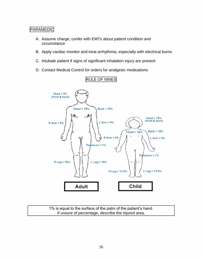
PARAMEDIC
A. Assume charge; confer with EMTs about patient condition and
circumstance
B. Apply cardiac monitor and treat arrhythmia, especially with electrical burns
C. Intubate patient if signs of significant inhalation injury are present
D. Contact Medical Control for orders for analgesic medications
RULE OF NINES
1% is equal to the surface of the palm of the patient’s hand. If unsure of percentage, describe the injured area.
27
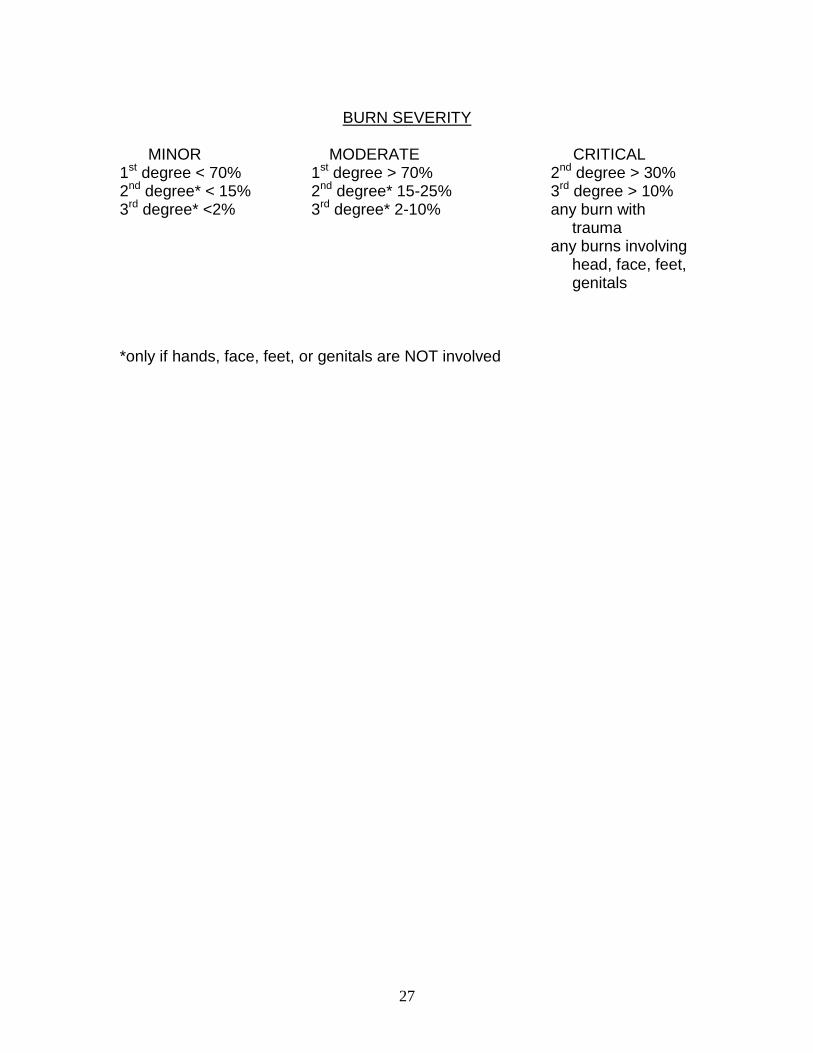
BURN SEVERITY
MINOR MODERATE CRITICAL 1st degree < 70% 1st degree > 70% 2nd degree > 30% 2nd degree* < 15% 2nd degree* 15-25% 3rd degree > 10% 3rd degree* <2% 3rd degree* 2-10% any burn with trauma any burns involving head, face, feet, genitals *only if hands, face, feet, or genitals are NOT involved
28
CARDIAC ARREST
GENERAL INSTRUCTIONS
A. CPR should not be interrupted for more than 15 seconds until
spontaneous pulse is established B. If IV cannot be established, epinephrine, atropine, lidocaine, Narcan, or
vasopressin may be administered through the endotracheal tube C. When a defibrillator (automated or manual) is immediately available,
electrical countershock should be administered as quickly as possible after ventricular fibrillation is identified
D. Each IV push medication should be followed by a 20 cc flush
EMT-B
A. If an Automated External Defibrillator (AED) is available:
1. Assess patient for respiratory and cardiac arrest 2. Apply AED and activate device
a. ―No Shock Advised‖ i. CPR as recommended by the American Heart
Association ii. Ventilate with 100% oxygen by bag-valve-mask
(BVM) or oxygen-powered manually triggered ventilation device and oral/nasal airway
iii. Ventilation should be delivered for one second. and cricoid pressure can be considered to help reduce gastric distention
iv. Intubate patient (per Endotracheal Intubation procedure) as soon as possible
v. Establish communications with Medical Control and advise of cardiac arrest
vi. Transport IMMEDIATELY b. ―Shock Advised‖
i. Deliver one Shock ii. After the shock -CPR as recommended by the
American Heart Association for two minutes. iii. Ventilate with 100% oxygen by bag-valve-mask or
oxygen-powered manually triggered ventilation device and oral/nasal airway
iv. Ventilation should be delivered for one second and cricoid pressure should be considered to help reduce gastric distention
v. Intubate patient (per Endotracheal Intubation procedure) as soon as possible
vi. Establish communications with Medical Control and advise of cardiac arrest
29
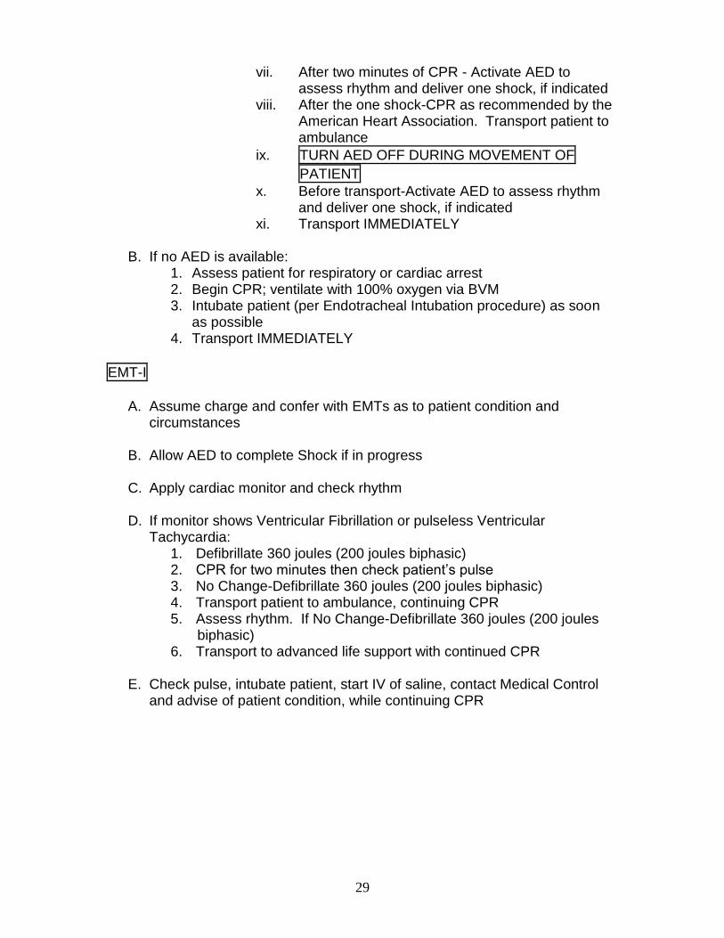
vii. After two minutes of CPR - Activate AED to assess rhythm and deliver one shock, if indicated
viii. After the one shock-CPR as recommended by the American Heart Association. Transport patient to ambulance
ix. TURN AED OFF DURING MOVEMENT OF
PATIENT
x. Before transport-Activate AED to assess rhythm and deliver one shock, if indicated
xi. Transport IMMEDIATELY
B. If no AED is available: 1. Assess patient for respiratory or cardiac arrest 2. Begin CPR; ventilate with 100% oxygen via BVM 3. Intubate patient (per Endotracheal Intubation procedure) as soon
as possible 4. Transport IMMEDIATELY
EMT-I
A. Assume charge and confer with EMTs as to patient condition and
circumstances
B. Allow AED to complete Shock if in progress
C. Apply cardiac monitor and check rhythm
D. If monitor shows Ventricular Fibrillation or pulseless Ventricular Tachycardia:
1. Defibrillate 360 joules (200 joules biphasic) 2. CPR for two minutes then check patient’s pulse 3. No Change-Defibrillate 360 joules (200 joules biphasic) 4. Transport patient to ambulance, continuing CPR 5. Assess rhythm. If No Change-Defibrillate 360 joules (200 joules
biphasic) 6. Transport to advanced life support with continued CPR
E. Check pulse, intubate patient, start IV of saline, contact Medical Control
and advise of patient condition, while continuing CPR
30
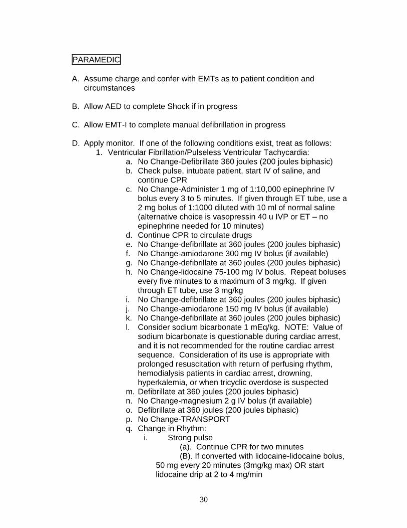
PARAMEDIC
A. Assume charge and confer with EMTs as to patient condition and
circumstances
B. Allow AED to complete Shock if in progress
C. Allow EMT-I to complete manual defibrillation in progress
D. Apply monitor. If one of the following conditions exist, treat as follows: 1. Ventricular Fibrillation/Pulseless Ventricular Tachycardia:
a. No Change-Defibrillate 360 joules (200 joules biphasic) b. Check pulse, intubate patient, start IV of saline, and
continue CPR c. No Change-Administer 1 mg of 1:10,000 epinephrine IV
bolus every 3 to 5 minutes. If given through ET tube, use a 2 mg bolus of 1:1000 diluted with 10 ml of normal saline (alternative choice is vasopressin 40 u IVP or ET – no epinephrine needed for 10 minutes)
d. Continue CPR to circulate drugs e. No Change-defibrillate at 360 joules (200 joules biphasic) f. No Change-amiodarone 300 mg IV bolus (if available) g. No Change-defibrillate at 360 joules (200 joules biphasic) h. No Change-lidocaine 75-100 mg IV bolus. Repeat boluses
every five minutes to a maximum of 3 mg/kg. If given through ET tube, use 3 mg/kg
i. No Change-defibrillate at 360 joules (200 joules biphasic) j. No Change-amiodarone 150 mg IV bolus (if available) k. No Change-defibrillate at 360 joules (200 joules biphasic) l. Consider sodium bicarbonate 1 mEq/kg. NOTE: Value of
sodium bicarbonate is questionable during cardiac arrest, and it is not recommended for the routine cardiac arrest sequence. Consideration of its use is appropriate with prolonged resuscitation with return of perfusing rhythm, hemodialysis patients in cardiac arrest, drowning, hyperkalemia, or when tricyclic overdose is suspected
m. Defibrillate at 360 joules (200 joules biphasic) n. No Change-magnesium 2 g IV bolus (if available) o. Defibrillate at 360 joules (200 joules biphasic) p. No Change-TRANSPORT q. Change in Rhythm:
i. Strong pulse (a). Continue CPR for two minutes
(B). If converted with lidocaine-lidocaine bolus, 50 mg every 20 minutes (3mg/kg max) OR start lidocaine drip at 2 to 4 mg/min
31
ii. Hypotension and bradycardia---see Arrhythmia Protocol
iii. No pulse-CPR
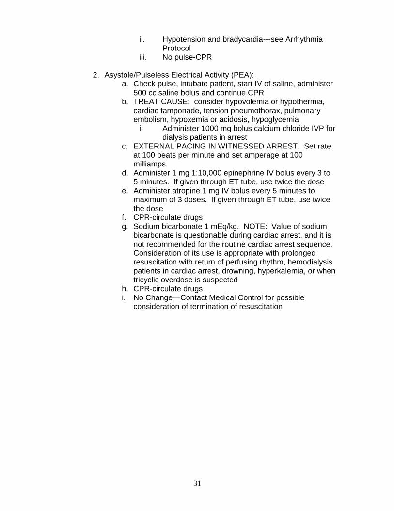
2. Asystole/Pulseless Electrical Activity (PEA): a. Check pulse, intubate patient, start IV of saline, administer
500 cc saline bolus and continue CPR b. TREAT CAUSE: consider hypovolemia or hypothermia,
cardiac tamponade, tension pneumothorax, pulmonary embolism, hypoxemia or acidosis, hypoglycemia
i. Administer 1000 mg bolus calcium chloride IVP for dialysis patients in arrest
c. EXTERNAL PACING IN WITNESSED ARREST. Set rate at 100 beats per minute and set amperage at 100 milliamps
d. Administer 1 mg 1:10,000 epinephrine IV bolus every 3 to 5 minutes. If given through ET tube, use twice the dose
e. Administer atropine 1 mg IV bolus every 5 minutes to maximum of 3 doses. If given through ET tube, use twice the dose
f. CPR-circulate drugs g. Sodium bicarbonate 1 mEq/kg. NOTE: Value of sodium
bicarbonate is questionable during cardiac arrest, and it is not recommended for the routine cardiac arrest sequence. Consideration of its use is appropriate with prolonged resuscitation with return of perfusing rhythm, hemodialysis patients in cardiac arrest, drowning, hyperkalemia, or when tricyclic overdose is suspected
h. CPR-circulate drugs i. No Change—Contact Medical Control for possible
consideration of termination of resuscitation

32
33
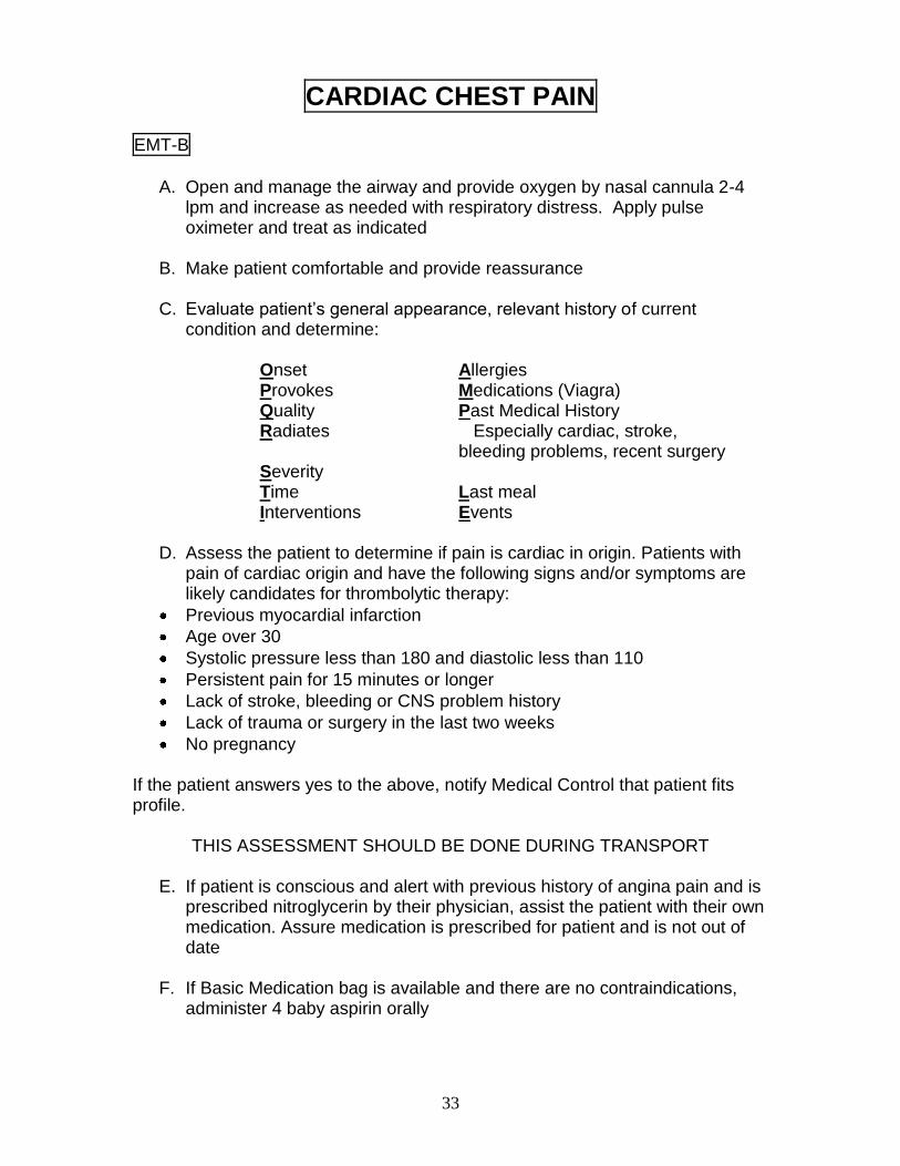
CARDIAC CHEST PAIN
EMT-B
A. Open and manage the airway and provide oxygen by nasal cannula 2-4
lpm and increase as needed with respiratory distress. Apply pulse oximeter and treat as indicated
B. Make patient comfortable and provide reassurance
C. Evaluate patient’s general appearance, relevant history of current
condition and determine:
Onset Allergies Provokes Medications (Viagra) Quality Past Medical History Radiates Especially cardiac, stroke,
bleeding problems, recent surgery Severity Time Last meal Interventions Events
D. Assess the patient to determine if pain is cardiac in origin. Patients with
pain of cardiac origin and have the following signs and/or symptoms are likely candidates for thrombolytic therapy:
Previous myocardial infarction
Age over 30
Systolic pressure less than 180 and diastolic less than 110
Persistent pain for 15 minutes or longer
Lack of stroke, bleeding or CNS problem history
Lack of trauma or surgery in the last two weeks
No pregnancy If the patient answers yes to the above, notify Medical Control that patient fits profile.
THIS ASSESSMENT SHOULD BE DONE DURING TRANSPORT
E. If patient is conscious and alert with previous history of angina pain and is
prescribed nitroglycerin by their physician, assist the patient with their own medication. Assure medication is prescribed for patient and is not out of date
F. If Basic Medication bag is available and there are no contraindications,
administer 4 baby aspirin orally
34
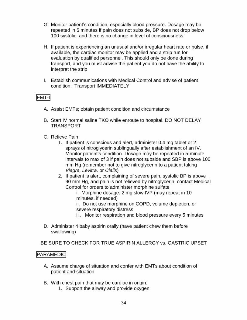
G. Monitor patient’s condition, especially blood pressure. Dosage may be repeated in 5 minutes if pain does not subside, BP does not drop below 100 systolic, and there is no change in level of consciousness
H. If patient is experiencing an unusual and/or irregular heart rate or pulse, if
available, the cardiac monitor may be applied and a strip run for evaluation by qualified personnel. This should only be done during transport, and you must advise the patient you do not have the ability to interpret the strip
I. Establish communications with Medical Control and advise of patient
condition. Transport IMMEDIATELY
EMT-I
A. Assist EMTs; obtain patient condition and circumstance
B. Start IV normal saline TKO while enroute to hospital. DO NOT DELAY
TRANSPORT
C. Relieve Pain 1. If patient is conscious and alert, administer 0.4 mg tablet or 2
sprays of nitroglycerin sublingually after establishment of an IV. Monitor patient’s condition. Dosage may be repeated in 5-minute intervals to max of 3 if pain does not subside and SBP is above 100 mm Hg (remember not to give nitroglycerin to a patient taking Viagra, Levitra, or Cialis)
2. If patient is alert, complaining of severe pain, systolic BP is above 90 mm Hg, and pain is not relieved by nitroglycerin, contact Medical Control for orders to administer morphine sulfate
i. Morphine dosage: 2 mg slow IVP (may repeat in 10 minutes, if needed) ii. Do not use morphine on COPD, volume depletion, or severe respiratory distress iii. Monitor respiration and blood pressure every 5 minutes
D. Administer 4 baby aspirin orally (have patient chew them before
swallowing)
BE SURE TO CHECK FOR TRUE ASPIRIN ALLERGY vs. GASTRIC UPSET
PARAMEDIC
A. Assume charge of situation and confer with EMTs about condition of
patient and situation
B. With chest pain that may be cardiac in origin: 1. Support the airway and provide oxygen
35
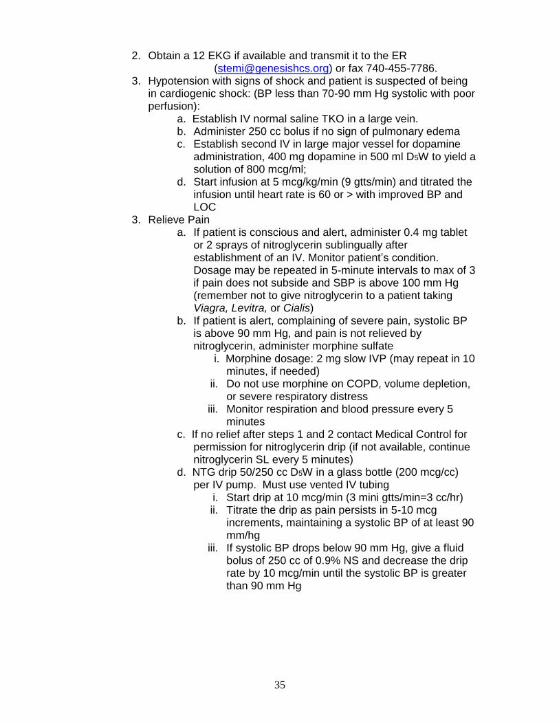
2. Obtain a 12 EKG if available and transmit it to the ER ([email protected]) or fax 740-455-7786.
3. Hypotension with signs of shock and patient is suspected of being in cardiogenic shock: (BP less than 70-90 mm Hg systolic with poor perfusion):
a. Establish IV normal saline TKO in a large vein. b. Administer 250 cc bolus if no sign of pulmonary edema c. Establish second IV in large major vessel for dopamine
administration, 400 mg dopamine in 500 ml D5W to yield a solution of 800 mcg/ml;
d. Start infusion at 5 mcg/kg/min (9 gtts/min) and titrated the infusion until heart rate is 60 or > with improved BP and LOC
3. Relieve Pain a. If patient is conscious and alert, administer 0.4 mg tablet
or 2 sprays of nitroglycerin sublingually after establishment of an IV. Monitor patient’s condition. Dosage may be repeated in 5-minute intervals to max of 3 if pain does not subside and SBP is above 100 mm Hg (remember not to give nitroglycerin to a patient taking Viagra, Levitra, or Cialis)
b. If patient is alert, complaining of severe pain, systolic BP is above 90 mm Hg, and pain is not relieved by nitroglycerin, administer morphine sulfate
i. Morphine dosage: 2 mg slow IVP (may repeat in 10 minutes, if needed)
ii. Do not use morphine on COPD, volume depletion, or severe respiratory distress
iii. Monitor respiration and blood pressure every 5 minutes
c. If no relief after steps 1 and 2 contact Medical Control for permission for nitroglycerin drip (if not available, continue nitroglycerin SL every 5 minutes)
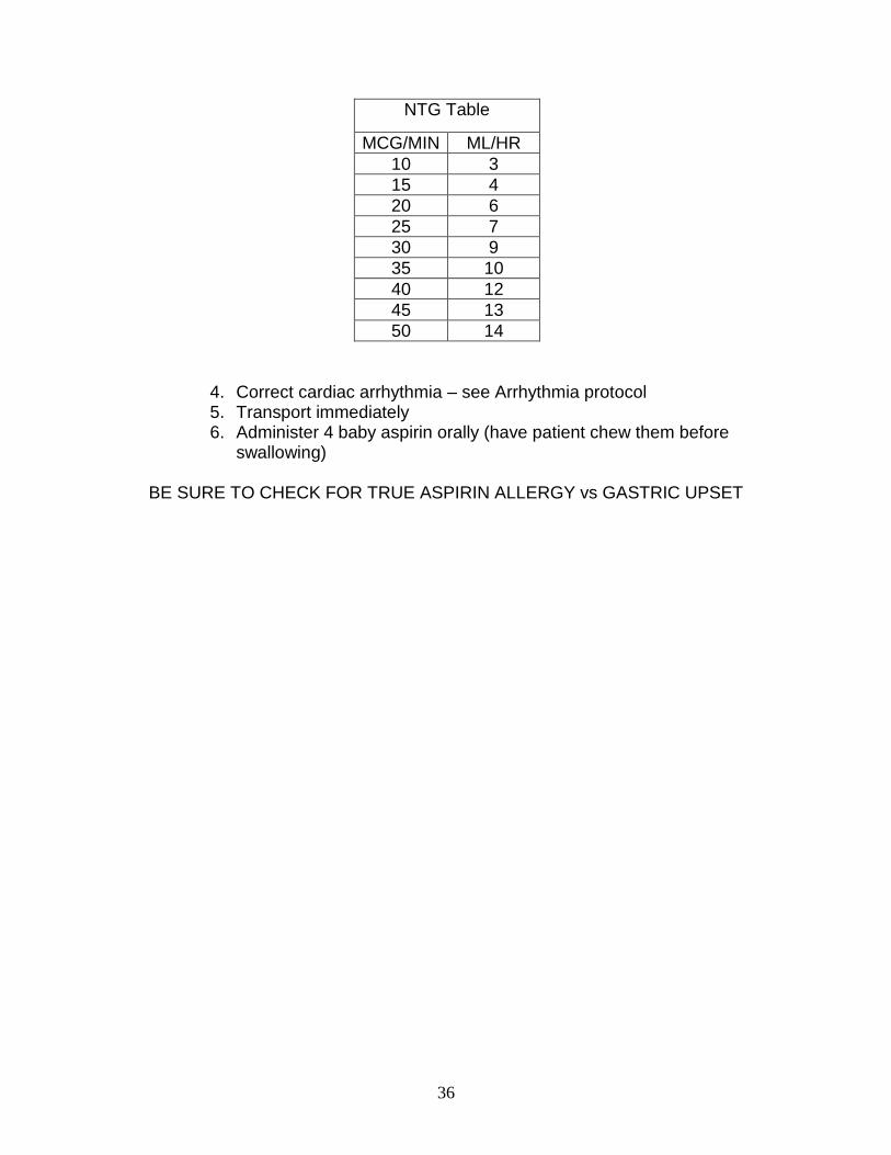
d. NTG drip 50/250 cc D5W in a glass bottle (200 mcg/cc) per IV pump. Must use vented IV tubing
i. Start drip at 10 mcg/min (3 mini gtts/min=3 cc/hr) ii. Titrate the drip as pain persists in 5-10 mcg
increments, maintaining a systolic BP of at least 90 mm/hg
iii. If systolic BP drops below 90 mm Hg, give a fluid bolus of 250 cc of 0.9% NS and decrease the drip rate by 10 mcg/min until the systolic BP is greater than 90 mm Hg
36
NTG Table
MCG/MIN ML/HR
10 3
15 4
20 6
25 7
30 9
35 10
40 12
45 13
50 14
4. Correct cardiac arrhythmia – see Arrhythmia protocol 5. Transport immediately 6. Administer 4 baby aspirin orally (have patient chew them before
swallowing)
BE SURE TO CHECK FOR TRUE ASPIRIN ALLERGY vs GASTRIC UPSET
37
CHILDBIRTH
GENERAL INSTRUCTIONS
A. Unless delivery is imminent, transport to a hospital with obstetrical
capabilities B. Imminent delivery is when the baby’s head is visible in the vaginal opening
during a contraction (crowning) C. A visual inspection of the perineal area should only be done when
contractions are less than 5 minutes apart and/or there is bleeding or fluid discharge
D. The EMT should not place a gloved hand inside the vagina except in the case of breech delivery with entrapped head or a prolapsed umbilical cord
E. During delivery, gentle pressure with a flat hand on the baby’s head should be applied to prevent an explosive delivery
F. A mother in active labor should be placed on the cot or floor to prevent the newborn from falling after delivery
G. An internal vaginal exam should never be done in the case of late pregnancy bleeding
EMT-B
A. Obtain history of patient condition and pregnancy: contraction duration
and interval, due date, number of pregnancies and number of live children, prenatal care, possible complications, breakage of amniotic fluid
B. Determine transport or delivery. Transport unless crowning is present
during a contraction; contact Medical Control
C. Always try to transport mother to her hospital designated for delivery. Transport mother lying on left side with head slightly elevated to relieve pressure on mother’s vena cava created by baby. Pressure could cause a decrease in mother’s and baby’s heart rate
D. If delivery is imminent, prepare equipment and follow guidelines for
delivery 1. Equipment: OB Kit, oxygen and BVM, towels and blankets, cot,
large dressings, pediatric airway and IV equipment
E. After delivery, transport mother on cot and baby in car seat, if available, or have parent or EMT hold baby during transport
F. Keep mother and baby warm. Monitor airway and observe for signs of
shock.
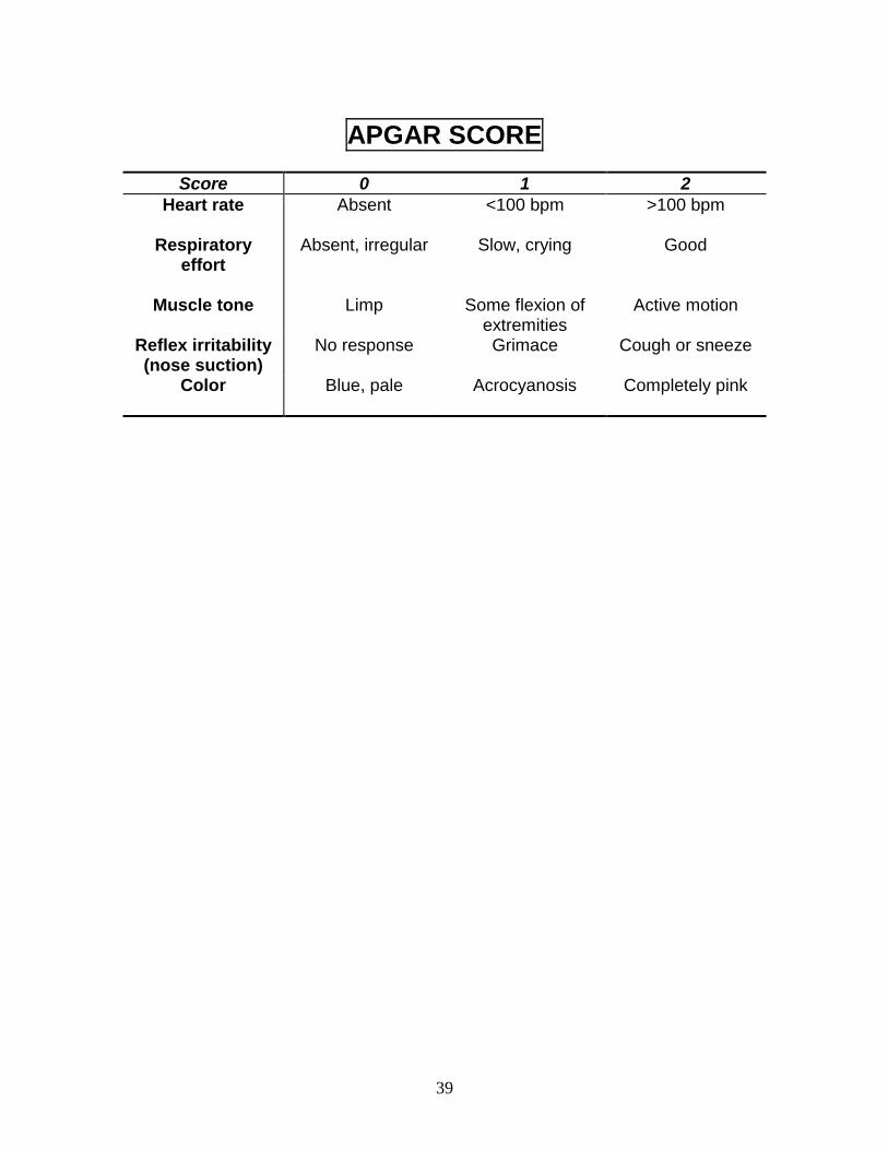
G. Obtain APGAR score at one and five minutes
38
EMT-I/PARAMEDIC
A. Assist EMT, obtain patient condition and circumstance
B. Start IV normal saline TKO on mother (run wide open if excessive
bleeding is present)
DELIVERY PROCEDURE
A. Prepare all equipment as above B. Position mother on floor or other flat surface (i.e. bed, stretcher)
C. Encourage mother to push with contractions
D. As head is delivering, apply gentle pressure to perineum to prevent
explosive delivery and reduce the incidence of vaginal tears
E. Once the head has delivered, it should rotate to the side
F. Use bulb syringe to suction baby’s mouth and nose while still at the perineum
G. Apply gentle downward traction on the baby’s head to assist delivery of
the anterior shoulder
H. Once the anterior shoulder has delivered, the rest of the baby will deliver rapidly
I. Suction the mouth and nose with bulb syringe
J. Clamp cord 10-12 cm away from the baby’s body, and again 2-3 cm
further. Cut cord between clamps if clean scissors are available
K. Dry and stimulate baby with soft cloths or blankets
L. Obtain APGAR score at one and five minutes
M. If baby exhibits signs of respiratory distress, shock, or lethargy, follow Newborn Resuscitation protocol
39
APGAR SCORE
Score 0 1 2
Heart rate
Absent <100 bpm >100 bpm
Respiratory effort
Absent, irregular Slow, crying Good
Muscle tone
Limp Some flexion of extremities
Active motion
Reflex irritability (nose suction)
No response Grimace Cough or sneeze
Color
Blue, pale Acrocyanosis Completely pink
40
DELIVERY COMPLICATIONS
CONTACT MEDICAL CONTROL AS SOON AS ANY COMPLICATION IS
DISCOVERED
A. Cord Around Baby’s Neck: 1. As baby’s head passes out the vaginal opening, feel for the cord.
Initially try to slide cord over baby’s head. If it is too tight, clamp cord in two places and cut between clamps
B. Breech Delivery:
1. Footling breech, which is one or both feet delivered first with knees flexed
2. Frank breech, which is the buttocks first presentation a. When the feet or buttocks first become visible, there is
normally time to transport patient to nearest facility b. If upper thighs or the buttocks have come out of the
vagina, delivery is imminent c. If the child’s body has delivered and the head appears
caught in the vagina, the EMT must support the infant’s body and insert two fingers into the vagina along the infant’s neck until the chin is located. At this point, the two fingers should be placed between the chin and the vaginal canal and then advanced past the mouth and nose
d. After achieving this position a passage for air must be created by pushing the vaginal canal away from the infant’s face. This air passage must be maintained until the infant is completely delivered
C. Excessive Bleeding Pre-delivery:
1. If bleeding is excessive during this time and delivery is imminent, in addition to normal delivery procedures, the EMT should follow the Shock protocol
2. If delivery is not imminent, patient should be transported lying on her left side and Shock protocol should be followed
D. Excessive Bleeding Post-delivery:
1. If bleeding appears to be excessive, open IV of normal saline and start second IV
2. If placenta has been delivered, massage uterus and put baby to mother’s breast
3. Follow hypovolemic shock guidelines
41
E. Prolapsed Cord:
1. When the umbilical cord passes through the vagina and is exposed, the EMT should check cord for a pulse
2. The patient should be transported with hips elevated or in the knee-chest position and a moist dressing around cord
3. If umbilical cord is seen or felt in the vagina, insert two fingers to elevate presenting part away from cord and distribute pressure evenly when occiput presents
4. DO NOT attempt to push the cord back 5. High-flow oxygen and transport IMMEDIATELY
F. Shoulder Dystocia
1. Occurs when the head has delivered and the shoulders appear to be stuck in the vagina (Turtle sign)
2. Initial maneuver to assist delivery is placing the mother in knee-chest position (McRobert’s maneuver)
3. Firm suprapubic pressure may also help to allow the anterior shoulder to deliver
4. Once one of the shoulders delivers, the rest of the body will deliver rapidly
5. Transport IMMEDIATELY
G. Limb Presentation 1. If the presenting part is an arm or an outstretched leg, field delivery
is virtually impossible 2. Encourage the mother NOT to push 3. High-flow oxygen and transport IMMEDIATELY
42
DIABETIC EMERGENCIES
EMT-B
A. Secure and maintain airway. Support with appropriate level of 02 B. Obtain relevant medical history: OPQRST
1. Has patient eaten today 2. Has patient vomited in the past 12 hours 3. Onset 4. Medication – type and time taken
C. Determine blood sugar level by glucometer stick
1. Blood sugar less than 70, administer 15 g oral glucose to conscious and alert patients only
2. Unable to obtain blood sugar, transport and contact Medical Control for guidance
D. Establish communications with Medical Control and advise of patient
condition. Transport IMMEDIATELY E. Apply monitor and run strip for interpretation by qualified personnel
EMT-I
A. Assist EMT, obtain patient condition and circumstance B. Start IV normal saline TKO, while enroute to hospital (provide 500 cc
fluid bolus if blood sugar is greater than 400)
C. Determine blood sugar level by glucometer stick 1. Blood sugar less than 70, administer 50 cc D50 IV push immediately
or glucagon 1mg IM/SQ/MAD 2. Blood sugar greater than 400, infuse patient with 500 cc bolus of
normal saline 3. Unable to obtain blood sugar, transport and contact Medical Control
for guidance
D. If patient has altered level of consciousness, follow Altered LOC Protocol
43
PARAMEDIC
A. Assume charge of situation and confer with EMTs about condition of
patient and situation B. Apply monitor and check rhythm
C. Start IV normal saline TKO
D. Determine blood sugar level by glucometer stick
1. Blood sugar less than 70, administer 50 cc D50 IV push immediately or glucagon 1mg IM/SQ/MD.
2. Blood sugar greater than 400, infuse patient with 500 cc bolus of normal saline
3. Unable to obtain blood sugar, transport and contact Medical Control for guidance
E. If patient has altered level of consciousness, follow Altered LOC Protocol

44
EYE INJURIES
GENERAL CONSIDERATIONS
TRAUMA
A. Do not allow eye injury to distract you from the basics of trauma care B. Do not remove any foreign body imbedded in the eye or orbit. Stabilize
any large protruding foreign bodies
C. With blunt trauma to the eye, if time permits, examine the globe briefly for gross laceration as the lid may be swollen tightly shut later. Scleral rupture may lie beneath an intact conjunctiva
1. Exert no pressure on the globe when doing the exam or when covering for transport
2. A light sterile wet dressing may be used to cover the eye for transport—avoid pressure directly to the eye by covering with a protective shield (metal patch, drinking cup)
3. Do not delay transport by covering the eye if the patient has other life-threatening injuries
D. Covering both eyes when only one eye is injured may help to minimize
trauma to the injured eye, but in some cases the patient is too anxious to tolerate this
E. Transport patient sitting upright unless other injuries prohibit this
CHEMICAL BURNS
A. When possible determine type of chemical involved first. The eye should be irrigated with copious amounts of water or saline, using IV tubing wide open for a minimum of 15 minutes. This should be started as soon as possible. Any delay may result in serious damage to the eye
B. Consider the use of topical ophthalmic anesthetic prior to irrigation.
Always check to determine if the patient has any allergy to anesthetic agents
C. Always obtain name, and if possible, a sample of the contaminant; ask
that sample be brought to the hospital as soon as possible if not available
45
CONTACT LENSES
A. If possible, contact lenses should be removed from the eye; be sure to transport lenses to the hospital with patient. If the lenses cannot be removed, notify the ED personnel as soon as possible
B. If the patient is conscious and alert, it is much safer and easier to have the
patient remove his lenses ACUTE, UNILATERAL VISION LOSS
A. If patient suddenly has painless vision loss in one eye, must consider central retinal artery occlusion. Emergent transport and treatment is necessary
B. Transport patient in supine position
EMT-B/EMT-I
A. Keep patient calm and lying flat, unless otherwise indicated B. Obtain history of injury: Type, Where, When, How
C. Establish communication with Medical Control and advise of patient
condition. Transport immediately
PARAMEDIC
A. Assume charge of situation and confer with EMTs about condition of
patient and situation B. In cases where eyes may need irrigation, administer two (2) drops of
topical ophthalmic anesthetic (i.e. tetracaine) to eyes
46
GASTROINTESTINAL BLEEDING
Gastrointestinal bleeding can be caused by a number of different pathologic processes. It is typically divided into upper and lower GI bleeding. Upper GI bleeding is characterized either by bloody emesis (either bright red or coffee grounds) or by melena (black, tarry stools). This can be very mild or can be quite severe. Lower GI bleeding is usually less severe and is characterized by bright red rectal bleeding. Common causes of upper GI bleeding include peptic ulcer disease, gastritis, and esophageal varices, among others. Common causes of lower GI bleeding include hemorrhoids, colon carcinoma, diverticulitis, ulcerative colitis, and rectal fissure, among others. Proper field treatment involves recognition of severe bleeding and possible impending hypovolemic shock.
EMT-B
A. Secure airway
1. administer oxygen as needed 2. apply pulse oximeter
B. Evaluate patient’s general appearance, relevant history of condition, and
determine the following: Onset Allergies Provokes Medication Quality Past medical history (especially recent Radiates surgery, any abnormal ingestion, Severity previous trauma, related conditions)
Time Last meal Interventions Events leading to present illness
C. Transport in position of comfort
D. Give nothing by mouth
E. Apply cardiac monitor and run strip for interpretation by qualified personnel
EMT-I
A. Initiate two normal saline IVs. If blood pressure is normal, run at a keep
open rate. If the patient is hypotensive, run the IV wide open to maintain perfusion
47
EMT-P
A. Administer Phenergan 12.5 mg IV or 25 mg IM. B. Another option would be to administer Zofran (Ondansertron) 4mg SLOW
IVP, or deep IM, as needed for nausea/vomiting.
MEDICAL CONTROL
A. If severe upper GI bleeding, contact Medical Control to see if the physician
wishes to administer a vasopressin infusion. If so, mix 40 units of vasopressin in 500 cc normal saline. Run at approximately 150 cc/hr (approx. 0.2 units/minute). Can be titrated upward per Medical Control
48
HEAT EXPOSURE
GENERAL CONSIDERATIONS
A. Recognize that the very old, very young, and patients with a history of
spinal injury are most likely to suffer heat-related illnesses. Other contributory factors may include heart medications, diuretics, cold medications and/or psychiatric medications
B. Heat exposure can occur either due to increased environmental temperatures or prolonged exercise or a combination of both. Environments with temperatures above 90 degrees Fahrenheit and humidity greater than 60% present the greatest risk
C. Types of heat-related illness 1. Heat Stroke—The most serious type of exposure illness, usually
due to prolonged exposure to heat, inadequate fluid replacement and deficient thermoregulatory function. The patient will often experience inadequate perspiration with body temperatures reaching over 105 degrees Fahrenheit. The skin is usually hot and dry. Some level of neurologic dysfunction will be present (altered LOC, seizures). Cardiovascular collapse is the usual cause of death
2. Heat Exhaustion—A more moderate form of heat exposure associated with dehydration combined with overexertion. The skin is cooler and the core temperature is below 105 degrees Fahrenheit. The patient may have orthostatic hypotension and may experience syncope
3. Heat Cramps—The mildest form of heat exposure caused by dehydration, overexertion and electrolyte abnormalities. The skin is moist with muscle cramps, usually affecting large muscle groups
D. When altered mental status is present, consider other causes, such as hypoglycemia, stroke, shock
EMT-B
A. Secure airway and consider cervical spine injury
1. Administer oxygen, maintaining a 95% SpO2
B. Move patient to cool environment and remove any tight clothing C. Evaluate patient’s general appearance, relevant history. Especially
important to determine length of exposure, unconsciousness, drugs/alcohol, possible ingestions
D. Assess vital signs every 15 minutes, including mental status and
temperature
49
E. Determine type of exposure: 1. Heat Stroke (hot with insufficient sweating)
a. Patient with altered LOC—transport and: i. Cool with mist or cool wet sheet with fan, air
conditioning and/or open windows ii. Apply cold packs to axilla, groin and neck, but
avoid shivering 2. Heat Exhaustion (pale, moist, may be orthostatic)
a. Patient alert and oriented, may give fluid orally if there is no nausea/vomiting
b. Patient with altered LOC, transport and: i. Apply cold packs to axilla, groin and neck, but
avoid shivering 3. Heat Cramps
a. Patient alert and oriented, may give fluid orally if there is no nausea/vomiting
F. Apply cardiac monitor and run strip for interpretation by qualified
personnel G. Transport IMMEDIATELY
EMT-I
A. Confer with EMT-Bs and confirm assessment B. Initiate IV normal saline wide open
C. Treat seizures or altered LOC via appropriate protocol
PARAMEDIC
A. Confer with EMTs and confirm assessment B. During transport
1. Apply cardiac monitor, check rhythm, and treat according to Arrhythmia protocol as needed
2. Intubate and oxygenate with 100% O2 if indicated 3. Initiate IV normal saline wide open 4. Contact Medical Control 5. Treat seizures or altered LOC via appropriate protocol
50
HYPOTHERMIA/FROSTBITE
GENERAL CONSIDERATIONS
A. These guidelines were written to assist those instances of hypothermic
injury involving long evacuation and transport times. When possible, all treatment should be left for a hospital setting
B. Generalized Hypothermia: 1. The most common mechanism of death in hypothermia is
ventricular fibrillation. If the hypothermia victim is in ventricular fibrillation, CPR should be initiated. If ventricular fibrillation is NOT present, then all treatment and transport decisions should be made with understanding that ventricular fibrillation can be precipitated by rough handling, noxious stimuli, or even minor mechanical disturbances. All interventions, such as intubation and respiratory support, should be performed as gently as possible
2. In the absence of monitor-confirmed ventricular fibrillation, the decision to initiate CPR must consider the following:
a. Hypothermia may produce bradycardia; therefore, the pulse should be checked for at least 60 seconds before concluding that the patient is pulseless
b. Hypothermia can exert a protective effect on body tissues. The hypothermic patient’s own cardiac activity, even if profoundly bradycardic, may be preferable to CPR, especially when the possibility of CPR precipitating ventricular fibrillation is considered
3. The heart is most likely to fibrillate between 85-88 degrees Fahrenheit (29-31 degrees Celsius.) Follow VF/VT guidelines.
4. Since fibrillation is difficult to convert without rewarming, measures to rewarm should be instituted in any hypothermic patient with ventricular fibrillation. The decision to rewarm should be made in consultation with Medical Control and should consider the following factors:
a. Method of rewarming available b. Time/distance to hospital c. Squad capability of treating ventricular fibrillation
5. Shivering stops below 90 degrees Fahrenheit (32 degrees Celsius) 6. Consider hypoglycemia in the hypothermic patient 7. Wet clothing robs heat from the body and should be removed.
Care should be taken to protect patient from wind 8. Never give hot liquids by mouth 9. Generalized hypothermia can occur whenever the ambient
temperature is less than body temperature. Suspect hypothermia in the injured, elderly, or debilitated patient
C. Local Hypothermia (Frostbite):

51
1. Thawing should be done under controlled conditions. IT IS EXTREMELY PAINFUL
2. Complete rewarming requires active heating for prolonged period. Partial rewarming is worse than none; therefore, rewarming should rarely be done in the field
EMT-B
A. Secure airway and consider cervical spine injury
1. Administer warmed 100% oxygen, if available, by NRB mask and/or BVM
B. Move patient to warm environment, remove wet clothing, and cover with
blankets C. Evaluate patient’s general appearance, relevant history of condition.
Particularly important are details about length of exposure, loss of consciousness, thawing and refreezing of injured areas, and drug/alcohol ingestion
D. Assess vital signs, mental status, temperature of patient and environment,
and evidence of local injury
E. Apply cardiac monitor and run strip for interpretation by qualified personnel
F. Generalized Hypothermia with Arrest
1. CPR and transport IMMEDIATELY 2. If an AED is available:
a. Assess patient for respiratory and cardiac arrest b. Apply AED and activate device
i. ―No Shock Advised‖ (a) CPR as recommended by the American Heart
Association (b) Establish communications with Medical
Control and advise of situation (c) Transport IMMEDIATELY
ii. ―Shock Advised‖ (a) Deliver three stacked shocks (shocks without
pulse checks [1] Defibrillate [2] No Change—Second defibrillation [3] No Change—Third defibrillation
(b) Further defibrillation attempts will be futile until patient has been rewarmed; initiate CPR, intubate patient per Endotracheal Intubation procedure, contact Medical Control, and transport IMMEDIATELY
52
3. If an AED is not available: a. CPR as recommended by the American Heart Association b. Intubate patient per Endotracheal Intubation procedure c. Establish communications with Medical Control and advise
of situation d. Transport IMMEDIATELY
G. Generalized Hypothermia Without Arrest 1. Do NOT initiate CPR if there is any pulse present, no matter how
bradycardic 2. Use oxygen, high flow. Do not hyperventilate. Do not use
adjunctive airway equipment unless absolutely necessary. If needed, use least intrusive method to adequately ensure airway and ventilation
3. Avoid rough handling and unnecessary stimulation 4. If rewarming is undertaken, rewarm rapidly by applying warm packs
or hot water bottles to trunk, neck and groin only 5. Do not allow conscious patient to ambulate or move about
H. Local Hypothermia (Frostbite) 1. Protect the injured areas from pressure, trauma or friction. Remove
all covering from injured parts and do not rub. Do not break blisters if present
2. Do not thaw injured parts with local heat in excess of 100-110 degrees Fahrenheit (comfortably warm to touch)
3. Do not allow limb to thaw at all if there is a chance that the limb may refreeze before evacuation and transport is complete
4. Maintain core temperature by keeping patient warm with blankets, warm fluids, etc.
5. Transport and contact Medical Control
EMT-I
A. Confer with EMT-Bs and confirm assessment B. During Transport:
1. Apply cardiac monitor, check rhythm, and treat arrhythmia according to proper protocol. Maximum defibrillation is three shocks
2. Intubate and oxygenate with 100% oxygen that is warmed/humidified, if possible
3. IV with warm normal saline, if possible. If hypotension develops, push 500 cc bolus. Contact Medical Control
4. Evaluate blood sugar and administer D50 IV or 1mg Gulcagon IM, MAD if indicated.
53
PARAMEDIC
A. Confer with EMTs and confirm assessment B. Apply cardiac monitor, check rhythm, and treat according to Cardiac
Arrest or Arrhythmia protocols
C. Intubate, if necessary, and oxygenate with 100% oxygen that is warmed and humidified, if possible
D. IV with warm normal saline if available. If hypotension develops, push 500
cc bolus
E. Evaluate blood sugar and administer D50 IV or 1mg Gulcagon IM, MAD if indicated.
F. One round of ACLS medications if indicated
G. Consider pain relief when rewarming. Contact Medical Control
54
OBSTETRICAL EMERGENCIES
GENERAL CONSIDERATIONS
A. Miscarriage: Premature termination of a pregnancy
1. May cause heavy vaginal bleeding 2. Assess for shock and treat per Shock protocol 3. Mother may not know she was pregnant 4. Give psychological support to patient and/or family 5. Be sure to take all expelled tissue with you to the hospital, if
possible
B. Ectopic Pregnancy: When growth and development of a fertilized egg occurs outside the uterus (usually in Fallopian tubes)
1. Patient may experience severe abdominal pain 2. May have intra-abdominal and/or vaginal bleeding and discharge 3. Patient may not know she is pregnant 4. Assess for shock and treat per Shock protocol 5. Transport supine with knees flexed 6. Take any expelled tissue with you to the hospital
C. Cardiac Arrest: Cardiac resuscitation of the expectant mother is unique due to the changes in the maternal cardiovascular and respiratory physiology
1. Precipitating events for cardiac arrest include: pulmonary embolism, trauma, hemorrhage or congenital/acquired cardiac disease
2. Standard resuscitative guidelines should be carried out 3. When the mother is supine, the fetus may compress the iliac
vessels, the inferior vena cava, and the abdominal aorta. To minimize effects of the fetus’ pressure on venous return:
a. Place a wedge (pillow) under the right abdominal flank and hip to tilt mother to the left or
b. Apply continuous manual displacement of the uterus to the left
4. Resuscitation of the fetus consists of resuscitation of the mother
D. Third Trimester Bleeding 1. Abruptio placenta—premature separation of placenta from uterine
wall. Characterized by abdominal pain and vaginal bleeding a. Bleeding may be dark b. Uterus is usually tender
2. Placenta previa—placenta partially or completely covers the cervical (birth) canal. Characterized by painless vaginal bleeding
a. Bleeding may be bright red b. Uterus is usually non-tender

55
3. NEVER DO VAGINAL EXAM 4. Assess for shock and treat per Shock protocol

56
ORTHOPEDIC EMERGENCIES
EMT-B
A. Assess injuries, neurovascular compromise B. Control c-spine, if necessary
C. Assess vital signs
D. Apply pulse oximeter and provide supplemental oxygen, if necessary
E. Splint injury in position found
F. For mid-shaft femur fractures, apply traction splint
G. Transport; contact Medical Control
EMT-I
A. Confer with EMT-Bs about patient condition and situation
B. Reassess patient condition, neurovascular status, level of pain
C. If no injuries other than Orthopedic trauma are found (no sign of injury to
the head, chest, or abdomen) and the patient has normal vital signs and level of consciousness, contact Medical Control for orders to administer Nubain 10 mg IV/IM or morphine sulfate 2-4 mg IV/IM
PARAMEDIC
A. Confer with EMTs about patient condition and situation B. Reassess patient condition, neurovascular status, level of pain
C. If no injuries other than Orthopedic trauma are found (no sign of injury to
the head, chest, or abdomen) and the patient has normal vital signs and level of consciousness, administer Nubain 10 mg IV/IM or morphine sulfate 2-4 mg IV/IM
D. Contact Medical Control to notify of patient arrival and to request further
doses of analgesic medications
57
POISONING/OVERDOSE
GENERAL CONSIDERATIONS
EMTs and Paramedics will consider the possibility of accidental or self-poisoning under the following conditions:
A. History of observed or admitted accidental or intentional ingestion B. Coma without known cause C. History of known suicide gesture D. Suggestive intoxicated behavior (hyperactive, hypoactive, unsteady
gait, lethargic) E. Needle track marks
EMT-B
A. Secure airway; provide supplemental oxygen as needed B. Obtain relevant history
a. What, when, why taken (if known) b. Quantity taken (if known) c. Victim’s age and weight
C. Take whatever container the substance came from to the hospital along with readily obtainable samples of medication unless this results in an unreasonable delay of transport
D. Evaluate patient’s
a. Breath sounds b. LOC c. Pupil size/reactivity d. Evidence of head injury
E. Apply monitor and run strip for interpretation by qualified personnel F. Depending on route poison entered body apply the following:
a. Ingested Poisons – i. Contact Medical Control ii. If Basic Medication Bag is available, administer activated
charcoal 50 g orally if requested by Medical Control iii. Transport IMMEDIATELY
b. Inhaled Poisons – i. Patient removed from toxic area by qualified team ii. Secure airway, support with 100% O2 iii. Assist with ventilation if necessary iv. Contact Medical Control v. Transport IMMEDIATELY
c. Absorbed Poisons
58
i. Remove victims clothing ii. Identify substance iii. Flush skin with water before and during transport if possible
– at least 10-15 minutes iv. If eyes are involved flush with copious amounts of water or
saline for 10-15 minutes v. Contact Medical Control vi. Transport IMMEDIATELY
d. Injected Poisons i. Secure and maintain airway; support with 100% O2 ii. Find substance and introduction system, if possible iii. Contact Medical Control iv. Transport IMMEDIATELY
EMT-I
A. Assist EMT, obtain patient condition and circumstance B. Start IV normal saline, TKO, while enroute to hospital (provide 500 cc
fluid bolus if signs of hypoperfusion are present)
C. If patient has an altered level of consciousness, follow the Altered LOC Protocol
D. In cases of a known or suspected overdose of an opiate (heroin,
morphine, Percocet, Vicodin, Darvocet, etc.) and there is a decreased level of consciousness, administer 2 mg Narcan slow IV push or MAD.
DO NOT DELAY TRANSPORT
PARAMEDIC
A. Assume charge of situation and confer with EMTs about condition of
patient and situation B. If patient has an altered level of consciousness, follow the Altered LOC
Protocol
C. Start IV normal saline TKO (provide 500 cc fluid bolus if signs of hypoperfusion are present)
D. Contact Medical Control
E. Activated charcoal 50 g orally (if patient has normal mental status and
administration is requested by Medical Control)
59
CYANIDE TOXICITY Cyanide toxicity can produce a spectrum of clinical effects ranging from headache to bradycardia and obtundation. Frequently, the identity of the offending substance is not known. The detection of an odor of almonds can provide a clue to the presence of cyanide gas.
EMT-B
A. Patient removal from toxic area by qualified team
B. Secure airway; administer 100% supplemental oxygen
C. Contact Medical Control
D. Transport IMMEDIATELY
E. Apply monitor and run strip for interpretation by qualified personnel
EMT-I
A. Assist EMT, obtain patient condition and circumstance
B. Start IV normal saline, TKO, while enroute to hospital (provide 500 cc
fluid bolus if signs of hypoperfusion are present)
DO NOT DELAY TRANSPORT
PARAMEDIC
A. Assume charge of situation and confer with EMTs about condition of
patient and situation
B. Secure airway as needed (intubate if indicated); provide 100% supplemental oxygen
C. Crush one amyl nitrite inhaler and have patient inhale vapors for 15-30 seconds. Repeat every 2-3 minutes. Discontinue if blue tint to skin develops
D. Contact Medical Control
E. Transport IMMEDIATELY
60
TRICYCLIC ANTIDEPRESSANT OVERDOSE Overdoses of tricyclic antidepressants tend to be severe overdoses with significant clinical sequelae. These patients can look fine one moment and be in a seizure the next moment. Prompt treatment can prevent some of the dangerous consequences. Members of this class of medications include:
Amitriptyline (Elavil, Vanatrip)
Amoxapine (Asendin)
Clomipramine (Anafranil)
Desipramine (Norpramin)
Doxepin (Sinequan, Zonalon)
Imipramine (Tofranil)
Maprotiline (Ludiomil)
Nortriptyline (Aventyl, Pamelor)
Protriptyline (Vivactil)
Trimipramine (Surmontil)
EMT-B
A. Secure airway; provide supplemental oxygen as needed B. Obtain relevant history
1. What, when, why taken (if known) 2. Quantity taken (if known) 3. Victim’s age and weight
C. Take whatever container the substance came from to the hospital
along with readily obtainable samples of medication unless this results in an unreasonable delay of transport
D. Evaluate patient’s
1. Breath sounds 2. LOC 3. Pupil size/reactivity 4. Evidence of head injury
E. Apply monitor and run strip for interpretation by qualified personnel
EMT-I
A. Assist EMT, obtain patient condition and circumstance B. Start IV normal saline, TKO, while enroute to hospital (provide 500 cc
fluid bolus if signs of hypoperfusion are present)
61
C. Treat seizures per Seizure protocol
PARAMEDIC
A. Assist EMT, obtain patient condition and circumstance B. Secure airway (intubate if indicated)
C. If known overdose of tricyclic antidepressants, administer sodium
bicarbonate 1 amp IV push
D. Treat seizures or arrhythmias per appropriate protocol
ORGANOPHOSPHATES/NERVE GASES These agents represent a very toxic class of compounds that have both commercial and military uses. Many common insecticides (i.e. Diazinon, Wasp & Hornet Killer) are mild to moderate strength organophosphates. The chemical warfare nerve gases (i.e. Tabun, Sarin, Soman, VX) represent highly powerful organophosphate agents. These agents are readily absorbed through the skin, GI tract, or respiratory tree. The mechanism of action is via inhibition of acetylcholinesterase, which produces an excess of acetylcholine in the body and a resulting overstimulation of the nervous system. Symptoms include hypersalivation, tearing of the eyes, pinpoint pupils, vomiting, diarrhea, urinary incontinence, muscle spasms, dyspnea, and seizures. Field treatment involves decontamination, supportive care, and large doses of atropine.
EMT-B
A. Patient removal from toxic area by qualified team
B. Remove clothing and flush exposed skin thoroughly
C. Secure airway; administer 100% supplemental oxygen
D. Suction excess secretions as needed
E. Contact Medical Control
F. Transport IMMEDIATELY
G. Apply monitor and run strip for interpretation by qualified personnel
62
EMT-I
A. Assist EMT, obtain patient condition and circumstance
B. Start IV normal saline, TKO, while enroute to hospital (provide 500 cc
fluid bolus if signs of hypoperfusion are present)
DO NOT DELAY TRANSPORT
PARAMEDIC
A. Assume charge of situation and confer with EMTs about condition of
patient and situation B. Secure airway as needed (intubate if indicated); provide 100%
supplemental oxygen
C. Administer atropine 2-5 mg IV every 5 minutes until signs of reversal of toxic effects begin to occur (i.e. control of hypersecretion, dilation of pupils) --- large doses will be needed so call other squads to meet you with their meds.
D. Contact Medical Control
E. Transport RAPIDLY to Emergency Department
63
POST-CARDIAC ARREST CARE
EMT-B
A. Continue ventilation with 100% oxygen source B. Monitor pulse and blood pressure closely
C. Keep AED nearby in case pulses are lost
D. Transport rapidly to ED
EMT-I
A. Assume charge and confer with EMT-Bs concerning patient condition and
circumstance B. Continue ventilation with 100% oxygen source
C. Monitor pulse and blood pressure closely
D. Continue to monitor cardiac rhythm for deterioration to ventricular
fibrillation or ventricular tachycardia
E. Initiate IV normal saline TKO (provide 500 cc fluid bolus if patient is hypotensive and no signs of pulmonary edema are present)
F. Transport rapidly to ED
PARAMEDIC
A. Assume charge and confer with EMTs concerning patient condition and
circumstance B. Continue ventilation with 100% oxygen source
C. Monitor pulse and blood pressure closely
D. Continue to monitor cardiac rhythm for deterioration
E. Initiate IV normal saline TKO (provide 500 cc fluid bolus if patient is
hypotensive and no signs of pulmonary edema are present)
F. If lidocaine was successfully used to convert ventricular fibrillation or ventricular tachycardia, initiate lidocaine drip at 2-4 mg/min
G. If bradycardic, treat per Bradycardia section of Arrhythmia protocol
64
H. If hypotensive, initiate dopamine drip at 10 ug/kg/min and titrate to systolic
BP greater than 90 (with Medical Control approval)
I. Transport rapidly to ED

65
PSYCHIATRIC EMERGENCIES
EMT-B/EMT-I/PARAMEDIC
A. Obtain relevant history
1. Previous psychiatric hospitalizations, when and where 2. Where does patient receive psychiatric care 3. What drugs does patient take, including alcohol and illicit drugs
B. Calm the patient C. Evaluate patient’s
1. Vital signs 2. General appearance
D. Contact Medical Control and advise of patient condition E. Transport patient to appropriate facility
F. Contact local law enforcement for assistance with violent patients.
RESTRAINTS MAY BE USED TO PROTECT THE PATIENT, TECHNICIANS, AND BYSTANDERS. SEE RESTRAINT POLICY
G. ALL patients who are not making rational decisions should be transported
for medical evaluation (refer to Patient Refusal guidelines). Threat of suicide, overdose of medication, drugs, alcohol, and/or threats to the health and well-being of others are all considered IRRATIONAL behaviors
MEDICAL CONTROL
A. Haldol 5mg deep IM (Paramedic only) B. Ativan 1-2 mg IM for sedation (EMT-I or Paramedic)
C. Versed 2-5 mg IM for sedation (Paramedic only)
66
RESPIRATORY DISTRESS
EMT-B
A. Open airway and check for breathing
1. Airway obstructed a. Manual clearing b. Abdominal or chest thrust c. Suction d. If airway cannot be cleared in 60 seconds
i. Transport immediately to nearest hospital ii. Do not take history iii. Do not make further physical assessment
2. Airway is open, breathing absent, pulse present a. Ventilate patient with 100% oxygen by two-person BVM or
oxygen-powered manually-triggered ventilation device with nasal or oral airway once every five seconds
b. Ventilation should be delivered over two seconds and cricoid pressure should be considered to help reduce gastric distention
3. Airway is open and patient is in distress: a. Administer 100% oxygen by NRB mask b. Be prepared to assist ventilations as necessary c. Evaluate breath sounds
i. Clear breath sounds—treat cause (MI, pulmonary embolism, metabolic disturbance, hyperventilation) and transport IMMEDIATELY
ii. Wheezes present (a) Patient with COPD (emphysema, bronchitis,
asthma) (i.) Minor distress—put patient in
position of comfort and support with low flow oxygen
(ii.) Severe distress (aa) Sit patient up and assist
ventilations with high flow oxygen
(bb) Ask patient or bystanders if bronchodilator by inhaler has been prescribed for these situations
(cc) Assist patient with inhaler, if available. May use inhaler from Basic Medication Bag, if available.
(dd) If COPD Pt. ,If available and If you have been approved by the
67
Medical Director to do so, you may consider CPAP for these patients
(ee) Contact Medical Control
(b) Severe allergic reaction (i.) Treat per Anaphylaxis protocol
iii. Rales present (pulmonary edema) (a) Sit patient up (b) Administer high flow oxygen (c) Assist ventilations, if necessary (d) Transport IMMEDIATELY
iv. Breath sounds absent (a) Treat cause: pneumothorax, hemothorax,
lower airway obstruction (b) Administer high flow oxygen (c) Assist ventilations, if necessary (d) Transport IMMEDIATELY
B. Apply pulse oximeter and monitor patient condition; treat accordingly C. Evaluate patient’s general appearance, relevant history
D. Apply cardiac monitor and run strip for interpretation by qualified
personnel
E. Contact Medical Control, advise of patient condition, and transport
EMT-I
A. Assist EMT; obtain patient condition and circumstance
B. Reassess breath sounds and treat as follows 1. Airway open, breath sounds absent
a. Provide 100% oxygen by BVM b. Treat cause and transport IMMEDIATELY
2. Airway obstructed: Try to visualize obstruction with laryngoscope if basic procedures are unsuccessful. Remove foreign body with Magill forceps if possible
3. Wheezes present: a. Severe systemic allergic reaction
i. Treat per Anaphylaxis protocol b. Patient with asthma:
i. Minor distress 1. Put patient in position of comfort and
support with oxygen 2. Consider albuterol breathing treatment:
2.5 mg (3 cc) of albuterol in nebulizer
68
with oxygen flow at 8 liters per minute. You may mix in Atrovent
ii. Severe distress 1. Sit patient up, assist ventilation with high
flow oxygen 2. Albuterol breathing treatment: 2.5 mg (3
cc) of albuterol in nebulizer with oxygen flow at 8 liters per minute. Mix in Atrovent
3. Epinephrine 0.3-0.5 mg (1:1000) subcutaneously if no contraindications
4. Start IV normal saline TKO c. Patient with COPD
i. Minor distress: 1. Put patient in position of comfort,
support with low flow oxygen 2. Albuterol breathing treatment: 2.5 mg (3
cc) of albuterol in nebulizer with oxygen flow at 8 lpm. Mix in Atrovent
ii. Severe distress: 1. Sit patient up and assist ventilations with
high flow oxygen 2. Albuterol breathing treatment: 2.5 mg (3
cc) of albuterol in nebulizer with oxygen flow at 8 lpm. Mix in Atrovent
3. If available and If you have been approved by the Medical Director to do so, you may consider CPAP for these patients.
4. Rales present a. Pulmonary edema:
i. Look for and note cyanosis, hypotension, coughing, wheezing, labored breathing, diaphoresis, pitting edema, tachypnea, apprehension, and inability to talk
ii. Administer high flow oxygen iii. Patient has normal blood pressure or is hypertensive:
(i.) Administer sublingual nitroglycerin 0.4 mg at five-minute intervals (tablet or spray). Maintain systolic BP above 100. Remember not to give nitroglycerin to patients taking Viagra, Levitra, or Cialis
(ii.) Transport patient (iii.) Consider morphine sulfate for
analgesia as well as hemodynamic response. Contact Medical Control for orders
(aa) Dosage: 2 mg IV
69
(bb) Do not use in COPD, volume depletion, hypotension, or severe respiratory distress
(cc) Monitor vital signs, especially respirations and blood pressure, every 5 minutes
5. Breath sounds asymmetrical or absent
a. Spontaneous Pneumothorax: Administer high flow oxygen and transport in position of comfort
b. Sucking chest wound: Administer high flow oxygen and seal open wound on 3 sides, monitor for development of tension pneumothorax
c. Tension Pneumothorax: Administer high flow oxygen and perform needle decompression.
d. Lower airway obstruction: Administer high flow oxygen and transport in position of comfort
C. Start IV normal saline TKO, while enroute to hospital. DO NOT DELAY
TRANSPORT
PARAMEDIC
A. Assume charge of situation and confer with EMTs about condition of
patient and situation B. Reassess breath sounds and treat as follows:
a. Airway open, breath sounds absent i. Endotracheal intubation ii. Provide 100% oxygen by BVM or PPV iii. Treat cause and transport
b. Airway obstructed: i. Try to visualize obstruction with laryngoscope if basic
procedures are unsuccessful; remove foreign body using Magill forceps if possible
ii. If airway cannot be cleared, perform a cricothyroidotomy c. Spontaneous breathing with breath sounds:
i. Clear breath sounds: treat cause (MI, pulmonary embolism, metabolic disturbance, hyperventilation)
ii. Wheezes present: 1. Severe systemic allergic reaction
a. Treat per Anaphylaxis protocol 2. Patient with asthma:
a. Minor distress i. Put patient in position of comfort and
support with oxygen ii. Consider albuterol breathing treatment:
2.5 mg (3 cc) of albuterol in nebulizer
70
with oxygen flow at 8 liters per minute. Mix in Atrovent
b. Severe distress i. Sit patient up, assist ventilation with high
flow oxygen ii. Albuterol breathing treatment: 2.5 mg (3
cc) of albuterol in nebulizer with oxygen flow at 8 liters per minute. You may mix in Atrovent
iii. Epinephrine 0.3-0.5 mg (1:1000) subcutaneously if no contraindications
iv. If available you may consider CPAP for these patients.
v. Start IV normal saline TKO 3. Patient with COPD
a. Minor distress: i. Put patient in position of comfort,
support with low flow oxygen ii. Albuterol breathing treatment: 2.5 mg (3
cc) of albuterol in nebulizer with oxygen flow at 8 lpm. Mix in Atrovent
b. Severe distress: i. Sit patient up and assist ventilations with
high flow oxygen ii. Albuterol breathing treatment: 2.5 mg (3
cc) of albuterol in nebulizer with oxygen flow at 8 lpm. Mix in Atrovent
iii. If available you may consider CPAP for these patients.
iv. Start IV normal saline iii. Rales present
1. Pulmonary edema: a. Look for and note cyanosis, hypotension,
coughing, wheezing, labored breathing, diaphoresis, pitting edema, tachypnea, apprehension, and inability to talk
b. Administer high flow oxygen c. Patient has normal blood pressure or is
hypertensive: i. Administer sublingual nitroglycerin 0.4
mg at five minute intervals (tablet or spray). Maintain systolic BP above 100. Consider nitroglycerin drip (contact Medical Control). Remember not to give nitroglycerin to patients taking Viagra, Levitra, or Cialis
ii. Establish IV and administer Lasix (furosemide) over 1 to 2 minutes. Give
71
Lasix 40 mg IV if patient is not on medication at home; if patient does take Lasix at home, give twice the home dose (i.e. patient takes 80 mg Lasix per dose --- give 160 mg IV) – maximum dose 160 mg IV
iii. Transport patient iv. Consider morphine sulfate for analgesia
as well as hemodynamic response 1. Dosage: 2 mg IV 2. Do not use in COPD, volume
depletion, hypotension, or severe respiratory distress
3. Monitor vital signs, especially respirations and blood pressure, every 5 minutes
iv. Breath sounds asymmetrical or absent: 1. Spontaneous Pneumothorax: Administer high flow
oxygen and transport in position of comfort 2. Sucking chest wound: Administer high flow oxygen
and seal open wound on 3 sides, monitor for development of tension pneumothorax
3. Tension Pneumothorax: Administer high flow oxygen and perform needle decompression
4. Lower airway obstruction: Administer high flow oxygen and transport in position of comfort
Paramedic Note: If available you may consider CPAP for patients that present with conditions that are listed in the indications for the use of this equipment. See page 162.
72
SEIZURE
GENERAL CONSIDERATIONS
A. The seizure has usually stopped by the time the EMS personnel arrive
(postictal state) B. The basic rule with seizures is to ―protect and support‖ the patient; if
trauma, consider cervical immobilization C. Aspiration precautions include:
1. Coma position: a side lying position with the head lowered 15 to 30 degrees
2. Suction readily available 3. If possible, mouth cleared of foreign bodies (food, gum, dentures)
EMT-B
A. Place patient away from objects on which they might injure
themselves; protect but do not restrain them B. Clear and maintain airway, consider cervical spine injury
C. Administer 100% O2 with NRB or BVM
D. Obtain history from bystanders:
1. Seizure history 2. Description of onset of seizure 3. Medications 4. Other known medical history (especially, head trauma, diabetes,
drugs, alcohol, stroke, heart disease)
E. Evaluate: 1. Evidence of head trauma 2. Drug abuse 3. Possibility of accidental/intentional overdose
F. Bring medication with patient if available G. Determine blood sugar level
H. Apply cardiac monitor and run strip for interpretation by qualified
personnel
I. Establish communications with Medical Control and advise of patient condition. Transport IMMEDIATELY
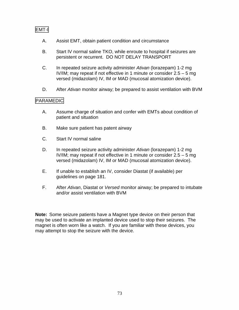
73
EMT-I
A. Assist EMT, obtain patient condition and circumstance B. Start IV normal saline TKO, while enroute to hospital if seizures are
persistent or recurrent. DO NOT DELAY TRANSPORT
C. In repeated seizure activity administer Ativan (lorazepam) 1-2 mg IV/IM; may repeat if not effective in 1 minute or consider 2.5 – 5 mg versed (midazolam) IV, IM or MAD (mucosal atomization device).
D. After Ativan monitor airway; be prepared to assist ventilation with BVM
PARAMEDIC
A. Assume charge of situation and confer with EMTs about condition of
patient and situation B. Make sure patient has patent airway
C. Start IV normal saline
D. In repeated seizure activity administer Ativan (lorazepam) 1-2 mg
IV/IM; may repeat if not effective in 1 minute or consider 2.5 – 5 mg versed (midazolam) IV, IM or MAD (mucosal atomization device).
E. If unable to establish an IV, consider Diastat (if available) per
guidelines on page 181. F. After Ativan, Diastat or Versed monitor airway; be prepared to intubate
and/or assist ventilation with BVM
Note: Some seizure patients have a Magnet type device on their person that may be used to activate an implanted device used to stop their seizures. The magnet is often worn like a watch. If you are familiar with these devices, you may attempt to stop the seizure with the device.
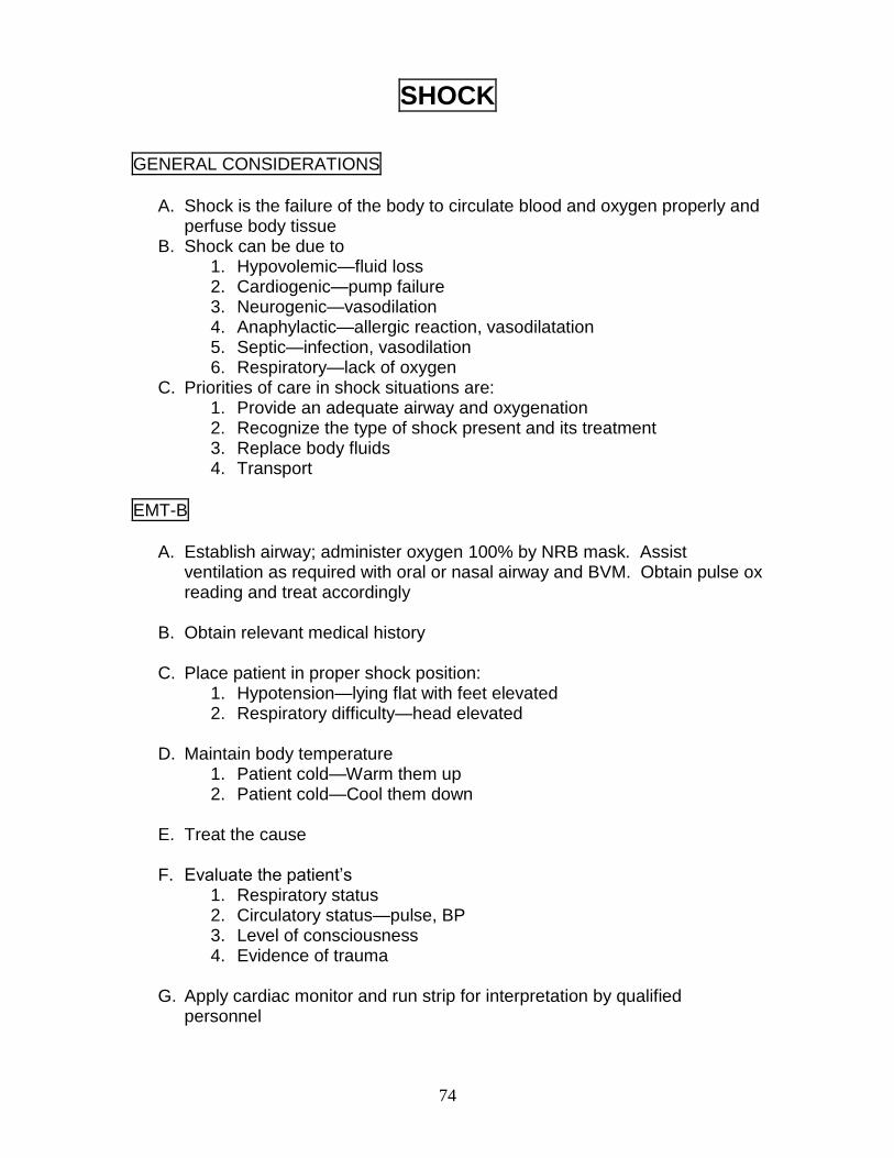
74
SHOCK
GENERAL CONSIDERATIONS
A. Shock is the failure of the body to circulate blood and oxygen properly and
perfuse body tissue B. Shock can be due to
1. Hypovolemic—fluid loss 2. Cardiogenic—pump failure 3. Neurogenic—vasodilation 4. Anaphylactic—allergic reaction, vasodilatation 5. Septic—infection, vasodilation 6. Respiratory—lack of oxygen
C. Priorities of care in shock situations are: 1. Provide an adequate airway and oxygenation 2. Recognize the type of shock present and its treatment 3. Replace body fluids 4. Transport
EMT-B
A. Establish airway; administer oxygen 100% by NRB mask. Assist
ventilation as required with oral or nasal airway and BVM. Obtain pulse ox reading and treat accordingly
B. Obtain relevant medical history
C. Place patient in proper shock position:
1. Hypotension—lying flat with feet elevated 2. Respiratory difficulty—head elevated
D. Maintain body temperature 1. Patient cold—Warm them up 2. Patient cold—Cool them down
E. Treat the cause F. Evaluate the patient’s
1. Respiratory status 2. Circulatory status—pulse, BP 3. Level of consciousness 4. Evidence of trauma
G. Apply cardiac monitor and run strip for interpretation by qualified personnel
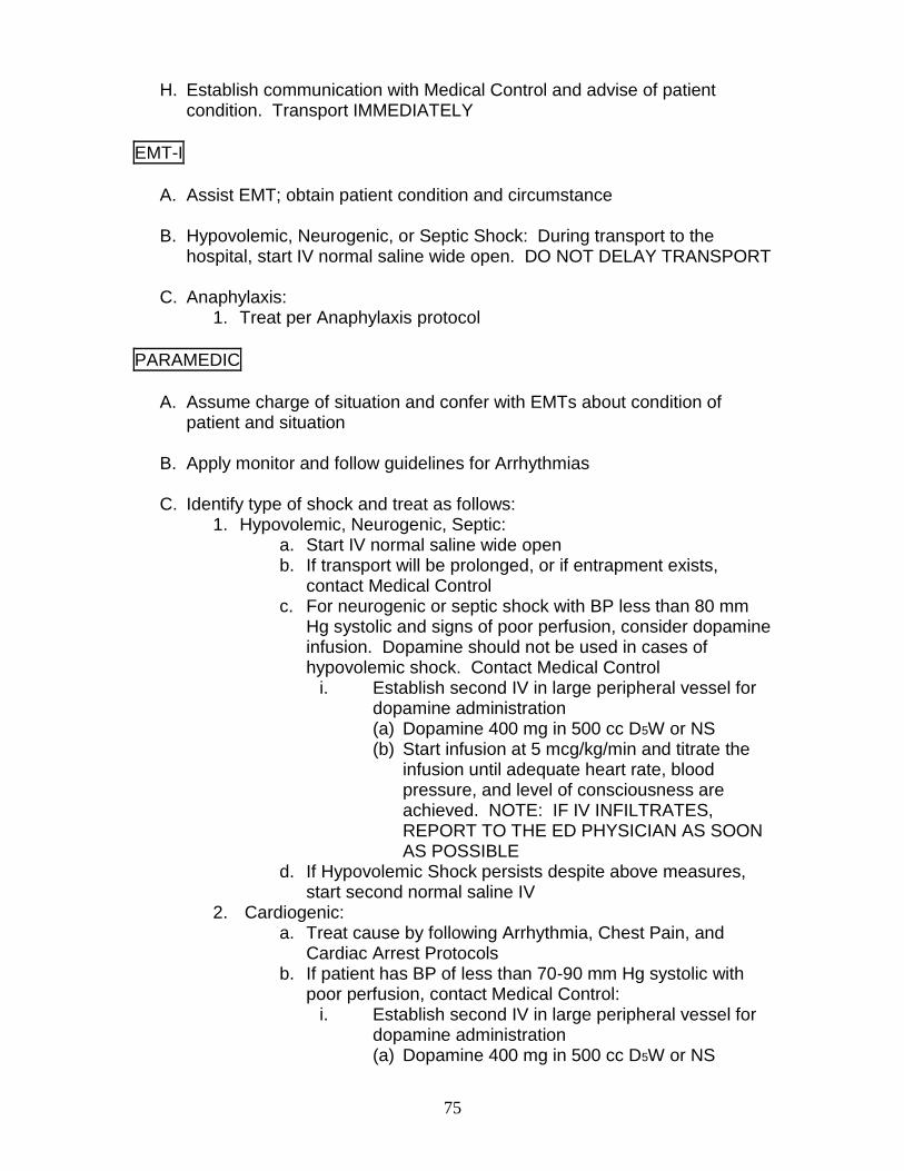
75
H. Establish communication with Medical Control and advise of patient condition. Transport IMMEDIATELY
EMT-I
A. Assist EMT; obtain patient condition and circumstance B. Hypovolemic, Neurogenic, or Septic Shock: During transport to the
hospital, start IV normal saline wide open. DO NOT DELAY TRANSPORT
C. Anaphylaxis: 1. Treat per Anaphylaxis protocol
PARAMEDIC
A. Assume charge of situation and confer with EMTs about condition of
patient and situation B. Apply monitor and follow guidelines for Arrhythmias
C. Identify type of shock and treat as follows:
1. Hypovolemic, Neurogenic, Septic: a. Start IV normal saline wide open b. If transport will be prolonged, or if entrapment exists,
contact Medical Control c. For neurogenic or septic shock with BP less than 80 mm
Hg systolic and signs of poor perfusion, consider dopamine infusion. Dopamine should not be used in cases of hypovolemic shock. Contact Medical Control
i. Establish second IV in large peripheral vessel for dopamine administration (a) Dopamine 400 mg in 500 cc D5W or NS (b) Start infusion at 5 mcg/kg/min and titrate the
infusion until adequate heart rate, blood pressure, and level of consciousness are achieved. NOTE: IF IV INFILTRATES, REPORT TO THE ED PHYSICIAN AS SOON AS POSSIBLE
d. If Hypovolemic Shock persists despite above measures, start second normal saline IV
2. Cardiogenic: a. Treat cause by following Arrhythmia, Chest Pain, and
Cardiac Arrest Protocols b. If patient has BP of less than 70-90 mm Hg systolic with
poor perfusion, contact Medical Control: i. Establish second IV in large peripheral vessel for
dopamine administration (a) Dopamine 400 mg in 500 cc D5W or NS
76
(b) Start infusion at 5 mcg/kg/min and titrate the infusion until adequate heart rate, blood pressure, and level of consciousness are achieved. NOTE: IF IV INFILTRATES, REPORT TO THE ED PHYSICIAN AS SOON AS POSSIBLE
3. Anaphylactic: a. Treat per Anaphylaxis protocol
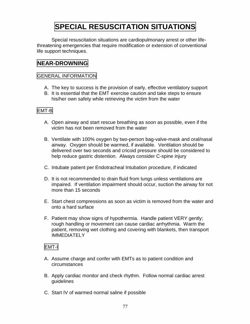
77
SPECIAL RESUSCITATION SITUATIONS Special resuscitation situations are cardiopulmonary arrest or other life-threatening emergencies that require modification or extension of conventional life support techniques.
NEAR-DROWNING
GENERAL INFORMATION
A. The key to success is the provision of early, effective ventilatory support B. It is essential that the EMT exercise caution and take steps to ensure
his/her own safety while retrieving the victim from the water
EMT-B
A. Open airway and start rescue breathing as soon as possible, even if the
victim has not been removed from the water B. Ventilate with 100% oxygen by two-person bag-valve-mask and oral/nasal
airway. Oxygen should be warmed, if available. Ventilation should be delivered over two seconds and cricoid pressure should be considered to help reduce gastric distention. Always consider C-spine injury
C. Intubate patient per Endotracheal Intubation procedure, if indicated
D. It is not recommended to drain fluid from lungs unless ventilations are
impaired. If ventilation impairment should occur, suction the airway for not more than 15 seconds
E. Start chest compressions as soon as victim is removed from the water and
onto a hard surface
F. Patient may show signs of hypothermia. Handle patient VERY gently; rough handling or movement can cause cardiac arrhythmia. Warm the patient, removing wet clothing and covering with blankets, then transport IMMEDIATELY
EMT-I
A. Assume charge and confer with EMTs as to patient condition and
circumstances B. Apply cardiac monitor and check rhythm. Follow normal cardiac arrest
guidelines
C. Start IV of warmed normal saline if possible
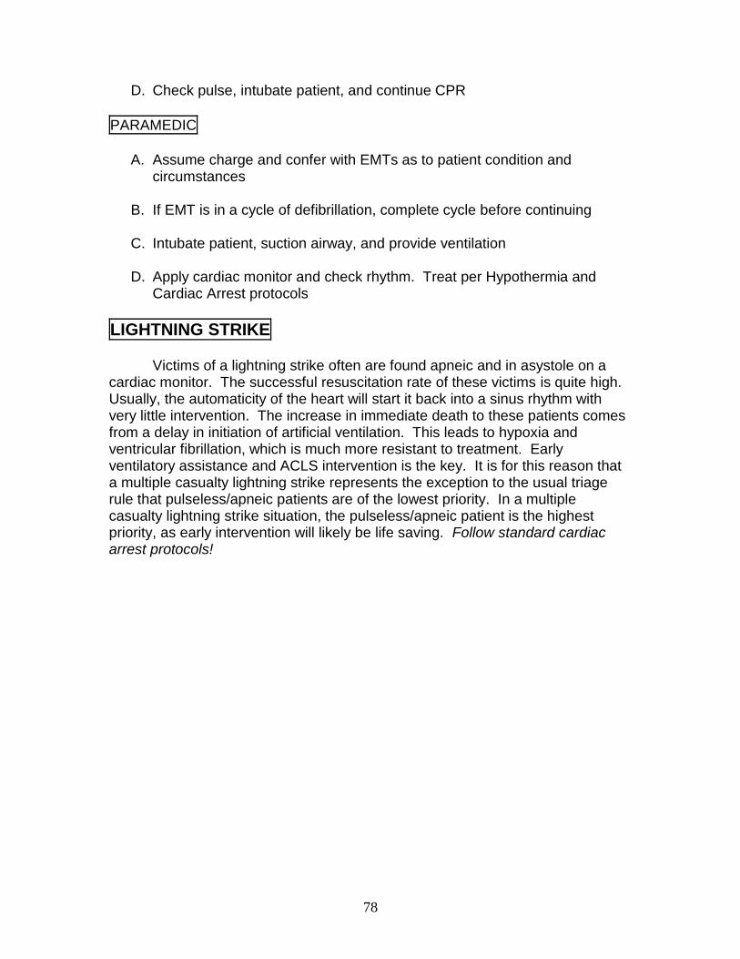
78
D. Check pulse, intubate patient, and continue CPR
PARAMEDIC
A. Assume charge and confer with EMTs as to patient condition and
circumstances B. If EMT is in a cycle of defibrillation, complete cycle before continuing
C. Intubate patient, suction airway, and provide ventilation
D. Apply cardiac monitor and check rhythm. Treat per Hypothermia and
Cardiac Arrest protocols
LIGHTNING STRIKE Victims of a lightning strike often are found apneic and in asystole on a cardiac monitor. The successful resuscitation rate of these victims is quite high. Usually, the automaticity of the heart will start it back into a sinus rhythm with very little intervention. The increase in immediate death to these patients comes from a delay in initiation of artificial ventilation. This leads to hypoxia and ventricular fibrillation, which is much more resistant to treatment. Early ventilatory assistance and ACLS intervention is the key. It is for this reason that a multiple casualty lightning strike represents the exception to the usual triage rule that pulseless/apneic patients are of the lowest priority. In a multiple casualty lightning strike situation, the pulseless/apneic patient is the highest priority, as early intervention will likely be life saving. Follow standard cardiac arrest protocols!
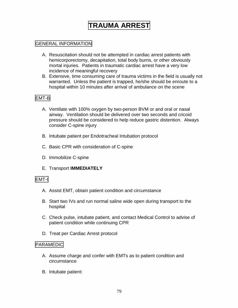
79
TRAUMA ARREST
GENERAL INFORMATION
A. Resuscitation should not be attempted in cardiac arrest patients with
hemicorporectomy, decapitation, total body burns, or other obviously mortal injuries. Patients in traumatic cardiac arrest have a very low incidence of meaningful recovery
B. Extensive, time consuming care of trauma victims in the field is usually not warranted. Unless the patient is trapped, he/she should be enroute to a hospital within 10 minutes after arrival of ambulance on the scene
EMT-B
A. Ventilate with 100% oxygen by two-person BVM or and oral or nasal
airway. Ventilation should be delivered over two seconds and cricoid pressure should be considered to help reduce gastric distention. Always consider C-spine injury
B. Intubate patient per Endotracheal Intubation protocol
C. Basic CPR with consideration of C-spine
D. Immobilize C-spine
E. Transport IMMEDIATELY
EMT-I
A. Assist EMT, obtain patient condition and circumstance B. Start two IVs and run normal saline wide open during transport to the
hospital
C. Check pulse, intubate patient, and contact Medical Control to advise of patient condition while continuing CPR
D. Treat per Cardiac Arrest protocol
PARAMEDIC
A. Assume charge and confer with EMTs as to patient condition and
circumstance
B. Intubate patient:
80
1. Patients should be intubated orotracheally without movement of the C-spine
2. If orotracheal intubation is not possible, or an obstruction is present, then cricothyroidotomy may be necessary
C. Assess cause of patient’s condition and treat according to appropriate
guidelines
D. Continue treatment per Cardiac Arrest protocol
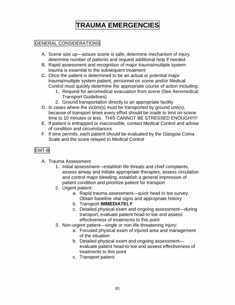
81
TRAUMA EMERGENCIES
GENERAL CONSIDERATIONS
A. Scene size up—assure scene is safe, determine mechanism of injury,
determine number of patients and request additional help if needed B. Rapid assessment and recognition of major trauma/multiple system
trauma is essential to the subsequent treatment C. Once the patient is determined to be an actual or potential major
trauma/multiple system patient, personnel on scene and/or Medical Control must quickly determine the appropriate course of action including:
1. Request for aeromedical evacuation from scene (See Aeromedical Transport Guidelines)
2. Ground transportation directly to an appropriate facility D. In cases where the victim(s) must be transported by ground unit(s),
because of transport times every effort should be made to limit on-scene time to 10 minutes or less. THIS CANNOT BE STRESSED ENOUGH!!!!!
E. If patient is entrapped or inaccessible, contact Medical Control and advise of condition and circumstances
F. If time permits, each patient should be evaluated by the Glasgow Coma Scale and the score relayed to Medical Control
EMT-B
A. Trauma Assessment
1. Initial assessment—establish life threats and chief complaints, assess airway and initiate appropriate therapies, assess circulation and control major bleeding, establish a general impression of patient condition and prioritize patient for transport
2. Urgent patient: a. Rapid trauma assessment—quick head to toe survey.
Obtain baseline vital signs and appropriate history b. Transport IMMEDIATELY c. Detailed physical exam and ongoing assessment—during
transport, evaluate patient head-to-toe and assess effectiveness of treatments to this point
3. Non-urgent patient—single or non-life-threatening injury: a. Focused physical exam of injured area and management
of the situation b. Detailed physical exam and ongoing assessment—
evaluate patient head-to-toe and assess effectiveness of treatments to this point
c. Transport patient
82
B. Urgent trauma treatment
1. Establish airway, breathing and circulation; maintain C-spine immobilization
2. Administer 100% oxygen and apply pulse oximeter 3. Control hemorrhage by appropriate method 4. Transport IMMEDIATELY 5. During transportation
a. Splint individual fracture(s) b. Evaluate patient’s:
i. Pulses distal to the fracture site ii. Distal skin color, temperature, neurological status
c. Obtain relevant history i. Where, When, How ii. Mechanism of injury
6. Establish communications with Medical Control and advise of patient condition and need for Trauma Team
C. Non-urgent trauma treatment
1. Establish airway, breathing and circulation; maintain C-spine immobilization
2. Administer 100% oxygen and apply pulse oximeter 3. Control hemorrhage by appropriate method 4. Splint all fracture(s)
a. Evaluate patient’s: i. Pulses distal to the fracture site ii. Distal skin color, temperature, neurological status
5. Obtain relevant history: a. Where, When, How b. Mechanism of injury
6. Establish communications with Medical Control and advise of patient condition
EMT-I
A. Assist EMTs; obtain patient condition and circumstance B. Start two IVs of normal saline to maintain perfusion and SBP of 90; IV
MUST NOT DELAY TRANSPORTATION
83
C. If the patient is conscious and alert and complaining of severe pain,
contact Medical Command to administer Morphine Sulfate as follows: 1. Small frequent doses of 2-4 mg every 5 minutes and
titrate to patient condition 2. DO NOT USE ON HEAD TRAUMA, CHEST INJURY,
RESPIRATORY DISTRESS DUE TO TRAUMA, OR ON ANY PATIENT WITH VOLUME DEPLETION OF ANY CAUSE
PARAMEDIC
A. Assume charge of situation and confer with EMTs about condition of
patient and situation B. Treat for shock per Shock Protocol
C. If the patient is conscious and alert and complaining of severe pain,
contact Medical Command to administer Morphine Sulfate as follows: 1. Small frequent doses of 2-4 mg every 5 minutes and
titrate to patient condition 2. DO NOT USE ON HEAD TRAUMA, CHEST INJURY,
RESPIRATORY DISTRESS DUE TO TRAUMA, OR ON ANY PATIENT WITH VOLUME DEPLETION OF ANY CAUSE
SPECIFIC INJURIES
A. Chest Wounds: 1. For sucking chest wounds or open pneumothorax, always cover the
wound with a non-porous dressing and seal 3 sides 2. Stabilize flail chest with trauma dressing
B. Evisceration: 1. Cover organs with sterile dressing moistened with saline 2. Lay the patient flat and elevate the knees
C. Complete Amputations: 1. Control bleeding by the most appropriate method; remember that
tourniquet is a last resort 2. Always take time to find the avulsed part, but do not delay patient
transport; transport avulsed part in a cool, dry sterile dressing
D. Pneumothorax/Hemothorax/Tension Pneumothorax: 1. Transport patient in position of comfort and watch for signs of a
tension pneumothorax 2. Symptoms of tension pneumothorax (Significant tension
pneumothorax may present exhibiting any or all of the following):
84
a. Chest pain or evidence of trauma b. Tachypnea c. Tachycardia d. JVD e. May initially exhibit hypertension progressing to
hypotension f. Hyperresonance on affected side g. Diminished or absent breath sounds on affected side h. Audible wheeze i. Tracheal deviation away from affected side (late sign)
3. Needle decompression per procedure
E. Head Injury: 1. Evaluate patient condition:
a. Level of Consciousness b. Pupillary size and reaction c. Glasgow Coma Scale results
2. Transport with head elevated 8-10 inches by tilting backboard—keep C-spine immobilized
3. Maintain airway, support with 100% oxygen by NRB mask and/or BVM
a. Orotracheal, nasotracheal or digital intubation may be indicated and should be accomplished gently with in-line C-spine immobilization (only if unable to ventilate/oxygenate via noninvasive measures)
b. Hyperoxygenate when there are signs of cerebral herniation: blown pupils, bradycardia, posturing
F. Spinal Injury
1. Immobilize spine 2. Cervical Immobilization Assessment
a. Cervical immobilization should be used if the following criteria are met:
i. The patient complains of neck pain ii. The patient has pain on palpation of neck iii. The patient complains of neurologic deficits or is
found upon physical exam to have neurologic deficits, including subjective numbness, tingling, or weakness or objective diminished sensation or motor weakness
iv. The patient with altered LOC and impaired competence, whether from drugs, alcohol, or head injury in a situation with suggestive mechanism of injury for neck injury (refer to Refusal of Service for impaired competence criteria)
v. The patient with suggestive mechanism of injury for neck injury and the patient has other major distracting injuries
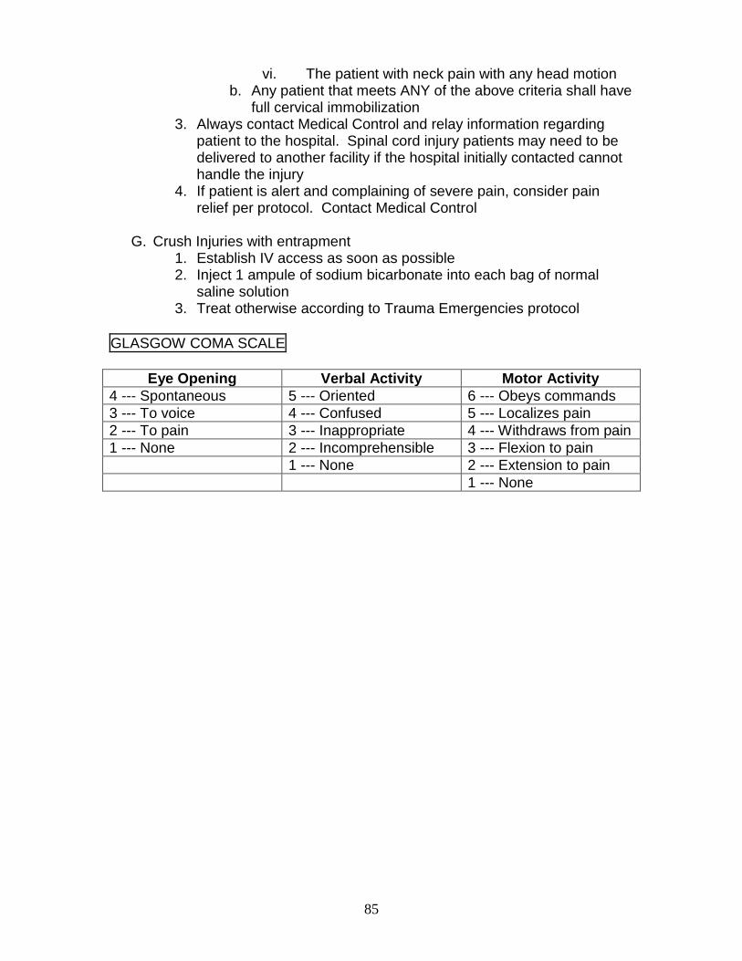
85
vi. The patient with neck pain with any head motion b. Any patient that meets ANY of the above criteria shall have
full cervical immobilization 3. Always contact Medical Control and relay information regarding
patient to the hospital. Spinal cord injury patients may need to be delivered to another facility if the hospital initially contacted cannot handle the injury
4. If patient is alert and complaining of severe pain, consider pain relief per protocol. Contact Medical Control
G. Crush Injuries with entrapment
1. Establish IV access as soon as possible 2. Inject 1 ampule of sodium bicarbonate into each bag of normal
saline solution 3. Treat otherwise according to Trauma Emergencies protocol
GLASGOW COMA SCALE
Eye Opening Verbal Activity Motor Activity
4 --- Spontaneous 5 --- Oriented 6 --- Obeys commands
3 --- To voice 4 --- Confused 5 --- Localizes pain
2 --- To pain 3 --- Inappropriate 4 --- Withdraws from pain
1 --- None 2 --- Incomprehensible 3 --- Flexion to pain
1 --- None 2 --- Extension to pain
1 --- None

86
87
Pediatric
Treatment
Protocols

88
89
ALTERED LEVEL OF CONSCIOUSNESS
(PEDIATRIC)
EMT-B
A. ABCs. Manually stabilize cervical spine as per Multiple Trauma Protocol if
cause of unconsciousness is unknown B. Administer 100% oxygen by NRB mask
C. If apneic, insert oral or nasal airway and ventilate with BVM
D. Evaluate patient’s general appearance, relevant history of condition, and
past medical history, especially history of diabetes, seizures, stroke, head injury, drug abuse
E. Determine blood sugar level
1. If blood sugar is less than 70, administer oral glucose if patient is awake and alert. May be repeated in 10 minutes if blood sugar remains below 70
2. If blood sugar is greater than 400, TRANSPORT
F. If unable to check blood sugar, or if blood sugar is between 70 and 400, establish communication with Medical Control and advise of patient condition
F. Apply cardiac monitor and run strip for interpretation by qualified
personnel
G. Transport IMMEDIATELY
EMT-I
A. Assist EMTs; obtain patient condition and circumstance B. Start IV/IO normal saline TKO
C. IV/IO fluid bolus 20 cc/kg of saline if any of the following are present:
unresponsive, appears dry, low BP, or poor capillary refill
D. DO NOT DELAY TRANSPORT
E. Determine blood sugar level a. Blood sugar less than 70, administer IV/IO bolus:
90
i. For children under 50 pounds (25 kg), 2 ml/kg of 25% dextrose (D25) or 1 ml/kg of 50% dextrose (D50) diluted with 1 ml/kg of sterile water
ii. For children over 50 pounds (25 kg), 1 ml/kg of 50% dextrose (D50) – maximum dose 50 ml or glucagon 1mg IM or MAD.
iii. D50 may be repeated in 10 minutes if blood sugar remains below 70.
b. Blood sugar greater than 400 and signs of hypoperfusion are present, administer an IV/IO fluid bolus:
i. 20 cc/kg of normal saline ii. May be repeated if no response in 10 minutes
F. If blood sugar is normal, respirations are impaired, or patient does not
respond to dextrose or fluid bolus, administer Narcan. If patient improves but does not fully awaken with Narcan, contact Medical Control for repeat dose
a. Patients under 5 years of age: 0.1 mg/kg slow IV/IO push b. Patient over 5 years of age or greater than 20 kg in weight: 2 mg
slow IV/IO push
PARAMEDIC
A. Assume charge of situation and confer with EMTs about condition of
patient and situation B. If patient does not have a gag reflex, intubate patient per Endotracheal
Intubation procedure
C. Apply cardiac monitor and check rhythm
D. Start IV/IO normal saline. Administer fluid bolus of 20 cc/kg if any of the following are present: unresponsive, appears dry, has a low BP or poor capillary refill
E. Determine blood sugar level
a. Blood sugar less than 70, administer IV/IO bolus: i. For children under 50 pounds (25 kg), 2 ml/kg of 25%
dextrose (D25) or 1 ml/kg of 50% dextrose (D50) diluted with 1 ml/kg of sterile water
i. For children over 50 pounds (25 kg), 1 ml/kg of 50% dextrose (D50) – maximum dose 50 ml or glucagon 1mg IM or MAD.
ii. D50 may be repeated in 10 minutes if blood sugar remains below 70.
91
b. Blood sugar greater than 400 and signs of hypoperfusion are
present, administer an IV/IO fluid bolus: i. 20 cc/kg of normal saline ii. May be repeated if no response in 10 minutes
F. If blood sugar is normal, respirations are impaired, or patient does not
respond to dextrose or fluid bolus, administer Narcan. If patient improves but does not fully awaken with Narcan, contact Medical Control for repeat dose
a. Patients under 5 years of age: 0.1 mg/kg slow IV/IO push b. Patient over 5 years of age or greater than 20 kg in weight: 2 mg
slow IV/IO push
G. Re-evaluate patient condition, contact Medical Control, and transport to hospital
H. In some cases, patient may require restraint; patient should not be
transported until completely restrained
92
ARRHYTHMIAS (PEDIATRIC)
GENERAL CONSIDERATIONS
A. In the treatment of cardiac arrhythmia, current American Heart Association
guidelines were consulted for protocol development B. Life-threatening cardiac rhythm disturbances in children are more
frequently the result rather than the cause of acute cardiopulmonary emergencies
C. In infants and children, arrhythmia should be treated as an emergency only if:
1. The arrhythmia compromises cardiac output, or 2. The arrhythmia has the potential for degenerating into a rhythm that
compromises cardiac output D. Initial therapy in children will consist of proper ventilation and oxygenation,
along with the assessment of cardiac output E. Transport is essential when advanced cardiac life support is not
immediately available F. Refer to length-based drug treatment guide (e.g. BROSELOW
PEDIATRIC EMERGENCY TAPE) when unsure about patient weight, age, and/or drug dosage
EMT-B
A. Assist ventilation with BVM while administering 100% oxygen or provide
mouth-to-mouth ventilation using barrier device B. Immobilize cervical spine, if indicated
C. Evaluate patient’s general appearance and determine:
1. Vital signs 2. Level of consciousness 3. Cardiac output 4. Lung sounds
D. Obtain relevant history of current condition E. Establish communication with Medical Control and advise of patient
condition. Transport IMMEDIATELY
F. If cardiac monitor is available, and patient has an unusual and/or irregular heart rate or pulse, apply cardiac monitor and run a strip for interpretation by qualified personnel
93
EMT-I
A. Initiate IV/IO normal saline TKO (provide 20 cc/kg fluid bolus if signs of
hypoperfusion are present)
PARAMEDIC
A. Assume charge of situation and confer with EMTs about patient condition
and situation B. Assess airway adequacy and intubate if indicated
C. Apply monitor and determine rhythm
D. Start IV/IO normal saline TKO using pediatric IV tubing set-up if available
E. Treat arrhythmias as follows:
1. Bradycardia. Treat only if: a. Infant or Child’s heart rate is less than 60/min despite
adequate ventilation, and patient has decreased cardiac output
b. Administer epinephrine IV, IO, or ET every three minutes or until cardiac output improves
i. When IV or IO routes are available, administer 0.1 mg/kg (0.1 ml/kg) of 1:1000 (maximum single dose 1 mg)
ii. When administering through ET tube, use twice the IV or IO dose. ET epinephrine must be diluted with sterile water or saline
iii. If no response, administer atropine 0.02 mg/kg. MINIMUM DOSE: 0.1 MG. MAXIMUM SINGLE DOSE: 1.0 MG
iv. TRANSPORT IMMEDIATELY v. If no response, contact Medical Control for
possible cardiac pacing. Do not delay transport 2. Supraventricular Tachycardia (SVT)
a. If patient is asymptomatic, do not treat. Transport immediately
b. Consider hypovolemia and follow Shock protocol c. If patient is symptomatic (signs of CHF, poor capillary refill,
hypotension, respiratory distress) and rate is greater than 240:
i. Administer adenosine, 0.1 mg/kg (maximum 6 mg) RAPID IV bolus over 1 to 2 seconds followed IMMEDIATELY with a 10 cc bolus of saline (within 5 seconds)

94
ii. If no conversion, repeat adenosine in 1-2 minutes, 0.2 mg/kg (maximum 12 mg) RAPID IV bolus followed IMMEDIATELY with a 10 cc bolus of saline (within 5 seconds)
iii. If no conversion, repeat adenosine in 1-2 minutes, 0.2 mg/kg (maximum 12 mg) RAPID IV bolus followed IMMEDIATELY with a 10 cc bolus of saline (within 5 seconds). MAXIMUM OF 3 DOSES (30 mg) OF ADENOSINE
iv. Contact Medical Control for possible synchronized cardioversion at 0.5 joules/kg (use same setting for biphasic) – maximum initial energy 50 joules
v. Transport IMMEDIATELY
95
CARDIOPULMONARY ARREST (PEDIATRIC)
GENERAL CONSIDERATIONS
A. Cardiac arrest in children is primarily due to the lack of an adequate
airway, resulting in hypoxia B. All EMT personnel must concentrate on opening and maintaining the
airway and ventilating with 100% oxygen C. When using BVM ventilation, cricoid pressure can be applied to occlude
the esophagus and prevent gastric distention. Cricoid pressure can be applied until an ET tube can be inserted
D. Transport immediately when excessive hemorrhage or hypothermia is present. Advanced life support measures should be carried out during transportation
E. If peripheral IVs cannot be established, vascular access should be obtained by intraosseous route
F. If IV or IO access cannot be established, administer appropriate medications through the endotracheal tube
G. If Sudden Infant Death Syndrome (SIDS) is suspected: 1. Initiate basic and advanced life support, unless apparent rigor
mortis or signs of lividity are present 2. Be supportive of family 3. Encourage family to have friends or neighbors accompany them to
the hospital 4. If infant is not resuscitated, refer parents to Social Services at the
Emergency Department to initiate counseling H. Refer to length-based drug treatment guide (e.g. BROSELOW
PEDIATRIC EMERGENCY TAPE) when unsure about patient weight, age and/or drug dosage
EMT-B
A. Open and maintain airway with sniffing position B. Ventilate with 100% oxygen, via bag-valve-mask with oxygen reservoir
C. Intubate per Endotracheal Intubation procedure
D. Initiate cardiac compressions in accordance with American Heart
Association guidelines
E. Apply cardiac monitor and run strip for interpretation by qualified personnel
F. Establish communication with Medical Control and advise of patient
condition. Transport IMMEDIATELY
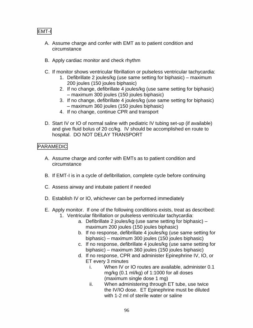
96
EMT-I
A. Assume charge and confer with EMT as to patient condition and
circumstance B. Apply cardiac monitor and check rhythm
C. If monitor shows ventricular fibrillation or pulseless ventricular tachycardia:
1. Defibrillate 2 joules/kg (use same setting for biphasic) – maximum 200 joules (150 joules biphasic)
2. If no change, defibrillate 4 joules/kg (use same setting for biphasic) – maximum 300 joules (150 joules biphasic)
3. If no change, defibrillate 4 joules/kg (use same setting for biphasic) – maximum 360 joules (150 joules biphasic)
4. If no change, continue CPR and transport
D. Start IV or IO of normal saline with pediatric IV tubing set-up (if available) and give fluid bolus of 20 cc/kg. IV should be accomplished en route to hospital. DO NOT DELAY TRANSPORT
PARAMEDIC
A. Assume charge and confer with EMTs as to patient condition and
circumstance B. If EMT-I is in a cycle of defibrillation, complete cycle before continuing
C. Assess airway and intubate patient if needed
D. Establish IV or IO, whichever can be performed immediately
E. Apply monitor. If one of the following conditions exists, treat as described:
1. Ventricular fibrillation or pulseless ventricular tachycardia: a. Defibrillate 2 joules/kg (use same setting for biphasic) –
maximum 200 joules (150 joules biphasic) b. If no response, defibrillate 4 joules/kg (use same setting for
biphasic) – maximum 300 joules (150 joules biphasic) c. If no response, defibrillate 4 joules/kg (use same setting for
biphasic) – maximum 360 joules (150 joules biphasic) d. If no response, CPR and administer Epinephrine IV, IO, or
ET every 3 minutes i. When IV or IO routes are available, administer 0.1
mg/kg (0.1 ml/kg) of 1:1000 for all doses (maximum single dose 1 mg)
ii. When administering through ET tube, use twice the IV/IO dose. ET Epinephrine must be diluted with 1-2 ml of sterile water or saline
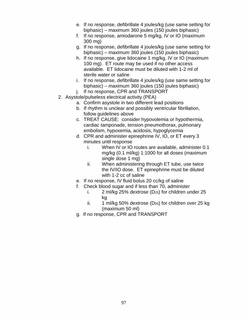
97
e. If no response, defibrillate 4 joules/kg (use same setting for biphasic) – maximum 360 joules (150 joules biphasic)
f. If no response, amiodarone 5 mg/kg, IV or IO (maximum 300 mg)
g. If no response, defibrillate 4 joules/kg (use same setting for biphasic) – maximum 360 joules (150 joules biphasic)
h. If no response, give lidocaine 1 mg/kg, IV or IO (maximum 100 mg). ET route may be used if no other access available. ET lidocaine must be diluted with 1-2 ml of sterile water or saline
i. If no response, defibrillate 4 joules/kg (use same setting for biphasic) – maximum 360 joules (150 joules biphasic)
j. If no response, CPR and TRANSPORT 2. Asystole/pulseless electrical activity (PEA)
a. Confirm asystole in two different lead positions b. If rhythm is unclear and possibly ventricular fibrillation,
follow guidelines above c. TREAT CAUSE: consider hypovolemia or hypothermia,
cardiac tamponade, tension pneumothorax, pulmonary embolism, hypoxemia, acidosis, hypoglycemia
d. CPR and administer epinephrine IV, IO, or ET every 3 minutes until response
i. When IV or IO routes are available, administer 0.1 mg/kg (0.1 ml/kg) 1:1000 for all doses (maximum single dose 1 mg)
ii. When administering through ET tube, use twice the IV/IO dose. ET epinephrine must be diluted with 1-2 cc of saline
e. If no response, IV fluid bolus 20 cc/kg of saline f. Check blood sugar and if less than 70, administer
i. 2 ml/kg 25% dextrose (D25) for children under 25 kg
ii. 1 ml/kg 50% dextrose (D50) for children over 25 kg (maximum 50 ml)
g. If no response, CPR and TRANSPORT
98
CHILD ABUSE/NEGLECT
GENERAL CONSIDERATIONS
A. Child abuse/neglect is widespread enough that nearly all EMTs and
Paramedics will see these problems at some time. Often, people refuse to believe that anyone could intentionally harm a child. The first step in recognizing abuse or neglect is to accept that it happens
B. Initiate treatment as necessary for situation using established protocols C. If possible, remove child from scene, transporting to hospital even if there
is no medical reason for transport D. If parents refuse permission to transport, notify law enforcement for
appropriate disposition. If patient is in immediate danger, let law enforcement officials handle scene
E. Advise parents to go to hospital. AVOID ACCUSATIONS as this may delay transport. Adult present at scene may not be the abuser
F. Carefully document findings and report to physicians at the hospital. Document conditions of environment at scene, if relevant. An EMT must also report or assure that actual or suspected child abuse/neglect is reported to the local law enforcement agency or the Department of Children and Family Services
DOCUMENT THIS NOTIFICATION
DO NOT JEOPARDIZE YOUR SAFETY
99
CHILDREN WITH SPECIAL NEEDS
SPECIAL CONSIDERATIONS
A. Children formerly cared for in hospitals and chronic care facilities are now
often cared for in homes. These children may have self-limited or chronic diseases. Many are relatively unstable and may frequently involve the EMS system for evaluation, stabilization, and transport
B. Knowing which children in a given area have special needs and keeping a log book can be useful
C. Parents and caretakers are usually trained in emergency management of these children and can be of assistance to EMS personnel
D. Special needs children include children with tracheostomy tubes with or without assisted ventilation, children with gastrostomy tubes, and children with indwelling central lines. Most serious complications of these devices are related to tracheostomy problems
EMERGENCIES IN CHILDREN WITH TRACHEOSTOMIES
EMT-B/EMT-I
A. Examine the child quickly for possible causes of distress which may be
easily correctable, such as a detached oxygen source B. Try to establish the child’s baseline
C. Document pulse ox, if possible
D. If on a ventilator, remove child from the ventilator and bag the child with a
secure oxygen source; there may be a problem with the ventilator or oxygen source
E. Suction the child, as accumulation of debris is a common cause of
obstruction; if the tracheostomy tube has a cannula, remove it (if it is the cause of obstruction, there will be immediate improvement)
F. If still no improvement, immediately transport to the nearest medical
facility; initiate appropriate resuscitation as needed
100
PARAMEDIC
A. If there is no improvement and the child is in severe respiratory distress,
the tube should be removed. Attempt BVM ventilation. If another tube is available, insert into the stoma and resume ventilation
B. If there is still no improvement, see the Pediatric Respiratory Distress
Protocol
EMERGENCIES IN CHILDREN WITH INDWELLING CENTRAL LINES
GENERAL CONSIDERATIONS
A. Children may have central lines in several possible locations: some
complications are due to location. Remember that some central lines are located under the skin and can be felt but not easily seen
B. The most common emergencies with central lines include blockage of the line, complete or partial accidental removal, and complete or partial laceration of the line
EMT-B/EMT-I/PARAMEDIC
A. Always evaluate child for cardiovascular stability as some complications
may be life-threatening B. Children may be experiencing complications from their underlying medical
condition; ask caretakers about the child’s condition
C. If line is blocked, do not attempt to force the catheter open; transport
D. For complete removal, do not attempt to reinsert; transport to the nearest Emergency Department. Because of risk of infection, do not attempt to push a line back in even if it is only slightly out
E. For partial or complete laceration of the line, clamp proximal to laceration
and transport to nearest Emergency Department
F. For children with sudden deterioration, begin basic resuscitation and transport to nearest Emergency Department (consider pneumothorax or internal bleeding)
101
EMERGENCIES IN CHILDREN WITH GASTROSTOMY TUBES
GENERAL CONSIDERATIONS
A. Children with gastrostomy tubes may have complications of obstruction or
dislodgement; these complications are easily recognizable and require transport
B. The child should be examined for any other possible problems
EMT-B/EMT-I/PARAMEDIC
A. Children who have problems with their tubes may have complications of
regurgitation or aspiration B. Be aware of and address any other possible problems related to
underlying medical condition
C. Transport the child and the tube to the nearest facility capable of replacing the tube; this is not an emergency transport. If caretaker has extra tubes, bring them. Do not attempt to replace the tube
102
FEVER
GENERAL CONSIDERATIONS
A. DO NOT sponge child or apply ice packs, as this can cause shivering
which can increase temperature B. Transport infants less than 8 weeks of age with a reported temperature of
greater than 100.4 degrees Fahrenheit (38 degrees Celsius) or less than 96 degrees Fahrenheit (35.5 degrees Celsius)
C. Obtain history, including feeding, level of consciousness, degree of
temperature, medications or therapies administered, and immunizations
D. Fevers less than 105 degrees Fahrenheit (40.5 degrees Celsius) are the body’s natural response to infection (either bacterial or viral) and are NOT dangerous
E. In children with a history of febrile seizures, be aware of the possibility of
another seizure, as these commonly recur in children who have had them before. These are not necessarily related to degree of fever
F. Most children who have had febrile seizures will grow out of them by the
age of 5.
103
FLUID AND DRUG ADMINISTRATION (PEDIATRIC)
EMT-I/PARAMEDIC
A. Peripheral venous access lines will be the first route for fluid and drug
administration for a life or limb threatening emergency situation B. Unless there are compelling factors, no more that two attempts at
peripheral access should be made in the pediatric patient
C. In life-threatening situations where venous access appears futile, immediately establish intraosseous access (in children age 2 and under). Intraosseous access should only be done when absolutely necessary (fluid and/or medication administration necessary, medications cannot be given IM, and transport time is more than a few minutes)
D. Intraosseous infusion
1. The following are guidelines for the UNSTABLE child requiring alternative vascular access AFTER ensuring airway and ventilation are established:
a. Indications: Route of choice for fluid and/or drug administration when peripheral IV access cannot be obtained with two quick attempts in less than 90 seconds and child is unstable (severely altered vital signs, markedly decreased level of consciousness)
b. Contraindications: Recently fractured bone near planned access site, known bone disorder, unsuccessful prior attempt at site. Relative contraindications: cellulitis or infected burn at site
c. Equipment: Bone marrow aspiration needle, iodine and alcohol preps, 5 cc syringe, local anesthetic (optional)
2. Procedure: a. Select site. Tibia (preferred): anteromedial aspect of
proximal tibial shaft, 1-3 cm below tibial tuberosity. Femur: distal 1/3 of femur, midline, 3 cm above condyle
b. Prep skin with iodine and alcohol c. After penetration of the skin, direct the needle at 90 degree
angle OR at a slight 10-15 degree vertical angle away from knee while applying gentle pressure, using a twisting motion
d. After penetration through the cortex, as marrow cavity is entered, operator may feel a ―pop‖ or less resistance. Remove the inner stylet and attach 5 cc syringe
e. Placement usually confirmed by aspiration of marrow followed by successful fluid administration without edema or swelling. Also, IO line should stand in place with minimal movement
104
f. Connect to conventional IV tubing and infuse fluids, blood or drugs as per protocol. If infusion fails to run, or runs slowly, flush needle with 5 cc of isotonic solution
g. Secure as needed; immobilize extremity and observe site frequently for extravasation of fluid
h. Infusion may require pressure bag to maintain patency or 60 cc syringe to provide bolus dosing
i. Document procedure and child’s response
E. Fluid of choice is normal saline, utilizing a macrodrip administration set. If child is less than 2 years old, a microdrip set should be used if available
F. Medications may be administered via either the IV or IO route G. When peripheral or IO access is not available for administering
medications: 1. If an ET tube is in place, the ET tube should be the route of
administration for Lidocaine, Atropine, Narcan, Epinephrine (Paramedic only)
2. Intramuscular (IM) route may be used for: a. Ativan (EMT-I or Paramedic) b. Atropine (Paramedic only) c. Benadryl (EMT-I or Paramedic) d. Glucagon (EMT-I or Paramedic) e. Inapsine (Paramedic only) – may only be used in children
over age 12 f. Morphine (EMT-I or Paramedic) g. Phenergan (Paramedic only) h. Versed (Paramedic only)
3. Mucosal Atomization Device (MAD) may be used for: a. Versed b. Narcan c. Glucagon
105
MULTI-TRAUMA
GENERAL CONSIDERATIONS
A. Pediatric Trauma care should primarily follow the Adult Protocol B. Areas where special focus should occur
1. May involve both respiratory failure and shock 2. Assessment and support of cardiopulmonary function is
fundamental
C. Common errors of pediatric trauma resuscitation are: 1. Failure to open and maintain the airway 2. Failure to provide appropriate fluid resuscitation to children with
head injury 3. Failure to recognize and treat internal hemorrhage 4. Reliance on normal vital signs as a predictor of normal volume
status
D. Intraosseous (IO) infusion is indicated in the trauma setting when shock needs to be treated and rapid venous access is unobtainable (see Intraosseous Access procedure)
E. The proper size equipment is very important to resuscitation care. Refer
to length-based drug treatment guide (e.g. BROSELOW PEDIATRIC EMERGENCY TAPE) when unsure about patient weight, age and/or drug dosage and when choosing equipment size
106
NEWBORN RESUSCITATION
GENERAL CONSIDERATIONS
A. Body heat must always be maintained. As soon as the baby is born, wipe
the baby dry and place in a warm environment. The following are ways to maintain heat:
1. Cover infant’s head, place infant against mother’s skin, and cover both
2. Use child seat with heat packs under and beside infant. Be sure to place towels between heat packs and infant
3. Swaddle infant tightly in blankets B. Always position infant in the ―sniffing‖ position (1 inch rolled towel under
shoulders). This allows for maximal open airway and drainage of secretions
C. Intermittently suction infant if airway has secretions. Prolonged or deep suctioning may cause bradycardia
1. Meconium aspiration is a major cause of death and morbidity among infants. If thick meconium is present and not removed adequately, a high percentage (60%) of these infants will aspirate the meconium
2. If meconium is present, suction the mouth and nose thoroughly, preferably prior to infant’s first breath. It MAY be necessary to suction the lower airway. This should be done only in cases of thick meconium and a non-vigorous infant. Lower airway suctioning is achieved by intubating the infant with an endotracheal tube and suctioning directly through the tube. Mechanical suction may be used only if suction pressure does not exceed –100 mmHg; bulb suctioning is preferred. The ET tube should be removed immediately after suctioning. If repeat suctioning is required, a new ET tube should be used
D. If drying and bulb suction has not provided enough stimulation, the infant’s feet may be flicked or the back rubbed. DO NOT hit the infant’s back or buttocks
E. If additional stimulation is needed, a BVM may be used to provide oxygen F. American Heart Association standards will be used as a guideline for both
Basic and Advanced Life Support procedures G. Refer to length-based drug treatment guide (e.g. BROSELOW
PEDIATRIC EMERGENCY TAPE) when unsure about patient weight, age and/or drug dosage
107
EMT-B
A. After delivery of the newborn’s head, but prior to delivery of the body,
quickly and thoroughly suction the mouth and oropharynx, followed by the nose, with a bulb syringe
B. After delivery of the infant, assess airway and breathing while drying and
stimulating the infant. If amniotic fluid is NOT clear, continue to suction PRIOR to stimulation
C. Clamp cord 10-12 centimeters from abdomen and again 2-3 centimeters
distally with cord clamp. Cord may be cut between clamps if clean scissors are available
D. If infant is not breathing despite stimulation after 30 seconds, initiate
ventilation using BVM
E. If heart rate is less than 60 but rising, continue ventilation
F. If heart rate is less than 60 but not rising, initiate cardiac compressions
G. BVM ventilation is also indicated for apnea and persistent central cyanosis. It is not indicated for peripheral cyanosis if respirations and heart rate are adequate
H. BVM ventilation rate should be between 40-60 breaths per minute.
Cardiac compression rate should be at a rate of 120 times per minute (COMPRESSION TO BREATH RATIO OF 3:1)
I. Establish communications with Medical Control and advise of patient
condition. Transport IMMEDIATELY
EMT-I
A. Monitor heart rate through auscultation or palpation
B. Establish IV or IO access if any signs of lethargy, respiratory difficulty,
poor perfusion, or bradycardia C. If infant shows signs of hypovolemia (capillary refill greater than 2
seconds, history of blood loss during delivery), administer saline 10 cc/kg over 5 minutes
D. Check blood sugar level and administer 2 cc/kg of 10% Dextrose (D10) if
level is below 40
E. TRANSPORT IMMEDIATELY
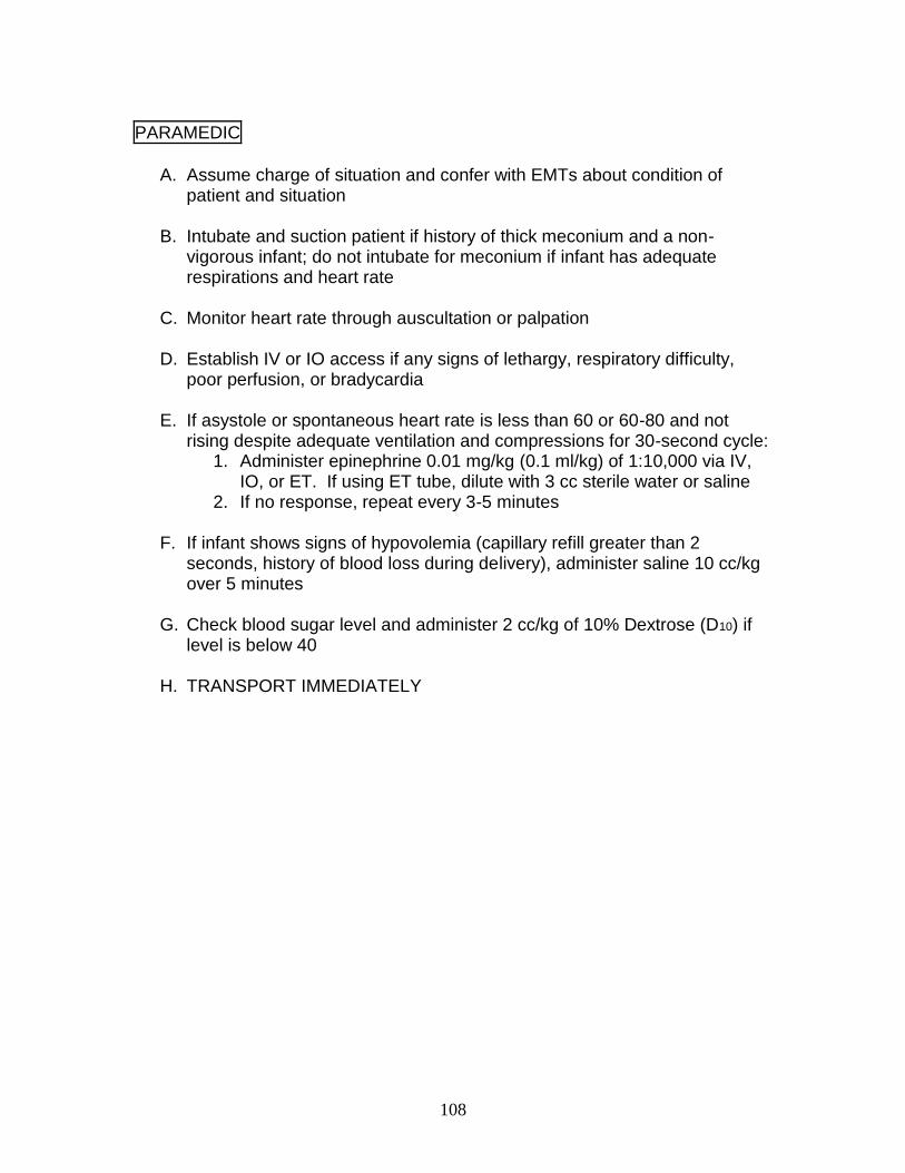
108
PARAMEDIC
A. Assume charge of situation and confer with EMTs about condition of
patient and situation B. Intubate and suction patient if history of thick meconium and a non-
vigorous infant; do not intubate for meconium if infant has adequate respirations and heart rate
C. Monitor heart rate through auscultation or palpation
D. Establish IV or IO access if any signs of lethargy, respiratory difficulty,
poor perfusion, or bradycardia
E. If asystole or spontaneous heart rate is less than 60 or 60-80 and not rising despite adequate ventilation and compressions for 30-second cycle:
1. Administer epinephrine 0.01 mg/kg (0.1 ml/kg) of 1:10,000 via IV, IO, or ET. If using ET tube, dilute with 3 cc sterile water or saline
2. If no response, repeat every 3-5 minutes F. If infant shows signs of hypovolemia (capillary refill greater than 2
seconds, history of blood loss during delivery), administer saline 10 cc/kg over 5 minutes
G. Check blood sugar level and administer 2 cc/kg of 10% Dextrose (D10) if
level is below 40
H. TRANSPORT IMMEDIATELY
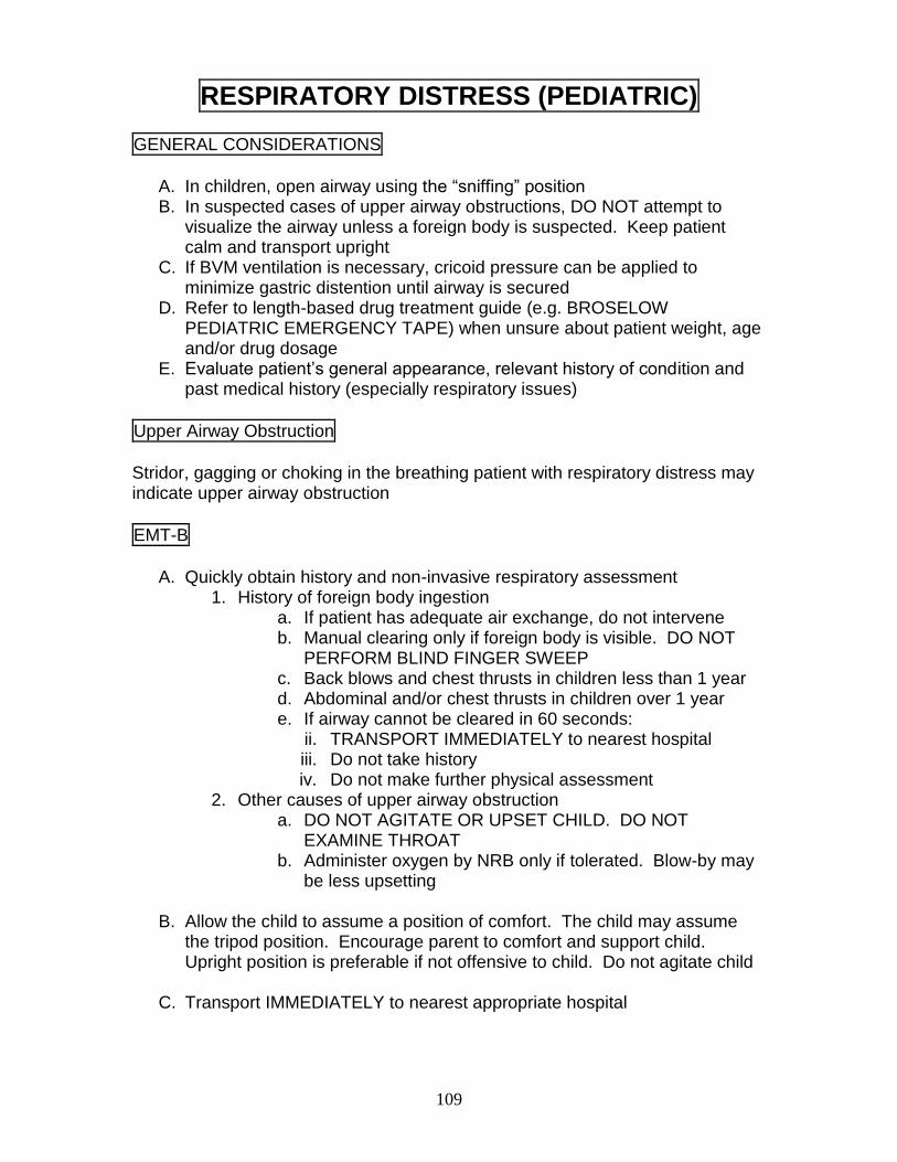
109
RESPIRATORY DISTRESS (PEDIATRIC)
GENERAL CONSIDERATIONS
A. In children, open airway using the ―sniffing‖ position B. In suspected cases of upper airway obstructions, DO NOT attempt to
visualize the airway unless a foreign body is suspected. Keep patient calm and transport upright
C. If BVM ventilation is necessary, cricoid pressure can be applied to minimize gastric distention until airway is secured
D. Refer to length-based drug treatment guide (e.g. BROSELOW PEDIATRIC EMERGENCY TAPE) when unsure about patient weight, age and/or drug dosage
E. Evaluate patient’s general appearance, relevant history of condition and past medical history (especially respiratory issues)
Upper Airway Obstruction
Stridor, gagging or choking in the breathing patient with respiratory distress may indicate upper airway obstruction
EMT-B
A. Quickly obtain history and non-invasive respiratory assessment
1. History of foreign body ingestion a. If patient has adequate air exchange, do not intervene b. Manual clearing only if foreign body is visible. DO NOT
PERFORM BLIND FINGER SWEEP c. Back blows and chest thrusts in children less than 1 year d. Abdominal and/or chest thrusts in children over 1 year e. If airway cannot be cleared in 60 seconds:
ii. TRANSPORT IMMEDIATELY to nearest hospital iii. Do not take history iv. Do not make further physical assessment
2. Other causes of upper airway obstruction a. DO NOT AGITATE OR UPSET CHILD. DO NOT
EXAMINE THROAT b. Administer oxygen by NRB only if tolerated. Blow-by may
be less upsetting
B. Allow the child to assume a position of comfort. The child may assume the tripod position. Encourage parent to comfort and support child. Upright position is preferable if not offensive to child. Do not agitate child
C. Transport IMMEDIATELY to nearest appropriate hospital
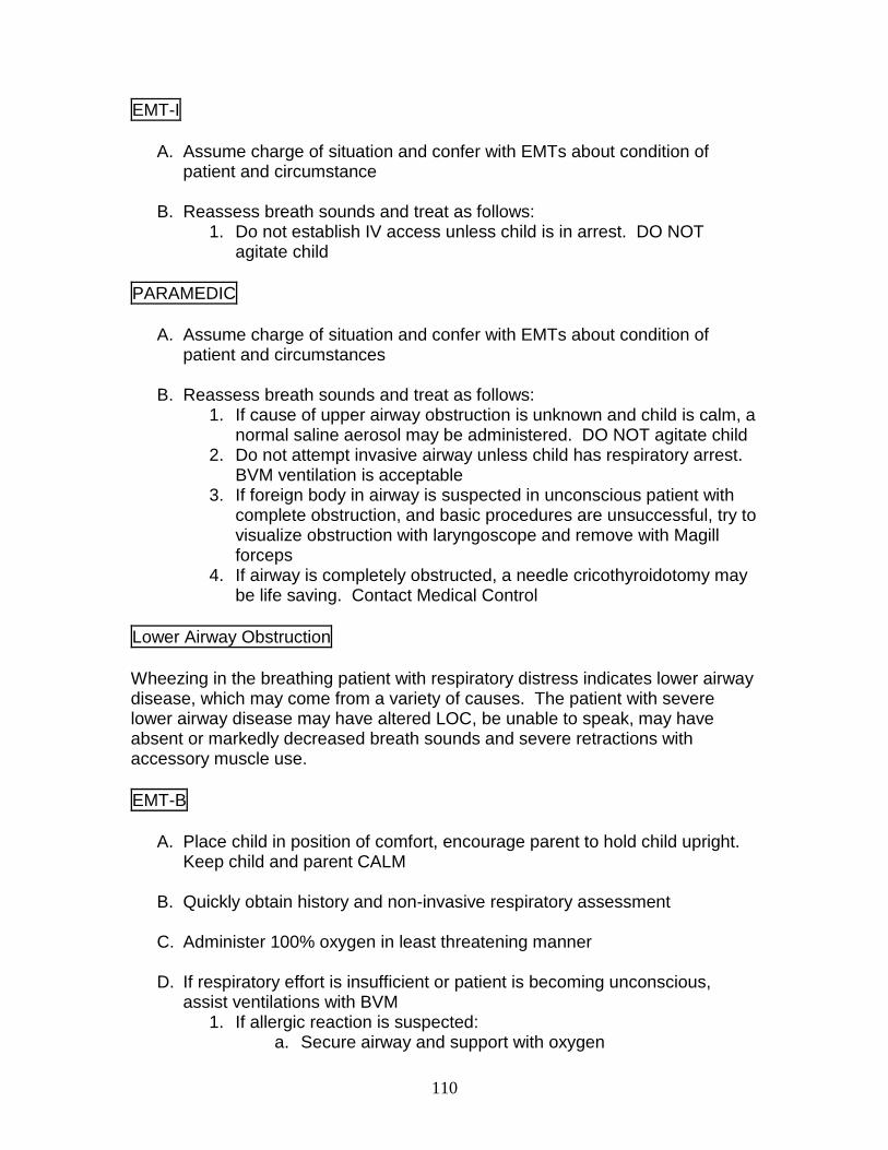
110
EMT-I
A. Assume charge of situation and confer with EMTs about condition of
patient and circumstance B. Reassess breath sounds and treat as follows:
1. Do not establish IV access unless child is in arrest. DO NOT agitate child
PARAMEDIC
A. Assume charge of situation and confer with EMTs about condition of
patient and circumstances B. Reassess breath sounds and treat as follows:
1. If cause of upper airway obstruction is unknown and child is calm, a normal saline aerosol may be administered. DO NOT agitate child
2. Do not attempt invasive airway unless child has respiratory arrest. BVM ventilation is acceptable
3. If foreign body in airway is suspected in unconscious patient with complete obstruction, and basic procedures are unsuccessful, try to visualize obstruction with laryngoscope and remove with Magill forceps
4. If airway is completely obstructed, a needle cricothyroidotomy may be life saving. Contact Medical Control
Lower Airway Obstruction
Wheezing in the breathing patient with respiratory distress indicates lower airway disease, which may come from a variety of causes. The patient with severe lower airway disease may have altered LOC, be unable to speak, may have absent or markedly decreased breath sounds and severe retractions with accessory muscle use.
EMT-B
A. Place child in position of comfort, encourage parent to hold child upright.
Keep child and parent CALM B. Quickly obtain history and non-invasive respiratory assessment
C. Administer 100% oxygen in least threatening manner
D. If respiratory effort is insufficient or patient is becoming unconscious,
assist ventilations with BVM 1. If allergic reaction is suspected:
a. Secure airway and support with oxygen
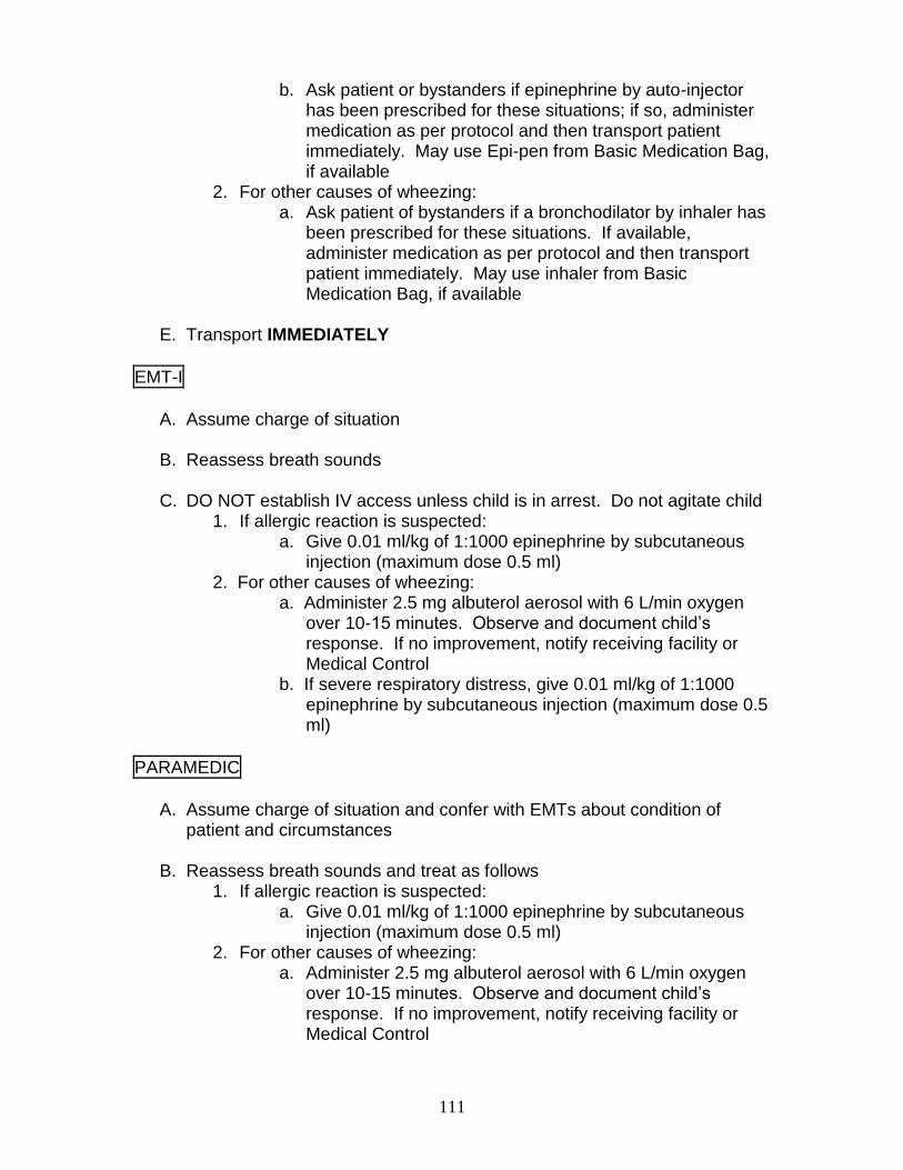
111
b. Ask patient or bystanders if epinephrine by auto-injector has been prescribed for these situations; if so, administer medication as per protocol and then transport patient immediately. May use Epi-pen from Basic Medication Bag, if available
2. For other causes of wheezing: a. Ask patient of bystanders if a bronchodilator by inhaler has
been prescribed for these situations. If available, administer medication as per protocol and then transport patient immediately. May use inhaler from Basic Medication Bag, if available
E. Transport IMMEDIATELY
EMT-I
A. Assume charge of situation B. Reassess breath sounds
C. DO NOT establish IV access unless child is in arrest. Do not agitate child
1. If allergic reaction is suspected: a. Give 0.01 ml/kg of 1:1000 epinephrine by subcutaneous
injection (maximum dose 0.5 ml) 2. For other causes of wheezing:
a. Administer 2.5 mg albuterol aerosol with 6 L/min oxygen over 10-15 minutes. Observe and document child’s response. If no improvement, notify receiving facility or Medical Control
b. If severe respiratory distress, give 0.01 ml/kg of 1:1000 epinephrine by subcutaneous injection (maximum dose 0.5 ml)
PARAMEDIC
A. Assume charge of situation and confer with EMTs about condition of
patient and circumstances B. Reassess breath sounds and treat as follows
1. If allergic reaction is suspected: a. Give 0.01 ml/kg of 1:1000 epinephrine by subcutaneous
injection (maximum dose 0.5 ml) 2. For other causes of wheezing:
a. Administer 2.5 mg albuterol aerosol with 6 L/min oxygen over 10-15 minutes. Observe and document child’s response. If no improvement, notify receiving facility or Medical Control

112
b. If severe respiratory distress, give 0.01 ml/kg of 1:1000 epinephrine by subcutaneous injection (maximum dose 0.5 ml)
c. DO NOT attempt invasive airway unless child has respiratory arrest and ventilation with BVM is unsuccessful
113
SEIZURE (PEDIATRIC)
GENERAL CONSIDERATIONS
A. The seizure has usually stopped by the time the EMS personnel arrive.
The patient will normally be in a postictal state B. The basic rule with seizures is to ―protect and support‖ the patient C. Aspiration precautions should include:
1. Coma position: a left side-lying position with the head lowered 15-30 degrees
2. Suction readily available 3. Clear mouth of foreign bodies (food, gum, etc)
D. Febrile seizures (seizures due to fever) are common in children and should be treated like other seizures
EMT-B
A. Place patient away from objects on which they might injure themselves:
protect but do not restrain them B. Clear and maintain airway; consider C-spine injury
C. Administer 100% oxygen with NRB as needed for ventilation
D. Obtain history from family and/or bystanders:
1. Seizure history 2. Description of onset of seizure 3. Medication and medication compliance 4. Other known medical history, especially fever, recent illness, head
trauma, diabetes, drugs
E. Evaluate any evidence of injury, especially head trauma F. Bring any medications with child to the hospital
G. Check blood sugar
H. Apply cardiac monitor and run strip for interpretation by qualified
personnel
I. Establish communications with Medical Control and advised of patient condition. Transport IMMEDIATELY

114
EMT-I
A. Assist EMTs; obtain patient condition and circumstances B. Initiate IV/IO normal saline TKO
C. If seizure activity persists, determine blood sugar level and treat
accordingly: a. Blood sugar less than 70, administer IV/IO bolus
i. 2 ml/kg of 25% Dextrose (D25) for children under 50 pounds (25 kg)
ii. 1 ml/kg of 50% Dextrose (D50) for children over 50 pounds (25 kg) – maximum dose 50 ml
b. Administer Ativan (lorazepam) 0.1 mg/kg slow IV/IO push over 3 minutes (maximum 2 mg). If IV/IO not available, can administer Ativan IM using same dosage
PARAMEDIC
A. Assume charge of the situation and confer with EMTs about patient
condition and situation B. Make sure patient has good airway. Check pulse ox. Consider intubation
C. Start IV/IO normal saline TKO
D. If seizure activity persists, determine blood sugar level and treat
accordingly: a. Blood sugar less than 70, administer IV/IO bolus
i. 2 ml/kg of 25% Dextrose (D25) for children under 50 pounds (25 kg)
ii. 1 ml/kg of 50% Dextrose (D50) for children over 50 pounds (25 kg) – maximum dose 50 ml
b. Administer Ativan (lorazepam) 0.1 mg/kg slow IV/IO push over 3 minutes (maximum 2 mg). If IV/IO not available, can administer Ativan IM using same dosage
c. If unable to establish an IV, consider Diastat (if available) per guidelines on page 181.
d. If Ativan or Diastat is not available, administer Versed (midazolam) 0.1 mg/kg IV/IM/IO/MAD (maximum 2.5 mg)
e. After meds monitor airway closely and be prepared to intubate and/or assist ventilation with a BVM.
115
SHOCK (PEDIATRIC)
GENERAL CONSIDERATIONS
A. Shock has many causes. The EMT must evaluate for fluid loss from blood
loss, excessive vomiting and/or diarrhea, heat exposure, severe infection, severe allergic reaction, spinal trauma, and heart failure
B. TACHYCARDIA IS THE FIRST SIGN OF SHOCK IN CHILDREN. Do not depend on hypotension, as this is a late finding. When evaluating for shock, look for low body temperature, poor capillary refill, decreased level of consciousness, and poor skin color or turgor
C. Transport should not be delayed. The airway must be secured, and the patient transported immediately. It is preferable for IV or IO access to be obtained during transportation
EMT-B
A. Open and maintain the airway with ―sniffing‖ position and the use of an
oral airway if needed. Consider C-spine precautions B. Control all external bleeding and evaluate for internal hemorrhage and/or
dehydration
C. Provide 100% oxygen through NRB mask, and assist ventilations if needed with BVM
D. Obtain vital signs, especially pulse and respirations
E. Apply cardiac monitor and run strip for interpretation by qualified
personnel
F. Establish communication with Medical Control and advise of patient condition. Transport IMMEDIATELY
EMT-I/PARAMEDIC
A. Assume charge of situation and confer with EMTs about condition of
patient and circumstances B. Apply monitor and follow protocol for Arrhythmia
C. Identify type of shock and treat as follows:
1. Hypovolemic, Neurogenic, Septic Shock: a. Start IV or IO saline and administer fluid bolus of 20 cc/kg if
signs of hypoperfusion or dehydration are present b. Repeat bolus during transport c. Check blood sugar; if less than 70, administer IV/IO bolus
116
i. 2 ml/kg of 25% Dextrose (D25) for children under 50 pounds (25 kg)
ii. 1 ml/kg of 50% Dextrose (D50) for children over 50 pounds (25 kg) – maximum dose 50 ml
2. Anaphylactic: a. Respiratory distress
i. Give 0.01 ml/kg) epinephrine subcutaneously (maximum dose 0.5 ml)
ii. Administer Benadryl (diphenhydramine) 1 mg/kg IV/IM/IO (especially when drug reaction is suspected) – maximum dose 50 mg
iii. When wheezing is present and cleared by epinephrine, provide albuterol breathing treatment 2.5 mg by aerosol mask over 10-15 minutes
b. Hives, itching, and/or swelling with normal blood pressure: i. Administer Benadryl (diphenhydramine) 1 mg/kg IV/IM
(especially when drug reaction is suspected) – maximum dose 50 mg
ii. Give 0.01 ml/kg 1:1000 epinephrine by injection subcutaneously (maximum dose 0.5 ml) if any sign of tongue or throat swelling
117
Administrative
Guidelines

118
119
AEROMEDICAL TRANSPORT
GENERAL CONSIDERATIONS
The decision to summon an aeromedical unit should be one that is made by the senior member of the EMS crew on scene. This decision should be made in order to minimize the out-of-hospital time of the victim. One an EMS unit is dispatched to the scene of an emergency, the ultimate responsibility for the treatment and transport of the patient(s) rests with the on-line Medical Control Physician and/or the off-line Medical Director. The most important factor in the overall outcome of the patient, especially in a traumatic injury, is the amount of time that passes until definitive care is obtained. Definitive care begins in the Emergency Department. Unnecessary delays on the scene of an emergency should be avoided, including waiting time for a helicopter to arrive. If the helicopter cannot arrive in less time than it would take to arrive at the nearest hospital by ground transport, then ground transport to the nearest hospital should be initiated as quickly as possible. It is not necessary to contact Medical Control prior to contacting an aeromedical unit, provided that one of the following conditions exists:
1. The patient will require an extended extrication from the scene of the emergency that exceeds the estimated arrival time of the aeromedical unit.
2. The estimated arrival time of an aeromedical unit is less than the estimated ground transport time to the nearest hospital.
3. The emergency scene involves numerous seriously injured persons, and it is believed that the number of patients would overwhelm the resources of the local hospital(s).
4. The scene of the emergency is inaccessible to ground vehicles but is accessible to aeromedical units. It is necessary to contact Medical Control if the landing zone for the
helicopter is the helipad at the local hospital. This presents a legal issue in that the Emergency Physician assumes greater legal responsibility for the patient because the patient has traversed onto hospital property. Radio contact with the Emergency Physician at the landing zone site should be made and the case discussed. Any on-line orders given should be followed and properly documented.
If the aeromedical unit has a physician on board, that physician shall assume responsibility for patient care upon his/her arrival and will assume Medical Control of the scene. If there is no physician on board the aeromedical unit, Medical Control for the EMS unit continues to rest with the on-line Medical Control Physician.
All communication with the aeromedical unit should be done on the hospital band frequency (155.22). This will ensure adequate communication capabilities and will allow the receiving and/or Medical Control facility to monitor communications.
120
BABY ABANDONMENT
GENERAL INSTRUCTIONS – ALL PROVIDERS
State law requires prehospital providers to assume temporary emergency custody of any infant under 72 hours of age if the parent(s) intent is to give up custody if the infant and not return to reclaim custody. At that time, the child is considered to be in emergency custody of the State of Ohio (no parental consent is required for medical treatment). At the time of the presentation of the child, an attempt should be made to obtain medical/family history of the child, although the parent(s) are not legally required to provide this information. The parent(s) have the right to remain anonymous and are immune from criminal charges. An abbreviated physical examination of the infant should be done at the time of initial presentation to look for signs of abuse or neglect. If such signs are found, the parent(s) lose the right to anonymity and the immunity from criminal charges. Attempts should be made to identify the person(s) and description(s) of the person(s) should be recorded. The Police Department should be notified immediately and given the necessary information. Any infant accepted into temporary emergency custody by a prehospital provider should be transported to the Emergency Department at Bethesda Hospital for further evaluation and proper placement by the social worker. Any prehospital provider who accepts an infant into temporary emergency custody is immune from civil or criminal legal action unless he/she acts in bad faith or with malicious purpose.
121
COMMUNICATIONS
A member of the prehospital team must contact the receiving hospital at the earliest possible time that will not interfere with good and efficient patient care. If on-line Medical Control is needed, this should be communicated at the beginning of the radio report so that the physician can be summoned to the radio. Patient reports should be thorough but concise. In the case of a multiple casualty incident, a preliminary report should be made, including estimates of number of victims, severity of injuries, arrival times, and further assistance needed. Patients appropriate for assignment to the triage area will be so assigned upon agreement between the EMS crew and the Emergency Department staff member receiving the radio report. High band Hospital – 155.220 Marcs channel Bethesda - Hos - 6001 Marcs channel Good Sam - Hos - 6002 Report Phone Good Sam – 740-455-7576 Report Phone Bethesda – 740-454-4290
122
DEAD ON ARRIVAL (DOA)
When a DOA is encountered, the EMS crew members should avoid disturbing the scene or body as much as possible, unless it is necessary to do so in order to care for other victims. Once it is determined that the victim is dead, the management of the scene should be conferred as quickly as possible to either the Police Department or the Coroner’s Office. It is the EMS crew’s responsibility contact the Coroner’s Office or to ensure that the Coroner’s Office has been or will be notified by the police officer on the scene. The determination that the victim is dead rests with the senior EMS crew member on the scene, but must follow these guidelines:
1. There is an injury incompatible with life (i.e. decapitation, burned beyond recognition, etc.).
2. The victim exhibits signs of decomposition, rigor mortis, or dependent lividity.
3. The patient has been in cardiac arrest for more than 20 minutes and is found in asystole (not due to cold water drowning or hypothermia).
4. A valid Ohio DNR form of identification is found or produced by family members (see DNR guidelines).
5. The patient has a terminal condition, the family refuses resuscitation, and permission to pronounce the patient dead is given via on-line Medical Control.
CAUTION: IF ANY DOUBT EXISTS THAT THE VICTIM IS DEAD AT THE TIME OF ARRIVAL, RESUSCITATIVE MEASURES SHOULD BE INSTITUTED IMMEDIATELY. WHENEVER RESUSCITATIVE MEASURES ARE STARTED, THEY MUST BE CONTINUED UNTIL ARRIVAL AT THE HOSPITAL, A PHYSICIAN PRONOUNCES THE PATIENT DEAD, OR A VALID OHIO DNR FORM OF IDENTIFICATION IS PRODUCED.
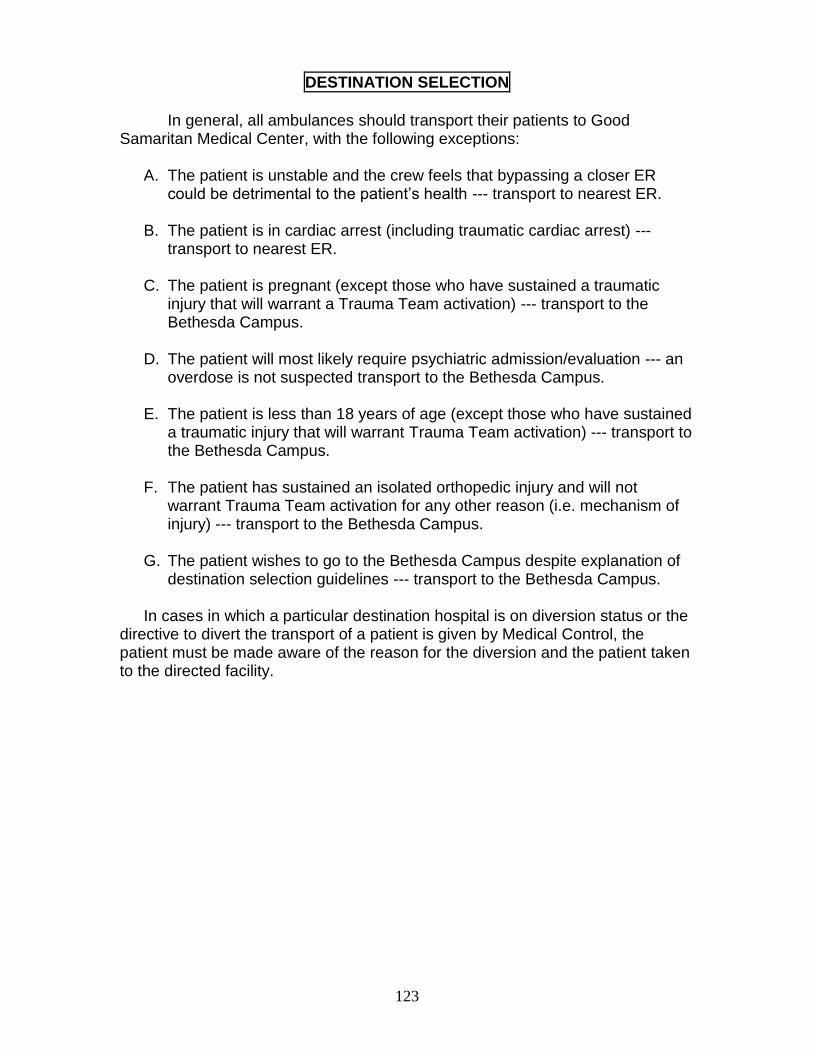
123
DESTINATION SELECTION
In general, all ambulances should transport their patients to Good
Samaritan Medical Center, with the following exceptions:
A. The patient is unstable and the crew feels that bypassing a closer ER could be detrimental to the patient’s health --- transport to nearest ER.
B. The patient is in cardiac arrest (including traumatic cardiac arrest) ---
transport to nearest ER.
C. The patient is pregnant (except those who have sustained a traumatic injury that will warrant a Trauma Team activation) --- transport to the Bethesda Campus.
D. The patient will most likely require psychiatric admission/evaluation --- an
overdose is not suspected transport to the Bethesda Campus.
E. The patient is less than 18 years of age (except those who have sustained a traumatic injury that will warrant Trauma Team activation) --- transport to the Bethesda Campus.
F. The patient has sustained an isolated orthopedic injury and will not
warrant Trauma Team activation for any other reason (i.e. mechanism of injury) --- transport to the Bethesda Campus.
G. The patient wishes to go to the Bethesda Campus despite explanation of
destination selection guidelines --- transport to the Bethesda Campus.
In cases in which a particular destination hospital is on diversion status or the directive to divert the transport of a patient is given by Medical Control, the patient must be made aware of the reason for the diversion and the patient taken to the directed facility.
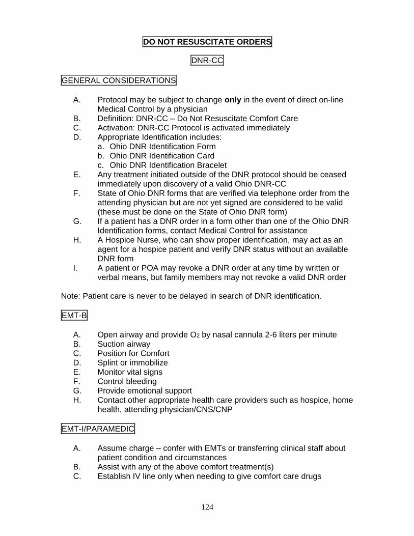
124
DO NOT RESUSCITATE ORDERS
DNR-CC
GENERAL CONSIDERATIONS
A. Protocol may be subject to change only in the event of direct on-line
Medical Control by a physician B. Definition: DNR-CC – Do Not Resuscitate Comfort Care C. Activation: DNR-CC Protocol is activated immediately D. Appropriate Identification includes:
a. Ohio DNR Identification Form b. Ohio DNR Identification Card c. Ohio DNR Identification Bracelet
E. Any treatment initiated outside of the DNR protocol should be ceased immediately upon discovery of a valid Ohio DNR-CC
F. State of Ohio DNR forms that are verified via telephone order from the attending physician but are not yet signed are considered to be valid (these must be done on the State of Ohio DNR form)
G. If a patient has a DNR order in a form other than one of the Ohio DNR Identification forms, contact Medical Control for assistance
H. A Hospice Nurse, who can show proper identification, may act as an agent for a hospice patient and verify DNR status without an available DNR form
I. A patient or POA may revoke a DNR order at any time by written or verbal means, but family members may not revoke a valid DNR order
Note: Patient care is never to be delayed in search of DNR identification.
EMT-B
A. Open airway and provide O2 by nasal cannula 2-6 liters per minute B. Suction airway C. Position for Comfort D. Splint or immobilize E. Monitor vital signs F. Control bleeding G. Provide emotional support H. Contact other appropriate health care providers such as hospice, home
health, attending physician/CNS/CNP
EMT-I/PARAMEDIC
A. Assume charge – confer with EMTs or transferring clinical staff about
patient condition and circumstances B. Assist with any of the above comfort treatment(s) C. Establish IV line only when needing to give comfort care drugs
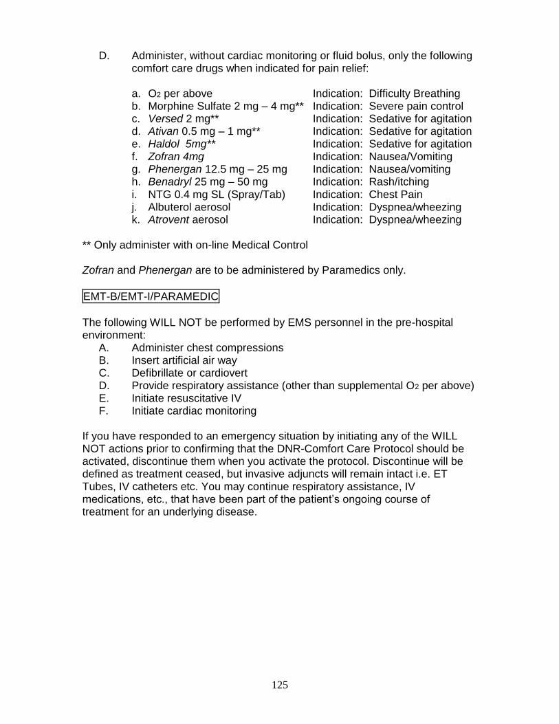
125
D. Administer, without cardiac monitoring or fluid bolus, only the following comfort care drugs when indicated for pain relief:
a. O2 per above Indication: Difficulty Breathing b. Morphine Sulfate 2 mg – 4 mg** Indication: Severe pain control c. Versed 2 mg** Indication: Sedative for agitation d. Ativan 0.5 mg – 1 mg** Indication: Sedative for agitation e. Haldol 5mg** Indication: Sedative for agitation f. Zofran 4mg Indication: Nausea/Vomiting g. Phenergan 12.5 mg – 25 mg Indication: Nausea/vomiting h. Benadryl 25 mg – 50 mg Indication: Rash/itching i. NTG 0.4 mg SL (Spray/Tab) Indication: Chest Pain j. Albuterol aerosol Indication: Dyspnea/wheezing k. Atrovent aerosol Indication: Dyspnea/wheezing
** Only administer with on-line Medical Control Zofran and Phenergan are to be administered by Paramedics only.
EMT-B/EMT-I/PARAMEDIC
The following WILL NOT be performed by EMS personnel in the pre-hospital environment:
A. Administer chest compressions B. Insert artificial air way C. Defibrillate or cardiovert D. Provide respiratory assistance (other than supplemental O2 per above) E. Initiate resuscitative IV F. Initiate cardiac monitoring
If you have responded to an emergency situation by initiating any of the WILL NOT actions prior to confirming that the DNR-Comfort Care Protocol should be activated, discontinue them when you activate the protocol. Discontinue will be defined as treatment ceased, but invasive adjuncts will remain intact i.e. ET Tubes, IV catheters etc. You may continue respiratory assistance, IV medications, etc., that have been part of the patient’s ongoing course of treatment for an underlying disease.
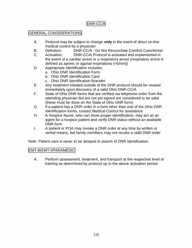
126
DNR-CC/A
GENERAL CONSIDERATIONS
A. Protocol may be subject to change only in the event of direct on-line
medical control by a physician B. Definition: DNR-CC/A - Do Not Resuscitate Comfort Care/Arrest C. Activation: DNR-CC/A Protocol is activated and implemented in
the event of a cardiac arrest or a respiratory arrest (respiratory arrest is defined as apneic or agonal respirations [<6/min])
D. Appropriate Identification includes: a. Ohio DNR Identification Form b. Ohio DNR Identification Card c. Ohio DNR Identification Bracelet
E. Any treatment initiated outside of the DNR protocol should be ceased immediately upon discovery of a valid Ohio DNR-CC/A
F. State of Ohio DNR forms that are verified via telephone order from the attending physician but are not yet signed are considered to be valid (these must be done on the State of Ohio DNR form)
G. If a patient has a DNR order in a form other than one of the Ohio DNR Identification forms, contact Medical Control for assistance
H. A Hospice Nurse, who can show proper identification, may act as an agent for a hospice patient and verify DNR status without an available DNR form
I. A patient or POA may revoke a DNR order at any time by written or verbal means, but family members may not revoke a valid DNR order
Note: Patient care is never to be delayed in search of DNR identification.
EMT-B/EMT-I/PARAMEDIC
A. Perform assessment, treatment, and transport at the respective level of
training as determined by protocol up to the above activation period
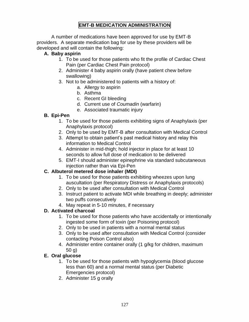
127
EMT-B MEDICATION ADMINISTRATION
A number of medications have been approved for use by EMT-B providers. A separate medication bag for use by these providers will be developed and will contain the following:
A. Baby aspirin 1. To be used for those patients who fit the profile of Cardiac Chest
Pain (per Cardiac Chest Pain protocol) 2. Administer 4 baby aspirin orally (have patient chew before
swallowing) 3. Not to be administered to patients with a history of:
a. Allergy to aspirin b. Asthma c. Recent GI bleeding d. Current use of Coumadin (warfarin) e. Associated traumatic injury
B. Epi-Pen 1. To be used for those patients exhibiting signs of Anaphylaxis (per
Anaphylaxis protocol) 2. Only to be used by EMT-B after consultation with Medical Control 3. Attempt to obtain patient’s past medical history and relay this
information to Medical Control 4. Administer in mid-thigh; hold injector in place for at least 10
seconds to allow full dose of medication to be delivered 5. EMT-I should administer epinephrine via standard subcutaneous
injection rather than via Epi-Pen C. Albuterol metered dose inhaler (MDI)
1. To be used for those patients exhibiting wheezes upon lung auscultation (per Respiratory Distress or Anaphylaxis protocols)
2. Only to be used after consultation with Medical Control 3. Instruct patient to activate MDI while breathing in deeply; administer
two puffs consecutively 4. May repeat in 5-10 minutes, if necessary
D. Activated charcoal 1. To be used for those patients who have accidentally or intentionally
ingested some form of toxin (per Poisoning protocol) 2. Only to be used in patients with a normal mental status 3. Only to be used after consultation with Medical Control (consider
contacting Poison Control also) 4. Administer entire container orally (1 g/kg for children, maximum
50 g) E. Oral glucose
1. To be used for those patients with hypoglycemia (blood glucose less than 60) and a normal mental status (per Diabetic Emergencies protocol)
2. Administer 15 g orally
128
EMT-INTERMEDIATE CURRICULUM UPDATE
In 2003, the State of Ohio introduced updates to the EMT-I curriculum, which included training in administration of selected medications, interpretation of selected cardiac rhythms, and performance of needle chest decompression. In November 2003, these protocols were updated to include these changes. It is intended that the performance of these listed skills included in the updated curriculum be only by those EMT-I personnel who have undergone the requisite training. The requisite training is either completion of an approved full EMT-I course that includes the new curriculum or completion of an approved EMT-I Bridge course. Those EMT-I personnel who have not completed the requisite training may still function temporarily using the skills that they had learned in their previous EMT-I curriculum course, but they may not apply those skills included in the curriculum update. As of July 1, 2005, any EMT-I who has not completed one of the two forms of requisite training will be considered to be at the EMT-B level.
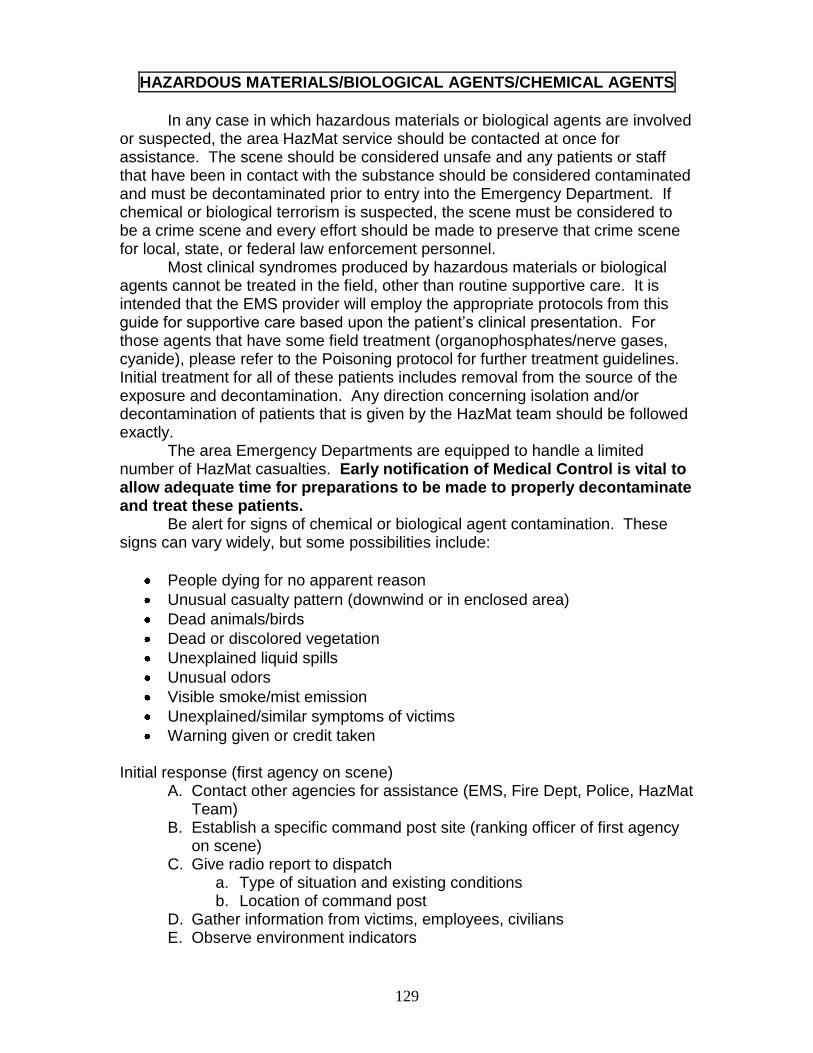
129
HAZARDOUS MATERIALS/BIOLOGICAL AGENTS/CHEMICAL AGENTS
In any case in which hazardous materials or biological agents are involved or suspected, the area HazMat service should be contacted at once for assistance. The scene should be considered unsafe and any patients or staff that have been in contact with the substance should be considered contaminated and must be decontaminated prior to entry into the Emergency Department. If chemical or biological terrorism is suspected, the scene must be considered to be a crime scene and every effort should be made to preserve that crime scene for local, state, or federal law enforcement personnel. Most clinical syndromes produced by hazardous materials or biological agents cannot be treated in the field, other than routine supportive care. It is intended that the EMS provider will employ the appropriate protocols from this guide for supportive care based upon the patient’s clinical presentation. For those agents that have some field treatment (organophosphates/nerve gases, cyanide), please refer to the Poisoning protocol for further treatment guidelines. Initial treatment for all of these patients includes removal from the source of the exposure and decontamination. Any direction concerning isolation and/or decontamination of patients that is given by the HazMat team should be followed exactly. The area Emergency Departments are equipped to handle a limited number of HazMat casualties. Early notification of Medical Control is vital to allow adequate time for preparations to be made to properly decontaminate and treat these patients. Be alert for signs of chemical or biological agent contamination. These signs can vary widely, but some possibilities include:
People dying for no apparent reason
Unusual casualty pattern (downwind or in enclosed area)
Dead animals/birds
Dead or discolored vegetation
Unexplained liquid spills
Unusual odors
Visible smoke/mist emission
Unexplained/similar symptoms of victims
Warning given or credit taken Initial response (first agency on scene)
A. Contact other agencies for assistance (EMS, Fire Dept, Police, HazMat Team)
B. Establish a specific command post site (ranking officer of first agency on scene)
C. Give radio report to dispatch a. Type of situation and existing conditions b. Location of command post
D. Gather information from victims, employees, civilians E. Observe environment indicators
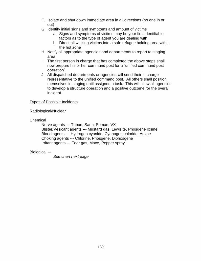
130
F. Isolate and shut down immediate area in all directions (no one in or out)
G. Identify initial signs and symptoms and amount of victims a. Signs and symptoms of victims may be your first identifiable
factors as to the type of agent you are dealing with b. Direct all walking victims into a safe refugee holding area within
the hot zone H. Notify all appropriate agencies and departments to report to staging
area I. The first person in charge that has completed the above steps shall
now prepare his or her command post for a ―unified command post operation‖
J. All dispatched departments or agencies will send their in charge representative to the unified command post. All others shall position themselves in staging until assigned a task. This will allow all agencies to develop a structure operation and a positive outcome for the overall incident.
Types of Possible Incidents Radiological/Nuclear Chemical Nerve agents --- Tabun, Sarin, Soman, VX Blister/Vesicant agents --- Mustard gas, Lewisite, Phosgene oxime Blood agents --- Hydrogen cyanide, Cyanogen chloride, Arsine Choking agents --- Chlorine, Phosgene, Diphosgene Irritant agents --- Tear gas, Mace, Pepper spray Biological --- See chart next page
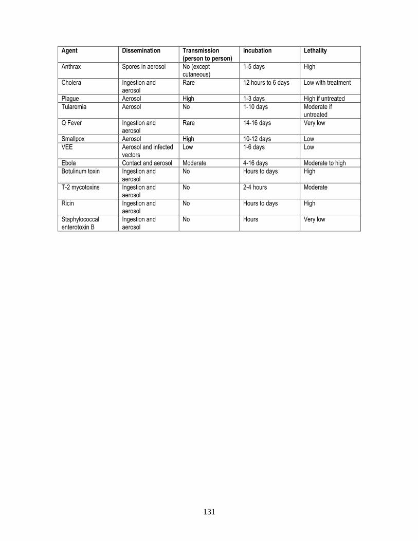
131
Agent Dissemination Transmission
(person to person) Incubation Lethality
Anthrax Spores in aerosol No (except cutaneous)
1-5 days High
Cholera Ingestion and aerosol
Rare 12 hours to 6 days Low with treatment
Plague Aerosol High 1-3 days High if untreated
Tularemia Aerosol No 1-10 days Moderate if untreated
Q Fever Ingestion and aerosol
Rare 14-16 days Very low
Smallpox Aerosol High 10-12 days Low
VEE Aerosol and infected vectors
Low 1-6 days Low
Ebola Contact and aerosol Moderate 4-16 days Moderate to high
Botulinum toxin Ingestion and aerosol
No Hours to days High
T-2 mycotoxins Ingestion and aerosol
No 2-4 hours Moderate
Ricin Ingestion and aerosol
No Hours to days High
Staphylococcal enterotoxin B
Ingestion and aerosol
No Hours Very low
132
HEAVY PATIENTS
In any community, there may be one or more individuals with excessive
weight. Obviously, the medical complications that accompany this extreme weight only compound an already difficult extrication situation. In order to facilitate the safe and effective treatment and transport of these patients, the following guidelines have been established.
A. In managing a patient with weight deemed to be unmanageable by the crew members on scene, it is suggested that at least 6 individuals assist in the transfer. If 6 EMS crew members are not available, other personnel (i.e. police or fire personnel) should be enlisted to assist. Mutual aid services should be called in if necessary.
B. It may be necessary to remove doors, walls, or windows. This situation is no different than extrication from a vehicle, although property damage may be higher. The life of the patient must remain the highest priority in this matter.
C. The patient is to be placed on double backboards or another adequate transfer device for support.
D. The patient is to be loaded onto a cot that is in the down position, and the cot is to be kept in the down position at all times.
E. If the patient’s weight exceeds the weight limit of the cot, the local fire department should be contacted for additional equipment.
F. At the Emergency Department, a special bed designed to support the patient’s weight will be made available at the ambulance entrance. It is necessary to notify the ED well in advance so that an appropriate bed can be obtained in a timely fashion.
133
MULTIPLE CASUALTY INCIDENTS
GENERAL CONSIDERATIONS
In cases of incidents involving multiple casualties, every attempt should be made to divide the patients between receiving hospitals so that one facility is not overwhelmed. Triage guidelines in respect to trauma patients must never be violated, however, even if this means transporting all patients to the same facility. Proper incident command procedures should be followed and it is the responsibility of the Transportation Officer to ensure that one facility is not overloaded with patients from the same incident.
134
NON-TRANSPORT EMS AGENCIES
GENERAL CONSIDERATIONS
EMS agencies that do not function to transport patients, but rather serve solely as first responders, may employ these protocols to the limits that their equipment and the training levels of their personnel may allow. These agencies are not required to carry any specific quota of equipment and may use their available resources within the scope of these protocols to the benefit of overall patient care. In cases where the level of certification of the personnel of the first responder agency exceeds the level of training of the personnel of the transporting agency, the highest level provider from the first responder agency must accompany the patient to the receiving hospital unless it is determined that the patient’s condition does not warrant this action. Under new state guidelines an EMT-FR may transport with an EMT and count as one of the required two EMTs for transport purposes.
135
ON SCENE EMT INTERVENER
On an EMS run where an EMT from another service happens upon the scene and offers assistance in the care of patients:
A. If no further assistance is needed, the offer should be politely declined. B. If the EMT’s assistance is needed or would be beneficial to patient care,
the offer may be accepted. The crew should obtain identification in the form of a valid EMT card. The EMT’s name and certification number should be documented on the run report.
C. If the intervening EMT represents the highest trained provider on the
scene, he/she must accompany the patient to the hospital. The crew should obtain identification in the form of a valid EMT card. The EMT’s name and certification number should be documented on the run report.
D. Under new state guidelines an EMT-FR may transport with an EMT and
count as one of the required two EMTs for transport purposes.
136
PATIENT REFUSAL OF TREATMENT OR TRANSPORT
It is not necessary to contact Medical Control concerning patient refusal unless there is some question regarding the patient’s capacity to make that decision. Direct communication between the patient and physician can often resolve many questions and convince the patient of the importance of treatment and transport.
A. Consent 1. The patient has the legal right to consent to or refuse treatment,
even if that does not seem to be in his/her best interest. If he or she is unable to do so, a legal guardian has this right.
2. When waiting to obtain consent from a legal guardian would present a possible harm to the patient, treatment may be undertaken to avoid those risks without consent. In no event should legal consent procedures be permitted to delay immediately required treatment.
3. In non-emergency cases involving minors, consent should be obtained from the parent or legal guardian prior to initiating any treatment. All children must be properly evaluated for acuity of illness or injury, regardless of parental consent.
4. An ―emancipated‖ minor is permitted to consent or refuse treatment. An ―emancipated‖ minor is defined as a child less than 18 years of age who is married or is living away from home and is financially independent from his/her parents. These patients may consent or refuse care for themselves or their child.
5. If the patient is under age 18, consent should be obtained from: a. legal guardian b. natural parent c. adopted parent
B. Mental Competence
1. A person is considered to be mentally competent if he/she: a. is capable of understanding the nature and consequences
of their decision to consent or refuse treatment b. has sufficient emotional control, judgment, and discretion
to manage his/her own affairs 2. Ascertaining that the patient is oriented, has an understanding of
what has happened and what might happen if treated or not treated, and a plan of action for the immediate future (i.e. whom they will call for a ride home), should be adequate to document mental competence
3. Patients determined to be intoxicated are not considered to be mentally competent.
137
4. If the patient is not mentally competent under these guidelines, consent should be obtained from another responsible party (must be mentally competent and 21 years of age) in the following order of preference:
a. legal guardian b. spouse c. adult son or daughter d. parent e. adult brother or sister
5. If the patient is not mentally competent and none of the above persons can be reached, the patient should be transported to a medical facility for evaluation. The concurrence of a police officer and Medical Control should be obtained.
6. If the patient is not competent and one of the above persons can be reached and is competent, that person has the same right to consent or refuse treatment as the patient himself/herself.
7. If, after evaluation of a minor and the absence of a parent or legal guardian, the EMS crew and Medical Control agree that the illness/injury is minor and does not require urgent treatment/transport, that minor can be left in the care of a responsible adult that is not the parent or legal guardian (i.e. family friend, teacher, school bus driver, etc.).
PROCEDURE FOR REFUSAL
A. The benefits of treatment and/or transport as well as the risks of refusal of
treatment and/or transport will be thoroughly explained to the patient. B. The patient must be able to relate in his/her own words what these risks
and benefits are. C. The patient will be provided with a refusal of care release form. This sheet
will be signed and dated by the patient and will be kept with the patient’s prehospital record.
D. If the patient refuses to sign the refusal of care release form, this, along with the discussion of risks/benefits, will be thoroughly documented by the EMS crew. A member of the EMS crew will sign the refusal of care form as a witness that the patient was unwilling to sign.
138
PHYSICIAN AT THE SCENE
MEDICAL CONTROL PHYSICIAN
If the physician on the scene is the EMS service’s Medical Director or one
of the Medical Control physicians, that physician assumes medical control of the scene and all patient care issues. Operational issues at the scene will continue to be controlled by the Incident Commander.
GOOD SAMARITAN PHYSICIAN
This is a physician with no previous relationship to the patient, who is not
the patient’s private physician, but is offering assistance in caring for the patient. The following criteria must be met for this physician to assume any responsibility for the care of the patient:
A. Medical Control must be informed and give approval. B. The physician must produce a valid medical license. The physician’s
name and medical license number should be documented on the run report.
C. The physician must not require the EMT to perform any procedures or institute any treatment that is outside their scope of training.
D. Once the patient has been transferred into the squad, the patient’s care comes under Medical Control (on-line or protocol), unless the physician accompanies the patient to the hospital and continues to assume medical control.
If the physician is unwilling or unable to comply with the above requirements, then his/her assistance must be courteously declined.
PHYSICIAN IN HIS/HER OFFICE OR URGENT CARE CENTER PATIENT’S PRIVATE PHYSICIAN
A. EMS should perform its duties as usual under the supervision of Medical
Control or by protocol. B. The physician may elect to treat the patient in his/her office. C. The EMT should not provide any treatment under the physician’s direction
that varies from protocol. If asked, the EMT should decline until contact is made with Medical Control. If the physician wishes to assume medical control of the scene, he/she must contact the Medical Control Physician at the receiving hospital. If the Medical Control Physician permits the other physician to take over medical control, the EMT may follow orders given by that physician. This process should be described to the physician clearly and early in the course of the incident. If the crew has information cards describing this process, one of these should be given to the on scene physician as early as possible.
139
D. Once the patient has been transferred into the squad, the patient’s care comes under Medical Control (on-line or protocol), unless the physician accompanies the patient to the hospital and continues to assume medical control.
140
NURSES STAFFING OF EMS UNITS
Nurses are permitted to staff EMS units under these protocols as long as they meet the following requirements:
A. Possess at least an EMT-B certification B. Obtain written approval from the Medical Director (this may include a
demonstration of necessary skills, depending on the prior experience of the Nurse)
A Nurse staffing an EMS unit may perform any of the duties listed in these
protocols for which they have received prior training. As such, a Registered Nurse, with written approval from the Medical Director, may function to the level of a Paramedic, omitting procedures for which they have not been trained.

141
REQUEST FOR TERMINATION OF RESUSCITATION EFFORTS
―Resuscitation may be discontinued in the prehospital setting when the
patient is nonresuscitable after an adequate trial of ACLS.‖
In accordance with the Journal of the American Medical Association’s guidelines for cardiopulmonary resuscitation and emergency cardiac care, the above statement encourages local Medical Directors to develop guidelines for prehospital care providers to terminate resuscitation efforts when the patient’s chance of survival approaches zero. A trial of ACLS, according to the guidelines, occurs when:
A. adequate BLS has been provided for a reasonable length of time B. endotracheal intubation has been successfully accomplished C. intravenous access has been achieved and rhythm-appropriate
medications and countershocks for ventricular fibrillation have been administered according to protocol
D. persistent asystole or agonal electrocardiographic patterns are present and no reversible causes are identified
The State of Ohio Regional Physician Advisory Board has adopted the
following criteria for termination of resuscitation efforts at the scene following unmonitored, out of hospital, adult, primary cardiac arrest. Paramedic personnel under local Medical Control authority may terminate resuscitation when:
A. adult cardiopulmonary arrest (not associated with trauma, body temperature aberration, respiratory etiology, or drug overdose)
B. standard ACLS in accordance with American Heart Association guidelines has been carried out for 20 minutes or more
C. no restoration of circulation (spontaneous pulse rate of greater that 60 beats per minute for at least a 5 minute period)
D. absence of persisting, recurring, or refractory ventricular fibrillation/tachycardia or any continuous neurological activity (e.g. spontaneous respirations, eye opening, or motor response)
When the above conditions have been met, the paramedic should contact Medical Control and request termination of resuscitation. Documentation should be completed and forwarded to the appropriate Medical Control authority within 48 hours of the run.
142
RESTRAINT POLICY
A. Soft restraints are to be used only when necessary in situations where the
patient is potentially violent and may be of danger to themselves or others. EMS providers must remember that aggressive, violent behavior may be a symptom of medical conditions such as but not limited to:
1. Head trauma 2. Alcohol/drug related problems 3. Metabolic disorders (i.e. hypoglycemia, hypoxia, etc.) 4. Psychiatric/stress related disorders
B. Patient health care management remains the responsibility of the EMS provider. The method of restraint shall not restrict the adequate monitoring of vital signs or the ability to protect the patient’s airway, compromise peripheral neurovascular status, or otherwise prevent appropriate and necessary therapeutic measures. It is recognized that evaluation of many patient parameters requires patient cooperation and thus may be difficult or impossible.
C. All restraints should have the ability to be quickly released, if necessary.
D. Restraints applied by law enforcement (i.e. handcuffs) require a law enforcement officer to remain available to adjust restraints as necessary for the patient’s safety. This policy is not intended to negate the need for law enforcement personnel to use appropriate restraint equipment to establish scene control.
E. Patients shall not be transported in a face down position to ensure adequate respiratory and circulatory monitoring and management.
F. Restrained extremities should be monitored for color, nerve and motor function, pulse quality, and capillary refill at the time of application and every 15 minutes thereafter.
G. Restraint documentation on the EMS report shall include: a. Reason for restraint b. Agency responsible for restraint application (i.e. EMS, police) c. Documentation of cardio-respiratory status and peripheral
neurovascular status
143
TRAUMA TRIAGE GUIDELINES
The following guidelines have been taken from the published Ohio EMS Field Trauma Triage Criteria and have been designed to fit within the scope of Ohio House Bill 138:
A. Neurological
GCS < 13
LOC > 5 minutes
Decreasing LOC at the scene or during transport
B. Respiratory
Respiratory rate < 10 or > 29
Requires endotracheal intubation
Requires relief of tension pneumothorax
Evidence of respiratory distress or failure (stridor, grunting, retractions, cyanosis, hoarseness, difficulty speaking)
C. Circulatory
Pulse > 120 with signs of shock
SBP < 90 or no radial pulse with carotid pulse present
Evidence of poor perfusion (weak distal pulse, pallor, cyanosis, delayed capillary refill, tachycardia)
D. Suspected Injury
Penetrating trauma to the head, neck, or torso
Significant, penetrating trauma to extremities proximal to the knee or elbow with evidence of neurovascular compromise
Visible crush injury to head, neck, chest, abdomen, or extremity
Abdominal tenderness, distention, or seat belt sign
Pelvic fracture
Flail chest
Amputations proximal to the wrist or ankle
Fractures of two or more proximal long bones
Neurovascular compromise in an extremity
Signs or symptoms of spinal cord injury
Second or third degree burns > 10% BSA
Significant burns involving the face, feet, hands, genitalia, or airway

144
Consideration to mechanism of injury and co-morbid factors should be given in
the decision process for transport of the trauma patient. The following factors, among others, could indicate the necessity for specialized trauma care:
High speed crash
Ejection of victim from vehicle
Fall > 15 feet
Pedestrian struck by vehicle
Death of another occupant of vehicle
Age < 5 or > 55
History of other significant medical conditions
Current use of anticoagulant medications
Current pregnancy Patients that meet one or more of the above criteria should be transported
directly to a qualified trauma center unless one or more of the following exceptions apply:
It is medically necessary to transport to another hospital for stabilization
It is unsafe or inappropriate due to excessive ground transport time or adverse weather
Would cause a shortage of local EMS resources
No trauma center is able to accept the patient
Patient or guardian requests transport to a specific hospital
145
Special
Procedures
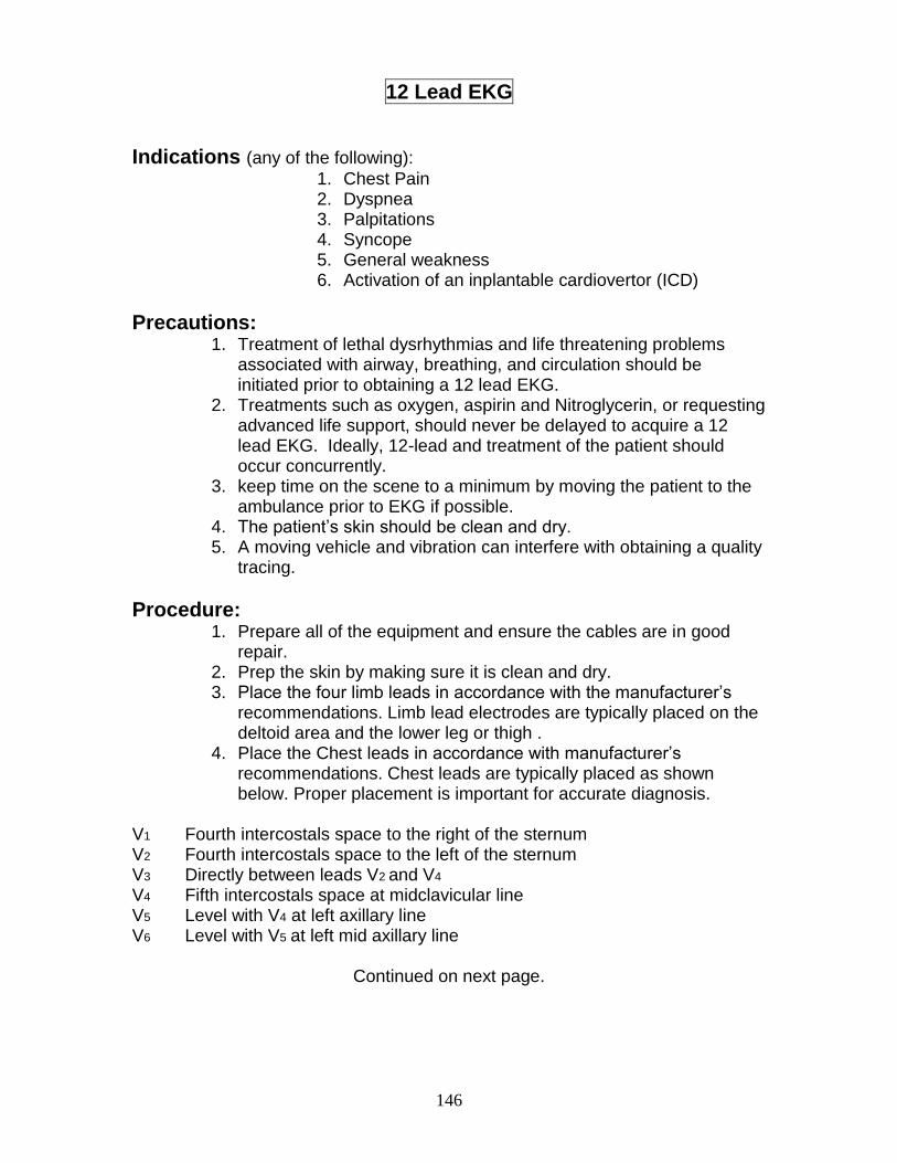
146
12 Lead EKG
Indications (any of the following):
1. Chest Pain 2. Dyspnea 3. Palpitations 4. Syncope 5. General weakness 6. Activation of an inplantable cardiovertor (ICD)
Precautions: 1. Treatment of lethal dysrhythmias and life threatening problems
associated with airway, breathing, and circulation should be initiated prior to obtaining a 12 lead EKG.
2. Treatments such as oxygen, aspirin and Nitroglycerin, or requesting advanced life support, should never be delayed to acquire a 12 lead EKG. Ideally, 12-lead and treatment of the patient should occur concurrently.
3. keep time on the scene to a minimum by moving the patient to the ambulance prior to EKG if possible.
4. The patient’s skin should be clean and dry. 5. A moving vehicle and vibration can interfere with obtaining a quality
tracing.
Procedure: 1. Prepare all of the equipment and ensure the cables are in good
repair. 2. Prep the skin by making sure it is clean and dry. 3. Place the four limb leads in accordance with the manufacturer’s
recommendations. Limb lead electrodes are typically placed on the deltoid area and the lower leg or thigh .
4. Place the Chest leads in accordance with manufacturer’s recommendations. Chest leads are typically placed as shown below. Proper placement is important for accurate diagnosis.
V1 Fourth intercostals space to the right of the sternum V2 Fourth intercostals space to the left of the sternum V3 Directly between leads V2 and V4
V4 Fifth intercostals space at midclavicular line V5 Level with V4 at left axillary line V6 Level with V5 at left mid axillary line
Continued on next page.
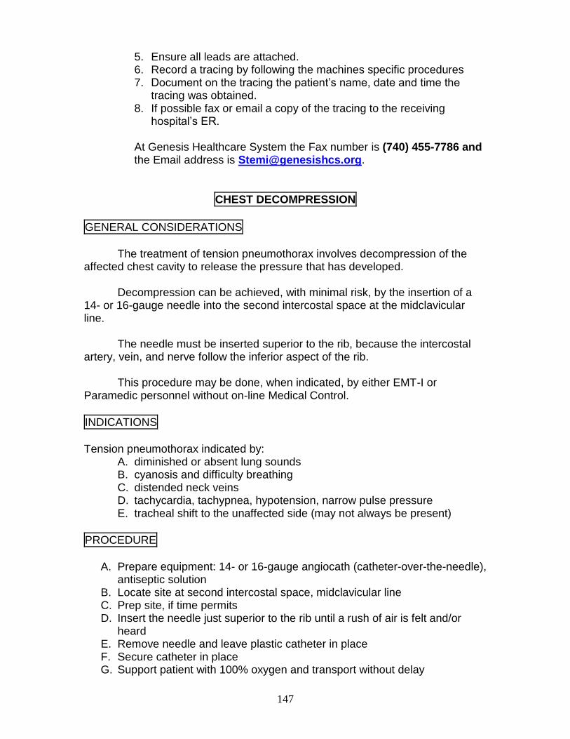
147
5. Ensure all leads are attached. 6. Record a tracing by following the machines specific procedures 7. Document on the tracing the patient’s name, date and time the
tracing was obtained. 8. If possible fax or email a copy of the tracing to the receiving
hospital’s ER. At Genesis Healthcare System the Fax number is (740) 455-7786 and the Email address is [email protected].
CHEST DECOMPRESSION
GENERAL CONSIDERATIONS
The treatment of tension pneumothorax involves decompression of the affected chest cavity to release the pressure that has developed. Decompression can be achieved, with minimal risk, by the insertion of a 14- or 16-gauge needle into the second intercostal space at the midclavicular line. The needle must be inserted superior to the rib, because the intercostal artery, vein, and nerve follow the inferior aspect of the rib. This procedure may be done, when indicated, by either EMT-I or Paramedic personnel without on-line Medical Control.
INDICATIONS
Tension pneumothorax indicated by:
A. diminished or absent lung sounds B. cyanosis and difficulty breathing C. distended neck veins D. tachycardia, tachypnea, hypotension, narrow pulse pressure E. tracheal shift to the unaffected side (may not always be present)
PROCEDURE
A. Prepare equipment: 14- or 16-gauge angiocath (catheter-over-the-needle),
antiseptic solution B. Locate site at second intercostal space, midclavicular line C. Prep site, if time permits D. Insert the needle just superior to the rib until a rush of air is felt and/or
heard E. Remove needle and leave plastic catheter in place F. Secure catheter in place G. Support patient with 100% oxygen and transport without delay
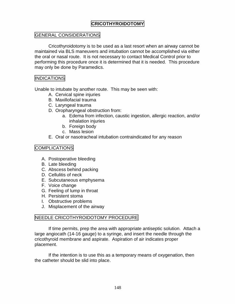
148
CRICOTHYROIDOTOMY
GENERAL CONSIDERATIONS
Cricothyroidotomy is to be used as a last resort when an airway cannot be
maintained via BLS maneuvers and intubation cannot be accomplished via either the oral or nasal route. It is not necessary to contact Medical Control prior to performing this procedure once it is determined that it is needed. This procedure may only be done by Paramedics.
INDICATIONS
Unable to intubate by another route. This may be seen with:
A. Cervical spine injuries B. Maxillofacial trauma C. Laryngeal trauma D. Oropharyngeal obstruction from:
a. Edema from infection, caustic ingestion, allergic reaction, and/or inhalation injuries
b. Foreign body c. Mass lesion
E. Oral or nasotracheal intubation contraindicated for any reason
COMPLICATIONS
A. Postoperative bleeding B. Late bleeding C. Abscess behind packing D. Cellulitis of neck E. Subcutaneous emphysema F. Voice change G. Feeling of lump in throat H. Persistent stoma I. Obstructive problems J. Misplacement of the airway
NEEDLE CRICOTHYROIDOTOMY PROCEDURE
If time permits, prep the area with appropriate antiseptic solution. Attach a
large angiocath (14-16 gauge) to a syringe, and insert the needle through the cricothyroid membrane and aspirate. Aspiration of air indicates proper placement.
If the intention is to use this as a temporary means of oxygenation, then the catheter should be slid into place.
149
If the needle is going to be used as a guide for surgical cricothyroidotomy, then the catheter should not be used in order to prevent the possibility of shearing off the catheter when the scalpel is used.
A jet ventilator should be used to provide sufficient volume of oxygen at a pressure of no more than 30 psi.
Needle cricothyroidotomy is the preferred method in children less than 12 years of age.
SURGICAL CRICOTHYROIDOTOMY PROCEDURE
Locate the cricothyroid membrane and make a 2-4 cm vertical incision to
the skin overlying the membrane. Once the membrane has been exposed, make a 1.5-2 cm horizontal incision into the membrane and through to the trachea. Maintain a slight caudal direction with the blade to avoid damage to the vocal cords.
Use forceps or dilator to spread the opening in the cricothyroid membrane. Again, angle the instrument inferiorly to minimize the risk of vocal cord damage. If time does not allow or equipment is not available, the blunt end of the scalpel can be placed in the incision and rotated to dilate the opening.
Insert a 6 mm endotracheal tube. Advance the tube and inflate the balloon. Once the tube is in place, check breath sounds and secure the tube. A tracheostomy tube may be used, if available. Surgical cricothyroidotomy should not be done in children less than 12 years of age.
150
ENDOTRACHEAL INTUBATION
INDICATION
Endotracheal intubation is to be utilized for any victim with respiratory arrest
and/or insufficient capability to maintain a patent airway. Field intubation should only be done in a patient in respiratory arrest or without a gag reflex. Studies have shown that field intubations have a higher complication rate not because of lower personnel capabilities, but due to the uncontrolled situation surrounding the intubation. It is for this reason that it is preferred that intubation be done in the Emergency Department, if possible. This does not mean that a patient who emergently needs intubation should be denied this possibly life-saving intervention. Endotracheal intubation protects the airway from aspiration of foreign material and it allows for intermittent positive pressure ventilation with 100% oxygen. It makes the trachea and respiratory tract available for suctioning, and also eliminates the problem of gastric distention.
Endotracheal intubation may be done by those at the EMT-B or EMT-I training level only in the case of respiratory or cardiac arrest.
COMPLICATIONS
A. Esophageal intubation B. Tracheal rupture C. Right mainstem bronchus intubation D. Broken teeth E. Laryngospasm F. Trauma to the oropharynx G. Trauma or puncture of trachea due to misplacement of stylet
OROTRACHEAL INTUBATION PROCEDURE
A. Always begin artificial ventilation as soon as possible using a bag-valve-
mask. B. Assemble and ready equipment.
1. endotracheal tubes of various sizes 2. laryngoscope and blades 3. malleable stylet 4. Magill forceps 5. 10 cc syringe 6. suction apparatus and catheters 7. ET tube tape 8. oropharyngeal airway
C. Check cuff on tube for leaks. Insert stylet into tube. D. Assemble laryngoscope and check bulb. E. Put victim’s head in sniffing position (slight backward tilt). Do not tilt head
if spinal injury is a possibility.
151
F. Holding the laryngoscope in the left hand, insert the blade into the mouth to the right of the midline and sweep to the left, moving the tongue up and to the left and giving clear visualization of the glottic opening.
G. Suction the mouth and pharynx, if necessary. H. Visualize the glottis and vocal cords. I. Select the proper size tube and insert it with the right hand through the
vocal cords and into the trachea. Insert the tube approximately 2 inches past the trachea.
J. Remove the laryngoscope and stylet while holding the tube securely with the right hand.
K. Attempt to ventilate with a bag-valve-mask and check for breath sounds in both lungs.
L. If breath sounds are heard on both sides, inflate the tube’s cuff with approximately 10 cc of air and secure the tube in place with tape. Insert the oropharyngeal airway as a bite block. If breath sounds are only heard on one side, pull the tube back a few centimeters and reassess.
M. Maintain ventilation until adequate respirations resume or the victim is delivered to the ED.
N. Recheck breath sounds and verify tube placement each time the patient is moved or every 10 minutes.
O. Document the intubation by noting the following: 1. number of attempts 2. person(s) making attempts 3. size of tube used 4. type of laryngoscope blade used on each attempt 5. lung sounds before intubation 6. lung sounds after intubation and time of each check 7. measurement on tube at lips of patient when lung sounds are
present 8. any complications
NASOTRACHEAL INTUBATION PROCEDURE
A. Nasotracheal intubation of the airway may be used when the patient has
an unprotected, inadequate airway creating hypoxia. B. Nasotracheal intubation is indicated when:
1. Airway control is needed due to loss of gag reflex or inability to adequately oxygenate the patient.
2. The patient cannot successfully be intubated via the orotracheal route.
C. Nasotracheal intubation is contraindicated in the apneic patient or those with suspected fractures to the base of the skull or to the face.
D. Complications of nasotracheal intubation include: 1. nasal hemorrhage 2. laryngeal damage due to increased manipulation 3. rupture of cuff balloon from use of Magill forceps 4. sinus infection
E. When attempting nasotracheal intubation:
152
1. always begin basic airway control and oxygenation as soon as possible
2. assemble and ready equipment: a. endotracheal tubes of various sizes (ENDOTROL type) b. Magill forceps c. 10 cc syringe d. water soluble lubricant
3. determine size of tube based on size of nasal opening 4. check tube cuff for leaks and lubricate tube; seat 15 mm connector
firmly in tube 5. holding tube in dominant hand, place thumb against the 15 mm
connector and index finger in the ring loop 6. insert the tube into the right nostril and advance tube gradually,
anterior to posterior, avoiding superior movement which will be met with resistance and could cause injury
7. as the tube enters the pharynx, listen for breathing and pull on the tip control ring loop to turn the tip of the tube anteriorly toward the trachea
8. when the patient takes a breath, advance the tube into the trachea 9. listen for lung sounds, inflate the tube’s cuff, maintain ventilation
and oxygenation, confirm tube placement via auscultation of breath sounds
10. intubation attempt should not take longer than 30 seconds 11. if any resistance is encountered during insertion, abandon
procedure and utilize another method of airway control and oxygenation
12. recheck lung sounds and verify tube placement each time patient is moved and every 10 minutes
13. document the intubation by noting the following: a. number of attempts b. person(s) making attempts c. size of tube used d. lung sounds before intubation e. lung sounds after intubation and time of each check f. measurement on tube at nose of patient when lung sounds
are present g. any complications
TUBE SIZING
The size of tube that can be easily passed into the trachea of most adults is
8.0 mm. Therefore this tube should be selected first on the average adult. A smaller female adult may require a 7.5 mm tube. The size of the tube is judged by the size of the patient, not by the age.
For children, the proper tube size can be approximated by the size of the child’s little finger. A Broselow tape can also be used to assist in tube size selection. The following can also be used as a guide to tube sizing:
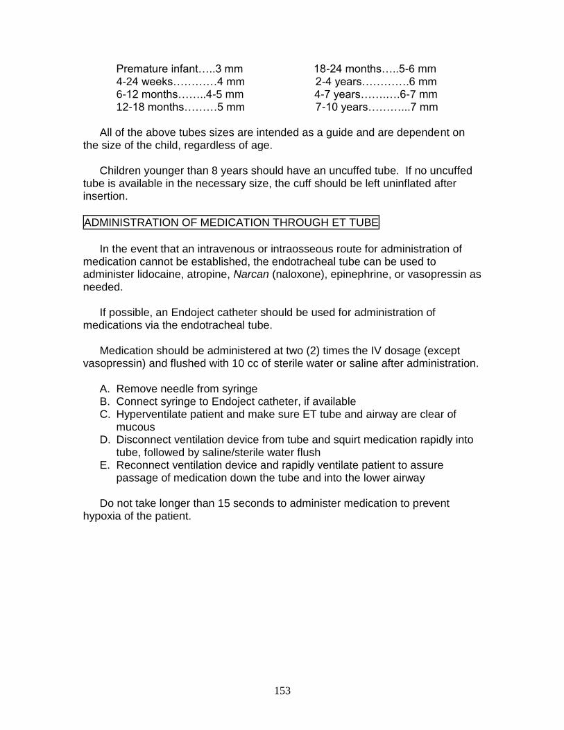
153
Premature infant…..3 mm 18-24 months…..5-6 mm 4-24 weeks…………4 mm 2-4 years………….6 mm 6-12 months……..4-5 mm 4-7 years…….….6-7 mm 12-18 months………5 mm 7-10 years………...7 mm
All of the above tubes sizes are intended as a guide and are dependent on the size of the child, regardless of age.
Children younger than 8 years should have an uncuffed tube. If no uncuffed tube is available in the necessary size, the cuff should be left uninflated after insertion.
ADMINISTRATION OF MEDICATION THROUGH ET TUBE
In the event that an intravenous or intraosseous route for administration of
medication cannot be established, the endotracheal tube can be used to administer lidocaine, atropine, Narcan (naloxone), epinephrine, or vasopressin as needed.
If possible, an Endoject catheter should be used for administration of medications via the endotracheal tube.
Medication should be administered at two (2) times the IV dosage (except vasopressin) and flushed with 10 cc of sterile water or saline after administration.
A. Remove needle from syringe B. Connect syringe to Endoject catheter, if available C. Hyperventilate patient and make sure ET tube and airway are clear of
mucous D. Disconnect ventilation device from tube and squirt medication rapidly into
tube, followed by saline/sterile water flush E. Reconnect ventilation device and rapidly ventilate patient to assure
passage of medication down the tube and into the lower airway
Do not take longer than 15 seconds to administer medication to prevent hypoxia of the patient.
154
COMBITUBE INSERTION PROCEDURE
In the event that a patient cannot be intubated endotracheally and a
Combitube is available, this device may be used to temporarily establish a means for ventilation. This device should only be used in the case of cardiac or respiratory arrest without a gag reflex. It should only be used if standard endotracheal intubation cannot be accomplished. Medications should not be administered via the Combitube until it is confirmed that it has been placed in the trachea rather than the esophagus. It is permissible for those personnel at the EMT-B or EMT-I level to use this device if they have been trained in its use.
A. Manually open the airway and suction the oropharynx as needed B. Begin positive pressure ventilation via BVM with 100% oxygen C. Assemble Combitube with large syringe (100 cc) attached to the blue cuff
and small syringe (15 cc) attached to the white cuff D. Test both cuffs for leaks E. Lubricate tip of tube and advance tube into oropharynx until upper
teeth/gums are aligned between the two black rings F. Inflate the balloon attached to the white cuff (small syringe) with 12 cc of
air G. Begin ventilating through the white tube (tube #2) H. Assess tube placement by listening for air over lungs and epigastrium and
watching for chest rise I. If there are bilateral lung sounds, absent epigastric sounds, and good
chest rise, this indicates tracheal placement. Continue ventilating with 100% oxygen via tube #2
J. If chest rise and lung sounds are absent and/or if air is heard gurgling in the stomach, inflate the balloon attached to the blue cuff (large syringe) with 50-75 cc of air, move ventilation device to tube #1, and reassess placement as above
K. If there are bilateral lung sounds, absent epigastric sounds, and good chest rise while ventilating through tube #2, this indicates esophageal placement. Continue ventilating with 100% oxygen via tube #2
L. Once tube placement is confirmed, secure tube and continue ventilation M. If tube placement cannot be definitively confirmed, remove the Combitube
and ventilate via BVM N. Note that expulsion of stomach contents through tube #2 may occur if
initial placement is in the esophagus
155
EXTERNAL PACEMAKER
INDICATIONS
An external pacemaker may be used in the following situations:
A. Patients who are in full arrest with asystole confirmed in two leads, and not responding to medication therapy
B. Patients with symptomatic bradycardia, unresponsive to atropine C. Additional patients at the discretion of the on-line Medical Control
Physician
APPLICATION
In the patient with bradycardia, the rate is to be set at 80 beats per minute
and the current at 20 milliamperes, which is to be increased by 20 milliamperes every 10 seconds until capture is obtained.
In asystole, the device is to be set at 100 beats per minute and 100 milliamperes. Reduce the output as far as possible while still maintaining mechanical capture.
Once electrical capture is obtained, check for mechanical capture (pulse).
The external pacemaker is only to be used on pediatric patients with on-line medical direction.
Nitroglycerine patches are to be removed before pacing.
156
INTRAOSSEOUS INFUSION
INDICATIONS
A. To establish parenteral means to administer fluids, blood products, and
parenteral medications B. May be used in any instance that an IV route would be appropriate C. Its use should be considered after two IV attempts have failed or if no
peripheral IV sites are found D. This procedure is indicated primarily in children but accepted in adults. E. This procedure should only be employed in cases of critically ill or injured
children when no alternatives exist for medication/fluid administration and transport time is more than a few minutes
CONTRAINDICATIONS
A. Osteomyelitis or cellulitis at proposed site B. Fracture at or above the proposed site C. Previous IO attempt at the proposed site
EQUIPMENT
A. Proper size Intraosseous Needle with driving device (manual or power
such as EZ IO) B. Betadine and Alcohol C. IV set-up D. Syringe for aspiration E. Lidocaine prn
PROCEDURE
A. Prepare as for a surgical procedure, using sterile technique B. Attempt to have feet in flexed position against board or sandbag C. If the patient is alert, consider using a local anesthetic D. The preferred site is the proximal anteromedial tibia, 1-3 cm below the
tibial tuberosity. Secondary site is the distal femur, midline, 3 cm above condyle. Sternal IO are also acceptable.
E. Needle insertion varies between 70 and 90 degree angle to the skin surface, approximately one-two finger-breadths distal to the tibial tuberosity. With a straight, steady push and/or rotary motion, push needle through subcutaneous tissue and bone until a drop or pop is felt
1. With the EZ IO follow recommended guidelines.
F. Once the needle has reached the bone marrow, saline should be injected via syringe to clear needle and then aspiration should be attempted. The infusion should flow freely without evidence of subcutaneous infiltration
157
G. The needle should feel firm in position and stand upright without support H. Infusion via this route is the same as venous access without limit to rate of
administration, drugs pushed, or fluid type infused I. After removing needle (for successful or unsuccessful attempt), apply
pressure to area for five minutes and apply dressing to area J. Intraosseous infusions of fluid may cause subcutaneous infiltration,
subcutaneous infection, or osteomyelitis if proper techniques are not followed
158
IV PROCEDURES
GENERAL CONSIDERATIONS
IVs will be started by the EMT-Intermediate and/or the Paramedic as allowed
by each patient care protocol.
IV placement must not delay transport of any critical patient involved in trauma.
Generally, no more than two (2) attempts or more than five minutes should be spent attempting an IV. If unable to initiate IV line, transport patient and notify hospital IV was not able to be started.
IVs may be started on patients of any age providing there are adequate veins and patient’s condition warrants an IV.
Blood draws for hospital laboratory testing will not be required under this protocol.
IV SOLUTION
0.9% Sodium Chloride (Normal Saline) will be the only fluid used in the pre-
hospital setting under this protocol. The amount of solution to be infused is as directed by specific treatment
protocols. Saline Locks may be used on non-critical patients if properly trained.
MECHANICS FOR STARTING PERIPHERAL IV
A. Prepare equipment B. Select IV site; do not use the antecubital fossa unless necessary C. Apply tourniquet D. Cleanse site with alcohol prep E. First attempt at insertion on an adult patient should be:
1. 14-16ga IV catheter for trauma patients 2. 18-20ga IV catheter for medical patients
F. Attach IV tubing G. Secure IV using appropriate measures to insure stability of the line H. Check for signs of infiltration I. Adjust flow rate J. Document IV procedure on run sheet
159
MECHANICS FOR STARTING EXTERNAL JUGULAR IV LINE
A. Locate external jugular vein B. Cleans site with Betadine solution and alcohol. (The only time Betadine is
not required is when the patient has an allergy to Betadine-type solutions) C. Select IV catheter
1. On adults, a large bore (16ga or 18ga) may be used 2. Use 2 inch IV catheter when available
D. Position yourself at patient’s head E. Turn patient’s head so as to maximally expose vein and minimize
interference of jaw F. Cannulate the vein by directing the needle inferiorly at an angle nearly
parallel to the neck G. Attach IV tubing H. Secure IV using appropriate measures to ensure stability of the line I. Check for signs of infiltration J. Adjust flow rate K. Document IV procedure on run sheet
ACCESSING DIALYSIS/INDWELLING CATHETERS
Personnel with adequate training or experience in accessing dialysis
catheters or other indwelling catheters may do so instead of initiating peripheral venous access. Proper sterile technique should be followed at all times to reduce the risk of infection to the patient. A surgically implanted A-V dialysis shunt that has already been accessed by the dialysis center staff may be used for fluid or medication administration provided that it is properly secured and the fluid is flowing smoothly and easily. Access of these shunts be prehospital providers should only be done in the case of a critically ill patient in whom no other access can be obtained.
DOCUMENTATION
ALL IV attempts must be recorded on run sheet and include the following:
A. When successful: i. Time IV was started ii. Type and amount of solution hung and infused during run iii. Flow rate iv. Size of catheter or needle used v. Location of IV site vi. Initials of all EMTs who attempted and/or started IV
B. When unsuccessful: i. Time IV was attempted ii. Type of solution iii. Size of catheter or needle used iv. Location of attempted site

160
v. Initials of all EMTs who attempted and/or started IV C. Record all IV medications given:
i. Name of medication ii. Dosage and amount given iii. Time ordered (if applicable) iv. Time given v. Initials of all EMT-Is or Paramedics who administered
medication
161
OROGASTRIC TUBE PLACEMENT
GENERAL CONSIDERATIONS
Field orogastric (OG) tube placement is intended for alleviation of gastric
distention caused by ventilation via bag-valve-mask (BVM). It is only indicated in those patients with impaired ventilation due to gastric distention. It should only be done in intubated patients with no gag reflex. This is considered to be an optional skill for those Paramedics who are interested in undertaking the training.
PARAMEDIC
A. Ensure that the endotracheal (ET) tube is in proper position and is
secured. B. Assess adequacy of ventilation and extent of gastric distention. C. Prepare equipment (OG tube and large syringe with Toomey tip) D. Insert tube into posterior pharynx and advance tube into stomach. E. Verify tube placement.
1. Attach Toomey syringe to end of OG tube 2. Insufflate stomach with air while listening over epigastrium for air
sounds F. Cap end of OG tube after gastric distention alleviated. G. Secure OG tube in place. H. Laryngoscope may be used to elevate tongue and allow visualization of
esophagus in cases of difficulty with placement of OG tube.
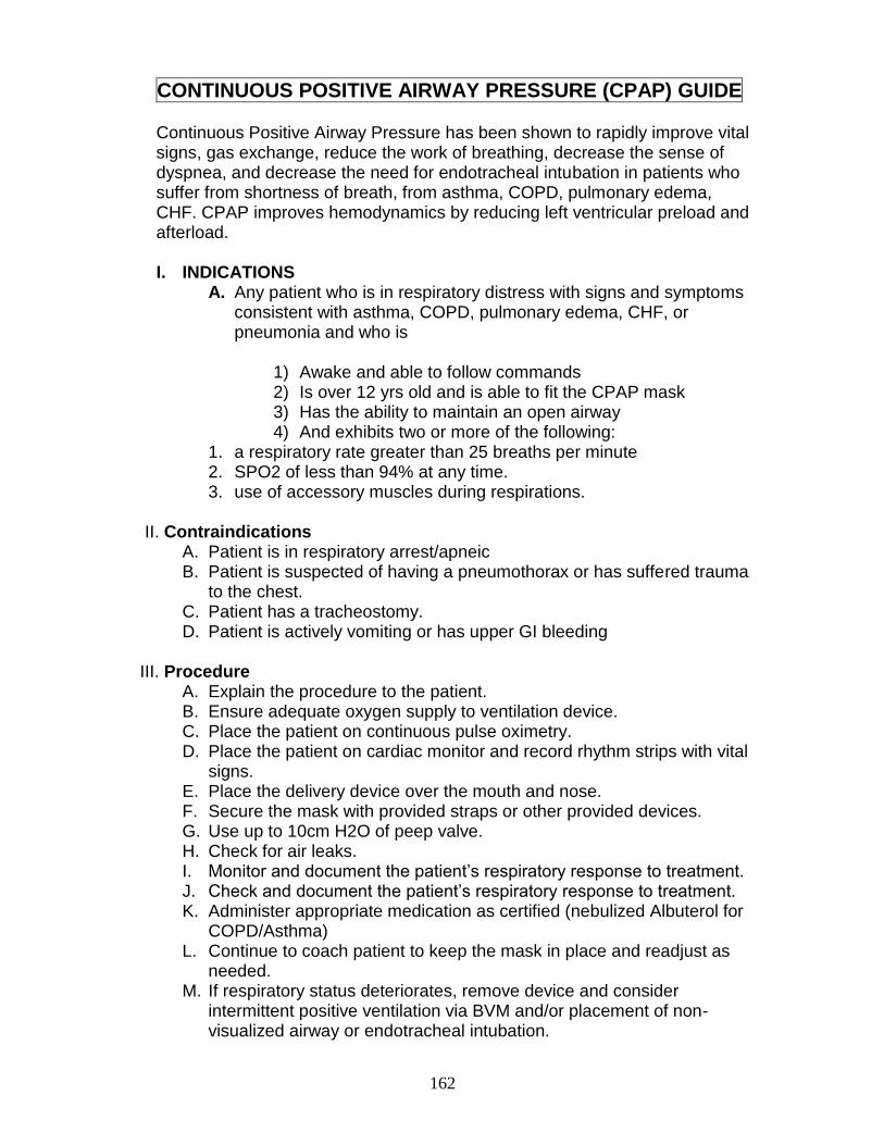
162
CONTINUOUS POSITIVE AIRWAY PRESSURE (CPAP) GUIDE Continuous Positive Airway Pressure has been shown to rapidly improve vital signs, gas exchange, reduce the work of breathing, decrease the sense of dyspnea, and decrease the need for endotracheal intubation in patients who suffer from shortness of breath, from asthma, COPD, pulmonary edema, CHF. CPAP improves hemodynamics by reducing left ventricular preload and afterload. I. INDICATIONS
A. Any patient who is in respiratory distress with signs and symptoms consistent with asthma, COPD, pulmonary edema, CHF, or pneumonia and who is
1) Awake and able to follow commands 2) Is over 12 yrs old and is able to fit the CPAP mask 3) Has the ability to maintain an open airway 4) And exhibits two or more of the following:
1. a respiratory rate greater than 25 breaths per minute 2. SPO2 of less than 94% at any time. 3. use of accessory muscles during respirations.
II. Contraindications A. Patient is in respiratory arrest/apneic B. Patient is suspected of having a pneumothorax or has suffered trauma
to the chest. C. Patient has a tracheostomy. D. Patient is actively vomiting or has upper GI bleeding
III. Procedure
A. Explain the procedure to the patient. B. Ensure adequate oxygen supply to ventilation device. C. Place the patient on continuous pulse oximetry. D. Place the patient on cardiac monitor and record rhythm strips with vital
signs. E. Place the delivery device over the mouth and nose. F. Secure the mask with provided straps or other provided devices. G. Use up to 10cm H2O of peep valve. H. Check for air leaks. I. Monitor and document the patient’s respiratory response to treatment. J. Check and document the patient’s respiratory response to treatment. K. Administer appropriate medication as certified (nebulized Albuterol for
COPD/Asthma) L. Continue to coach patient to keep the mask in place and readjust as
needed. M. If respiratory status deteriorates, remove device and consider
intermittent positive ventilation via BVM and/or placement of non-visualized airway or endotracheal intubation.
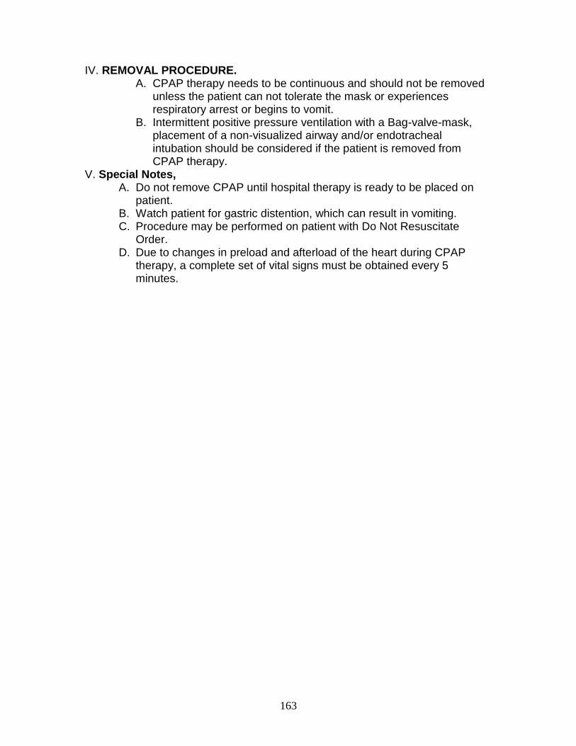
163
IV. REMOVAL PROCEDURE.
A. CPAP therapy needs to be continuous and should not be removed unless the patient can not tolerate the mask or experiences respiratory arrest or begins to vomit.
B. Intermittent positive pressure ventilation with a Bag-valve-mask, placement of a non-visualized airway and/or endotracheal intubation should be considered if the patient is removed from CPAP therapy.
V. Special Notes, A. Do not remove CPAP until hospital therapy is ready to be placed on
patient. B. Watch patient for gastric distention, which can result in vomiting. C. Procedure may be performed on patient with Do Not Resuscitate
Order. D. Due to changes in preload and afterload of the heart during CPAP
therapy, a complete set of vital signs must be obtained every 5 minutes.
164
Medication
Reference
Guide
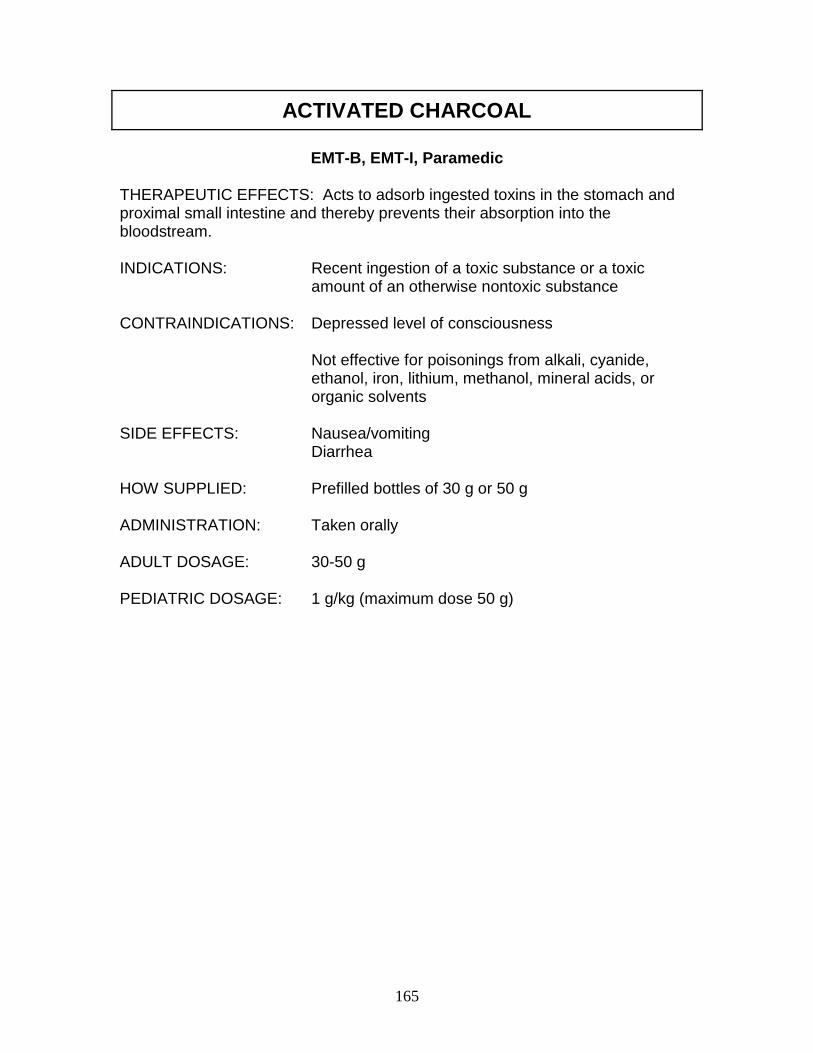
165
ACTIVATED CHARCOAL
EMT-B, EMT-I, Paramedic
THERAPEUTIC EFFECTS: Acts to adsorb ingested toxins in the stomach and proximal small intestine and thereby prevents their absorption into the bloodstream. INDICATIONS: Recent ingestion of a toxic substance or a toxic
amount of an otherwise nontoxic substance CONTRAINDICATIONS: Depressed level of consciousness
Not effective for poisonings from alkali, cyanide, ethanol, iron, lithium, methanol, mineral acids, or organic solvents
SIDE EFFECTS: Nausea/vomiting Diarrhea HOW SUPPLIED: Prefilled bottles of 30 g or 50 g ADMINISTRATION: Taken orally ADULT DOSAGE: 30-50 g PEDIATRIC DOSAGE: 1 g/kg (maximum dose 50 g)
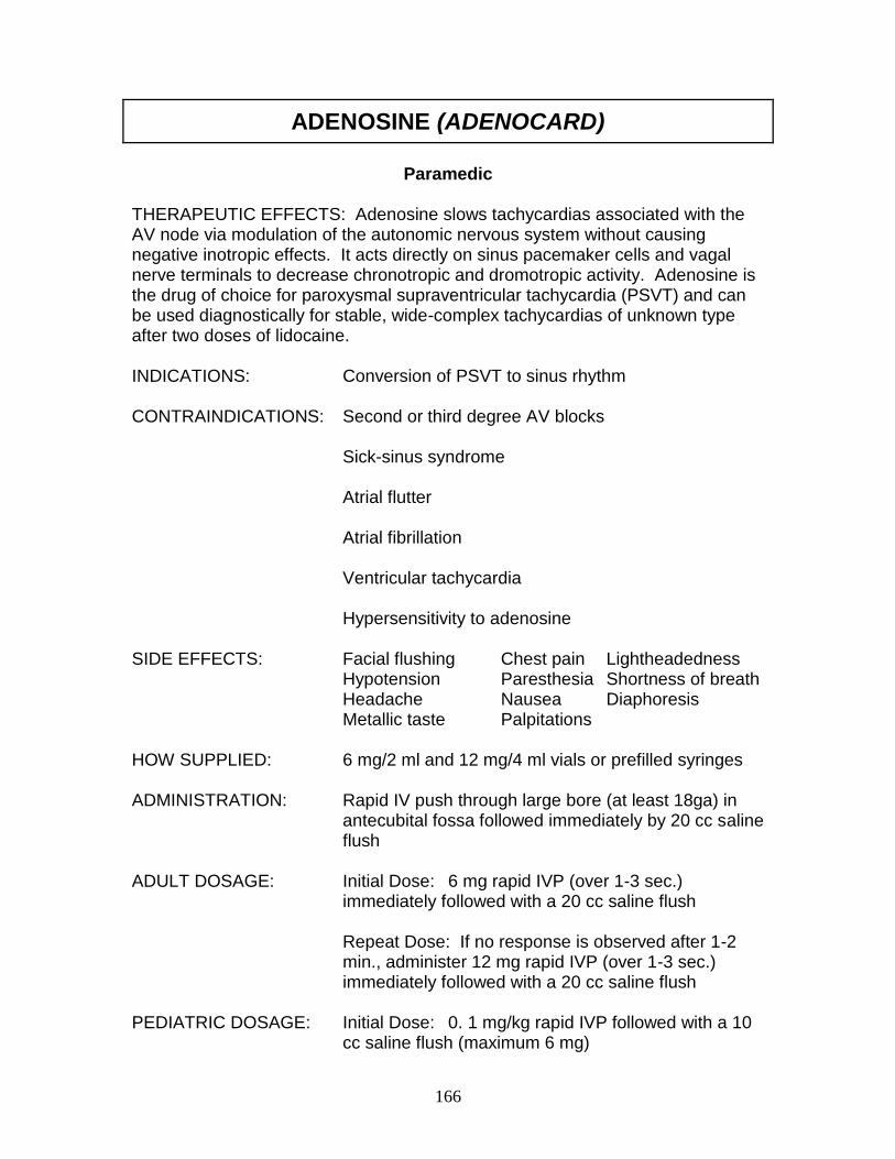
166
ADENOSINE (ADENOCARD)
Paramedic
THERAPEUTIC EFFECTS: Adenosine slows tachycardias associated with the AV node via modulation of the autonomic nervous system without causing negative inotropic effects. It acts directly on sinus pacemaker cells and vagal nerve terminals to decrease chronotropic and dromotropic activity. Adenosine is the drug of choice for paroxysmal supraventricular tachycardia (PSVT) and can be used diagnostically for stable, wide-complex tachycardias of unknown type after two doses of lidocaine. INDICATIONS: Conversion of PSVT to sinus rhythm CONTRAINDICATIONS: Second or third degree AV blocks
Sick-sinus syndrome
Atrial flutter Atrial fibrillation Ventricular tachycardia Hypersensitivity to adenosine SIDE EFFECTS: Facial flushing Chest pain Lightheadedness Hypotension Paresthesia Shortness of breath Headache Nausea Diaphoresis Metallic taste Palpitations HOW SUPPLIED: 6 mg/2 ml and 12 mg/4 ml vials or prefilled syringes ADMINISTRATION: Rapid IV push through large bore (at least 18ga) in
antecubital fossa followed immediately by 20 cc saline flush
ADULT DOSAGE: Initial Dose: 6 mg rapid IVP (over 1-3 sec.)
immediately followed with a 20 cc saline flush
Repeat Dose: If no response is observed after 1-2 min., administer 12 mg rapid IVP (over 1-3 sec.) immediately followed with a 20 cc saline flush
PEDIATRIC DOSAGE: Initial Dose: 0. 1 mg/kg rapid IVP followed with a 10
cc saline flush (maximum 6 mg)

167
Repeat Dose: If no response is observed after 1-2
min., administer 0.2 mg/kg rapid IVP followed with a 10 cc saline flush (maximum 12 mg)
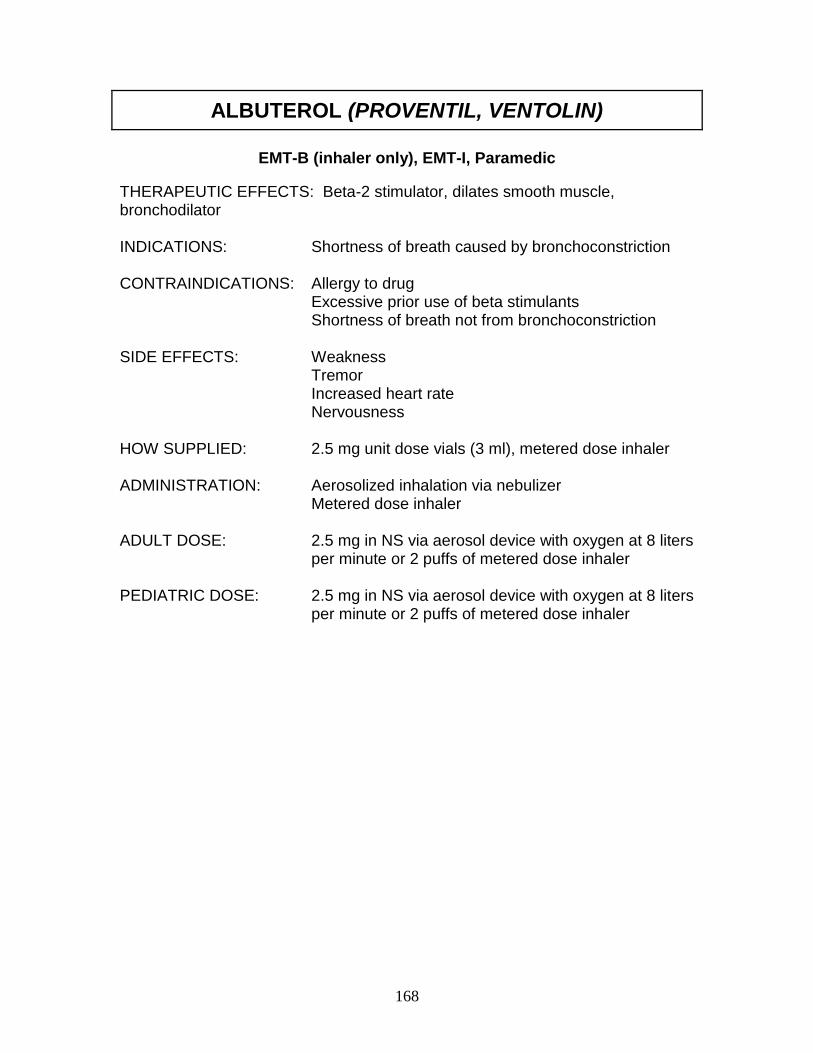
168
ALBUTEROL (PROVENTIL, VENTOLIN)
EMT-B (inhaler only), EMT-I, Paramedic
THERAPEUTIC EFFECTS: Beta-2 stimulator, dilates smooth muscle, bronchodilator INDICATIONS: Shortness of breath caused by bronchoconstriction CONTRAINDICATIONS: Allergy to drug Excessive prior use of beta stimulants Shortness of breath not from bronchoconstriction SIDE EFFECTS: Weakness Tremor Increased heart rate Nervousness HOW SUPPLIED: 2.5 mg unit dose vials (3 ml), metered dose inhaler ADMINISTRATION: Aerosolized inhalation via nebulizer Metered dose inhaler ADULT DOSE: 2.5 mg in NS via aerosol device with oxygen at 8 liters
per minute or 2 puffs of metered dose inhaler PEDIATRIC DOSE: 2.5 mg in NS via aerosol device with oxygen at 8 liters
per minute or 2 puffs of metered dose inhaler
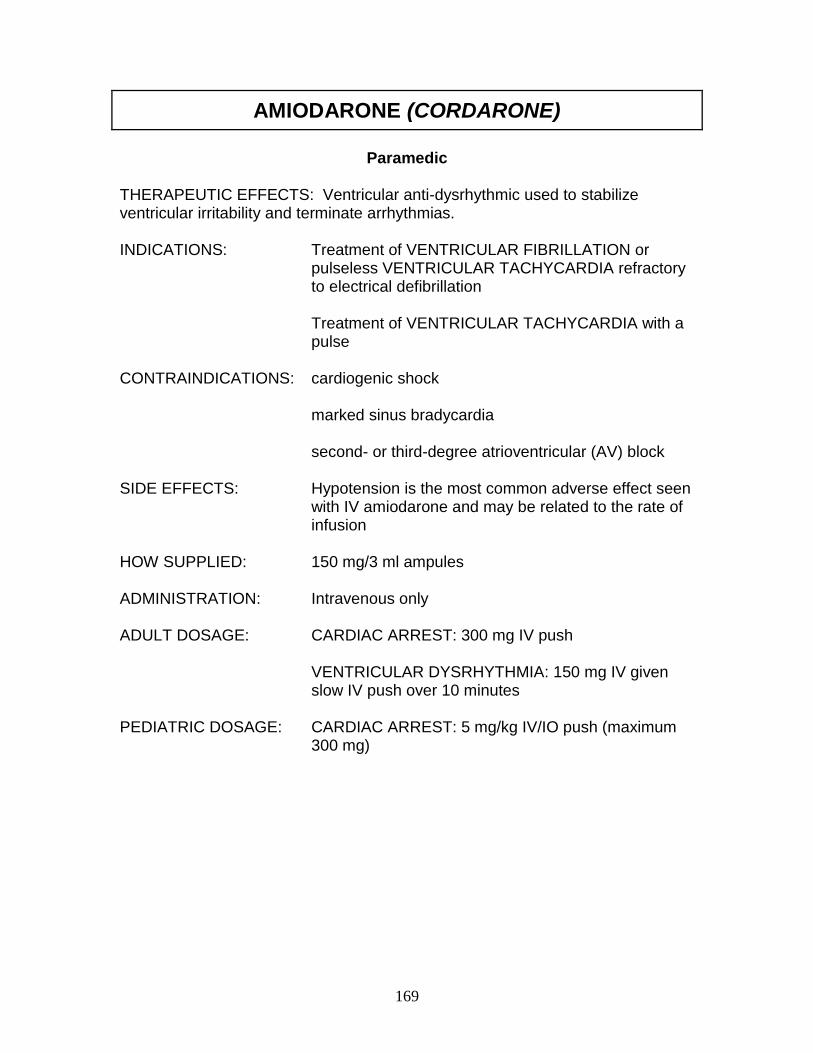
169
AMIODARONE (CORDARONE)
Paramedic
THERAPEUTIC EFFECTS: Ventricular anti-dysrhythmic used to stabilize ventricular irritability and terminate arrhythmias. INDICATIONS: Treatment of VENTRICULAR FIBRILLATION or
pulseless VENTRICULAR TACHYCARDIA refractory to electrical defibrillation
Treatment of VENTRICULAR TACHYCARDIA with a pulse
CONTRAINDICATIONS: cardiogenic shock
marked sinus bradycardia second- or third-degree atrioventricular (AV) block
SIDE EFFECTS: Hypotension is the most common adverse effect seen
with IV amiodarone and may be related to the rate of infusion
HOW SUPPLIED: 150 mg/3 ml ampules ADMINISTRATION: Intravenous only ADULT DOSAGE: CARDIAC ARREST: 300 mg IV push
VENTRICULAR DYSRHYTHMIA: 150 mg IV given slow IV push over 10 minutes
PEDIATRIC DOSAGE: CARDIAC ARREST: 5 mg/kg IV/IO push (maximum 300 mg)
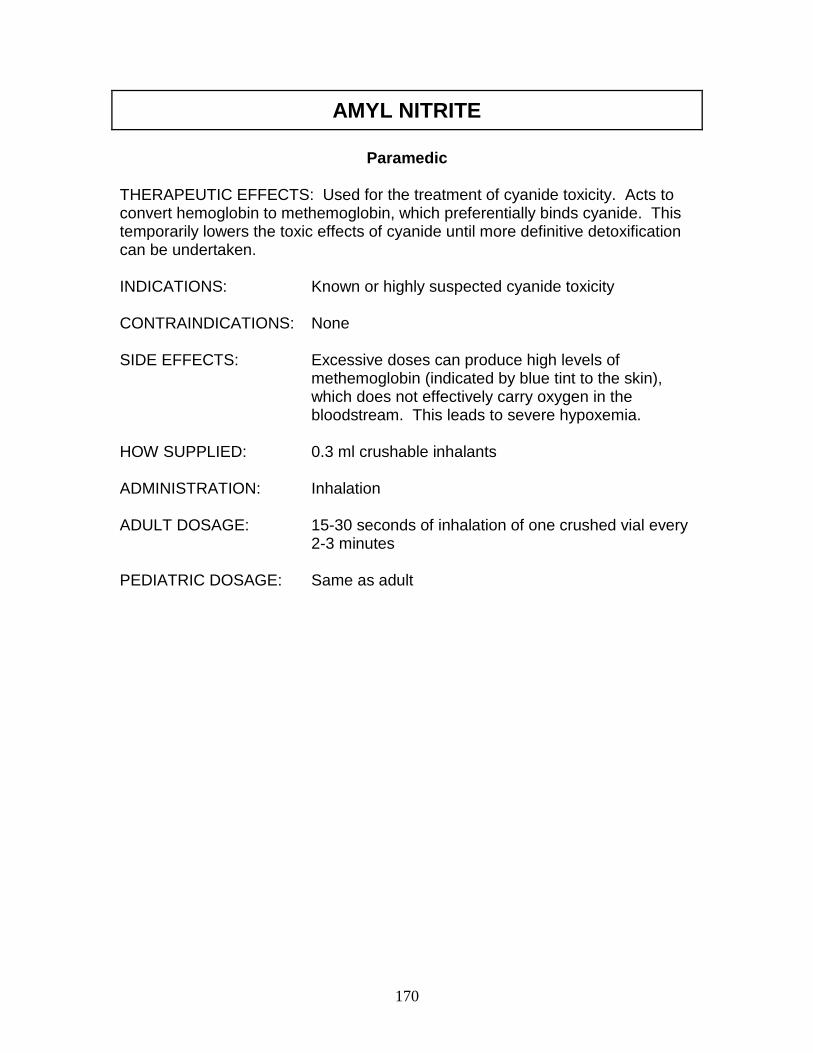
170
AMYL NITRITE
Paramedic
THERAPEUTIC EFFECTS: Used for the treatment of cyanide toxicity. Acts to convert hemoglobin to methemoglobin, which preferentially binds cyanide. This temporarily lowers the toxic effects of cyanide until more definitive detoxification can be undertaken. INDICATIONS: Known or highly suspected cyanide toxicity CONTRAINDICATIONS: None SIDE EFFECTS: Excessive doses can produce high levels of
methemoglobin (indicated by blue tint to the skin), which does not effectively carry oxygen in the bloodstream. This leads to severe hypoxemia.
HOW SUPPLIED: 0.3 ml crushable inhalants ADMINISTRATION: Inhalation ADULT DOSAGE: 15-30 seconds of inhalation of one crushed vial every
2-3 minutes PEDIATRIC DOSAGE: Same as adult
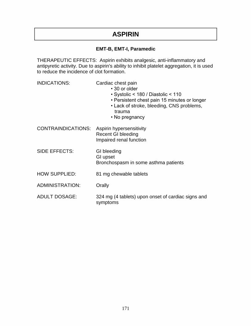
171
ASPIRIN
EMT-B, EMT-I, Paramedic
THERAPEUTIC EFFECTS: Aspirin exhibits analgesic, anti-inflammatory and antipyretic activity. Due to aspirin's ability to inhibit platelet aggregation, it is used to reduce the incidence of clot formation. INDICATIONS: Cardiac chest pain
• 30 or older • Systolic < 180 / Diastolic < 110 • Persistent chest pain 15 minutes or longer • Lack of stroke, bleeding, CNS problems,
trauma • No pregnancy
CONTRAINDICATIONS: Aspirin hypersensitivity
Recent GI bleeding Impaired renal function
SIDE EFFECTS: GI bleeding GI upset
Bronchospasm in some asthma patients HOW SUPPLIED: 81 mg chewable tablets ADMINISTRATION: Orally ADULT DOSAGE: 324 mg (4 tablets) upon onset of cardiac signs and
symptoms
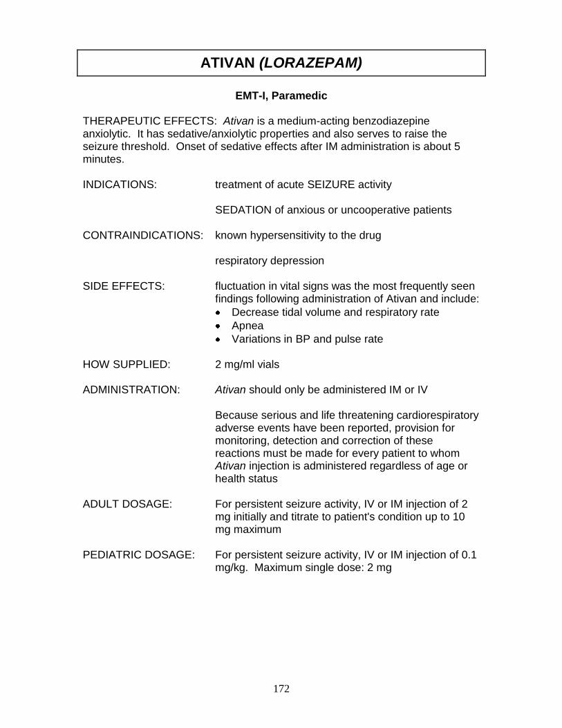
172
ATIVAN (LORAZEPAM)
EMT-I, Paramedic
THERAPEUTIC EFFECTS: Ativan is a medium-acting benzodiazepine anxiolytic. It has sedative/anxiolytic properties and also serves to raise the seizure threshold. Onset of sedative effects after IM administration is about 5 minutes. INDICATIONS: treatment of acute SEIZURE activity SEDATION of anxious or uncooperative patients CONTRAINDICATIONS: known hypersensitivity to the drug respiratory depression SIDE EFFECTS: fluctuation in vital signs was the most frequently seen
findings following administration of Ativan and include:
Decrease tidal volume and respiratory rate
Apnea
Variations in BP and pulse rate HOW SUPPLIED: 2 mg/ml vials ADMINISTRATION: Ativan should only be administered IM or IV
Because serious and life threatening cardiorespiratory adverse events have been reported, provision for monitoring, detection and correction of these reactions must be made for every patient to whom Ativan injection is administered regardless of age or health status
ADULT DOSAGE: For persistent seizure activity, IV or IM injection of 2
mg initially and titrate to patient's condition up to 10 mg maximum
PEDIATRIC DOSAGE: For persistent seizure activity, IV or IM injection of 0.1
mg/kg. Maximum single dose: 2 mg
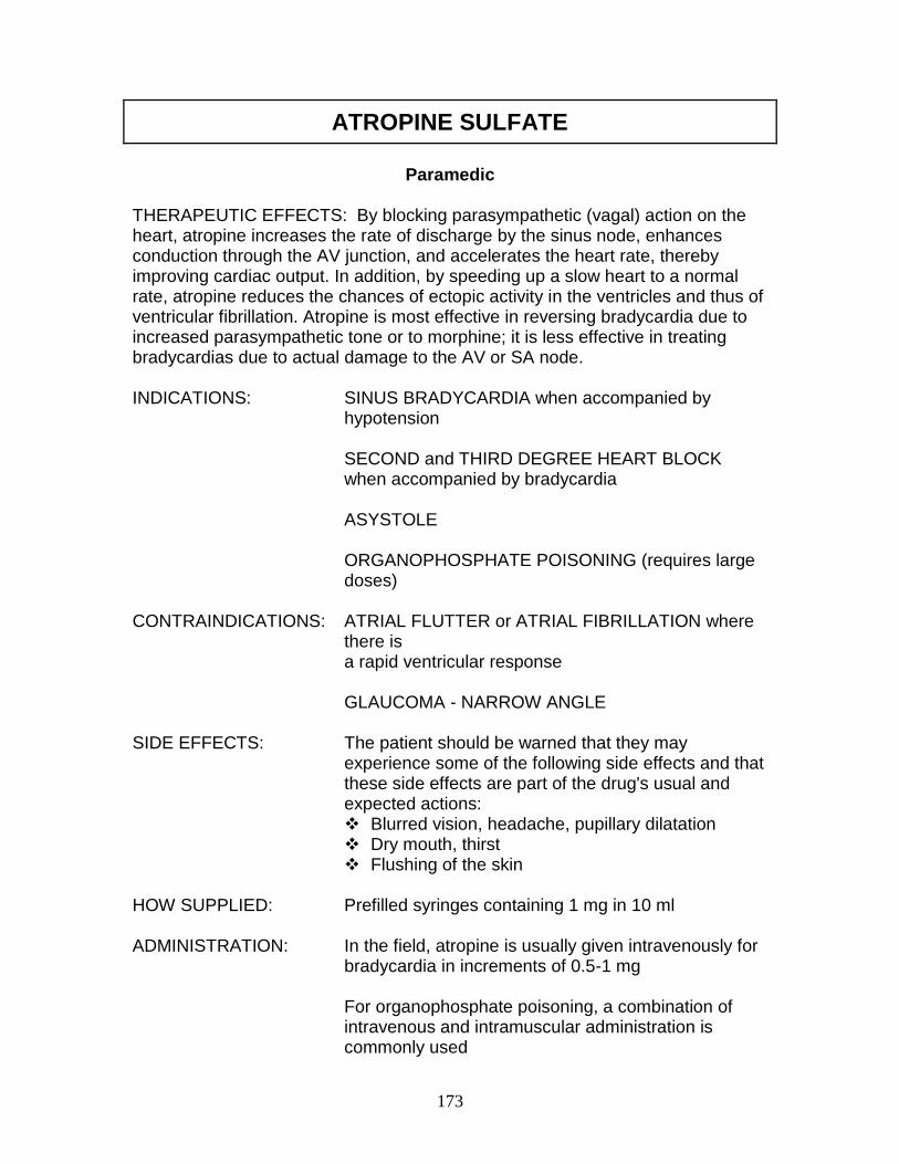
173
ATROPINE SULFATE
Paramedic
THERAPEUTIC EFFECTS: By blocking parasympathetic (vagal) action on the heart, atropine increases the rate of discharge by the sinus node, enhances conduction through the AV junction, and accelerates the heart rate, thereby improving cardiac output. In addition, by speeding up a slow heart to a normal rate, atropine reduces the chances of ectopic activity in the ventricles and thus of ventricular fibrillation. Atropine is most effective in reversing bradycardia due to increased parasympathetic tone or to morphine; it is less effective in treating bradycardias due to actual damage to the AV or SA node. INDICATIONS: SINUS BRADYCARDIA when accompanied by
hypotension
SECOND and THIRD DEGREE HEART BLOCK when accompanied by bradycardia
ASYSTOLE
ORGANOPHOSPHATE POISONING (requires large doses)
CONTRAINDICATIONS: ATRIAL FLUTTER or ATRIAL FIBRILLATION where
there is a rapid ventricular response
GLAUCOMA - NARROW ANGLE SIDE EFFECTS: The patient should be warned that they may
experience some of the following side effects and that these side effects are part of the drug's usual and expected actions: Blurred vision, headache, pupillary dilatation Dry mouth, thirst Flushing of the skin
HOW SUPPLIED: Prefilled syringes containing 1 mg in 10 ml ADMINISTRATION: In the field, atropine is usually given intravenously for
bradycardia in increments of 0.5-1 mg
For organophosphate poisoning, a combination of intravenous and intramuscular administration is commonly used
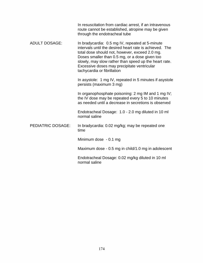
174
In resuscitation from cardiac arrest, if an intravenous route cannot be established, atropine may be given through the endotracheal tube
ADULT DOSAGE: In bradycardia: 0.5 mg IV, repeated at 5-minute
intervals until the desired heart rate is achieved. The total dose should not, however, exceed 2.0 mg. Doses smaller than 0.5 mg, or a dose given too slowly, may slow rather than speed up the heart rate. Excessive doses may precipitate ventricular tachycardia or fibrillation
In asystole: 1 mg IV, repeated in 5 minutes if asystole persists (maximum 3 mg)
In organophosphate poisoning: 2 mg IM and 1 mg IV; the IV dose may be repeated every 5 to 10 minutes as needed until a decrease in secretions is observed Endotracheal Dosage: 1.0 - 2.0 mg diluted in 10 ml normal saline
PEDIATRIC DOSAGE: In bradycardia: 0.02 mg/kg; may be repeated one
time Minimum dose - 0.1 mg Maximum dose - 0.5 mg in child/1.0 mg in adolescent Endotracheal Dosage: 0.02 mg/kg diluted in 10 ml normal saline
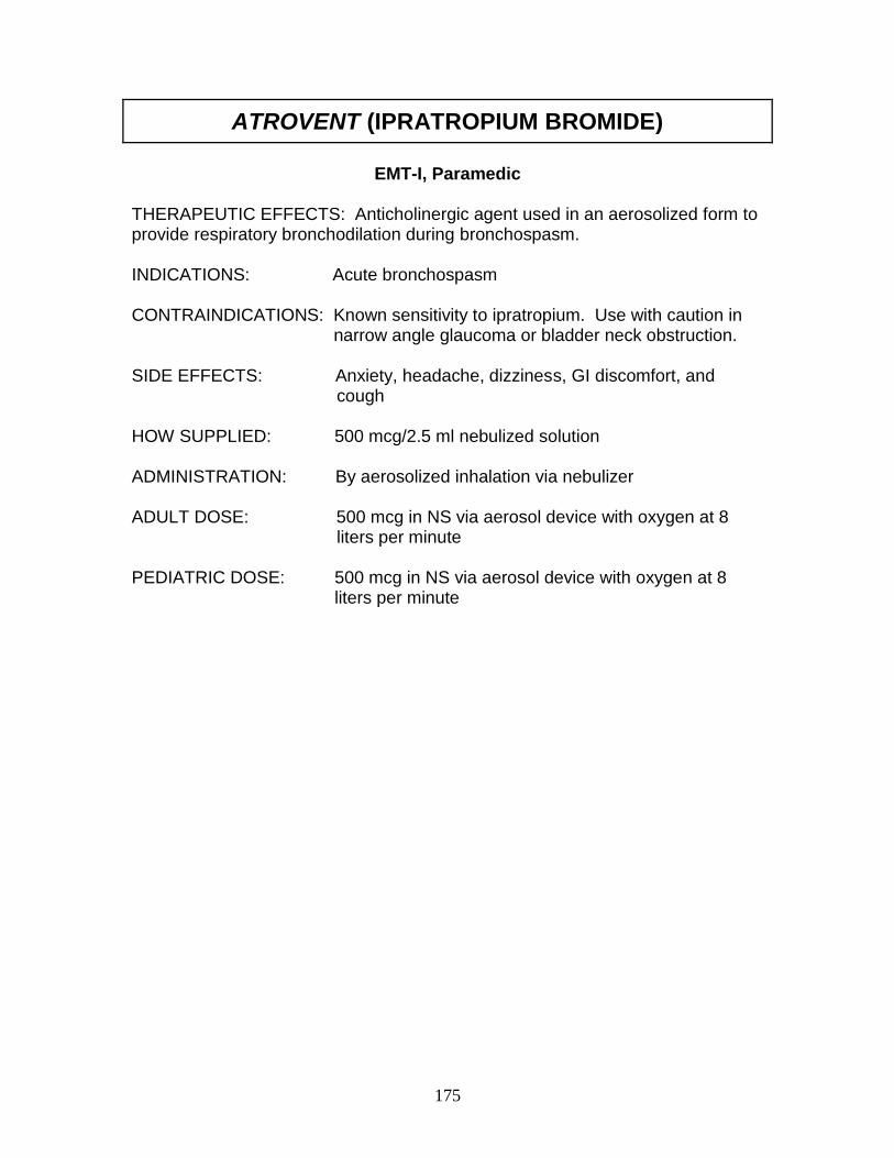
175
ATROVENT (IPRATROPIUM BROMIDE)
EMT-I, Paramedic
THERAPEUTIC EFFECTS: Anticholinergic agent used in an aerosolized form to provide respiratory bronchodilation during bronchospasm. INDICATIONS: Acute bronchospasm CONTRAINDICATIONS: Known sensitivity to ipratropium. Use with caution in
narrow angle glaucoma or bladder neck obstruction. SIDE EFFECTS: Anxiety, headache, dizziness, GI discomfort, and cough HOW SUPPLIED: 500 mcg/2.5 ml nebulized solution ADMINISTRATION: By aerosolized inhalation via nebulizer ADULT DOSE: 500 mcg in NS via aerosol device with oxygen at 8 liters per minute PEDIATRIC DOSE: 500 mcg in NS via aerosol device with oxygen at 8
liters per minute
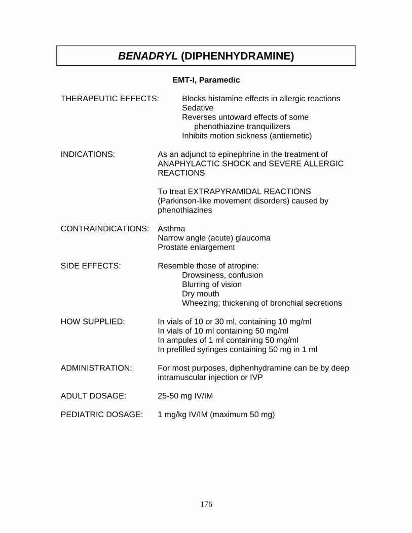
176
BENADRYL (DIPHENHYDRAMINE)
EMT-I, Paramedic
THERAPEUTIC EFFECTS: Blocks histamine effects in allergic reactions
Sedative Reverses untoward effects of some
phenothiazine tranquilizers Inhibits motion sickness (antiemetic)
INDICATIONS: As an adjunct to epinephrine in the treatment of
ANAPHYLACTIC SHOCK and SEVERE ALLERGIC REACTIONS
To treat EXTRAPYRAMIDAL REACTIONS (Parkinson-like movement disorders) caused by phenothiazines
CONTRAINDICATIONS: Asthma
Narrow angle (acute) glaucoma Prostate enlargement
SIDE EFFECTS: Resemble those of atropine:
Drowsiness, confusion Blurring of vision Dry mouth Wheezing; thickening of bronchial secretions
HOW SUPPLIED: In vials of 10 or 30 ml, containing 10 mg/ml
In vials of 10 ml containing 50 mg/ml In ampules of 1 ml containing 50 mg/ml In prefilled syringes containing 50 mg in 1 ml
ADMINISTRATION: For most purposes, diphenhydramine can be by deep
intramuscular injection or IVP ADULT DOSAGE: 25-50 mg IV/IM PEDIATRIC DOSAGE: 1 mg/kg IV/IM (maximum 50 mg)
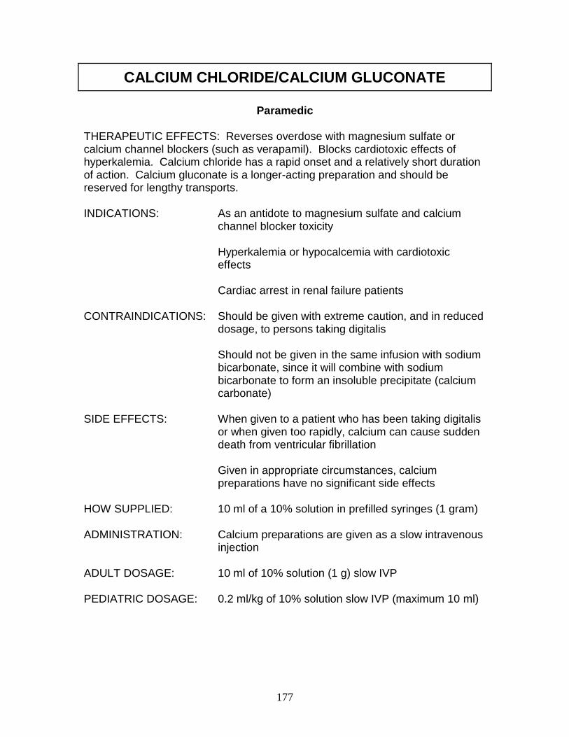
177
CALCIUM CHLORIDE/CALCIUM GLUCONATE
Paramedic
THERAPEUTIC EFFECTS: Reverses overdose with magnesium sulfate or calcium channel blockers (such as verapamil). Blocks cardiotoxic effects of hyperkalemia. Calcium chloride has a rapid onset and a relatively short duration of action. Calcium gluconate is a longer-acting preparation and should be reserved for lengthy transports.
INDICATIONS: As an antidote to magnesium sulfate and calcium
channel blocker toxicity Hyperkalemia or hypocalcemia with cardiotoxic effects
Cardiac arrest in renal failure patients
CONTRAINDICATIONS: Should be given with extreme caution, and in reduced
dosage, to persons taking digitalis
Should not be given in the same infusion with sodium bicarbonate, since it will combine with sodium bicarbonate to form an insoluble precipitate (calcium carbonate)
SIDE EFFECTS: When given to a patient who has been taking digitalis
or when given too rapidly, calcium can cause sudden death from ventricular fibrillation
Given in appropriate circumstances, calcium preparations have no significant side effects
HOW SUPPLIED: 10 ml of a 10% solution in prefilled syringes (1 gram) ADMINISTRATION: Calcium preparations are given as a slow intravenous
injection ADULT DOSAGE: 10 ml of 10% solution (1 g) slow IVP PEDIATRIC DOSAGE: 0.2 ml/kg of 10% solution slow IVP (maximum 10 ml)
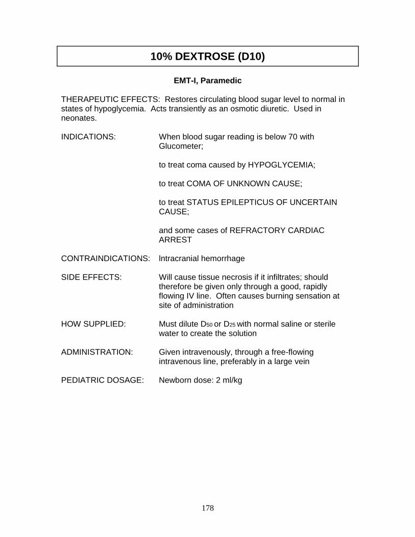
178
10% DEXTROSE (D10)
EMT-I, Paramedic
THERAPEUTIC EFFECTS: Restores circulating blood sugar level to normal in states of hypoglycemia. Acts transiently as an osmotic diuretic. Used in neonates. INDICATIONS: When blood sugar reading is below 70 with
Glucometer;
to treat coma caused by HYPOGLYCEMIA; to treat COMA OF UNKNOWN CAUSE; to treat STATUS EPILEPTICUS OF UNCERTAIN CAUSE; and some cases of REFRACTORY CARDIAC ARREST
CONTRAINDICATIONS: lntracranial hemorrhage SIDE EFFECTS: Will cause tissue necrosis if it infiltrates; should
therefore be given only through a good, rapidly flowing IV line. Often causes burning sensation at site of administration
HOW SUPPLIED: Must dilute D50 or D25 with normal saline or sterile
water to create the solution ADMINISTRATION: Given intravenously, through a free-flowing
intravenous line, preferably in a large vein PEDIATRIC DOSAGE: Newborn dose: 2 ml/kg
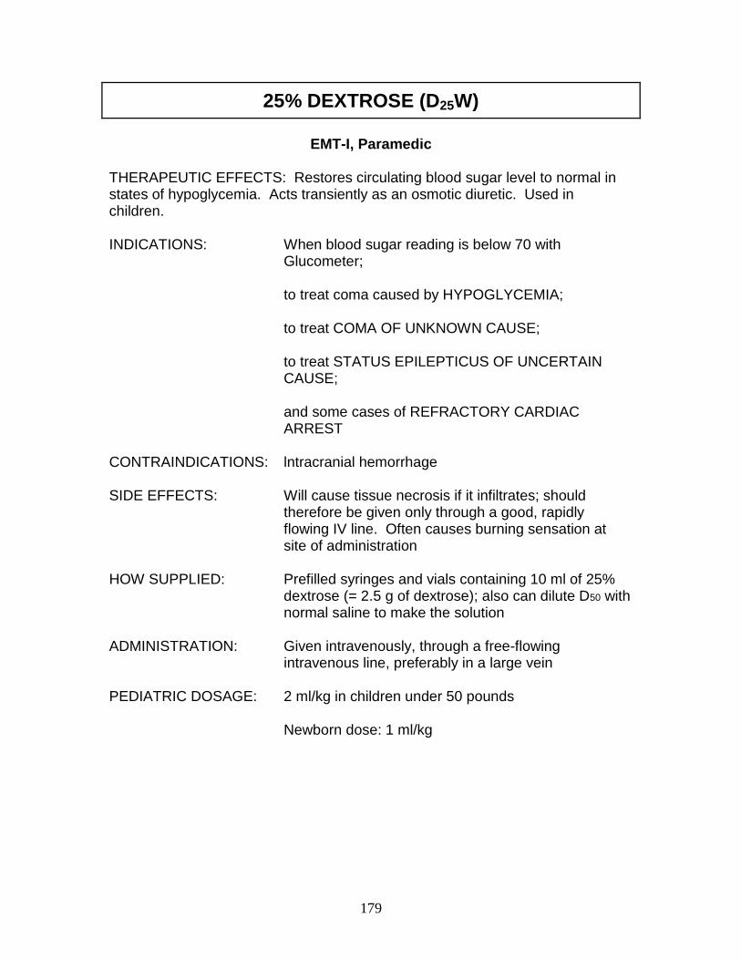
179
25% DEXTROSE (D25W)
EMT-I, Paramedic
THERAPEUTIC EFFECTS: Restores circulating blood sugar level to normal in states of hypoglycemia. Acts transiently as an osmotic diuretic. Used in children. INDICATIONS: When blood sugar reading is below 70 with
Glucometer;
to treat coma caused by HYPOGLYCEMIA; to treat COMA OF UNKNOWN CAUSE; to treat STATUS EPILEPTICUS OF UNCERTAIN CAUSE; and some cases of REFRACTORY CARDIAC ARREST
CONTRAINDICATIONS: lntracranial hemorrhage SIDE EFFECTS: Will cause tissue necrosis if it infiltrates; should
therefore be given only through a good, rapidly flowing IV line. Often causes burning sensation at site of administration
HOW SUPPLIED: Prefilled syringes and vials containing 10 ml of 25%
dextrose (= 2.5 g of dextrose); also can dilute D50 with normal saline to make the solution
ADMINISTRATION: Given intravenously, through a free-flowing
intravenous line, preferably in a large vein PEDIATRIC DOSAGE: 2 ml/kg in children under 50 pounds
Newborn dose: 1 ml/kg
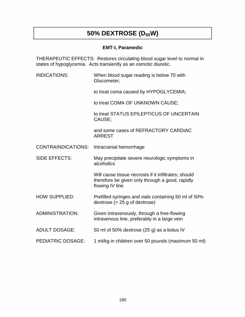
180
50% DEXTROSE (D50W)
EMT-I, Paramedic
THERAPEUTIC EFFECTS: Restores circulating blood sugar level to normal in states of hypoglycemia. Acts transiently as an osmotic diuretic. INDICATIONS: When blood sugar reading is below 70 with
Glucometer;
to treat coma caused by HYPOGLYCEMIA; to treat COMA OF UNKNOWN CAUSE; to treat STATUS EPILEPTICUS OF UNCERTAIN CAUSE; and some cases of REFRACTORY CARDIAC ARREST
CONTRAINDICATIONS: lntracranial hemorrhage SIDE EFFECTS: May precipitate severe neurologic symptoms in
alcoholics Will cause tissue necrosis if it infiltrates; should therefore be given only through a good, rapidly flowing IV line
HOW SUPPLIED: Prefilled syringes and vials containing 50 ml of 50%
dextrose (= 25 g of dextrose) ADMINISTRATION: Given intravenously, through a free-flowing
intravenous line, preferably in a large vein ADULT DOSAGE: 50 ml of 50% dextrose (25 g) as a bolus IV PEDIATRIC DOSAGE: 1 ml/kg in children over 50 pounds (maximum 50 ml)
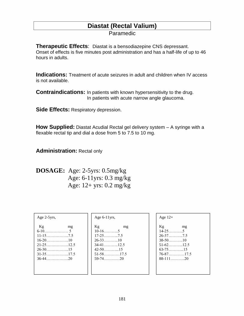
181
Diastat (Rectal Valium) Paramedic
Therapeutic Effects: Diastat is a bensodiazepine CNS depressant.
Onset of effects is five minutes post administration and has a half-life of up to 46 hours in adults.
Indications: Treatment of acute seizures in adult and children when IV access
is not available.
Contraindications: In patients with known hypersensitivity to the drug.
In patients with acute narrow angle glaucoma.
Side Effects: Respiratory depression.
How Supplied: Diastat Acudial Rectal gel delivery system – A syringe with a
flexable rectal tip and dial a dose from 5 to 7.5 to 10 mg.
Administration: Rectal only
DOSAGE: Age: 2-5yrs: 0.5mg/kg
Age: 6-11yrs: 0.3 mg/kg
Age: 12+ yrs: 0.2 mg/kg
Age 12+
Kg mg
14-25………..5
26-37………..7.5
38-50………..10
51-62………..12.5
63-75………...15
76-87…………17.5
88-111………..20
Age 6-11yrs,
Kg mg
10-16………..5
17-25………..7.5
26-33………..10
34-41………..12.5
42-50………...15
51-58…………17.5
59-74…………20
Age 2-5yrs,
Kg mg
6-10……………… 5
11-15……………..7.5
16-20……………..10
21-25……………..12.5
26-30……………..15
31-35……………..17.5
36-44……………..20
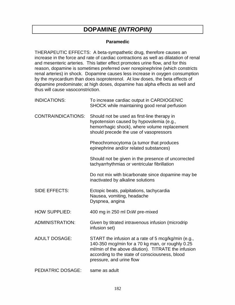
182
DOPAMINE (INTROPIN)
Paramedic THERAPEUTIC EFFECTS: A beta-sympathetic drug, therefore causes an increase in the force and rate of cardiac contractions as well as dilatation of renal and mesenteric arteries. This latter effect promotes urine flow, and for this reason, dopamine is sometimes preferred over norepinephrine (which constricts renal arteries) in shock. Dopamine causes less increase in oxygen consumption by the myocardium than does isoproterenol. At low doses, the beta effects of dopamine predominate; at high doses, dopamine has alpha effects as well and thus will cause vasoconstriction. INDICATIONS: To increase cardiac output in CARDIOGENIC
SHOCK while maintaining good renal perfusion CONTRAINDICATIONS: Should not be used as first-line therapy in
hypotension caused by hypovolemia (e.g., hemorrhagic shock), where volume replacement should precede the use of vasopressors
Pheochromocytoma (a tumor that produces epinephrine and/or related substances) Should not be given in the presence of uncorrected tachyarrhythmias or ventricular fibrillation Do not mix with bicarbonate since dopamine may be inactivated by alkaline solutions
SIDE EFFECTS: Ectopic beats, palpitations, tachycardia
Nausea, vomiting, headache Dyspnea, angina
HOW SUPPLIED: 400 mg in 250 ml D5W pre-mixed ADMINISTRATION: Given by titrated intravenous infusion (microdrip
infusion set) ADULT DOSAGE: START the infusion at a rate of 5 mcg/kg/min (e.g.,
140-350 mcg/min for a 70 kg man, or roughly 0.25 ml/min of the above dilution). TITRATE the infusion according to the state of consciousness, blood pressure, and urine flow
PEDIATRIC DOSAGE: same as adult
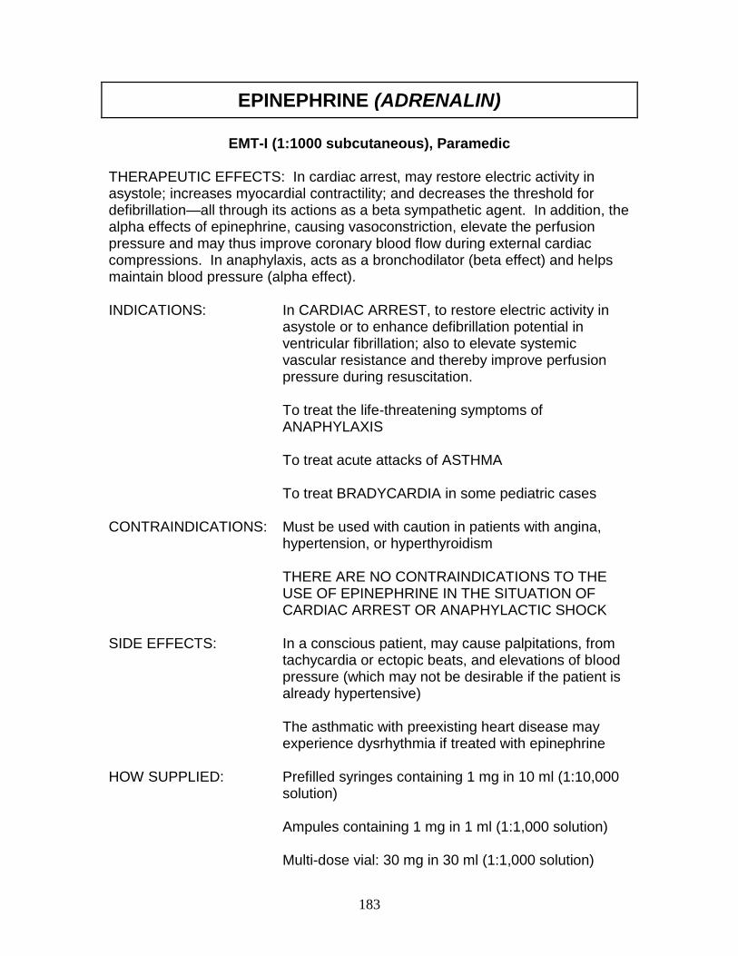
183
EPINEPHRINE (ADRENALIN)
EMT-I (1:1000 subcutaneous), Paramedic
THERAPEUTIC EFFECTS: In cardiac arrest, may restore electric activity in asystole; increases myocardial contractility; and decreases the threshold for defibrillation—all through its actions as a beta sympathetic agent. In addition, the alpha effects of epinephrine, causing vasoconstriction, elevate the perfusion pressure and may thus improve coronary blood flow during external cardiac compressions. In anaphylaxis, acts as a bronchodilator (beta effect) and helps maintain blood pressure (alpha effect). INDICATIONS: In CARDIAC ARREST, to restore electric activity in
asystole or to enhance defibrillation potential in ventricular fibrillation; also to elevate systemic vascular resistance and thereby improve perfusion pressure during resuscitation.
To treat the life-threatening symptoms of ANAPHYLAXIS To treat acute attacks of ASTHMA To treat BRADYCARDIA in some pediatric cases
CONTRAINDICATIONS: Must be used with caution in patients with angina,
hypertension, or hyperthyroidism
THERE ARE NO CONTRAINDICATIONS TO THE USE OF EPINEPHRINE IN THE SITUATION OF CARDIAC ARREST OR ANAPHYLACTIC SHOCK
SIDE EFFECTS: In a conscious patient, may cause palpitations, from
tachycardia or ectopic beats, and elevations of blood pressure (which may not be desirable if the patient is already hypertensive)
The asthmatic with preexisting heart disease may experience dysrhythmia if treated with epinephrine
HOW SUPPLIED: Prefilled syringes containing 1 mg in 10 ml (1:10,000
solution) Ampules containing 1 mg in 1 ml (1:1,000 solution) Multi-dose vial: 30 mg in 30 ml (1:1,000 solution)
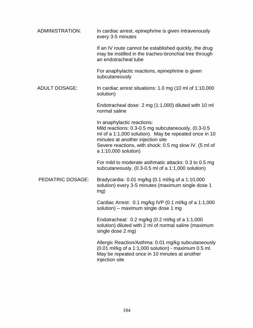
184
ADMINISTRATION: In cardiac arrest, epinephrine is given intravenously
every 3-5 minutes If an IV route cannot be established quickly, the drug
may be instilled in the tracheo-bronchial tree through an endotracheal tube
For anaphylactic reactions, epinephrine is given subcutaneously
ADULT DOSAGE: In cardiac arrest situations: 1.0 mg (10 ml of 1:10,000
solution) Endotracheal dose: 2 mg (1:1,000) diluted with 10 ml normal saline
In anaphylactic reactions: Mild reactions: 0.3-0.5 mg subcutaneously, (0.3-0.5 ml of a 1:1,000 solution). May be repeated once in 10 minutes at another injection site Severe reactions, with shock: 0.5 mg slow IV. (5 ml of a 1:10,000 solution) For mild to moderate asthmatic attacks: 0.3 to 0.5 mg subcutaneously, (0.3-0.5 ml of a 1:1,000 solution)
PEDIATRIC DOSAGE: Bradycardia: 0.01 mg/kg (0.1 ml/kg of a 1:10,000
solution) every 3-5 minutes (maximum single dose 1 mg)
Cardiac Arrest: 0.1 mg/kg IVP (0.1 ml/kg of a 1:1,000 solution) – maximum single dose 1 mg
Endotracheal: 0.2 mg/kg (0.2 ml/kg of a 1:1,000 solution) diluted with 2 ml of normal saline (maximum single dose 2 mg) Allergic Reaction/Asthma: 0.01 mg/kg subcutaneously (0.01 ml/kg of a 1:1,000 solution) - maximum 0.5 ml. May be repeated once in 10 minutes at another injection site
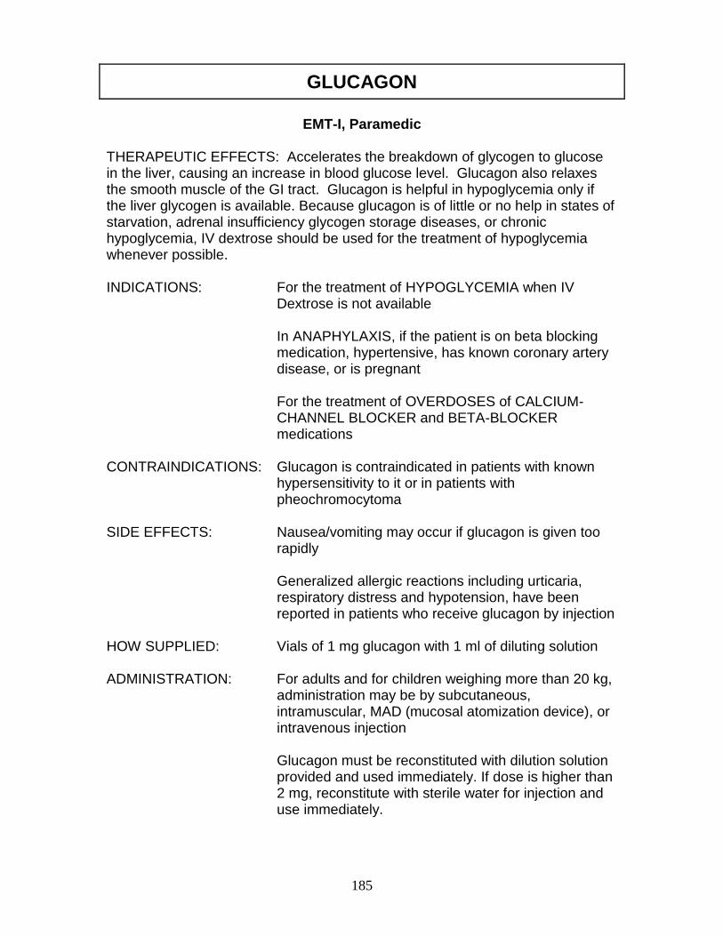
185
GLUCAGON
EMT-I, Paramedic
THERAPEUTIC EFFECTS: Accelerates the breakdown of glycogen to glucose in the liver, causing an increase in blood glucose level. Glucagon also relaxes the smooth muscle of the GI tract. Glucagon is helpful in hypoglycemia only if the liver glycogen is available. Because glucagon is of little or no help in states of starvation, adrenal insufficiency glycogen storage diseases, or chronic hypoglycemia, IV dextrose should be used for the treatment of hypoglycemia whenever possible. INDICATIONS: For the treatment of HYPOGLYCEMIA when IV
Dextrose is not available
In ANAPHYLAXIS, if the patient is on beta blocking medication, hypertensive, has known coronary artery disease, or is pregnant
For the treatment of OVERDOSES of CALCIUM-CHANNEL BLOCKER and BETA-BLOCKER medications
CONTRAINDICATIONS: Glucagon is contraindicated in patients with known
hypersensitivity to it or in patients with pheochromocytoma
SIDE EFFECTS: Nausea/vomiting may occur if glucagon is given too
rapidly
Generalized allergic reactions including urticaria, respiratory distress and hypotension, have been reported in patients who receive glucagon by injection
HOW SUPPLIED: Vials of 1 mg glucagon with 1 ml of diluting solution ADMINISTRATION: For adults and for children weighing more than 20 kg,
administration may be by subcutaneous, intramuscular, MAD (mucosal atomization device), or intravenous injection
Glucagon must be reconstituted with dilution solution provided and used immediately. If dose is higher than 2 mg, reconstitute with sterile water for injection and use immediately.
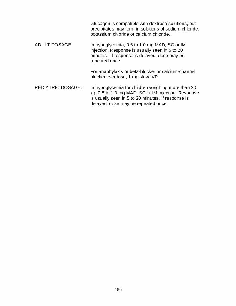
186
Glucagon is compatible with dextrose solutions, but precipitates may form in solutions of sodium chloride, potassium chloride or calcium chloride.
ADULT DOSAGE: In hypoglycemia, 0.5 to 1.0 mg MAD, SC or IM
injection. Response is usually seen in 5 to 20 minutes. If response is delayed, dose may be repeated once
For anaphylaxis or beta-blocker or calcium-channel
blocker overdose, 1 mg slow IVP PEDIATRIC DOSAGE: In hypoglycemia for children weighing more than 20
kg, 0.5 to 1.0 mg MAD, SC or IM injection. Response is usually seen in 5 to 20 minutes. If response is delayed, dose may be repeated once.

187
Haldol (Haloperidol)
Paramedic
THERAPEUTIC EFFECTS: Sedation INDICATIONS: This medication is used to treat symptoms of certain
types of mental conditions, and to control severe behavioral problems. This medication is considered a chemical restraint at sedating doses and as such, must be ordered by Medical Control prior to administration.
CONTRAINDICATIONS: Severe intoxication with alcohol or other central depressant drugs
Known allergy against haloperidol or other butyrophenones or other drug ingredients
Known heart disease; when combined will tend towards cardiac arrest
SIDE EFFECTS: Dry mouth, lethargy, muscle stiffness, muscle
cramping, restlessness, and tremors. ADMINISTRATION: IM ADULT DOSAGE: 5mg deep IM PEDIATRIC DOSAGE: Medical control only (rarely used)
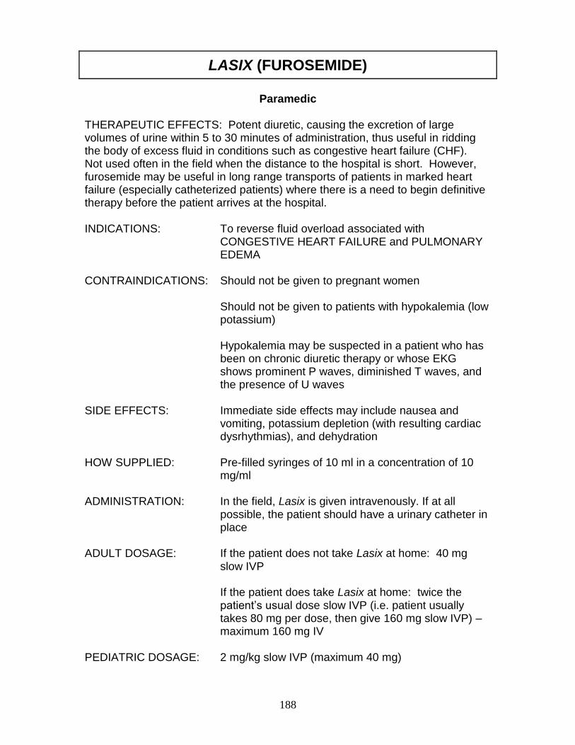
188
LASIX (FUROSEMIDE)
Paramedic
THERAPEUTIC EFFECTS: Potent diuretic, causing the excretion of large volumes of urine within 5 to 30 minutes of administration, thus useful in ridding the body of excess fluid in conditions such as congestive heart failure (CHF). Not used often in the field when the distance to the hospital is short. However, furosemide may be useful in long range transports of patients in marked heart failure (especially catheterized patients) where there is a need to begin definitive therapy before the patient arrives at the hospital. INDICATIONS: To reverse fluid overload associated with
CONGESTIVE HEART FAILURE and PULMONARY EDEMA
CONTRAINDICATIONS: Should not be given to pregnant women
Should not be given to patients with hypokalemia (low potassium) Hypokalemia may be suspected in a patient who has been on chronic diuretic therapy or whose EKG shows prominent P waves, diminished T waves, and the presence of U waves
SIDE EFFECTS: Immediate side effects may include nausea and
vomiting, potassium depletion (with resulting cardiac dysrhythmias), and dehydration
HOW SUPPLIED: Pre-filled syringes of 10 ml in a concentration of 10
mg/ml ADMINISTRATION: In the field, Lasix is given intravenously. If at all
possible, the patient should have a urinary catheter in place
ADULT DOSAGE: If the patient does not take Lasix at home: 40 mg
slow IVP
If the patient does take Lasix at home: twice the patient’s usual dose slow IVP (i.e. patient usually takes 80 mg per dose, then give 160 mg slow IVP) – maximum 160 mg IV
PEDIATRIC DOSAGE: 2 mg/kg slow IVP (maximum 40 mg)
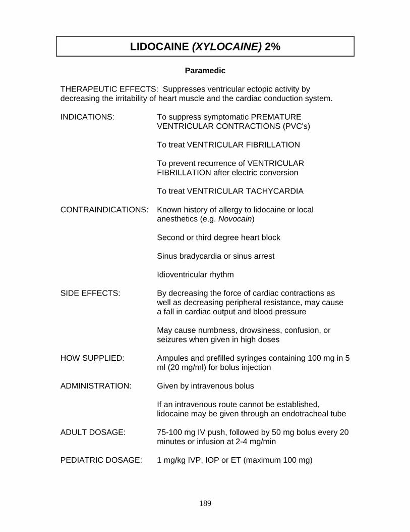
189
LIDOCAINE (XYLOCAINE) 2%
Paramedic
THERAPEUTIC EFFECTS: Suppresses ventricular ectopic activity by decreasing the irritability of heart muscle and the cardiac conduction system. INDICATIONS: To suppress symptomatic PREMATURE
VENTRICULAR CONTRACTIONS (PVC's) To treat VENTRICULAR FIBRILLATION To prevent recurrence of VENTRICULAR FIBRILLATION after electric conversion To treat VENTRICULAR TACHYCARDIA
CONTRAINDICATIONS: Known history of allergy to lidocaine or local
anesthetics (e.g. Novocain) Second or third degree heart block Sinus bradycardia or sinus arrest Idioventricular rhythm
SIDE EFFECTS: By decreasing the force of cardiac contractions as
well as decreasing peripheral resistance, may cause a fall in cardiac output and blood pressure
May cause numbness, drowsiness, confusion, or seizures when given in high doses
HOW SUPPLIED: Ampules and prefilled syringes containing 100 mg in 5
ml (20 mg/ml) for bolus injection ADMINISTRATION: Given by intravenous bolus
If an intravenous route cannot be established, lidocaine may be given through an endotracheal tube
ADULT DOSAGE: 75-100 mg IV push, followed by 50 mg bolus every 20
minutes or infusion at 2-4 mg/min PEDIATRIC DOSAGE: 1 mg/kg IVP, IOP or ET (maximum 100 mg)
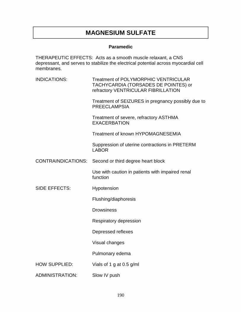
190
MAGNESIUM SULFATE
Paramedic
THERAPEUTIC EFFECTS: Acts as a smooth muscle relaxant, a CNS depressant, and serves to stabilize the electrical potential across myocardial cell membranes. INDICATIONS: Treatment of POLYMORPHIC VENTRICULAR
TACHYCARDIA (TORSADES DE POINTES) or refractory VENTRICULAR FIBRILLATION
Treatment of SEIZURES in pregnancy possibly due to
PREECLAMPSIA Treatment of severe, refractory ASTHMA
EXACERBATION Treatment of known HYPOMAGNESEMIA Suppression of uterine contractions in PRETERM
LABOR CONTRAINDICATIONS: Second or third degree heart block Use with caution in patients with impaired renal
function SIDE EFFECTS: Hypotension Flushing/diaphoresis Drowsiness
Respiratory depression Depressed reflexes Visual changes Pulmonary edema HOW SUPPLIED: Vials of 1 g at 0.5 g/ml ADMINISTRATION: Slow IV push
191
ADULT DOSAGE: 2 grams PEDIATRIC DOSAGE: 25 mg/kg (maximum 2 g)
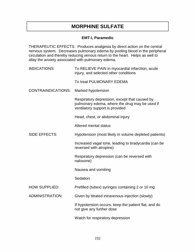
192
MORPHINE SULFATE
EMT-I, Paramedic
THERAPEUTIC EFFECTS: Produces analgesia by direct action on the central nervous system. Decreases pulmonary edema by pooling blood in the peripheral circulation and thereby reducing venous return to the heart. Helps as well to allay the anxiety associated with pulmonary edema. INDICATIONS: To RELIEVE PAIN in myocardial infarction, acute
injury, and selected other conditions
To treat PULMONARY EDEMA
CONTRAINDICATIONS: Marked hypotension
Respiratory depression, except that caused by pulmonary edema, where the drug may be used if ventilatory support is provided Head, chest, or abdominal injury Altered mental status
SIDE EFFECTS: Hypotension (most likely in volume depleted patients)
Increased vagal tone, leading to bradycardia (can be reversed with atropine) Respiratory depression (can be reversed with naloxone) Nausea and vomiting
Sedation HOW SUPPLIED: Prefilled (tubex) syringes containing 2 or 10 mg ADMINISTRATION: Given by titrated intravenous injection (slowly)
If hypotension occurs, keep the patient flat, and do not give any further dose Watch for respiratory depression
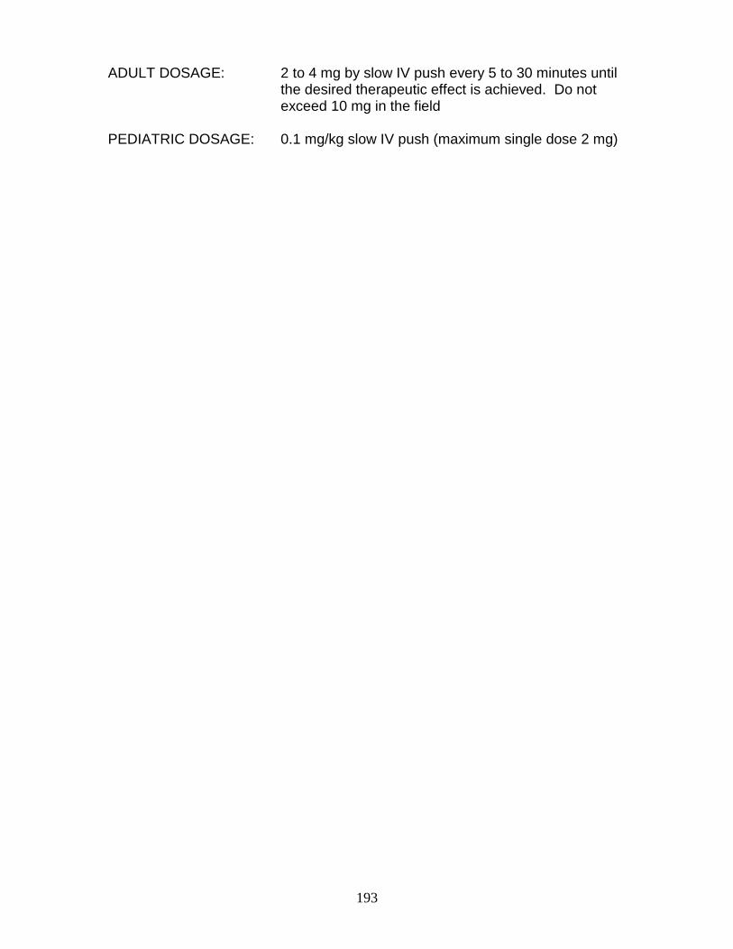
193
ADULT DOSAGE: 2 to 4 mg by slow IV push every 5 to 30 minutes until the desired therapeutic effect is achieved. Do not exceed 10 mg in the field
PEDIATRIC DOSAGE: 0.1 mg/kg slow IV push (maximum single dose 2 mg)
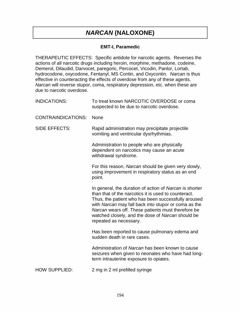
194
NARCAN (NALOXONE)
EMT-I, Paramedic
THERAPEUTIC EFFECTS: Specific antidote for narcotic agents. Reverses the actions of all narcotic drugs including heroin, morphine, methadone, codeine, Demerol, Dilaudid, Darvocet, paregoric, Percocet, Vicodin, Panlor, Lortab, hydrocodone, oxycodone, Fentanyl, MS Contin, and Oxycontin. Narcan is thus effective in counteracting the effects of overdose from any of these agents. Narcan will reverse stupor, coma, respiratory depression, etc. when these are due to narcotic overdose. INDICATIONS: To treat known NARCOTIC OVERDOSE or coma
suspected to be due to narcotic overdose. CONTRAINDICATIONS: None SIDE EFFECTS: Rapid administration may precipitate projectile
vomiting and ventricular dysrhythmias.
Administration to people who are physically dependent on narcotics may cause an acute withdrawal syndrome. For this reason, Narcan should be given very slowly, using improvement in respiratory status as an end point. In general, the duration of action of Narcan is shorter than that of the narcotics it is used to counteract. Thus, the patient who has been successfully aroused with Narcan may fall back into stupor or coma as the Narcan wears off. These patients must therefore be watched closely, and the dose of Narcan should be repeated as necessary. Has been reported to cause pulmonary edema and sudden death in rare cases. Administration of Narcan has been known to cause seizures when given to neonates who have had long-term intrauterine exposure to opiates.
HOW SUPPLIED: 2 mg in 2 ml prefilled syringe
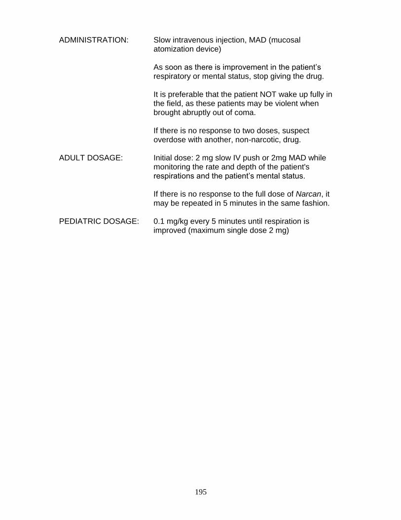
195
ADMINISTRATION: Slow intravenous injection, MAD (mucosal atomization device)
As soon as there is improvement in the patient’s respiratory or mental status, stop giving the drug. It is preferable that the patient NOT wake up fully in the field, as these patients may be violent when brought abruptly out of coma. If there is no response to two doses, suspect overdose with another, non-narcotic, drug.
ADULT DOSAGE: Initial dose: 2 mg slow IV push or 2mg MAD while
monitoring the rate and depth of the patient's respirations and the patient’s mental status.
If there is no response to the full dose of Narcan, it may be repeated in 5 minutes in the same fashion.
PEDIATRIC DOSAGE: 0.1 mg/kg every 5 minutes until respiration is
improved (maximum single dose 2 mg)

196
NITROGLYCERIN
EMT-I, Paramedic
THERAPEUTIC EFFECTS: The primary pharmacologic effect of nitroglycerin and related drugs is to relax smooth muscle, and the effects of nitroglycerin on the cardiovascular system are chiefly due to relaxation of vascular smooth muscle (hence vasodilatation). Nitroglycerin provides relief of pain in angina, probably by dilating coronary arteries and thereby increasing blood flow through them as well as by decreasing myocardial oxygen demand. Through its vasodilatation action on peripheral vessels, nitroglycerin promotes pooling of the blood in the systemic circulation and decreases the resistance against which the heart has to pump (the afterload); these effects may be useful in treating congestive heart failure. INDICATIONS: To relieve the pain of ANGINA
To treat PULMONARY EDEMA CONTRAINDICATIONS: Use with caution in myocardial infarction
Increased intracranial pressure Use of Viagra, Levitra, or Cialis in past 48 hours
SIDE EFFECTS: Transient, throbbing headache
Hypotension Dizziness, weakness Flushing
HOW SUPPLIED: Many forms, including ointment, spray, tablets,
sustained release capsules, and IV solution (50 mg/250 cc D5W in glass bottle --- 200 mg/cc)
For use in the field, tablets of 0.4 mg, metered-dose spray bottles (200 mcg), or IV solution
ADMINISTRATION: Given sublingually (under tongue) or IV
The patient should be semi-sitting or recumbent IV access should be established prior to administration
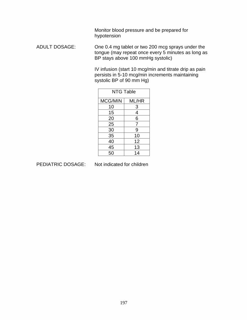
197
Monitor blood pressure and be prepared for hypotension
ADULT DOSAGE: One 0.4 mg tablet or two 200 mcg sprays under the
tongue (may repeat once every 5 minutes as long as BP stays above 100 mmHg systolic) IV infusion (start 10 mcg/min and titrate drip as pain persists in 5-10 mcg/min increments maintaining systolic BP of 90 mm Hg)
NTG Table
MCG/MIN ML/HR
10 3
15 4
20 6
25 7
30 9
35 10
40 12
45 13
50 14
PEDIATRIC DOSAGE: Not indicated for children
198
NORMAL SALINE IV SOLUTION (0.9%)
EMT-I, Paramedic
THERAPEUTIC EFFECTS: Used as standard IV access for all patients. Normal saline solution is isotonic and as such, will not easily cross into the extravascular space. This action provides good short term volume expansion as well as a versatile medium for parenteral drug administration. INDICATIONS: Can be used as a KEEP OPEN LINE for patients of
any age that may require fluid or medication administration by the IV route.
HYPOVOLEMIA secondary to bleeding, burns,
sepsis, hyperglycemia, etc. SYMPTOMATIC HYPOTENSION caused by
dehydration, vasodilators, vagal tone, etc. CONTRAINDICATIONS: None
Careless or overaggressive administration to children
and patients with congestive heart failure or renal insufficiency can precipitate volume overload and pulmonary edema.
Use with caution and in limited volume in head injury
patients. SIDE EFFECTS: Hypernatremia/Hyperchloremia if overload is present,
otherwise, none. HOW SUPPLIED: 500 ml and 1,000 ml bags; 100 ml and 250 ml mixers;
various size vials for flushing saline locks. DOSAGE: ―KEEP OPEN‖: 10 ml/hr for all ages
Adults: ―WIDE OPEN‖ to 500 ml bolus, then reassess clinical state of patient. May repeat twice if transport time requires. MONITOR LUNG SOUNDS IN PATIENTS WITH CHF & RENAL FAILURE.
199
Children: Syringe bolus at 20 ml/kg per IV or IO route. Reassess the patient for improvement after the bolus and repeat once prior to contacting Medical Control for authorization of further fluid administration.
Neonates to One Month of age: Syringe bolus at 10 ml/kg per IV or IO route. Reassess patient’s condition and contact Medical Control for further fluid administration orders.
200
NUBAIN (NALBUPHINE)
EMT-I, Paramedic
THERAPEUTIC EFFECTS: Narcotic agonist-antagonist with potency similar to morphine. When given following or concurrently with morphine, it can block or partially reverse narcotic-induced respiratory depression. INDICATIONS: Relief of moderate to severe pain. This is also an
appropriate agent for pain relief during labor and delivery.
CONTRAINDICATIONS: Known sensitivity to Nubain SIDE EFFECTS: Mild respiratory depression with 10 mg dosage;
however, higher doses do not induce further respiratory depression.
Sweatiness, nausea and vomiting, dizziness
Nubain may precipitate withdrawal in patients with opioid dependence.
HOW SUPPLIED: 10 mg/ml in 1 ml ampules ADMINISTRATION: Intravenously or intramuscularly ADULT DOSAGE: 10 mg IV or IM PEDIATRIC DOSAGE: not for use in children
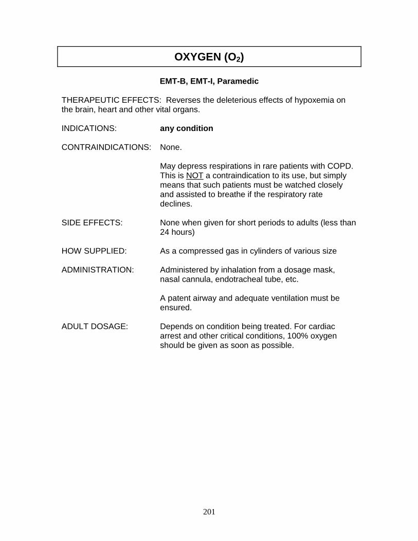
201
OXYGEN (O2)
EMT-B, EMT-I, Paramedic
THERAPEUTIC EFFECTS: Reverses the deleterious effects of hypoxemia on the brain, heart and other vital organs.
INDICATIONS: any condition CONTRAINDICATIONS: None.
May depress respirations in rare patients with COPD. This is NOT a contraindication to its use, but simply means that such patients must be watched closely and assisted to breathe if the respiratory rate declines.
SIDE EFFECTS: None when given for short periods to adults (less than 24 hours)
HOW SUPPLIED: As a compressed gas in cylinders of various size ADMINISTRATION: Administered by inhalation from a dosage mask,
nasal cannula, endotracheal tube, etc. A patent airway and adequate ventilation must be
ensured. ADULT DOSAGE: Depends on condition being treated. For cardiac
arrest and other critical conditions, 100% oxygen should be given as soon as possible.
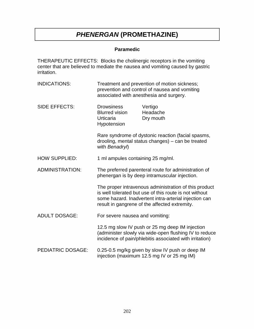
202
PHENERGAN (PROMETHAZINE)
Paramedic
THERAPEUTIC EFFECTS: Blocks the cholinergic receptors in the vomiting center that are believed to mediate the nausea and vomiting caused by gastric irritation. INDICATIONS: Treatment and prevention of motion sickness;
prevention and control of nausea and vomiting associated with anesthesia and surgery.
SIDE EFFECTS: Drowsiness Vertigo
Blurred vision Headache Urticaria Dry mouth Hypotension Rare syndrome of dystonic reaction (facial spasms, drooling, mental status changes) – can be treated with Benadryl)
HOW SUPPLIED: 1 ml ampules containing 25 mg/ml. ADMINISTRATION: The preferred parenteral route for administration of
phenergan is by deep intramuscular injection. The proper intravenous administration of this product
is well tolerated but use of this route is not without some hazard. Inadvertent intra-arterial injection can result in gangrene of the affected extremity.
ADULT DOSAGE: For severe nausea and vomiting: 12.5 mg slow IV push or 25 mg deep IM injection
(administer slowly via wide-open flushing IV to reduce incidence of pain/phlebitis associated with irritation)
PEDIATRIC DOSAGE: 0.25-0.5 mg/kg given by slow IV push or deep IM
injection (maximum 12.5 mg IV or 25 mg IM)
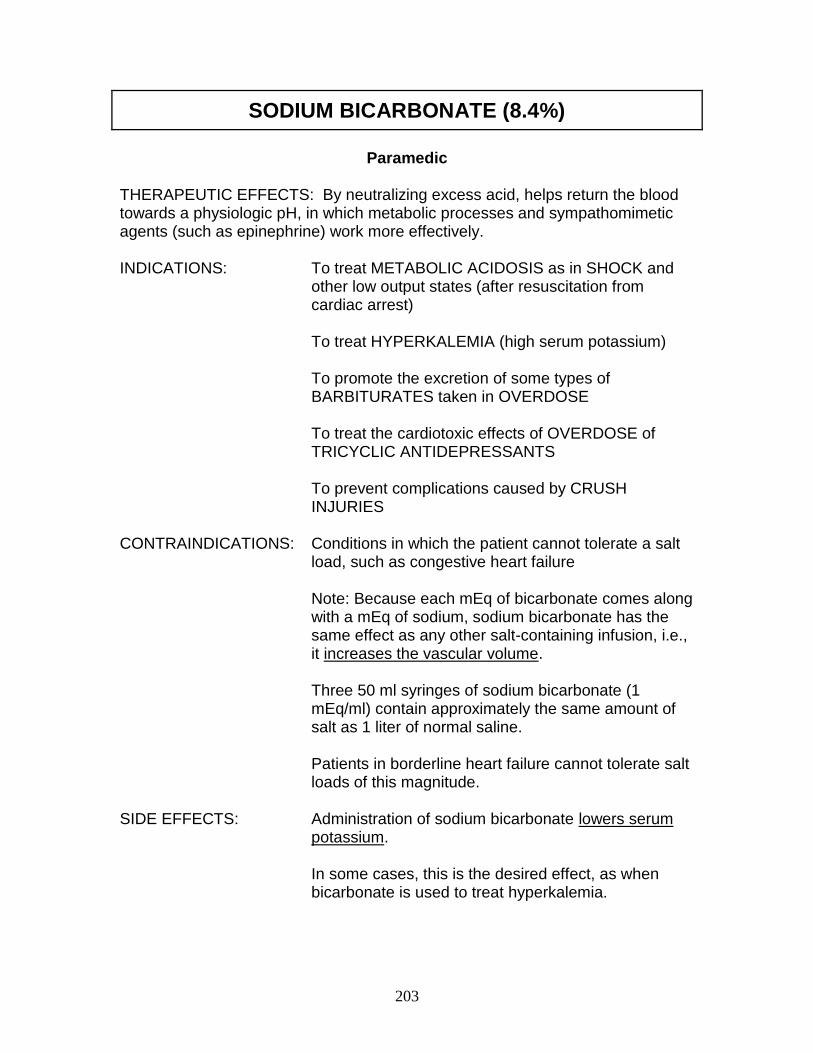
203
SODIUM BICARBONATE (8.4%)
Paramedic
THERAPEUTIC EFFECTS: By neutralizing excess acid, helps return the blood towards a physiologic pH, in which metabolic processes and sympathomimetic agents (such as epinephrine) work more effectively. INDICATIONS: To treat METABOLIC ACIDOSIS as in SHOCK and
other low output states (after resuscitation from cardiac arrest)
To treat HYPERKALEMIA (high serum potassium) To promote the excretion of some types of
BARBITURATES taken in OVERDOSE To treat the cardiotoxic effects of OVERDOSE of
TRICYCLIC ANTIDEPRESSANTS To prevent complications caused by CRUSH
INJURIES CONTRAINDICATIONS: Conditions in which the patient cannot tolerate a salt
load, such as congestive heart failure Note: Because each mEq of bicarbonate comes along
with a mEq of sodium, sodium bicarbonate has the same effect as any other salt-containing infusion, i.e., it increases the vascular volume.
Three 50 ml syringes of sodium bicarbonate (1
mEq/ml) contain approximately the same amount of salt as 1 liter of normal saline.
Patients in borderline heart failure cannot tolerate salt
loads of this magnitude. SIDE EFFECTS: Administration of sodium bicarbonate lowers serum
potassium. In some cases, this is the desired effect, as when
bicarbonate is used to treat hyperkalemia.
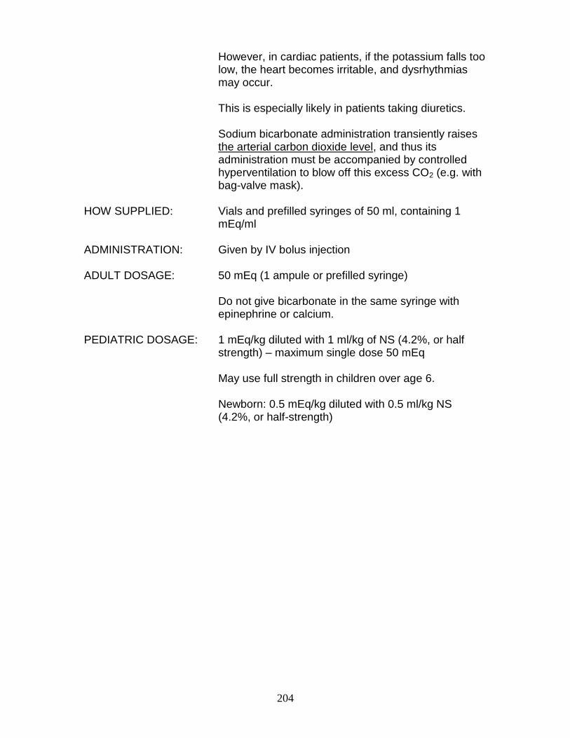
204
However, in cardiac patients, if the potassium falls too low, the heart becomes irritable, and dysrhythmias may occur.
This is especially likely in patients taking diuretics. Sodium bicarbonate administration transiently raises
the arterial carbon dioxide level, and thus its administration must be accompanied by controlled hyperventilation to blow off this excess CO2 (e.g. with bag-valve mask).
HOW SUPPLIED: Vials and prefilled syringes of 50 ml, containing 1
mEq/ml ADMINISTRATION: Given by IV bolus injection ADULT DOSAGE: 50 mEq (1 ampule or prefilled syringe)
Do not give bicarbonate in the same syringe with epinephrine or calcium.
PEDIATRIC DOSAGE: 1 mEq/kg diluted with 1 ml/kg of NS (4.2%, or half strength) – maximum single dose 50 mEq
May use full strength in children over age 6.
Newborn: 0.5 mEq/kg diluted with 0.5 ml/kg NS (4.2%, or half-strength)
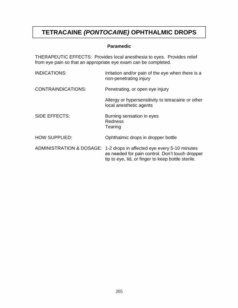
205
TETRACAINE (PONTOCAINE) OPHTHALMIC DROPS
Paramedic
THERAPEUTIC EFFECTS: Provides local anesthesia to eyes. Provides relief from eye pain so that an appropriate eye exam can be completed. INDICATIONS: Irritation and/or pain of the eye when there is a
non-penetrating injury CONTRAINDICATIONS: Penetrating, or open eye injury
Allergy or hypersensitivity to tetracaine or other local anesthetic agents
SIDE EFFECTS: Burning sensation in eyes
Redness Tearing
HOW SUPPLIED: Ophthalmic drops in dropper bottle ADMINISTRATION & DOSAGE: 1-2 drops in affected eye every 5-10 minutes
as needed for pain control. Don’t touch dropper tip to eye, lid, or finger to keep bottle sterile.
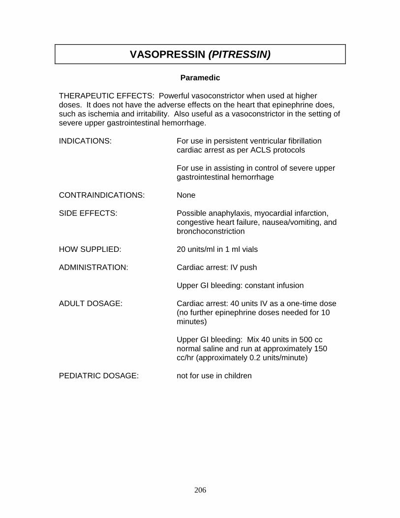
206
VASOPRESSIN (PITRESSIN)
Paramedic
THERAPEUTIC EFFECTS: Powerful vasoconstrictor when used at higher doses. It does not have the adverse effects on the heart that epinephrine does, such as ischemia and irritability. Also useful as a vasoconstrictor in the setting of severe upper gastrointestinal hemorrhage. INDICATIONS: For use in persistent ventricular fibrillation
cardiac arrest as per ACLS protocols For use in assisting in control of severe upper
gastrointestinal hemorrhage CONTRAINDICATIONS: None SIDE EFFECTS: Possible anaphylaxis, myocardial infarction,
congestive heart failure, nausea/vomiting, and bronchoconstriction
HOW SUPPLIED: 20 units/ml in 1 ml vials ADMINISTRATION: Cardiac arrest: IV push Upper GI bleeding: constant infusion ADULT DOSAGE: Cardiac arrest: 40 units IV as a one-time dose
(no further epinephrine doses needed for 10 minutes)
Upper GI bleeding: Mix 40 units in 500 cc
normal saline and run at approximately 150 cc/hr (approximately 0.2 units/minute)
PEDIATRIC DOSAGE: not for use in children
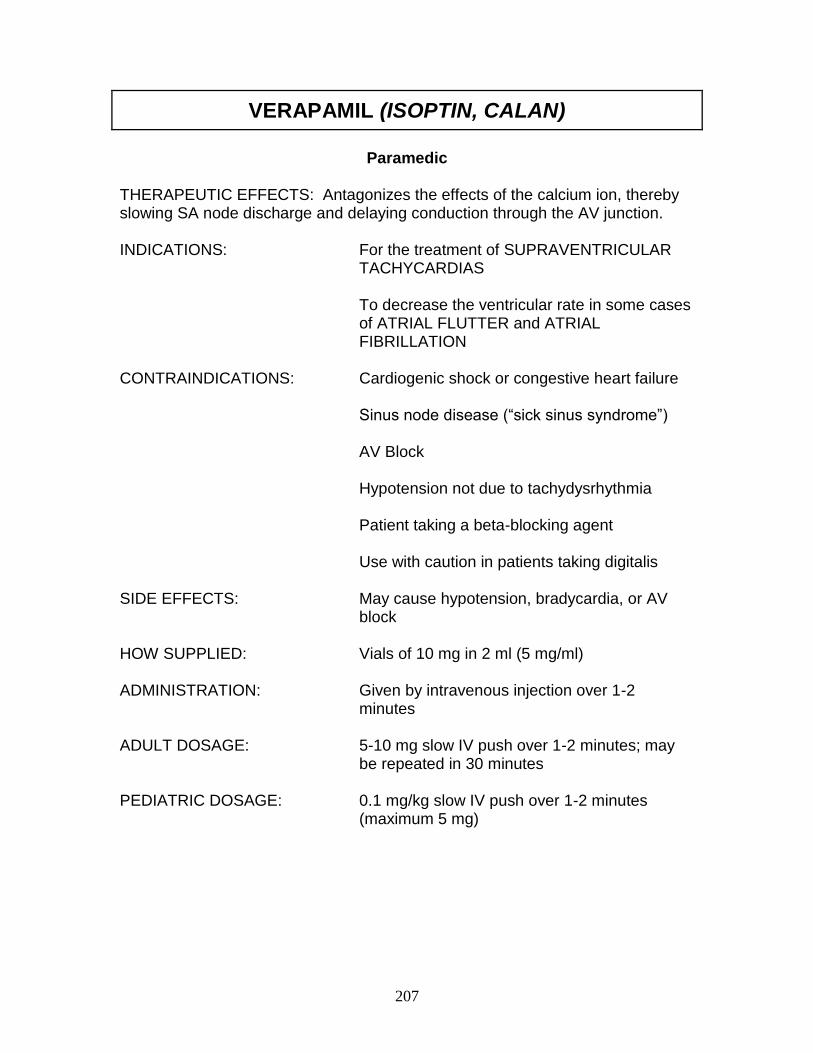
207
VERAPAMIL (ISOPTIN, CALAN)
Paramedic
THERAPEUTIC EFFECTS: Antagonizes the effects of the calcium ion, thereby slowing SA node discharge and delaying conduction through the AV junction. INDICATIONS: For the treatment of SUPRAVENTRICULAR
TACHYCARDIAS To decrease the ventricular rate in some cases
of ATRIAL FLUTTER and ATRIAL FIBRILLATION
CONTRAINDICATIONS: Cardiogenic shock or congestive heart failure Sinus node disease (―sick sinus syndrome‖) AV Block Hypotension not due to tachydysrhythmia Patient taking a beta-blocking agent Use with caution in patients taking digitalis SIDE EFFECTS: May cause hypotension, bradycardia, or AV
block HOW SUPPLIED: Vials of 10 mg in 2 ml (5 mg/ml) ADMINISTRATION: Given by intravenous injection over 1-2
minutes ADULT DOSAGE: 5-10 mg slow IV push over 1-2 minutes; may
be repeated in 30 minutes PEDIATRIC DOSAGE: 0.1 mg/kg slow IV push over 1-2 minutes
(maximum 5 mg)
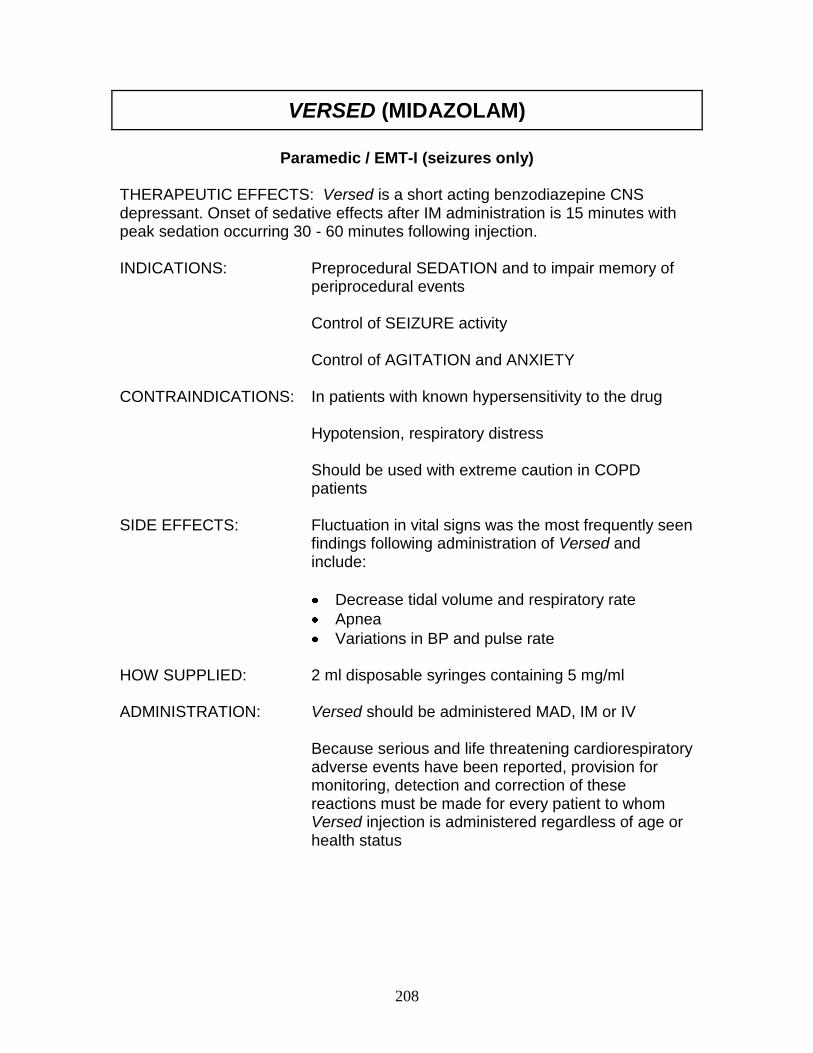
208
VERSED (MIDAZOLAM)
Paramedic / EMT-I (seizures only)
THERAPEUTIC EFFECTS: Versed is a short acting benzodiazepine CNS depressant. Onset of sedative effects after IM administration is 15 minutes with peak sedation occurring 30 - 60 minutes following injection. INDICATIONS: Preprocedural SEDATION and to impair memory of
periprocedural events Control of SEIZURE activity
Control of AGITATION and ANXIETY
CONTRAINDICATIONS: In patients with known hypersensitivity to the drug Hypotension, respiratory distress
Should be used with extreme caution in COPD patients
SIDE EFFECTS: Fluctuation in vital signs was the most frequently seen
findings following administration of Versed and include:
Decrease tidal volume and respiratory rate
Apnea
Variations in BP and pulse rate HOW SUPPLIED: 2 ml disposable syringes containing 5 mg/ml ADMINISTRATION: Versed should be administered MAD, IM or IV
Because serious and life threatening cardiorespiratory adverse events have been reported, provision for monitoring, detection and correction of these reactions must be made for every patient to whom Versed injection is administered regardless of age or health status
209
ADULT DOSAGE: For persistent seizure activity, MAD, IV or IM injection
of 5 mg initially and titrate to patient's condition up to 10 mg maximum
For preprocedural sedation, IV or IM injection of 2.5
mg For agitation/anxiety control, IV or IM injection of 2.5
mg PEDIATRIC DOSAGE: For persistent seizure activity, IV or IM injection of 0.1
mg/kg (maximum single dose 2.5 mg)
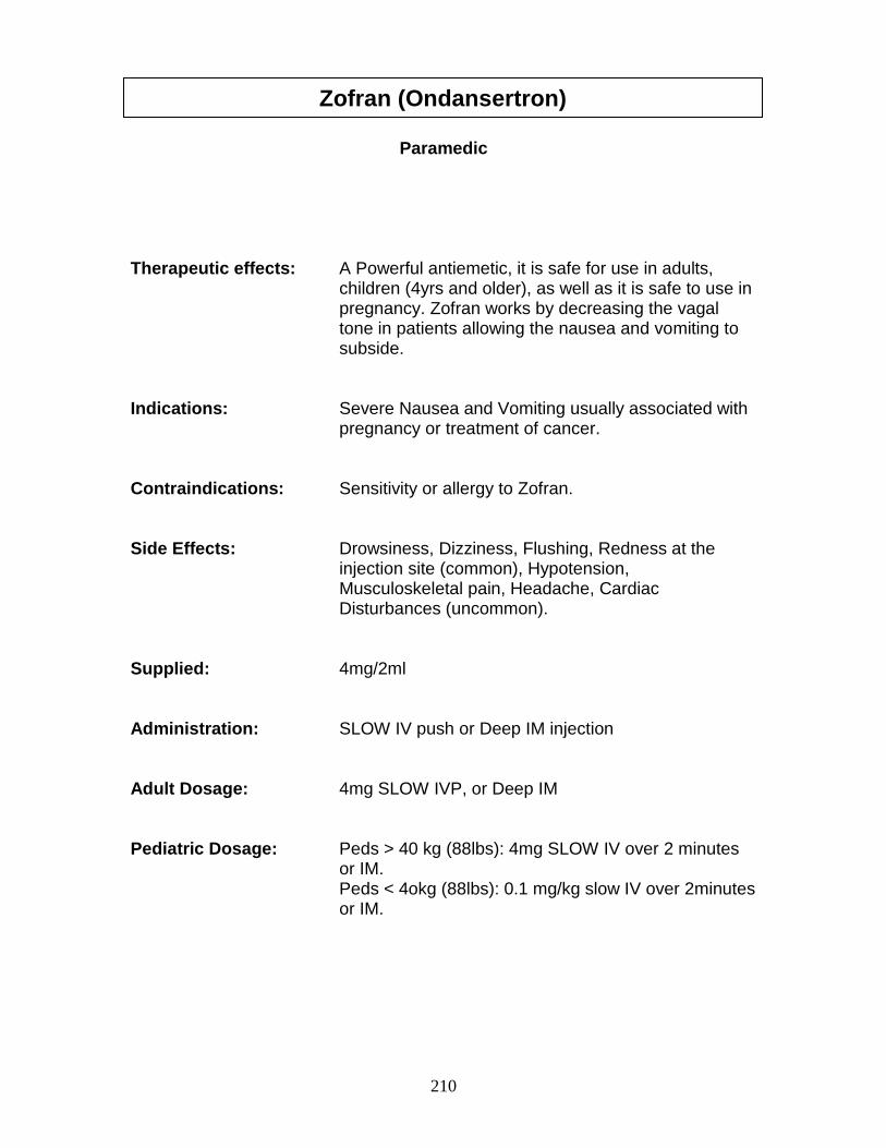
210
Paramedic Therapeutic effects: A Powerful antiemetic, it is safe for use in adults,
children (4yrs and older), as well as it is safe to use in pregnancy. Zofran works by decreasing the vagal tone in patients allowing the nausea and vomiting to subside.
Indications: Severe Nausea and Vomiting usually associated with
pregnancy or treatment of cancer. Contraindications: Sensitivity or allergy to Zofran. Side Effects: Drowsiness, Dizziness, Flushing, Redness at the
injection site (common), Hypotension, Musculoskeletal pain, Headache, Cardiac Disturbances (uncommon).
Supplied: 4mg/2ml Administration: SLOW IV push or Deep IM injection Adult Dosage: 4mg SLOW IVP, or Deep IM Pediatric Dosage: Peds > 40 kg (88lbs): 4mg SLOW IV over 2 minutes
or IM. Peds < 4okg (88lbs): 0.1 mg/kg slow IV over 2minutes
or IM.
Zofran (Ondansertron)