Embed Size (px)
Citation preview
REVISED COPY 1
Invasion-inhibitory antibodies elicited by immunization with 2
Plasmodium vivax apical membrane antigen-1 expressed 3
in Pichia pastoris yeast 4
5
Elaine C. Vicentina, Kátia S. Françosoa, Mariana V. Rochaa, 6
Dmitri Iourtovb, Fernanda L. dos Santosb, Flávia S. Kubruslyb, Maria A. Sakauchib, 7
Isaias Rawb, Francois Nostenc,d,e, Laurent Réniaf, 8
Mauricio M. Rodriguesg, Bruce Russellf,h and Irene S. Soaresa# 9
10 aDepartamento de Análises Clínicas e Toxicológicas, Faculdade de Ciências 11
Farmacêuticas, Universidade de São Paulo, 05508-900, São Paulo, SP, Brazil; 12 bInstituto Butantan, São Paulo, SP, Brazil; 13 cShoklo Malaria Research Unit (SMRU), Mae Sot, Tak Province, Thailand; 14 dMahidol-Oxford-University Research Unit, Bangkok, Thailand; 15 eCentre for Tropical Medicine, University of Oxford, Churchill Hospital, Oxford, United 16
Kingdom; 17 fSingapore Immunology Network, Biopolis, Agency for Science Technology and 18
Research, Singapore; 19 gCTCMOL, Departamento de Microbiologia, Imunologia e Parasitologia, Universidade 20
Federal de São Paulo-Escola Paulista de Medicina, Rua Mirassol, 207, São Paulo 21
04044-010, SP, Brazil; 22 hDepartment of Microbiology, Yong Loo Lin School of Medicine, 23
National University of Singapore, Singapore. 24
25
Running title: Plasmodium vivax AMA-1 vaccine candidate. 26
Address correspondence and reprint requests to Dr. Irene S. Soares 27
Departamento de Análises Clínicas e Toxicológicas, Faculdade de 28
Ciências Farmacêuticas, Universidade de São Paulo, Av. Prof. Lineu 29
Prestes, 580, Cidade Universitária, São Paulo, SP, 05508-900, Brazil; E-30
mail: [email protected]. Tel.: + 55-11-3091-3641; Fax: + 55-11-3813-2197. 31
IAI Accepts, published online ahead of print on 30 December 2013Infect. Immun. doi:10.1128/IAI.01169-13Copyright © 2013, American Society for Microbiology. All Rights Reserved.
on February 6, 2020 by guest
http://iai.asm.org/
Dow
nloaded from
Vicentin et al., 2013
2
This work was supported by funds from Fundação de Amparo à Pesquisa do Estado
de São Paulo (FAPESP 2008/05613-2) and The National Institute for Vaccine
Development and Technology (CNPq - INCTV). ECV, MMR, and ISS are supported
by fellowships from CNPq. MVR was supported by fellowship from FAPESP. This
study received funding from SIgN and from the Horizontal Programme on Infectious
Diseases under the Agency for Science, Technology and Research (A*STAR,
Singapore). SMRU is sponsored by The Wellcome Trust of Great Britain, as part of
the Oxford Tropical Medicine Research Programme of Wellcome Trust-Mahidol
University.
32 33
on February 6, 2020 by guest
http://iai.asm.org/
Dow
nloaded from
Vicentin et al., 2013
3
Abstract 34 35
In a recent vaccine trial performed with African children, immunization with a 36
recombinant protein based on Plasmodium falciparum apical membrane antigen 1 37
(AMA-1) conferred a significant degree of strain-specific resistance against malaria. 38
To contribute to the efforts of generating a vaccine against P. vivax malaria, we 39
expressed the ectodomain of P. vivax AMA-1 (PvAMA-1) as a secreted soluble 40
protein in the methylotrophic yeast Pichia pastoris. Recognized by a high percentage 41
of sera from individuals infected by P. vivax, this recombinant protein was found to 42
have maintained its antigenicity. The immunogenicity of this protein was evaluated in 43
mice using immunization protocols that included homologous and heterologous 44
prime/boost strategies with plasmid DNA and recombinant protein. We used the 45
following formulations containing different adjuvants: aluminum salts (Alum), 46
Bordetella pertussis monophosphoryl lipid A (MPLA), flagellin FliC from Salmonella 47
Typhimurium, saponin Quil A, or Incomplete Freund Adjuvant (IFA). The formulations 48
containing the adjuvants Quil A or IFA elicited the highest IgG antibody titers. 49
Significant antibody titers were also obtained using a formulation developed for 50
human use containing MPLA or Alum plus MPLA. Recombinant PvAMA-1 produced 51
under conditions of good laboratory practice provided a good yield, high purity, low 52
endotoxin levels, no microbial contaminants, and reproduced the experimental 53
immunizations. Most relevant for vaccine development was the fact that immunization 54
with PvAMA-1 elicited invasion-inhibitory antibodies against different Asian isolates of 55
P. vivax. Our results show that AMA-1 expressed in P. pastoris is a promising antigen 56
for use in future pre-clinical and clinical studies. 57
Keywords: Malaria, P. vivax, recombinant vaccine. 58
59
on February 6, 2020 by guest
http://iai.asm.org/
Dow
nloaded from
Vicentin et al., 2013
4
Introduction 60
The pursuit of a P. vivax vaccine remains a great challenge. Furthermore, 61
despite the widespread distribution of the disease worldwide and increasing reports 62
of morbidity and mortality, research on P. vivax malaria has been neglected for many 63
years (1, 2). In spite of its importance and in contrast to P. falciparum malaria, only 64
three clinical trials based on subunit P. vivax vaccines have been completed to date 65
(http://www.clinicaltrials.gov/). 66
One of the leading candidates for the development of a vaccine against malaria 67
is the transmembrane protein apical membrane antigen-1 (AMA-1), which is 68
characteristic of Plasmodium sp. and formed by a cysteine-rich ectodomain, 69
transmembrane region, and C-terminal region (3). AMA-1 is initially expressed in 70
sporozoites (4); at the end of asexual reproduction inside hepatocytes or 71
erythrocytes, the expression of AMA-1 increases and the protein is translocated to 72
the micronemes in the apical pole (5). Recent studies have shown that the 73
hydrophobic regions located in domain II of P. falciparum AMA-1 bind to rhoptry neck 74
protein 2 (RON2) (6) to form a complex, a process that is inhibited by antibodies (7) 75
and peptides (8), thereby preventing invasion. These data suggest that the AMA1-76
RON complex is essential for parasite invasion. Although experiments of conditional 77
gene deletion have confirmed that AMA-1 is required for merozoite invasion of red 78
blood cells, it has been found to be dispensable for sporozoite invasion of 79
hepatocytes (9). 80
Many significant variations (alleles) have been observed in P. falciparum and 81
P. vivax isolates (10–17). The majority of PvAMA-1 polymorphisms are described in 82
domain I (13–15), whereas domain II is more conserved, suggesting an important 83
function (16, 17). 84
on February 6, 2020 by guest
http://iai.asm.org/
Dow
nloaded from
Vicentin et al., 2013
5
A number of phase II clinical trials using recombinant proteins or viruses 85
based on PfAMA-1 have been performed to date (18–21). Recently, a vaccine trial 86
was conducted in 400 African children using the malaria vaccine FMP2.1/AS02a. 87
This vaccine is a recombinant prokaryotic protein based on PfAMA1 from the 3D7 88
strain of P. falciparum and is administered as a formulation containing the adjuvant 89
system AS02A (oil-in-water emulsion with 3-deacylated-monophosphoryl lipid A 90
from Salmonella minnesota and a highly purified saponin, QS-21). However, the 91
results of the primary analyses revealed an efficacy against malaria of only 17.4%. 92
Due to the possibility of strain-specific immunity, a secondary analysis was performed 93
and described a much higher efficacy (64.3%) against malaria caused by parasites 94
with the pfama1 gene, corresponding to the 3D7 strain. This results led to the 95
conclusion that vaccination with FMP2.1/AS02a elicited a significant strain-specific 96
resistance against P. falciparum malaria (20). 97
Very recently (this year), the results of clinical trials were published on genetic 98
immunization with the pfcsp and pfama-1 genes in a heterologous prime-boost 99
vaccination regimen. This protocol consisted of priming with recombinant plasmid 100
DNA, followed by a booster immunization with human type 5 replication-deficient 101
adenovirus (AdHu5), both expressing the pfcsp and pfama-1 genes from P. 102
falciparum strain 3D7. The results showed that 27% of the individuals were sterilely 103
protected upon experimental challenge by exposure to the bite of mosquitos infected 104
with the homologous parasite strain (22). 105
In previous studies, we have shown that recombinant proteins based on P. 106
vivax AMA-1 are immunogenic in natural infection (23–26). Furthermore, a prime-107
boost strategy using recombinant AMA-1 administered in Montanide ISA720, 108
on February 6, 2020 by guest
http://iai.asm.org/
Dow
nloaded from
Vicentin et al., 2013
6
followed by booster injection of AdHu5 expressing PvAMA-1, produced high titers 109
and long-lasting antibodies and specific memory T cells (27). 110
The disadvantage of prokaryotic systems for recombinant protein production is 111
the fact that the protein based on PvAMA-1 representing the entire ectodomain was 112
insoluble (26). In spite of efforts for the standardization of an efficient protocol for 113
solubilization/refolding, the yield was low, and endotoxin contamination was reported 114
(23, 26). In addition, the recognition of conformational epitopes may be critical for 115
protective antibodies. Accordingly, the expression of recombinant proteins using 116
eukaryotic systems may represent a long-term advantage in an effort to solve these 117
problems. Indeed, a previous study expressed the PvAMA-1 ectodomain in Pichia 118
pastoris, and this antigen was immunogenic in Rhesus monkeys when administered 119
with the SBSA2 adjuvant (28). 120
Based on the promising results of vaccination with PfAMA-1 described above, 121
we expressed and characterized the immunogenic properties of recombinant 122
PvAMA-1 expressed as a soluble protein in the yeast Pichia pastoris, aiming at the 123
development of a vaccine against P. vivax malaria. 124
125
on February 6, 2020 by guest
http://iai.asm.org/
Dow
nloaded from
Vicentin et al., 2013
7
Materials and Methods 126 127
Synthesis, cloning, and yeast expression. 128
The synthetic gene encoding amino acids 43-487 of the PvAMA-1 ectodomain was 129
synthetized by GenScript USA Inc. (Piscataway, NJ) with codon optimization to 130
improve expression in P. pastoris. The amino acid sequence was based on a 131
Brazilian P. vivax ama-1 isolate (23). Three potential N-glycosylation sites were 132
altered to prevent unwanted glycosylation (178 N→S, 226 N→D, and 441 N→Q) by 133
using substituent amino acids from other available AMA-1 sequences of malaria 134
parasites (28). The constructs were designed with appropriate restriction sites and a 135
carboxyl-terminal His6 tag to enable purification. The synthetic gene cloned in the 136
pUC57 vector was removed by digestion with an NotI enzyme mix (New England 137
Biolabs) and subcloned into the NotI site of the P. pastoris expression vector pPIC9K 138
(Invitrogen). This expression vector contains the nucleotide sequence encoding the 139
α-factor signal peptide of Saccharomyces cerevisiae for protein secretion, the AOX1 140
promoter for the control of gene expression, and the HIS4 gene for selection of the 141
recombinant yeast clones. A clone was selected containing the pvama-1 in the 142
correct orientation. The plasmid pPIC9K-pvama-1 was linearized with SalI to 143
transform P. pastoris GS115 strain (his4–) by electroporation. Approximately 350 His+ 144
clones transformed with the plasmid pPIC9K-pvama-1 were screened for high copy-145
number integration by G418 selection; of these clones, two were resistant to 2 mg/ml 146
G418. Based on an immunoblotting analysis with mouse polyclonal anti-E. coli 147
PvAMA-1, a clone secreting high levels of PvAMA-1 and possessing a Mut+ 148
phenotype was selected. 149
The expression and purification of the recombinant protein PvAMA-1 was 150
performed as previously described, with some modifications (29). A Mut+ 151
on February 6, 2020 by guest
http://iai.asm.org/
Dow
nloaded from
Vicentin et al., 2013
8
transformant was initially grown overnight in 200 ml BMGY medium (1% w/v yeast 152
extract, 2% w/v peptone, 1.34% w/v yeast nitrogen base without amino acids, 4 x 10⁻⁵ 153
% w/v biotin, 1% w/v glycerol, and 0.1 M potassium phosphate, pH 6.0) at 28-30ºC 154
with vigorous shaking. The cells were harvested, resuspended in 2 L BMMY (BMGY 155
with glycerol replaced by 0.5% v/v methanol) and incubated again for 72 h. Methanol 156
was added at a final concentration of 1% v/v every 24 h. After induction for 72 h, the 157
cells were removed by centrifugation, and the culture supernatant was concentrated 158
by ultrafiltration with an Amicon Ultracel 30,000 MWCO membrane (Millipore) and 159
extensively dialyzed at 4ºC against 20 mM sodium phosphate buffer (pH 8.0)/0.2 M 160
NaCl. The supernatant was applied to a column with Ni2+-NTA agarose resin 161
(Qiagen), which was previously equilibrated (20 mM sodium phosphate buffer [pH 162
8.0]/0.5 M NaCl). The bound proteins were eluted with a 15 to 400 mM imidazole 163
(Sigma) gradient in wash buffer (sodium phosphate buffer 20 mM [pH 8.0], 0.5 M 164
NaCl, 1 mM PMSF, and 10% glycerin). Fractions containing the protein were pooled 165
and used in a second-step purification of anionic exchange chromatography using Q 166
FF resin (GE Healthcare) coupled to an ÄKTA prime plus (GE Healthcare). The 167
protein was eluted using a 0 to 1 M NaCl linear gradient. The peak corresponding to 168
PvAMA1 with a high degree of purity was collected and dialyzed against PBS. The 169
protein concentration was determined by the Bradford method (BioRad) using bovine 170
serum albumin (BSA, Sigma) as the standard. 171
Purified PvAMA-1 was analyzed by reverse-phase high-performance liquid 172
chromatography (RP-HPLC) using a Vydac C4 column (4.6 mm×250 mm; 300 μm 173
particle size) and a Shimadzu LC Solution (Shimadzu Corp., Kyoto, Japan) HPLC 174
system. The HPLC procedure was performed using an acetonitrile gradient from 0 to 175
100% in 0.1% trifluoroacetic acid (TFA)/90% acetonitrile at room temperature (≈24°C) 176
on February 6, 2020 by guest
http://iai.asm.org/
Dow
nloaded from
Vicentin et al., 2013
9
at 1 ml/min for 40 min. The elution was monitored with a UV–Visible absorbance 177
detector (Shimadzu SPD M20A) at 214 nm. 178
The protocol of PvAMA-1 described above was validated in accordance with 179
the requirements of Good Laboratory Practices (GLP) under contract with the 180
Company Farmacore Biotecnologia Ltda (Ribeirão Preto, São Paulo, Brazil). In this 181
system, the final product is subjected to analytical tests, SDS-PAGE and isoelectric 182
focusing (IEF), western blotting, BCA quantification, endotoxin determination, and 183
sterility. Vials containing the purified recombinant protein were stored at -80°C. 184
Expression and purification of the PvAMA1 ectodomain in Escherichia coli has 185
been described elsewhere (23). The recombinant proteins were purified by Ni-affinity 186
following published protocols. 187
188
Circular dichroism. 189
Circular dichroism was performed using a JASCO-J720 spectropolarimeter. 190
Recombinant PvAMA-1 diluted to 9.96 µM in PBS was loaded into a 5-mm quartz 191
cuvette. Far-UV measurements (8 scans) were performed over wavelengths of 260-192
200 nm with a 1-nm bandwidth, 1 second response time, and 20 nm/min scan speed. 193
The spectra were corrected by subtraction of the buffer signal. The molecular 194
ellipticity [�]MRW was calculated (30), and the secondary structure was estimated by 195
computer analysis using CDNN software (Applied Photophysics Ltd). 196
197
Recombinant plasmid used for immunizations. 198
The gene encoding amino acids 43 to 487 of the PvAMA-1 ectodomain was obtained 199
by PCR using the plasmid pMOS-ama-1 as the target DNA (23). The two synthetic 200
on February 6, 2020 by guest
http://iai.asm.org/
Dow
nloaded from
Vicentin et al., 2013
10
oligonucleotide primers were (i) 5’ – GGA GGT ACC CCT ACC GTT GAG AGA AGC 201
– 3’ (forward) and 5’ – AGT GGA TCC TAG TAG CAT CTG CTT GTT CGA – 3’ 202
(reverse) (Invitrogen). The oligonucleotide primers were synthesized with a KpnI 203
(forward) or BamHI (reverse) restriction site (underlined). The resulting PCR 204
amplification products were cloned into pGEM®-T Easy (Promega), and positive 205
clones were selected by DNA restriction endonuclease analysis and further 206
confirmed by nucleotide sequence analysis. The pvama-1 gene was removed from 207
the pGEM®-T Easy vector by digestion with KpnI and BamHI and cloned into the 208
vector pIgSP (31) digested with these same enzymes. A colony was selected with a 209
plasmid containing the insert in the correct orientation. This plasmid contains the 210
sequence encoding the signal peptide of the mouse immunoglobulin kappa chain in 211
the commercial vector pcDNA3 (Invitrogen). The plasmids were grown in E. coli 212
DH5α and purified using the Plasmid Giga Kit (Qiagen). The DNA concentration was 213
estimated at 260 nm. 214
215
Mice and immunization protocol. 216
Female BALB/c (H-2d) mice at 6-8 weeks old were used in all the experiments; the 217
animals were purchased from University of São Paulo, Brazil. Study protocol no. 112 218
was approved by the Ethics Committee of the Faculty of Pharmaceutical Sciences of 219
the University of São Paulo. The immunogenicity of the recombinant PvAMA-1 220
protein was evaluated in mice using homologous prime-boost protocols (DNA 221
prime/DNA boost and protein prime/protein boost) and heterologous prime/boost 222
strategies (DNA prime/protein boost or protein prime/DNA boost). For the DNA 223
immunizations, the pIgSP empty (Dctrl) or pIgSP-ama-1 (D) plasmid was injected as 224
previously described (31, 32). Briefly, both tibialis anterioris muscles were injected 225
on February 6, 2020 by guest
http://iai.asm.org/
Dow
nloaded from
Vicentin et al., 2013
11
with 7 μg cardiotoxin (Sigma, St. Louis, MO); 5 days later, 100 μg plasmid DNA was 226
injected intramuscularly at the same sites as the previous cardiotoxin injections (D*). 227
The effect of DNA administration in the absence of cardiotoxin also was evaluated 228
(D). Each mouse received four intramuscular doses of plasmid DNA injected at 0, 3, 229
6, and 9 weeks (D/D); in parallel, other groups of mice received DNA prime/protein 230
boost (D/P) or protein prime/DNA boost (P/D). The immunizations with the 231
recombinant protein (P) were performed by a subcutaneous (s.c.) route with 10 μg of 232
the protein in the absence of any adjuvant or in the presence of incomplete Freund's 233
adjuvant (IFA, Sigma). A volume of 50 μl was injected into each footpad. For 234
comparison, one group was immunized with the protein emulsified in complete 235
Freund's adjuvant (CFA, Sigma), and the animals received a booster injection of 10 236
μg of the same protein emulsified in IFA, injected s.c. at the base of the tail (P/P) 237
after 3, 6, and 9 weeks. The controls received only PBS emulsified in adjuvant. 238
Twenty days after each immunization, blood was collected from the tail, and the sera 239
were analyzed for the presence of antibodies against PvAMA-1 recombinant protein. 240
Subsequently, the immunizations were performed with the recombinant 241
PvAMA-1 protein formulated in different adjuvants, including 100 μg Alhydrogel 242
(Superfos Biosector, Denmark), 10 μg Bordetella pertussis monophosphoryl lipid A 243
(MPLA), 5 μg flagellin FliC from Salmonella Typhimurium, 25 μg saponin Quil A 244
(Superfos Biosector, Denmark), and incomplete Freund adjuvant (IFA). MPLA was 245
produced using LPS from previously detoxified whole-cell pertussis vaccine, as 246
previously described (33). The PvAMA-1 protein was also co-administered in Alum 247
plus FliC or Alum + MPLA. These adjuvants were administered at the same doses 248
used for the immunizations with single adjuvants. The controls received only PBS 249
emulsified in adjuvant. The immunization schedule was the same as that used in the 250
on February 6, 2020 by guest
http://iai.asm.org/
Dow
nloaded from
Vicentin et al., 2013
12
heterologous prime-boost protocol, except that the interval between the doses was 2 251
weeks for all the different adjuvant formulations. In summary, groups of BALB/c mice 252
were immunized with one of the following regimens: 253
i) 4 doses of plasmid DNA (plgSP-pvama-1) without (D/D) and with cardiotoxin 254
(D*/D); 255
ii) 4 doses of PvAMA-1 protein in the absence or presence of CFA/IFA (P/P); 256
iii) one dose of PvAMA-1 protein in the absence or presence of IFA, followed by 3 257
doses of plgSP-pvama-1 (P/D); 258
iv) one dose of plgSP-pvama-1 immunization, followed by 3 doses of PvAMA-1 259
protein in presence of IFA (D*/P); or 260
v) one dose of plasmid plgSP followed by 3 doses of PvAMA-1 protein emulsified in 261
IFA (Dctrl*/P). 262
263
Immunological assays. 264
(i) Immunoblotting analysis. Protein fractions were fractionated by 12% SDS-PAGE 265
under reducing or non-reduced conditions and transferred from the gel to 266
nitrocellulose membranes (Hybond N, GE Healthcare) with the aid of a Mini Trans-267
Blot apparatus (BioRad). The membranes were saturated for 2 h at room 268
temperature (rt) in PBS-milk-BSA (5% w/v non-fat milk and 2.5% w/v bovine serum 269
albumin). The membranes were then incubated with a mouse monoclonal anti-270
histidine tag (anti-His tag, GE Healthcare) at a final dilution of 1:1,000 or with 271
monoclonal antibodies against PvAMA-1 domain II (34). After 1 h at rt, the 272
membranes were washed three times with PBS-Tween (0.05% v/v Tween 20), and 273
goat anti-mouse IgG coupled to peroxidase was added to the membranes at a final 274
on February 6, 2020 by guest
http://iai.asm.org/
Dow
nloaded from
Vicentin et al., 2013
13
dilution of 1:2,000 (KPL, Gaithersburg, MD). After 1 h incubation at rt, the reaction 275
was developed using a chemiluminescence detection assay (ECL, GE Healthcare). 276
(ii) Detection of human anti-PvAMA-1 antibodies by ELISA. Serum samples were 277
previously collected from 208 individuals with patent P. vivax malaria living in 278
endemic regions in the state of Pará (northern Brazil). Clinical and laboratory data 279
have been reported elsewhere for all the individuals (35). Study protocol was 280
approved by the Ethics Committee of the Faculty of Pharmaceutical Sciences of the 281
University of São Paulo (CEP no. 354/2006). The detection of human IgG antibodies 282
specific for the recombinant proteins was performed by ELISA, as described (26). 283
The ELISA plates were coated with 100 ng/well of PvAMA-1; for comparison, a 284
bacterial recombinant protein previously produced (23, 26) was also tested against 285
the same sera. A 50-μl aliquot of each solution was added to each well of 96-well 286
plates (High binding, Costar). After an overnight incubation at rt, the plates were 287
washed with PBS-0.05% Tween-20 (PBS-Tween) and blocked with 5% nonfat milk in 288
PBS (PBS-milk) for 2 h at 37oC. Serum samples were added to each well in duplicate 289
at a final dilution of 1:100. After incubation for 2 h at rt and washes with PBS-0.05% 290
Tween-20, we added 50 μl of a solution containing peroxidase-conjugated goat anti-291
human IgG (Fc specific, Sigma) at a final dilution of 1:5,000 to each well. The 292
enzymatic reaction was developed with o-phenylenediamine (1 mg/ml, Sigma) diluted 293
in phosphate-citrate buffer (pH 5.0) containing hydrogen peroxide (0.03%, v/v). The 294
enzymatic reaction was stopped by the addition of 50 μl of a solution containing 4 N 295
H2SO4. The absorbance was measured at 492 nm (OD492) using an ELISA reader 296
(Awareness Technology, mod. Stat Fax 3200, EUA). The cutoff points were set at 297
three standard deviations above the mean OD492 value of the sera from 25 healthy 298
individuals from the city of São Paulo never exposed to malaria (35). 299
on February 6, 2020 by guest
http://iai.asm.org/
Dow
nloaded from
Vicentin et al., 2013
14
(iii) Determination of mouse antibody titers against PvAMA-1. Antibodies against 300
PvAMA-1 in mouse sera were detected by ELISA, essentially as described previously 301
(26, 34). The recombinant PvAMA-1 proteins were employed as solid phase-bound 302
antigen (100 ng/well), and a volume of 50 μl of each solution was added to each well 303
of 96-well plates. After overnight incubation at rt, the plates were washed with PBS-304
Tween and blocked with PBS-milk-2.5% BSA for 2 h at 37°C.The polyclonal sera of 305
the mice were tested at serial dilutions in a final volume of 50 μl of sample added to 306
each well in duplicate, followed by incubation for 1 h at rt. After washes with PBS-307
Tween, 50 μl of a solution containing the secondary antibody conjugated to 308
peroxidase (goat anti-mouse IgG, KPL) diluted 1:3,000 was added to each well. The 309
enzymatic reaction was developed as described above. The anti-PvAMA-1 titers were 310
determined as the highest dilution yielding an OD492 higher than 0.1. The results are 311
expressed as means of IgG titers (log10) ± SEM detected at 2 weeks after each 312
immunizing dose. 313
(iv) Indirect immunofluorescence assay. Immunofluorescence assays were 314
performed as described (27). Merozoite preparations were made from ex vivo 315
matured and concentrated schizonts as described in the methods section (v) below. 316
The thin-smear preparations of free merozoites and mature schizont-infected 317
erythrocytes were fixed with cold acetone for 15 min and blocked with 3% BSA in 318
PBS for 30 min at 37.5°C in a humidified incubator. Sera from animals immunized 319
with AMA-1 + Quil A or AMA-1 + MPLA (dilution 1:100) were applied to the slides and 320
incubated for 1 h. The slides were then washed 3 times with PBS before the addition 321
of anti-mouse IgG conjugated to Alexa Fluor 568 (Molecular Probes) diluted 1:500 322
with 3% BSA in PBS or DAPI (Invitrogen). Binding was visualized using a Nikon TS 323
100 epifluorescence microscope. 324
on February 6, 2020 by guest
http://iai.asm.org/
Dow
nloaded from
Vicentin et al., 2013
15
(v) Inhibition invasion assay. Four clinical isolates of P. vivax infected blood from 325
malaria patients attending the clinics of the Shoklo Malaria Research Unit (SMRU), 326
Mae Sot region northwest of Thailand, were collected after written informed consent 327
[OXTREC 027-025 (University of Oxford, Centre for Clinical Vaccinology and Tropical 328
Medicine, UK)]. These blood samples were collected by venipuncture in 5 ml volume 329
lithium heparinized tubes, which were transported to the laboratory at SMRU within 5 330
h of collection. White blood cells and platelets were removed using a CF11 column 331
(36). The P. vivax infected erythrocytes were cultured to the late schizont stage in a 332
2% Hct, McCoy’s 5A medium supplemented with 2.4 g/liter D-glucose, 40 mg/ml 333
gentamycin sulphate and 20% heat-inactivated human AB serum, in an atmosphere 334
of 5% O2 at 37.5 °C for ~44 hrs2. The mature schizonts were concentrated on a 335
cushion of 45% Percoll (isotonic) centrifuged for 15 min at 1600 x g3(37). After 336
washing twice in McCoy’s 5A medium, thin-smear preparations of the schizont 337
concentrate were split into two portions(~5 μl each), the first portion was smeared 338
onto glass slides, air driedand fixed with cold acetone for 15 min and stored at -20oC 339
until needed (These were used in the IFA methods section above). The remainder of 340
the schizont concentrate was then utilized in a P. vivax invasion assay which utilized 341
reticulocytes enriched from one isolate of human cord blood (38). In addition to the 342
treatment (AMA-1 + Quil A, 1:100) and untreated control it is vital to use the positive 343
control (25 μg/ml of anti 2C3, a monoclonal antibody against the Duffy antigen 344
receptor or DARC) which almost always blocks P. vivax invasion in this isolates (King 345
gift of Drs Yves Colin and Olivier Bertrand; INSERM UMR-S665 and Institut National 346
de la Transfusion Sanguine, 75015 Paris, France). To provide for an objective and 347
quantitative measure of P. vivax invasion in the treatments and controls after 24 h of 348
incubation we used the tri-color flow cytometry method using a field deployable flow 349
on February 6, 2020 by guest
http://iai.asm.org/
Dow
nloaded from
Vicentin et al., 2013
16
cytometer (BD Accuri® C6 Flow Cytometer) was used (39). In addition, the flow 350
cytometry data was cross-checked with Giemsa-stained smears using microscopy. 351
Statistical analysis. 352
Correlations were determined by the nonparametric Spearman correlation 353
coefficient. A one-way Anova was used to compare possible differences in the mean 354
values, with the level of significance set at p<0.05. 355
356
357
on February 6, 2020 by guest
http://iai.asm.org/
Dow
nloaded from
Vicentin et al., 2013
17
RESULTS 358
Expression, purification, and biochemical characterization of recombinant 359
PvAMA1. 360
To improve protein expression, we used a codon-optimized gene for secreted 361
expression in the methylotrophic yeast Pichia pastoris based on the previously 362
described pvama-1 sequence (23). Only the ectodomain of PvAMA-1 was 363
synthesized, representing amino acids 43 to 487. This amino acid sequence has 364
three putative N-linked glycosylation sites, and we performed conservative mutations 365
to remove these sites (178 N→S, 226 N→D, and 441 N→Q) using substituent amino 366
acids from available AMA-1 sequences of other malaria parasites (28). The final 367
construct also encodes six amino acids that include the N-terminal hexa-histidine tag 368
for Ni2+-chelating chromatography. The synthetic pvama-1 gene was subcloned into 369
the commercial expression vector pPIC9K in frame with the nucleotides encoding the 370
yeast α-factor secretion signal peptide. 371
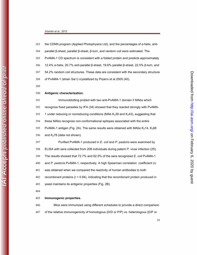
The protein was expressed as a secreted, soluble protein, and the yield (7 372
mg/L) was superior to that obtained previously in E. coli (23). The protein was purified 373
as described in the Methods section, and the final protein purity was >90% according 374
to SDS-PAGE and Coomassie blue staining, which revealed a predominant band that 375
migrated at approximately 53 kDa under reducing conditions (Fig. 1A). Using an 376
immunoblotting analysis, the protein was detected by an anti-His tag mAb, indicating 377
that the His6-tag had been preserved (Fig. 1B). 378
The homogeneity of the recombinant protein was confirmed by reverse-phase 379
chromatography on a C4 column, and a single peak was observed, as shown in 380
Figure 1C. To investigate the folding of recombinant PvAMA-1, we examined the 381
protein by far-UV CD spectroscopy (Fig. 1D). The CD data were deconvoluted using 382
on February 6, 2020 by guest
http://iai.asm.org/
Dow
nloaded from
Vicentin et al., 2013
18
the CDNN program (Applied Photophysics Ltd), and the percentages of α-helix, anti-383
parallel β-sheet, parallel β-sheet, β-turn, and random coil were estimated. The 384
PvAMA-1 CD spectrum is consistent with a folded protein and predicts approximately 385
12.4% α-helix, 20.7% anti-parallel β-sheet, 19.6% parallel β-sheet, 22.5% β-turn, and 386
54.2% random coil structures. These data are consistent with the secondary structure 387
of PvAMA-1 (strain Sal I) crystallized by Pizarro et al 2005 (40). 388
389
Antigenic characterization. 390
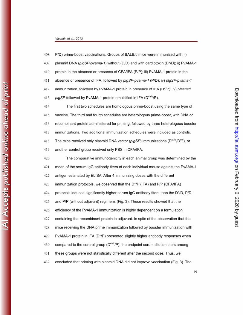
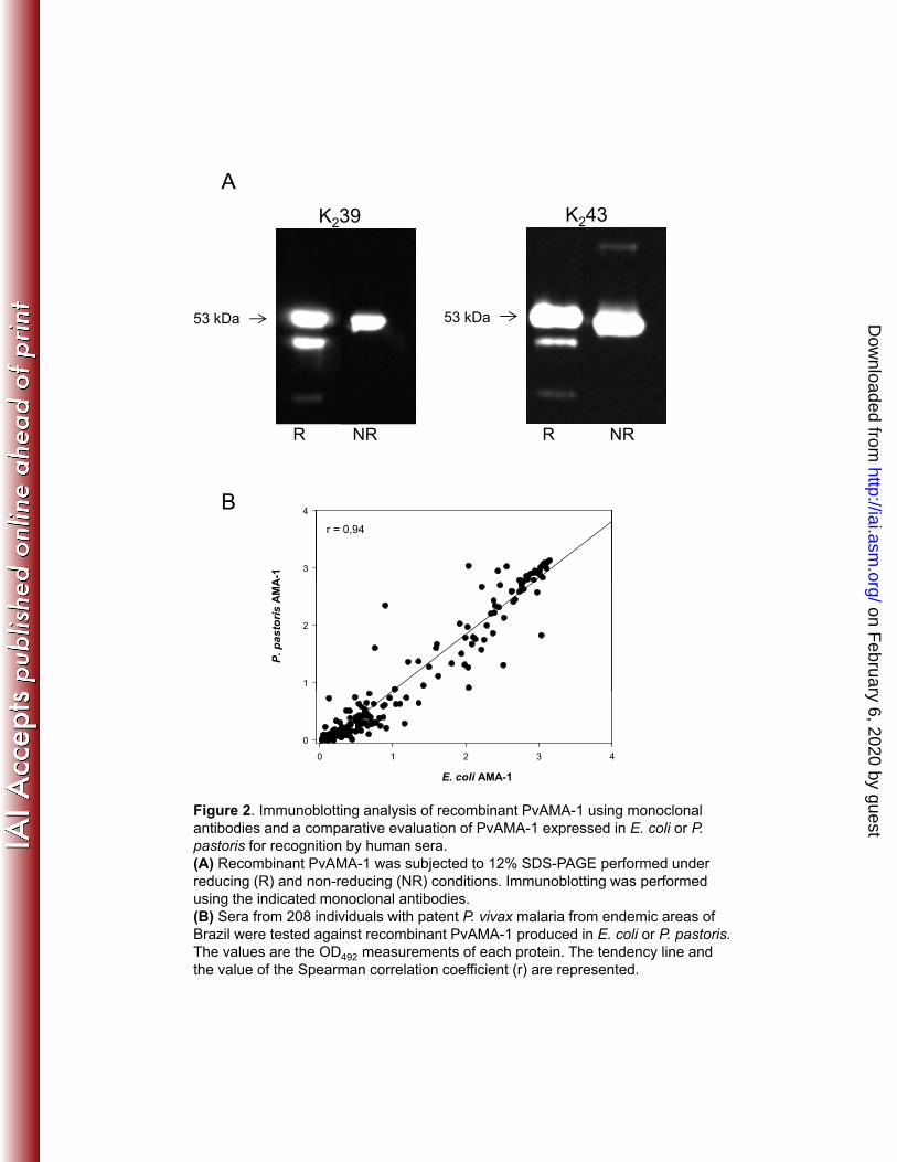
Immunoblotting probed with two anti-PvAMA-1 domain II MAbs which 391
recognize fixed parasites by IFA (34) showed that they reacted strongly with PvAMA-392
1 under reducing or nonreducing conditions (MAb K239 and K243), suggesting that 393
these MAbs recognize non-conformational epitopes associated with the entire 394
PvAMA-1 antigen (Fig. 2A). The same results were obtained with MAbs K214, K268 395
and K278 (data not shown). 396
Purified PvAMA-1 produced in E. coli and P. pastoris were examined by 397
ELISA with sera collected from 208 individuals during patent P. vivax infection (25). 398
The results showed that 72.7% and 62.5% of the sera recognized E. coli PvAMA-1 399
and P. pastoris PvAMA-1, respectively. A high Spearman correlation coefficient (r) 400
was obtained when we compared the reactivity of human antibodies to both 401
recombinant proteins (r = 0.94), indicating that the recombinant protein produced in 402
yeast maintains its antigenic properties (Fig. 2B). 403
404
Immunogenic properties. 405
Mice were immunized using different schedules to provide a direct comparison 406
of the relative immunogenicity of homologous (D/D or P/P) vs. heterologous (D/P or 407
on February 6, 2020 by guest
http://iai.asm.org/
Dow
nloaded from
Vicentin et al., 2013
19
P/D) prime-boost vaccinations. Groups of BALB/c mice were immunized with: i) 408
plasmid DNA (plgSP-pvama-1) without (D/D) and with cardiotoxin (D*/D); ii) PvAMA-1 409
protein in the absence or presence of CFA/IFA (P/P); iii) PvAMA-1 protein in the 410
absence or presence of IFA, followed by plgSP-pvama-1 (P/D); iv) plgSP-pvama-1 411
immunization, followed by PvAMA-1 protein in presence of IFA (D*/P); v) plasmid 412
plgSP followed by PvAMA-1 protein emulsified in IFA (Dctrl*/P). 413
The first two schedules are homologous prime-boost using the same type of 414
vaccine. The third and fourth schedules are heterologous prime-boost, with DNA or 415
recombinant protein administered for priming, followed by three heterologous booster 416
immunizations. Two additional immunization schedules were included as controls. 417
The mice received only plasmid DNA vector (plgSP) immunizations (Dctrl*/Dctrl), or 418
another control group received only PBS in CFA/IFA. 419
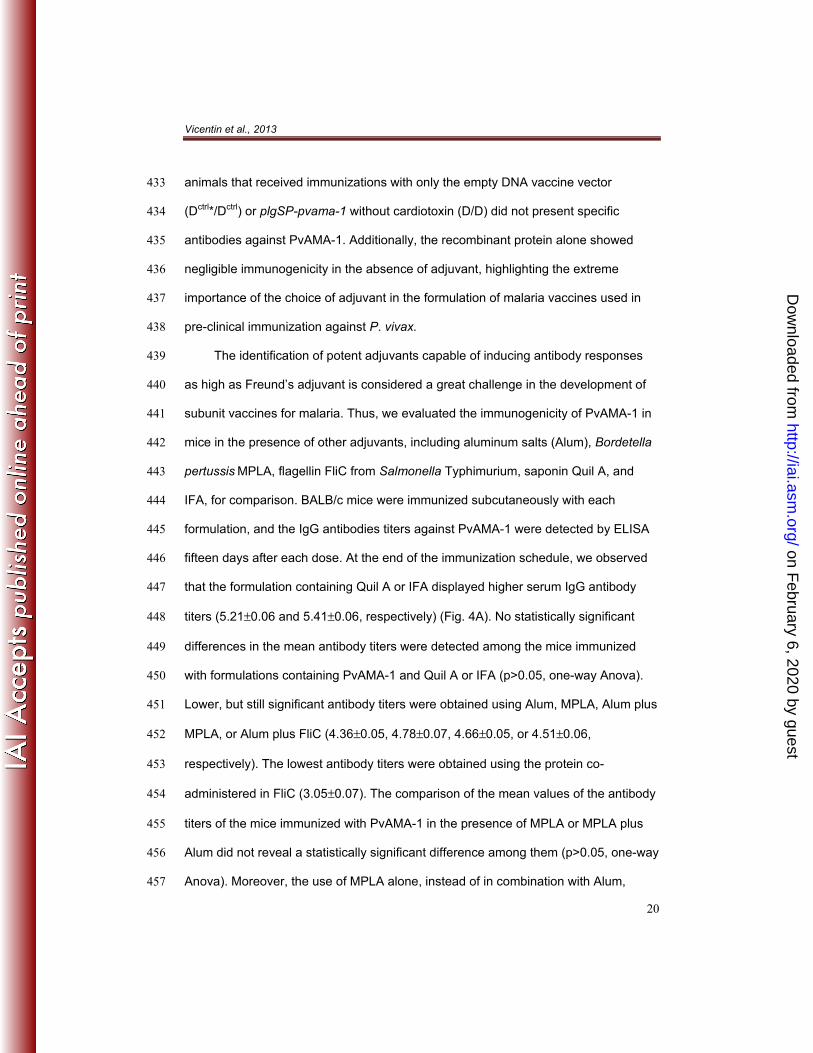
The comparative immunogenicity in each animal group was determined by the 420
mean of the serum IgG antibody titers of each individual mouse against the PvAMA-1 421
antigen estimated by ELISA. After 4 immunizing doses with the different 422
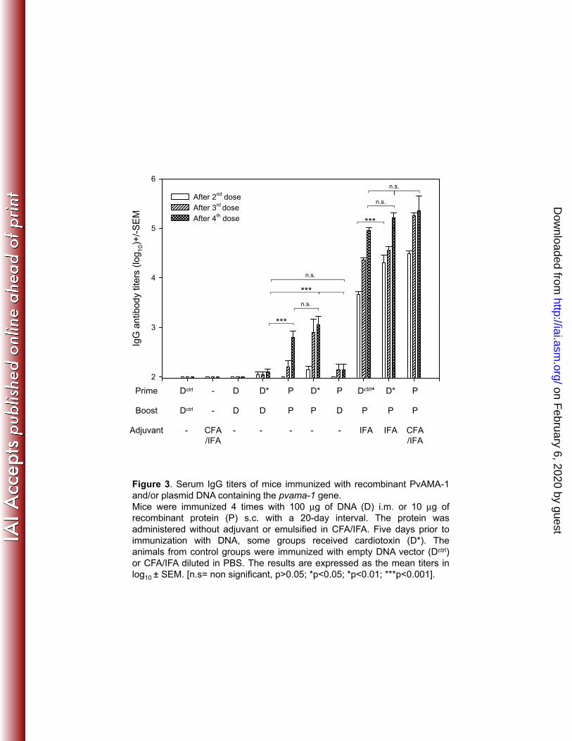
immunization protocols, we observed that the D*/P (IFA) and P/P (CFA/IFA) 423
protocols induced significantly higher serum IgG antibody titers than the D*/D, P/D, 424
and P/P (without adjuvant) regimens (Fig. 3). These results showed that the 425
efficiency of the PvAMA-1 immunization is highly dependent on a formulation 426
containing the recombinant protein in adjuvant. In spite of the observation that the 427
mice receiving the DNA prime immunization followed by booster immunization with 428
PvAMA-1 protein in IFA (D*/P) presented slightly higher antibody responses when 429
compared to the control group (Dctrl*/P), the endpoint serum dilution titers among 430
these groups were not statistically different after the second dose. Thus, we 431
concluded that priming with plasmid DNA did not improve vaccination (Fig. 3). The 432
on February 6, 2020 by guest
http://iai.asm.org/
Dow
nloaded from
Vicentin et al., 2013
20
animals that received immunizations with only the empty DNA vaccine vector 433
(Dctrl*/Dctrl) or plgSP-pvama-1 without cardiotoxin (D/D) did not present specific 434
antibodies against PvAMA-1. Additionally, the recombinant protein alone showed 435
negligible immunogenicity in the absence of adjuvant, highlighting the extreme 436
importance of the choice of adjuvant in the formulation of malaria vaccines used in 437
pre-clinical immunization against P. vivax. 438
The identification of potent adjuvants capable of inducing antibody responses 439
as high as Freund’s adjuvant is considered a great challenge in the development of 440
subunit vaccines for malaria. Thus, we evaluated the immunogenicity of PvAMA-1 in 441
mice in the presence of other adjuvants, including aluminum salts (Alum), Bordetella 442
pertussis MPLA, flagellin FliC from Salmonella Typhimurium, saponin Quil A, and 443
IFA, for comparison. BALB/c mice were immunized subcutaneously with each 444
formulation, and the IgG antibodies titers against PvAMA-1 were detected by ELISA 445
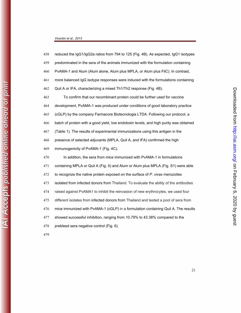
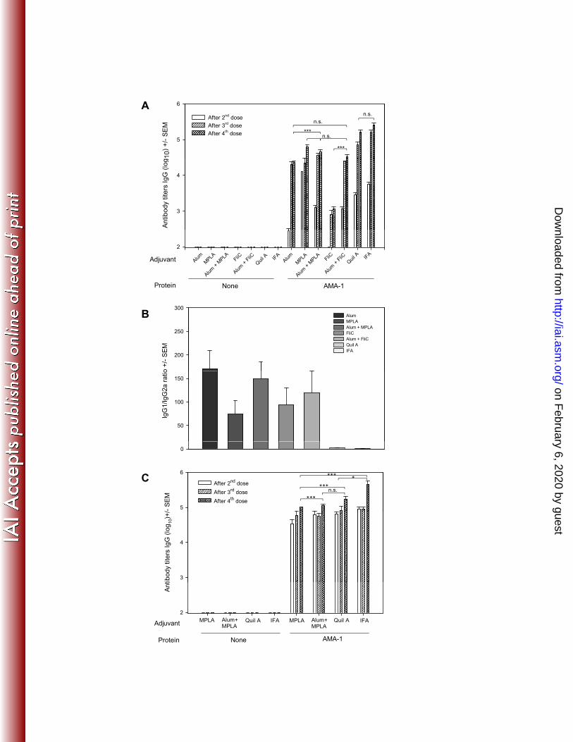
fifteen days after each dose. At the end of the immunization schedule, we observed 446
that the formulation containing Quil A or IFA displayed higher serum IgG antibody 447
titers (5.21±0.06 and 5.41±0.06, respectively) (Fig. 4A). No statistically significant 448
differences in the mean antibody titers were detected among the mice immunized 449
with formulations containing PvAMA-1 and Quil A or IFA (p>0.05, one-way Anova). 450
Lower, but still significant antibody titers were obtained using Alum, MPLA, Alum plus 451
MPLA, or Alum plus FliC (4.36±0.05, 4.78±0.07, 4.66±0.05, or 4.51±0.06, 452
respectively). The lowest antibody titers were obtained using the protein co-453
administered in FliC (3.05±0.07). The comparison of the mean values of the antibody 454
titers of the mice immunized with PvAMA-1 in the presence of MPLA or MPLA plus 455
Alum did not reveal a statistically significant difference among them (p>0.05, one-way 456
Anova). Moreover, the use of MPLA alone, instead of in combination with Alum, 457
on February 6, 2020 by guest
http://iai.asm.org/
Dow
nloaded from
Vicentin et al., 2013
21
reduced the IgG1/IgG2a ratios from 794 to 125 (Fig. 4B). As expected, IgG1 isotypes 458
predominated in the sera of the animals immunized with the formulation containing 459
PvAMA-1 and Alum (Alum alone, Alum plus MPLA, or Alum plus FliC). In contrast, 460
more balanced IgG isotype responses were induced with the formulations containing 461
Quil A or IFA, characterizing a mixed Th1/Th2 response (Fig. 4B). 462
To confirm that our recombinant protein could be further used for vaccine 463
development, PvAMA-1 was produced under conditions of good laboratory practice 464
(cGLP) by the company Farmacore Biotecnologia LTDA. Following our protocol, a 465
batch of protein with a good yield, low endotoxin levels, and high purity was obtained 466
(Table 1). The results of experimental immunizations using this antigen in the 467
presence of selected adjuvants (MPLA, Quil A, and IFA) confirmed the high 468
immunogenicity of PvAMA-1 (Fig. 4C). 469
In addition, the sera from mice immunized with PvAMA-1 in formulations 470
containing MPLA or Quil A (Fig. 5) and Alum or Alum plus MPLA (Fig. S1) were able 471
to recognize the native protein exposed on the surface of P. vivax merozoites 472
isolated from infected donors from Thailand. To evaluate the ability of the antibodies 473
raised against PvAMA1 to inhibit the reinvasion of new erythrocytes, we used four 474
different isolates from infected donors from Thailand and tested a pool of sera from 475
mice immunized with PvAMA-1 (cGLP) in a formulation containing Quil A. The results 476
showed successful inhibition, ranging from 10.79% to 43.38% compared to the 477
prebleed sera negative control (Fig. 6). 478
479
on February 6, 2020 by guest
http://iai.asm.org/
Dow
nloaded from
Vicentin et al., 2013
22
DISCUSSION 480 481
In previous studies, we showed that recombinant proteins expressed in E. coli 482
based on the P. vivax AMA-1 sequence were recognized by IgG antibodies of a large 483
fraction of malaria-infected individuals (23–26). We also described that these 484
recombinant proteins can induce high antibody titers in mice following a homologous 485
(protein/protein) or heterologous (protein/adenovirus) regimen of vaccination (27). 486
Unfortunately, when produced in E. coli, the ectodomain of PvAMA-1 is insoluble and 487
requires denaturation and refolding for purification (23, 26). Thus, to attempt to solve 488
this problem, the present study was designed to express a soluble form of the 489
ectodomain of PvAMA-1 as a secreted polypeptide in the yeast P. pastoris. 490
Using this system, we successfully generated a soluble antigenic protein under 491
cGLP conditions that exhibited a high degree of purity and low endotoxin and 492
microbial contents. The yeast-derived PvAMA1 protein retained its antigenicity, as it 493
was recognized by the IgG antibodies from 62.5% of individuals infected with P. 494
vivax. The recognition was comparable to the E. coli-derived PvAMA1 protein 495
recombinant protein (r =0.94), indicating the presence of epitopes in both proteins 496
shared with native PvAMA-1. 497
The evaluation of immunogenicity in mice showed that IFA and Quil A induced 498
higher IgG titers and a more balanced Th1/Th2 response. Relevant for vaccine 499
development was the observation that lower but also significant titers were obtained 500
in the presence of the adjuvants licensed for human use: Alum, MPLA, or the 501
combination of Alum plus MPLA. Formulations with the adjuvants Alum or Alum plus 502
MPLA predominantly induced a Th2 response, and the use of MPLA alone balanced 503
this response. Formulations containing MPLA can be used for human vaccination 504
trials, and the MPLA we used has recently been developed and investigated in 505
on February 6, 2020 by guest
http://iai.asm.org/
Dow
nloaded from
Vicentin et al., 2013
23
human trials as an adjuvant for an influenza vaccine (41). Formulations containing 506
either MPLA, Alum and squalene, or MPLA plus Alum have been proven to be safe 507
and immunogenic for humans (41). 508
Although high antibody titers are important, protective immunity against 509
infection will only be achieved if these antibodies recognize the native protein and 510
inhibit parasite invasion of reticulocytes. The immunofluorescence results confirmed 511
the recognition of native protein from P. vivax isolates from Thailand by the sera of 512
immunized mice. Because our protein was based on an Amazonian isolate of P. 513
vivax, these results reflects the presence of cross-reactive epitopes. 514
It has long been known that antibodies against Plasmodium sp. AMA-1 have a 515
strong invasion-inhibitory activity (3). However, one of the greatest limitations for P. 516
vivax vaccine development has been the lack of a functional in vitro assay to 517
routinely assess the invasion-inhibitory activity of the antibodies. To overcome this 518
limitation, we took advantage of a recently described ex vivo reinvasion assay (38) to 519
test whether the sera of mice immunized with PvAMA-1 presented invasion-inhibitory 520
activity. We found that the antibodies obtained from mice immunized with PvAMA-1 521
in the presence of Quil A inhibited the reticulocyte invasion of four different isolates 522
from Thailand, results that were for the first time obtained with P. vivax. Our results of 523
parasite inhibition are compatible with our own previous studies using immune IgG 524
against region II of the Duffy Binding Protein (38). Essentially, these findings confirm 525
and extend the previous studies in other species of Plasmodium, providing further 526
support for the implementation of PvAMA-1 as a vaccine candidate against P. vivax 527
malaria. 528
on February 6, 2020 by guest
http://iai.asm.org/
Dow
nloaded from
Vicentin et al., 2013
24
However, the main problem of using AMA-1 as a malaria vaccine component is 529
the known allelic polymorphism, which may generate allele-specific invasion-530
inhibitory antibodies. In fact, as mentioned in the Introduction section, the results 531
obtained in a phase II trial using the vaccine FMP2.1/AS02a, a recombinant protein 532
of PfAMA-1 based on the 3D7 allele, strongly argue in favor of the interpretation of 533
strain-specific resistance against malaria infection (20). 534
Obviously, because P. vivax research has been highly neglected, to our 535
knowledge, nothing is known regarding the immunological impact of the allelic 536
differences of PvAMA-1. Indeed, it is unknown whether a similar strain-specific 537
immunity will be induced by vaccination with the recombinant protein we described 538
herein. Therefore, it would not be proper to speculate at this time. 539
In spite of the problems faced by allelic polymorphisms, the potential of AMA-1 540
as a vaccine component against P. falciparum malaria continues to attract a number 541
of important research groups in this field. These laboratories are investing different 542
approaches to overcome the problem imposed by the allelic polymorphism of PfAMA-543
1 (42–44). In addition, recent studies using parasites isolated from individuals 544
vaccinated with FMP2.1/AS02a mapped few amino acid variations as the cause of 545
strain-specific resistance (45). Such limited polymorphism raises the possibility that a 546
few recombinant proteins representing key alleles would cover the entire population 547
of P. falciparum. In the worst-case scenario, if similar problems of strain-specific 548
immunity arise due to allelic polymorphism of the PvAMA-1 protein, perhaps solutions 549
similar to those developed for P. falciparum AMA-1 can be adopted for P. vivax. 550
551
552
on February 6, 2020 by guest
http://iai.asm.org/
Dow
nloaded from
Vicentin et al., 2013
25
Acknowledgments 553
We are particularly grateful to Isabel de Fátima Correia Batista e Rafael
Marques Porto (Instituto Butantan, São Paulo, Brazil) for help us with RP-HPLC
analysis and Cristiane Gozzo (University of São Paulo, São Paulo, Brazil) for
assistance with the CD measurements. Luis Carlos de Souza Ferreira (University of
São Paulo, São Paulo, Brazil) provided the recombinant flagellin FliC. We also would
like to thank all of the patients and staff of SMRU for their contribution to this study.
554
555
556 557 558 559
560 on February 6, 2020 by guest
http://iai.asm.org/
Dow
nloaded from
Vicentin et al., 2013
26
REFERENCES 561
1. Mueller I, Galinski MR, Baird JK, Carlton JM, Kochar DK, Alonso PL, Del 562 Portillo H a. 2009. Key gaps in the knowledge of Plasmodium vivax, a 563 neglected human malaria parasite. The Lancet infectious diseases 9:555–66. 564
2. Lacerda MVG, Mourão MPG, Alexandre MAA, Siqueira AM, Magalhães 565 BML, Martinez-Espinosa FE, Filho FSS, Brasil P, Ventura AMRS, Tada MS, 566 Couto VSCD, Silva AR, Silva RSU, Alecrim MGC. 2012. Understanding the 567 clinical spectrum of complicated Plasmodium vivax malaria: a systematic 568 review on the contributions of the Brazilian literature. Malaria journal 11:12. 569
3. Remarque EJ, Faber BW, Kocken CHM, Thomas AW. 2008. Apical 570 membrane antigen 1: a malaria vaccine candidate in review. Trends in 571 parasitology 24:74–84. 572
4. Silvie O, Franetich JF, Charrin S, Mueller MS, Siau A, Bodescot M, 573 Rubinstein E, Hannoun L, Charoenvit Y, Kocken CH, Thomas AW, Van 574 Gemert GJ, Sauerwein RW, Blackman MJ, Anders RF, Pluschke G, Mazier 575 D, Gemert G Van, Curie M, Inserm U, Andre I. 2004. A role for apical 576 membrane antigen 1 during invasion of hepatocytes by Plasmodium falciparum 577 sporozoites. The Journal of biological chemistry 279:9490–6. 578
5. Bannister LH, Hopkins JM, Dluzewski AR, Margos G, Williams IT, 579 Blackman MJ, Kocken CH, Thomas AW, Mitchell GH. 2003. Plasmodium 580 falciparum apical membrane antigen 1 (PfAMA-1) is translocated within 581 micronemes along subpellicular microtubules during merozoite development. 582 Journal of cell science 116:3825–34. 583
6. Srinivasan P, Beatty WL, Diouf A, Herrera R, Ambroggio X, Moch JK, 584 Tyler JS, Narum DL, Pierce SK, Boothroyd JC, Haynes JD, Miller LH. 2011. 585 Binding of Plasmodium merozoite proteins RON2 and AMA1 triggers 586 commitment to invasion. Proceedings of the National Academy of Sciences of 587 the United States of America 108:13275–80. 588
7. Collins CR, Withers-Martinez C, Hackett F, Blackman MJ. 2009. An 589 inhibitory antibody blocks interactions between components of the malarial 590 invasion machinery. PLoS pathogens 5:e1000273. 591
8. Richard D, MacRaild CA, Riglar DT, Chan J-A, Foley M, Baum J, Ralph S 592 a, Norton RS, Cowman AF, Raymond S. 2010. Interaction between 593 Plasmodium falciparum apical membrane antigen 1 and the rhoptry neck 594 protein complex defines a key step in the erythrocyte invasion process of 595 malaria parasites. The Journal of biological chemistry 285:14815–22. 596
9. Giovannini D, Späth S, Lacroix C, Perazzi A, Bargieri D, Lagal V, Lebugle 597 C, Combe A, Thiberge S, Baldacci P, Tardieux I, Ménard R. 2011. 598
on February 6, 2020 by guest
http://iai.asm.org/
Dow
nloaded from
Vicentin et al., 2013
27
Independent roles of apical membrane antigen 1 and rhoptry neck proteins 599 during host cell invasion by apicomplexa. Cell host & microbe 10:591–602. 600
10. Kocken CH, Dubbeld MA, Van Der Wel A, Pronk JT, Waters AP, 601 Langermans JA, Thomas AW. 1999. High-level expression of Plasmodium 602 vivax apical membrane antigen 1 (AMA-1) in Pichia pastoris: strong 603 immunogenicity in Macaca mulatta immunized with P. vivax AMA-1 and 604 adjuvant SBAS2. Infection and immunity 67:43–9. 605
11. Polley SD, Conway DJ. 2001. Strong diversifying selection on domains of the 606 Plasmodium falciparum apical membrane antigen 1 gene. Genetics 158:1505–607 12. 608
12. Cortés A, Mellombo M, Masciantonio R, Murphy VJ, Reeder JC, Anders 609 RF. 2005. Allele specificity of naturally acquired antibody responses against 610 Plasmodium falciparum apical membrane antigen 1. Infection and immunity 611 73:422–30. 612
13. Chenet SM, Tapia LL, Escalante AA, Durand S, Lucas C, Bacon DJ. 2012. 613 Genetic diversity and population structure of genes encoding vaccine candidate 614 antigens of Plasmodium vivax. Malaria journal 11:68. 615
14. Thakur A, Alam MT, Bora H, Kaur P, Sharma YD. 2008. Plasmodium vivax: 616 sequence polymorphism and effect of natural selection at apical membrane 617 antigen 1 (PvAMA1) among Indian population. Gene 419:35–42. 618
15. Lopez AC, Ortiz A, Coello J, Sosa-Ochoa W, Torres REM, Banegas EI, 619 Jovel I, Fontecha GA. 2012. Genetic diversity of Plasmodium vivax and 620 Plasmodium falciparum in Honduras. Malaria journal 11:391. 621
16. Putaporntip C, Jongwutiwes S, Grynberg P, Cui L, Hughes AL. 2009. 622 Nucleotide sequence polymorphism at the apical membrane antigen-1 locus 623 reveals population history of Plasmodium vivax in Thailand. Infection, genetics 624 and evolution 2009:1295–1300. 625
17. Zakeri S, Sadeghi H, Mehrizi AA, Djadid ND. 2013. Population genetic 626 structure and polymorphism analysis of gene encoding apical membrane 627 antigen-1 (AMA-1) of Iranian Plasmodium vivax wild isolates. Acta tropica 628 126:269–79. 629
18. Ouattara A, Mu J, Takala-Harrison S, Saye R, Sagara I, Dicko A, Niangaly 630 A, Duan J, Ellis RD, Miller LH, Su X, Plowe C V, Doumbo OK. 2010. Lack of 631 allele-specific efficacy of a bivalent AMA1 malaria vaccine. Malaria journal 632 9:175. 633
19. Duncan CJ a, Sheehy SH, Ewer KJ, Douglas AD, Collins K a, Halstead FD, 634 Elias SC, Lillie PJ, Rausch K, Aebig J, Miura K, Edwards NJ, Poulton ID, 635 Hunt-Cooke A, Porter DW, Thompson FM, Rowland R, Draper SJ, Gilbert 636 SC, Fay MP, Long CA, Zhu D, Wu Y, Martin LB, Anderson CF, Lawrie AM, 637 Hill AVS, Ellis RD. 2011. Impact on malaria parasite multiplication rates in 638
on February 6, 2020 by guest
http://iai.asm.org/
Dow
nloaded from
Vicentin et al., 2013
28
infected volunteers of the protein-in-adjuvant vaccine AMA1-639 C1/Alhydrogel+CPG 7909. PloS one 6:e22271. 640
20. Thera MA, Doumbo OK, Coulibaly D, Laurens MB, Ouattara A, Kone AK, 641 Guindo AB, Traore K, Traore I, Kouriba B, Diallo D a, Diarra I, Daou M, 642 Dolo A, Tolo Y, Sissoko MS, Niangaly A, Sissoko M, Takala-Harrison S, 643 Lyke KE, Wu Y, Blackwelder WC, Godeaux O, Vekemans J, Dubois M-C, 644 Ballou WR, Cohen J, Thompson D, Dube T, Soisson L, Diggs CL, House 645 B, Lanar DE, Dutta S, Heppner DG, Plowe C V. 2011. A field trial to assess a 646 blood-stage malaria vaccine. The New England journal of medicine 365:1004–647 13. 648
21. Sheehy SH, Duncan CJA, Elias SC, Biswas S, Collins KA, O’Hara GA, 649 Halstead FD, Ewer KJ, Mahungu T, Spencer AJ, Miura K, Poulton ID, 650 Dicks MDJ, Edwards NJ, Berrie E, Moyle S, Colloca S, Cortese R, Gantlett 651 K, Long CA, Lawrie AM, Gilbert SC, Doherty T, Nicosia A, Hill AVS, Draper 652 SJ. 2012. Phase Ia clinical evaluation of the safety and immunogenicity of the 653 Plasmodium falciparum blood-stage antigen AMA1 in ChAd63 and MVA 654 vaccine vectors. PloS one 7:e31208. 655
22. Chuang I, Sedegah M, Cicatelli S, Spring M, Polhemus M, Tamminga C, 656 Patterson N, Guerrero M, Bennett JW, McGrath S, Ganeshan H, Belmonte 657 M, Farooq F, Abot E, Banania JG, Huang J, Newcomer R, Rein L, Litilit D, 658 Richie NO, Wood C, Murphy J, Sauerwein R, Hermsen CC, McCoy AJ, 659 Kamau E, Cummings J, Komisar J, Sutamihardja A, Shi M, Epstein JE, 660 Maiolatesi S, Tosh D, Limbach K, Angov E, Bergmann-Leitner E, Bruder 661 JT, Doolan DL, King CR, Carucci D, Dutta S, Soisson L, Diggs C, 662 Hollingdale MR, Ockenhouse CF, Richie TL. 2013. DNA prime/Adenovirus 663 boost malaria vaccine encoding P. falciparum CSP and AMA1 induces sterile 664 protection associated with cell-mediated immunity. PloS one 8:e55571. 665
23. Rodrigues MHC, Rodrigues KM, Oliveira TR, Cômodo AN, Rodrigues MM, 666 Kocken CHM, Thomas AW, Soares IS. 2005. Antibody response of naturally 667 infected individuals to recombinant Plasmodium vivax apical membrane 668 antigen-1. International journal for parasitology 35:185–92. 669
24. Morais CG, Soares IS, Carvalho LH, Fontes CJF, Krettli AU, Braga EM. 670 2006. Antibodies to Plasmodium vivax apical membrane antigen 1: persistence 671 and correlation with malaria transmission intensity. The American journal of 672 tropical medicine and hygiene 75:582–7. 673
25. Barbedo MB, Ricci R, Jimenez MCS, Cunha MG, Yazdani SS, Chitnis CE, 674 Rodrigues MM, Soares IS. 2007. Comparative recognition by human IgG 675 antibodies of recombinant proteins representing three asexual erythrocytic 676 stage vaccine candidates of Plasmodium vivax. Memórias do Instituto Oswaldo 677 Cruz 102:335–9. 678
26. Múfalo BC, Gentil F, Bargieri DY, Costa FTM, Rodrigues MM, Soares IS. 679 2008. Plasmodium vivax apical membrane antigen-1: comparative recognition 680
on February 6, 2020 by guest
http://iai.asm.org/
Dow
nloaded from
Vicentin et al., 2013
29
of different domains by antibodies induced during natural human infection. 681 Microbes and infection / Institut Pasteur 10:1266–73. 682
27. Bouillet LÉM, Dias MO, Dorigo NA, Moura AD, Russell B, Nosten F, Renia 683 L, Braga EM, Gazzinelli RT, Rodrigues MM, Soares IS, Bruna-Romero O. 684 2011. Long-term humoral and cellular immune responses elicited by a 685 heterologous Plasmodium vivax apical membrane antigen 1 protein 686 prime/adenovirus boost immunization protocol. Infection and immunity 687 79:3642–52. 688
28. Kocken CHM, Dubbeld MA, Van Der Wel A, Pronk JT, Waters AP, 689 Langermans JAM, Thomas AW. 1999. High-Level Expression of Plasmodium 690 vivax Apical Membrane Antigen 1 (AMA-1) in Pichia pastoris: Strong 691 Immunogenicity in Macaca mulatta Immunized with P. vivax AMA-1 and 692 Adjuvant SBAS2. Infection and Immunity 67:43–49. 693
29. Soares IS, Rodrigues MM. 2002. Immunogenic properties of the Plasmodium 694 vivax vaccine candidate MSP1(19) expressed as a secreted non-glycosylated 695 polypeptide from Pichia pastoris. Parasitology 124:237–246. 696
30. Ramos CHI. 2004. A spectroscopic-based laboratory experiment for protein 697 conformational studies*. Biochemistry and molecular biology education�: a 698 bimonthly publication of the International Union of Biochemistry and Molecular 699 Biology 32:31–4. 700
31. Boscardin SB, Kinoshita SS, Fujimura AE, Rodrigues MM. 2003. 701 Immunization with cDNA expressed by amastigotes of Trypanosoma cruzi 702 elicits protective immune response against experimental infection. Infection and 703 Immunity 71:2744–2757. 704
32. Costa F, Franchin G, Pereira-Chioccola VL, Ribeirão M, Schenkman S, 705 Rodrigues MM. 1998. Immunization with a plasmid DNA containing the gene 706 of trans-sialidase reduces Trypanosoma cruzi infection in mice. Vaccine 707 16:768–774. 708
33. Quintilio W, Kubrusly FS, Iourtov D, Miyaki C, Sakauchi MA, Lúcio F, Dias 709 SDC, Takata CS, Miyaji EN, Higashi HG, Leite LCC, Raw I. 2009. Bordetella 710 pertussis monophosphoryl lipid A as adjuvant for inactivated split virion 711 influenza vaccine in mice. Vaccine 27:4219–24. 712
34. Gentil F, Bargieri DY, Leite JA, Françoso KS, Patricio MBM, Espíndola 713 NM, Vaz AJ, Palatnik-de-Sousa CB, Rodrigues MM, Costa FTM, Soares IS. 714 2010. A recombinant vaccine based on domain II of Plasmodium vivax Apical 715 Membrane Antigen 1 induces high antibody titres in mice. Vaccine 28:6183–90. 716
35. Rodrigues MHC, Cunha MG, Machado RL, Ferreira OC, Rodrigues MM, 717 Soares IS. 2003. Serological detection of Plasmodium vivax malaria using 718 recombinant proteins corresponding to the 19-kDa C-terminal region of the 719 merozoite surface protein-1. Malaria journal 2:39. 720
on February 6, 2020 by guest
http://iai.asm.org/
Dow
nloaded from
Vicentin et al., 2013
30
36. Sriprawat K, Kaewpongsri S, Suwanarusk R, Leimanis ML, Lek-Uthai U, 721 Phyo AP, Snounou G, Russell B, Renia L, Nosten F. 2009. Effective and 722 cheap removal of leukocytes and platelets from Plasmodium vivax infected 723 blood. Malaria journal 8:115. 724
37. Russell B, Suwanarusk R, Malleret B, Costa FTM, Snounou G, Kevin Baird 725 J, Nosten F, Rénia L. 2012. Human ex vivo studies on asexual Plasmodium 726 vivax: the best way forward. International journal for parasitology 42:1063–70. 727
38. Russell B, Suwanarusk R, Borlon C, Costa FTM, Chu CS, Rijken MJ, 728 Sriprawat K, Warter L, Koh EGL, Malleret B, Colin Y, Bertrand O, Adams 729 JH, D’Alessandro U, Snounou G, Nosten F, Rénia L. 2011. A reliable ex vivo 730 invasion assay of human reticulocytes by Plasmodium vivax. Blood 118:e74–731 81. 732
39. Malleret B, Claser C, Ong ASM, Suwanarusk R, Sriprawat K, Howland SW, 733 Russell B, Nosten F, Rénia L. 2011. A rapid and robust tri-color flow 734 cytometry assay for monitoring malaria parasite development. Scientific reports 735 1:118. 736
40. Pizarro JC, Vulliez Le Normand B, Chesne-Seck ML, Collins CR, Withers-737 Martinez C, Hackett F, Blackman MJ, Faber BW, Remarque EJ, Kocken 738 CHM, Thomas AW, Bentley GA. 2005. Crystal structure of the malaria 739 vaccine candidate apical membrane antigen 1. Science (New York, N.Y.) 740 308:408–11. 741
41. Precioso AR, Miraglia JL, Campos LMA, Goulart AC, Timenetsky MDCST, 742 Cardoso MRA, Luna E, Mondini G, Guedes JDS, Raw I. 2011. A phase I 743 randomized, double-blind, controlled trial of 2009 influenza A (H1N1) 744 inactivated monovalent vaccines with different adjuvant systems. Vaccine 745 29:8974–81. 746
42. Drew DR, Hodder AN, Wilson DW, Foley M, Mueller I, Siba PM, Dent AE, 747 Cowman AF, Beeson JG. 2012. Defining the antigenic diversity of 748 Plasmodium falciparum apical membrane antigen 1 and the requirements for a 749 multi-allele vaccine against malaria. PloS one 7:e51023. 750
43. Faber BW, Younis S, Remarque EJ, Rodriguez Garcia R, Riasat V, 751 Walraven V, Van der Werff N, Van der Eijk M, Cavanagh DR, Holder AA, 752 Thomas AW, Kocken CHM. 2013. Diversity covering AMA1-MSP119 fusion 753 proteins as malaria vaccines. Infection and immunity 81:1479–90. 754
44. Miura K, Herrera R, Diouf A, Zhou H, Mu J, Hu Z, MacDonald NJ, Reiter K, 755 Nguyen V, Shimp RL, Singh K, Narum DL, Long CA, Miller LH. 2013. 756 Overcoming allelic specificity by immunization with five allelic forms of 757 Plasmodium falciparum apical membrane antigen 1. Infection and immunity 758 81:1491–501. 759
45. Ouattara A, Takala-Harrison S, Thera MA, Coulibaly D, Niangaly A, Saye 760 R, Tolo Y, Dutta S, Heppner DG, Soisson L, Diggs CL, Vekemans J, Cohen 761
on February 6, 2020 by guest
http://iai.asm.org/
Dow
nloaded from

Vicentin et al., 2013
31
J, Blackwelder WC, Dube T, Laurens MB, Doumbo OK, Plowe C V. 2013. 762 Molecular basis of allele-specific efficacy of a blood-stage malaria vaccine: 763 vaccine development implications. The Journal of infectious diseases 207:511–764 9. 765
766
on February 6, 2020 by guest
http://iai.asm.org/
Dow
nloaded from
Vicentin et al., 2013
32
FIGURE LEGENDS 767
Figure 1. The recombinant protein was expressed in P. pastoris with a hexa-histidine 768
tag and purified from the supernatant by affinity chromatography, followed by anion 769
exchange chromatography, as described in the Methods section. 770
(A) SDS-PAGE analysis of purified recombinant PvAMA-1 stained with Coomassie 771
blue (1 μg of protein per lane). 772
(B) Immunoblotting analysis of purified recombinant PvAMA-1 using an anti-histidine 773
tag antibody. 774
(C) RP-HPLC profile of purified recombinant PvAMA-1. Purified PvAMA-1 produced 775
under cGLP was analyzed by RP-HPLC using a C4 column, as described in the 776
Methods section. The plot represents the mean residue ellipticity (m.r.e.) of the 777
recombinant protein. 778
(D) Circular dichroism spectrum of recombinant PvAMA-1. The spectrum was 779
recorded from 200 to 260 nm using a JASCO-J720 spectropolarimeter. The plot 780
represents the mean residue ellipticity (m.r.e.) of the recombinant protein. 781
782
Figure 2. Immunoblotting analysis of recombinant PvAMA-1 using monoclonal 783
antibodies and a comparative evaluation of PvAMA-1 expressed in E. coli or P. 784
pastoris for recognition by human sera. 785
(A) Recombinant PvAMA-1 was subjected to 12% SDS-PAGE performed under 786
reducing (R) and non-reducing (NR) conditions. Immunoblotting was performed using 787
the indicated monoclonal antibodies. 788
(B) Sera from 208 individuals with patent P. vivax malaria from endemic areas of 789
Brazil were tested against recombinant PvAMA-1 produced in E. coli or P. pastoris. 790
The values are the OD492 measurements of each protein. The tendency line and the 791
value of the Spearman correlation coefficient (r) are represented. 792
793
Figure 3. Serum IgG titers of mice immunized with recombinant PvAMA-1 and/or 794
plasmid DNA containing the pvama-1 gene. 795
Mice were immunized 4 times with 100 μg of DNA (D) i.m. or 10 μg of recombinant 796
protein (P) s.c. with a 20-day interval. The protein was administered without adjuvant 797
on February 6, 2020 by guest
http://iai.asm.org/
Dow
nloaded from
Vicentin et al., 2013
33
or emulsified in CFA/IFA. Five days prior to immunization with DNA, some groups 798
received cardiotoxin (D*). The animals from control groups were immunized with 799
empty DNA vector (Dctrl) or CFA/IFA diluted in PBS. The results are expressed as the 800
mean titers in log10 ± SEM. [n.s.= non significant, p>0.05; *p<0.05; *p<0.01; 801
***p<0.001]. 802
803
Figure 4. Serum IgG titers of mice immunized with recombinant PvAMA-1 using 804
formulations containing different adjuvants. 805
(A) Groups of BALB/c mice were immunized s.c. with 10 μg of the PvAMA-1 protein 806
in the presence of different adjuvants, as described in the Methods section. These 807
adjuvants were tested alone or in combination (Alum + MPLA and Alum + FliC). The 808
results are expressed as the mean titers in log10 ± SEM. [n.s.= non significant, 809
p>0.05; *p<0.05; **p<0.01; ***p<0.001]. 810
(B) Comparative serum IgG subclass profile after mouse immunization with PvAMA-1 811
using different adjuvants. The results are expressed as the IgG1/IgG2a ratio of titer ± 812
SEM. 813
(C) Immunogenicity of the PvAMA-1 protein produced under conditions of Good 814
Laboratory Practice (cGLP). The results are expressed as the means ± SEM of 815
antibody titer (log10). [n.s.= non significant, p>0.05; *p<0.05; **p<0.01; ***p<0.001]. 816
817
Figure 5. Indirect immunofluorescence analysis using sera from BALB/c mice 818
immunized with the cGLP PvAMA-1 protein in formulations containing different 819
adjuvants. The second row of panels show a magnified region of the corresponding 820
panels above. These zoomed in images highlight the apical staining pattern of the 821
anti-PvAMA-1 + MPLA. Microscope slides containing fixed P. vivax obtained from 822
patients from Thailand were incubated with sera from mice immunized with PvAMA-1 823
in the presence of MPLA or Quil A (diluted 1:100). 824
825
Figure 6. Invasion-inhibitory activity of sera from mice immunized with PvAMA-1 826
(cGLP) in the presence of Quil A (1:100) against merozoites from four Thai isolates of 827
P. vivax (Mean Inhibition=25.15% +/- SD 15.77). A flow cytometry method using a 828
field deployable flow cytometer (BD Accuri® C6 Flow Cytometer) was used to 829
quantify the amount of invasion inhibition relative to the treatment free control (1:100 830
on February 6, 2020 by guest
http://iai.asm.org/
Dow
nloaded from
Vicentin et al., 2013
34
prebleed mouse sera) (FL1-A= Sybr Green and FL2-A = Dihydroethidium). Each 831
assay was compared to a positive control (anti DARC (2C3 region)) (Mean 832
Inhibition=85.81% +/- SD 3.72). Surrounding the central graph we provide FACs 833
scatter plots of the inhibition assays. The FACs plot on the left side of the Y-axis 834
shows a gate containing 2.42% rings (post invasion) in the untreated control (1:100 835
prebleed mouse sera) compared to 1.37% and 0.26% in the AMA-1 treatment and 836
DARC positive control, respectively. 837
838
on February 6, 2020 by guest
http://iai.asm.org/
Dow
nloaded from

A
116 kDa ⎯
B
66 kDa ⎯
45 kDa ⎯
53 kDa
35 kDa ⎯
25 kDa ⎯
CC
D
mol
e-1) 2000
e E
llip
ticity
(de
gxc
m2 xd
ecim
-6000
-4000
-2000
0
Wavelength (nm)
200 210 220 230 240 250 260
Mea
n R
esid
ue
-10000
-8000
on February 6, 2020 by guest
http://iai.asm.org/
Dow
nloaded from
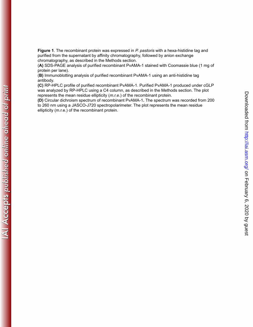
Figure 1. The recombinant protein was expressed in P. pastoris with a hexa-histidine tag and purified from the supernatant by affinity chromatography, followed by anion exchange chromatography, as described in the Methods section. (A) SDS-PAGE analysis of purified recombinant PvAMA-1 stained with Coomassie blue (1 mg of protein per lane)protein per lane). (B) Immunoblotting analysis of purified recombinant PvAMA-1 using an anti-histidine tag antibody.(C) RP-HPLC profile of purified recombinant PvAMA-1. Purified PvAMA-1 produced under cGLPwas analyzed by RP-HPLC using a C4 column, as described in the Methods section. The plot represents the mean residue ellipticity (m.r.e.) of the recombinant protein.(D) Circular dichroism spectrum of recombinant PvAMA-1. The spectrum was recorded from 200(D) Circular dichroism spectrum of recombinant PvAMA 1. The spectrum was recorded from 200 to 260 nm using a JASCO-J720 spectropolarimeter. The plot represents the mean residue ellipticity (m.r.e.) of the recombinant protein.
on February 6, 2020 by guest
http://iai.asm.org/
Dow
nloaded from
A
K243K239
53 kDa53 kDa
R NRR NR
B
3
4
A-1
r = 0,94
1
2
P. p
asto
ris A
MA
0 1 2 3 4
0
E. coli AMA-1
Figure 2. Immunoblotting analysis of recombinant PvAMA-1 using monoclonal antibodies and a comparative evaluation of PvAMA-1 expressed in E. coli or P. pastoris for recognition by human sera. (A) Recombinant PvAMA-1 was subjected to 12% SDS-PAGE performed under reducing (R) and non-reducing (NR) conditions. Immunoblotting was performed using the indicated monoclonal antibodies. (B) S f 208 i di id l ith t t P i l i f d i f(B) Sera from 208 individuals with patent P. vivax malaria from endemic areas of Brazil were tested against recombinant PvAMA-1 produced in E. coli or P. pastoris. The values are the OD492 measurements of each protein. The tendency line and the value of the Spearman correlation coefficient (r) are represented.
on February 6, 2020 by guest
http://iai.asm.org/
Dow
nloaded from
SE
M
5
6
After 2nd dose After 3rd dose After 4th dose ***
n.s.
n.s.
dy
titer
s (lo
g 10)+
/-S
4
5
***
n.s.
IgG
ant
ibod
3***
n.s.
2
Prime
Boost
Adjuvant
Dctrl
Dctrl
-
-
-
CFA/IFA
D
D
-
D*
D
-
P
P
-
D*
P
-
P
D
-
Dctrl*
P
IFA
D*
P
IFA
P
P
CFA/IFA/IFA /IFA
Figure 3. Serum IgG titers of mice immunized with recombinant PvAMA-1and/or plasmid DNA containing the pvama-1 gene.Mice were immunized 4 times with 100 μg of DNA (D) i.m. or 10 μg ofμg ( ) μgrecombinant protein (P) s.c. with a 20-day interval. The protein wasadministered without adjuvant or emulsified in CFA/IFA. Five days prior toimmunization with DNA, some groups received cardiotoxin (D*). Theanimals from control groups were immunized with empty DNA vector (Dctrl)or CFA/IFA diluted in PBS. The results are expressed as the mean titers inlog10 ± SEM. [n.s= non significant, p>0.05; *p<0.05; *p<0.01; ***p<0.001].
on February 6, 2020 by guest
http://iai.asm.org/
Dow
nloaded from
g 10)
+/-
SE
M
5
6
After 2nd dose
After 3rd dose
After 4th dose
An.s.
n.s.
***
n.s.***
Ant
ibod
y tit
ers
IgG
(lo
g
3
4
Alum
MPLA
Alum
+ M
PLA FliC
Alum
+ F
liC
Qui
l A IFA
Alum
MPLA
Alum
+ M
PLA FliC
Alum
+ F
liCQ
uil A IF
AAdjuvant
AMA-1Protein None
2
200
250
300AlumMPLAAlum + MPLAFliCAlum + FliCQuil AIFA
o +
/- S
EM
B
50
100
150
IgG
1/Ig
G2a
ra
tio
0
/- S
EM
5
6
After 2nd dose
After 3rd dose
After 4th dose
C***
n.s.***
****
tibo
dy t
iters
IgG
(lo
g10
)+/
3
4
An
t
MPLA Quil A IFA MPLA Quil A IFA
None AMA-1
Alum+MPLA
Alum+MPLA
2
Adjuvant
Protein
on February 6, 2020 by guest
http://iai.asm.org/
Dow
nloaded from
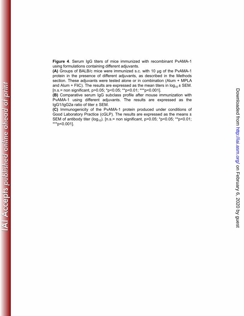
Figure 4. Serum IgG titers of mice immunized with recombinant PvAMA-1using formulations containing different adjuvants.g g j(A) Groups of BALB/c mice were immunized s.c. with 10 μg of the PvAMA-1protein in the presence of different adjuvants, as described in the Methodssection. These adjuvants were tested alone or in combination (Alum + MPLAand Alum + FliC). The results are expressed as the mean titers in log10 ± SEM.[n.s.= non significant, p>0.05; *p<0.05; **p<0.01; ***p<0.001].(B) Comparative serum IgG subclass profile after mouse immunization withPvAMA-1 using different adjuvants. The results are expressed as theIgG1/IgG2a ratio of titer ± SEM.(C) Immunogenicity of the PvAMA-1 protein produced under conditions ofGood Laboratory Practice (cGLP). The results are expressed as the means ±SEM of antibody titer (log10). [n.s.= non significant, p>0.05; *p<0.05; **p<0.01;***p<0.001].
on February 6, 2020 by guest
http://iai.asm.org/
Dow
nloaded from
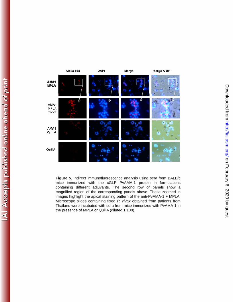
Figure 5. Indirect immunofluorescence analysis using sera from BALB/cg y gmice immunized with the cGLP PvAMA-1 protein in formulationscontaining different adjuvants. The second row of panels show amagnified region of the corresponding panels above. These zoomed inimages highlight the apical staining pattern of the anti-PvAMA-1 + MPLA.Microscope slides containing fixed P. vivax obtained from patients fromThailand were incubated with sera from mice immunized with PvAMA-1 inthe presence of MPLA or Quil A (diluted 1:100).
on February 6, 2020 by guest
http://iai.asm.org/
Dow
nloaded from
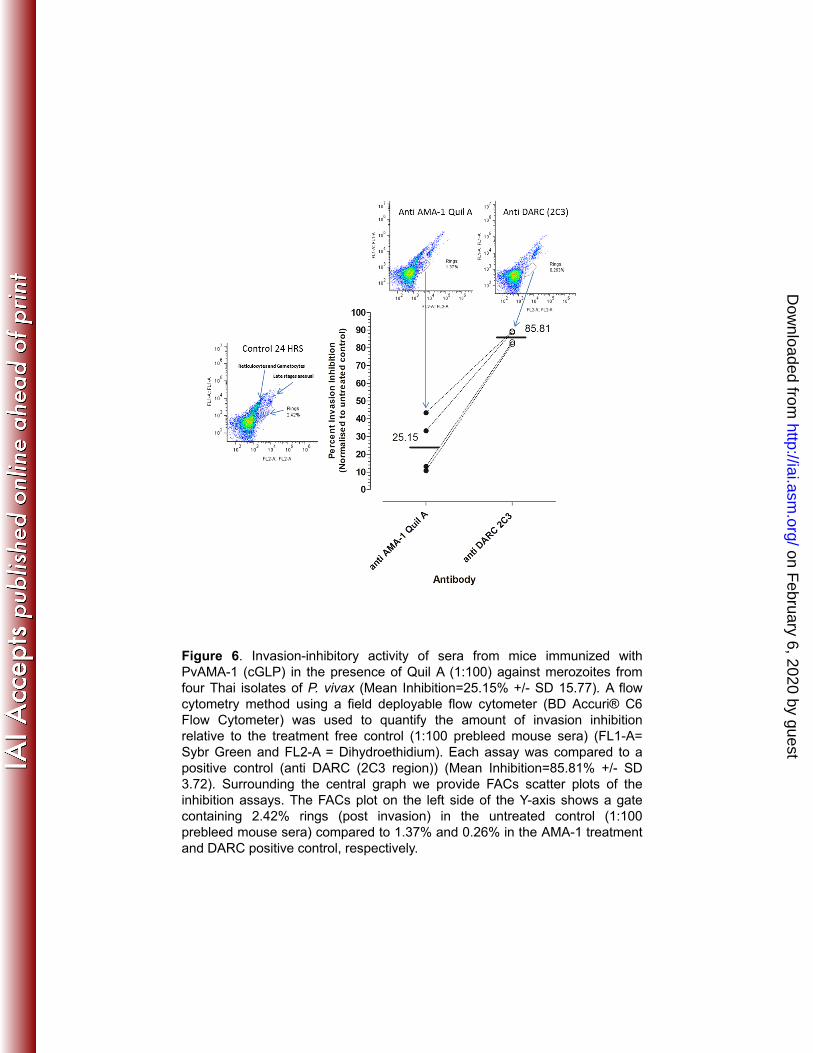
Figure 6. Invasion-inhibitory activity of sera from mice immunized withPvAMA-1 (cGLP) in the presence of Quil A (1:100) against merozoites fromfour Thai isolates of P. vivax (Mean Inhibition=25.15% +/- SD 15.77). A flowcytometry method using a field deployable flow cytometer (BD Accuri® C6Flow Cytometer) was used to quantify the amount of invasion inhibitiony ) q yrelative to the treatment free control (1:100 prebleed mouse sera) (FL1-A=Sybr Green and FL2-A = Dihydroethidium). Each assay was compared to apositive control (anti DARC (2C3 region)) (Mean Inhibition=85.81% +/- SD3.72). Surrounding the central graph we provide FACs scatter plots of theinhibition assays. The FACs plot on the left side of the Y-axis shows a gatecontaining 2.42% rings (post invasion) in the untreated control (1:100prebleed mouse sera) compared to 1.37% and 0.26% in the AMA-1 treatmentand DARC positive control, respectively.
on February 6, 2020 by guest
http://iai.asm.org/
Dow
nloaded from
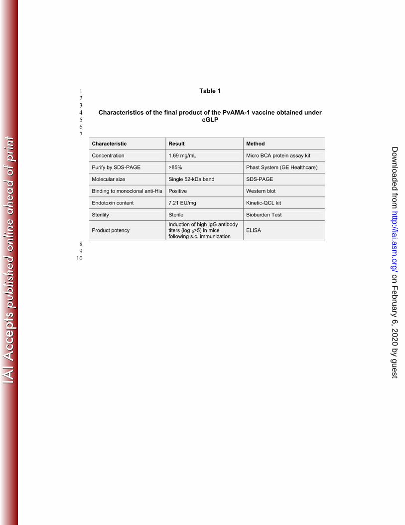
Table 1 1 2 3
Characteristics of the final product of the PvAMA-1 vaccine obtained under 4 cGLP 5
6 7 Characteristic Result Method
Concentration 1.69 mg/mL Micro BCA protein assay kit
Purify by SDS-PAGE >85% Phast System (GE Healthcare)
Molecular size Single 52-kDa band SDS-PAGE
Binding to monoclonal anti-His Positive Western blot
Endotoxin content 7.21 EU/mg Kinetic-QCL kit
Sterility Sterile Bioburden Test
Product potency Induction of high IgG antibody titers (log10>5) in mice following s.c. immunization
ELISA
8 9 10
on February 6, 2020 by guest
http://iai.asm.org/
Dow
nloaded from