Embed Size (px)
Citation preview

Physics of adherent cells
Ulrich S. Schwarz*
BioQuant and Institute for Theoretical Physics, Heidelberg University,Philosophenweg 19, 69120 Heidelberg, Germany
Samuel A. Safran†
Department of Materials and Interfaces, Weizmann Institute, Rehovot 76100, Israel
(published 27 August 2013)
One of the most unique physical features of cell adhesion to external surfaces is the active
generation of mechanical force at the cell-material interface. This includes pulling forces generated
by contractile polymer bundles and networks, and pushing forces generated by the polymerization
of polymer networks. These forces are transmitted to the substrate mainly by focal adhesions, which
are large, yet highly dynamic adhesion clusters. Tissue cells use these forces to sense the physical
properties of their environment and to communicate with each other. The effect of forces is
intricately linked to the material properties of cells and their physical environment. Here a review is
given of recent progress in our understanding of the role of forces in cell adhesion from the
viewpoint of theoretical soft matter physics and in close relation to the relevant experiments.
DOI: 10.1103/RevModPhys.85.1327 PACS numbers: 87.17.Aa, 87.17.Rt, 05.20.�y, 62.20.�x
CONTENTS
I. Introduction 1327
II. Physics Background 1329
A. Soft matter in biological systems 1329
B. Liquid crystals 1330
C. Semiflexible polymers 1331
D. Polymer gels 1332
E. Elements of elasticity 1333
F. Adhesion of vesicles and capsules 1334
III. Biology Background 1336
A. Actin cytoskeleton and cell adhesion 1336
B. Actin filaments and their assemblies 1338
C. Actomyosin contractility 1340
D. Focal adhesions 1341
IV. Physics of Cell-matrix Adhesions 1343
A. Physical motivation 1343
B. Stability of stationary adhesion clusters under
force 1344
C. Adhesion between moving surfaces 1345
D. Load localization and fracture in adhesions 1348
E. Adsorption kinetics for growing adhesions 1348
F. Force-induced growth of adhesions 1350
1. Symmetry breaking due to activation of
mechanosensors (within the adhesion) by force 1350
2. Symmetry breaking due to the geometrical
structure of the adhesion 1351
V. Cell Shape and Forces 1351
A. Physical motivation 1351
B. Contour models for cell shape 1352
C. Whole-cell models 1354
D. Force from shape 1356
VI. Active Response of Cells 1357
A. Mechanical response of force dipoles 1357
B. Force dipoles and their interactions 1358
C. Elastic model for cells on a substrate 1359
1. Cellular strain for soft substrates 1360
2. Substrate elastic energy 1361
D. Cell polarization guided by substrate rigidity 1361
E. Single cell response to rigidity gradients 1362
F. Dynamical response of cells to mechanical stress 1363
1. CSK disassembly and reassembly 1364
2. CSK stiffness changes in response to applied
stress 1364
3. CSK and cellular reorientation in response
to cyclic stretch 1365
4. Theory of cell response to applied stress 1365
5. Molecularly based models 1365
6. Coarse-grained models based on force dipoles 1366
VII. Cell Assemblies 1367
A. Matrix-mediated cell interactions 1367
B. Elastic interactions of force dipoles 1367
C. Myofibril registry modulated by substrate elasticity 1368
VIII. Conclusions and Outlook 1370
Acknowledgments 1371
References 1372
I. INTRODUCTION
Observations of swimming bacteria, crawling animal cells,
or developing organisms dramatically indicate that physical
force and movement are central to the behavior of biological
systems (Thompson, 1992; Huang and Ingber, 1999; Lecuit
and Lenne, 2007; Kollmannsberger et al., 2011). The func-
tions of cells have evolved in the context of very specific
physical environments leading to a close coupling between
cells and their surroundings (Alberts et al., 2007; Phillips,
Kondev, and Theriot, 2008). This is especially true for animal
cells, which have evolved in the controlled environment
provided by multicellular organisms and therefore appear to*[email protected]†[email protected]
REVIEWS OF MODERN PHYSICS, VOLUME 85, JULY–SEPTEMBER 2013
13270034-6861=2013=85(3)=1327(55) � 2013 American Physical Society

be more sensitive to environmental cues than, e.g., unicellular
organisms that can sometimes live in very harsh surround-
ings. Therefore it is an essential element of understanding
animal cells to consider their physical interactions with the
environment.During recent years, it has become increasingly clear that the
cell-material interface determines the behavior and fate of
biological cells to a much larger extent than was formerly
appreciated (Discher, Janmey, and Wang, 2005; Schwarz and
Bischofs, 2005; Vogel and Sheetz, 2006; Discher, Mooney, and
Zandstra, 2009; Geiger, Spatz, and Bershadsky, 2009; Janmey
et al., 2009; Mitragotri and Lahann, 2009; De, Zemel, and
Safran, 2010; Hoffman, Grashoff, and Schwartz, 2011;
Ladoux and Nicolas, 2012; Schwarz and Gardel, 2012). For
example, it has been shown that the differentiation of stem cells
can be guided by the mechanical or adhesive properties of the
substrate (Engler et al., 2006; Fu et al., 2010; Kilian et al.,
2010). Such observations can lead the way to exciting new
applications for regenerative medicine and tissue engineering,
because physical signals are easier to control and can be more
permanent than biochemical or genetic manipulations. On the
scientific side, however, the fundamentals of these processes
are puzzling and not yet well understood, despite their impor-
tance in development, health, and disease (DuFort, Paszek, and
Weaver, 2011; Janmey and Miller, 2011).From a physical point of view, the most important aspect of
the cellular response to the physical properties of the environ-
ment is the observation that cells show a controlled response
only if they are able to actively generate force and to transmit
this force to the surroundings. This finding makes sense
because cells must actively sense the passive properties of
their environment. For rigidity sensing, for example, cells
must actively strain their surroundings to probe their elastic
properties (similar to a bat that senses the geometry of its
environment by sending out ultrasound). Cells have evolved
special sensory systems for this purpose. For example, it has
been found that the size of the contacts between cells and
their environment grows with physical force (Chrzanowska-
Wodnicka and Burridge, 1996; Choquet, Felsenfeld, and
Sheetz, 1997; Balaban et al., 2001; Riveline et al., 2001;
Tan et al., 2003; Paul et al., 2008; Colombelli et al., 2009;
Trichet et al., 2012). Although this finding makes sense from
a biological viewpoint, it is at the same time puzzling to the
physicist, since in materials science force usually disrupts
adhesion contacts.Physicists have traditionally been reluctant to study living
systems due to their molecular complexity. However, this has
recently changed in many ways. An important development
that has led to physics approaches to describe cells and tissue
is the maturation of soft matter physics into an independent
and very active field of research. Soft matter physics tradi-
tionally has focused on the properties of liquid crystals,
colloidal dispersions, emulsions, fluid membranes, polymer
gels, and other complex fluids (de Gennes, 1992; Chaikin and
Lubensky, 2000; Jones, 2002; Safran, 2003). These systems
are soft since the interaction energies are of the same order as
the thermal energy. They are thus very sensitive to thermal
fluctuations and concepts from both mechanics and statistical
mechanics must be employed to understand phenomena such
as, e.g., conformational changes of membranes (Seifert,
1997; Powers, 2010) or deformations of polymer networks
(Bausch and Kroy, 2006; Daniel T. N. Chen et al., 2010).
While soft matter physics has established a firm physical
basis of the building blocks of biological cells, their behavior
critically depends on additional elements, most prominently
active remodeling controlled bygenetic and signaling networks.
Meeting the challenge of combining the physics of soft matter
physics with active processes to describe active matter enables
insight intomany biological processes, guides the design of new
types of materials, and further extends the range of phenomena
that can be analyzed by concepts and methods from physics
(Fletcher and Geissler, 2009; MacKintosh and Schmidt, 2010;
Ramaswamy, 2010; Gonzalez-Rodriguez et al., 2012; Huber
et al., 2013; Marchetti et al., 2013).To understand the physical aspects of cell adhesion, soft
matter physics provides useful model reference systems, such
as the wetting of substrates by droplets (de Gennes, 1985),
adhesion of vesicles made of fluid membranes (Seifert, 1997),
or the adhesion of capsules that comprise thin polymer shells
(Pozrikidis and Pozrikidis, 2003). The challenge is to com-
bine these reference systems with the molecularly specific
and active processes that they support at the cell-material
interface, such as force generation by polymerization
(Mogilner, 2006) or the binding and unbinding of transmem-
brane adhesion receptors (Evans and Calderwood, 2007).
Over the last decade, several soft matter systems have been
revisited with a focus on this particular point of view. The
physical understanding of the properties of active materials is
rapidly growing; particular attention has been paid to active
membranes (Manneville et al., 2001; Gov, 2004) and active
gels (Liverpool and Marchetti, 2003; Kruse et al., 2004;
Julicher et al., 2007; Marchetti et al., 2013).Cell adhesion is a multiscale problem because the molecu-
lar processes at the cell-material interface are dramatically
amplified on the scale of cells. Cellular processes such as
spreading, adhesion, migration, and proliferation are in turn
dramatically amplified on the scale of tissues (Gonzalez-
Rodriguez et al., 2012). Interestingly, similar concepts have
been successfully applied to different levels in this hierarchy.
In order to address the role of cellular forces in the context of
connective tissue, whose mechanical properties are domi-
nated by the extracellular matrix, one can build on traditional
approaches from condensed matter physics for force-
generating centers in a continuum matrix, such as the theory
of elastic defects and their interactions (Eshelby, 1957, 1959;
Siems, 1968; Wagner and Horner, 1974; Lau and Kohn, 1977;
Safran and Hamann, 1979). Motivated by experimental mea-
surements of cellular traction patterns (Dembo and Wang,
1999; Butler et al., 2002; Schwarz et al., 2002), it was
suggested that the contractile activity of cells can be modeled
as anisotropic force contraction dipoles (Schwarz et al., 2002;
Schwarz and Safran, 2002) and that cell orientation and
positioning can be predicted by minimizing the energy in-
vested by the cell into straining its environment for a given
level of force generation (Bischofs and Schwarz, 2003;
Bischofs, Safran, and Schwarz, 2004). Similar concepts
have been used to predict the contractile action of molecular
motors in the cytoskeleton (Dasanayake, Michalski, and
Carlsson, 2011; Soares e Silva et al., 2011), the orientation
response of single cells to externally applied stress
1328 Ulrich S. Schwarz and Samuel A. Safran: Physics of adherent cells
Rev. Mod. Phys., Vol. 85, No. 3, July–September 2013
(De, Zemel, and Safran, 2007), the collective response of
contractile cells in an elastic medium (Zemel, Bischofs, and
Safran, 2006), the polarization and registry of cells as a
function of external rigidity (Zemel et al., 2010b; Friedrich
and Safran, 2011, 2012), and the growth of tissue where
dividing cells correspond to force dipoles (Ranft et al.,
2010). Thus the concept of force dipoles is very general,
with applications to molecular, cellular, and tissue scales.
However, the details of these different applications strongly
depend on the biological situation of interest.For epithelial tissue dominated by direct cell-cell contacts,
other approaches are adequate, most prominently vertex
models starting from the fact that cell walls are strongly
contractile (Farhadifar et al., 2007; Hufnagel et al., 2007;
Rauzi et al., 2008; Landsberg et al., 2009; Aegerter-Wilmsen
et al., 2010; Canela-Xandri et al., 2011; Aliee et al., 2012).
Although this situation is somehow reminiscent of foams, due
to the presence of cell proliferation and death we are dealing
with an active material (Shraiman, 2005; Basan et al., 2009;
Ranft et al., 2010). This shows again that, within the over-
arching framework of active materials, different physics con-
cepts have to be used depending on the biological context.Because biological systems are very complex, meaningful
mathematical models must be selective and focus on phe-
nomena that can be treated in a tractable manner in order to
yield physical insight. The role of forces at the cell-material
interface is certainly a phenomenon which can only be fully
understood with concepts and tools from physics. For future
progress, it is essential to choose the appropriate parameters
and formulate models that are sufficiently simple to be
analyzed in detail, but predictive enough to be verified or
falsified by experiments. A theoretical analysis has many
benefits. Apart from providing deeper insight and quantitative
predictions, it usually reveals relations between quantities or
phenomena that would go unnoticed without a theoretical
model. For example, the interplay between cell adhesion and
mechanics leads to interesting predictions regarding the cou-
pling of cell shape and forces (Bar-Ziv et al., 1999;
Deshpande, McMeeking, and Evans, 2006; Bischofs et al.,
2008; Bischofs, Schmidt, and Schwarz, 2009; Guthardt
Torres, Bischofs, and Schwarz, 2012). A major focus of this
review is to point out the relations between cell shape,
structure, adhesion, and force as they emerge from our grow-
ing physical understanding of the role of physical forces at
the cell-material interface.This review is organized as follows. We start with a survey
of the relevant soft matter physics that describes and quanti-
fies those parts of cells that are involved in force transmission.
In particular, we review the properties of liquid crystals,
flexible and semiflexible chains and gels, and elements of
elasticity theory for both bulk systems such as elastic solids as
well as for finite-sized systems such as vesicles and capsules.
We then present the minimally required cell biology back-
ground, including a general discussion of the cytoskeleton
and the properties of actin polymers and networks, myosin
molecular motors that endow these networks with active
contractility, and the membrane-based adhesion structures
that connect cells to their environment. The main part of
this review then covers recent developments in the physics
of adherent cells. In the spirit of a multiscale approach, we
start on a relatively small scale with simple models for thephysics of adhesion clusters. We then progress to models for
cell shape and structure, which in turn form the basis forcoarse-grained models for entire cells as force dipoles. In
particular, we use this framework to discuss cell response tomechanical stress as well as actin network polarization and itsdependence on the elasticity of the underlying matrix. Finally
we address the physics of matrix-mediated cell assembliesfrom the viewpoint of cellular forces. We close with someconclusions and an outlook on future perspectives.
II. PHYSICS BACKGROUND
A. Soft matter in biological systems
This review on physical forces at the cell-material interface
focuses on a view of animal cells as complex, composite, softmaterials comprising fluid membranes that are coupled to two
types of elastic and often contracile polymer networks. Insidethe cell, there exists a highly cross-linked and entanglednetwork of three different types of polymers (actin filaments,
microtubules, and intermediate filaments) collectively calledthe cytoskeleton (CSK). On the outside, the cell is coupled toanother multicomponent, gel-like network (including fibrous
protein components such as collagen or fibronectin) called theextracellular matrix (ECM). If subjected to mechanical
forces, the biological material initially responds like a passiveelastic body; thus elasticity theory is an essential element ofthe physics of cells and tissues. At longer time scales, the cell
responds to mechanical perturbations by actively reorganiz-ing the structure of its CSK (and, to a certain extent, its ECMas well).
Experiments suggest that cells in solution respond elasti-
cally up to times on the order of a few seconds (Wottawahet al., 2005). The same is true for tissue on a time scale ofseconds and minutes (Gonzalez-Rodriguez et al., 2012). The
deformation of an elastic body induces both stress and strain.For example, for a simple, one-dimensional stretch of an
elastic slab, the stress � is the force per area applied to theslab on its top and bottom faces, while the strain � is theresulting relative change in length. (For a more detailed
introduction to the tensorial theory of elasticity see below.)The simplest constitutive relation that relates stress and strainis a linear one in which � ¼ E�. The elastic constant Eintroduced in this way is known as Young’s modulus and isoften called the stiffness or rigidity. The larger the Young’s
modulus, the more stress is required to stretch the material tothe same extent. Because strain � is dimensionless, theYoung’s modulus has the same physical dimensions as stress
�, that is N=m2 ¼ Pa. Physically this means that the elasticmodulus is a measure of the mechanical energy density of the
system. The corresponding spring constant k ¼ EA=L0 alsodepends on two geometrical quantities: the cross-sectionalarea A and the rest length L0 of the spring.
For much of our discussion of cell elasticity, it is essential
to note that the elastic modulus of a typical tissue cell is in therange of 10 kPa. This should be contrasted with the muchhigher values of crystal moduli of 100 GPa. With a typical
size of the supramolecular assembly of 10 nm, simple scalingpredicts that the typical energy scale for cells is in the range
Ulrich S. Schwarz and Samuel A. Safran: Physics of adherent cells 1329
Rev. Mod. Phys., Vol. 85, No. 3, July–September 2013

of 10 kPað10 nmÞ3 ¼ 10�20 J, which is close to the thermal
energy scale kBT ¼ 4:1� 10�21 J ¼ 4:1 pN nm. (Here we
used T ¼ 300 K since most of biology operates at room or
body temperature.) Although somehow simplistic, this argu-
ment nevertheless correctly indicates that the cohesive inter-
actions that stabilize cells are weak. These are mainly
electrostatic attractions between charges or charge distribu-
tions (mainly dipoles) that are screened by water and rela-
tively high salt concentration (100 mM corresponding to a
Debye screening length of about 1 nm), hydrogen bridges,
hydrophobic interactions due to the special properties of
water, and entropic forces such as depletion interactions, all
of which operate on an energy scale of a few kBT (Dill and
Bromberg, 2010; Israelachvili, 2011).The relatively weak cohesive energies are also related to
the large length scales that characterize soft matter, since it is
often the energy density (energy per unit volume) that is
relevant. For example, the large-scale structures of linear
macromolecules (polymers) in solution can be described by
disordered, bloblike structures where the typical blob size
can be hundreds of angstroms (de Gennes, 1979). Water-
amphiphile dispersions can exhibit disordered, spongelike
structures consisting of bilayer sheets of amphiphilic mole-
cules whose sizes can be 100 times the size of an individual
molecule (Schwarz and Gompper, 2002; Safran, 2003). Even
those soft materials that show solidlike elasticity, such as gels
(de Gennes, 1979; Boal and David, 2012) or colloidal crystals
(Pieranski, 1983), have mesh or lattice constants that are in
the range of hundreds to thousands of angstroms. In addition,
the overall weak nature of the interactions (e.g., gels with
dilute cross-links separated by large distances, colloidal par-
ticles with small surface charges) results in shear elastic
constants that can be many orders of magnitude weaker
than those of hard matter. The weak, noncovalent nature of
the interactions in soft matter often competes with the entropy
of the system and leads to large responses and variations in
the structures and phases as the temperature or composition is
varied. The soft matter topics that are most closely related to
cellular forces are liquid crystals, polymers and gels, elastic-
ity and the adhesion of fluid drops, amphiphilic vesicles, and
polymerized capsules.In practice, the mechanical properties of cells are described
by linear elasticity only in a limited regime, and there exists a
hierarchy of length and energy scales that determine how
cells respond to force. In fact, the viscoelastic response of
cells has been measured using various techniques in different
situations and over a large range of frequencies (Fung, 1993).
These measurements show that cells share many of the
features of the viscoelasticity of in vitro, reconstituted net-
works of biopolymers (Bausch and Kroy, 2006; Daniel T. N.
Chen et al., 2010).While synthetic soft matter systems are subject and sensi-
tive to thermal disorder, biological cells exhibit far more
noisy behavior (Pearson, 2008) due to the stochastic nature
of many of the biomolecular processes that take place; in our
context such nonthermal noise occurs mainly in the context of
force generation by molecular motors (Howard, 2001) and
actin polymerization (Mogilner, 2006). If these processes are
correlated only on molecular length and time scales, we can
regard them as active white noise with regard to modeling
cellular behavior on much longer scales. In that case, themolecular processes can be approximated as being deltacorrelated in both space and time the results of which(Haken, 1983) resemble an effective temperature that deter-mines the width of a Boltzmann-like distribution. However,this cannot be generalized to the role of an effective tempera-ture in a true thermodynamic sense (Ben-Isaac et al., 2011).In particular, the fluctuation-dissipation theorem (Chaikinand Lubensky, 2000) is not obeyed (Mizuno et al., 2007) asit is in thermal systems. With these caveats, we use theconcept of effective temperature and the resultingBoltzmann distribution of cellular energies in situations inwhich the molecular noise can be regarded as deltacorrelated.
B. Liquid crystals
We begin our review of relevant physical systems withliquid crystals, which comprise anisotropic (e.g., rodlike)molecules that can show orientational (nematic) order, butnot necessarily positional (translational) order (de Gennesand Prost, 1995). At lower temperatures, nematically orderedsystems show a type of one-dimensional (smectic) order inwhich the molecules form well-defined layers with the mo-lecular axis oriented parallel to the layer normal (smectic A).The layers themselves are fluid with no translational, in-planeorder. The relevance of liquid crystal ordering to cells lies inthe fact that, under external forces (shear flow or elasticdeformation) or in elastic environments of appropriate rigid-ity, the polymer networks inside cells can show nematic andeven smectic order; these applications are discussed later andhere we outline the relevant liquid crystal physics.
The cooperative, orientational interactions betweenrodlike molecules can give rise to phase transitions in whichthey all align in a given direction, as in Fig. 1(a) (de Gennesand Prost, 1995). Such nematic ordering transitions canarise when the temperature is lowered in systems governedby microscopic interactions that promote order.Microscopically, each molecule is characterized by its ther-mally fluctuating orientation angle �, where the z axis that
(a) (b)
(c) (d)
FIG. 1. Passive bulk soft matter examples that are important
model systems for the understanding of the material properties of
cells and tissues. (a) Liquid crystals often form nematic phases, with
no positional but orientational order. (b) In a dilute polymer
solution, each polymer forms a globule that is well separated
from the other polymers. (c) A cross-linked polymer gel can behave
like an elastic solid but with much weaker rigidity. (d) Lipids self-
assemble into fluid bilayers that at high concentration in turn tend to
self-assemble into stacks, the so-called lamellar phase.
1330 Ulrich S. Schwarz and Samuel A. Safran: Physics of adherent cells
Rev. Mod. Phys., Vol. 85, No. 3, July–September 2013

defines the angle can be defined by convention or by somemacroscopic, symmetry-breaking field such as the (nonspher-ical) shape of the system. Because the rodlike molecules haveup-down symmetry, the interaction energy cannot be an oddfunction of the angle since that changes sign when the rod isflipped. Instead, the energies must be even functions of �.
These symmetry considerations allow the definition of thelocal value of the nematic order parameter Si of a givenmolecule labeled by i as
Si ¼ 12ð3 cos�i2 � 1Þ: (1)
A simple mean-field theory for nematic ordering was formu-lated by Maier and Saupe (1959). The Hamiltonian ofthe interacting system of rods in which the energy dependson the local orientations of nearby molecules is replacedby a one-body approximation Ums in which the energy of agiven molecule is proportional to the product of its orderparameter with the thermal average of the average orderparameter of the system hSi. The mean-field nature of thisassumption lies in the fact that the orientations of neighbor-ing, interacting molecules are approximated by the averageorder parameter
Ums ¼ BhSiXi
Si; (2)
where B is a constant that characterizes the interactions. Theorder parameter is determined self-consistently from thestatistical mechanical definition of the thermal average inwhich the probability distribution is proportional to theBoltzmann factor exp½�Ums=kBT�:
hSi ¼ 1
Z
Zd�Si exp½�BhSiSi=kBT�; (3)
where � is the solid angle and the normalization Z is theaverage of the exp½�Ums=kBT� over all solid angles.
This approximation correctly predicts a first-order phasetransition at which the average order parameter hSi jumpsfrom a value of zero to a value of approximately 0.4 whenB=kBT ¼ 4:6. As the temperature is lowered, the order pa-rameter increases until it reaches its saturation value of unity.The molecules are still in the fluid state; there is no transla-tional order, but only orientational order.
C. Semiflexible polymers
Semiflexible polymers (also known as wormlike chains)(Sait, Takahashi, and Yunoki, 1967; Marko and Siggia, 1995)are long, one-dimensional chains ofN molecules (monomericunits) whose intermolecular bonds resist bending; this is incontrast to flexible chains (de Gennes, 1979), where there isno energetic penalty for bending (at scales that are compa-rable to the size of a monomer) and which are completelygoverned by entropy. Both types of polymers form globules,as in Fig. 1(b), but with different typical sizes. The physics offlexible chains are well known (de Gennes, 1979; Doi, 1996;Rubinstein and Colby, 2003) and their resistance to changesof their ‘‘size’’ (end-to-end distance or radius of gyration R)away from their ‘‘random walk’’ or Gaussian conformationwhere R� N1=2 is characterized in a mean-field treatment bya free energy per chain
f ¼ 3kBT
2
R2
Na2; (4)
where N is the number of monomers in the chain and a is themonomer size. Self-avoidance of the chain due to excludedvolume interactions among the monomers leads to additionalinteractions and in a mean-field treatment the scaling of Rwith N is modified so that R� N3=5. Many biopolymersincluding DNA and various cytoskeletal filaments such asactin and microtubules discussed later are semiflexible andbend only on length scales of 50 nm (DNA) through micro-meters (actin) or even millimeters (microtubules), while syn-thetic polymers such as polystyrene in organic solvents areflexible and easily bend on nanometric scales.
On a coarse-grained, continuum level the bending resist-ance of a semiflexible polymer is similar to that of an elasticrod (Landau and Lifshitz, 1970). Bending is geometricallycharacterized by the curvature of the position vector of the
rod ~RðsÞ which is a function of the monomer distance s alongthe contour (0 � s � L, where L is the contour length of therod). For systems where positive and negative curvatures areequivalent by symmetry, there can be no terms in the energythat are linear in curvature, so that in a small curvatureexpansion (appropriate when the radius of curvature ismuch larger than a monomer size), the energyHb is quadraticin the chain curvature (Landau and Lifshitz, 1970):
Hb ¼ �
2
Z L
0ds
�d2 ~R
ds2
�2; (5)
where � is the bending modulus that characterizes the elasticresistance to bending. The lowest energy deformations of therod are the bending modes that do not result in an overallvolume change of the rod and involve only relative extensionand compression of its upper and lower surfaces (Landau andLifshitz, 1970). For most purposes one assumes that the rod isinextensible and neglects any stretching of the center of massdistances between molecules. This is expressed by the inex-tensibility constraint that leaves the rod length unchanged:
L ¼Z L
0ds
��������d~R
ds
�������� (6)
and is equivalent to the requirement that the tangent vector
given by d ~R=ds is a unit vector.In equilibrium, a semiflexible polymer represented by such
a rod undergoes thermally driven motion that is resisted bythe bending energy. The inextensibility constraint makes thisproblem difficult to treat exactly (Sait, Takahashi, andYunoki, 1967; Marko and Siggia, 1995; Rubinstein andColby, 2003). For small deformations of a chain oriented inthe z direction, one can approximate s � z and describe the
chain position by ~R ¼ ðXðzÞ; YðzÞ; zÞ. The deformations canbe resolved into their Fourier components XðqÞ ¼RdzXðzÞeiqz that are the normal modes which diagonalize
the bending Hamiltonian. Using the equipartition theoremone finds that hjXðqÞj2i ¼ kBT=�q
4 and one can show that the
tangent vectors t ¼ d ~R=dz of neighboring points are nearlyequal with
h½tðzÞ � tð0Þ�2i � kBTZ
dq1� cosðqzÞ
�q2� kBT
�z: (7)
Ulrich S. Schwarz and Samuel A. Safran: Physics of adherent cells 1331
Rev. Mod. Phys., Vol. 85, No. 3, July–September 2013
The tangent correlations diverge as the distance between thepoints along the rod increases and for large z this invalidatesthe approximation of small fluctuations and inextensibility(equivalent to a unit tangent vector). However, one can findthe value of z at which the tangent correlations first become oforder unity; this defines the persistence length (Rubinsteinand Colby, 2003; Phillips, Kondev, and Theriot, 2008) � ofthe chain and one finds � � �=kBT. At scales smaller than thepersistence length, the chain shows rigid-rodlike behaviorwith relatively small bending fluctuations; at longer scales,the fluctuations are large and a random walk (or excludedvolume random walk) picture is more appropriate.
D. Polymer gels
While single cytoskeletal proteins such as actin filamentsor microtubules can be modeled as semiflexible polymers, theCSK often contains cross-linked assemblies (gels) compris-ing these proteins, as in Fig. 1(c). The assemblies can benetworklike (macroscopically isotropic) or ordered intobundlelike filaments. Here we review the response of semi-flexible polymers to applied, static forces that stretch thechains and determine the regimes in which the chains respondlinearly or nonlinearly to applied force.
For simplicity, we focus on a chain whose projected lengthis less than or of the order of its persistence length. In thecontext of a cross-linked gel, the projected length is deter-mined by the distance between the cross-links, assumingpermanent cross-links at whose positions the polymer isrigidly held fixed. The dissociation of the cross-links disruptsthe network and can lead to nonelastic (e.g., viscous flow)response to stress; however, we focus on the early-time (tensof seconds and possibly more in strongly adherent cells)behavior where the network response to force is elastic innature (Wottawah et al., 2005). We consider the elasticresponse of a single, semiflexible polymer. Naively, onemight think that this response will be typical of a polymersegment in the gel whose projected length is the averagespacing between cross-links; the distribution of cross-linksin the gel implies a distribution of polymer segment lengthsbetween cross-links. This would indeed be true for affinedeformations in which each chain is stretched in the sameproportions as the macroscopically applied stress or strain.However, for large deformations, where the elastic responseis highly nonlinear, the distribution of stresses among thechains with varying segment lengths can be length dependent;the stresses will not be affine and it is harder to associate thegel with the response of one chain of average segment length(Head, Levine, and MacKintosh, 2003a, 2003b; Wilhelm andFrey, 2003; Heussinger and Frey, 2006; Heussinger, Schaefer,and Frey, 2007).
Before treating the case of semiflexible polymers, webriefly derive the elastic modulus that characterizes theresponse of flexible polymers to applied forces (so-calledrubber elasticity). The modulus is completely determinedby the changes in the chain entropy that are due to theapplied strain �i ¼ �i � 1 (i ¼ x, y, z) that changes themacroscopic dimensions of the sample from ðLx; Ly; LzÞ toð�xLx; �yLy; �zLzÞ. Incompressibility of the chains and sol-
vent implies that the volume must remain unchanged, so that
the product �x�y�z ¼ 1. The free energy per chain in the
unstressed system is given by Eq. (5) and for affine strains
where ~R ¼ ðX; Y; ZÞ ! ð�xX; �yY; �zZÞ, the free energy per
chain becomes
f ¼ kBT
2ð�2
x þ �2y þ �2
z � 3Þ: (8)
We consider a uniaxial deformation in the x direction, �x ¼ �
and by incompressibility �y ¼ �z ¼ 1=ffiffiffiffi�
p. The force applied
to a single chain is @f=@Lx and the stress in the entire systemof chains � is the total force applied per unit area � ¼�kBTð�2 � 1=�Þ, where � is the number of chain segmentsper unit volume. For small deformations � � 1, an expansionof the expression for � shows that the stress is proportional tothe product of the strain and �kBT, similar to the pressure ofan ideal gas. For large strains, the stress is nonlinearly relatedto the strain but this arises from the incompressibility condi-tion and not from any specific properties of the chains.Fluctuations of the cross-links further reduce the strain(Rubinstein and Colby, 2003).
Semiflexible chains have a more complex response toapplied forces and one can use the model described aboveto predict their stress-dependent elastic modulus. When semi-flexible chains are stretched near their limit, the additionalforce to stretch them further tends to diverge and this resultsin an elastic modulus that is intrinsically stress dependent.One considers a Hamiltonian that includes the bending en-ergy as well as an energy that tends to equalize the projected
length Lp and contour length L¼RLp
0 dzffiffiffiffiffiffiffiffiffiffiffiffiffiffiffiffiffiffiffiffiffiffiffiffiffiffiffiffiffiffiffiffiffiffiffiffiffiffi1þX0ðzÞ2þY0ðzÞ2p
.
This arises from a tension (energy per unit length) � thatcouples to the difference L� Lp. In the approximation that
the fluctuations are small, one can expand the square root toobtain
H�¼�
2
Z L
0dz½X00ðzÞ2þY00ðzÞ2�2þ�
2
Z L
0dz½X0ðzÞ2þY0ðzÞ2�2:
(9)
Using equipartition of the Fourier modes of the chain fluctu-ations (Landau and Lifshitz, 1970; Safran, 2003) one cancalculate (Mackintosh, 2006) ‘, which is the increase in thechain extension compared to its zero-tension, fluctuatingvalue:
‘ ¼ L2
6�
�1þ 3
2�� 3 cothð ffiffiffiffi
�p Þ
ffiffiffiffi�
p�; (10)
where � ¼ �L2=�2 is a dimensionless measure of theapplied force and � is the persistence length defined above.
For small forces ‘� �L4=��; the excess strain ‘=L isproportional to the force and the system is harmonic. Forlarge forces, ‘ approaches the value for full extension of‘0 ¼ L2=ð6�Þ and the difference ‘0 � ‘� 1=
ffiffiffi�
p. This
nonlinear relationship between extension and applied forceexpresses the fact that, as the chain approaches its maximumextension, a very large force must be applied. The measuredelastic constant of the cross-linked, semiflexible polymer gelis then stress dependent as discussed in the context of actingels.
1332 Ulrich S. Schwarz and Samuel A. Safran: Physics of adherent cells
Rev. Mod. Phys., Vol. 85, No. 3, July–September 2013

E. Elements of elasticity
In the previous discussion, we employed scalar definitionsof the stress and strain developed in an elastic system that issubject to applied forces. While liquids and gases also resistcompression, they do not show an elastic response to externalforces that act only to change the shape of the system; suchforces (per unit area) that do not induce any volume changeare called shear stresses. The elastic response (restoringforce) to shear stresses is characteristic of solids. Cross-linkedgels, while being disordered, are indeed classified as solidssince they resist shape changes and can be described byelasticity theory. This is true when the cross-links are perma-nent, or very long lived; otherwise, one must deal with time-(or frequency-)dependent elastic constants (Fung, 1993; Boaland David, 2012). Biological gels are typically not perma-nently cross-linked (Lieleg et al., 2008) and at long times oneexpects liquidlike flow instead of an elastic response to shearforces. This indeed is the time regime in which the CSK ismodeled (Liverpool and Marchetti, 2003; Kruse et al., 2004;Julicher et al., 2007; Marchetti et al., 2013) as an active gelthat flows in response to internal forces generated by cellactivity which is fueled by energy consumption. Here werestrict our focus to the early-time (tens of seconds) behaviorof the CSK (Kollmannsberger and Fabry, 2011), where thecross-linkers still maintain the elastic response of the CSK toboth internal and external forces. The elastic approach is alsomore appropriate for the ECM which remodels much lessthan the CSK.
In the presence of external or internal forces that are notpart of the elastic network themselves (including thermalforces that change the positions of the particles), and in acontinuum picture, the material particles that comprise anelastic system are assumed to be displaced from their equi-librium positions by a smooth displacement field ~uð~rÞ where~r ¼ ðr1; r2; r3Þ ¼ ðx; y; zÞ. The elastic energy arises from in-terparticle interactions and is thus a function not of ~uð~rÞ, butof its spatial gradients that represent changes in the relativepositions of the particles. This is true in the absence of anyexternal ‘‘pinning’’ forces for which translations of the sys-tem [where ~uð~rÞ is constant] have no energy cost. The elasticenergy is thus a function of the strain tensor uij defined
(Landau and Lifshitz, 1970) as
uij ¼ 1
2
�@uið~rÞ@rj
þ @ujð~rÞ@ri
þ @ulð~rÞ@ri
@ulð~rÞ@rj
�; (11)
where summation over the repeated index l is implied. Thenonlinear term on the right can be neglected for small strains.The local change in a small length element dx is dxð1þ uxxÞso that the local volume change (given by the product dxdydzminus the initial volume) is determined to first order in thestrain by trðuijÞ ¼ uii ¼ uxx þ uyy þ uzz. These are coupled
to isotropic compressions or expansions while shear forcesthat change the shape of the system couple to the off-diagonalstrain components such as @ux=@y that represent changes inthe interparticle spacing in the x direction that vary in the ydirection.
Displacing the particles from their equilibrium positionscreates strains that are resisted by internal restoring forcesthat originate in the intermolecular interactions (and in the
case of polymeric gels, entropy) that provide shape memoryand hence elasticity. The forces that arise from the elasticityare described by a stress tensor �ijð~rÞ. This is the force per
unit area in the i direction that acts on the surfaces whosenormal is in the j direction of an infinitesimal volume ele-ment. The pressure is the negative of one-third of the trace ofthe stress. In the absence of motion, the difference of thestresses on two surfaces separated by a distance d~r is attrib-
uted to the presence of a local force density ~fð~rÞ within thatvolume element so that (Landau and Lifshitz, 1970), inequilibrium, fið~rÞ ¼ �Pj@�ij=@rj. It is important to note
that the force per unit volume fi is attributed to forces that arenot included in the system’s elastic response and arise eitherfrom active internal elements or from macroscopic forces thatact on the system boundaries. In the absence of such forces,mechanical equilibrium thus dictates that the divergence ofthe stress tensor vanishes.
For an isotropic body, rotational symmetry implies thatthere are two tensor components that must be considered forthe strain and stress: (i) the trace that describes the localvolume change u0ð~rÞ ¼ uijð~rÞij or the hydrostatic pressure
��0ð~rÞ=3 ¼ ��ijð~rÞij=3 (where one sums over the re-
peated index), and (ii) the traceless shear, defined as usijð~rÞ ¼uijð~rÞ � ð1=3Þu0ð~rÞij with a similar expression for the shear
stress. Since the internal forces that resist deformations canalso include thermal effects at the intramolecular level (suchas changes in the conformations of polymers in gel networks),one considers the elastic free energy per unit volume (Landauand Lifshitz, 1970) fe. The free energy associated with elasticdeformations is a scalar and can be written from the followingsymmetry considerations. (i) The free energy depends only onthe strains and not on the displacements. (ii) There is no termlinear in strain since the deformation free energy representsan expansion about equilibrium where the free energy isminimal. (iii) The free energy is a scalar and cannot dependon the coordinate system. Since u0iju
sij ¼ 0, the free energy
written up to quadratic order in the strains can contain onlyterms with ðu0ijÞ2 and usiju
sij:
fe ¼ K
2
�Xi
uii
�2 þ�
Xij
�uij � 1
3ij
Xl
ull
�2; (12)
where uij denotes the local strain uijð~rÞ. The first term
accounts for the free energy associated with volumechanges and is proportional to the bulk modulus K, whilethe second term accounts for the shear response, proportionalto the shear modulus �. These two elastic constants thathave the dimensions of energy per unit volume (the sameas pressure, measured in Pa) are material dependentand can also be expressed (in three dimensions) by theYoung’s modulus E ¼ 9K�=ð3Kþ�Þ and Poisson ratio ¼ ð3K � 2�Þ=2ð3K þ�Þ. As mentioned, the Young’smodulus is the elastic constant that appears naturally for aone-dimensional stretching experiment. Tensorial elasticityshows that, even in the simplest case of linear isotropicelasticity, two elastic constants exist, with the Poisson ratioacting as a second elastic constant that accounts for howdifferent dimensions are coupled to each other. TheYoung’s modulus can show tremendous variation dependingon the strength of the interparticle interactions and the typical
Ulrich S. Schwarz and Samuel A. Safran: Physics of adherent cells 1333
Rev. Mod. Phys., Vol. 85, No. 3, July–September 2013

particle spacing: diamond or carbon sheets have E� TPa,metals have E� 100 GPa, and rubber has E�MPa, whiletissue cells typically have E� 10 kPa. The large differencesbetween the rigidities of molecular and cellular systems aremostly determined by the different length scales involved: themodulus (with dimensions of energy per unit volume) scalesas the inverse of the cube of the characteristic length thatdetermines the interactions. Materials whose cohesive energyis due to interatomic or intermolecular interactions on the nmscale can therefore have elastic moduli that are 6 orders ofmagnitude larger than the biopolymer gels that comprise theCSK or ECM where the cross-link distance can be 100 nm ormore. For incompressible materials K=� ! 1 and ! 1=2,while in the opposite limit of highly compressible materials ! �1. Most biological gels are fairly incompressible dueto the presence of water that solubilizes the biopolymericelastic elements, with in the range of 1=3 to 1=2.
The strains in an elastic material result in forces that tendto restore the equilibrium, unstrained state. These are mostconveniently given by the stress tensor �ij (force per unit
area) that is derived from derivative of the free energy withrespect to the strains (analogous to force given by the deriva-tive of the energy with respect to displacement): �ij ¼@fe=@uij. This relationship implies that the elastic deforma-
tion energy per unit volume can also be written as
fe ¼ 1
2
Xij
�ijuij: (13)
Using the expression for the force balance in mechanicalequilibrium, Eq. (13) for the free energy (for small strains),and the relationship between stress and strain, one finds
fið~rÞ ¼ � @�ijð~rÞ@rj
¼ � ~E
�
1� 2
@u‘‘@ri
þ @uij@rj
�
¼ � ~E
2
�@2ui@r2j
þ 1
1� 2
@2uj@ri@rj
�; (14)
where ~E ¼ E=ð1þ Þ and a summation is implied by re-peated indices. The second and third equalities in Eq. (14) areobtained from the definitions of the stress and strain tensors.
The solution of such linear differential equations with a
source term [the internal force distribution ~fð~rÞ] is given bythe convolution of the source (the force at position ~r0) with theGreen’s function Gijð~r; ~r0Þ of the system (Arfken and Weber,
1995). In our case this predicts the displacement
uið~rÞ ¼Z
d~r0Gijð~r; ~r0Þfjð~r0Þ: (15)
The Green’s function itself is given by the solution of Eq. (14)for uið~rÞ for the case of a delta-function, point force locatedat ~r0. For an infinite elastic domain, the Green’s function
depends only on ~R ¼ ~r� ~r0 and is written as (Landau andLifshitz, 1970)
Gijð ~RÞ ¼ 1
8 ~Eð1� ÞR�ð3� 4 Þij þ
RiRj
R2
�: (16)
While the angular dependence is complex and resembles thatof an electric dipole, the distance dependence of 1=R issimilar to the potential due to a point charge. Similar to
electrostatics, elastic stresses and strains due to localizedforces are long ranged. As we see later, this allows cells tocommunicate with each other and with the boundaries of theirphysical environment over relatively large distances.
F. Adhesion of vesicles and capsules
Until now we have discussed bulk phases of soft matter andbiomaterials. We next address finite-sized model systems thatcan account for some aspects (mainly passive responses) ofcells, namely, fluid droplets, elastic spheres, vesicles, andcapsules, as in Fig. 2. We consider the case where thesebodies are in contact with an attractive surface that favorsadhesion. While fluid droplets and elastic spheres are bothchemically homogeneous, with the same chemical species atboth the surface and in the bulk, vesicles and capsules (alsoknown as polymerized vesicles) are characterized by surfaceswhose composition differs from that of the bulk. Vesiclestypically consist of fluid, amphiphilic bilayers that enclose aspherical water core. Although the thermodynamic stablephase is usually the lamellar phase depicted in Fig. 1(d),vesicles are metastable over very long time scales and ubiq-uitous in biological systems. The bilayers respond to forcesthat couple to their curvature (bending response). The sur-faces of capsules are typically thin polymer films with bothbending and elastic responses. Because of their membrane-like nature that is sensitive to bending and/or elastic forces,the adhesions of vesicles and capsules are interesting refer-ence cases for the adhesion of cells.
The interface of a fluid droplet is defined as the regionwhere two coexisting phases overlap (e.g., fluid and vapor).The interfacial energy therefore scales to first order with theproduct of the geometrical area and the surface tension �(Safran, 2003). The interfacial Hamiltonian is simply
Ui ¼ �Z
dA: (17)
Variation of this surface functional with a Lagrange parame-ter �p that enforces the conservation of volume (�p simply
(a)
(c)
(b)
(d)
FIG. 2. Simple soft matter models relevant to the passive features
of cell adhesion to a flat substrate. (a) A liquid droplet adhering to a
surface is governed by surface tension. (b) A solid elastic sphere
gains adhesion energy by forming a contact region whose size is
determined by the balance of the adhesion and shear deformation
energies. (c) A closed shell of a fluid, lipid bilayer (vesicle) is
governed by bending energy. (d) A polymeric capsule has both
bending and stretching energy; their interplay can lead to buckling
in the contact area.
1334 Ulrich S. Schwarz and Samuel A. Safran: Physics of adherent cells
Rev. Mod. Phys., Vol. 85, No. 3, July–September 2013

corresponds to the pressure difference between the inside andoutside of the sphere) yields the Laplace law H ¼ �p=ð2�Þ,where H ¼ ð1=R1 þ 1=R2Þ=2 is the mean curvature of thesurface and R1 and R2 are the two principal radii of curvature(Safran, 2003). For a free droplet, the solution will be simplya sphere, with the mean curvature H ¼ 1=R everywhere. Foran adherent droplet, the Laplace law is valid for the free partof the droplet, which therefore will be a spherical cap ofradius R as in Fig. 2(a). The exact dimensions of this spheri-cal cap are determined by the overall volume and the contactangle �, which in turn is determined by the interfacial en-ergies according to Young’s law:
cos� ¼ �SG � �SL
�; (18)
where �SG is the interfacial energy between the substrate andthe gas phase and �SL is the interfacial energy between thesubstrate and the liquid phase, respectively. The contact angleaccording to Young’s law also determines the direction inwhich the interface is pulling as expressed by its surfacetension. With a typical contact angle around 90�, the pullingforce is mainly normal to the substrate. The horizontal com-ponent of this pulling force is balanced by the surface en-ergies associated with adhesion.
A filled elastic sphere of homogeneous composition andwith radius R that adheres to a surface forms a finite-sizedcontact region of radius a as in Fig. 2(b). The size of theadhesion region is determined from the balance of the gain inadhesion energy per unit area W and the elastic energypenalty from the deformation that accumulates in the spheredue to the shape change upon adhesion. For a material thatobeys linear elasticity, this depends on the Young’s modulusE and the Poisson ratio . The balance of the adhesion andelastic forces is treated in contact mechanics and was firstsolved by Johnson, Kendall, and Roberts (JKR theory)(Johnson, Kendall, and Roberts, 1971; Johnson, 1985), whocalculated
a3 ¼ 9ð1� 2Þ2E
R2W: (19)
Thus, the linear dimension of the adhesion area increases withadhesion energy (due to the gain in adhesion energy) anddecreases with Young’s modulus (since it tends to oppose theshape deformation induced by the attractive adhesion en-ergy). Note that these calculations assume only normalforces. Contact mechanics predicts that in order to detachthe elastic sphere in the normal direction, a critical forceFc ¼ 3WR=2 is required, which surprisingly depends onlyon the adhesion energy and is independent of the elasticconstants. For an elastic sphere pushed onto the substrateby a normal force, one has the Hertzian stress profile �ðrÞ ¼�0½1� ðr=aÞ2�1=2, where r is the radial coordinate. In markedcontrast to this, the JKR solution, which applies to a self-adhered elastic sphere, has an additional contribution½1� ðr=aÞ2��1=2 that diverges at the boundary. The localiza-tion of the stress to the boundary makes the contact prone tofracture from the periphery due to crack nucleation.
In contrast to droplets and elastic spheres, the interfacialenergy of vesicles and capsules is determined by the forceresponse of the molecules on the surface. For thin, elastic
shells (capsules) that obey linear, isotropic elasticity withbulk Young’s modulus E and Poisson ratio , there are threemain deformation modes: out of plane bending as well as in-plane shear and stretching. The bending energy reads
Ub ¼ 2�Z
dAH2; (20)
where H is the mean curvature as above and � is the bendingrigidity which is related to the elastic properties of thematerial by � ¼ Eh3=12ð1� 2Þ (Landau and Lifshitz,1970). A simple material law for the in-plane contributionsis (Lim H.W. et al., 2002)
Up ¼Z
dA
��ð�1 � �2Þ22�1�2
þ K
2ð�1�2 � 1Þ2
�; (21)
where � and K are two-dimensional shear and bulk moduli,respectively, which are related to the three-dimensionalmoduli by multiplication by the shell thickness h; here �i ¼1þ uii are the principal extension ratios.
For vesicles, comprising amphiphilic bilayers that aregenerally fluid, the in-plane deformations are not relevantfor two reasons. Because of the fluid nature of the lipidbilayer, the shear modulus vanishes, and the bulk modulusis so large that the system is effectively incompressible.Therefore, only the bending energy is relevant; the form ofthe bending energy is the same as in Eq. (20), but the origin ofthe bending energy depends on the molecular characteristics;for systems with long chain molecules, the entropy, which is afunction of chain length, can play an important role (Safran,1999). The typical bending rigidity of amphiphilic lipids thatcomprise biological membranes is � ¼ 20 kBT. A detailedshape analysis of the bending Hamiltonian equation (20) andits extensions to account for each of the monolayers thatcomprise the bilayer has shown that free vesicles can adopta large variety of often surprising shapes (Canham, 1970;Helfrich, 1973; Seifert, Berndl, and Lipowsky, 1991; Miaoet al., 1994; Seifert, 1997). In order to calculate vesicle shapeupon adhesion, as in Fig. 2(c), one must consider the com-petition of the bending energy with the adhesion energywhere W is the adhesion energy per unit area (Seifert andLipowsky, 1990; Seifert, 1997). For weak adhesion or smallradii of curvature, the bending energy dominates and thevesicle maintains it spherical shape without deforming toadhere to the surface. However, in the case of strong adhesionWR2
0=� � 1 (where R0 is the equivalent sphere radius de-
fined by the vesicle volume V ¼ 4R30=3), the vesicle shape
effectively approaches a spherical cap with a well-definedcontact radius. In this case, the adhesion forces will again bemostly normal and localized to the rim of the adhesion region.
As we see later, an important aspect of cell adhesion is thatadhesion molecules are mobile in the lipid bilayers and canform local clusters. This has indeed been demonstrated ex-perimentally in vesicular systems by incorporating such ad-hesion molecules within the lipid bilayers (Albersdorfer,Feder, and Sackmann, 1997). Theoretical models have shownthat membrane fluctuations lead to an effective attractiveinteraction between the adhesion molecules which can ex-plain this clustering (Zuckerman and Bruinsma, 1995;Lipowsky, 1996; Menes and Safran, 1997; Weikl andLipowsky, 2001; Smith and Seifert, 2005; Smith et al.,
Ulrich S. Schwarz and Samuel A. Safran: Physics of adherent cells 1335
Rev. Mod. Phys., Vol. 85, No. 3, July–September 2013

2008) and there is experimental evidence that indeed this
mechanism also operates in biological cells (Delano-Ayari
et al., 2004). However, despite the presence of this local
clustering, the contact zone of vesicles that adhere to a
surface through specific adhesion molecules tends to remain
rather homogeneous.In contrast to vesicle adhesion, capsule adhesion also
involves in-plane elastic energies. It is well known that, in
particular, the stretching energy cannot be neglected when
dealing with the shape of capsules, because the ratio of
stretching and bending energies for spherical shells scales
as ðR=hÞ2 (where R is the radius of curvature and h is the shell
thickness) and is therefore always large (Landau and Lifshitz,
1970). An important consequence of this fact is that thin
elastic capsules buckle inward when a critical pressure of
pc � Eðh=RÞ2 is exceeded (Landau and Lifshitz, 1970). In
general, the interplay between stretching and bending (pos-
sibly complemented by sheet adhesion to itself) leads to a
very rich phase diagram of possible shapes (Knoche and
Kierfeld, 2011). A rich variety of phenomena also arises for
forced crumpling of planar sheets such as paper or graphene
(Lobkovsky et al., 1995; Vliegenthart and Gompper, 2006a)
or closed shells such as ping pong balls, fullerenes, or virus
capsids (Schwarz, Komura, and Safran, 2000; Vliegenthart
and Gompper, 2006b). For red blood cells, one must combine
the elasticity of thin shells with the bending energy; one then
finds good agreement between simulated and observed
shapes, both for free cells (Lim H.W. et al., 2002) and for
cells in hydrodynamic shear flow (Noguchi and Gompper,
2005).Because attraction to a flat substrate results in deformations
that are similar to those induced by external pressure or forces,
adhesion also can lead to the inward buckling of an adherent
capsule as in Fig. 2(d) (Fery and Weinkamer, 2007). A com-
puter simulation for spherical shells adhering to a flat substrate
has shown that, as the adhesion energy increases, the shell first
flattens like an elastic sphere, then buckles in a radially sym-
metric manner, and finally develops a polygonal adhesion
region through the formation of elastic ridges running in
parallel to the substrate (Komura, Tamura, and Kato, 2005).
This shows that capsules in adhesion can develop very inho-
mogeneous adhesion regions and suggests that interfacial
stresseswillmainly be localized at the rimof the adhesion area.Here we focused on the competition of adhesion and
deformation energies in determining the shapes of adhering
bodies as relevant background to understand the specific
features of cell adhesion. As seen in the next section, how-
ever, cell adhesion is characterized by additional and mainly
active features that do not exist in the passive systems dis-
cussed so far. Adherent cells tend to develop very inhomoge-
neous contact areas, with adhesion molecules strongly
clustered along the periphery of the adhesion region. In
particular, the inward buckling characteristic for homogene-
ously adhering capsules is not observed for cells. The stress
localization expected for capsules is weakened by remodeling
processes at the cell periphery, which are, in turn, closely
coupled to the growth and stabilization of the adhesions. Most
importantly, the adhesion structures of cells are extremely
dynamic, with a constant flow of material from the cell
periphery toward the cell center.
III. BIOLOGY BACKGROUND
A. Actin cytoskeleton and cell adhesion
Cells are the smallest units of life and widely vary in theirshape, structure, and function (Bray, 2001; Alberts et al.,2007; Phillips, Kondev, and Theriot, 2008; Boal and David,2012). For simplicity we focus here on animal cells, therebyexcluding, e.g., bacteria, protists, and plant cells from ourdiscussion. Typical cell sizes are of the order of tens ofmicrometers and there are roughly 1014 cells in humans.They can be classified into 200 major cell types rangingfrom connective tissue cells through epithelial and musclecells to nerve cells (Alberts et al., 2007). All cells in anorganism carry the same genome, but as a result of differen-tiation, different cell types have different gene expressionpatterns, i.e., different cell types produce different proteins. Ifviewed from the point of view of soft materials, however, allanimal cells are similar, including a spatial organizationdetermined by lipid bilayers and the polymer networks ofthe cytoskeleton.
Figure 3 shows a schematic representation of the mainstructural elements of an animal tissue cell in suspension. Thecell is separated from its surroundings by a plasma mem-brane, which is a bilayer that comprises different lipid mole-cules and is enriched by additional components such ascholesterol. The plasma membrane is fluid in nature (no fixedtopological relations of neighboring molecules, flow under
FIG. 3 (color online). Schematic drawing of an animal cell in
suspension. Such a cell is essentially round due to its effective
surface tension. Important cellular organelles responsible for its
internal structure and mechanical properties include (1) the plasma
membrane, a lipid bilayer that envelopes the entire cell and carries
different proteins, including transmembrane receptors; (2) other
membrane structures (thin lines) such as the two membranes around
the nucleus containing the genes, the endoplasmic reticulum, the
Golgi apparatus, and different kinds of vesicles; (3) the actin cortex,
a thin shell comprising a polymer network underlying the plasma
membrane; and (4) the microtubule system (thick lines), a system of
relatively stiff polymers that radiate outward from the microtubule
organizing center that is attached to the nucleus.
1336 Ulrich S. Schwarz and Samuel A. Safran: Physics of adherent cells
Rev. Mod. Phys., Vol. 85, No. 3, July–September 2013
shear deformations) and acts as a carrier for a large variety of
membrane-bound proteins and sugars. Underneath the plasma
membrane is the actin cortex, a relatively thin (100 nm)
dynamic layer of cross-linked actin filaments whose me-
chanical properties dominate the elastic response in reaction
to deformations of the cell. The plasma membrane and the
actin cortex are coupled through a variety of linker molecules
that are separated by relatively large distances, so that the
membrane between them can fluctuate relatively freely, lead-
ing to the phenomenon of membrane flickering. The cyto-
plasm of the cell refers to the cellular volume (excluding the
nucleus containing the generic material in the form of DNA)
delimited by the plasma membrane. It contains several organ-
elles important for cell function, including a variety of addi-
tional membrane systems (such as the endoplasmic reticulum
and the Golgi apparatus) and polymer networks. There are
three important types of polymer networks, based on actin
filaments, microtubules, and intermediate filaments (Howard,
2001; Alberts et al., 2007; Phillips, Kondev, and Theriot,
2008; Boal and David, 2012). Collectively, they are called
the cytoskeleton. For a cell in suspension, only the micro-
tubule network is well developed in the cytoplasm.Animal cells in suspension are usually round as depicted in
Fig. 3, indicating an effective surface tension arising from the
combined effect of plasma membrane and actin cortex. The
round shape of a cell changes once it adheres to an external
surface. If a cell encounters an external surface covered with
specific ligand, it undergoes a multistep process that deter-
mines whether or not it eventually will adhere (Bershadsky,
Balaban, and Geiger, 2003; Cohen et al., 2004). In general,
cells use different mechanisms to avoid nonspecific adhesion
(e.g., due to van der Waals forces), including a repulsive sugar
layer anchored in the membrane (glycocalix) as well as the
steric (entropic) repulsion due to membrane fluctuations
(Safran, 2003). Adhesion is induced only if it is promoted by
specific molecular signals that are found on the substrate. The
specificity of cell-matrix adhesion is implemented by trans-
membrane adhesion receptors (in humans, these aremainly the
24 variants of the integrin family), which bind to complemen-
tary ligands of the extracellular matrix (including collagen,
fibronectin, vitronectin, and laminin). Similar to passive
vesicles or capsules, the early stages of cell adhesion and
spreading can be strongly determined by viscoelastic pro-
cesses, e.g., the deformation of the rim of the developing
contact region (Cuvelier et al., 2007). Later stages are more
strongly determined by remodeling of the cytoskeleton and the
establishment of localized sites of specific adhesion. During
the remodeling process, the actin system is organized into
additional networks extending throughout the cytoplasm.
Because these networks are cross-linked, the actin cytoskele-
ton provides the cell with elastic restoring forces that resist
shear deformations and is thus essential in determining the
shape, stability, and mechanical response of cells. While the
volume of a cell tends to stay constant during adhesion and
spreading, the surface can increase by up to 50%,which occurs
via the flattening of the undulated membrane as well as by the
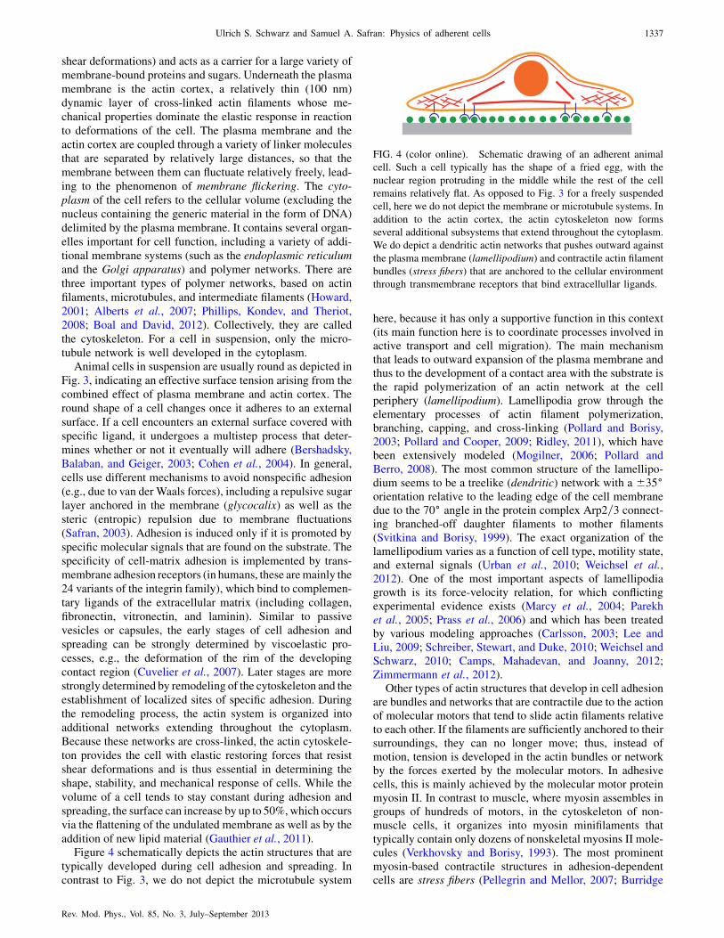
addition of new lipid material (Gauthier et al., 2011).Figure 4 schematically depicts the actin structures that are
typically developed during cell adhesion and spreading. In
contrast to Fig. 3, we do not depict the microtubule system
here, because it has only a supportive function in this context(its main function here is to coordinate processes involved inactive transport and cell migration). The main mechanismthat leads to outward expansion of the plasma membrane andthus to the development of a contact area with the substrate isthe rapid polymerization of an actin network at the cellperiphery (lamellipodium). Lamellipodia grow through theelementary processes of actin filament polymerization,branching, capping, and cross-linking (Pollard and Borisy,2003; Pollard and Cooper, 2009; Ridley, 2011), which havebeen extensively modeled (Mogilner, 2006; Pollard andBerro, 2008). The most common structure of the lamellipo-dium seems to be a treelike (dendritic) network with a 35�orientation relative to the leading edge of the cell membranedue to the 70� angle in the protein complex Arp2=3 connect-ing branched-off daughter filaments to mother filaments(Svitkina and Borisy, 1999). The exact organization of thelamellipodium varies as a function of cell type, motility state,and external signals (Urban et al., 2010; Weichsel et al.,2012). One of the most important aspects of lamellipodiagrowth is its force-velocity relation, for which conflictingexperimental evidence exists (Marcy et al., 2004; Parekhet al., 2005; Prass et al., 2006) and which has been treatedby various modeling approaches (Carlsson, 2003; Lee andLiu, 2009; Schreiber, Stewart, and Duke, 2010; Weichsel andSchwarz, 2010; Camps, Mahadevan, and Joanny, 2012;Zimmermann et al., 2012).
Other types of actin structures that develop in cell adhesionare bundles and networks that are contractile due to the actionof molecular motors that tend to slide actin filaments relativeto each other. If the filaments are sufficiently anchored to theirsurroundings, they can no longer move; thus, instead ofmotion, tension is developed in the actin bundles or networkby the forces exerted by the molecular motors. In adhesivecells, this is mainly achieved by the molecular motor proteinmyosin II. In contrast to muscle, where myosin assembles ingroups of hundreds of motors, in the cytoskeleton of non-muscle cells, it organizes into myosin minifilaments thattypically contain only dozens of nonskeletal myosins II mole-cules (Verkhovsky and Borisy, 1993). The most prominentmyosin-based contractile structures in adhesion-dependentcells are stress fibers (Pellegrin and Mellor, 2007; Burridge
FIG. 4 (color online). Schematic drawing of an adherent animal
cell. Such a cell typically has the shape of a fried egg, with the
nuclear region protruding in the middle while the rest of the cell
remains relatively flat. As opposed to Fig. 3 for a freely suspended
cell, here we do not depict the membrane or microtubule systems. In
addition to the actin cortex, the actin cytoskeleton now forms
several additional subsystems that extend throughout the cytoplasm.
We do depict a dendritic actin networks that pushes outward against
the plasma membrane (lamellipodium) and contractile actin filament
bundles (stress fibers) that are anchored to the cellular environment
through transmembrane receptors that bind extracellullar ligands.
Ulrich S. Schwarz and Samuel A. Safran: Physics of adherent cells 1337
Rev. Mod. Phys., Vol. 85, No. 3, July–September 2013
and Wittchen, 2013) shown schematically as thick lines inFig. 4. One can distinguish different types of stress fibers(Hotulainen and Lappalainen, 2006). Dorsal stress fibersconnect to an adhesion site at one end and have their otherend connected to other actin structures in the cell that are farfrom the substrate. Ventral stress fibers are connected toadhesion sites at both of their ends and thus run parallel tothe substrate. In contrast to dorsal and ventral stress fibers,transverse arcs are usually not straight, are not connected toadhesion sites, and typically extend parallel to the leadingedge. Stress fibers are thought to serve as the main sources ofcellular forces that are exerted on the substrate, since theirend points are often found at large adhesion sites that corre-late with large forces (Balaban et al., 2001). Laser cuttingexperiments demonstrated that stress fibers are under largetension, since they retract over large distances when being cut(Kumar et al., 2006; Colombelli et al., 2009). Stress fibers aredistinguished from retraction fibers, which are noncontractileactin bundles that specifically serve to anchor the cell to theECM during cell division (Thery et al., 2007).
The lamellipodium and stress fibers are actin assembliesthat create pushing and pulling forces, respectively; hence,they are the two main force-generating mechanisms for cellsthat adhere to flat substrates. Although its effect is ratherindirect, the plasma membrane plays an important role in thiscontext. Apart from acting as host for the transmembranereceptors from the integrin family, it also controls the poly-merization of the lamellipodium and the contraction of thestress fibers by triggering biochemical signals that regulatethese processes (Ridley, 2011). Equally important, the plasmamembrane plays an important role in the overall force balancein the cell, since its tension and curvature elasticity providethe counterforces to actin-generated forces that tend to extendand deform the membrane. An imbalance in these forces isespecially important in cell migration (Lauffenburger andHorwitz, 1996; Fletcher and Theriot, 2004).
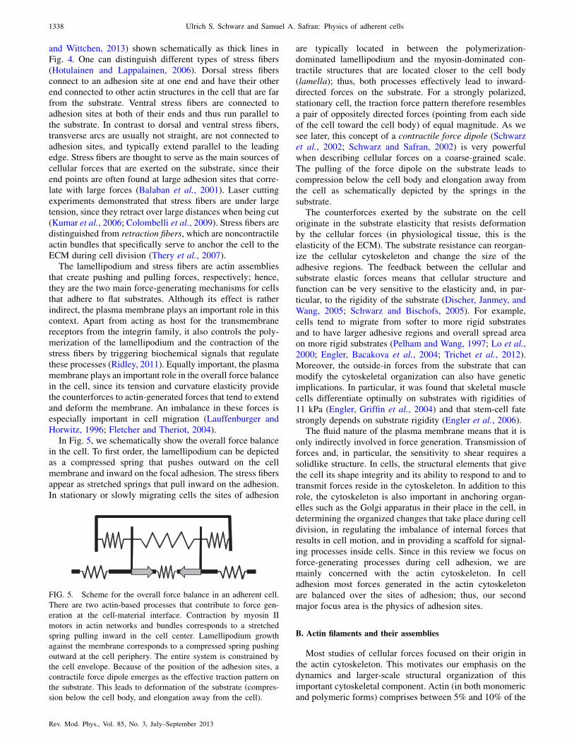
In Fig. 5, we schematically show the overall force balancein the cell. To first order, the lamellipodium can be depictedas a compressed spring that pushes outward on the cellmembrane and inward on the focal adhesion. The stress fibersappear as stretched springs that pull inward on the adhesion.In stationary or slowly migrating cells the sites of adhesion
are typically located in between the polymerization-
dominated lamellipodium and the myosin-dominated con-tractile structures that are located closer to the cell body
(lamella); thus, both processes effectively lead to inward-directed forces on the substrate. For a strongly polarized,
stationary cell, the traction force pattern therefore resembles
a pair of oppositely directed forces (pointing from each sideof the cell toward the cell body) of equal magnitude. As we
see later, this concept of a contractile force dipole (Schwarzet al., 2002; Schwarz and Safran, 2002) is very powerful
when describing cellular forces on a coarse-grained scale.
The pulling of the force dipole on the substrate leads tocompression below the cell body and elongation away from
the cell as schematically depicted by the springs in thesubstrate.
The counterforces exerted by the substrate on the cell
originate in the substrate elasticity that resists deformationby the cellular forces (in physiological tissue, this is the
elasticity of the ECM). The substrate resistance can reorgan-ize the cellular cytoskeleton and change the size of the
adhesive regions. The feedback between the cellular and
substrate elastic forces means that cellular structure andfunction can be very sensitive to the elasticity and, in par-
ticular, to the rigidity of the substrate (Discher, Janmey, andWang, 2005; Schwarz and Bischofs, 2005). For example,
cells tend to migrate from softer to more rigid substrates
and to have larger adhesive regions and overall spread areaon more rigid substrates (Pelham and Wang, 1997; Lo et al.,
2000; Engler, Bacakova et al., 2004; Trichet et al., 2012).Moreover, the outside-in forces from the substrate that can
modify the cytoskeletal organization can also have genetic
implications. In particular, it was found that skeletal musclecells differentiate optimally on substrates with rigidities of
11 kPa (Engler, Griffin et al., 2004) and that stem-cell fatestrongly depends on substrate rigidity (Engler et al., 2006).
The fluid nature of the plasma membrane means that it is
only indirectly involved in force generation. Transmission offorces and, in particular, the sensitivity to shear requires a
solidlike structure. In cells, the structural elements that give
the cell its shape integrity and its ability to respond to and totransmit forces reside in the cytoskeleton. In addition to this
role, the cytoskeleton is also important in anchoring organ-elles such as the Golgi apparatus in their place in the cell, in
determining the organized changes that take place during celldivision, in regulating the imbalance of internal forces that
results in cell motion, and in providing a scaffold for signal-
ing processes inside cells. Since in this review we focus onforce-generating processes during cell adhesion, we are
mainly concerned with the actin cytoskeleton. In celladhesion most forces generated in the actin cytoskeleton
are balanced over the sites of adhesion; thus, our second
major focus area is the physics of adhesion sites.
B. Actin filaments and their assemblies
Most studies of cellular forces focused on their origin inthe actin cytoskeleton. This motivates our emphasis on the
dynamics and larger-scale structural organization of this
important cytoskeletal component. Actin (in both monomericand polymeric forms) comprises between 5% and 10% of the
FIG. 5. Scheme for the overall force balance in an adherent cell.
There are two actin-based processes that contribute to force gen-
eration at the cell-material interface. Contraction by myosin II
motors in actin networks and bundles corresponds to a stretched
spring pulling inward in the cell center. Lamellipodium growth
against the membrane corresponds to a compressed spring pushing
outward at the cell periphery. The entire system is constrained by
the cell envelope. Because of the position of the adhesion sites, a
contractile force dipole emerges as the effective traction pattern on
the substrate. This leads to deformation of the substrate (compres-
sion below the cell body, and elongation away from the cell).
1338 Ulrich S. Schwarz and Samuel A. Safran: Physics of adherent cells
Rev. Mod. Phys., Vol. 85, No. 3, July–September 2013
protein in eukaryotic cells and is of great importance in cell
structure and motility (Fletcher and Mullins, 2010; Stricker,
Falzone, and Gardel, 2010). We begin with a discussion of the
growth of actin polymers. In contrast to self-assembling,
equilibrium polymerization, these are catalyzed by the bind-
ing of adenosine triphosphate (ATP) to monomeric (globular)
actin (G actin). While many synthetic polymers are nonpolar,
actin polymers are chiral with each macromolecule compris-
ing two helical, interlaced strands of monomeric subunits.
The two-filament assembly is thus polar so that the two ends
are therefore not equivalent; hence polymerization rates at
one end are not necessarily equal to those at the other. Actin
polymerization is therefore a polar, energy-consuming, non-
equilibrium process (Phillips, Kondev, and Theriot, 2008).A dynamical model for the growth of an actin filament
takes into account that the polymer is polar and the dynamics
of association of monomers at the two ends differ. That is,
kþ;�on and kþ;�
off are, respectively, the rates for monomers to
associate with þ (generally growing) or � (generally shrink-
ing) ends and to dissociate from those ends. For equilibrium
polymerization the association or dissociation energy itself
must be the same at either end since, although the monomers
are asymmetric at the two ends, the molecular bonds that are
formed are the same. Hence, by detailed balance, kþoff=kþon ¼
k�off=k�on. Thus, for such equilibrium polymers one can show
that (Phillips, Kondev, and Theriot, 2008) there is no state in
which one end is growing and the other is shrinking; the
polymer either grows or shrinks from both ends—albeit with
different on and off rates for the two ends of polar chains.
However, for the polymerization of actin in cells, detailed
balance does not apply and a richer set of behaviors is found
as we now discuss.In living systems, polymerization is often a dynamic pro-
cess that involves chemical changes that may differ at the two
ends of a polar chain such as actin so that the polymerization
and depolymerization rates differ. The chemical changes are
catalyzed by an input of energy from the conversion of ATP
(adenosine triphosphate with three phosphate bonds) to ADP
(adenosine diphosphate with two phosphate bonds) (Alberts
et al., 2007; Phillips, Kondev, and Theriot, 2008). This
conversion is known as hydrolysis since one phosphate group
dissociates from ATP to remain solubilized in water; the
breaking of one of the phosphate bonds releases about
ð10–20ÞkBT of energy since the hydration bonds between
ADP and water and the released phosphate group and water
are energetically more favorable than the bonds between the
phosphate bonds in ATP. The energy released by hydrolysis
of ATP can be used to modify the conformations of mole-
cules, such as actin that is bound to ATP in its lowest energy
state. The resulting conformational changes result in in-
creased or decreased bonding of the molecules to other
molecules; in the case of actin, hydrolysis destabilizes poly-
merization at its plus end.The nonequilibrium nature of actin polymerization in cells
is related to the conformational changes in the monomers that
are catalyzed by ATP; G-actin monomers bound to ATP join
the plus end of the actin polymer (Alberts et al., 2007;
Phillips, Kondev, and Theriot, 2008). Within a time of about
2 s, however, ATP is hydrolyzed to form ADP which reduces
the binding strength of the monomers in the chain, thus
destabilizing the polymer. There is therefore a nonequilib-
rium competition between growth and shrinkage of the poly-
mer. Note that, in solution, the G-actin monomers that have
dissociated from the chain can dissociate from ADP and
reassociate with ATP to rejoin the polymer; this turnover
makes the process highly dynamic. Since the actin polymer
is polar due to its double helical structure, the growth and
shrinkage at the þ and � ends is different, and, in principle,
one would need four rate constants to describe the on and off
rates of the ATP- and ADP-bound monomers at each of the
ends. Filaments elongate about 10 times faster at theirþ ends
compared with their � ends and this leads to an apparent
motion of theþ end known as treadmilling (Phillips, Kondev,
and Theriot, 2008). Typical values (Boal and David, 2012) are
kþon=k�on � 10 for ATP-bound actin and about 70 for the
predominant situation of ATP-bound actin at the þ end and
ADP-bound actin at the � end. The ratio kþoff=k�off � 5 for
ATP-bound actin at the þ end and ADP-bound actin at the
� end, with typical values of koff � 0:3–7:0 s�1 depending
on which end is being considered and whether the actin is
ATP or ADP bound. One can show (Phillips, Kondev, and
Theriot, 2008) that there is a monomer concentration range
for which the þ ends are growing while the � ends are
shrinking. Note that the treadmilling velocity can be finite
while the total filament length remains the same. Whether the
filament can move or not depends on its environment; for
example, treadmilling actin filaments in the vicinity of the
cell membrane have their motion impeded by the restoring
forces (due to surface tension and curvature energy) of the
membrane. This then leads to flow of the actin in the direction
opposite to treadmilling, that is, away from the cell mem-
brane (retrograde flow) as can be measured with speckle
fluorescence microscopy (Ponti et al., 2004).The larger-scale organization of actin can take several
forms. In vitro studies have shown (Tempel, Isenberg, and
Sackmann, 1996) that in some cases, alpha-actinin cross-
linkers can result in relatively thick actin bundles; in other
cases, a cross-linked, isotropic gel is formed. The detailed
phase diagram depends on both the actin and cross-linker
concentration (Zilman and Safran, 2003). In vivo, many
proteins can become involved in actin bundling which is
utilized by the cell in maintaining relatively stable (Gov,
2006), fingerlike protrusions called microvilli. These proteins
also participate in more dynamical protrusions called filopo-
dia (Mogilner and Rubinstein, 2005) that exert polymeriza-
tion forces on the cell membrane and play a role in cellular
motion and shape changes. Actin bundling is also an impor-
tant characteristic of stress fibers (Hotulainen and
Lappalainen, 2006) that typically range over some fraction
of the cell size and provide structural stability to the cell
while transmitting contractile forces to its surroundings.Because of the dynamics of the cross-links and the tread-
milling of actin, the cytoskeleton can be remodeled and is
therefore not permanently cross-linked. However, experi-
ments in which cells are subject to time-varying strains that
cause cytoskeleton reorganization show that the overall time
scale for reassembly and reorientation of stress fibers can be
several hours (Brown et al., 1998; Wang et al., 2001;
Jungbauer et al., 2008). Entropic fluctuations occur on a
time scale shorter than 0.01 s (Deng et al., 2006), while on
Ulrich S. Schwarz and Samuel A. Safran: Physics of adherent cells 1339
Rev. Mod. Phys., Vol. 85, No. 3, July–September 2013
longer time scales the elastic response to time-varying strain
has been characterized as glassy. We first consider the elastic
modulus of actin networks in vitro (Boal and David, 2012) on
time scales shorter than those at which the shear modulus
vanishes due to the cross-link disconnections (Lieleg et al.,
2008); on these time scales, the system in some average sense
can be regarded as being permanently cross-linked.The simplest model for the elastic constant of a perma-
nently cross-linked polymeric network predicts a shear modu-
lus �� �kBT, where � is the number density of cross-links
(Rubinstein and Colby, 2003). The typical spacing between
cross-links with a 1:100 ratio of linker to actin monomers is
of the order of 0:1 �m. This yields a shear modulus of about
1 J=m3 at room temperature which is equivalent to 1 Pa. The
measured value (Janmey et al., 1990) in the presence of the
cross-linker ABP (actin binding proten) (at a ratio of ABP:
actin of the order of 1:100) is about 1 order of magnitude
larger and is sensitive to the length of the actin segments; the
observations also depend on the history of the sample, since
shear can disrupt actin filaments and give misleadingly low
values for the modulus. Higher values of the modulus than
expected from simple considerations of cross-linked polymer
networks can be due to the stiffening effects of the cross-links
themselves, the semiflexible (as opposed to Gaussian) nature
of biopolymers such as actin, and to nonlinear shear stiffen-
ing. On the other hand, the analogy with permanently cross-
linked gels must be reconsidered in light of the finite lifetime
of the cross-links. Alpha actinin has a dissociation rate of
about 1 s�1 (Xu, Wirtz, and Pollard, 1998; Gardel, Kasza
et al., 2008) which may explain why in vitro experiments
using this cross-linker (Lieleg et al., 2008) in actin gels yield
a low frequency elastic modulus of about 1 Pa only at the very
highest cross-linker concentrations (alpha-actinin:actin ratios
of 1:15). The dissociation rate may be different in different
geometries; in isotropically cross-linked gels, the cross-
linkers can more effectively dissociate compared with their
relatively tighter packing in actin bundles where neighboring
filaments are nearby. The dissociation rate is also strongly
temperature dependent and of course very different for differ-
ent cross-linkers (Xu, Wirtz, and Pollard, 1998).In addition to the finite residence time of the cross-linkers
at the network junctions, another important difference be-
tween the elastic modulus of cross-linked biopolymers such
as actin and synthetic polymer gels is the observation that the
actin cytoskeleton shows a nonlinear elastic response in a
nonperturbative manner (Gardel et al., 2004; Storm et al.,
2005). For small stresses, the elastic stress in actin gels is
proportional to the strain, while for larger stresses, of the
order of 0.2 Pa, the effective modulus varies as the 3=2 powerof the applied stress due to the entropically dominated me-
chanical response of semiflexible polymers of finite extensi-
bility as described above (Storm et al., 2005). This entropic
nonlinearity is particularly interesting because the strains
may still be relatively small (Gardel et al., 2004; Storm
et al., 2005) even though the medium responds very non-
linearly; this is quite different from analytical nonlinearities
(e.g., due to additional quadratic terms in the stress-strain
relationship or due to geometrical nonlinearity) that arise
when the strains become large in nonpolymeric systems.
Interestingly, cells can regulate the regime in which they
function by changing their internal stress state through varia-
tion of the activity of molecular motors. Since the cell elasticmodulus is of the order of 1–10 kPa (reflecting the types of
stresses that cells can maintain), the cytoskeletal elasticresponse can easily operate in the nonlinear regime. Finally
we note that many other important biopolymers (Storm et al.,2005; Klotzsch et al., 2009) also show similar nonlinear
response, including collagen which is an important part ofthe ECM.
C. Actomyosin contractility
In Sec. III.B, we summarized the properties of cross-linkedactin gels based on information obtained from in vitro experi-
ments. However, one important aspect regarding actin net-works and bundles in cells is the fact that these networks are
under tension due to the contractile activity of myosin motors(Howard, 2001). Contractile actin networks (Mizuno et al.,
2007; Koenderink et al., 2009; Kohler, Schaller, and Bausch,
2011; Soares e Silva et al., 2011; Murrell and Gardel, 2012)and bundles (Thoresen, Lenz, and Gardel, 2011, 2013) have
recently been reconstituted in biomimetic assays. In cells, themyosin motors generate internal forces in the actin network
which are transmitted to its surroundings due to the ‘‘glue’’the cell produces in the form of proteins that assemble into
focal contacts or focal complexes (Geiger, Spatz, andBershadsky, 2009). The production of force is a nonequilib-
rium process that requires energy input via ATP hydrolysis
that causes conformational changes in the myosin molecularmotors (Howard, 2001). The internal forces generated by
molecular motors that act upon the cross-linked actin assem-blies in cells distinguish them from ‘‘dead,’’ nonactive gels
and allows cells to pull on their environment and on eachother. Motor activity also means that the cell can exert forces
on itself and this, along with polymerization of actin, plays animportant role in cell motility. Motors can also influence the
conformations of the actin filaments in a manner that has to
do with the motor and motor-actin dynamics. The stochasticnature of the motor-actin coupling in which the motor is
associated with the actin for a finite time (Howard, 2001;Boal and David, 2012) after which it can detach and diffuse
affects the fluctuations of the filaments. These motor-drivenfluctuations are distinct from the thermal fluctuations of the
actin that are driven by Brownian motion of its aqueousenvironment (Mizuno et al., 2007; MacKintosh and
Schmidt, 2010). This leads to a breakdown of the
fluctuation-dissipation theorem that relates the thermal fluc-tuations of an equilibrium system to its response to a deter-
ministic force as discussed in Sec. II on the physicsbackground.
Because stress fibers are an important element of the force-
generating apparatus of cells adhering to flat substrates, alarge variety of models has been developed to describe their
physical properties. Dynamical models show that actin fila-ments can be sorted by myosin II motors into a tensile state
(Kruse and Julicher, 2000a, 2003; Ziebert and Zimmermann,2004; Yoshinaga et al., 2010; Stachowiak, Matthew et al.,
2012). Models for mature fibers are often motivated byperturbation experiments on stress fibers, such as studies of
contraction dynamics after activation (Peterson et al., 2004) or
1340 Ulrich S. Schwarz and Samuel A. Safran: Physics of adherent cells
Rev. Mod. Phys., Vol. 85, No. 3, July–September 2013
relaxation dynamics after laser cutting (Kumar et al., 2006;
Colombelli et al., 2009). They usually assume a sarcomeric
organization of the stress fiber (Friedrich et al., 2012) and
couple elastic, viscous, and contractile elements in a unit cell
(Besser and Schwarz, 2007; Luo et al., 2008; Stachowiak,
Matthew and O’Shaughnessy, 2008, 2009; Russell et al.,
2009; Besser et al., 2011). Recently it was demonstrated that
such sarcomeric models predict some of the central physical
properties of reconstituted contractile actin bundles (e.g.,
retraction velocity is proportional to length) (Thoresen,
Lenz, and Gardel, 2013). On a very coarse-grained scale,
such one-dimensionalmodels can be regarded asmore detailed
versions of the force-dipole model introduced in Fig. 5. In
particular, they usually obey force balance on the substrate by
construction. These types of one-dimensional models can also
be used to predict the cellular response to substrate stiffness
(Mitrossilis et al., 2009; Besser and Schwarz, 2010; Marcq,
Yoshinaga, and Prost, 2011; Crow et al., 2012).An important reference case for the physics of stress fibers
is sarcomeric muscle, in which actin filaments, passive cross-
linkers, and myosin II motors are arranged in a very ordered
fashion. The action of myosin II molecular motors can be
modeled either with a generic two-state theory (Julicher and
Prost, 1995; Placais et al., 2009) or with more detailed cross-
bridge models that go back to the seminal work of Huxley
(Huxley, 1957; Duke, 1999; Vilfan and Duke, 2003; Erdmann
and Schwarz, 2012), where the cross bridges refer to the
actomyosin coupling. This model accounts for the fact that
a myosin II motor loaded with ATP goes through a cycle
where it first weakly binds to the actin filament. Release of
the inorganic phosphate (after hydrolysis of ATP to ADP)
causes the myosin motor to make a power stroke that creates
force and motion. Finally, after the release of the ADP and
binding of a new ATP molecule, the myosin II motor unbinds
from the filament and is ready for the next motor cycle. The
effective force-velocity relation has been measured both in
the context of muscle (Pate et al., 1994) and in single-
molecule experiments (Veigel et al., 2003). At vanishing
force, the motors move with an ATP-dependent free velocity
of about 1 �m=s. As the external counterforce increases, thevelocity of the motor along the filament drops in a hyperbolic
manner, until it vanishes at a stall force of a few pN. This
close coupling between force and sliding velocity was first
noted by Hill in 1938, who described it using a phenomeno-
logical equation (called the Hill equation), which can be
explained in detail by the cross-bridge models (McMahon,
1984; Howard, 2001). Alternatively, as we see later, Hill-type
relations can be used directly as an assumption in coarse-
grained models. In particular, such a model has been used to
argue that rigidity sensing is based on the same principles
similar to muscle contraction (Mitrossilis et al., 2009).Contractility is also observed in biological systems with no
apparent sarcomeric order. For example, the contractility of the
actin ring during cell division may rely on depolymerization
forces (Pinto et al., 2012). It has been shown theoretically that,
in one-dimensional actin bundles containing myosin, contrac-
tility can occur even in the absence of spatial organization of
the bundle, due to bundle shortening (Kruse and Julicher,
2000b; Kruse, Zumdieck, and Julicher, 2003). The existence
of net contractility is related to the assumption that a myosin
motor which binds to or arrives at the plus end (but not at the
minus end) of a filament remains attached for some time. Thismodel does not contain additional cross-linkers (similar to
Z bodies found in sarcomeres) that may tend to associatewith only one end of the polar actin molecules. Another recent
suggestion (Lenz et al., 2012) for how contractility can arise inbundles without sarcomeric order is based on the asymmetric
response of the filaments to longitudinally applied stresses,e.g., a tendency to yield under compression while resisting
extension. Such buckling has been observed in in vitro experi-
ments containing actin, smooth muscle myosin, and ATP. Wenote that additional cross-linkers, such as those found in
sarcomeres and possibly stress fibers, that may break thesymmetry were not included.Whether stress fibers, contractile
rings, or smooth muscle fibers are ordered and function in amanner similar to sarcomeres should be investigated by future
experiments. The sarcomeric order discussed below in terms ofsmectic ordering of force dipoles is meant to apply to nascent
muscle cells where striations have indeed been observed
(Engler, Griffin et al., 2004; Friedrich et al., 2011). In asomewhat similar manner, striations have also been reported
in studies of nonmuscle stress fibers (Peterson et al., 2004).Here the microscopic, antiparallel arrangement of adjacent
actin polymers was not directly observed, but the striationsmeasured suggest a sarcomeric analogy.
D. Focal adhesions
Understanding the mechanical response of stationary cellsinvolves analysis of the internal elastic response of contractile
cells as well as their mechanical coupling to their surround-ings. While the response of cells to external forces or other
mechanical perturbations can necessitate the disassembly andrebuilding of the actin cytoskeleton, the stable coupling of the
cell to the surrounding elastic matrix is due to sites ofadhesion called focal adhesions that connect the actin cytos-
keleton to transmembrane adhesion receptors from the integ-
rin family. These are then connected, on the extracellularside, to the substrate or extracellular matrix.
In contrast to the adhesion of passive vesicles or capsules,the spatial distribution of the adhesion structure of cells is
very heterogeneous. It is mainly localized at the cell periph-
ery, because it is strongly coupled to the growth processes ofthe lamellipodium. Figure 6 depicts the spatial coordination
between the growth of adhesions and the actin cytoskeleton.Nascent adhesions are initiated close to the leading edge and
then move toward the cell center. This movement is mainlydriven by the flow of actin away from the leading edge
(retrograde flow) due to the counterforces exerted on thepolymerizing actin by the membrane. As they move toward
the cell center, the small adhesions either mature into
micrometer-sized focal adhesions or decay again. This switchtypically occurs near the lamellipodium-lamella boundary,
where a more condensed and myosin II-rich actin networkreplaces the dendritic network of the lamellipodium (Schwarz
and Gardel, 2012; Shemesh, Bershadsky, and Kozlov, 2012).Whether the adhesion grows and matures or whether it decays
is strongly coupled to the mechanics of the system. Theadhesions are stable only if sufficient force is exerted upon
them and this is not possible on very soft substrates. This
Ulrich S. Schwarz and Samuel A. Safran: Physics of adherent cells 1341
Rev. Mod. Phys., Vol. 85, No. 3, July–September 2013

force is mainly applied by contractile stress fibers and net-
works connecting them to the focal adhesions in the lamella,
although the force resulting from retrograde flow also might
play an important role. Because focal adhesions are con-
nected to the matrix, these forces are transmitted to the
substrate and can be measured there with traction force
microscopy on flat elastic substrates (Dembo and Wang,
1999; Butler et al., 2002; Schwarz et al., 2002; Sabass
et al., 2008; Plotnikov et al., 2012; Legant et al., 2013) or
with fields of elastic pillars (Tan et al., 2003; Saez et al.,
2005; Trichet et al., 2012). These studies have shown that
force and protein assembly are linearly coupled at focal
adhesions, resulting in a constant stress for adhesions of about
5 nN=nm2 ¼ 5 kPa (Balaban et al., 2001; Tan et al., 2003).
For elastic substrates, recently it was suggested that this
relation holds only for growing adhesions (Stricker et al.,
2011). For pillar assays, recently it was reported that there
exists a constant stress, but that it depends on extracellular
stiffness due to global feedback (Trichet et al., 2012). At any
rate, the typical stress at single focal adhesions is close to the
value of the physiological stiffness of matrix and cells; this
suggests that these forces are used for mechanosensing in the
physiological environment of the cell.The detailed molecular organization of focal adhesions is
an active area of research that is challenging due to the large
number (more than 180) of different components that are
involved (Zaidel-Bar et al., 2007; Kuo et al., 2011). Recent
progress includes the use of electron tomography (Patla et al.,
2010) and superresolution microscopy (Shtengel et al., 2009)
to discern focal adhesion structure at scales smaller than
the optical resolution, and the use of high throughput
RNA-interference screens to dissect the regulatory hierarchy
of focal adhesions (Prager-Khoutorsky et al., 2011).
Kinetic models have been used to describe the temporal
and spatial coordination of the different components
(Civelekoglu-Scholey et al., 2005; Macdonald, Horwitz,
and Lauffenburger, 2008; Hoffmann and Schwarz, 2013).
Figure 7 shows a schematic representation of a focal adhe-
sion. In general, focal adhesions have a layered structure
determined by the two-dimensional nature of the plasma
membrane. The transmembrane adhesion receptors of the
integrin family consists of two subunits, with relatively large
headpieces that bind to the matrix and relatively small cyto-
plasmic tails. In the absence of special signals, the integrins
have a low affinity for matrix binding. However, due to
inside-out signaling (related to the cytoskeletal forces), the
integrins can become activated and are then primed for matrix
binding. This in turn leads to stabilization of the intracellular
part of the adhesion complex that through the binding of a
variety of cytoplasmic proteins forms a two-dimensional
plaque that reinforces the attachment of the cytoskeleton
and the integrin layer. One of the main molecules responsible
for cross-linking neighboring integrins is talin (Alberts et al.,
2007), which extends over 60 nm. Since talin also binds actin,
it connects the integrins in the focal adhesion to the actin
cytoskeleton. As the adhesion matures, this cross-linking is
strengthened by additional proteins such as vinculin and
paxillin, whose recruitment seems to be increased by force.Focal adhesions act not only as mechanical linkers that
anchor the cell to its substrate, but also as prominent signaling
centers that activate biochemical signaling molecules that
diffuse into the cytoplasm and toward the nucleus (Zaidel-
Bar et al., 2007; Vogel and Sheetz, 2009). In our context, the
most important signaling molecules are the small GTPases
from the Rho family (Rho, Rac, and Cdc42) that regulate the
assembly and activity of the actomyosin system. Each of these
molecules acts like a molecular switch which is activated by
exchanging guanosine diphosphate (GDP) by guanosine tri-
phosphate (GTP) (analogous to ADP to ATP conversion); the
active form then diffuses in the cytoplasma and activates
downstream targets. For example,mature adhesions are known
for Rho signaling, which upregulates both actin polymeriza-
tion (through the formin mDia1) and contractility of non-
muscle myosin II motors (through the Rho-associated
kinase, ROCK). Moreover, many signaling molecules respon-
sible for cell migration, differentiation, and fate are localized
to focal adhesions, most prominently the focal adhesion kinase
FAK, which is known to be important in many types of cancer
(Mitra, Hanson, and Schlaepfer, 2005).
LP
LM
FIG. 6 (color online). Spatial organization of focal adhesion
growth and the actin cytoskeleton. The actin lamellipodium (LP)
is assembled at the leading edge and flows from there toward the
cell center. Small adhesions are formed along the way and mature
into focal adhesion as they move with the actin flow. At the
boundary with the myosin-dominated lamella (LM), only a few
mature focal adhesions persist; those are stabilized by large con-
tractile forces which are mainly due to the activity of myosin II
minifilaments in stress fibers.
FIG. 7 (color online). Schematic view of a focal adhesion. The
transmembrane adhesion receptors from the integrin family (a
heterodimer with two subunits) bind to the extracellular matrix
(for example, collagen) on the outside and are cross-linked by
cytoplasmic proteins such as talin in the inside. Talin binds to actin
and this binding is further strengthened by proteins such as vinculin.
The contractility of the actin cytoskeleton is determined by the
activity of myosin II minifilaments.
1342 Ulrich S. Schwarz and Samuel A. Safran: Physics of adherent cells
Rev. Mod. Phys., Vol. 85, No. 3, July–September 2013
Adhesion proteins localized to focal adhesions coexist withthe same proteins in relatively dilute solution in the cytoplasm
or the membrane. The domains as a whole have relatively longlifetimes (tens of minutes) which suggests that the two coex-isting phases might be at equilibrium. However, fluorescence
studies (Wolfenson et al., 2011) show that the adhesionscontinuously exchange proteins with the cytoplasm eventhough the large-scale composition and structure seem to
remain unchanged. Thermodynamic equilibrium dictatesmuch larger domain sizes (or no domains at all) and why the
adhesions are stable on the micrometer scale is not obvious(Lenne and Nicolas, 2009). This puzzle might be resolved bynoting that in addition to diffusion, energy-consuming, active
processes may transport free proteins from the dilute phase inthe cytoplasm or membrane to the adhesion sites. These activeprocesses involve molecular motor proteins and are highly
regulated by the cell (Kawakami, Tatsumi, and Sokabe,2001). This suggests that nonequilibrium effects may be im-portant in stabilizing these finite-size domains, similar to the
situation with treadmilling actin filaments.Naively, one might try to understand cellular adhesions by
analogy with physical adhesion (e.g., of a synthetic vesiclecoated with ligands that are attracted to an appropriate sur-
face, see Sec. II.F). However, while physical adhesion is apassive process, cell adhesion involves molecular motors thatgenerate internal stresses. Force generation consumes ATP
and results in the fact that, in addition to passive contacts thatresult in forces mainly directed in the normal direction, cellsalso exert contractile forces that act mainly in the lateral
direction, i.e., parallel to the substrate. These contractileforces have been observed in experiments that measure sur-face deformation (Dembo and Wang, 1999; Butler et al.,
2002; Schwarz et al., 2002; Tan et al., 2003; Saez et al.,2005; Sabass et al., 2008). Although recent experiments alsoprovided evidence for vertical forces (Hur et al., 2009;
Delano-Ayari, Rieu, and Sano, 2010), most of these experi-ments were done with rather weakly adhering cells, whichshare some similarities with the passive reference cases dis-
cussed above (droplets, vesicles, capsules). Thus on a planarsubstrate it is the actively generated and often tangential
forces that are used by the cell to regulate its response tothe physical environment. This conclusion is supported by thefact that use of the myosin blocker blebbistatin not only leads
to the disappearance of these forces, but also eliminates thecellular response to stiffness. Thus the main challenge is tounderstand how actively generated forces allow the cell to
probe the physical properties of its environment.
IV. PHYSICS OF CELL-MATRIX ADHESIONS
A. Physical motivation
As explained in Sec. III.D, cell adhesion does not occurhomogeneously at the cell-material interface, but instead ischaracterized by the local assembly of specific adhesion
molecules into supramolecular adhesion sites, the so-calledfocal adhesions. For cell adhesion to a flat substrate, thesefocal adhesions are mainly situated at the cell periphery.
During the last decade, it was shown that the protein assem-bly that comprises the focal adhesion is strongly coupled to
mechanical force. Experimental studies inducing changes in
mechanical stress at adhesions used shear flow (Davies,
Robotewskyj, and Griem, 1994; Zaidel-Bar et al., 2003),
optical tweezers (Choquet, Felsenfeld, and Sheetz, 1997),
micromanipulators (Riveline et al., 2001; Paul et al., 2008;
Heil and Spatz, 2010), laser nanosurgery (Kumar et al., 2006;
Lele et al., 2006; Colombelli et al., 2009), or pharmacologi-
cal drugs (Chrzanowska-Wodnicka and Burridge, 1996; Kuo
et al., 2011; Wolfenson et al., 2011) to perturb and hence
study the nature of these contacts. In all cases, it was observed
that focal adhesions respond to changes in mechanical load
by growth as evidenced by changes in focal adhesion mor-
phology and size. Quantitative correlation indicated a linear
relation between force and adhesion size (Balaban et al.,
2001; Tan et al., 2003), although the history of the adhesion
and global determinants also play an important role (Stricker
et al., 2011; Trichet et al., 2012).From the physical viewpoint, it is interesting to note that
tensile mechanical deformation, such as the shear induced by
the tangential displacement of a pipette, leads to growth of
focal adhesions in the direction of applied force (Riveline
et al., 2001). Deactivation of actomyosin contractility reduces
the adhesion size and eventually leads to its complete dis-
ruption (Balaban et al., 2001). Other forces, such as hydro-
dynamic flow (Zaidel-Bar et al., 2003) or stretching forces
applied to the substrate (Kaunas et al., 2005), also cause
growth of focal adhesions in the direction of the force. The
anisotropy of focal adhesion growth under force is charac-
terized both by overall growth of the adhesion (in which the
number of molecules involved increases) and by treadmilling
(or sliding) of the center of mass of the adhesion toward the
direction of the applied force. These anomalies cannot be
explained with standard models of nucleation and growth of
molecules adsorbed from solution onto surfaces. The role of
force in stabilizing and promoting growth of focal adhesions
(FAs) has been discussed from several different points of
view with a focus on predictions of the growth of focal
adhesions in the direction of actomyosin or externally applied
forces (Bershadsky, Kozlov, and Geiger, 2006).However, before discussing the growth response of focal
adhesions under load, it is instructive to consider why this
response evolved in the first place. From the physical point of
view, it seems obvious that in this way the system avoids
material failure. In general, failure under mechanical load is
a phenomenon relevant to many systems of practical interest
and on widely different scales, from muscle proteins (on the
nanometer scale) through bone on a centimeter scale to bridges
and buildings (on the meter scale) and earthquakes (on the
kilometer scale) (Buehler and Keten, 2010). In contrast to the
macroscopic systems considered in traditional fracture me-
chanics, biomolecular adhesions do not usually break at sharp,
deterministic stability thresholds, but can unbind and rebind
from the surface to which they adhere in a stochastic manner.
This important observation is related to the weak interaction
scales relevant to soft matter comparedwith hardmatter. Thus,
if one considers the stability of such assemblies (defined by
either their average lifetime or fracture strength), one must
include stochastic effects, in contrast with traditional fracture
mechanics of macroscopic systems where these effects are not
relevant. Before we address the issue of growth, we therefore
Ulrich S. Schwarz and Samuel A. Safran: Physics of adherent cells 1343
Rev. Mod. Phys., Vol. 85, No. 3, July–September 2013
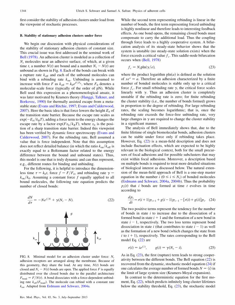
first consider the stability of adhesion clusters under load fromthe viewpoint of stochastic processes.
B. Stability of stationary adhesion clusters under force
We begin our discussion with physical considerations ofthe stability of stationary adhesion clusters of constant size.This crucial issue was first addressed in the seminal work ofBell (1978). An adhesion cluster is modeled as a collection ofNt molecules near an adhesive surface, of which, at a giventime t, a number NðtÞ are bound and a number Nt � NðtÞ areunbound as shown in Fig. 8. Each of the bonds can break witha rupture rate koff and each of the unbound molecules canbind with a rebinding rate kon. Unbinding is assumed toincrease with force F as koff ¼ k0e
F=F0 , where F0 denotes amolecular-scale force (typically of the order of pN). WhileBell used this expression as a phenomenological ansatz, itwas later motivated by Kramers theory (Hanggi, Talkner, andBorkovec, 1990) for thermally assisted escape from a meta-stable state (Evans and Ritchie, 1997; Evans and Calderwood,2007). Here the basic idea is that force lowers the height Eb ofthe transition state barrier. Because the escape rate scales asexpð�Eb=kBTÞ, adding a force term to the energy changes theescape rate by a factor expðFxb=kBTÞ, where xb is the posi-tion of a sharp transition state barrier. Indeed this viewpointhas been verified by dynamic force spectroscopy (Evans andCalderwood, 2007). For the rebinding rate, Bell assumed avalue that is force independent. Note that this assumptiondoes not reflect detailed balance (in which the ratio kon=koff isexactly equal to a Boltzmann factor related to the energydifference between the bound and unbound states). Thus,this model is one that is truly dynamic and can thus represent,e.g., different routes for binding and unbinding.
For the following, it is helpful to introduce the dimension-less time � ¼ k0t, force f ¼ F=Fb, and rebinding rate � ¼kon=k0. Assuming a constant force f equally applied to allbound molecules, the following rate equation predicts thenumber of closed bonds:
dN
d�¼ �Nef=N þ �ðNt � NÞ: (22)
While the second term representing rebinding is linear in thenumber of bonds, the first term representing forced unbindingis highly nonlinear and therefore leads to interesting feedbackeffects. As one bond opens, the remaining closed bonds mustcompensate to carry the additional load. Thus the couplingthrough force leads to a highly cooperative system. A bifur-cation analysis of its steady-state behavior shows that thesystem is unstable (no steady-state solution exists) when theforce exceeds a critical value fc. This saddle-node bifurcationoccurs when (Bell, 1978)
fc ¼ Ntplnð�=eÞ; (23)
where the product logarithm plnðaÞ is defined as the solutionof xex ¼ a. Therefore an adhesion characterized by a finitenumber of bonded molecules is stable only up to a criticalforce fc. For small rebinding rate �, the critical force scaleslinearly with �. Thus an adhesion cluster is completelyunstable if the rebinding rate is zero; for finite rebindingthe cluster stability (i.e., the number of bonds formed) growsin proportion to the degree of rebinding. For large rebindingrates, the scaling becomes logarithmic; that is, once therebinding rate exceeds the force-free unbinding rate, verylarge changes in � are required to change the cluster stabilityin a significant manner.
The analysis of Bell immediately shows that, due to thefinite lifetime of single biomolecular bonds, adhesion clusterscan be stable under force only if rebinding takes place.However, Eq. (22) is a mean-field description and does notinclude fluctuation effects, which are expected to be highlyrelevant in the biological context, both for the small precur-sors of focal adhesions and for possible subclusters that mayexist within focal adhesions. Moreover, a description basedon multiple bonds is required to treat more detailed situationsof biological interest as discussed below. The natural exten-sion of the mean-field approach of Bell is a one-step masterequation in the number i (0 � i � Nt) of bonded molecules(Erdmann and Schwarz, 2004a, 2004b). Thus the probabilitypiðtÞ that i bonds are formed at time t evolves in timeaccording to
dpi
dt¼ rðiþ1Þpiþ1þgði�1Þpi�1�½rðiÞþgðiÞ�pi: (24)
The two positive terms represent the tendency for the numberof bonds in state i to increase due to the dissociation of aformed bond in state iþ 1 and the formation of a new bond instate i� 1, respectively. The two loss terms represent bonddissociation in state i (that contributes to state i� 1) as wellas the formation of a new bond (which changes the state fromi to iþ 1), respectively. The rates corresponding to the Bellmodel Eq. (22) are
rðiÞ ¼ ief=i; gðiÞ ¼ �ðNt � iÞ: (25)
As in Eq. (23), the first (rupture) term leads to strong cooper-ativity between the different bonds. The Bell equation (22) isrecovered from the dynamic, stochastic model equation (24) ifone calculates the average number of formed bondsN ¼ hii inthe limit of large system size (Kramers-Moyal expansion).
In contrast to the deterministic equation for the first mo-ment, Eq. (22), which predicts infinitely long cluster lifetimesbelow the stability threshold, Eq. (23), the stochastic model
F
k koffon
FIG. 8. Minimal model for an adhesion cluster under force: Nt
adhesion receptors are arranged along the membrane. Because of
this geometry, they share the load. At any time, NðtÞ bonds are
closed and Nt � NðtÞ bonds are open. The applied force F is equally
distributed over the closed bonds due to the parallel architecture
Fbond ¼ F=NðtÞ. A bond dissociates with a force-dependent unbind-
ing rate koffðFbondÞ. The molecule can rebind with a constant rate
kon. Adapted from Erdmann and Schwarz, 2004a.
1344 Ulrich S. Schwarz and Samuel A. Safran: Physics of adherent cells
Rev. Mod. Phys., Vol. 85, No. 3, July–September 2013

equation (24) predicts finite lifetimes for any value of theforce. The average lifetime of a cluster with i ¼ Nt bonds attime t ¼ 0 can be identified with the mean time T for thiscluster to stochastically evolve to the state i ¼ 0 (no boundmolecules). For a one-step master equation, this mean firstpassage time can be easily calculated (van Kampen, 1992):
T ¼ XNt
i¼1
1
rðiÞ þXNt�1
i¼1
XNt
j¼iþ1
Qj�1k¼j�i gðkÞQjk¼j�i rðkÞ
: (26)
The first term in Eq. (26) is the result of vanishing rebinding� ¼ 0. For small force f < 1, it is analogous to the simplecase of proportional (or radioactive) decay (for �¼f¼0, onebasically deals with the stochastic version of dN=d� ¼ �N).Using that approximation one finds that the cluster lifetimescales as T � lnNt; that is, the cluster size Nt has only arelatively weak effect on the stability of the cluster, becausethe bonds dissociate in parallel with no cooperativity. Finiteforce exponentially decreases the lifetime due to the Bellequation. The second term in Eq. (26) increases the lifetimeas a polynomial of order Nt � 1 in � and approximatelyexponentially with increasing cluster size Nt. For Nt ¼ 2,Eq. (26) reads
T ¼ 12ðe�f=2 þ 2e�f þ �e�3f=2Þ: (27)
This simple but instructive formula shows how force f ex-ponentially suppresses the cluster lifetime, while rebinding �increases it in a polynomial fashion.
In order to study the cluster lifetime as a function of force,in Fig. 9 we plot T as calculated from Eq. (26) as a function off=Nt for � ¼ 1 and different values of the cluster size Nt. Forsmall force f < Nt, the mean lifetime increases exponentiallywith the cluster size Nt. For large force f > Nt, rebindingbecomes irrelevant and all the curves for different valuesof Nt are very similar. One interesting aspect here is thetransition region, which is characterized by strong amplifica-tion (a small change in force f has a strong effect on the meanlifetime T). This transition region corresponds to the stabilitythreshold from Eq. (23) (vertical line). Above this threshold, adeterministic lifetime can be defined by numerically solvingEq. (22) with Nð0Þ ¼ Nt and solving for the time at which
N ¼ 1 (this replaces the criterion i ¼ 0 from the stochasticcase, which cannot be used in the deterministic case due toexponential approach of N ¼ 0). The result (dashed line)corresponds well to the mean cluster lifetime from the sto-chastic model, but diverges at the threshold.
Equation (26) can be used to make interesting estimates forexperimental situations of interest. For example, in the case ofzero rebinding rate (� ¼ 0) and zero applied force (f ¼ 0),for a single bond lifetime of 1 s (k0 ¼ 1=s), a cluster lifetimeT of 1 min could be achieved only with the absurdly largenumber of 1026 bonds; this is because in this case the clusterlifetime scales only logarithmically with cluster size.However, for a finite rebinding rate of � ¼ 1 (kon ¼ k0, stillat zero force), only Nt ¼ 10 bonds are necessary, because thelifetime scales strongly with rebinding rate T � �Nt�1.Increasing the dimensionless force to f ¼ 10 (correspondingto 40 pN for Fb ¼ 4 pN) would decrease the lifetime to T ¼0:05 s, because T is an exponentially decreasing function off. To reach a cluster lifetime of 1 min in this case, the clustersize must be increased to 50 or the rebinding rate must beincreased by a factor of 10.
One of the strongest advantages of the stochastic model isthat it can be used to simulate single trajectories, which sharemany similarities with experimental realizations of individualexperiments. In Fig. 10 we show selected simulated trajecto-ries for forces below [Fig. 10(a)] and above [Fig. 10(b)] thecritical force. We note that failure is rather abrupt due tocooperative effects: once sufficiently many bonds have bro-ken, e.g., due to a fluctuation that leads to a smaller number ofbonds, the force on the remaining ones is so high that rebindingbecomes very unlikely and the cluster fails in a cascade ofdissociated bonds that leads to rupture of the adhesion cluster(cascading failure). Interestingly, this effect is not apparent ifone calculates only the averages, which are shown as lines.Wefurther note that, even below the stability threshold inFig. 10(a), small clusters are likely to fail due to the finiteprobability for a devastating fluctuation that takes the systemto a state with a small number of closed bonds. Above thethreshold in Fig. 10(b), clusters of any size are unstable.
The conceptual framework introduced by Bell shows howan adhesion site can at the same time be highly dynamic andyet be stable up to some maximal force: while some bondscan dissociate and then rebind, the remaining adhesion bondsallow the transfer of force from the cell to the substrate. Thesimple model also shows that this mechanism leads to strongcooperativity, because each bond that forms or breaks leads toa fast redistribution of the force, thereby affecting all theother bonds. The same cooperative mechanism operates inmany other biologically relevant situations, e.g., during forcegeneration in muscle (Huxley, 1957; Duke, 1999) or duringcargo transport by multiple molecular motors (Klumpp andLipowsky, 2005; Gurin et al., 2010). The stochastic extensionof the Bell model demonstrates that adhesions are not onlyunstable for large applied forces, but are also unstable atsmall sizes for which fluctuations to smaller numbers ofclosed bonds can be detrimental.
C. Adhesion between moving surfaces
Adhesion not only occurs via molecular binding of twostationary surfaces, but also frequently bridges two surfaces
10-2
100
102
104
106
10-3 10-2 10-1 100 101
T
f / Nt
FIG. 9. Average cluster lifetime T for Nt ¼ 1, 2, 5, 10, 15, and 25
(from bottom to top) as a function of F=FbNt for kon ¼ k0 as
follows from Eq. (26). The vertical line is the critical force predicted
by Eq. (23) and the dashed line is the lifetime predicted by Eq. (22).
Adapted from Erdmann and Schwarz, 2004b.
Ulrich S. Schwarz and Samuel A. Safran: Physics of adherent cells 1345
Rev. Mod. Phys., Vol. 85, No. 3, July–September 2013

that move relative to each other. This is especially relevant for
matrix adhesion underneath the cellular lamellipodium,
where actin retrograde flow can transport individual binding
molecules from the cell edge toward the cell body and where
nascent adhesions form and mature in the region between the
cell and the substrate. It has been found experimentally that in
this case a biphasic relation exists between the traction force
on the substrate and the flow velocity in the cell: while the
traction force and flow velocity are linear proportional in
the case of mature adhesions, they are inversely related in
the case of fast flow over nascent adhesions (Gardel, Sabass
et al., 2008). Experimentally, the threshold value of the
velocity at which this change occurs has been found to be
10 nm=s and to be insensitive to various perturbations of the
cellular system. Surprisingly, the simple conceptual frame-
work introduced above for stationary adhesion sites can be
extended to explain these experimental findings (Srinivasan
and Walcott, 2009; Li, Bhimalapuram, and Dinner, 2010;
Sabass and Schwarz, 2010).Figure 11 shows the minimal model for the situation of
interest. The upper surface moves with a velocity v relative to
the lower surface. Each bond is modeled as a spring with
spring constant � that immediately gets elongated with ve-
locity v once it is formed. In contrast to the minimal model
for a stationary adhesion cluster (see Fig. 8), there are two
essential differences. First, the governing model parameter is
the relative velocity v rather than the external force F. Thisalso implies that different bonds are not coupled by force and
a theoretical description can be constructed using a single
bond model. Second, each bond is characterized by its dy-
namic length, namely, its extension compared with the case of
zero velocity, which we denote by x. This is in addition to theproperty of the bonds to dynamically form or dissociate. We
therefore now introduce a probability that depends on bothtime and bond elongation pbðx; tÞ that describes the like-lihood that at time t a given bond is closed and has elongationx. The complementary probability that the molecule is dis-sociated is not related to the elongation x and we denote it bypuðtÞ. From normalization we get
puðtÞ ¼ 1�Z þ1
�1dxpbðx; tÞ ¼ 1� PbðtÞ; (28)
where we introduced the abbreviation PbðtÞ for the overallprobability of a bond to be closed with some elongation x.When we assume harmonic springs with spring constants �,the average traction on the substrate is determined by the firstmoment of pbðx; tÞ:
FT ¼ Nt�Z þ1
�1dxxpbðxÞ: (29)
We next consider the evolution equation for pbðx; tÞ. Incontrast to the equation for the fraction of bound bonds for thestationary adhesion cluster, Eq. (22), we now have a con-vective derivative that accounts for the change in extensiondue to the fact that the molecules (one end of which are fixedto the moving surface) are moving with velocity v:
@pb
@tþ v
@pb
@x¼ �pbkoff þ ð1� PbÞkonðxÞ: (30)
Here pu has been replaced by the right-hand side of Eq. (28).The delta function represents the assumption that a new bondforms with vanishing elongation x. For the single bond un-binding rate koff and rebinding rate kon, we make assumptionssimilar to those made for the bonds in a stationary cluster,namely, a Bell model koff ¼ k0e
rx for the unbinding rate and aconstant rebinding rate kon ¼ const. Here the reactive com-pliance r ¼ �=Fb is a typical inverse length scale of the bond.With these simple forms of the rates, the steady state with@pb=@t ¼ 0 can be calculated analytically (Srinivasan andWalcott, 2009; Sabass and Schwarz, 2010).
We first note that because bonds form with vanishingelongation and are then stretched by the motion of the uppersurface (so that the elongation is positive), the probability ofnegative elongation vanishes, pbðx < 0Þ ¼ 0 (the time t doesnot appear because we consider steady state). From Eq. (30)we see that for x ¼ 0 we have
log N
0 5 10 15 20 25
0 1 2 3 4
τ
τ
3
2
1
0
3
2
1
0
log N
(a)
(b)
FIG. 10. Selected trajectories simulated with the stochastic master
equation model. Rebinding rate � ¼ 1. (a) Force below threshold
f=Nt ¼ 0:25. Small clusters (Nt ¼ 10 and 100) are unstable due to
fluctuations. (b) Force above threshold f=Nt ¼ 0:3. Now all cluster
sizes are unstable. Lines are first moments. Adapted from Erdmann
and Schwarz, 2004a.
v
k koffon
FIG. 11. Minimal model for an adhesion cluster that bridges two
surfaces that move relative to each other with velocity v. Here we
discuss this case in the limit of continuous ligand and receptor
coverage. The situation described here is very similar to sliding
friction as usually studied for macroscopic objects.
1346 Ulrich S. Schwarz and Samuel A. Safran: Physics of adherent cells
Rev. Mod. Phys., Vol. 85, No. 3, July–September 2013
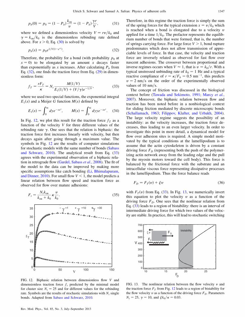
pbð0Þ ¼ p0 ¼ ð1� PbÞ konv ¼ ð1� PbÞ�rV ; (31)
where we defined a dimensionless velocity V ¼ rv=k0 and� ¼ kon=k0 is the dimensionless rebinding rate definedabove. For x > 0, Eq. (30) is solved by
pbðxÞ ¼ p0e1=Vð1�erxÞ: (32)
Therefore, the probability for a bond (with probability p0 atx ¼ 0) to be elongated by an amount x decays fasterthan exponentially as x increases. After calculating Pb fromEq. (32), one finds the traction force from Eq. (29) in dimen-sionless form:
fT ¼ rFT
�¼ Nt
Mð1=VÞE1ð1=VÞ þ ðV=�Þe�1=V
; (33)
where we used two special functions, the exponential integralE1ðxÞ and a Meijer G function MðxÞ defined by
E1ðxÞ ¼Z 1
0dye�xey ; MðxÞ ¼
Z 1
0dyye�xey : (34)
In Fig. 12, we plot this result for the traction force fT as afunction of the velocity V for three different values of therebinding rate �. One sees that the relation is biphasic: thetraction force first increases linearly with velocity, but thendecays again after going through a maximum value. Thesymbols in Fig. 12 are the results of computer simulationsfor stochastic models with the same number of bonds (Sabassand Schwarz, 2010). The analytical result from Eq. (33)agrees with the experimental observation of a biphasic rela-tion in retrograde flow (Gardel, Sabass et al., 2008). The fit ofthe model to the data can be improved by making morespecific assumptions like catch bonding (Li, Bhimalapuram,and Dinner, 2010). For small flow V < 1, the model predicts alinear relation between flow speed and traction force asobserved for flow over mature adhesions:
Ft ¼ Ntkonkon þ k0
�v
k0: (35)
Therefore, in this regime the traction force is simply the sumof the spring forces for the typical extension x ¼ v=k0 whichis reached when a bond is elongated due to a velocity vapplied for a time 1=k0. The prefactor represents the equilib-rium number of bonds that were formed, that is, the numberof springs carrying force. For large force V > 1, bond rupturepredominates which does not allow transmission of appre-ciable levels of force. In that case, the velocity and tractionforce are inversely related as observed for fast flow overnascent adhesions. The crossover between proportional andinverse regimes occurs when V � 1, that is v ¼ k0=r. With atypical unstressed unbinding rate of k0 ¼ 1 Hz and a typicalreactive compliance of r ¼ �=Fb ¼ 0:5 nm�1, this predictsv ¼ 2 nm=s on the order of the experimentally observedvalues of 10 nm=s.
The concept of friction was discussed in the biologicalcontext before (Tawada and Sekimoto, 1991; Marcy et al.,2007). Moreover, the biphasic relation between flow andtraction has been noted before in a nonbiological contextfor sliding friction mediated by discrete microscopic bonds(Schallamach, 1963; Filippov, Klafter, and Urbakh, 2004).The large velocity regime suggests the possibility of aninstability: as the velocity increases, the traction force de-creases, thus leading to an even larger velocity. In order toinvestigate this point in more detail, a dynamical model forflow over adhesion sites is required. A simple model moti-vated by the typical conditions at the lamellipodium is toassume that the actin cytoskeleton is driven by a constantdriving force FD (representing both the push of the polymer-izing actin network away from the leading edge and the pullby the myosin motors toward the cell body). This force isbalanced by the frictional force with the substrate and anintracellular viscous force representing dissipative processesin the lamellipodium. Thus the force balance reads
FD ¼ FTðvÞ þ �v (36)
with FTðvÞ from Eq. (33). In Fig. 13, we numerically invertthis equation to plot the velocity v as a function of thedriving force FD. One sees that the nonlinear relation fromEq. (33) leads to a region of bistability: there is an interval ofintermediate driving force for which two values of the veloc-ity are stable. In practice, this will lead to stochastic switching
γ γ γ
fT
V
FIG. 12. Biphasic relation between dimensionless flow V and
dimensionless traction force ft predicted by the minimal model
for cluster size Nt ¼ 25 and for different values for the rebinding
rate. Symbols are the results of stochastic simulations with Nt single
bonds. Adapted from Sabass and Schwarz, 2010.
10 15 20 250
100
200
300
400
500
600
700
FD
V
FDξ v
FT
bistability
FIG. 13. The nonlinear relation between the flow velocity v and
the traction force FT from Fig. 12 leads to a region of bistability for
the flow velocity v as a function of the driving force FD. Parameters
Nt ¼ 25, � ¼ 10, and �k0=� ¼ 0:03.
Ulrich S. Schwarz and Samuel A. Safran: Physics of adherent cells 1347
Rev. Mod. Phys., Vol. 85, No. 3, July–September 2013
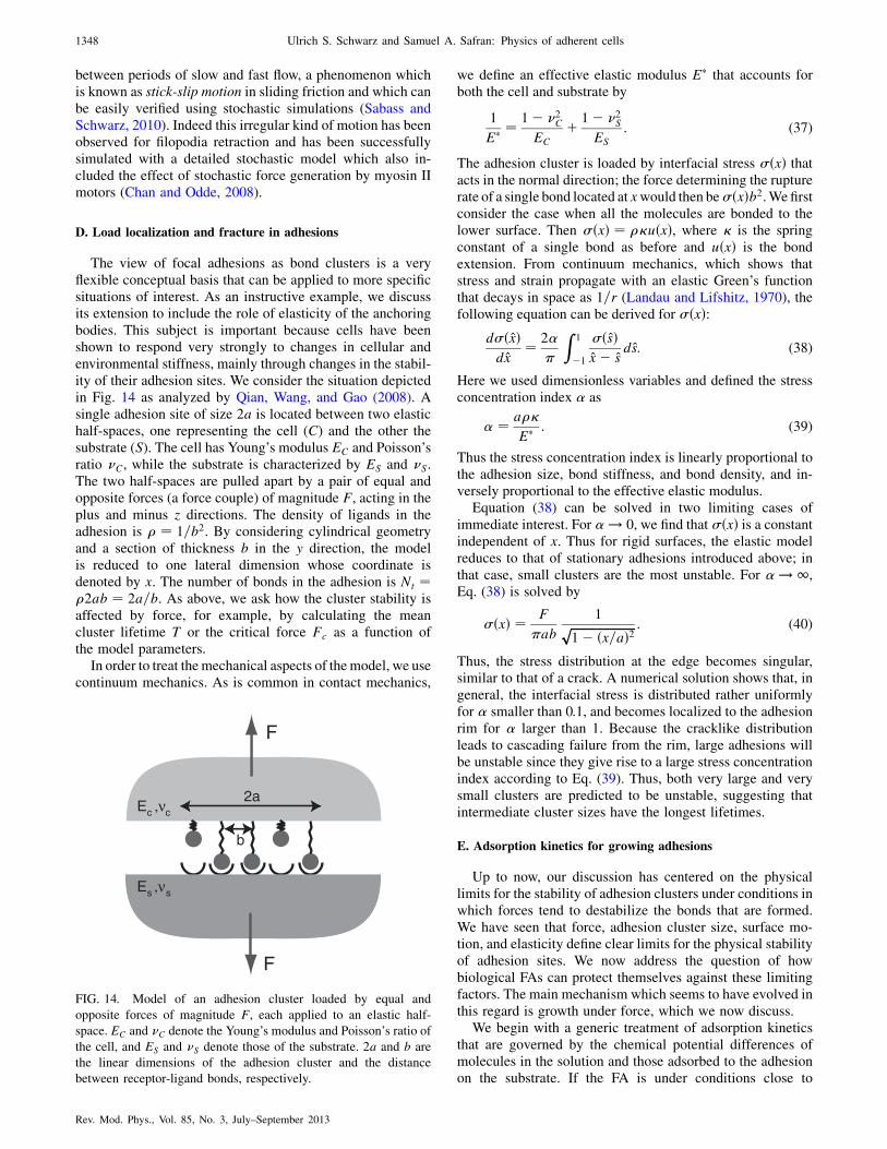
between periods of slow and fast flow, a phenomenon whichis known as stick-slip motion in sliding friction and which canbe easily verified using stochastic simulations (Sabass andSchwarz, 2010). Indeed this irregular kind of motion has beenobserved for filopodia retraction and has been successfullysimulated with a detailed stochastic model which also in-cluded the effect of stochastic force generation by myosin IImotors (Chan and Odde, 2008).
D. Load localization and fracture in adhesions
The view of focal adhesions as bond clusters is a veryflexible conceptual basis that can be applied to more specificsituations of interest. As an instructive example, we discussits extension to include the role of elasticity of the anchoringbodies. This subject is important because cells have beenshown to respond very strongly to changes in cellular andenvironmental stiffness, mainly through changes in the stabil-ity of their adhesion sites. We consider the situation depictedin Fig. 14 as analyzed by Qian, Wang, and Gao (2008). Asingle adhesion site of size 2a is located between two elastichalf-spaces, one representing the cell (C) and the other thesubstrate (S). The cell has Young’s modulus EC and Poisson’sratio C, while the substrate is characterized by ES and S.The two half-spaces are pulled apart by a pair of equal andopposite forces (a force couple) of magnitude F, acting in theplus and minus z directions. The density of ligands in theadhesion is � ¼ 1=b2. By considering cylindrical geometryand a section of thickness b in the y direction, the modelis reduced to one lateral dimension whose coordinate isdenoted by x. The number of bonds in the adhesion is Nt ¼�2ab ¼ 2a=b. As above, we ask how the cluster stability isaffected by force, for example, by calculating the meancluster lifetime T or the critical force Fc as a function ofthe model parameters.
In order to treat themechanical aspects of themodel, we usecontinuum mechanics. As is common in contact mechanics,
we define an effective elastic modulus E that accounts forboth the cell and substrate by
1
E ¼1� 2
C
EC
þ 1� 2S
ES
: (37)
The adhesion cluster is loaded by interfacial stress �ðxÞ thatacts in the normal direction; the force determining the rupturerate of a single bond located at xwould then be�ðxÞb2.We firstconsider the case when all the molecules are bonded to thelower surface. Then �ðxÞ ¼ ��uðxÞ, where � is the springconstant of a single bond as before and uðxÞ is the bondextension. From continuum mechanics, which shows thatstress and strain propagate with an elastic Green’s functionthat decays in space as 1=r (Landau and Lifshitz, 1970), thefollowing equation can be derived for �ðxÞ:
d�ðxÞdx
¼ 2�
Z 1
�1
�ðsÞx� s
ds: (38)
Here we used dimensionless variables and defined the stressconcentration index � as
� ¼ a��
E : (39)
Thus the stress concentration index is linearly proportional tothe adhesion size, bond stiffness, and bond density, and in-versely proportional to the effective elastic modulus.
Equation (38) can be solved in two limiting cases ofimmediate interest. For � ! 0, we find that �ðxÞ is a constantindependent of x. Thus for rigid surfaces, the elastic modelreduces to that of stationary adhesions introduced above; inthat case, small clusters are the most unstable. For � ! 1,Eq. (38) is solved by
�ðxÞ ¼ F
ab
1ffiffiffiffiffiffiffiffiffiffiffiffiffiffiffiffiffiffiffiffiffiffiffi1� ðx=aÞ2p : (40)
Thus, the stress distribution at the edge becomes singular,similar to that of a crack. A numerical solution shows that, ingeneral, the interfacial stress is distributed rather uniformlyfor � smaller than 0.1, and becomes localized to the adhesionrim for � larger than 1. Because the cracklike distributionleads to cascading failure from the rim, large adhesions willbe unstable since they give rise to a large stress concentrationindex according to Eq. (39). Thus, both very large and verysmall clusters are predicted to be unstable, suggesting thatintermediate cluster sizes have the longest lifetimes.
E. Adsorption kinetics for growing adhesions
Up to now, our discussion has centered on the physicallimits for the stability of adhesion clusters under conditions inwhich forces tend to destabilize the bonds that are formed.We have seen that force, adhesion cluster size, surface mo-tion, and elasticity define clear limits for the physical stabilityof adhesion sites. We now address the question of howbiological FAs can protect themselves against these limitingfactors. The main mechanism which seems to have evolved inthis regard is growth under force, which we now discuss.
We begin with a generic treatment of adsorption kineticsthat are governed by the chemical potential differences ofmolecules in the solution and those adsorbed to the adhesionon the substrate. If the FA is under conditions close to
F
F
2aEc c
b
,ν
Es s,ν
FIG. 14. Model of an adhesion cluster loaded by equal and
opposite forces of magnitude F, each applied to an elastic half-
space. EC and C denote the Young’s modulus and Poisson’s ratio of
the cell, and ES and S denote those of the substrate. 2a and b are
the linear dimensions of the adhesion cluster and the distance
between receptor-ligand bonds, respectively.
1348 Ulrich S. Schwarz and Samuel A. Safran: Physics of adherent cells
Rev. Mod. Phys., Vol. 85, No. 3, July–September 2013
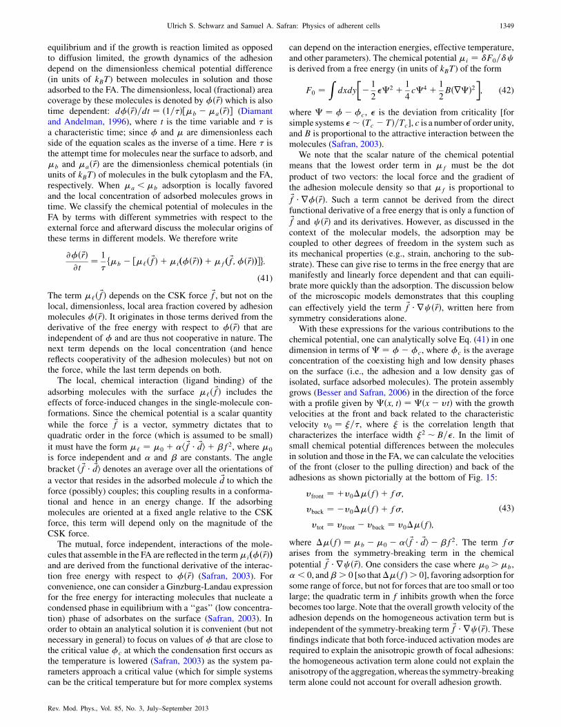
equilibrium and if the growth is reaction limited as opposedto diffusion limited, the growth dynamics of the adhesiondepend on the dimensionless chemical potential difference(in units of kBT) between molecules in solution and thoseadsorbed to the FA. The dimensionless, local (fractional) areacoverage by these molecules is denoted by �ð~rÞ which is alsotime dependent: d�ð~rÞ=dt ¼ ð1=�Þ½�b ��að~rÞ� (Diamantand Andelman, 1996), where t is the time variable and � isa characteristic time; since � and � are dimensionless eachside of the equation scales as the inverse of a time. Here � isthe attempt time for molecules near the surface to adsorb, and�b and �að~rÞ are the dimensionless chemical potentials (inunits of kBT) of molecules in the bulk cytoplasm and the FA,respectively. When �a < �b adsorption is locally favoredand the local concentration of adsorbed molecules grows intime. We classify the chemical potential of molecules in theFA by terms with different symmetries with respect to theexternal force and afterward discuss the molecular origins ofthese terms in different models. We therefore write
@�ð~rÞ@t
¼ 1
�f�b � ½�‘ð ~fÞ þ�ið�ð~rÞÞþ�fð ~f;�ð~rÞÞ�g:
(41)
The term �‘ð ~fÞ depends on the CSK force ~f, but not on thelocal, dimensionless, local area fraction covered by adhesionmolecules �ð~rÞ. It originates in those terms derived from thederivative of the free energy with respect to �ð~rÞ that areindependent of � and are thus not cooperative in nature. Thenext term depends on the local concentration (and hencereflects cooperativity of the adhesion molecules) but not onthe force, while the last term depends on both.
The local, chemical interaction (ligand binding) of the
adsorbing molecules with the surface �‘ð ~fÞ includes theeffects of force-induced changes in the single-molecule con-formations. Since the chemical potential is a scalar quantity
while the force ~f is a vector, symmetry dictates that toquadratic order in the force (which is assumed to be small)
it must have the form �‘ ¼ �0 þ �h ~f � ~di þ �f2, where �0
is force independent and � and � are constants. The angle
bracket h ~f � ~di denotes an average over all the orientations of
a vector that resides in the adsorbed molecule ~d to which theforce (possibly) couples; this coupling results in a conforma-tional and hence in an energy change. If the adsorbingmolecules are oriented at a fixed angle relative to the CSKforce, this term will depend only on the magnitude of theCSK force.
The mutual, force independent, interactions of the mole-cules that assemble in the FA are reflected in the term�ið�ð~rÞÞand are derived from the functional derivative of the interac-tion free energy with respect to �ð~rÞ (Safran, 2003). Forconvenience, one can consider a Ginzburg-Landau expressionfor the free energy for interacting molecules that nucleate acondensed phase in equilibrium with a ‘‘gas’’ (low concentra-tion) phase of adsorbates on the surface (Safran, 2003). Inorder to obtain an analytical solution it is convenient (but notnecessary in general) to focus on values of � that are close tothe critical value �c at which the condensation first occurs asthe temperature is lowered (Safran, 2003) as the system pa-rameters approach a critical value (which for simple systemscan be the critical temperature but for more complex systems
can depend on the interaction energies, effective temperature,and other parameters). The chemical potential�i ¼ F0=cis derived from a free energy (in units of kBT) of the form
F0 ¼Z
dxdy
�� 1
2��2 þ 1
4c�4 þ 1
2Bðr�Þ2
�; (42)
where � ¼ ���c, � is the deviation from criticality [forsimple systems �� ðTc � TÞ=Tc], c is a number of order unity,and B is proportional to the attractive interaction between themolecules (Safran, 2003).
We note that the scalar nature of the chemical potentialmeans that the lowest order term in �f must be the dot
product of two vectors: the local force and the gradient ofthe adhesion molecule density so that �f is proportional to~f � r�ð~rÞ. Such a term cannot be derived from the directfunctional derivative of a free energy that is only a function of~f and c ð~rÞ and its derivatives. However, as discussed in thecontext of the molecular models, the adsorption may becoupled to other degrees of freedom in the system such asits mechanical properties (e.g., strain, anchoring to the sub-strate). These can give rise to terms in the free energy that aremanifestly and linearly force dependent and that can equili-brate more quickly than the adsorption. The discussion belowof the microscopic models demonstrates that this coupling
can effectively yield the term ~f � rc ð~rÞ, written here fromsymmetry considerations alone.
With these expressions for the various contributions to thechemical potential, one can analytically solve Eq. (41) in onedimension in terms of� ¼ ���c, where �c is the averageconcentration of the coexisting high and low density phaseson the surface (i.e., the adhesion and a low density gas ofisolated, surface adsorbed molecules). The protein assemblygrows (Besser and Safran, 2006) in the direction of the forcewith a profile given by �ðx; tÞ ¼ �ðx� vtÞ with the growthvelocities at the front and back related to the characteristicvelocity v0 ¼ �=�, where � is the correlation length thatcharacterizes the interface width �2 � B=�. In the limit ofsmall chemical potential differences between the moleculesin solution and those in the FA, we can calculate the velocitiesof the front (closer to the pulling direction) and back of theadhesions as shown pictorially at the bottom of Fig. 15:
vfront ¼ þv0��ðfÞ þ f�;
vback ¼ �v0��ðfÞ þ f�;
vtot ¼ vfront � vback ¼ v0��ðfÞ;(43)
where ��ðfÞ ¼ �b ��0 � �h ~f � ~di � �f2. The term f�arises from the symmetry-breaking term in the chemical
potential ~f � rc ð~rÞ. One considers the case where �0 >�b,� < 0, and�> 0 [so that��ðfÞ> 0], favoring adsorption forsome range of force, but not for forces that are too small or toolarge; the quadratic term in f inhibits growth when the forcebecomes too large. Note that the overall growth velocity of theadhesion depends on the homogeneous activation term but is
independent of the symmetry-breaking term ~f � rc ð~rÞ. Thesefindings indicate that both force-induced activation modes arerequired to explain the anisotropic growth of focal adhesions:the homogeneous activation term alone could not explain theanisotropy of the aggregation,whereas the symmetry-breakingterm alone could not account for overall adhesion growth.
Ulrich S. Schwarz and Samuel A. Safran: Physics of adherent cells 1349
Rev. Mod. Phys., Vol. 85, No. 3, July–September 2013
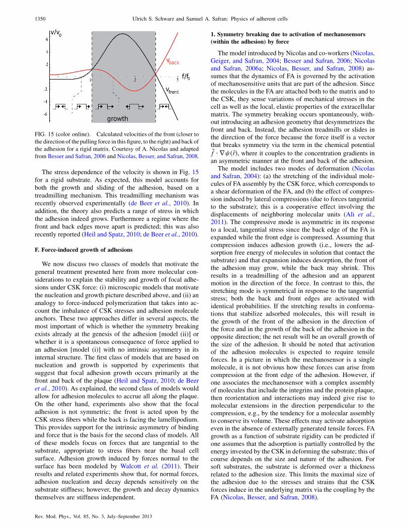
The stress dependence of the velocity is shown in Fig. 15for a rigid substrate. As expected, this model accounts forboth the growth and sliding of the adhesion, based on atreadmilling mechanism. This treadmilling mechanism wasrecently observed experimentally (de Beer et al., 2010). Inaddition, the theory also predicts a range of stress in whichthe adhesion indeed grows. Furthermore a regime where thefront and back edges move apart is predicted; this was alsorecently reported (Heil and Spatz, 2010; de Beer et al., 2010).
F. Force-induced growth of adhesions
We now discuss two classes of models that motivate thegeneral treatment presented here from more molecular con-siderations to explain the stability and growth of focal adhe-sions under CSK force: (i) microscopic models that motivatethe nucleation and growth picture described above, and (ii) ananalogy to force-induced polymerization that takes into ac-count the imbalance of CSK stresses and adhesion moleculeanchors. These two approaches differ in several aspects, themost important of which is whether the symmetry breakingexists already at the genesis of the adhesion [model (ii)] orwhether it is a spontaneous consequence of force applied toan adhesion [model (i)] with no intrinsic asymmetry in itsinternal structure. The first class of models that are based onnucleation and growth is supported by experiments thatsuggest that focal adhesion growth occurs primarily at thefront and back of the plaque (Heil and Spatz, 2010; de Beeret al., 2010). As explained, the second class of models wouldallow for adhesion molecules to accrue all along the plaque.On the other hand, experiments also show that the focaladhesion is not symmetric; the front is acted upon by theCSK stress fibers while the back is facing the lamellipodium.This provides support for the intrinsic asymmetry of bindingand force that is the basis for the second class of models. Allof these models focus on forces that are tangential to thesubstrate, appropriate to stress fibers near the basal cellsurface. Adhesion growth induced by forces normal to thesurface has been modeled by Walcott et al. (2011). Theirresults and related experiments show that, for normal forces,adhesion nucleation and decay depends sensitively on thesubstrate stiffness; however, the growth and decay dynamicsthemselves are stiffness independent.
1. Symmetry breaking due to activation of mechanosensors
(within the adhesion) by force
The model introduced by Nicolas and co-workers (Nicolas,Geiger, and Safran, 2004; Besser and Safran, 2006; Nicolasand Safran, 2006a; Nicolas, Besser, and Safran, 2008) as-sumes that the dynamics of FA is governed by the activationof mechanosensitive units that are part of the adhesion. Sincethe molecules in the FA are attached both to the matrix and tothe CSK, they sense variations of mechanical stresses in thecell as well as the local, elastic properties of the extracellularmatrix. The symmetry breaking occurs spontaneously, with-out introducing an adhesion geometry that desymmetrizes thefront and back. Instead, the adhesion treadmills or slides inthe direction of the force because the force itself is a vectorthat breaks symmetry via the term in the chemical potential~f � rc ð~rÞ, where it couples to the concentration gradients inan asymmetric manner at the front and back of the adhesion.
The model includes two modes of deformation (Nicolasand Safran, 2004): (a) the stretching of the individual mole-cules of FA assembly by the CSK force, which corresponds toa shear deformation of the FA, and (b) the effect of compres-sion induced by lateral compressions (due to forces tangentialto the substrate); this is a cooperative effect involving thedisplacements of neighboring molecular units (Ali et al.,2011). The compressive mode is asymmetric in its responseto a local, tangential stress since the back edge of the FA isexpanded while the front edge is compressed. Assuming thatcompression induces adhesion growth (i.e., lowers the ad-sorption free energy of molecules in solution that contact thesubstrate) and that expansion induces desorption, the front ofthe adhesion may grow, while the back may shrink. Thisresults in a treadmilling of the adhesion and an apparentmotion in the direction of the force. In contrast to this, thestretching mode is symmetrical in response to the tangentialstress; both the back and front edges are activated withidentical probabilities. If the stretching results in conforma-tions that stabilize adsorbed molecules, this will result inthe growth of the front of the adhesion in the direction ofthe force and in the growth of the back of the adhesion in theopposite direction; the net result will be an overall growth ofthe size of the adhesion. It should be noted that activationof the adhesion molecules is expected to require tensileforces. In a picture in which the mechanosensor is a singlemolecule, it is not obvious how these forces can arise fromcompression at the front edge of the adhesion. However, ifone associates the mechanosensor with a complex assemblyof molecules that include the integrins and the protein plaque,then reorientation and interactions may indeed give rise tomolecular extensions in the direction perpendicular to thecompression, e.g., by the tendency for a molecular assemblyto conserve its volume. These effects may activate adsorptioneven in the absence of externally generated tensile forces. FAgrowth as a function of substrate rigidity can be predicted ifone assumes that the adsorption is partially controlled by theenergy invested by the CSK in deforming the substrate; this ofcourse depends on the size and nature of the adhesion. Forsoft substrates, the substrate is deformed over a thicknessrelated to the adhesion size. This limits the maximal size ofthe adhesion due to the stresses and strains that the CSKforces induce in the underlying matrix via the coupling by theFA (Nicolas, Besser, and Safran, 2008).
FIG. 15 (color online). Calculated velocities of the front (closer to
the direction of the pulling force in this figure, to the right) and back of
the adhesion for a rigid matrix. Courtesy of A. Nicolas and adapted
from Besser and Safran, 2006 and Nicolas, Besser, and Safran, 2008.
1350 Ulrich S. Schwarz and Samuel A. Safran: Physics of adherent cells
Rev. Mod. Phys., Vol. 85, No. 3, July–September 2013

A related point of viewwas recently presented byGarikipatiand collaborators (Olberding et al., 2010) who used a general
thermodynamic argument to motivate the symmetry-breakingterm in �f. Instead of assuming that the adhesion molecules
are activated to adsorb near regions of compression and desorbnear regions of expansion as in Nicolas and Safran (2006a) andNicolas, Besser, and Safran (2008), the ‘‘negative work’’ done
by the CSK pulling forces is included in the energetics at theoutset. The resulting energy is decreased when the adhesionmoves in the direction of the force (toward the nucleus); this
apparent sliding occurs by the adsorption of molecules at theedge of the adhesion that is closest to the nucleus (or directionof applied force) and desorption at the other end. We note that
this mechanism can lead to growth all along the adhesionsurface and not just at the edges as discussed in relation tothe work of Shemesh et al. (2005).
Another model that focuses on the integrin binding with theadhesion was suggested by Deshpande and co-workers
(Deshpande, McMeeking, and Evans, 2006, 2007; Pathaket al., 2008; McGarry et al., 2009) who developed a thermody-namically motivated computational approach that has three
essential features: (i) coexistence of both low and high affinityintegrins in thermodynamic equilibrium, (ii) mobility of thelow affinity integrins within the plasma membrane, and(iii) mechanical equilibrium of the contractile forces generated
by the stress fibers—these forces affect the free energies of theintegrins and give rise to a coupled thermomechanical response.An initial prediction based on this is the correlation of the
distributions of the normalized focal adhesion densities (asparametrized by the high affinity integrin concentration) andcontours of the stress-fiber density. However, this model does
not contain spontaneous symmetry breaking and the growth ofadhesions in the direction of the applied force requires thead hoc inclusion of anisotropic activation signals.
Another recent model of focal adhesion growth focuses onthe bond attachment-reattachment dynamics previously dis-
cussed and combines this with considerations related to clus-tering. The stochastic elastic model combines theory andMonte Carlo simulations (Gao, Qian, and Chen, 2011) and
suggests that FA growth is self-limiting since growth eventu-ally leads to cracklike delamination failure near the adhesionedges. Very soft substrates tend to diminish the adaptivecapability of cells by suppressing bond rebinding irrespective
of the cytoskeleton stiffness, which may prevent short-lived,small focal contacts from maturing into stable FA.
2. Symmetry breaking due to the geometrical structure of the
adhesion
The model introduced by Kozlov and co-workers (Shemeshet al., 2005) uses a general thermodynamic argument to write
�f ¼ � ~f � ~d, where ~f is the force applied by the CSK to one
adhesion molecule and ~d is a particular bond in the moleculethat is stretched by that force. In the one-dimensional versionof the model described by Shemesh et al. (2005), the force and
bond are in the same directions. This energy represents thenegative work done by the CSK against the adhesion when anadditional protein is allowed to adsorb. In the absence of thisadditional protein, the adhesion is stretched and deformed by
the CSK force; the presence of an additional protein adsorbedfrom solution relaxes this deformation to some extent and thus
lowers the free energy of the adhesion, hence the negative signin the expression for �f.
To predict the growth of the adhesion in the direction of theapplied force, several assumptions are made. First, the FA isassumed to be capable of adsorbing and releasing adhesionmolecules at every point along its surface; this differs frommodels of polymerization where the monomer exchangeoccurs only at the ends of a linear polymer. For the FA plaque,plausibility of this unusual property has some experimentalsupport (von Wichert et al., 2003). However, other studieshave shown that the growth primarily occurs at the two endsof the adhesion and not uniformly along its surface (Heil andSpatz, 2010; de Beer et al., 2010). Second, the pulling forces~f are assumed to be applied to the FA surface at discretepoints that are distributed along the FA length with a particu-lar density. Finally, the FA plaque is taken to be anchored to arigid external substrate by discrete linkers spread over theadhesions length with a density that differs from the densityof the pulling forces. This intrinsic asymmetry in the geome-try of the adhesion leads to its asymmetric growth.
The interplay between the distribution of the pulling forcesand the anchors leads to an inhomogeneous stretching stresswithin the FA and, consequently, to an uneven distribution ofthe chemical potential �f along the adhesion. Hence, the
tendency to assemble or disassemble can vary along the FAresulting in different regimes of the overall molecular ex-change between the FAand the adhesionmolecules in solution.
The model predicts different modes of the FA assembly thatare largely consistent with the experimentally observed FAbehavior. As a function of the chemical potential difference,the FA can exhibit unlimited growth, complete disintegration,or reach a stable force-dependent steady-state dimension. Thesliding of the adhesion in the direction of the force is explainedby an explicit symmetry breaking introduced in this model dueto an imbalance of local forces and anchors; this requiresadditional specific modifications of the generic model andmay be due to the presence of the lamellipodium on the farside (close to the cell membrane) of the adhesion and the stressfibers on its near side (closer to the nucleus). In contrast to othermodels of FA mechanosensing, the thermodynamic modeldoes not require any special conformational changes of pro-teins upon force application. At the same time, the modelassumes that the FA plaque is an elastic body able to accumu-late mechanical stresses. Moreover, the plaque must possessa mechanism to accommodate new FA proteins withoutundergoing stretch-induced rupture. This may require thepresence of delicate molecular mechanisms having propertiessimilar to those of the members of the formin protein family(Kovar, 2006), which are able to maintain a stable connectionto an associated protein complex (the growing ends of actinfilaments) and at the same time, enable insertion of the newprotein monomers into the complex and stabilization of thegrowing structure.
V. CELL SHAPE AND FORCES
A. Physical motivation
We now move from the level of cellular adhesions to thelevel of whole cells and address the question of how theforces in an adherent cell are balanced over the entire cell.
Ulrich S. Schwarz and Samuel A. Safran: Physics of adherent cells 1351
Rev. Mod. Phys., Vol. 85, No. 3, July–September 2013

From Sec. IV, we learned that cells adhere through relatively
few but stable sites of focal adhesion. At the same time, they
tend to be very contractile provided that the environment is
sufficiently rigid to balance these forces. The interplay be-
tween contractility and spatially localized adhesions leads to
interesting phenomena which can be understood best by first
considering the shape of the cells.Depending on cell type and environment, cells adopt a
large variety of different shapes (Bray, 2001; Mogilner and
Keren, 2009; Kollmannsberger et al., 2011). For cells in the
human body, for example, we observe such diverse shapes as
the biconcave disk of the red blood cell, the invaginated
shapes of single fibroblasts in connective tissue, the polygo-
nal shapes of cells in densely packed epithelial tissue, and the
highly branched networks formed by neurons in the brain. In
fact the term cell was coined by Robert Hooke who in his
1665 book Micrographia was the first to report on the many
shapes visible under the microscope. He chose this term cell
because the near rectangular shapes of the building blocks of
cork reminded him of monk cells in a monastery. Because of
the evolutionary process, the shape of cells is closely related
to their function. For example, red blood cells are optimized
to squeeze through narrow capillaries, fibroblasts are suffi-
ciently contractile to deform and remodel extracellular ma-
trix, epithelial cells pack tightly to seal certain regions, and
neurons form a highly connected communication network.In 1917 D’Arcy Thompson suggested in his book On
Growth and Form that the shapes of cells and organisms
must be closely related to physical forces (Thompson,
1992). The interest in shape somehow declined in the wake
of the molecular revolution in biology, but has recently been
reinvigorated by the finding that cell shape strongly effects
cellular function. By using microcontact printing of adhesive
patterns to constrain cells into predefined shapes, it has been
shown that it is not the total amount of adhesive ligand
available to the cell, but rather its spatial distribution that
determines cell fate (Chen et al., 1997). In fact, relatively
little ligand is sufficient to ensure cell survival if it is arranged
in such a way as to allow the cell to spread over a large area
on the substrate. In contrast, a cell constrained to occupy a
small area or volume goes into programmed cell death even if
many ligands are available in that small space. More recently,
it has also been shown that stem-cell differentiation depends
on cell shape (Kilian et al., 2010).During the last decade, the use of micropatterned sub-
strates has developed into a standard technique employed to
investigate many details of the spatial organization of cells.
For example, it was shown that the spatial coordination of
lamellipodia (Parker et al., 2002), stress fibers (Thery et al.,
2006), spindle formation (Thery et al., 2007; Fink et al.,
2011), and cell-cell adhesions (Tseng et al., 2012) strongly
depends on the geometry of the extracellular environment.
Moreover, micropatterned substrates can also be used to
quantitatively analyze cell shape. It has long been noticed
that on flat substrates most tissue cell types adopt shapes that
are indicative of cell contraction, often characterized by
invaginations between pinning sites (Zand and Albrecht-
Buehler, 1989). The tendency to invaginate becomes even
more pronounced when the actin cytoskeleton is disrupted,
which leads to a raylike morphology of adhering cells
(Bar-Ziv et al., 1999). Recently micropatterning and imageprocessing have been combined to quantitatively study therelation between cell shape and adhesive geometry (Bischofset al., 2008).
In general, many of the observed cell shapes can be under-stood with concepts borrowed from soft matter physics. Theround shape of cells in solution, the invaginated shapes ofsingle tissue cells on flat substrates, and the foamlike packingof cells in epithelial tissue point to a strong role of cellulartension; see Figs. 16(a)–16(c). Tension also plays an impor-tant role in many dynamical situations, such as the pearling ofcell extensions after changes in pressure (Pullarkat et al.,2006) or cell elasticity (Bar-Ziv et al., 1999), as inFig. 16(d). Tensions that contract cell-cell junctions haveemerged as a key factor that determines the dynamics oftissues (Farhadifar et al., 2007; Lecuit and Lenne, 2007;Paluch and Heisenberg, 2009; Aliee et al., 2012). However,in order to quantitatively explain the experimental data forsingle cells, tension arguments must be combined with ele-ments of elasticity (Bar-Ziv et al., 1999; Bischofs et al.,2008). During recent years, different modeling approacheshave been suggested to describe the interplay betweenmyosin II contractility which is balanced by elastic forcesexerted by the cytoskeleton and the adhesions (that couple tothe substrate). In the following we discuss and compare someof the suggested approaches. This then forms a basis thatallows us to consider even more coarse-grained models ofcells (cellular force dipoles) in the next section.
B. Contour models for cell shape
Because adhering cells on flat substrates spread to becomevery thin compared with their lateral extensions, it is appro-priate to describe them as approximately two-dimensionalobjects; see Fig. 17(a). The simplest approach is to focus onlyon cell shape and to consider only the two-dimensionalcontour ~rðsÞ describing the cell boundary with s (which hasunits of length) defined as the distance coordinate along thecontour. At any point s along the boundary, we define thetangent vector ~tðsÞ ¼ ½d~rðsÞ=ds�=jd~rðsÞ=dsj and the normal
(a) (b)
(c) (d)
FIG. 16. Cell shapes are often dominated by tension. (a) Single
cells in solution are usually round. (b) Isolated tissue cells like
fibroblasts commonly show an invaginated shape between distinct
sites of adhesion. (c) Epithelial cells in closely packed tissue are
usually polygonal, in both two and three dimensions (here a
polyhedron is schematically depicted for the 3D case).
(d) Cylindrical cell extensions such as axons tend to pearl when
the tension is increased.
1352 Ulrich S. Schwarz and Samuel A. Safran: Physics of adherent cells
Rev. Mod. Phys., Vol. 85, No. 3, July–September 2013

vector ~nðsÞ perpendicular to it. These two unit vectors areconnected by the geometrical relation d~tðsÞ=ds ¼ ~nðsÞ=RðsÞ,where RðsÞ is the local radius of curvature. We first considerthe simplest model geometry possible, namely, a contourwhich is pulled in toward the cell in the region betweentwo adhesion points, as shown in Fig. 17(b). We relate thisto the forces that arise from actomyosin contractility in cells.
We begin with the most elementary example, the simpletension model, in which these forces arise from the energiesassociated with changes in the contour length and surfacearea. In the simple tension model, a constant surface tension� pulls in the contour (thereby reducing the surface area) andis balanced by the effects of a constant line tension � whichtends to straighten the contour (thereby reducing the linelength); see Fig. 17(b). The surface tension acts on a lineelement and points in the normal direction, leading to a force~F ¼ � ~nðsÞds, while the line tension acts on every point of thecontour and in the tangential direction with a force ~T ¼ �~tðsÞ.This situation is depicted in Fig. 17(c). The force balance on acontour element ds then leads to a Laplace law:
~F ¼ ~Tðsþ dsÞ � ~TðsÞ ) � ~n ¼ �d~t
ds¼ �
R~n ) R ¼ �
�:
(44)
Note that this result is expected from dimensional analysis.The simple tension model thus predicts that the contour formsperfect circular arcs, which indeed is often observed in cellexperiments. In Figs. 18(a) and 18(b), this is demonstrated forcells on a dot micropattern and on a pillar array, respectively.Note that, due to its local nature, the simple tension modeldoes not obey total momentum conservation that is expectedfor a closed system like a single cell.
The last equality in Eq. (44) resembles the Laplace lawR ¼ ð2�Þ=�p for a sphere (e.g., a soap bubble) whosesurface is contracted by a surface tension � and stabilizedby a pressure difference �p (compare Sec. II.F). We note thatin three dimensions � and � (related to line and surface) arereplaced by � and �p (related to surface and volume),respectively, and that a factor of 2 appears (compareSec. II.F). These two effects obviously result from the differ-ent dimensionalities. However, a more fundamental differ-ence is the fact that while the sphere stabilizes itself withoutany need for attachment, the simple tension model for theinvaginated contour makes sense only in the presence of thetwo adhesion sites. Without the adhesions, both tensionswould work in the same direction and the contour wouldsimply contract to a point.
In single cells adhered to a flat substrate, the two tensionshave contributions from different processes. The surfacetension � mainly results from the pull of the myosin motorsin the actin cytoskeleton (including the actin cortex), but canalso have a contribution from the tension in the plasmamembrane. The line tension � is expected to primarily arisefrom the elastic pull of the thick and contractile actin bundleslining the cell periphery. If cells are treated with pharmaco-logical drugs that disrupt the actin cytoskeleton, they tend toinvaginate more strongly, indicating that effectively the linetension � is reduced more than the surface tension � (Bar-Zivet al., 1999; Bischofs et al., 2008).
Interestingly, very strong invaginations necessarily lead totubelike extensions connecting the retracted cell body to thesites of adhesions (here we assume a three-dimensional view-point again). A cylindrical tube in which the surface tension isthe only relevant force undergoes a Rayleigh-Plateau insta-bility (Safran, 2003). Thus one also expects pearling of thecellular tubes. Indeed this is exactly what has been observedexperimentally (Bar-Ziv et al., 1999) once the elasticity of thetube is suppressed, similar to the pearling which can beinduced, for example, in axons, by changing the osmoticpressure (Pullarkat et al., 2006).
For contractile cells, the force due to the surface tension� is mainly balanced by the elasticity of the actin cortex
dL
R
n(s)dst(s)λ
σF=
T=
(b)σ λ
λ
(a)
(c) (d)
FIG. 17. Simple tension model for cell shape on a flat substrate.
(a) The cell is very flat and therefore effectively two dimensional.
The outlined region is shown in (b) with more detail. (b) Along the
contour between two neighboring adhesion sites, surface tension �pulls inward, while line tension � pulls tangentially. (c) Along the
contour, surface tension and line tension pull in normal and
tangential directions, respectively. For a circular arc, radius R,contour length L, and spanning distance d are geometrically related
to each other. (d) For a house-shaped cell, all arcs have the same
radius, although the spanning distance is larger on the diagonals.
Also shown are the traction forces derived from the shape model.
Adapted from Bischofs, Schmidt, and Schwarz, 2009 and Guthardt
Torres, Bischofs, and Schwarz, 2012.
(a) (b)
FIG. 18 (color online). (a) House-shaped cell on an adhesive
micropattern created by microcontact printing. Note the circular
arcs with radii that are larger at the two diagonals. From Fig. 1(B)
from Bischofs et al., 2008. (b) Cell on a pillar array that allows a
simple readout of local forces from measurements of the deflection
of each pillar. Circular arcs describe most of the cell contour, but
not, for example, at the upper left corner, where an internal fiber
distorts the contour. From Bischofs, Schmidt, and Schwarz, 2009.
Ulrich S. Schwarz and Samuel A. Safran: Physics of adherent cells 1353
Rev. Mod. Phys., Vol. 85, No. 3, July–September 2013

underlying the plasma membrane. Experimentally, it wasfound that thinner tubes are unstable whereas thicker onesare not. This can be explained as follows. One considersa cylindrical tube with undulations by defining a localradius that varies along the tube axis in the z direction:RðzÞ ¼ R0 þ �R cosð2z=�Þ. There are two energy contri-butions, the change in surface energy and an elastic energythat includes bending and stretching energies. Using theconstraint of volume conservation the sum of these twoenergies reads (Bar-Ziv et al., 1999)
Et ¼ �1
4u2ðk2 � 1Þ þ 1
4
3ER0
1þ u2: (45)
Here k ¼ 2R0=� and u ¼ �R=R0 are the dimensionlesswave number and amplitude of a perturbation of wave vectork, respectively, while E and are the Young’s modulus andPoisson’s ratio of the cortex, respectively. Because both termshave the same scaling with u, these terms cannot determinethe amplitude. However, the wavelength at which the systemis unstable is determined by this expression. The first termcan become negative for large wavelengths k < 1. In thiscase, the energy is negative if the tension exceeds a criticalthreshold of
�c ¼ 3ER0
1þ (46)
which increases with R0. This makes thicker tubes morestable than thinner ones as observed experimentally.
The pearling study indicates that in cellular systems ten-sion and elasticity are strongly coupled. A similar result wasobtained by a quantitative study of cell shape on micropat-terned substrates (Bischofs et al., 2008). Although this analy-sis revealed that invaginations of the cell contour are usuallyclose to circular as predicted by the Laplace law fromEq. (44), it also showed that the arc radius R varies withthe spanning distance d (defined in Fig. 17) between the twoneighboring adhesion sites, while the Laplace law predicts aconstant radius independent of spanning distance. Again thiscan be explained by an elastic analysis, the tension-elasticitymodel (Bischofs et al., 2008). The circular nature of the arcssuggests that a modified Laplace law must hold. While thesurface tension � is expected to be determined mainly bymyosin motor activity in the bulk cytoskeleton and thereforeshould be the same and constant for all arcs, the line tension �might be determined locally by the mechanics of the periph-eral bundles. The simplest possible model is to take intoaccount the fact that the actin cortex, localized near theline, can behave elastically. The line tension then has acontribution from the stretching of this cortex relative to itsrelaxed state:
� ¼ EAL� L0
L0
; (47)
where the product of three-dimensional modulus E and cross-sectional area A is the effective one-dimensional modulus ofthe bundle, and L and L0 are its actual stretched length andrelaxed length, respectively. Together with the geometricalrelation
sin
�L
2R
�¼ d
2R(48)
between the contour length L, radius R and spanning distanced [compare Fig. 17(c)], one obtains a self-consistent equationfor the arc radius R:
R ¼ EA
�
�2R
L0
arcsin
�d
2R
�� 1
�: (49)
The simplest model assumption for the resting length isL0 ¼ d. A numerical solution of Eq. (49) shows that R is amonotonically increasing function of d as observed experi-mentally. For small invaginations d=R � 1 and one canexpand Eq. (49) to obtain
R ¼ ðEA=24�Þ1=3d2=3: (50)
Thus R increases with the contour rigidity EA and spanningdistance d, but decreases with increasing surface tension �.Note that compared with the Laplace law from Eq. (44), onestill has an inverse relation between radius and surface ten-sion, but now with a different exponent.
C. Whole-cell models
As shown in Fig. 18, contour models can also be used toanalyze the shape of whole cells that are characterized bygeometrically prominent features like circular arcs. Onemodel class that addresses whole cells by construction arecellular Potts models, which have been successfully used toevaluate and predict cell shape on dotlike micropatterns(Vianay et al., 2010), with an example shown in Fig. 19(a).In essence, the cellular Potts model is very similar to thesimple tension model, because its main ingredient is tensionat the interface. There are many situations of interest, how-ever, for which such a simple approach is not sufficient toaccount for cell behavior. One example is the case where theactin cytoskeleton locally reorganizes into contractile bundles(in addition to those that line the cell periphery). However, atheory based only on structural elements visible with standardmicroscopy procedures might not be sufficient as many ob-servations in single cell experiments point to the existence ofa much finer-scale network of additional fibers that coexistwith the stress fibers. The natural theoretical framework forstudying this situation is continuum mechanics. Although
FIG. 19 (color online). Two model approaches for whole cells.
(a) Cellular Potts models use spins on a lattice to simulate the
contour between the cell and its surroundings. From Vianay et al.,
2010. (b) Finite element methods (FEM) can be used to implement
any material law of interest. Contractility is implemented by
thermoelasticity. The vector field represents the direction and
activation level of stress fibers. From Deshpande, McMeeking,
and Evans, 2006.
1354 Ulrich S. Schwarz and Samuel A. Safran: Physics of adherent cells
Rev. Mod. Phys., Vol. 85, No. 3, July–September 2013
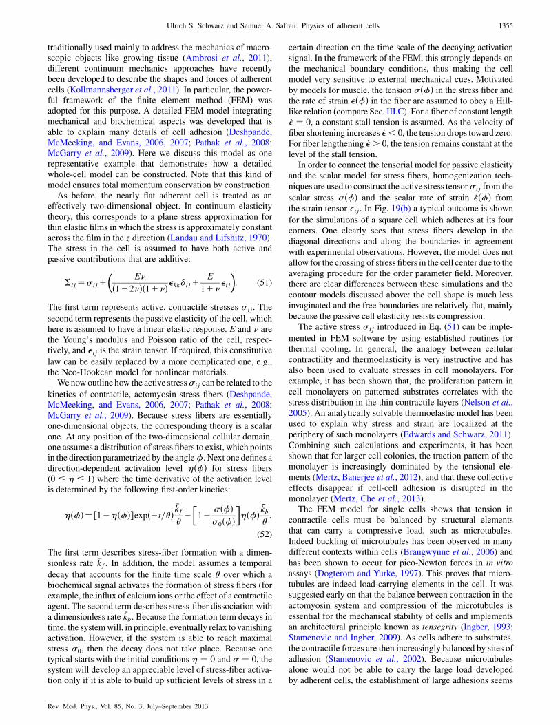
traditionally used mainly to address the mechanics of macro-scopic objects like growing tissue (Ambrosi et al., 2011),different continuum mechanics approaches have recentlybeen developed to describe the shapes and forces of adherentcells (Kollmannsberger et al., 2011). In particular, the power-ful framework of the finite element method (FEM) wasadopted for this purpose. A detailed FEM model integratingmechanical and biochemical aspects was developed that isable to explain many details of cell adhesion (Deshpande,McMeeking, and Evans, 2006, 2007; Pathak et al., 2008;McGarry et al., 2009). Here we discuss this model as onerepresentative example that demonstrates how a detailedwhole-cell model can be constructed. Note that this kind ofmodel ensures total momentum conservation by construction.
As before, the nearly flat adherent cell is treated as aneffectively two-dimensional object. In continuum elasticitytheory, this corresponds to a plane stress approximation forthin elastic films in which the stress is approximately constantacross the film in the z direction (Landau and Lifshitz, 1970).The stress in the cell is assumed to have both active andpassive contributions that are additive:
�ij¼�ijþ�
E
ð1�2 Þð1þ Þ�kkijþ E
1þ �ij
�: (51)
The first term represents active, contractile stresses �ij. The
second term represents the passive elasticity of the cell, whichhere is assumed to have a linear elastic response. E and arethe Young’s modulus and Poisson ratio of the cell, respec-tively, and �ij is the strain tensor. If required, this constitutive
law can be easily replaced by a more complicated one, e.g.,the Neo-Hookean model for nonlinear materials.
We now outline how the active stress�ij can be related to the
kinetics of contractile, actomyosin stress fibers (Deshpande,McMeeking, and Evans, 2006, 2007; Pathak et al., 2008;McGarry et al., 2009). Because stress fibers are essentiallyone-dimensional objects, the corresponding theory is a scalarone. At any position of the two-dimensional cellular domain,one assumes a distribution of stress fibers to exist, which pointsin the direction parametrized by the angle�. Next one defines adirection-dependent activation level �ð�Þ for stress fibers(0 � � � 1) where the time derivative of the activation levelis determined by the following first-order kinetics:
_�ð�Þ¼ ½1��ð�Þ�expð�t=�Þ�kf���1� �ð�Þ
�0ð�Þ��ð�Þ
�kb�:
(52)
The first term describes stress-fiber formation with a dimen-sionless rate �kf. In addition, the model assumes a temporal
decay that accounts for the finite time scale � over which abiochemical signal activates the formation of stress fibers (forexample, the influx of calcium ions or the effect of a contractileagent. The second term describes stress-fiber dissociation witha dimensionless rate �kb. Because the formation term decays intime, the systemwill, in principle, eventually relax to vanishingactivation. However, if the system is able to reach maximalstress �0, then the decay does not take place. Because onetypical starts with the initial conditions � ¼ 0 and � ¼ 0, thesystem will develop an appreciable level of stress-fiber activa-tion only if it is able to build up sufficient levels of stress in a
certain direction on the time scale of the decaying activation
signal. In the framework of the FEM, this strongly depends on
the mechanical boundary conditions, thus making the cell
model very sensitive to external mechanical cues. Motivated
by models for muscle, the tension �ð�Þ in the stress fiber andthe rate of strain _�ð�Þ in the fiber are assumed to obey a Hill-
like relation (compare Sec. III.C). For a fiber of constant length_� ¼ 0, a constant stall tension is assumed. As the velocity of
fiber shortening increases _� < 0, the tension drops toward zero.For fiber lengthening _� > 0, the tension remains constant at the
level of the stall tension.In order to connect the tensorial model for passive elasticity
and the scalar model for stress fibers, homogenization tech-
niques are used to construct the active stress tensor�ij from the
scalar stress �ð�Þ and the scalar rate of strain _�ð�Þ from
the strain tensor �ij. In Fig. 19(b) a typical outcome is shown
for the simulations of a square cell which adheres at its four
corners. One clearly sees that stress fibers develop in the
diagonal directions and along the boundaries in agreement
with experimental observations. However, the model does not
allow for the crossing of stress fibers in the cell center due to the
averaging procedure for the order parameter field. Moreover,
there are clear differences between these simulations and the
contour models discussed above: the cell shape is much less
invaginated and the free boundaries are relatively flat, mainlybecause the passive cell elasticity resists compression.
The active stress �ij introduced in Eq. (51) can be imple-
mented in FEM software by using established routines for
thermal cooling. In general, the analogy between cellular
contractility and thermoelasticity is very instructive and has
also been used to evaluate stresses in cell monolayers. For
example, it has been shown that, the proliferation pattern in
cell monolayers on patterned substrates correlates with the
stress distribution in the thin contractile layers (Nelson et al.,
2005). An analytically solvable thermoelastic model has been
used to explain why stress and strain are localized at the
periphery of such monolayers (Edwards and Schwarz, 2011).
Combining such calculations and experiments, it has been
shown that for larger cell colonies, the traction pattern of the
monolayer is increasingly dominated by the tensional ele-
ments (Mertz, Banerjee et al., 2012), and that these collective
effects disappear if cell-cell adhesion is disrupted in the
monolayer (Mertz, Che et al., 2013).The FEM model for single cells shows that tension in
contractile cells must be balanced by structural elements
that can carry a compressive load, such as microtubules.
Indeed buckling of microtubules has been observed in many
different contexts within cells (Brangwynne et al., 2006) and
has been shown to occur for pico-Newton forces in in vitro
assays (Dogterom and Yurke, 1997). This proves that micro-
tubules are indeed load-carrying elements in the cell. It was
suggested early on that the balance between contraction in the
actomyosin system and compression of the microtubules is
essential for the mechanical stability of cells and implements
an architectural principle known as tensegrity (Ingber, 1993;
Stamenovic and Ingber, 2009). As cells adhere to substrates,
the contractile forces are then increasingly balanced by sites of
adhesion (Stamenovic et al., 2002). Because microtubules
alone would not be able to carry the large load developed
by adherent cells, the establishment of large adhesions seems
Ulrich S. Schwarz and Samuel A. Safran: Physics of adherent cells 1355
Rev. Mod. Phys., Vol. 85, No. 3, July–September 2013

to be a necessary condition for the development of contrac-
tility in adherent cells. In contrast to FEM models that couple
continuum elasticity to discrete, actively contractile stress
fibers, the tensegrity models consider discrete structural ele-
ments such as compression struts connected by tensed cables
as a model of single stress fibers (Luo et al., 2008). Because
they model discrete elements and do not require homogeni-
zation, tensegrity models can be compared more directly with
experiments, for example, when cutting discrete elements
with lasers (Kumar et al., 2006; Luo et al., 2008).Given the success of the contour models in explaining the
appearance of circular arcs of adherent cells, it is interesting to
ask if whole-cell models can predict the same shapes. Recently
it was suggested that this can indeed be achieved by modeling
cells as actively contracting cable networks (Bischofs et al.,
2008; Guthardt Torres, Bischofs, and Schwarz, 2012). This
approach combines elements fromFEMand tensegritymodels.
It considers the extreme case in which contractile forces are
balanced by the stretch response of the cytoskeletal polymers
and by forces from the elastic environment that couple to the
cell at the adhesion sites. The model does not include any
compressive response from within the cell or any area conser-
vationbecause it is assumed that cytoplasm is not constrained to
the 2D plane; the rest of the 3D cell therefore acts as a reservoir
for adhesion area. A 2D mechanical network is constructed
which consists of a set of nodes locally connected by mechani-
cal links. If the links are taken to be linear springs, the network
propagates compression similar to the FEMmodel.However, if
the links are taken to be cables, only tension and not compres-
sion is propagated. This representation is appropriate to the
polymeric nature of the cytoskeleton, whose filaments tend to
buckle or depolymerize under compressive strain (Coughlin
and Stamenovic, 2003). In principle, contraction can be mod-
eled by reducing the relaxed length of the springs or cables.
However, this does not represent the properties of actin bundles
contracted by myosin II minifilaments, which do not have a
well-defined reference state, but contract in a Hill-type fashion
until a certain stall force is reached. A simple way to achieve
this feature is to add a pair of constant forces (force dipole) to
each network link, thus creating permanent contraction be-
tween two connected nodes. With these very simple prescrip-
tions, actively contracting cable networks can be simulated. In
Fig. 20(a), we demonstrate that contracting spring networks do
not result in the circular arc morphology, but rather show a flat
contour as does the FEM model. Circular arcs appear for
actively contracting cable networks, independent of network
topology; compareFigs. 20(b) and20(c).Dependingon the link
density at the boundary, this translates directly into an effective
tension � that acts within the network. Figure 20(d) demon-
strates that the arc radius R scales with network tension � as
predicted by the tension-elasticity model (TEM); see Eq. (50).
Here different symbols correspond to different network top-
ologies and the three sets of curves correspond to three different
spanning distances d. Solid lines correspond to the numerical
solution of Eq. (49) while dashed lines are the analytical results
from Eq. (50). Deviations between computer simulations and
TEM occur only at very large tensions.The continuum mechanics approaches described here are
especially suited to investigate static situations relevant to
mature cell adhesion; however, to treat cell migration, one
must consider a dynamically changing cell shape. A naturalframework for this is hydrodynamics in the overdampedlimit, since cellular flows are characterized by very smallReynolds numbers. For example, the shape of migratingkeratocytes was investigated by a hydrodynamic modelrepresenting the flux of newly polymerized actin networks(Barnhart et al., 2011). Here the shear and compressive forcesin the viscoelastic fluid are balanced by forces arising frommyosin contractility and flow over adhesion sites. Membranetension enters as a boundary condition. A similar approach istaken by active-gel theory, which can be considered as ahydrodynamic theory for polarized active gels (Julicheret al., 2007) that is based only on the symmetries andconservation laws of the system and is independent of anyparticular molecular model. Alternative approaches are levelset (or phase field) models (Shao, Rappel, and Levine, 2010;Ziebert, Swaminathan, and Aranson, 2012) or models incor-porating discrete elements such as single focal adhesions andstress fibers (Shemesh et al., 2009; Loosli, Luginbuehl, andSnedeker, 2010; Shemesh, Bershadsky, and Kozlov, 2012).
D. Force from shape
The various elastic forces predicted from the shape modelscan be verified with experimental measurements, e.g., forcells on soft elastic substrates or on pillar arrays. The simplestpossible evaluation can be obtained by comparing measure-ments with the simple tension model (Bischofs, Schmidt, andSchwarz, 2009). We discuss the forces one might expect fordifferent adhesion geometries. Typical micropatterns can be
10−5
10−4
10−3
10−2
10−1
101
102
(a) (b)
(c) (d)
R
σ
FIG. 20 (color online). (a) A spring network which is constrained
to be located at the four adhesion points indicated (and in which the
equilibrium spring length is smaller than the distance between
neighboring adhesion points divided by the number of springs along
that line), relaxes to a shape similar to the FEM model—that is, one
with relatively flat free edges. Note also the variation in force along
the boundary indicated by the colors. (b) An actively contracting
cable network results in circular arcs and shows hardly any variation
in force along the boundary. (c) The results do not depend on
network topology as shown here for a disordered network. (d) Arc
radius R depends on network tension � as predicted by the tension-
elasticity model. Adapted from Guthardt Torres, Bischofs, and
Schwarz, 2012.
1356 Ulrich S. Schwarz and Samuel A. Safran: Physics of adherent cells
Rev. Mod. Phys., Vol. 85, No. 3, July–September 2013
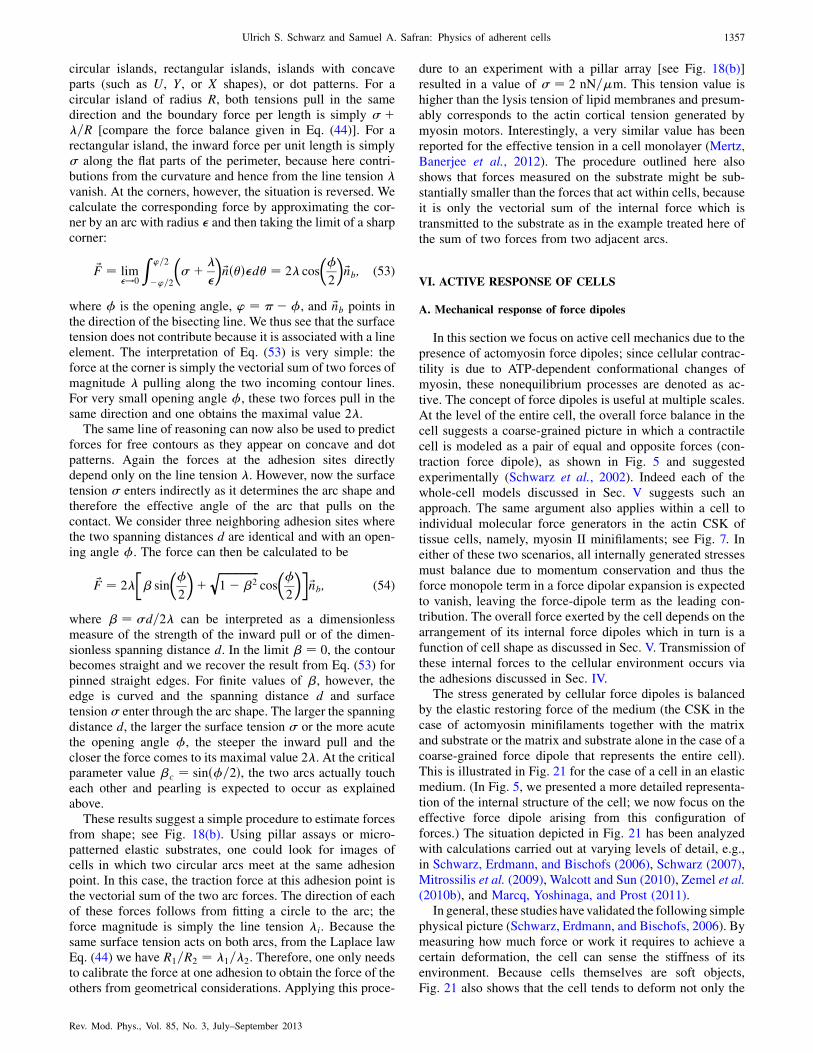
circular islands, rectangular islands, islands with concaveparts (such as U, Y, or X shapes), or dot patterns. For acircular island of radius R, both tensions pull in the samedirection and the boundary force per length is simply �þ�=R [compare the force balance given in Eq. (44)]. For arectangular island, the inward force per unit length is simply� along the flat parts of the perimeter, because here contri-butions from the curvature and hence from the line tension �vanish. At the corners, however, the situation is reversed. Wecalculate the corresponding force by approximating the cor-ner by an arc with radius � and then taking the limit of a sharpcorner:
~F ¼ lim�!0
Z ’=2
�’=2
��þ �
�
�~nð�Þ�d� ¼ 2� cos
��
2
�~nb; (53)
where � is the opening angle, ’ ¼ ��, and ~nb points inthe direction of the bisecting line. We thus see that the surfacetension does not contribute because it is associated with a lineelement. The interpretation of Eq. (53) is very simple: theforce at the corner is simply the vectorial sum of two forces ofmagnitude � pulling along the two incoming contour lines.For very small opening angle �, these two forces pull in thesame direction and one obtains the maximal value 2�.
The same line of reasoning can now also be used to predictforces for free contours as they appear on concave and dotpatterns. Again the forces at the adhesion sites directlydepend only on the line tension �. However, now the surfacetension � enters indirectly as it determines the arc shape andtherefore the effective angle of the arc that pulls on thecontact. We consider three neighboring adhesion sites wherethe two spanning distances d are identical and with an open-ing angle �. The force can then be calculated to be
~F ¼ 2�
�� sin
��
2
�þ
ffiffiffiffiffiffiffiffiffiffiffiffiffiffiffi1� �2
qcos
��
2
��~nb; (54)
where � ¼ �d=2� can be interpreted as a dimensionlessmeasure of the strength of the inward pull or of the dimen-sionless spanning distance d. In the limit � ¼ 0, the contourbecomes straight and we recover the result from Eq. (53) forpinned straight edges. For finite values of �, however, theedge is curved and the spanning distance d and surfacetension � enter through the arc shape. The larger the spanningdistance d, the larger the surface tension � or the more acutethe opening angle �, the steeper the inward pull and thecloser the force comes to its maximal value 2�. At the criticalparameter value �c ¼ sinð�=2Þ, the two arcs actually toucheach other and pearling is expected to occur as explainedabove.
These results suggest a simple procedure to estimate forcesfrom shape; see Fig. 18(b). Using pillar assays or micro-patterned elastic substrates, one could look for images ofcells in which two circular arcs meet at the same adhesionpoint. In this case, the traction force at this adhesion point isthe vectorial sum of the two arc forces. The direction of eachof these forces follows from fitting a circle to the arc; theforce magnitude is simply the line tension �i. Because thesame surface tension acts on both arcs, from the Laplace lawEq. (44) we have R1=R2 ¼ �1=�2. Therefore, one only needsto calibrate the force at one adhesion to obtain the force of theothers from geometrical considerations. Applying this proce-
dure to an experiment with a pillar array [see Fig. 18(b)]resulted in a value of � ¼ 2 nN=�m. This tension value is
higher than the lysis tension of lipid membranes and presum-ably corresponds to the actin cortical tension generated by
myosin motors. Interestingly, a very similar value has beenreported for the effective tension in a cell monolayer (Mertz,Banerjee et al., 2012). The procedure outlined here also
shows that forces measured on the substrate might be sub-stantially smaller than the forces that act within cells, becauseit is only the vectorial sum of the internal force which is
transmitted to the substrate as in the example treated here ofthe sum of two forces from two adjacent arcs.
VI. ACTIVE RESPONSE OF CELLS
A. Mechanical response of force dipoles
In this section we focus on active cell mechanics due to the
presence of actomyosin force dipoles; since cellular contrac-tility is due to ATP-dependent conformational changes ofmyosin, these nonequilibrium processes are denoted as ac-
tive. The concept of force dipoles is useful at multiple scales.At the level of the entire cell, the overall force balance in thecell suggests a coarse-grained picture in which a contractile
cell is modeled as a pair of equal and opposite forces (con-traction force dipole), as shown in Fig. 5 and suggested
experimentally (Schwarz et al., 2002). Indeed each of thewhole-cell models discussed in Sec. V suggests such anapproach. The same argument also applies within a cell to
individual molecular force generators in the actin CSK oftissue cells, namely, myosin II minifilaments; see Fig. 7. Ineither of these two scenarios, all internally generated stresses
must balance due to momentum conservation and thus theforce monopole term in a force dipolar expansion is expected
to vanish, leaving the force-dipole term as the leading con-tribution. The overall force exerted by the cell depends on thearrangement of its internal force dipoles which in turn is a
function of cell shape as discussed in Sec. V. Transmission ofthese internal forces to the cellular environment occurs via
the adhesions discussed in Sec. IV.The stress generated by cellular force dipoles is balanced
by the elastic restoring force of the medium (the CSK in thecase of actomyosin minifilaments together with the matrixand substrate or the matrix and substrate alone in the case of a
coarse-grained force dipole that represents the entire cell).This is illustrated in Fig. 21 for the case of a cell in an elastic
medium. (In Fig. 5, we presented a more detailed representa-tion of the internal structure of the cell; we now focus on theeffective force dipole arising from this configuration of
forces.) The situation depicted in Fig. 21 has been analyzedwith calculations carried out at varying levels of detail, e.g.,
in Schwarz, Erdmann, and Bischofs (2006), Schwarz (2007),Mitrossilis et al. (2009), Walcott and Sun (2010), Zemel et al.(2010b), and Marcq, Yoshinaga, and Prost (2011).
In general, these studies have validated the following simple
physical picture (Schwarz, Erdmann, and Bischofs, 2006). Bymeasuring how much force or work it requires to achieve acertain deformation, the cell can sense the stiffness of its
environment. Because cells themselves are soft objects,Fig. 21 also shows that the cell tends to deform not only the
Ulrich S. Schwarz and Samuel A. Safran: Physics of adherent cells 1357
Rev. Mod. Phys., Vol. 85, No. 3, July–September 2013

environment, but also itself. For two springs in series, theinverse of the effective spring constant is the sum of the inversespring constants. Therefore, if the environment is very stiff, the
cell deforms only itself and cannot sense its surroundings. Onthe other hand, if the environment is very soft, the cell caneasily deform it, but does not build upmuch force and thereforedoes not gain much information; in particular, any positive
feedback triggered by mechanosensors will not work well.Therefore the best working or set point for a cell seems to bea situation in which the two stiffnesses of cell and environmentare nearly matched. Indeed it was found experimentally that
cells tend to match their stiffness to that of the environment(Solon et al., 2007).
In this section, we discuss how active force generators suchas myosin II minifilaments in the actin cytoskeleton or entirecontractile cells in an elastic matrix interact with their me-
chanical environment. The deformations induced in the ma-trix by cell activity allow us to deduce effective interactionsbetween the force dipoles themselves via their mutual effectson the elastic environment. In the case of individual acto-
myosin minifilaments modeled as force dipoles, the elasticenvironment includes the CSK of the cell itself (in addition toits surroundings), while in the case of entire, contractile cells
modeled as force dipoles, the elastic environment is thematrix or substrate. The elastic properties of the environmentcan be easily controlled in experiments so that predictionsand measurements of their role in modulating cellular force-
dipole assemblies provide insight into these fundamentalprocesses.
In order to obtain analytical insight, linear elasticity theoryis applied to an isotropic medium (or substrate) to model themechanical properties of cells and their environment (Landau
and Lifshitz, 1970), although later on, we comment on pos-sible extensions to more detailed models for the elasticity ofbiomaterials, including nonlinear elasticity. Alternatively onecould also employ more microscopic models for the propa-
gation of stress and strain in polymer networks (Head,Levine, and MacKintosh, 2003a, 2003b; Wilhelm and Frey,2003; Heussinger and Frey, 2006; Heussinger, Schaefer, andFrey, 2007), but this may preclude the insight gained by the
use of elastic theory generalized to include force dipoles. Inthe framework employed here, cytoskeletal force generators(e.g., actomyosin minifilaments or entire cells) are viewed as
active, elastic inclusions in (or on) a homogeneous and iso-tropic medium. This suggests the use of Eshelby’s theory ofelastic inclusions that was originally developed in the contextof materials science (Eshelby, 1957, 1959). In the limit inwhich the inclusions are much smaller than the length scalesof interest, one can use the theory of elastic point defects,which also originated in materials science, and has been usedextensively to model the elastic interactions of hydrogen inmetal (Siems, 1968; Wagner and Horner, 1974).
There are a few important assumptions made in thefollowing treatment of force dipoles and cellular-matrixelasticity: (i) The buildup of force occurs on short enoughtime scales (tens of seconds) for which the CSK respondselastically to internal forces and does not flow. This wasdiscussed previously in a comparison of elastic versus flow-ing gel models of the CSK. Even if the matrix flows onlarger time scales, the cell will react on a shorter time scaleby building up new forces via a remodeled CSK. Elasticrelaxation after pharmacological inhibition of actomyosincontractility, laser cutting, or disruption of adhesion (e.g.,by trypsination) proves that adherent cells and matrix arecontinuously under elastic stress. (ii) The matrix-inducedforces that act to organize the cytoskeleton arise from thepotential energy that accounts for the deformation of thematrix by the dipoles. In this sense, matrix can refer either tothose parts of the CSK not included in the actomyosindipoles themselves and/or to the surrounding matrix orsubstrate. However, these forces are taken to determineonly the organization of the dipoles (e.g., nematic or smecticorder) but not their existence or magnitude which depends ina more complex manner on nonequilibrium cellular activity.These same forces would be present and act on artificialforce dipoles were they present in the CSK, matrix, orsubstrate. (iii) The time scale on which elastic signals arepropagated in the CSK or matrix or substrate is faster thaninternal relaxation times due to dissipation arising frominternal viscosity or fluid flow; for experimental measure-ments of these time scales see Fig. 3 of Kollmannsberger andFabry (2011). This allows us to predict force-dipole organi-zation by elastic forces from energetic or force balancearguments. We recognize that this is a crude approximationthat must be tested under various circumstances; a dynami-cal theory of elastic signal propagation in the CSK or matrixor substrate is a topic of current research. (iv) The theoriesbelow focus on cytoskeletal force generation and predictdipole arrangements for a fixed cell shape. Experimentsthat measure the response of cells to time-varying stressesshow (Faust et al., 2011) that cell shape follows cytoskeletalreorientation by several hours.
B. Force dipoles and their interactions
A simplified model of a contractile actomyosin unit (or in acoarse-grained picture, an entire, polarized cell) is that of a
source of two equal and opposite forces ~F, separated by a
nanoscale (or, for cells, micrometer scale) distance ~d. Byanalogy with electrostatics, these are termed force dipoles.The dyadic product of the force and the distance defines alocal elastic dipole Pij ¼ diFj. In contrast to electric dipoles
that are vectors given by products of the scalar charge andthe distance, the elastic dipole is a tensor. In a continuum
FIG. 21. Two-spring model for cell-substrate interactions. In ad-
dition to springs that characterize the cellular and substrate defor-
mations, the cell exerts active, contractile forces shown by the
double arrows. This simple cartoon shows that cells can measure
the stiffness of their environment, which is, however, convoluted
with their own stiffness. In contrast to Fig. 5, here we do not
consider compression of the substrate, because we consider only the
far field.
1358 Ulrich S. Schwarz and Samuel A. Safran: Physics of adherent cells
Rev. Mod. Phys., Vol. 85, No. 3, July–September 2013
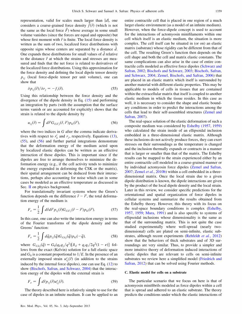
representation, valid for scales much larger than jdj, oneconsiders a coarse-grained force density ~fð~rÞ (which is not
the same as the local force ~F) whose average in some smallvolume vanishes (since the forces are equal and opposite) butwhose first moment with ~r is finite. The local force density iswritten as the sum of two, localized force distributions with
opposite signs whose centers are separated by a distance ~d.
One expands these distributions for small values of ~d relativeto the distance ~r at which the strains and stresses are mea-sured and finds that the net force is related to derivatives ofthe localized force distributions. Using this approximation forthe force density and defining the local dipole tensor densitypij (local force-dipole tensor per unit volume), one can
show that
@pijð~rÞ=@rj ¼ �fið~rÞ: (55)
Using this relationship between the force density and thedivergence of the dipole density in Eq. (15) and performingan integration by parts (with the assumption that the surfaceterms vanish or are accounted for explicitly) shows that thestrain is related to the dipole density by
uijð~rÞ ¼Z
d~r0Gil;k0jð~r; ~r0Þpklð~r0Þ; (56)
where the two indices in G after the comma indicate deriva-tives with respect to r0k and rj, respectively. Equations (13),
(55), and (56) and further partial integrations demonstratethat the deformation energy of the medium acted uponby localized elastic dipoles can be written as an effectiveinteraction of those dipoles. This is important since if thedipoles are free to arrange themselves to minimize the de-formation energy (e.g., if the cell activity tends to minimizethe energy expended in deforming the CSK or the matrix),their spatial arrangement can be deduced from their interac-tions, perhaps also accounting for noise which can in somecases be modeled as an effective temperature as discussed inSec. II on physics background.
For translationally invariant systems where the Green’sfunction depends on the difference ~r� ~r0, the total deforma-tion energy of the medium is
Fe ¼ 1
2
Zd~rd~r0pijð~rÞGil;k0jð~r� ~r0Þpklð~r0Þ: (57)
In this case, one can also write the interaction energy in termsof the Fourier transforms of the dipole density and theGreens’ function:
Fe ¼ 1
2
Zd ~qpijð ~qÞGil;kjð ~qÞpklð� ~qÞ; (58)
where Gil;kjð ~qÞ ¼ G0ðqkqj=q2Þ½il þ qiql=2q2ð1� Þ� fol-
lows from the exact (Kelvin) solution for a full elastic spaceandG0 is a constant proportional to 1=E. In the presence of anexternally imposed strain ueijð~rÞ (in addition to the strains
induced by the internal force dipoles), one can use Eq. (12) toshow (Bischofs, Safran, and Schwarz, 2004) that the interac-tion energy of the dipoles with the external strain is
Fe ¼Z
d~rpijð~rÞueijð~rÞ: (59)
The theory described here is relatively simple to use for thecase of dipoles in an infinite medium. It can be applied to an
entire contractile cell that is placed in one region of a muchlarger elastic environment (as a model of an infinite medium).However, when the force-dipole concept is used to accountfor the interactions of actomyosin minifilaments within onecell which itself is an elastic medium, the situation is morecomplex. The cell itself can be situated in (or on) an elasticmatrix (substrate) whose rigidity can be different from that ofthe cell. The resulting Green’s function then depends on thecell shape and both the cell and matrix elastic constants. Thesame complications can also arise in the case of entire con-tractile cells modeled as effective force dipoles (Schwarz andSafran, 2002; Bischofs and Schwarz, 2003; Bischofs, Safran,and Schwarz, 2004; Zemel, Bischofs, and Safran, 2006) thatare placed in an elastic matrix which itself is surrounded byanother material with different elastic properties. This may beapplicable to models of cells in tissues that are containedwithin the extracellular matrix that itself is coupled to anotherelastic medium in which the tissue resides. In this case aswell, it is necessary to consider the shape and elastic bound-ary conditions in order to predict the interactions among thecells that lead to their self-assembled structures (Zemel andSafran, 2007).
The real-space solution of the elastic deformation of such acomposite medium was considered by Eshelby (1957, 1959)who calculated the strain inside of an ellipsoidal inclusionembedded in a three-dimensional elastic matrix. Althoughthese inclusions do not actively produce forces, they can exertstresses on their surroundings as the temperature is changedand the inclusion thermally expands or contracts in a mannerthat is larger or smaller than that of the matrix. The Eshelbyresults can be mapped to the strain experienced either by anentire contractile cell modeled in a coarse-grained manner orby individual actomyosin force dipoles (Zemel and Safran,2007; Zemel et al., 2010b) within a cell embedded in a three-dimensional matrix. Once the local strain due to a givendipole distribution is known, the dipole interactions are givenby the product of the local dipole density and the local strain.Later in this review, we consider specific predictions for theorientational and spatial organization of force dipoles incellular systems and summarize the results obtained fromthe Eshelby theory. However, this theory with its focus onthe real-space boundary conditions is complex (Eshelby,1957, 1959; Mura, 1991) and is also specific to systems ofellipsoidal inclusions whose dimensionality is the same asthat of the surrounding matrix. This is not quite the casestudied experimentally where well-spread (nearly two-dimensional) cells are plated on semi-infinite, elastic sub-strates, although recent experiments (Rehfeldt et al., 2012)show that the behaviors of thick substrates and of 3D sur-roundings are very similar. Thus, to provide a simpler andmore intuitive theory of deformation induced interactions ofelastic dipoles that are relevant to cells on semi-infinitesubstrates we review here a simplified model (Friedrich andSafran, 2012) that can be solved using Fourier methods.
C. Elastic model for cells on a substrate
The particular scenario that we focus on here is that ofactomyosin minifibrils modeled as force dipoles within a cellthat is spread and adhered to an elastic substrate. The theorypredicts the conditions under which the elastic interactions of
Ulrich S. Schwarz and Samuel A. Safran: Physics of adherent cells 1359
Rev. Mod. Phys., Vol. 85, No. 3, July–September 2013
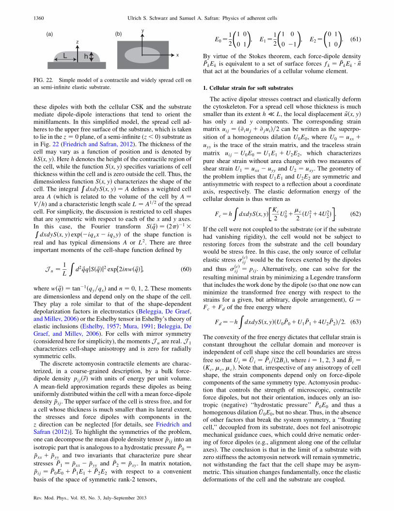
these dipoles with both the cellular CSK and the substratemediate dipole-dipole interactions that tend to orient theminifilaments. In this simplified model, the spread cell ad-heres to the upper free surface of the substrate, which is takento lie in the z ¼ 0 plane, of a semi-infinite (z < 0) substrate asin Fig. 22 (Friedrich and Safran, 2012). The thickness of thecell may vary as a function of position and is denoted byhSðx; yÞ. Here h denotes the height of the contractile region ofthe cell, while the function Sðx; yÞ specifies variations of cellthickness within the cell and is zero outside the cell. Thus, thedimensionless function Sðx; yÞ characterizes the shape of thecell. The integral
RdxdySðx; yÞ ¼ A defines a weighted cell
area A (which is related to the volume of the cell by A ¼V=h) and a characteristic length scale L ¼ A1=2 of the spreadcell. For simplicity, the discussion is restricted to cell shapesthat are symmetric with respect to each of the x and y axes.In this case, the Fourier transform Sð ~qÞ ¼ ð2Þ�1 �RdxdySðx; yÞ expð�iqxx� iqyyÞ of the shape function is
real and has typical dimensions A or L2. There are threeimportant moments of the cell-shape function defined by
J n ¼ 1
L
Zd2 ~qqjSð ~qÞj2 exp½2inwð ~qÞ�; (60)
where wð ~qÞ ¼ tan�1ðqy=qxÞ and n ¼ 0, 1, 2. These moments
are dimensionless and depend only on the shape of the cell.They play a role similar to that of the shape-dependentdepolarization factors in electrostatics (Beleggia, De Graef,and Millev, 2006) or the Eshelby tensor in Eshelby’s theory ofelastic inclusions (Eshelby, 1957; Mura, 1991; Beleggia, DeGraef, and Millev, 2006). For cells with mirror symmetry(considered here for simplicity), the moments J n are real. J 1
characterizes cell-shape anisotropy and is zero for radiallysymmetric cells.
The discrete actomyosin contractile elements are charac-terized, in a coarse-grained description, by a bulk force-dipole density pijð~rÞ with units of energy per unit volume.
A mean-field approximation regards these dipoles as beinguniformly distributed within the cell with a mean force-dipoledensity �pij. The upper surface of the cell is stress free, and for
a cell whose thickness is much smaller than its lateral extent,the stresses and force dipoles with components in thez direction can be neglected [for details, see Friedrich andSafran (2012)]. To highlight the symmetries of the problem,one can decompose the mean dipole density tensor �pij into an
isotropic part that is analogous to a hydrostatic pressure �P0 ¼�pxx þ �pyy and two invariants that characterize pure shear
stresses �P1 ¼ �pxx � �pyy and �P2 ¼ �pxy. In matrix notation,
�pij ¼ �P0E0 þ �P1E1 þ �P2E2 with respect to a convenient
basis of the space of symmetric rank-2 tensors,
E0¼1
2
1 0
0 1
!; E1¼1
2
1 0
0 �1
!; E2¼
0 1
1 0
!: (61)
By virtue of the Stokes theorem, each force-dipole density�PkEk is equivalent to a set of surface forces fk ¼ �PkEk � ~nthat act at the boundaries of a cellular volume element.
1. Cellular strain for soft substrates
The active dipolar stresses contract and elastically deformthe cytoskeleton. For a spread cell whose thickness is muchsmaller than its extent h � L, the local displacement ~uðx; yÞhas only x and y components. The corresponding strainmatrix uij ¼ ð@iuj þ @juiÞ=2 can be written as the superpo-
sition of a homogeneous dilation U0E0, where U0 ¼ uxx þuyy is the trace of the strain matrix, and the traceless strain
matrix uij � U0E0 ¼ U1E1 þ U2E2, which characterizes
pure shear strain without area change with two measures ofshear strain U1 ¼ uxx � uyy and U2 ¼ uxy. The geometry of
the problem implies that U1E1 and U2E2 are symmetric andantisymmetric with respect to a reflection about a coordinateaxis, respectively. The elastic deformation energy of thecellular domain is thus written as
Fc¼hZdxdySðx;yÞ
�Kc
2U2
0þ�c
2ðU2
1þ4U22Þ�: (62)
If the cell were not coupled to the substrate (or if the substratehad vanishing rigidity), the cell would not be subject torestoring forces from the substrate and the cell boundarywould be stress free. In this case, the only source of cellular
elastic stress �ðcÞij would be the forces exerted by the dipoles
and thus �ðcÞij ¼ pij. Alternatively, one can solve for the
resulting minimal strain by minimizing a Legendre transformthat includes the work done by the dipole (so that one now canminimize the transformed free energy with respect to thestrains for a given, but arbitrary, dipole arrangement), G ¼Fc þ Fd of the free energy where
Fd¼�hZdxdySðx;yÞðU0
�P0þU1�P1þ4U2
�P2Þ=2: (63)
The convexity of the free energy dictates that cellular strain isconstant throughout the cellular domain and moreover isindependent of cell shape since the cell boundaries are stress
free so that Ui �Ui ¼ �Pi=ð2BiÞ, where i ¼ 1, 2, 3 and ~Bi ¼ðKc;�c;�cÞ. Note that, irrespective of any anisotropy of cellshape, the strain components depend only on force-dipolecomponents of the same symmetry type. Actomyosin produc-tion that controls the strength of microscopic, contractileforce dipoles, but not their orientation, induces only an iso-tropic (negative) ‘‘hydrostatic pressure’’ �P0E0 and thus ahomogenous dilation �U0E0, but no shear. Thus, in the absenceof other factors that break the system symmetry, a ‘‘floatingcell,’’ decoupled from its substrate, does not feel anisotropicmechanical guidance cues, which could drive nematic order-ing of force dipoles (e.g., alignment along one of the cellularaxes). The conclusion is that in the limit of a substrate withzero stiffness the actomyosin network will remain symmetric,not withstanding the fact that the cell shape may be asym-metric. This situation changes fundamentally, once the elasticdeformations of the cell and the substrate are coupled.
(a) (b)z
x
y
L h
FIG. 22. Simple model of a contractile and widely spread cell on
an semi-infinite elastic substrate.
1360 Ulrich S. Schwarz and Samuel A. Safran: Physics of adherent cells
Rev. Mod. Phys., Vol. 85, No. 3, July–September 2013
Because of the coupling of the CSK forces to the focaladhesions, active cell contractility induces substrate deforma-tions ~vðx; y; zÞ. The substrate surface strain at z ¼ 0 isdecomposed into its symmetry components vijðx;y;z¼0Þ¼V0E0þV1E1þV2E2. For simplicity (Friedrich and Safran,2012) one can take the average strain inside the cell tobe equal to the average substrate strain underneath thecell �Vk ¼ �Uk.
2. Substrate elastic energy
The substrate deformations induced by its coupling to thecell can now be included. The elastic energy of the substrate iswritten in terms of the Fourier transforms of the substratedisplacements ~vðx; y; zÞ derived by Nicolas and Safran(2006a). This energy is proportional to�m, the shear modulusof the substrate, which is for simplicity taken to be incom-pressible. One next expresses the coupling condition in Fourierspace and determines the strains by minimizing (Friedrich andSafran, 2012) the Legendre transform G ¼ Fc þ Fm þ Fd ofthe free energy of both the cell and substrate subject to thecoupling condition �Vk ¼ �Uk. The cellular strain componentsUi are then found as a function of the dipole components Pi:Ui ¼ AijPj. The coupling coefficients Aij are functions of the
cell-shape moments [Eq. (60)] and of the cell and substrateelastic moduli (Friedrich and Safran, 2012).
Of particular interest is the fact that there are off-diagonalterms in this relationship. This means, for example, that ahomogeneous and isotropic dipole distribution characterizedby a nonzero value of P0 can induce a shear strain such as U1.The coefficient that quantifies this symmetry breaking A01
depends on both the cell shape and matrix rigidity. In par-ticular, it is proportional to J 1 (which vanishes for cells withcircular cross sections) and vanishes when the substratemodulus is either very small or very large.
The elastic energy is the product of the local strain andlocal force-dipole density and can be written in terms ofeffective interactions of the force dipoles:
Fi¼ðL2h=4ÞðA00�P20þ2A01
�P0�P1þA11
�P21þ4A22
�P22Þ:
(64)
The terms proportional to P20 and P2
1 are the ‘‘self-energies’’
of the isotropic and nematic components of the force dipole,respectively; they represent the energy to deform the cellitself together with the elastic substrate (Fernandez andBausch, 2009). In addition, the shear stress induced by theisotropic dipole component couples the �P1 nematic dipolecomponent to the isotropic contractility �P0 for cell shapes thatare anisotropic for which J 1 is nonzero. This implies that theeffects of cell shape on elastic interactions can induce ne-matic order of the cellular force dipoles even if local cellactivity results only in isotropic contractility.
The coupling of the isotropic component of the dipoletensor to the cellular shear allows the cytoskeletal shear topresent mechanical guidance cues for the polarization andalignment of cytoskeletal structures and eventually of cellulartraction forces. Initially isotropic cytoskeletal contractilitycan result from a local regulation of myosin activity withinthe cell that may tune �P0 to a set value �P
0. Considering�P1 as
an effective degree of freedom, the total elastic energy of cell
and substrate, Eq. (64), is minimized when �P1 is nonzero,
which corresponds to anisotropic cellular contractility.Physical insight into the coupling of symmetry modes can
be obtained considering the limiting cases of very soft and
very stiff substrates. Since the strain propagates (Banerjeeand Marchetti, 2012; Friedrich and Safran, 2012) into the
substrate a distance of order of the cell extent L (but only adistance of order the cell thickness h within the cell), the
effective Young’s modulus of the substrate is given by the
product of its Young’s modulus Em and a factor of L=h, whereh is the cell thickness ~�m ¼ EmL=h. This predicts that boththe stiffness ratio and the cell geometry (height and lateralextent) will determine cytoskeletal organization and ordering,
which can be tested by changing both substrate rigidity and
cell volume (Guo and Weitz, 2012).In the limit of a very soft substrate ~�m � �c, the situation
is that of an isolated cell; cellular strain components couple
only to force-dipole components of the same symmetry type.Cell activity that results in locally isotropic contractile di-
poles cannot give rise to nematic order. In the limit of a verystiff substrate, the cellular strain scales as 1= ~�m. For iso-
tropic, cellular contractility with only �P0 � 0: �U0 ¼J 0
�P0=8 ~�m, �U1 ¼ J 1�P0=8 ~�m, and �U2 ¼ 0. Thus, while
the symmetric part of the cell contractility P0 induces shear
strain due to the shape anisotropy, this shear strain attenuatesas ~�m ! 1. Similar conclusions are reached for the induc-
tion of nematic order (nonzero values of �P1) by locallyisotropic contractility. If, however, substrate stiffness and
cellular stiffness match, ~�m � Kc, �c, the symmetric shear
component �U1 is induced by the symmetric dipole component�U1 � J 1
�P0. This shear is a result of anisotropic, cell-shape
dependent, elastic restoring forces from the substrate. For anasymmetric cell shape that is elongated in the direction of the
x axis, J 1 < 0 and isotropic contractility with �P0 < 0 inducescellular shear strain �U1 > 0, which is expansive in thex direction (and compressive along the y direction). This
causes nematic ordering of the dipoles themselves along thex axis, characterized by negative values of �P1.
D. Cell polarization guided by substrate rigidity
These intuitive results were used in a more formal theoreti-cal model to predict that cells with anisotropic shapes on
substrates of intermediate rigidity will spontaneously showCSK nematic order (Zemel et al., 2010b; Friedrich and
Safran, 2011). A phenomenological model couched in terms
of active-gel theory was presented by Banerjee and Marchetti(2011). The case of stem cells is particularly applicable since,
at early times, the CSK is not yet well formed and oriented andone can study the genesis of CSK formation and orientation
starting with relatively short actomyosin minifilaments thatcan indeed be modeled as force dipoles contained within the
cell. Zemel et al. (2010b) used the real-space Eshelby formal-
ism (Eshelby, 1957, 1959) for an ellipsoidal inclusion tocalculate the real-space strains inside a cell contained in an
elastic medium of the same dimensionality (either 2D or 3D).The shear strains induced by the medium are nonzero for cells
that are not circular (2D) or spherical (3D) and are predicted to
give rise to orientational order of the internal force dipoles(short actomyosin minifilaments). The nematic order as
Ulrich S. Schwarz and Samuel A. Safran: Physics of adherent cells 1361
Rev. Mod. Phys., Vol. 85, No. 3, July–September 2013

expressed by �P1 is related to the shear strain by a susceptibility
whose form in the limit of large noise (expressed as an effective
temperature) was discussed by Zemel, Bischofs, and Safran
(2006). Friedrich and Safran (2011) provided a more general
statistical mechanical basis for the nematic ordering. An en-
ergy similar to Eq. (64) was obtained using the Eshelby theory;
this was used as a Hamiltonian in a Maier-Saupe (Maier and
Saupe, 1959) theory as described above in Eq. (2). This self-
consistently predicts the nematic order parameter �P1 as a
function of the cell and matrix elastic constants, cell shape,
and the noise (modeled as an effective temperature). In both
models, if the dipole density is dilute or if the noise is moder-
ately large, the nematic order of the CSK is maximal in some
optimal range of substrate rigidity and is small for very small or
very large rigidities.The dependence on the boundary conditions (i.e., the global
cell shape and substrate rigidity) highlights the importance of
the long-range elastic interactions in contrast to the general
situation for nematic ordering in molecular systems where the
interactions are short range. The Mair-Saupe theory predicts
that, for small noise (or large values of �P0), the nematic order
may increase monotonically as a function of substrate rigidity
due to the increasing importance of short-range interactions
such as the excluded volume of the dipoles themselves.Experiments were carried out (Zemel et al., 2010b) to
systematically analyze the alignment of stress fibers in human
mesenchymal stem cells as a function of the cell shape and
the rigidity of the environment. Cells were cultured on sub-
strates of varying stiffness and sorted by their aspect ratio. A
quantitative analysis of stress-fiber polarization in cells was
obtained by staining for both actin and nonmuscle myosin IIa
and applying a segmentation algorithm to map their spatial
organization in the cell. Both the magnitude of the dipoles as
measured by the number of actomyosin minifilaments (Zemel
et al., 2010a) and their orientation were measured. The results
are shown in Fig. 23. They suggest a generic mechanical
coupling between the cell shape, the rigidity of the surround-
ings, and the organization of stress fiber in the cytoskeleton of
stemcells, again pointing to the role of long-range interactions.
This identifies a mechanical property of cells (stress-fiber
polarization) that ismaximized at an optimal substrate rigidity,
analogous to the optimal rigidity found in stem-cell differen-
tiation (for example, to muscle cells) (Engler et al., 2006). The
fact that theCSK ismaximally polarized for substrate rigidities
of about 10 kPamay help explainwhy stem-cell differentiation
into muscle cells occurs optimally in this same rigidity range.
Stem cells on such substrates are musclelike in their CSK
structure and the resulting contractile forces; the latter may
play a role in nuclear deformations resulting in gene expres-
sion that is musclelike for precisely such contractile cells. We
note that the time scales observed for stress-fiber development
(Zemel et al., 2010b) and orientation (1–24 h) and for the
genetic changes in the cell (Engler et al., 2006) (about a week)
are quite different. In addition, recent experiments have shown
that differentiation may also depend on the mechanics of the
ligand molecule and not only on the bulk rigidity of the
substrate (Trappmann et al., 2012).The ordering of the cytoskeleton on substrates of different
rigidities eventually affects the overall mechanical response
of the cell. Recent studies (Janmey and Miller, 2011) have
shown that cell cortical stiffness increases as a function ofboth substrate stiffness and spread area. For soft substrates,the influence of substrate stiffness on cell cortical stiffness ismore prominent than that of cell shape, since increasingadherent area does not lead to cell stiffening. On the otherhand, for cells constrained to a small area, cell-shape effectsare more dominant than substrate stiffness, since increasingsubstrate stiffness no longer affects cell stiffness.
E. Single cell response to rigidity gradients
Section VI.D looked ‘‘inside the cell’’ and considered theelastic interactions and orientational ordering of short acto-myosin minifilaments modeled as force dipoles that areinternal to the cell. This is relevant to stem-cell developmentat relatively early times (1–24 h in which the stress fibers donot yet span the entire cell). Mature cells such as fibroblastsor muscle cells have long and well-ordered stress fibers(Hotulainen and Lappalainen, 2006; Thery et al., 2006) andin some cases the cell can be represented by a single, aniso-tropic force dipole (Schwarz and Safran, 2002; Bischofs and
(a)
(b)
FIG. 23 (color online). (a) The experimental values of the stress-
fiber order parameter S� �P1 (which indicates the extent to which
the nascent stress fibers are aligned along the long axis of the cell)
for three groups of stem cells (of aspect ratios 1.5, 2.5, and 3.5) as a
function of the Young’s modulus of the matrix Em. For the smallest
aspect ratio, the order is clearly maximal for Em � 11 kPa; for theother aspect ratios this trend may also be obeyed although it is less
clear. The fit is motivated by the theory described in the text. From
Zemel et al., 2010b. (b) Aspect ratio of stem cells as a function of
substrate elasticity. The point marked blebb refers to cells where
myosin activity has been suppressed by treatment with the drug
blebbistatin; this shows that the peak observed for untreated cells is
related to cell contractility. From Rehfeldt et al., 2012.
1362 Ulrich S. Schwarz and Samuel A. Safran: Physics of adherent cells
Rev. Mod. Phys., Vol. 85, No. 3, July–September 2013

Schwarz, 2003; Bischofs, Safran, and Schwarz, 2004; Pompe
et al., 2009). In the following we discuss the response of an
entire, polarized cell (modeled in a coarse-grained approxi-
mation as a single, anisotropic force dipole) to rigidity gra-
dients and in the next section, its response to dynamically
applied stress.Cell spreading, alignment, and locomotion are controlled
both by biochemical activity within the cell and by the rigidity
of the substrate on which the cell is plated (Pelham andWang,
1997; Lo et al., 2000; Discher, Janmey, and Wang, 2005; Saez
et al., 2007; Solon et al., 2007; Isenberg et al., 2009). In
general, these activities are enhanced onmore rigid substrates.
In addition, the forces that even static cells exert on substrates
have been shown to increase with substrate rigidity (Choquet,
Felsenfeld, and Sheetz, 1997; Saez et al., 2005; Yeung et al.,
2005; Zemel et al., 2010a). Moreover, recent studies indicate
that substrate viscoelasticity also plays a role in stem-cell
morphology and proliferation (Cameron, Frith, and Cooper-
White, 2011). Active-gel theory has been used to model iso-
tropic rigidity sensing in Marcq, Yoshinaga, and Prost (2011).
Here we review how models of contractility based on force
dipoles (Bischofs, Safran, and Schwarz, 2004; Nicolas and
Safran, 2006b; Zemel et al., 2010a) can provide insight into
these observations.Contractile cells are preprogrammed to exert force on their
surroundings. It has been argued that the experimental ob-
servation that most cells types prefer stiff over soft substrates
can be described by the assumption that cells effectively
minimize the elastic energy invested in deforming the matrix
(Bischofs and Schwarz, 2003; Bischofs, Safran, and Schwarz,
2004; Nicolas and Safran, 2006b). Neural cells, that prefer
soft over stiff substrates, are exceptions to this rule of thumb
(Janmey et al., 2009). This minimization can be the result of
evolution in producing optimized biological systems (Savir
et al., 2010), since the energy that the cell invests in deform-
ing its surrounding is not directly useful to the cell.
Alternatively, one can think of this approach as a convenient
framework for analytical progress. Considering the cell as a
uniform distribution of dipoles (that can be either ordered or
random in their orientation), one can use Eq. (64). The dipole
densities P0 and P1 are proportional to the number of dipoles
and the cell volume while for fairly rigid substrates A00 and
A11 scales inversely with ~�m ¼ �mL=h. Assuming that the
cell optimizes its activity to avoid investing energy in sub-
strate deformations, this predicts that cells will favor and
spread optimally on rigid substrates.This tendency of the cell to prefer rigid substrates is
particularly important for cells on substrates with rigidity
gradients or boundary regions (Lo et al., 2000; Allioux-
Guerin et al., 2009; Isenberg et al., 2009; Ladoux and
Nicolas, 2012). The limiting cases of a cell on a substrate
with a given rigidity near a boundary of a substrate with a
much larger or smaller rigidity can be understood by opti-
mizing the deformation energy of a single force dipole in a
medium with either clamped or free boundaries, respectively.
The corresponding elastic problem takes into account these
boundary conditions using the technique of ‘‘image dipoles’’
(Bischofs, Safran, and Schwarz, 2004). As shown there, the
preferred cell orientations close to the surface, as predicted
by the configurations of minimal deformation energy, are
parallel and perpendicular to the boundary line for free and
clamped boundaries, respectively.This leads to the prediction that cells preferentially loco-
mote toward a clamped boundary, but tend to migrate away
from a free boundary. One may think of a clamped (free)surface as the interface between the substrate on which the
cell is placed and an imaginary medium of infinite (vanish-ing) rigidity, which effectively rigidifies (softens) the bound-
ary region. Thus for clamped (free) boundary conditions, the
cell senses maximal stiffness in the direction normal to(parallel to) the boundary line. The cell exerts force on the
more rigid medium. For free boundaries, the substrate is morerigid and the cell orients parallel to the boundary to maximize
the deformation of the substrate. Near a clamped boundary,
there is less deformation if the cell orients perpendicular tothe boundary line. Indeed such behavior has been observed
experimentally, e.g., for cells close to the boundary betweensoft and rigid regions of a soft substrate (Lo et al., 2000). The
tendency to migrate toward stiffer regions has been termeddurotaxis. On a more microscopic level, this can be under-
stood from the preferred growth of focal adhesions on more
rigid substrates (see Sec. IV).
F. Dynamical response of cells to mechanical stress
Cells in tissues respond to a variety of mechanical forcesthat influence their behavior and alignment such as gravity,
muscle tension, and blood pressure as well as from local
active tractions of nearby cells (Ingber, 2003; Chen, 2008).The forces that act on cells can be static as well as time
varying, e.g., continuous loading occurs during developmentof long bone growth while cyclic loading occurs due to
periodic blood pressure variations. These mechanical signals
typically induce an active reorganization of the cell cytoske-leton and readjustment of the contractile forces exerted by the
cells (Deng et al., 2006; Stamenovic et al., 2007). The activenature of this mechanotransduction is demonstrated by the
fact that it often vanishes when actin-myosin contractility is
inhibited (Zhao et al., 2007).The response to mechanical stress is demonstrated by cells
that actively reorient and align themselves in preferred direc-
tions. It is interesting to note that, while in some studies cellswere shown to align parallel to the direction of a static or
quasistatic stress field (Samuel and Vandenburgh, 1990;Brown et al., 1998; Eastwood et al., 1998; Collinsworth
et al., 2000), other experiments find that cells remain ran-
domly oriented (Jungbauer et al., 2008). On the other hand,when subject to dynamically oscillating stress and strain
fields (designed originally to study the effects of heart beatand blood pressure), cells tend to orient away (nearly, but not
exactly perpendicularly) from the stress direction (Shirinskyet al., 1989; Wang and Grood, 2000; Hayakawa, Sato, and
Obinata, 2001; Wang et al., 2001; Kurpinski et al., 2006;
Jungbauer et al., 2008; Faust et al., 2011). As discussedbelow, this reorientation does not occur as a rigid body
motion of the cell, but rather involves the disassembly andthen reassembly of the CSK in directions determined by the
applied stress; it may also involve rotations of the stress fibers
(Deibler, Spatz, and Kemkemer, 2011). In some of the experi-ments on static stress (Brown et al., 1998), cells were placed
Ulrich S. Schwarz and Samuel A. Safran: Physics of adherent cells 1363
Rev. Mod. Phys., Vol. 85, No. 3, July–September 2013
in 3D collagen matrices and it is not clear whether remodel-ing of the matrix (Takakuda and Miyairi, 1996; Fernandezand Bausch, 2009) by the stress contributes to cellular ori-entation or if the orientation is solely a result of CSK reor-ganization within the cell in response to stretch. Theexperiments in Fig. 5 of Eastwood et al. (1998) indicaterandom collagen alignment even under tension. In general,the roles of passive (CSK elasticity) and active (actomyosincontractility) forces in determining cell response to appliedstress have yet to be fully elucidated (Nekouzadeh et al.,2008). In the following, we first review some recent mecha-nobiological measurements that provide new insights forunderstanding the response of cells to applied stress. Wethen summarize several theoretical approaches that quantify
these ideas.
1. CSK disassembly and reassembly
Recent experimental studies showed that cell stretch in-duces CSK fluidization (Bursac et al., 2005; Deng et al.,2006; Trepat et al., 2007; Krishnan et al., 2009; C. Chenet al., 2010) which occurs through direct physical effects ofphysical forces upon weak cytoskeletal cross-links. CSKfluidization is typified by marked decreases of CSK stiffness,CSK tension, and cellular traction forces, and marked in-creases in the rate of CSK remodeling dynamics (Trepatet al., 2007; Krishnan et al., 2009) and is accompanied byextremely rapid disassembly of actin bundles. These effectsdepend (Trepat et al., 2007; Krishnan et al., 2009) on theload, loading frequency, and magnitude of the prestretchactomyosin contractility.
To restore homeostasis in the cell (i.e., a fixed level ofcontractility), CSK fluidization is immediately succeeded byCSK reassembly, a signaling driven response that restoresmolecular interactions that were disrupted by fluidization(Trepat et al., 2007). CSK reassembly results in gradualincreases of CSK stiffness, CSK tension, and cellular tractionforces, and gradual decreases in the rate of CSK remodelingdynamics. These are driven by slow reassembly that actspredominantly on those spatial sites where traction forceswere markedly reduced by CSK fluidization (Trepat et al.,2007; Krishnan et al., 2009). These processes govern theresponse of cells to applied stress in which the reorientationis a result of the CSK fluidization and reassembly.
2. CSK stiffness changes in response to applied stress
While it is clear that the CSK reorganizes in response toapplied stretch, it is also important to know whether cellcontractility and stiffness is increased or decreased duringstress application. Experiments on fibroblasts in three-dimensional, collagen gels showed that overall cells reducetheir contractility during the stretch-relax cycles (Brownet al., 1998). This led Brown et al. (1998) to suggest thatcells have a homeostatic (or set point) contractility that isreduced when the surrounding medium is stretched. Thedynamics of this process were investigated in more detailby Nekouzadeh et al. (2008) who showed that, whenstretched for several minutes, contractile fibroblasts initiallydiminished the mechanical tractions they exert on their envi-ronment through depolymerization of actin filaments. The
cells then restored tissue tension and rebuilt actin stress fibers
through staged Ca dependent processes that consisted of a
rapid phase that ended less than a minute after stretching, a
plateau of inactivity, and a final gradual phase that required
several minutes to complete. Active contractile forces during
recovery scaled with the degree of rebuilding of the actin
cytoskeleton. The final cell stress following a stretch exceeds
the prestretch value; this is in contrast to the results reported
by Brown et al. (1998). However, the observations of cellular
ensembles might not be indicative of a ‘‘typical’’ cell; the
highly repeatable ensemble behaviors may represent a diver-
sity of responses at the level of individual cells.Trepat et al. (2004) developed an experimental system to
subject adherent cells to a global stretch while simultaneously
measuring the local complex shear modulus (G ¼ Gþ iG00)of the cells. They used this system to study the viscoelasticity
of alveolar epithelial cells in response to stepwise stretch and
found that with increasing levels of stepwise stress, both G0(elastic response) and G00 (viscous response) increased. Thesefindings indicate that the cytoskeletal response shows a non-
linear elastic response characteristic of strain stiffening and
that intracellular dissipation also increases with increasing
cytoskeletal tension. In addition, they found that the ratio
G00=G0 decreased with stretch, consistent with an increase in
the elastic rigidity of the CSK, corresponding to reassembly.
In a later study, they observed that when the cytoskeleton was
contracted with thrombin before application of a stepwise
stretch, the strain-stiffening response was abrogated (Trepat
et al., 2006), which suggests that the strain-stiffening regime
is restricted to a range of cytoskeletal tension. The same
experimental setup was used to test the viscoelastic response
of a broad variety of cell types that were subject to a transient
application of stretch or unstretch (Trepat et al., 2007).
Contrary to the case of a stepwise stretch, a transient stretch
that returns to zero strain caused a sharp drop in both G0 andG00 and a sudden increase in the ratio G00=G0. Thus, while a
stepwise stretch induces CSK rigidification, a transient
stretch induces cell softening and fluidization. To test whether
stretch-induced cell stiffening and softening were associated
with changes in cytoskeletal tension, Gavara et al. (2008)
developed a system to map traction forces at the cell-substrate
interface during application of stretch. They observed that
cytoskeletal tension increased with application of a stepwise
stretch, but decreased below baseline levels upon stretch
removal. Analysis of traction maps before, during, and after
stretch indicated that the regions of higher traction force
application were those that exhibited a larger relative drop
of traction after stress cessation, suggesting that those cellular
structures subjected to a higher tension are disrupted by
stretch. Taken together, these findings point to the existence
of two different mechanisms by which cells respond to
stretch. Strain stiffening during a stepwise stretch is likely
to arise from nonlinear stretching of single cytoskeletal fila-
ments. On the other hand, strain softening after a transient
stretch is probably caused by inelastic unbinding or unfolding
of cytoskeletal cross-links and actomyosin cross bridges. In
response to a constant stepwise stretch, filament stretching
appears to dominate over inelastic unbinding and unfolding
of cross-links and cross bridges. After stretch cessation,
however, the contribution of filament stretching becomes
1364 Ulrich S. Schwarz and Samuel A. Safran: Physics of adherent cells
Rev. Mod. Phys., Vol. 85, No. 3, July–September 2013

negligible and the effect of inelastic unbinding and unfoldingdominates.
3. CSK and cellular reorientation in response to cyclic stretch
In the introduction to this section, we mentioned severalstudies that showed that the cells orient away from the stressdirection of cyclically applied stress. Recent experimentsprovided quantitative measures of these effects.Experiments described in Jungbauer et al. (2008) andDeibler, Spatz, and Kemkemer (2011) investigated the dy-namic reorientation of rat embryonic and human fibroblastcells over a range of stretching frequencies from 0.0001 to20 s�1 and strain amplitudes from 1% to 15%. Their mea-surements show that the mean cell orientation changes ex-ponentially in time with a frequency-dependent characteristictime from 1 to 5 h. At subconfluent cell densities (at which thecells are not yet close packed), this characteristic time forreorientation shows two characteristic regimes as a functionof frequency. For frequencies below 1 s�1, the characteristictime decreases with a power law as the frequency increases.For frequencies above 1 s�1, it saturates at a constant value.In addition, a minimum threshold frequency was found belowwhich no significant cell reorientation occurs. The resultssuggest a saturation of molecular mechanisms of the mecha-notransduction response machinery for subconfluent cellswithin the frequency regime studied. One possible interpre-tation of these two time scales is given in the theoreticalmodel described in Secs. VI.F.4, VI.F.5, and VI.F.6.Interestingly, Zahn et al. (2011) recently showed that thetime scale is correlated with the amount of actin in the cell;aged cells with less actin show faster reorganization in re-sponse to uniaxial tensile stress compared with younger cellswhich contain more actin and are elastically more rigid. Inaddition, other biochemical changes can modify the responsetime of the cytoskeleton and thereby control its orientation inresponse to cyclically varying stress (Hoffman, Grashoff, andSchwartz, 2011).
To control the strains both parallel and perpendicular to thestress directions, Faust et al. (2011) used elastomeric cham-bers that were specifically designed and characterized todistinguish between zero strain and minimal stress directionsand to allow accurate theoretical modeling. Reorientation wasinduced only when the applied stretch exceeded a specificamplitude, suggesting a nonlinear response. However, on verysoft substrates no mechanoresponse occurs even for highstrain. This suggests an explanation for the necessity of ratherstiff environmental conditions to induce cellular reorientationin mammalian tissues. For all stretch amplitudes, the angulardistributions of reoriented cells could be modeled as dis-cussed in Secs. VI.F.4, VI.F.5, and VI.F.6. Cyclic stretchincreases the number of stress fibers and the coupling toadhesions. Changes in the cell shape follow the cytoskeletalreorientation with a significant temporal delay; this indicatesthat cell reorientation and shape is induced by CSK reassem-bly in response to stretch. In the frequency range studied of10–50 mHz, the stress induces cell reorientation (after about16 h) in the direction of zero strain. A recent study by Livneand Geiger (unpublished) analyzed the reorientation dynam-ics of cyclically stretched cells, over a wide range of stretchconfigurations, and observed a systematic deviation between
the measured cell and stress-fiber orientation and the zero-strain prediction (up to 10�). To address this discrepancy, anovel model that shifts the focus of the reorientation processto the FAs was developed.
4. Theory of cell response to applied stress
Models of cell response to applied stress are motivated bythe question of why stress fibers or cells orient nearly (but notalways exactly) perpendicular to the direction of the appliedstress. Macromolecular or biochemical models of cellularorientation and stress-fiber rearrangement in response toapplied forces have been discussed by Mogilner and
Rubinstein (2005), Wei et al. (2008), Hsu, Lee, and Kaunas(2009), Pirentis and Lazopoulos (2009), and Pirentis et al.(2011), while a more generic theoretical approach was givenby De, Zemel, and Safran (2007, 2008), De and Safran(2008), and Safran and De (2009). We first review moremolecularly based models that focus on the role of the stressfibers and then present a more phenomenological and generalapproach that in principle coarse grains over both stress fiberand focal adhesion response.
5. Molecularly based models
The work of Wei et al. (2008) predicts the orientation ofstress fibers in response to cyclic stretch based on abiochemical-mechanical model that relates the contractionand extension rate sensitivity of the stress fibers to the magni-tude and frequency of the applied stress. These kineticsdepend on a biochemical activation signal (the tension-dependent fiber dissociation rate) and the rate of force gen-eration bymyosin II motors. This assumes that the stress fibersare intact throughout the application of dynamically varyingstrain. Experiments (Bursac et al., 2005; Deng et al., 2006;Trepat et al., 2007) showed that cells respond to mechanicalstress via an initial, fast (sec time scale) fluidization of thestress fibers that then reassemble and reorganize. Motivatedby this, Pirentis and Lazopoulos (2009) and Pirentis et al.(2011) focused on a mathematical model that simulates the
effects of fluidization and reassembly driven rigidification(Krishnan et al., 2009; C. Chen et al., 2010) on cytoskeletalcontractile stress. They showed how these phenomena affectcytoskeletal realignment in response to pure uniaxial stretch-ing of the substrate. The model comprises individual elasticstress fibers anchored at the end points to an elastic substrateand predicts that in response to repeated stretch-unstretchcycles, stress fibers tend to realign in the direction perpen-dicular to stretching. They concluded that relaxation of cy-toskeletal contractile stress by means of fluidization andsubsequent stress recovery by means of CSK reassemblymay play a key role in reorganization of cytoskeletal stressfibers in response to uniaxial stretching of the substrate.
A somewhat more general approach to the mechanicalresponse of stress fibers that was taken by Kaunas andcolleagues (Hsu, Lee, and Kaunas, 2009; Kaunas, Hsu, andDeguchi, 2011) who developed a model that tracks the fate ofindividual, stretched stress fibers based on the hypothesis thatstress fibers have an optimal prestrain due to actomyosincontractility (Lu et al., 2008); perturbing the strain fromthat optimal value promotes stress-fiber disassembly.
Ulrich S. Schwarz and Samuel A. Safran: Physics of adherent cells 1365
Rev. Mod. Phys., Vol. 85, No. 3, July–September 2013

Motivated by experimental evidence of stress-fiber viscoelas-tic properties, stress fibers are assumed to relax at a rateproportional to the perturbation in fiber stretch away fromthis optimum. The dynamic turnover of stress fibers wasdescribed using a stochastic approach with the probabilityper unit time of stress-fiber disassembly expressed as aconstant plus a term quadratic in the deviation of the strainfrom its optimal value. The disassembly of a stress fiber isassumed to be immediately followed by the assembly of anew stress fiber at its optimal stretch and oriented in arandomly chosen direction. Model parameters were deter-mined by fitting experimentally measured time courses ofstress-fiber alignment performed at different rates of strain(i.e., 0.01 to 1 Hz). The model predicts that reorganization ofthe stress fibers is determined by the competition between therates of stress-fiber assembly and load-dependent disassem-bly. The stress fibers preferentially disassemble in the direc-tion of stretch, while stress fibers reassembling instochastically chosen directions gradually accumulate aboutthe direction of least perturbation in fiber tension. At lowstrain rates, the stress fibers are predicted to align withrandom orientations with respect to the applied stress direc-tion. While this has been reported in some cases for very slowcyclic stress, other experiments report alignment in the stressdirection as discussed above. Recent studies (Tondon, Hsu,and Kaunas, 2012) using nonsinusoidal waveforms show thatthe stress-fiber reorientation is most sensitive to the rate oflengthening; this provides support for the role of stretch of theactin filaments in cell reorientation under stress.
6. Coarse-grained models based on force dipoles
One goal of this more phenomenological approach (De,Zemel, and Safran, 2007, 2008; De and Safran, 2008; Safranand De, 2009) is to explain the observed frequency depen-dence of cell orientation previously mentioned. Another is tounderstand why the characteristic time for the cell to reach itssteady-state orientation �c � 103–104 s is strongly frequencydependent for stretch frequencies smaller than about 1 Hzwhile at higher frequencies, �c is frequency independent. Theexperiments were conducted on anisotropic cells such asfibroblasts so the theory focuses on needlelike cells in whichthe entire cell is modeled in a coarse-grained approximationas a single force dipole; for needlelike cells, the dipolecomponent P2 ¼ 0. The dipoles can then be characterizedby their magnitude P0 P < 0 (to signify contraction) anddirection, �¼arctan½ðP0�P1Þ=ðP0þP1Þ�¼arctan½pyy=pxx�,relative to the external stress.
It was suggested (Brown et al., 1998) that cells activelyadjust their contractility by reorganizing the FA and stressfibers to maintain an optimal (or set point) value of the stressor strain U? in the adjacent matrix (De, Zemel, and Safran,2007, 2008; De and Safran, 2008; Safran and De, 2009). Thistranslates via elastic theory into an optimal value of thecellular dipole P? > 0. The stresses are converted to energyunits by multiplying by the cell volume and the externallyapplied stretch is denoted as PaðtÞ> 0. In the presence ofsuch time-dependent stretch that acts at an angle � relative tothe cell axis, it is assumed that the homeostatic, set-point totallocal stress in the matrix is achieved when the cellular forcedipole obeys (Safran and De, 2009)
P ¼ �P? þ �0PaðtÞð���1Þ; (65)
where � ¼ cos2�. Two limiting cases are where (i) the cel-lular dipole is controlled by the matrix stress where �1 ¼ 0,and (ii) the dipole is controlled by the matrix strain and �1 ¼cos2�0 �0, where �0 is the zero-strain direction given bycos2�0 ¼ =ð1þ Þ. In general, �0 can be either positiveor negative corresponding to matrix stretch that causeseither a decrease or an increase in the cytoskeletal forces,respectively.
Deviations from the set point result in internal forceswithin the cell that reestablish the optimal stress condition.These forces can be derived from derivatives of an effective,harmonic ‘‘free energy’’ (more precisely, a cost functionwhose minimum represents the optimization of the cellularactivity) due to cell activity Fa that includes the activeprocesses within the cell that establish cellular response toits local environment,
Fa ¼ 12�½�Pþ �0PaðtÞð���1Þ � P?�2; (66)
where �P?2 (with units of energy) is a measure of cellactivity that establishes the set point.
In addition to the cell activity, the model also includes theeffect of mechanical matrix forces, Eq. (59), that yields anenergy Fe proportional to the product of PaðtÞ and P. Thegoal is to solve for the dipole magnitude and direction in thepresence of a time-varying stress PaðtÞ ¼ Pað1� cos!atÞ,where Pa > 0 for stretch. In general, the dynamics of thecytoskeleton are governed by complex, viscoelastic processesthat also involve liquification and reassembly of the stressfibers (Deng et al., 2006). In a coarse-grained picture, one canwrite relaxational equations for the dipole magnitude anddirection that are governed by the derivatives of F¼FaþFe:
dpðtÞdt
¼ � 1
�pfp;
d�ðtÞdt
¼ � 1
��f�; (67)
where p ¼ P=P, the dimensionless, effective free energyf ¼ F=�P2, and fp ¼ @f=@p and f� ¼ @f=@�. Noise
terms modeled as a dimensionless, effective temperature Ts
can also be included in this formalism (Safran and De, 2009);note the caveats on the use of effective temperature andBoltzmann distributions discussed in Sec. II on the physicsbackground.
Based on experiments (Deng et al., 2006; Gavara et al.,2008; Nekouzadeh et al., 2008), it has been suggested that theliquification and repolymerization of the actin stress fibersafter stretch is applied occur on a short time scale on the orderof several seconds, while the correlated reorientation occurson much longer time scales (on the order of many minutes)(Brown et al., 1998; Eastwood et al., 1998; Jungbauer et al.,2008). It is thus assumed that �p � ��: the time scale
associated with changes in the magnitude of the dipole ismuch faster than that associated with the dynamics of itshighly correlated reorientation. In this approximation, thedipole magnitude reaches a steady-state value in a shorttime; this value may be time dependent and oscillatory dueto the cyclic nature of the applied, time-dependent stress.
One therefore first solves for the dipole magnitude pðtÞ,treating the slowly varying dipole orientation �ðtÞ as aconstant; for details, see Safran and De (2009). The average
1366 Ulrich S. Schwarz and Samuel A. Safran: Physics of adherent cells
Rev. Mod. Phys., Vol. 85, No. 3, July–September 2013
value of � ¼ cos2� calculated as a function of the frequencydepends on the effective temperatures for the cases of both
stress and strain as set points. At high frequencies andlow effective temperatures, the average angle is nearlyperpendicular (or in the zero-strain direction �0, for cells
whose set point is determined by matrix strain), due to thedynamical frustration of the cell which is unable to adjust itsforce dipole to the time-dependent matrix stresses. At very
low frequencies, the average angle is nearly parallel and forthe case of both stress and strain as set points, consistent with
some of the experiments (Brown et al., 1998). At highereffective temperatures, the orientation distribution is randomand the average value of � (in two dimensions) is 1=2 for all
frequencies. At intermediate temperatures, one finds the in-teresting possibility of nearly perpendicular orientation forhigh frequencies, but nearly random orientation for low
frequencies [for details, see Safran and De (2009)].Biochemical changes can modify the response time of thecytoskeleton and thereby change cell orientation from nearly
perpendicular (when the CSK cannot follow the applied,cyclic stress) to parallel (when CSK remodeling time scalesare short enough) (Hoffman, Grashoff, and Schwartz, 2011).
In addition, experiments on 3D matrices (Riehl et al., 2012)indicate parallel orientation of stress fibers even at relativelyhigh frequencies. The systematic understanding of when cells
respond by orienting parallel compared to the relatively well-studied response of cells on relatively stiff substrates to cyclicstretch is a challenge that has yet to be met.
The dynamical theory is also used to calculate (Safran and
De, 2009) the characteristic time �c for a cell to attain itssteady-state orientation. At high frequencies, �c is frequencyindependent, while at low frequencies, �c � 1=!2; in both
regimes �c depends on the amplitude of the applied stress andthis is related to the fact that the zero-strain or zero-stressdirection orientation occurs only when the applied stress
exceeds a threshold value (De, Zemel, and Safran, 2007;De and Safran, 2008; Safran and De, 2009). Both the pre-dicted frequency and amplitude dependence are in qualitative
agreement with experiments (Jungbauer et al., 2008).
VII. CELL ASSEMBLIES
A. Matrix-mediated cell interactions
After treating the response of isolated cells to changes intheir elastic environment, we now discuss the elastic re-sponses of and interactions in ensembles of cells. We restrict
the analysis to the effects of elastic interactions on the relativepositions and orientations of cells. Moreover, we focus on the
case in which cells are well separated and interact with eachother only via the matrix and not through direct cell-cellinteractions. This is fundamentally different when modeling
growing epithelial tissue or tumors, when cell-cell interac-tions dominate. A popular model system for epithelial tissueformation is the Drosophila wing disk, which often is treated
using vertex models (Farhadifar et al., 2007; Hufnagel et al.,2007; Rauzi et al., 2008; Landsberg et al., 2009; Aegerter-Wilmsen et al., 2010; Canela-Xandri et al., 2011; Aliee et al.,
2012). In our focus here on matrix-mediated interactions, wedo not discuss the effect of cell proliferation and cell death
that also leads to interesting features in cell assembly
(Shraiman, 2005; Basan et al., 2009; Ranft et al., 2010).An early study that highlighted the effect of elastic sub-
strate deformations in modulating the relative positions of
cells placed far apart was presented by Korff and Augustin(1999). They observed that capillarylike structures formed by
two, initially separated groups of cells were associated withtensional remodeling of the collagen matrix and directional
sprouting of the outgrowing capillaries toward each other.These experiments presented evidence that tensional forces
on a fibrillar, extracellular matrix such as type I collagen, butnot fibrin, are sufficient to guide the directional outgrowth of
endothelial cells. More recently Reinhart-King, Dembo, and
Hammer (2008) and Califano and Reinhart-King (2010) usedmatrices of varying stiffness and measurements of endothelial
cell migration and traction stresses to show how cells candetect and respond to substrate strains created by the traction
stresses of a neighboring cell; they demonstrated that thisresponse is dependent on matrix stiffness. Other studies
suggest that, on some matrices, cells can sense each other(most probably via elastic deformations) at distances on the
order of 400 �m (Winer, Oake, and Janmey, 2009). Recently
it was also reported that cardiac cells can synchronize theirbeating through substrate deformations (Tang et al., 2011).
The various experiments imply that matrix mechanics canfoster tissue formation by correlating the relative motions or
even internal dynamics of cells, thereby promoting the for-mation of cell-cell contacts. The theoretical studies below
model an entire cell as a single, usually anisotropic, forcedipole. Interactions among cells are taken into account for
several simple geometries. A simple analogy to dielectric
media with predictions of the elastic susceptibilities and‘‘dielectric constants’’ of force-dipole assemblies was pre-
sented by Zemel, Bischofs, and Safran (2006), where theeffective elastic constants of materials containing force di-
poles are calculated as a function of the dipole density. Theresults, valid in the relatively dilute limit, indicate an effective
stiffening of the material due to the alignment of the con-tractile dipoles parallel to the applied stretch. It remains to be
seen how to take this analogy further to include dynamical,
tensorial, and nonlocal spatial effects as well as the develop-ment of a theory that is valid for both high and low force-
dipole concentrations. In addition, one must decide whetherthe dipoles are translationally mobile (as is the case for
counterion screening in electrostatics) or only orientationallymobile [as assumed by Zemel, Bischofs, and Safran (2006)].
B. Elastic interactions of force dipoles
The anisotropic and long-range nature of the interactions
of cell dipoles leads to a rich variety of self-assembled
structures (Bischofs and Schwarz, 2003, 2005, 2006;Bischofs, Safran, and Schwarz, 2004). Monte Carlo simula-
tions of these dipolar interactions in the presence of noise,modeled as an effective temperature, predicted cellular struc-
ture formation on elastic substrates as a function of the celldensity and Poisson ratio of the substrate. One interesting
situation considered was that of an infinitely extended stringof aligned force dipoles spaced at equal distances a (Bischofs
and Schwarz, 2005). An additional dipole is placed at a
Ulrich S. Schwarz and Samuel A. Safran: Physics of adherent cells 1367
Rev. Mod. Phys., Vol. 85, No. 3, July–September 2013
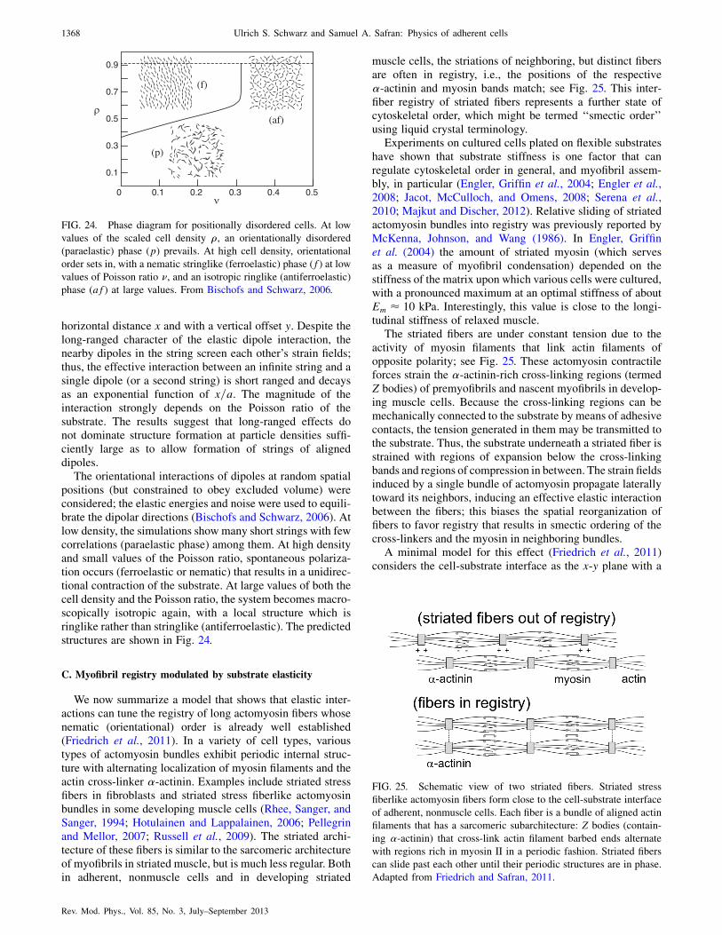
horizontal distance x and with a vertical offset y. Despite thelong-ranged character of the elastic dipole interaction, thenearby dipoles in the string screen each other’s strain fields;thus, the effective interaction between an infinite string and asingle dipole (or a second string) is short ranged and decaysas an exponential function of x=a. The magnitude of theinteraction strongly depends on the Poisson ratio of thesubstrate. The results suggest that long-ranged effects donot dominate structure formation at particle densities suffi-ciently large as to allow formation of strings of aligneddipoles.
The orientational interactions of dipoles at random spatialpositions (but constrained to obey excluded volume) wereconsidered; the elastic energies and noise were used to equili-brate the dipolar directions (Bischofs and Schwarz, 2006). Atlow density, the simulations show many short strings with fewcorrelations (paraelastic phase) among them. At high densityand small values of the Poisson ratio, spontaneous polariza-tion occurs (ferroelastic or nematic) that results in a unidirec-tional contraction of the substrate. At large values of both thecell density and the Poisson ratio, the system becomes macro-scopically isotropic again, with a local structure which isringlike rather than stringlike (antiferroelastic). The predictedstructures are shown in Fig. 24.
C. Myofibril registry modulated by substrate elasticity
We now summarize a model that shows that elastic inter-actions can tune the registry of long actomyosin fibers whosenematic (orientational) order is already well established(Friedrich et al., 2011). In a variety of cell types, varioustypes of actomyosin bundles exhibit periodic internal struc-ture with alternating localization of myosin filaments and theactin cross-linker �-actinin. Examples include striated stressfibers in fibroblasts and striated stress fiberlike actomyosinbundles in some developing muscle cells (Rhee, Sanger, andSanger, 1994; Hotulainen and Lappalainen, 2006; Pellegrinand Mellor, 2007; Russell et al., 2009). The striated archi-tecture of these fibers is similar to the sarcomeric architectureof myofibrils in striated muscle, but is much less regular. Bothin adherent, nonmuscle cells and in developing striated
muscle cells, the striations of neighboring, but distinct fibersare often in registry, i.e., the positions of the respective�-actinin and myosin bands match; see Fig. 25. This inter-fiber registry of striated fibers represents a further state ofcytoskeletal order, which might be termed ‘‘smectic order’’using liquid crystal terminology.
Experiments on cultured cells plated on flexible substrateshave shown that substrate stiffness is one factor that canregulate cytoskeletal order in general, and myofibril assem-bly, in particular (Engler, Griffin et al., 2004; Engler et al.,2008; Jacot, McCulloch, and Omens, 2008; Serena et al.,2010; Majkut and Discher, 2012). Relative sliding of striatedactomyosin bundles into registry was previously reported byMcKenna, Johnson, and Wang (1986). In Engler, Griffinet al. (2004) the amount of striated myosin (which servesas a measure of myofibril condensation) depended on thestiffness of the matrix upon which various cells were cultured,with a pronounced maximum at an optimal stiffness of aboutEm � 10 kPa. Interestingly, this value is close to the longi-tudinal stiffness of relaxed muscle.
The striated fibers are under constant tension due to theactivity of myosin filaments that link actin filaments ofopposite polarity; see Fig. 25. These actomyosin contractileforces strain the �-actinin-rich cross-linking regions (termedZ bodies) of premyofibrils and nascent myofibrils in develop-ing muscle cells. Because the cross-linking regions can bemechanically connected to the substrate by means of adhesivecontacts, the tension generated in them may be transmitted tothe substrate. Thus, the substrate underneath a striated fiber isstrained with regions of expansion below the cross-linkingbands and regions of compression in between. The strain fieldsinduced by a single bundle of actomyosin propagate laterallytoward its neighbors, inducing an effective elastic interactionbetween the fibers; this biases the spatial reorganization offibers to favor registry that results in smectic ordering of thecross-linkers and the myosin in neighboring bundles.
A minimal model for this effect (Friedrich et al., 2011)considers the cell-substrate interface as the x-y plane with a
FIG. 25. Schematic view of two striated fibers. Striated stress
fiberlike actomyosin fibers form close to the cell-substrate interface
of adherent, nonmuscle cells. Each fiber is a bundle of aligned actin
filaments that has a sarcomeric subarchitecture: Z bodies (contain-
ing �-actinin) that cross-link actin filament barbed ends alternate
with regions rich in myosin II in a periodic fashion. Striated fibers
can slide past each other until their periodic structures are in phase.
Adapted from Friedrich and Safran, 2011.
0.1
0.3
0.5
0.7
0.9
0 0.1 0.2 0.3 0.4 0.5ν
ρ(af)
(f)
(p)
FIG. 24. Phase diagram for positionally disordered cells. At low
values of the scaled cell density �, an orientationally disordered
(paraelastic) phase (p) prevails. At high cell density, orientational
order sets in, with a nematic stringlike (ferroelastic) phase (f) at lowvalues of Poisson ratio , and an isotropic ringlike (antiferroelastic)
phase (af) at large values. From Bischofs and Schwarz, 2006.
1368 Ulrich S. Schwarz and Samuel A. Safran: Physics of adherent cells
Rev. Mod. Phys., Vol. 85, No. 3, July–September 2013
single contractile fiber parallel to the x axis. The forcestransmitted by the fiber onto the substrate can be effectivelydescribed by a dipole distribution (with units of energy perunit area)�ijðx; yÞ ¼ �ðxÞðyÞixjx of force dipoles that are
localized to the adhesive contacts whose lateral extensionsare on the order of 100 nm and are thus much smaller than thespacing a � 1 �m of Z bodies. Here the force-dipole density(with units of energy per unit length) �ðxÞ is a periodicfunction of x due to the sarcomeric (i.e., periodic) architec-ture of a single striated fiber. For simplicity, the analysisfocuses on the principal Fourier mode �ðxÞ¼�0þ�1 cosð2x=aÞ, where a corresponds to the sarcomericperiodicity of the striated fiber.
The strain field uijðx; yÞ at the surface of the substrate withYoung’s modulus Em (and located at z ¼ 0) that is induced bythis periodic dipole ‘‘string’’ can be found from the Green’sfunction of Eq. (57). The parallel strain component u11ðx; yÞcan be written as a product of a ‘‘lateral propagation factor’’� that characterizes the propagation of strain in the lateral ydirection and a harmonic modulation in the x direction alongthe striated fiber
u11ðx; yÞ ¼ �ðjyj=a; Þ 2�1
Ema2cosð2x=aÞ: (68)
Thus the strain field u11 is periodic in the x direction withperiod a reflecting the periodicity of the striated fiber. Thefactor � characterizes the propagation of strain in the lateraldirection away from the center line of the fiber; this dependson the distance from the fiber as well as the Poisson ratio.The interaction between two such strings of dipoles is de-scribed by the elastic interaction energy, which is the localproduct of the dipole and the strain. The energy which a givenfiber (string of dipoles) must invest in order to deform thesubstrate is the sum of a self-energy of the first dipolar string
Wself ¼Rd2x�ð1Þ
ij uð1Þij , which accounts for the substrate defor-
mation energy in the absence of the second string of dipoles
and an interaction term Wint ¼Rd2x�ð1Þ
ij uð2Þij . The term Wint
characterizes an effective, substrate-mediated interaction be-tween the two contractile fibers and can guide their spatialreorganization.
Inserting the specific strain field induced by a singlestriated fiber, Eq. (68), into the general formula for elasticinteractions yields the elastic interaction energy between thetwo fibers (per minisarcomere) as a function of the phase shift�x and the separation of their center lines d:
Winteraction ¼ �ðd=a; Þ �21
aEm
cosð2�x=aÞ: (69)
Here W ¼ �21=ðaEmÞ � 10�18 J � 250kBT sets a typical
energy of the elastic interactions. Registry of fibers with�x ¼ 0 is favored for interbundle spacings where the propa-gation factor �< 0.
For incompressible substrates with Poisson ratio close to ¼ 1=2, such as those used in experiments (Engler, Griffinet al., 2004; Buxboim, Ivanovska, and Discher, 2010), it canbe shown (Friedrich et al., 2011) that the sign of the prefactor� of the elastic interaction energy is negative providedthat the lateral fiber spacing is larger than some threshold
d=a > d=a � 0:247. Hence, elastic interactions favor a
configuration where neighboring fibers are in registry with
�x ¼ 0. The opposite trend is found when � 0 (Bischofs
and Schwarz, 2005; Friedrich et al., 2011). It is therefore
possible that elastic interactions also set a preferred lateral
spacing of striated fibers. Additionally, steric interactions
may prevent neighboring fibers from getting too close and
could enforce the condition d > d.We previously discussed experiments and theory that dem-
onstrated that cells tend to prefer rigid substrates where the
elastic deformation energy cost is minimal. This also deter-
mines the optimal value of the cellular dipole magnitude in a
simple model that balances the ‘‘cell activity’’ [described in
Eq. (66) with zero applied field Pa ¼ 0] with the elastic
energy cost of deforming the substrate [analogous to the
expression in Eq. (64)] (De, Zemel, and Safran, 2007;
Safran and De, 2009; Friedrich and Safran, 2012). In terms
of the present model, this determines the optimal amplitude
of the dipole string �1 from the force balance given by
minimization with respect to the dipole amplitude �1 of the
sum of the two energies (per unit length of the bundle): the
activity optimization Wactive ¼ �ð�1 � �1Þ2=2 and the defor-
mation energy Wdeform � �21=ð2a2EmÞ, where Em is the
Young’s modulus of the substrate (Friedrich et al., 2011).
This predicts that �1 ¼ �1Em=ðEm þ E
mÞ, where Em ¼
1=a2�. The set-point value �1 corresponds to the amplitude
�1 of the dipole density on very stiff substrates with Em �Em. On soft substrates with Em � E
m, however, �1 can be
considerably smaller than �1.
Using this expression for the saturation of �1 on substrate
stiffness further predicts that the registry force between two
parallel striated fibers becomes a nonmonotonic function of
Em with a Lorentzian form and has maximal magnitude
for Em ¼ Em. Here it is assumed that the lateral spacing d
of the fibers is larger than the critical distance d and inde-
pendent of substrate stiffness.The theory was compared with recent experimental studies
of the interfiber registry in human mesenchymal stem cells
that were plated on polymeric gels of different stiffness
(ranging from 0.3 to 40 kPa) (Friedrich et al., 2011; Majkut
and Discher, 2012). Well-established, interfiber registry of
adjacent striated fibers was observed primarily for cells that
were cultured on 10 kPa gels as opposed to softer or more
rigid substrates. Myosin bands perpendicular to the axis of
nematic fiber organization were clearly visible and most
likely connect neighboring actomyosin bundles in registry.
Out of approximately 20 cells examined per gel, roughly
30%–50% exhibited aligned, striated fibers. The guidance
mechanism for the registry of striated fibers by elastic inter-
actions due to their elastic interactions predicts maximal
registry at an ‘‘optimal’’ value of the substrate rigidity and
represents a plausible mechanism for the establishment of
interfiber registry observed in the experiments. Further ex-
periments are needed to resolve the extent of striations within
one actomyosin bundle from the registry of striations in
neighboring bundles; the theory presented here addresses
the question of registry among bundles. It assumes that
each bundle is well ordered; a possible mechanism for the
development of such order was recently suggested by
Friedrich et al. (2012).
Ulrich S. Schwarz and Samuel A. Safran: Physics of adherent cells 1369
Rev. Mod. Phys., Vol. 85, No. 3, July–September 2013

VIII. CONCLUSIONS AND OUTLOOK
Research at the interface of physics and biology is anexciting adventure that has led and is still leading in severaldifferent directions. In this review with a theoretical focus, weappropriately modified soft matter physics approaches toanalyze how adherent cells mechanically interact with theirenvironment through forces at the cell-material interface.Although passive soft matter systems such as droplets, fullyelastic particles, vesicles, and polymeric capsules are impor-tant reference cases for the adhesion of single cells, ourdiscussion has shown that the main feature missing fromsuch theoretical frameworks are active processes. In thecontext of force generation and sensing of adherent cells,the most prominent active processes are the polymerization oflamellipodia at the cell edge and the myosin II generatedtensions in the actin cytoskeleton, including the contractilebundles (stress fibers) and networks that form during matureadhesion. These cytoskeletal processes are closely integratedwith the dynamics of spatially localized sites of focal adhe-sions. Together this system allows cells to sense and react tothe mechanical properties of their environment. Our reviewshows that the active and dynamic nature of cellular systemsmust be addressed on many different scales, from the model-ing of nanometer-scale molecular association and dissocia-tion events in adhesion clusters to force generation in largesupramolecular complexes and the shape and effective forcebalance at the 10 �m scale for animal cells. A further level ofcooperativity arises if one considers the tissue scale at whichcells can be further abstracted as discrete particles or defects.In the future, it is hoped that these different approaches willconverge into a system-level understanding of cellular sys-tems that not only includes genetic and biochemical aspects(which were not the focus of this review), but also structuraland mechanical ones; the latter are at least equally as influ-ential as biochemistry and genetics for the interactions ofcells with their environment.
An important aspect of this review is to point out in whichregard concepts from physics can be used to improve ourunderstanding of cellular systems. By focusing on the physi-cal constraints posed by the overall force balance in singlemyosin minifilaments and cells, we arrived at the notion offorce dipoles, which turns out to be a very powerful conceptto rationalize many important aspects of the interactions ofadherent cells with their physical environment. Motivated bypioneering experiments with adhesive micropatterns (Chenet al., 1997) and soft elastic substrates (Pelham and Wang,1997), during the last two decades or so, a growing body ofresearch has addressed the physical understanding of howcells sense and respond to the physical properties of theirsurroundings, including adhesive geometry, topography, andstiffness (Geiger, Spatz, and Bershadsky, 2009). The genericnature of the experimental observations (including the essen-tial role of active contractility) suggests that measurementsthat focus on mesoscale (tens of nanometers to micrometers)behavior along with ‘‘coarse-grained’’ models that capturethe physics with only a small number of molecular parame-ters can provide insight into the generic aspects of cellmechanosensitivity. In particular, we discussed modelsfor the observed force dependence of the initial stages ofcell adhesion in terms of either polymerlike elasticity or
nucleation and growth. The genesis of the CSK in stem cells
and its dependence on the rigidity of its elastic environment
can be understood in terms of models that focus on the
interactions of actomyosin force dipoles (within the cell)
through the elastic deformations they induce in the cytoske-
leton and the substrate. These deformations are long range
and the ordering that develops in the CSK is therefore
dependent on global boundary conditions such as the cell
shape and the substrate rigidity. This can be demonstrated
either by a rigorous treatment of the elasticity (that extends
known results for passive inclusions to the case of active
contractile elements) or by a simplified version based on the
approximation of the cell as a thin, actively contractile film
coupled to an elastic substrate. In the latter theory, the shape
dependence enters via moments of the Fourier transform of
the lateral spatial dependence of the cell height. The response
of cells to time-varying, externally applied stresses can be
understood either in terms of a specific elastic response of
stress fibers or in terms of a generic theory that treats the
entire cell as an elastic dipole that exerts forces on an elastic
substrate. When the dipole dynamics (the formation and
orientation of actomyosin bundles and their adhesions) are
fast enough to follow the applied field, the cell is predicted to
align parallel to the stress direction. However, if the applied
strains or stresses vary too rapidly, the cell cannot adjust and
orients its CSK in the zero-strain or zero-stress directions.Despite some success of these models in understanding
and in some cases, predicting the experimental findings,
many questions remain unresolved. Regarding the relation
between focal adhesions, actin cytoskeleton, and rigidity
sensing, recent experimental progress has posed new chal-
lenges to theory. Quantitative studies with elastic substrates
have shown that the size and traction force of focal adhesions
can be very variable, depending on the history and internal
structure of the adhesion (including a possible templating
effect for growth by the actin cytoskeleton) (Stricker et al.,
2011). Studies of cell forces with microplates (Mitrossilis
et al., 2010) and pillar assays (Trichet et al., 2012) have
suggested that rigidity sensing is a more global process
than formerly appreciated; however, models integrating focal
adhesion dynamics over entire cells present a great challenge.
Finally RNA-interference studies have revealed the regula-
tory complexity of rigidity sensing (Prager-Khoutorsky et al.,
2011), but a theoretical framework to integrate the biochemi-
cal, genetic, and mechanical features of focal adhesions on a
systems level is still missing.Another important challenge is improving our understand-
ing of cell behavior in three dimensions. The physiological
environment of tissue cells in three dimensions is a viscoelastic
porous matrix and it is thus not surprising that cell behavior in
three dimensions tends to be different from the one on flat
culture dishes (Cukierman et al., 2001;Baker andChen, 2012).
Surprisingly, however, if one cultures cells in open three-
dimensional scaffolds, many of the features known from
two-dimensional scaffolds seem to be conserved (in particular,
arclike stress fibers and focal adhesions) (Klein et al., 2010,
2011). Recent experiments with three-dimensional hydrogels
have shown that the dependence of cytoskeletal orientation on
the matrix rigidity is similar in both two and three dimensions
(Rehfeldt et al., 2012). In the future, a careful quantitative
1370 Ulrich S. Schwarz and Samuel A. Safran: Physics of adherent cells
Rev. Mod. Phys., Vol. 85, No. 3, July–September 2013

comparison should be made of those factors that are substan-
tially different in various experimental assays.From the mechanical point of view, many of the theoretical
models described here treat the CSK and the matrix as linear
elastic materials in which the stresses are proportional to the
strains. However, biopolymers that are important in either the
cytoskeleton or the extracellular matrix often show interest-
ing nonlinear responses to applied forces (Gardel et al., 2004;
Storm et al., 2005; Klotzsch et al., 2009) as discussed above.
Generalizing the theory of stress generation, response, and
interactions of elastic dipoles to nonlinear elastic environ-
ments, either within the CSK itself or via the coupling of
actomyosin forces to nonlinear substrates by focal adhesions,
is thus an important future goal. Some experiments report that
cells sense each other (most probably via elastic deforma-
tions) at distances on the order of 400 �m (Winer, Oake, and
Janmey, 2009) on nonlinear, elastic substrates. In addition,
those observations report that cell spreading becomes inde-
pendent of the (small-stress) elastic modulus, suggesting that
a mechanical ‘‘tug of war’’ persists until neither the cell nor
the nonlinear substrate can increase its resistance (Winer,
Oake, and Janmey, 2009). A recent theory (Shokef and
Safran, 2012a, 2012b) of the deformations induced by force
dipoles in nonlinear elastic media predicts a linear-type
response in the far-field regime, but with an amplitude that
is magnified by the nonlinearities important in the near field
where the stresses are large. The predicted amplification can
be quite large even for modest forces applied by the dipole.
This can also modify the interactions between dipoles. The
theory suggests further quantitative measurements of the
long-range effects reported by Winer, Oake, and Janmey
(2009) along with corresponding theoretical calculations of
the interactions of force dipoles in nonlinear elastic medium.
An experimental hint of some nonlinear effects was presented
by Pompe et al. (2009), where a nonquadratic dependence of
the deformation energy on the cellular force-dipole moment
was reported; linear elasticity predicts a quadratic depen-
dence as in Eqs. (57) and (64).Apart from making use of nonlinear elasticity, another
interesting avenue is the development of models for nontradi-
tional mechanics, such as the actively contracting cable net-
works discussed in Sec. V (Bischofs et al., 2008; Guthardt
Torres, Bischofs, and Schwarz, 2012). By focusing on two
essential physical aspects of biological materials, namely, the
asymmetric mechanical response of filaments and the gen-
eration of tension by molecular motors, these models capture
some of the essential physics but are still relatively easy to
handle. This allows the ideas to be used in new ways for
detailed comparison with experiments on micropatterned and
elastic substrates. Interestingly, these models also demon-
strate a close relation between elasticity and tension
(Bischofs et al., 2008; Edwards and Schwarz, 2011;
Guthardt Torres, Bischofs, and Schwarz, 2012; Mertz,
Banerjee et al., 2012), which recently has been confirmed
by experiments on cell layers (Mertz, Banerjee et al., 2012).Although here we focused on the physical aspects of
cellular systems, it is worth noting that some of the questions
addressed in this framework come quite close to central
questions currently studied in biology, for example, stem-
cell differentiation and development. Experiments that report
genetic effects of substrate rigidity and their implications forstem-cell differentiation (Engler et al., 2006) are based onobservations performed on the scale of several days, whilethose that report the physical effects of CSK nematic order inresponse to rigidity changes (Zemel et al., 2010b) are basedon observations performed on the scale of hours. Are thesetwo effects related and is CSK nematic order in stem cells andits optimization on substrates of particular rigidities a pre-cursor of differential of stem cells into muscle cells? While itis true that muscle cells show highly developed nematic orderof actomyosin bundles, it is not yet clear that the early-timedevelopment of nematic order in the same rigidity rangetriggers stem-cell differentiation into muscle. Further experi-ments and models that explore how CSK stresses translateinto nuclear stresses and possibly chromosomal rearrange-ments (Roopa et al., 2008; Wang, Tytell, and Ingber, 2009;Iyer et al., 2012; Zeng et al., 2012) are needed before con-clusions can be drawn.
The effects of elastic interactions, substrate rigidity, andapplied stresses on development are another related area.Understanding the role of elastic stresses on developmentinvolves not only an interplay of genetic expression con-trolled by CSK and nuclear deformations within a singlecell, but also the interactions of many developing cells viaboth chemical signals and elastic stresses. The spatial devel-opment of ‘‘order’’ as evidenced by differentiation within adeveloping tissue is influenced by both the diffusion ofsignaling morphogens (Ben-Zvi et al., 2008) and the long-range elastic interactions explored here in simpler contexts.The connection to morphogen diffusion requires an under-standing of the dynamical elastic interactions of cells and thismay involve both their elastic (‘‘speed of sound’’) and viscous(damping) dynamics and a complete theory that may bridgethe elastic nature of adherent cells to active-gel theories ofcytoskeletal flow and cell motility (Liverpool and Marchetti,2003; Kruse et al., 2004; Julicher et al., 2007; Marchettiet al., 2013).
ACKNOWLEDGMENTS
We are grateful to L. Addadi, N. Balaban, M. Bastmeyer,A. Bershadsky, A. Besser, D. Ben-Yaakov, I. Bischofs, A.Brown, A. Buxboim, K. Dasbiswas, R. De, D. Discher, C.Dunlop, T. Erdmann, J. Fredberg, B. Friedrich, F.Frischknecht, H. Gao, K. Garikpati, M. Gardel, B. Geiger,G. Genin, P. Guthardt Torres, B. Hoffmann, R. Kaunas, R.Kemkemer, M. Kozlov, E. Langbeheim, R. McMeeking, R.Merkel, D. Navajas, A. Nicolas, F. Rehfeldt, D. Riveline, B.Sabass, Y. Shokef, J. Spatz, X. Trepat, C. Waterman, J.Weichsel, Y. Yuval, and A. Zemel for useful discussionsand comments. U. S. S. is a member of the Heidelberg clusterof excellence CellNetworks and acknowledges support by theBMBF project MechanoSys and the EU project MEHTRICS.S. A. S. thanks the Israel Science Foundation, the MinervaFoundation, the Kimmel Stem Cell Institute, and theU.S.-Israel Binational Science Foundation for its support.
Note added in proof.—The following papers came to ourattention when reading the proofs: Banerjee and Giomi(2013), Chang and Kumar (2013), Muller and Pompe(2013), Qian et al. (2013), Rudnicki et al. (2013), Wen andJanmey (2013), and Ziebert and Aranson (2013).
Ulrich S. Schwarz and Samuel A. Safran: Physics of adherent cells 1371
Rev. Mod. Phys., Vol. 85, No. 3, July–September 2013

REFERENCES
Aegerter-Wilmsen, Tinri, Alister C. Smith, Alix J. Christen,
Christof M. Aegerter, Ernst Hafen, and Konrad Basler, 2010,
‘‘Exploring the effects of mechanical feedback on epithelial
topology,’’ Development 137, 499.
Albersdorfer, A., T. Feder, and E Sackmann, 1997, ‘‘Adhesion-
induced domain formation by interplay of longrange repulsion and
short-range attraction force: a model membrane study,’’ Biophys.
J. 73, 245.
Alberts, Bruce, Alexander Johnson, Peter Walter, Julian Lewis,
Martin Raff, and Keith Roberts, 2007, Molecular Biology of the
Cell (Taylor & Francis, London), 5th ed.
Ali, O., H. Guillou, O. Destaing, C. Albigs-Rizo, M. R. Block, and
B. Fourcade, 2011, ‘‘Cooperativity between integrin activation
and mechanical stress leads to integrin clustering,’’ Biophys. J.
100, 2595.
Aliee, Maryam, Jens-Christian Roper, Katharina P. Landsberg,
Constanze Pentzold, Thomas J. Widmann, Frank Julicher, and
Christian Dahmann, 2012, ‘‘Physical mechanisms shaping the dro-
sophila dorsoventral compartment boundary,’’ Curr. Biol. 22, 967.
Allioux-Guerin, Myriam, Delphine Icard-Arcizet, Christiane
Durieux, Sylvie Henon, Francois Gallet, Jean-Claude Mevel,
Marie-Jo Masse, Marc Tramier, and Maite Coppey-Moisan,
2009, ‘‘Spatiotemporal analysis of cell response to a rigidity
gradient: a quantitative study using multiple optical tweezers,’’
Biophys. J. 96, 238.
Ambrosi, D., et al., 2011, ‘‘Perspectives on biological growth and
remodeling,’’ J. Mech. Phys. Solids 59, 863.
Arfken, G., and H. Weber, 1995, Mathematical Methods for
Physicists (Academic Press, New York).
Baker, BrendonM., and Christopher S. Chen, 2012, ‘‘Deconstructing
the third dimension: how 3D culture micro environments alter
cellular cues,’’ J. Cell Sci. 125, 3015.
Balaban, N. Q., et al., 2001, ‘‘Force and focal adhesion assembly: a
close relationship studied using elastic micro-patterned sub-
strates,’’ Nat. Cell Biol. 3, 466.
Banerjee, S., and L. Giomi, 2013, ‘‘Polymorphism and bistability in
adherent cells,’’ Soft Matter 9, 5251.
Banerjee, S., and M.C. Marchetti, 2011, ‘‘Substrate rigidity deforms
and polarizes active gels,’’ Europhys. Lett. 96, 28 003.
Banerjee, Shiladitya, and M. Cristina Marchetti, 2012, ‘‘Contractile
stresses in cohesive cell layers on finite-thickness substrates,’’
Phys. Rev. Lett. 109, 108101.
Barnhart, Erin L., Kun-Chun Lee, Kinneret Keren, Alex Mogilner,
and Julie A. Theriot, 2011, ‘‘An adhesion-dependent switch
between mechanisms that determine motile cell shape,’’ PLoS
Biol. 9, e1001059.
Bar-Ziv, R., T. Tlusty, E. Moses, S. A. Safran, and A. Bershadsky,
1999, ‘‘Pearling in cells: A clue to understanding cell shape,’’
Proc. Natl. Acad. Sci. U.S.A. 96, 10 140.
Basan, M., T. Risler, J. F. Joanny, X. Sastre-Garau, and J. Prost,
2009, ‘‘Homeostatic competition drives tumor growth and meta-
stasis nucleation,’’ HFSP J. 3, 265.
Bausch, A. R., and K. Kroy, 2006, ‘‘A bottom-up approach to cell
mechanics,’’ Nat. Phys. 2, 231.
Beleggia, M., M. De Graef, and Y. Millev, 2006, ‘‘Demagnetization
factors of the general ellipsoid: An alternative to the maxwell
approach,’’ Philos. Mag. 86, 2451.
Bell, G. I., 1978, ‘‘Models for specific adhesion of cells to cells,’’
Science 200, 618.
Ben-Isaac, E., Y. Park, G. Popescu, F. Brown, N. S. Gov, and Y.
Shokef, 2011, ‘‘Effective temperature of red-blood-cell membrane
fluctuations,’’ Phys. Rev. Lett. 106, 283103.
Ben-Zvi, D., B. Shilo, A. Fainsod, and N. Barkai, 2008, ‘‘Scaling of
the bmp activation gradient in xenopus embryos,’’ Nature
(London) 453, 1205.
Bershadsky, Alexander, Michael Kozlov, and Benjamin Geiger,
2006, ‘‘Adhesion-mediated mechanosensitivity: a time to experi-
ment, and a time to theorize,’’ Curr. Opin. Cell Biol. 18, 472.
Bershadsky, Alexander D., Nathalie Q. Balaban, and Benjamin
Geiger, 2003, ‘‘Adhesion-Dependent cell mechanosensitivity,’’
Annu. Rev. Cell Dev. Biol. 19, 677.
Besser, A., and S. A. Safran, 2006, ‘‘Force-induced adsorption and
anisotropic growth of focal adhesions,’’ Biophys. J. 90, 3469.
Besser, A., and U. S. Schwarz, 2007, ‘‘Coupling biochemistry and
mechanics in cell adhesion: a model for inhomogeneous stress
fiber contraction,’’ New J. Phys. 9, 425.
Besser, A., and U. S. Schwarz, 2010, ‘‘Hysteresis in the cell response
to time-dependent substrate stiffness,’’ Biophys. J. 99, L10.
Besser, Achim, Julien Colombelli, Ernst H. K. Stelzer, and Ulrich S.
Schwarz, 2011, ‘‘Viscoelastic response of contractile filament
bundles,’’ Phys. Rev. E 83, 051902.
Bischofs, I. B., F.Klein,D.Lehnert,M.Bastmeyer, andU. S. Schwarz,
2008, ‘‘Filamentous network mechanics and active contractility
determine cell and tissue shape,’’ Biophys. J. 95, 3488.
Bischofs, I. B., S. A. Safran, and U. S. Schwarz, 2004, ‘‘Elastic
interactions of active cells with soft materials,’’ Phys. Rev. E 69,
021911.
Bischofs, I. B., S. S. Schmidt, and U. S. Schwarz, 2009, ‘‘Effect of
adhesion geometry and rigidity on cellular force distributions,’’
Phys. Rev. Lett. 103, 048101.
Bischofs, I. B., and U. S. Schwarz, 2003, ‘‘Cell organization in soft
media due to active mechanosensing,’’ Proc. Natl. Acad. Sci.
U.S.A. 100, 9274.
Bischofs, I. B., and U. S. Schwarz, 2005, ‘‘Effect of Poisson ratio on
cellular structure formation,’’ Phys. Rev. Lett. 95, 068102.
Bischofs, I. B., and U. S. Schwarz, 2006, ‘‘Collective effects in
cellular structure formation mediated by compliant environments:
a Monte Carlo study,’’ Acta Biomaterialia 2, 253.
Boal, David, 2012, Mechanics of the Cell (Cambridge University
Press, Cambridge, England), 2nd ed.
Brangwynne, Clifford P., Frederick C. MacKintosh, Sanjay Kumar,
Nicholas A. Geisse, Jennifer Talbot, L. Mahadevan, Kevin K.
Parker, Donald E. Ingber, and David A. Weitz, 2006,
‘‘Microtubules can bear enhanced compressive loads in living
cells because of lateral reinforcement,’’ J. Cell Biol. 173, 733.
Bray, D, 2001, Cell Movements: from Molecules to Motility
(Garland, New York), 2nd ed.
Brown, R.A., R. Prajapati, D.A. McGrouther, I. V. Yannas, and M.
Eastwood, 1998, ‘‘Tensional homeostasis in dermal fibroblasts:
Mechanical responses to mechanical loading in three-dimensional
substrates,’’ J. Cell. Physiol. 175, 323.
Buehler, Markus J., and Sinan Keten, 2010, ‘‘Colloquium: Failure of
molecules, bones, and the earth itself,’’ Rev. Mod. Phys. 82, 1459.
Burridge, Keith, and Erika S. Wittchen, 2013, ‘‘The tension mounts:
stress fibers as force-generating mechanotransducers,’’ J. Cell
Biol. 200, 9.
Bursac, P., G. Lenormand, B. Fabry, M. Oliver, D.A. Weitz, V.
Viasnoff, J. P. Butler, and J. Fredberg, 2005, ‘‘Cytoskeletal remod-
elling and slow dynamics in the living cell,’’ Nat. Mater. 4, 557.
Butler, James P., Iva Marija Toli-Nrrelykke, Ben Fabry, and Jeffrey
J. Fredberg, 2002, ‘‘Traction fields, moments, and strain energy
that cells exert on their surroundings,’’ Am. J. Physiol., Cell
Physiol. 282, C595.
Buxboim, A., I. L. Ivanovska, and D. E. Discher, 2010, ‘‘Matrix
elasticity, cytoskeletal forces and physics of the nucleus: how
deeply do cells ’feel’ outside and in?,’’ J. Cell Sci. 123, 297.
1372 Ulrich S. Schwarz and Samuel A. Safran: Physics of adherent cells
Rev. Mod. Phys., Vol. 85, No. 3, July–September 2013
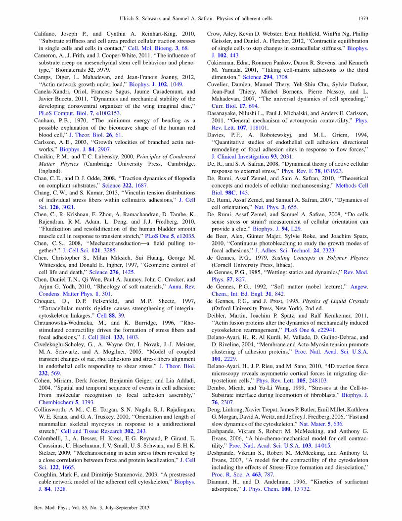
Califano, Joseph P., and Cynthia A. Reinhart-King, 2010,
‘‘Substrate stiffness and cell area predict cellular traction stresses
in single cells and cells in contact,’’ Cell. Mol. Bioeng. 3, 68.
Cameron, A., J. Frith, and J. Cooper-White, 2011, ‘‘The influence of
substrate creep on mesenchymal stem cell behaviour and pheno-
type,’’ Biomaterials 32, 5979.
Camps, Otger, L. Mahadevan, and Jean-Franois Joanny, 2012,
‘‘Actin network growth under load,’’ Biophys. J. 102, 1049.
Canela-Xandri, Oriol, Francesc Sagus, Jaume Casademunt, and
Javier Buceta, 2011, ‘‘Dynamics and mechanical stability of the
developing dorsoventral organizer of the wing imaginal disc,’’
PLoS Comput. Biol. 7, e1002153.
Canham, P. B., 1970, ‘‘The minimum energy of bending as a
possible explanation of the biconcave shape of the human red
blood cell,’’ J. Theor. Biol. 26, 61.
Carlsson, A. E., 2003, ‘‘Growth velocities of branched actin net-
works,’’ Biophys. J. 84, 2907.
Chaikin, P.M., and T. C. Lubensky, 2000, Principles of Condensed
Matter Physics (Cambridge University Press, Cambridge,
England).
Chan, C. E., and D. J. Odde, 2008, ‘‘Traction dynamics of filopodia
on compliant substrates,’’ Science 322, 1687.
Chang, C.W., and S. Kumar, 2013, ‘‘Vinculin tension distributions
of individual stress fibers within cellmatrix adhesions,’’ J. Cell
Sci. 126, 3021.
Chen, C., R. Krishnan, E. Zhou, A. Ramachandran, D. Tambe, K.
Rajendran, R.M. Adam, L. Deng, and J. J. Fredberg, 2010,
‘‘Fluidization and resolidification of the human bladder smooth
muscle cell in response to transient stretch,’’ PLoS One 5, e12035.
Chen, C. S., 2008, ‘‘Mechanotransduction—a field pulling to-
gether?,’’ J. Cell Sci. 121, 3285.
Chen, Christopher S., Milan Mrksich, Sui Huang, George M.
Whitesides, and Donald E. Ingber, 1997, ‘‘Geometric control of
cell life and death,’’ Science 276, 1425.
Chen, Daniel T. N., Qi Wen, Paul A. Janmey, John C. Crocker, and
Arjun G. Yodh, 2010, ‘‘Rheology of soft materials,’’ Annu. Rev.
Condens. Matter Phys. 1, 301.
Choquet, D., D. P. Felsenfeld, and M. P. Sheetz, 1997,
‘‘Extracellular matrix rigidity causes strengthening of integrin-
cytoskeleton linkages,’’ Cell 88, 39.
Chrzanowska-Wodnicka, M., and K. Burridge, 1996, ‘‘Rho-
stimulated contractility drives the formation of stress fibers and
focal adhesions,’’ J. Cell Biol. 133, 1403.
Civelekoglu-Scholey, G., A. Wayne Orr, I. Novak, J.-J. Meister,
M.A. Schwartz, and A. Mogilner, 2005, ‘‘Model of coupled
transient changes of rac, rho, adhesions and stress fibers alignment
in endothelial cells responding to shear stress,’’ J. Theor. Biol.
232, 569.
Cohen, Miriam, Derk Joester, Benjamin Geiger, and Lia Addadi,
2004, ‘‘Spatial and temporal sequence of events in cell adhesion:
From molecular recognition to focal adhesion assembly,’’
Chembiochem 5, 1393.
Collinsworth, A.M., C. E. Torgan, S. N. Nagda, R. J. Rajalingam,
W. E. Kraus, and G.A. Truskey, 2000, ‘‘Orientation and length of
mammalian skeletal myocytes in response to a unidirectional
stretch,’’ Cell and Tissue Research 302, 243.
Colombelli, J., A. Besser, H. Kress, E. G. Reynaud, P. Girard, E.
Caussinus, U. Haselmann, J. V. Small, U. S. Schwarz, and E.H. K.
Stelzer, 2009, ‘‘Mechanosensing in actin stress fibers revealed by
a close correlation between force and protein localization,’’ J. Cell
Sci. 122, 1665.
Coughlin, Mark F., and Dimitrije Stamenovic, 2003, ‘‘A prestressed
cable network model of the adherent cell cytoskeleton,’’ Biophys.
J. 84, 1328.
Crow, Ailey, Kevin D. Webster, Evan Hohlfeld, WinPin Ng, Phillip
Geissler, and Daniel. A. Fletcher, 2012, ‘‘Contractile equilibration
of single cells to step changes in extracellular stiffness,’’ Biophys.
J. 102, 443.
Cukierman, Edna, Roumen Pankov, Daron R. Stevens, and Kenneth
M. Yamada, 2001, ‘‘Taking cell-matrix adhesions to the third
dimension,’’ Science 294, 1708.
Cuvelier, Damien, Manuel Thery, Yeh-Shiu Chu, Sylvie Dufour,
Jean-Paul Thiery, Michel Bornens, Pierre Nassoy, and L.
Mahadevan, 2007, ‘‘The universal dynamics of cell spreading,’’
Curr. Biol. 17, 694.
Dasanayake, Nilushi L., Paul J. Michalski, and Anders E. Carlsson,
2011, ‘‘General mechanism of actomyosin contractility,’’ Phys.
Rev. Lett. 107, 118101.
Davies, P. F., A. Robotewskyj, and M. L. Griem, 1994,
‘‘Quantitative studies of endothelial cell adhesion. directional
remodeling of focal adhesion sites in response to flow forces,’’
J. Clinical Investigation 93, 2031.
De, R., and S. A. Safran, 2008, ‘‘Dynamical theory of active cellular
response to external stress,’’ Phys. Rev. E 78, 031923.
De, Rumi, Assaf Zemel, and Sam A. Safran, 2010, ‘‘Theoretical
concepts and models of cellular mechanosensing,’’ Methods Cell
Biol. 98C, 143.
De, Rumi, Assaf Zemel, and Samuel A. Safran, 2007, ‘‘Dynamics of
cell orientation,’’ Nat. Phys. 3, 655.
De, Rumi, Assaf Zemel, and Samuel A. Safran, 2008, ‘‘Do cells
sense stress or strain? measurement of cellular orientation can
provide a clue,’’ Biophys. J. 94, L29.
de Beer, Alex, Gunter Majer, Sylvie Roke, and Joachim Spatz,
2010, ‘‘Continuous photobleaching to study the growth modes of
focal adhesions,’’ J. Adhes. Sci. Technol. 24, 2323.
de Gennes, P. G., 1979, Scaling Concepts in Polymer Physics
(Cornell University Press, Ithaca).
de Gennes, P. G., 1985, ‘‘Wetting: statics and dynamics,’’ Rev. Mod.
Phys. 57, 827.
de Gennes, P. G., 1992, ‘‘Soft matter (nobel lecture),’’ Angew.
Chem., Int. Ed. Engl. 31, 842.
de Gennes, P. G., and J. Prost, 1995, Physics of Liquid Crystals
(Oxford University Press, New York), 2nd ed.
Deibler, Martin, Joachim P. Spatz, and Ralf Kemkemer, 2011,
‘‘Actin fusion proteins alter the dynamics of mechanically induced
cytoskeleton rearrangement,’’ PLoS One 6, e22941.
Delano-Ayari, H., R. Al Kurdi, M. Vallade, D. Gulino-Debrac, and
D. Riveline, 2004, ‘‘Membrane and Acto-Myosin tension promote
clustering of adhesion proteins,’’ Proc. Natl. Acad. Sci. U.S.A.
101, 2229.
Delano-Ayari, H., J. P. Rieu, and M. Sano, 2010, ‘‘4D traction force
microscopy reveals asymmetric cortical forces in migrating dic-
tyostelium cells,’’ Phys. Rev. Lett. 105, 248103.
Dembo, Micah, and Yu-Li Wang, 1999, ‘‘Stresses at the Cell-to-
Substrate interface during locomotion of fibroblasts,’’ Biophys. J.
76, 2307.
Deng, Linhong, Xavier Trepat, James P. Butler, EmilMillet, Kathleen
G.Morgan,DavidA.Weitz, and Jeffrey J. Fredberg, 2006, ‘‘Fast and
slow dynamics of the cytoskeleton,’’ Nat. Mater. 5, 636.
Deshpande, Vikram S, Robert M. McMeeking, and Anthony G.
Evans, 2006, ‘‘A bio-chemo-mechanical model for cell contrac-
tility,’’ Proc. Natl. Acad. Sci. U.S.A. 103, 14 015.
Deshpande, Vikram S., Robert M. McMeeking, and Anthony G.
Evans, 2007, ‘‘A model for the contractility of the cytoskeleton
including the effects of Stress-Fibre formation and dissociation,’’
Proc. R. Soc. A 463, 787.
Diamant, H., and D. Andelman, 1996, ‘‘Kinetics of surfactant
adsorption,’’ J. Phys. Chem. 100, 13 732.
Ulrich S. Schwarz and Samuel A. Safran: Physics of adherent cells 1373
Rev. Mod. Phys., Vol. 85, No. 3, July–September 2013

Dill, Ken A., and Sarina Bromberg, 2010, Molecular Driving
Forces: Statistical Thermodynamics in Chemistry and Biology
(Garland Publishing, New York).
Discher, D. E., D. J. Mooney, and P.W. Zandstra, 2009, ‘‘Growth
factors, matrices, and forces combine and control stem cells,’’
Science 324, 1673.
Discher, Dennis E., Paul Janmey, and Yu-li Wang, 2005, ‘‘Tissue
cells feel and respond to the stiffness of their substrate,’’ Science
310, 1139.
Dogterom, Marileen, and Bernard Yurke, 1997, ‘‘Measurement of the
force-velocity relation for growingmicrotubules,’’ Science278, 856.
Doi, M., 1996, Introduction to Polymer Physics (Oxford University,
New York).
DuFort, Christopher C., Matthew J. Paszek, and Valerie M. Weaver,
2011, ‘‘Balancing forces: architectural control of mechanotrans-
duction,’’ Nat. Rev. Mol. Cell Biol. 12, 308.
Duke, T. A. J., 1999, ‘‘Molecular model of muscle contraction,’’
Proc. Natl. Acad. Sci. U.S.A. 96, 2770.
Eastwood, M., V. C. Mudera, D.A. McGrouther, and R.A. Brown,
1998, ‘‘Effect of precise mechanical loading on fibroblast popu-
lated collagen lattices: Morphological changes,’’ Cell Motil.
Cytoskeleton 40, 13.
Edwards, Carina M., and Ulrich S. Schwarz, 2011, ‘‘Force
localization in contracting cell layers,’’ Phys. Rev. Lett. 107,
128101.
Engler, A., S. Sen, H. Sweeney, and D. Discher, 2006, ‘‘Matrix
elasticity directs stem cell lineage specification,’’ Cell 126, 677.
Engler, A. J., C. Carag-Krieger, C. P. Johnson, M. Raab, H.-Y.
Speicher, Tang D.W., J.W. Sanger, J.M. Sanger, and D. E.
Discher, 2008, ‘‘Embryonic cardiomyocytes beat best on a matrix
with heart-like elasticity: scar-like rigidity inhibits beating,’’ J.
Cell Sci. 121, 3794.
Engler, A. J., M.A. Griffin, S. Sen, C. G. Bonnetnann, H. L.
Sweeney, and D. E. Discher, 2004, ‘‘Myotubes differentiate opti-
mally on substrates with tissue-like stiffness: pathological impli-
cations for soft or stiff microenvironments,’’ J. Cell Biol. 166, 877.
Engler, Adam, Lucie Bacakova, Cynthia Newman, Alina Hategan,
Maureen Griffin, and Dennis Discher, 2004, ‘‘Substrate compli-
ance versus ligand density in cell on gel responses,’’ Biophys. J.
86, 617.
Erdmann, T., and U. S. Schwarz, 2004a, ‘‘Stability of adhesion
clusters under constant force,’’ Phys. Rev. Lett. 92, 108102.
Erdmann, T., and U. S. Schwarz, 2004b, ‘‘Stochastic dynamics of
adhesion clusters under shared constant force and with rebind-
ing,’’ J. Chem. Phys. 121, 8997.
Erdmann, Thorsten, and Ulrich S. Schwarz, 2012, ‘‘Stochastic force
generation by small ensembles of myosin ii motors,’’ Phys. Rev.
Lett. 108, 188101.
Eshelby, J. D., 1957, ‘‘The determination of the elastic field of an
ellipsoidal inclusion, and related problems,’’ Proc. R. Soc. A 241,
376.
Eshelby, J. D., 1959, ‘‘The field outside of an ellipsoidal inclusion,’’
Proc. R. Soc. A 252, 561.
Evans, E., and K. Ritchie, 1997, ‘‘Dynamic strength of molecular
adhesion bonds,’’ Biophys. J. 72, 1541.
Evans, Evan A., and David A. Calderwood, 2007, ‘‘Forces and bond
dynamics in cell adhesion,’’ Science 316, 1148.
Farhadifar, R., J. C. Roper, B. Algouy, S. Eaton, and F. Julicher,
2007, ‘‘The influence of cell mechanics, cell-cell interactions, and
proliferation on epithelial packing,’’ Curr. Biol. 17, 2095.
Faust, Uta, Nico Hampe, Wolfgang Rubner, Norbert Kirchgessner,
Sam Safran, Bernd Hoffmann, and Rudolf Merkel, 2011, ‘‘Cyclic
stress at mHz frequencies aligns fibroblasts in direction of zero
strain,’’ PLoS One 6, e28963.
Fernandez, P., and A. R. Bausch, 2009, ‘‘The compaction of gels by
cells: a case of collective mechanical activity,’’ Integr. Biol. 1,
252.
Fery, Andreas, and Richard Weinkamer, 2007, ‘‘Mechanical prop-
erties of micro- and nanocapsules: Single-capsule measurements,’’
Polymer 48, 7221.
Filippov, A. E., J. Klafter, and M. Urbakh, 2004, ‘‘Friction through
dynamical formation and rupture of molecular bonds,’’ Phys. Rev.
Lett. 92, 135503.
Fink, Jenny, et al., 2011, ‘‘External forces control mitotic spindle
positioning,’’ Nat. Cell Biol. 13, 771.
Fletcher, D. A., and P. L. Geissler, 2009, ‘‘Active biological mate-
rials,’’ Annu. Rev. Phys. Chem. 60, 469.
Fletcher, Daniel A., and R. Dyche Mullins, 2010, ‘‘Cell mechanics
and the cytoskeleton,’’ Nature (London) 463, 485.
Fletcher, Daniel A., and Julie A. Theriot, 2004, ‘‘An introduction to
cell motility for the physical scientist,’’ Phys. Biol. 1, T1.
Friedrich, B.M., E. Fischer-Friedrich, N. S. Gov, and S. A. Safran,
2012, ‘‘Sarcomeric pattern formation by actin cluster coales-
cence,’’ PLoS Comput. Biol. 8, e1002544.
Friedrich, B.M., and S. A. Safran, 2011, ‘‘Nematic order by elastic
interactions and cellular rigidity sensing,’’ Europhys. Lett. 93,
28007.
Friedrich, Benjamin M., Amnon Buxboim, Dennis E. Discher, and
Samuel A. Safran, 2011, ‘‘Striated Acto-Myosin fibers can reor-
ganize and register in response to elastic interactions with the
matrix,’’ Biophys. J. 100, 2706.
Friedrich, Benjamin M., and Samuel A. Safran, 2012, ‘‘How cells
feel their substrate: spontaneous symmetry breaking of active
surface stresses,’’ Soft Matter 8, 3223.
Fu, Jianping, Yang-Kao Wang, Michael T. Yang, Ravi A. Desai,
Xiang Yu, Zhijun Liu, and Christopher S. Chen, 2010,
‘‘Mechanical regulation of cell function with geometrically modu-
lated elastomeric substrates,’’ Nat. Methods 7, 733.
Fung, Y. C., 1993, Biomechanics: Mechanical Properties of Living
Tissues (Springer-Verlag, New York).
Gao, Huajian, Jin Qian, and Bin Chen, 2011, ‘‘Probing mechanical
principles of focal contacts in cellmatrix adhesion with a coupled
stochasticelastic modelling framework,’’ J. R. Soc. Interface 8,
1217.
Gardel, M. L., J. H. Shin, F. C. MacKintosh, L. Mahadevan, P.
Matsudaira, and D.A. Weitz, 2004, ‘‘Elastic behavior of cross-
linked and bundled actin networks,’’ Science 304, 1301.
Gardel, Margaret L., Karen E. Kasza, Clifford P. Brangwynne, Jiayu
Liu, and David A. Weitz, 2008, ‘‘Mechanical response of cytos-
keletal networks,’’ Methods Cell Biol. 89, 487.
Gardel, Margaret L., Benedikt Sabass, Lin Ji, Gaudenz Danuser,
Ulrich S. Schwarz, and Clare M. Waterman, 2008, ‘‘Traction
stress in focal adhesions correlates biphasically with actin retro-
grade flow speed,’’ J. Cell Biol. 183, 999.
Gauthier, Nils C., Marc Antoine Fardin, Pere Roca-Cusachs, and
Michael P. Sheetz, 2011, ‘‘Temporary increase in plasmamembrane
tension coordinates the activation of exocytosis and contraction
during cell spreading,’’ Proc. Natl. Acad. Sci. U.S.A. 108, 14 467.
Gavara, Nuria, Pere Roca-Cusachs, Raimon Sunyer, Ramon Farre,
and Daniel Navajas, 2008, ‘‘Mapping cell-matrix stresses during
stretch reveals inelastic reorganization of the cytoskeleton,’’
Biophys. J. 95, 464.
Geiger, Benjamin, Joachim P. Spatz, and Alexander D. Bershadsky,
2009, ‘‘Environmental sensing through focal adhesions,’’ Nat.
Rev. Mol. Cell Biol. 10, 21.
Gonzalez-Rodriguez, David, Karine Guevorkian, Stphane Douezan,
and Franoise Brochard-Wyart, 2012, ‘‘Soft matter models of
developing tissues and tumors,’’ Science 338, 910.
1374 Ulrich S. Schwarz and Samuel A. Safran: Physics of adherent cells
Rev. Mod. Phys., Vol. 85, No. 3, July–September 2013

Gov, N., 2004, ‘‘Membrane undulations driven by force fluctuations
of active proteins,’’ Phys. Rev. Lett. 93, 268104.
Gov, Nir S., 2006, ‘‘Dynamics and morphology of microvilli driven
by actin polymerization,’’ Phys. Rev. Lett. 97, 018101.
Guo, M., and D. Weitz, 2012 (unpublished).
Gurin, Thomas, Jacques Prost, Pascal Martin, and Jean-Franois
Joanny, 2010, ‘‘Coordination and collective properties of molecu-
lar motors: theory,’’ Curr. Opin. Cell Biol. 22, 14.
Guthardt Torres, P., I. B. Bischofs, and U. S. Schwarz, 2012,
‘‘Contractile network models for adherent cells,’’ Phys. Rev. E
85, 011913.
Haken, H., 1983, Synergetics, an Introduction: Nonequilibrium
Phase Transitions and Self-Organization in Physics, Chemistry,
and Biology (Springer-Verlag, Berlin).
Hanggi, Peter, Peter Talkner, and Michal Borkovec, 1990,
‘‘Reaction-rate theory: fifty years after Kramers,’’ Rev. Mod.
Phys. 62, 251.
Hayakawa, K., N. Sato, and T. Obinata, 2001, ‘‘Dynamic reorienta-
tion of cultured cells and stress fibers under mechanical stress
from periodic stretching,’’ Exp. Cell Res. 268, 104.
Head, D. A., A. J. Levine, and F. C. MacKintosh, 2003a, ‘‘Distinct
regimes of elastic response and deformation modes of cross-
linked cytoskeletal and semiflexible polymer networks,’’ Phys.
Rev. E 68, 061907.
Head, David A., Alex J. Levine, and F. C. MacKintosh, 2003b,
‘‘Deformation of cross-linked semiflexible polymer networks,’’
Phys. Rev. Lett. 91, 108102.
Heil, P., and J. P. Spatz, 2010, ‘‘Lateral shear forces applied to cells
with single elastic micropillars to influence focal adhesion
dynamics,’’ J. Phys. Condens. Matter 22, 194108.
Helfrich, W., 1973, ‘‘Elastic properties of lipid bilayers: theory and
possible experiments,’’ Z. Naturforsch. C 28, 693.
Heussinger, Claus, and Erwin Frey, 2006, ‘‘Stiff polymers, foams,
and fiber networks,’’ Phys. Rev. Lett. 96, 017802.
Heussinger, Claus, Boris Schaefer, and Erwin Frey, 2007,
‘‘Nonaffine rubber elasticity for stiff polymer networks,’’ Phys.
Rev. E 76, 031906.
Hoffman, Brenton D., Carsten Grashoff, and Martin A. Schwartz,
2011, ‘‘Dynamic molecular processes mediate cellular mechano-
transduction,’’ Nature (London) 475, 316.
Hoffmann, Max, and Ulrich S. Schwarz, 2013, ‘‘A kinetic model for
RNA-interference of focal adhesions,’’ BMC Systems Biology 7, 2.
Hotulainen, Pirta, and Pekka Lappalainen, 2006, ‘‘Stress fibers are
generated by two distinct actin assembly mechanisms in motile
cells,’’ J. Cell Biol. 173, 383.
Howard, J., 2001, Mechanics of Motor Proteins and the
Cytoskeleton (Sinauer Associates, Sunderland, MA).
Hsu, Hui-Ju, Chin-Fu Lee, and Roland Kaunas, 2009, ‘‘A dynamic
stochastic model of frequency-dependent stress fiber alignment
induced by cyclic stretch,’’ PLoS One 4, e4853.
Huang, S., and D. E. Ingber, 1999, ‘‘The structural and mechanical
complexity of cell-growth control,’’ Nat. Cell Biol. 1, E131.
Huber, F., J. Schnauss, S. Ronicke, P. Rauch, K. Muller, C. Futterer,
and J. Kas, 2013, ‘‘Emergent complexity of the cytoskeleton: from
single filaments to tissue,’’ Adv. Phys. 62, 1.
Hufnagel, L., A.A. Teleman, H. Rouault, S.M. Cohen, and B. I.
Shraiman, 2007, ‘‘On the mechanism of wing size determination
in fly development,’’ Proc. Natl. Acad. Sci. U.S.A. 104, 3835.
Hur, Sung Sik, Yihua Zhao, Yi-Shuan Li, Elliot Botvinick, and Shu
Chien, 2009, ‘‘Live cells exert 3-Dimensional traction forces on
their substrata,’’ Cell. Mol. Bioeng. 2, 425.
Huxley, A. F., 1957, ‘‘Muscle structure and theories of contraction,’’
Prog. Biophys. Biophys. Chem. 7, 255 [http://www.ncbi.nlm.nih.
gov/pubmed/13485191].
Ingber, D. E., 1993, ‘‘Cellular tensegrity—defining new rules of
biological design that govern the cytoskeleton,’’ J. Cell Sci. 104,
613.
Ingber, D. E., 2003, ‘‘Mechanobiology and diseases of mechano-
transduction,’’ Ann. Med. 35, 564.
Isenberg, B., P. DiMilla, M. Walker, S. Kim, and J. Wong, 2009,
‘‘Vascular smooth muscle cell durotaxis depends on substrate
stiffness gradient strength,’’ Biophys. J. 97, 1313.
Israelachvili, J. N., 2011, Intermolecular and Surface Forces
(Academic Press, Waltham), 3rd ed.
Iyer, K. Venkatesan, Shovamayee Maharana, Soumya Gupta, Albert
Libchaber, Tsvi Tlusty, and G.V. Shivashankar, 2012, ‘‘Modeling
and experimental methods to probe the link between global tran-
scription and spatial organization of chromosomes,’’ PLoS One 7,
e46628.
Jacot, Jeffrey G., Andrew D. McCulloch, and Jeffrey H. Omens,
2008, ‘‘Substrate stiffness affects the functional maturation of
neonatal rat ventricular myocytes,’’ Biophys. J. 95, 3479.
Janmey, P. A., S. Hvidt, J. Lamb, and T. P. Stossel, 1990,
‘‘Resemblance of actin-binding protein actin gels to covalently
cross-linked networks,’’ Nature (London) 345, 89.
Janmey, P. A., J. P. Winer, M. E. Murray, and Q. Wen, 2009, ‘‘The
hard life of soft cells,’’ Cell Motil. Cytoskeleton 66, 597.
Janmey, Paul A., and R. Tyler Miller, 2011, ‘‘Mechanisms of me-
chanical signaling in development and disease,’’ J. Cell Sci. 124, 9.
Johnson, K. L., 1985, Contact Mechanics (Cambridge University
Press, Cambridge, England).
Johnson, K. L., K. Kendall, and A.D. Roberts, 1971, ‘‘Surface
energy and the contact of elastic solids,’’ Proc. R. Soc. A 324, 301.
Jones, Richard A. L., 2002, Soft Condensed Matter (Oxford
University Press, New York).
Julicher, F., K. Kruse, J. Prost, and J. F. Joanny, 2007, ‘‘Active
behavior of the cytoskeleton,’’ Phys. Rep. 449, 3.
Julicher, Frank, and Jacques Prost, 1995, ‘‘Cooperative molecular
motors,’’ Phys. Rev. Lett. 75, 2618.
Jungbauer, S., H. J. Gao, J. P. Spatz, and R. Kemkemer, 2008, ‘‘Two
characteristic regimes in frequency-dependent dynamic reorienta-
tion of fibroblasts on cyclically stretched substrates,’’ Biophys. J.
95, 3470.
Kaunas, R., H. J. Hsu, and S. Deguchi, 2011, ‘‘Sarcomeric model of
stretch-induced stress fiber reorganization,’’ Cell Health and
Cytoskeleton 3, 13.
Kaunas, R., P. Nguyen, S. Usami, and S. Chien, 2005, ‘‘Cooperative
effects of rho and mechanical stretch on stress fiber organization,’’
Proc. Natl. Acad. Sci. U.S.A. 102, 15 895.
Kawakami, K., H. Tatsumi, and M. Sokabe, 2001, ‘‘Dynamics of
integrin clustering at focal contacts of endothelial cells studied by
multimode imaging microscopy,’’ J. Cell Sci. 114, 3125.
Kilian, K.A., B. Bugarija, B. T. Lahn, and M. Mrksich, 2010,
‘‘Geometric cues for directing the differentiation of mesenchymal
stem cells,’’ Proc. Natl. Acad. Sci. U.S.A. 107, 4872.
Klein, Franziska, Benjamin Richter, Thomas Striebel, Clemens M.
Franz, Georg von Freymann, Martin Wegener, and Martin
Bastmeyer, 2011, ‘‘Two-component polymer scaffolds for con-
trolled three-dimensional cell culture,’’ Adv. Mater. 23, 1341.
Klein, Franziska, Thomas Striebel, Joachim Fischer, Zhongxiang
Jiang, Clemens M. Franz, Georg von Freymann, Martin Wegener,
and Martin Bastmeyer, 2010, ‘‘Elastic fully three-dimensional
microstructure scaffolds for cell force measurements,’’ Adv.
Mater. 22, 868.
Klotzsch, Enrico, Michael L. Smith, Kristopher E. Kubow, Simon
Muntwyler, William C. Little, Felix Beyeler, Delphine Gourdon,
Bradley J. Nelson, and Viola Vogel, 2009, ‘‘Fibronectin forms
the most extensible biological fibers displaying switchable
Ulrich S. Schwarz and Samuel A. Safran: Physics of adherent cells 1375
Rev. Mod. Phys., Vol. 85, No. 3, July–September 2013

force-exposed cryptic binding sites,’’ Proc. Natl. Acad. Sci. U.S.A.
106, 18 267.
Klumpp, Stefan, and Reinhard Lipowsky, 2005, ‘‘Cooperative cargo
transport by several molecular motors,’’ Proc. Natl. Acad. Sci.
U.S.A. 102, 17 284.
Knoche, Sebastian, and Jan Kierfeld, 2011, ‘‘Buckling of spherical
capsules,’’ Phys. Rev. E 84, 046608.
Koenderink, Gijsje H., Zvonimir Dogic, Fumihiko Nakamura, Poul
M. Bendix, Frederick C. MacKintosh, John H. Hartwig, Thomas
P. Stossel, and David A. Weitz, 2009, ‘‘An active biopolymer
network controlled by molecular motors,’’ Proc. Natl. Acad. Sci.
U.S.A. 106, 15 192.
Kohler, Simone, Volker Schaller, and Andreas R. Bausch, 2011,
‘‘Structure formation in active networks,’’ Nat. Mater. 10, 462.
Kollmannsberger, P., C.M. Bidan, J.W. C. Dunlop, and P. Fratzl,
2011, ‘‘The physics of tissue patterning and extracellular matrix
organisation: how cells join forces,’’ Soft Matter 7, 9549.
Kollmannsberger, P., and B. Fabry, 2011, ‘‘Linear and nonlinear
rheology of living cells,’’ Annu. Rev. Mater. Res. 41, 75.
Komura, S., K. Tamura, and T. Kato, 2005, ‘‘Buckling of spherical
shells adhering onto a rigid substrate,’’ Eur. Phys. J. E 18, 343.
Korff, T., and H.G. Augustin, 1999, ‘‘Tensional forces in fibrillar
extracellular matrices control directional capillary sprouting,’’ J.
Cell Sci. 112, 3249.
Kovar, D. R., 2006, ‘‘Molecular details of formin-mediated actin
assembly,’’ Curr. Opin. Cell Biol. 18, 11.
Krishnan, R., et al., 2009, ‘‘Reinforcement versus fluidization in
cytoskeletal mechanoresponsiveness,’’ PLoS One 4, e5486.
Kruse, K., J. F. Joanny, F. Julicher, J. Prost, and K. Sekimoto, 2004,
‘‘Asters, vortices, and rotating spirals in active gels of polar
filaments,’’ Phys. Rev. Lett. 92, 078101.
Kruse, K., and F. Julicher, 2000a, ‘‘Actively contracting bundles of
polar filaments,’’ Phys. Rev. Lett. 85, 1778.
Kruse, K., and F. Julicher, 2000b, ‘‘Actively contracting bundles of
polar filaments,’’ Phys. Rev. Lett. 85, 1778.
Kruse, K., and F. Julicher, 2003, ‘‘Self-organization and mechanical
properties of active filament bundles,’’ Phys. Rev. E 67, 051913.
Kruse, K., A. Zumdieck, and F. Julicher, 2003, ‘‘Continuum theory
of contractile fibres,’’ Europhys. Lett. 64, 716.
Kumar, Sanjay, Iva Z. Maxwell, Alexander Heisterkamp, Thomas
R. Polte, Tanmay P. Lele, Matthew Salanga, Eric Mazur, and
Donald E. Ingber, 2006, ‘‘Viscoelastic retraction of single living
stress fibers and its impact on cell shape, cytoskeletal organiza-
tion, and extracellular matrix mechanics,’’ Biophys. J. 90, 3762.
Kuo, Jean-Cheng, Xuemei Han, Cheng-Te Hsiao, John R. Yates III,
and Clare M. Waterman, 2011, ‘‘Analysis of the myosin-II-
responsive focal adhesion proteome reveals a role for [beta]-Pix
in negative regulation of focal adhesion maturation,’’ Nat. Cell
Biol. 13, 383.
Kurpinski, Kyle, Julia Chu, Craig Hashi, and Song Li, 2006,
‘‘Anisotropic mechanosensing by mesenchymal stem cells,’’
Proc. Natl. Acad. Sci. U.S.A. 103, 16 095.
Ladoux, Benoit, and Alice Nicolas, 2012, ‘‘Physically based prin-
ciples of cell adhesion mechanosensitivity in tissues,’’ Rep. Prog.
Phys. 75, 116601.
Landau, L. D., and E.M. Lifshitz, 1970, Theory of Elasticity, Course
of Theoretical Physics (Pergamon Press, Oxford), Vol. 7, 2nd ed.
Landsberg, Katharina P., Reza Farhadifar, Jonas Ranft, Daiki
Umetsu, Thomas J. Widmann, Thomas Bittig, Amani Said,
Frank Jlicher, and Christian Dahmann, 2009, ‘‘Increased cell
bond tension governs cell sorting at the drosophila anteroposterior
compartment boundary,’’ Curr. Biol. 19, 1950.
Lau, K. H., and W. Kohn, 1977, ‘‘Elastic interaction of two atoms
adsorbed on a solid-surface,’’ Surf. Sci. 65, 607.
Lauffenburger, Douglas A., and Alan F. Horwitz, 1996, ‘‘Cell migra-
tion: A physically integrated molecular process,’’ Cell 84, 359.
Lecuit, Thomas, and Pierre-Francois Lenne, 2007, ‘‘Cell surface
mechanics and the control of cell shape, tissue patterns and
morphogenesis,’’ Nat. Rev. Mol. Cell Biol. 8, 633.
Lee, Kun-Chun, and Andrea J. Liu, 2009, ‘‘Force-Velocity relation
for Actin-Polymerization-Driven motility from brownian dynam-
ics simulations,’’ Biophys. J. 97, 1295.
Legant, Wesley R., Colin K. Choi, Jordan S. Miller, Lin Shao, Liang
Gao, Eric Betzig, and Christopher S. Chen, 2013, ‘‘Multidimensional
traction force microscopy reveals out-of-plane rotational moments
about focal adhesions,’’ Proc. Natl. Acad. Sci. U.S.A. 110, 881.
Lele, T. P., J. Pendse, S. Kumar, M. Salanga, J. Karavitis, and D. E.
Ingber, 2006, ‘‘Mechanical forces alter zyxin unbinding kinetics
within focal adhesions of living cells,’’ J. Cell. Physiol. 207, 187.
Lenne, Pierre-Francois, andAliceNicolas, 2009, ‘‘Physics puzzles on
membrane domains posed by cell biology,’’ Soft Matter 5, 2841.
Lenz, M., T. Thoresen, M. L. Gardel, and A. R. Dinner, 2012,
‘‘Contractile units in disordered actomyosin bundles arise from
f-actin buckling,’’ Phys. Rev. Lett. 108, 238107.
Li, Ying, Prabhakar Bhimalapuram, and Aaron R. Dinner, 2010,
‘‘Model for how retrograde actin flow regulates adhesion traction
stresses,’’ J. Phys. Condens. Matter 22, 194113.
Lieleg, O., M.M. Claessens, Y. Luan, and A. R. Bausch, 2008,
‘‘Transient binding and dissipation in cross-linked actin net-
works,’’ Phys. Rev. Lett. 101, 108101.
Lim H.W., Gerald, Michael Wortis, and Ranjan Mukhopadhyay,
2002, ‘‘Stomatocyte-discocyte-echinocyte sequence of the human
red blood cell: Evidence for the bilayer couple hypothesis from
membrane mechanics,’’ Proc. Natl. Acad. Sci. U.S.A. 99, 16766.
Lipowsky, Reinhard, 1996, ‘‘Adhesion of membranes via anchored
stickers,’’ Phys. Rev. Lett. 77, 1652.
Liverpool, T. B., andM.C. Marchetti, 2003, ‘‘Instabilities of isotropic
solutions of active polar filaments,’’ Phys. Rev. Lett. 90, 138102.
Lo, C.M., H. B. Wang, M. Dembo, and Y. L. Wang, 2000, ‘‘Cell
movement is guided by the rigidity of the substrate,’’ Biophys. J.
79, 144.
Lobkovsky, Alex, Sharon Gentges, Hao Li, David Morse, and T. A.
Witten, 1995, ‘‘Scaling properties of stretching ridges in a
crumpled elastic sheet,’’ Science 270, 1482.
Loosli, Y., R. Luginbuehl, and J. G. Snedeker, 2010, ‘‘Cytoskeleton
reorganization of spreading cells on Micro-Patterned islands: A
functional model,’’ Phil. Trans. R. Soc. A 368, 2629.
Lu, L., Y. Feng, W. J. Hucker, S. J. Oswald, G.D. Longmore, and
F. C. Yin, 2008, ‘‘Actin stress fiber preextension in human aortic
endothelial cells,’’ Cell Motil. Cytoskeleton 65, 281.
Luo, Y. Z., X. Xu, T. Lele, S. Kumar, and D. E. Ingber, 2008,
‘‘A multi-modular tensegrity model of an actin stress fiber,’’ J.
Biomech. 41, 2379.
Macdonald, Alice, A. Rick Horwitz, and Douglas A. Lauffenburger,
2008, ‘‘Kinetic model for lamellipodal actin-integrin ’clutch’
dynamics,’’ Cell Adhesion & Migration 2, 95.
Mackintosh, F., 2006, in Soft Condensed Matter Physics in
Molecular and Cell Biology (CRC Press, Boca Raton, FL).
MacKintosh, Frederick C., and Christoph F. Schmidt, 2010, ‘‘Active
cellular materials,’’ Curr. Opin. Cell Biol. 22, 29.
Maier, W., and A. Saupe, 1959, Z. Naturforschg. 14, 882.
Majkut, Stephanie F., andDennis E. Discher, 2012, ‘‘Cardiomyocytes
from late embryos and neonates do optimal work and striate best on
substrates with tissue-level elasticity: metrics and mathematics,’’
Biomech. Model. Mechanobiol. 11, 1219.
Manneville, J. B., P. Bassereau, S. Ramaswamy, and J. Prost, 2001,
‘‘Active membrane fluctuations studied by micropipet aspiration,’’
Phys. Rev. E 64, 021908.
1376 Ulrich S. Schwarz and Samuel A. Safran: Physics of adherent cells
Rev. Mod. Phys., Vol. 85, No. 3, July–September 2013
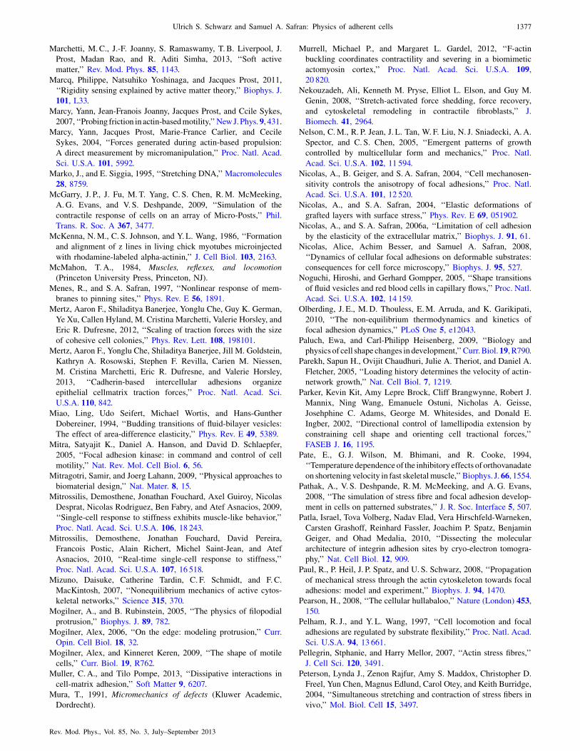
Marchetti, M. C., J.-F. Joanny, S. Ramaswamy, T. B. Liverpool, J.
Prost, Madan Rao, and R. Aditi Simha, 2013, ‘‘Soft active
matter,’’ Rev. Mod. Phys. 85, 1143.
Marcq, Philippe, Natsuhiko Yoshinaga, and Jacques Prost, 2011,
‘‘Rigidity sensing explained by active matter theory,’’ Biophys. J.
101, L33.
Marcy, Yann, Jean-Franois Joanny, Jacques Prost, and Ccile Sykes,
2007, ‘‘Probing friction in actin-basedmotility,’’NewJ.Phys.9, 431.
Marcy, Yann, Jacques Prost, Marie-France Carlier, and Cecile
Sykes, 2004, ‘‘Forces generated during actin-based propulsion:
A direct measurement by micromanipulation,’’ Proc. Natl. Acad.
Sci. U.S.A. 101, 5992.
Marko, J., and E. Siggia, 1995, ‘‘Stretching DNA,’’ Macromolecules
28, 8759.
McGarry, J. P., J. Fu, M. T. Yang, C. S. Chen, R.M. McMeeking,
A.G. Evans, and V. S. Deshpande, 2009, ‘‘Simulation of the
contractile response of cells on an array of Micro-Posts,’’ Phil.
Trans. R. Soc. A 367, 3477.
McKenna, N.M., C. S. Johnson, and Y. L. Wang, 1986, ‘‘Formation
and alignment of z lines in living chick myotubes microinjected
with rhodamine-labeled alpha-actinin,’’ J. Cell Biol. 103, 2163.
McMahon, T. A., 1984, Muscles, reflexes, and locomotion
(Princeton University Press, Princeton, NJ).
Menes, R., and S. A. Safran, 1997, ‘‘Nonlinear response of mem-
branes to pinning sites,’’ Phys. Rev. E 56, 1891.
Mertz, Aaron F., Shiladitya Banerjee, Yonglu Che, Guy K. German,
Ye Xu, Callen Hyland, M. Cristina Marchetti, Valerie Horsley, and
Eric R. Dufresne, 2012, ‘‘Scaling of traction forces with the size
of cohesive cell colonies,’’ Phys. Rev. Lett. 108, 198101.
Mertz, Aaron F., Yonglu Che, Shiladitya Banerjee, Jill M. Goldstein,
Kathryn A. Rosowski, Stephen F. Revilla, Carien M. Niessen,
M. Cristina Marchetti, Eric R. Dufresne, and Valerie Horsley,
2013, ‘‘Cadherin-based intercellular adhesions organize
epithelial cellmatrix traction forces,’’ Proc. Natl. Acad. Sci.
U.S.A. 110, 842.
Miao, Ling, Udo Seifert, Michael Wortis, and Hans-Gunther
Dobereiner, 1994, ‘‘Budding transitions of fluid-bilayer vesicles:
The effect of area-difference elasticity,’’ Phys. Rev. E 49, 5389.
Mitra, Satyajit K., Daniel A. Hanson, and David D. Schlaepfer,
2005, ‘‘Focal adhesion kinase: in command and control of cell
motility,’’ Nat. Rev. Mol. Cell Biol. 6, 56.
Mitragotri, Samir, and Joerg Lahann, 2009, ‘‘Physical approaches to
biomaterial design,’’ Nat. Mater. 8, 15.
Mitrossilis, Demosthene, Jonathan Fouchard, Axel Guiroy, Nicolas
Desprat, Nicolas Rodriguez, Ben Fabry, and Atef Asnacios, 2009,
‘‘Single-cell response to stiffness exhibits muscle-like behavior,’’
Proc. Natl. Acad. Sci. U.S.A. 106, 18 243.
Mitrossilis, Demosthene, Jonathan Fouchard, David Pereira,
Francois Postic, Alain Richert, Michel Saint-Jean, and Atef
Asnacios, 2010, ‘‘Real-time single-cell response to stiffness,’’
Proc. Natl. Acad. Sci. U.S.A. 107, 16 518.
Mizuno, Daisuke, Catherine Tardin, C. F. Schmidt, and F. C.
MacKintosh, 2007, ‘‘Nonequilibrium mechanics of active cytos-
keletal networks,’’ Science 315, 370.
Mogilner, A., and B. Rubinstein, 2005, ‘‘The physics of filopodial
protrusion,’’ Biophys. J. 89, 782.
Mogilner, Alex, 2006, ‘‘On the edge: modeling protrusion,’’ Curr.
Opin. Cell Biol. 18, 32.
Mogilner, Alex, and Kinneret Keren, 2009, ‘‘The shape of motile
cells,’’ Curr. Biol. 19, R762.
Muller, C. A., and Tilo Pompe, 2013, ‘‘Dissipative interactions in
cell-matrix adhesion,’’ Soft Matter 9, 6207.
Mura, T., 1991, Micromechanics of defects (Kluwer Academic,
Dordrecht).
Murrell, Michael P., and Margaret L. Gardel, 2012, ‘‘F-actin
buckling coordinates contractility and severing in a biomimetic
actomyosin cortex,’’ Proc. Natl. Acad. Sci. U.S.A. 109,
20 820.
Nekouzadeh, Ali, Kenneth M. Pryse, Elliot L. Elson, and Guy M.
Genin, 2008, ‘‘Stretch-activated force shedding, force recovery,
and cytoskeletal remodeling in contractile fibroblasts,’’ J.
Biomech. 41, 2964.
Nelson, C.M., R. P. Jean, J. L. Tan, W. F. Liu, N. J. Sniadecki, A.A.
Spector, and C. S. Chen, 2005, ‘‘Emergent patterns of growth
controlled by multicellular form and mechanics,’’ Proc. Natl.
Acad. Sci. U.S.A. 102, 11 594.
Nicolas, A., B. Geiger, and S. A. Safran, 2004, ‘‘Cell mechanosen-
sitivity controls the anisotropy of focal adhesions,’’ Proc. Natl.
Acad. Sci. U.S.A. 101, 12 520.
Nicolas, A., and S. A. Safran, 2004, ‘‘Elastic deformations of
grafted layers with surface stress,’’ Phys. Rev. E 69, 051902.
Nicolas, A., and S. A. Safran, 2006a, ‘‘Limitation of cell adhesion
by the elasticity of the extracellular matrix,’’ Biophys. J. 91, 61.
Nicolas, Alice, Achim Besser, and Samuel A. Safran, 2008,
‘‘Dynamics of cellular focal adhesions on deformable substrates:
consequences for cell force microscopy,’’ Biophys. J. 95, 527.
Noguchi, Hiroshi, and Gerhard Gompper, 2005, ‘‘Shape transitions
of fluid vesicles and red blood cells in capillary flows,’’ Proc. Natl.
Acad. Sci. U.S.A. 102, 14 159.
Olberding, J. E., M.D. Thouless, E.M. Arruda, and K. Garikipati,
2010, ‘‘The non-equilibrium thermodynamics and kinetics of
focal adhesion dynamics,’’ PLoS One 5, e12043.
Paluch, Ewa, and Carl-Philipp Heisenberg, 2009, ‘‘Biology and
physics of cell shape changes in development,’’Curr.Biol.19, R790.
Parekh, Sapun H., Ovijit Chaudhuri, Julie A. Theriot, and Daniel A.
Fletcher, 2005, ‘‘Loading history determines the velocity of actin-
network growth,’’ Nat. Cell Biol. 7, 1219.
Parker, Kevin Kit, Amy Lepre Brock, Cliff Brangwynne, Robert J.
Mannix, Ning Wang, Emanuele Ostuni, Nicholas A. Geisse,
Josehphine C. Adams, George M. Whitesides, and Donald E.
Ingber, 2002, ‘‘Directional control of lamellipodia extension by
constraining cell shape and orienting cell tractional forces,’’
FASEB J. 16, 1195.
Pate, E., G. J. Wilson, M. Bhimani, and R. Cooke, 1994,
‘‘Temperature dependence of the inhibitory effects of orthovanadate
on shortening velocity in fast skeletalmuscle,’’ Biophys. J.66, 1554.
Pathak, A., V. S. Deshpande, R.M. McMeeking, and A.G. Evans,
2008, ‘‘The simulation of stress fibre and focal adhesion develop-
ment in cells on patterned substrates,’’ J. R. Soc. Interface 5, 507.
Patla, Israel, Tova Volberg, Nadav Elad, Vera Hirschfeld-Warneken,
Carsten Grashoff, Reinhard Fassler, Joachim P. Spatz, Benjamin
Geiger, and Ohad Medalia, 2010, ‘‘Dissecting the molecular
architecture of integrin adhesion sites by cryo-electron tomogra-
phy,’’ Nat. Cell Biol. 12, 909.
Paul, R., P. Heil, J. P. Spatz, and U. S. Schwarz, 2008, ‘‘Propagation
of mechanical stress through the actin cytoskeleton towards focal
adhesions: model and experiment,’’ Biophys. J. 94, 1470.
Pearson, H., 2008, ‘‘The cellular hullabaloo,’’ Nature (London) 453,
150.
Pelham, R. J., and Y. L. Wang, 1997, ‘‘Cell locomotion and focal
adhesions are regulated by substrate flexibility,’’ Proc. Natl. Acad.
Sci. U.S.A. 94, 13 661.
Pellegrin, Stphanie, and Harry Mellor, 2007, ‘‘Actin stress fibres,’’
J. Cell Sci. 120, 3491.
Peterson, Lynda J., Zenon Rajfur, Amy S. Maddox, Christopher D.
Freel, Yun Chen, Magnus Edlund, Carol Otey, and Keith Burridge,
2004, ‘‘Simultaneous stretching and contraction of stress fibers in
vivo,’’ Mol. Biol. Cell 15, 3497.
Ulrich S. Schwarz and Samuel A. Safran: Physics of adherent cells 1377
Rev. Mod. Phys., Vol. 85, No. 3, July–September 2013

Phillips, Rob, Jane Kondev, and Julie Theriot, 2008, Physical
Biology of the Cell (Taylor & Francis, London), 1st ed.
Pieranski, P., 1983, ‘‘Colloidal crystals,’’ Contemp. Phys. 24, 25.
Pinto, Ines Mendes, Boris Rubinstein, Andrei Kucharavy, Jay R.
Unruh, and Rong Li, 2012, ‘‘Actin depolymerization drives
actomyosin ring contraction during budding yeast cytokinesis,’’
Dev. Cell 22, 1247.
Pirentis, A., and P. Lazopoulos, 2009, ‘‘On stress fiber reorientation
under plane substrate stretching,’’ Arch. Appl. Mech. 79, 263.
Pirentis, A., E. Peruski, A. Jordan, and D. Stamenovic, 2011, ‘‘A
model for stress fiber realignment caused by cytoskeletal fluid-
ization during cyclic stretching,’’ Cell. Mol. Bioeng. 4, 67.
Placais, P.-Y., M. Balland, T. Gurin, J.-F. Joanny, and P. Martin,
2009, ‘‘Spontaneous oscillations of a minimal actomyosin system
under elastic loading,’’ Phys. Rev. Lett. 103, 158102.
Plotnikov, Sergey V., Ana M. Pasapera, Benedikt Sabass, and Clare
M. Waterman, 2012, ‘‘Force fluctuations within focal adhesions
mediate ECM-Rigidity sensing to guide directed cell migration,’’
Cell 151, 1513.
Pollard, Thomas D., and Julien Berro, 2008, ‘‘Mathematical models
and simulations of cellular processes based on actin filaments,’’ J.
Biol. Chem. 284, 5433.
Pollard, Thomas D., and Gary G. Borisy, 2003, ‘‘Cellular motility
driven by assembly and disassembly of actin filaments,’’ Cell 112,
453.
Pollard, Thomas D., and John A. Cooper, 2009, ‘‘Actin, a central
player in cell shape and movement,’’ Science 326, 1208.
Pompe, T., S. Glorius, T. Bischoff, I. Uhlmann, M. Kaufmann, S.
Brenner, and C. Werner, 2009, ‘‘Dissecting the impact of matrix
anchorage and elasticity in cell adhesion,’’ Biophys. J. 97, 2154.
Ponti, A., M. Machacek, S. L. Gupton, C.M. Waterman-Storer, and
G. Danuser, 2004, ‘‘Two distinct actin networks drive the pro-
trusion of migrating cells,’’ Science 305, 1782.
Powers, Thomas R., 2010, ‘‘Dynamics of filaments and membranes
in a viscous fluid,’’ Rev. Mod. Phys. 82, 1607.
Pozrikidis, C., and Costas Pozrikidis, 2003, Modeling and
Simulation of Capsules and Biological Cells (CRC Press, Boca
Raton, FL).
Prager-Khoutorsky, Masha, Alexandra Lichtenstein, Ramaswamy
Krishnan, Kavitha Rajendran, Avi Mayo, Zvi Kam, Benjamin
Geiger, and Alexander D. Bershadsky, 2011, ‘‘Fibroblast polar-
ization is a matrix-rigidity-dependent process controlled by focal
adhesion mechanosensing,’’ Nat. Cell Biol. 13, 1457.
Prass, Marcus, Ken Jacobson, Alex Mogilner, and Manfred
Radmacher, 2006, ‘‘Direct measurement of the lamellipodial
protrusive force in a migrating cell,’’ J. Cell Biol. 174, 767.
Pullarkat, Pramod A., Paul Dommersnes, Pablo Fernndez, Jean-
Franois Joanny, and Albrecht Ott, 2006, ‘‘Osmotically driven
shape transformations in axons,’’ Phys. Rev. Lett. 96, 048104.
Qian, J., et al., 2013, ‘‘A mechanochemical model of cell
reorientation on substrates under cyclic stretch,’’ PLoS One 8,
e65864.
Qian, Jin, Jizeng Wang, and Huajian Gao, 2008, ‘‘Lifetime and
strength of adhesive molecular bond clusters between elastic
media,’’ Langmuir 24, 1262.
Ramaswamy, Sriram, 2010, ‘‘The mechanics and statistics of active
matter,’’ Annu. Rev. Condens. Matter Phys. 1, 323.
Ranft, Jonas, Markus Basan, Jens Elgeti, Jean-Francois Joanny,
Jacques Prost, and Frank Julicher, 2010, ‘‘Fluidization of tissues
by cell division and apoptosis,’’ Proc. Natl. Acad. Sci. U.S.A. 107,
20 863.
Rauzi, Matteo, Pascale Verant, Thomas Lecuit, and Pierre-Francois
Lenne, 2008, ‘‘Nature and anisotropy of cortical forces orienting
drosophila tissue morphogenesis,’’ Nat. Cell Biol. 10, 1401.
Rehfeldt, F., A. Brown, M. Raab, S. Cai, A. L. Zajac, A. Zemel, and
D. E. Discher, 2012, ‘‘Hyaluronic acid matrices show matrix
stiffness in 2d and 3d dictates cytoskeletal order and myosin-ii
phosphorylation within stem cells,’’ Integr. Biol. 4, 422.
Reinhart-King, Cynthia A., Micah Dembo, and Daniel A. Hammer,
2008, ‘‘Cell-cell mechanical communication through compliant
substrates,’’ Biophys. J. 95, 6044.
Rhee, D., J.M. Sanger, and J.W. Sanger, 1994, ‘‘The premyofibril:
evidence for its role in myofibrillogenesis,’’ Cell Motil.
Cytoskeleton 28, 1.
Ridley, Anne J., 2011, ‘‘Life at the leading edge,’’ Cell 145, 1012.
Riehl, B. D., J-H. Park, I. K. Kwon, and J. Y. Lim, 2012,
‘‘Mechanical stretching for tissue engineering: two-dimensional
and three-dimensional constructs,’’ Tissue Engineering Part B 18,
288.
Riveline, Daniel, Eli Zamir, Nathalie Q. Balaban, Ulrich S.
Schwarz, Toshimasa Ishizaki, Shuh Narumiya, Zvi Kam,
Benjamin Geiger, and Alexander D. Bershadsky, 2001, ‘‘Focal
contacts as mechanosensors: Externally applied local mechanical
force induces growth of focal contacts by an Mdia1-Dependent
and Rock-Independent mechanism,’’ J. Cell Biol. 153, 1175.
Roopa, T., A. Mazumder, A. Basu, L. Mahadevan, and G.V.
Shivashankar, 2008, ‘‘Dynamics of chromatin decondensation
reveals the structural integrity of a mechanically prestressed cell
nucleus,’’ Biophys. J. 95, 3028.
Rubinstein, M., and R. Colby, 2003, Polymer Physics (Oxford
University, New York).
Rudnicki, M. S., et al., 2013, ‘‘Nonlinear strain stiffening is not
suffcient to explain how far cells can feel on fibrous protein gels,’’
Biophys. J.. 105, 11.
Russell, Robert J., Shen-Ling Xia, Richard B. Dickinson, and
Tanmay P. Lele, 2009, ‘‘Sarcomere mechanics in capillary endo-
thelial cells,’’ Biophys. J. 97, 1578.
Sabass, B., M. L. Gardel, C.M. Waterman, and U. S. Schwarz, 2008,
‘‘High resolution traction force microscopy based on experimental
and computational advances,’’ Biophys. J. 94, 207.
Sabass, Benedikt, and Ulrich S. Schwarz, 2010, ‘‘Modeling cytos-
keletal flow over adhesion sites: competition between stochastic
bond dynamics and intracellular relaxation,’’ J. Phys. Condens.
Matter 22, 194112.
Saez, Alexandre, Axel Buguin, Pascal Silberzan, and Benoit
Ladoux, 2005, ‘‘Is the mechanical activity of epithelial cells
controlled by deformations or forces?,’’ Biophys. J. 89, L52.
Saez, Alexandre, Marion Ghibaudo, Axel Buguin, Pascal Silberzan,
and Benoit Ladoux, 2007, ‘‘Rigidity-driven growth and migration
of epithelial cells on microstructured anisotropic substrates,’’
Proc. Natl. Acad. Sci. U.S.A. 104, 8281.
Safran, S. A., 1999, ‘‘Curvature elasticity of thin films,’’ Adv. Phys.
48, 395.
Safran, S. A., and R. De, 2009, ‘‘Nonlinear dynamics of cell
orientation,’’ Phys. Rev. E 80, 060901.
Safran, S. A., and D. R. Hamann, 1979, ‘‘Long-range elastic inter-
actions and staging in graphite-intercalation compounds,’’ Phys.
Rev. Lett. 42, 1410.
Safran, Samuel, 2003, Statistical Thermodynamics of Surfaces,
Interfaces, and Membranes (Westview Press, Boulder, CO).
Saito, Nobuhiko, Kunihiko Takahashi, and Yasuo Yunoki, 1967,
‘‘Statistical mechanical theory of stiff chains,’’ J. Phys. Soc. Jpn.
22, 219.
Samuel, J. L., and H.H. Vandenburgh, 1990, ‘‘Mechanically in-
duced orientation of adult-rat cardiac myocytes invitro,’’ In Vitro
Cellular & Developmental Biology 26, 905.
Savir, Yonatan, Elad Noor, Ron Milo, and Tsvi Tlusty, 2010,
‘‘Cross-species analysis traces adaptation of rubisco toward
1378 Ulrich S. Schwarz and Samuel A. Safran: Physics of adherent cells
Rev. Mod. Phys., Vol. 85, No. 3, July–September 2013

optimality in a low-dimensional landscape,’’ Proc. Natl. Acad.
Sci. U.S.A. 107, 3475.
Schallamach, A., 1963, ‘‘A theory of dynamic rubber friction,’’
Wear 6, 375.
Schreiber, Christian H., Murray Stewart, and Thomas Duke, 2010,
‘‘Simulation of cell motility that reproduces the force-velocity
relationship,’’ Proc. Natl. Acad. Sci. U.S.A. 107, 9141.
Schwarz, U. S., 2007, ‘‘Soft matters in cell adhesion: rigidity
sensing on soft elastic substrates,’’ Soft Matter 3, 263.
Schwarz, U. S., N. Q. Balaban, D. Riveline, A. Bershadsky, B.
Geiger, and S. A. Safran, 2002, ‘‘Calculation of forces at focal
adhesions from elastic substrate data: the effect of localized force
and the need for regularization,’’ Biophys. J. 83, 1380.
Schwarz, U. S., and I. B. Bischofs, 2005, ‘‘Physical determinants of
cell organization in soft media,’’ Medical Engineering and Physics
27, 763.
Schwarz,U. S., T. Erdmann, and I. B.Bischofs, 2006, ‘‘Focal adhesions
as mechanosensors: the two-spring model,’’ BioSystems 83, 225.
Schwarz, U. S., and G. Gompper, 2002, in Morphology of
Condensed Matter: Physics and Geometry of Spatially Complex
Systems, Lecture Notes in Physics, edited by K. R. Mecke and D.
Stoyan (Springer, Heidelberg), Vol. 600, pp. 107–151.
Schwarz, U. S., S. Komura, and S. A. Safran, 2000, ‘‘Deformation
and tribology of multi-walled hollow nanoparticles,’’ Europhys.
Lett. 50, 762.
Schwarz, U. S., and S. A. Safran, 2002, ‘‘Elastic interactions of
cells,’’ Phys. Rev. Lett. 88, 048102.
Schwarz, Ulrich S., and Margaret L. Gardel, 2012, ‘‘United we
stand integrating the actin cytoskeleton and cellmatrix adhesions
in cellular mechanotransduction,’’ J. Cell Sci. 125, 3051.
Seifert, Udo, 1997, ‘‘Configurations of fluid membranes and
vesicles,’’ Adv. Phys. 46, 13.
Seifert, Udo, Karin Berndl, and Reinhard Lipowsky, 1991, ‘‘Shape
transformations of vesicles: Phase diagram for spontaneous-
curvature and bilayer-coupling models,’’ Phys. Rev. A 44, 1182.
Seifert, Udo, and Reinhard Lipowsky, 1990, ‘‘Adhesion of
vesicles,’’ Phys. Rev. A 42, 4768.
Serena, E., S. Zatti, E. Reghelin, A. Pasut, E. Cimetta, and N.
Elvasorre, 2010, ‘‘Soft substrates drive optimal differentiation of
human healthy and dystrophic myotubes,’’ Integr. Biol. 2, 193.
Shao, Danying, Wouter-Jan Rappel, and Herbert Levine, 2010,
‘‘Computational model for cell morphodynamics,’’ Phys. Rev.
Lett. 105, 108104.
Shemesh, T., B. Geiger, A. D. Bershadsky, and M.M. Kozlov, 2005,
‘‘Focal adhesions as mechanosensors: a physical mechanism,’’
Proc. Natl. Acad. Sci. U.S.A. 102, 12 383.
Shemesh, Tom, D. Alexander Bershadsky, and Michael
M. Kozlov, 2012, ‘‘Physical model for Self-Organization of actin
cytoskeleton and adhesion complexes at the cell front,’’ Biophys.
J. 102, 1746.
Shemesh, Tom, Alexander B. Verkhovsky, Tatyana M. Svitkina,
Alexander D. Bershadsky, and Michael M. Kozlov, 2009, ‘‘Role
of focal adhesions and mechanical stresses in the formation and
progression of the lamellum interface,’’ Biophys. J. 97, 1254.
Shirinsky, V. P., A. S. Antonov, K.G. Birukov, A.A. Sobolevsky,
Y. A. Romanov, N. V. Kabaeva, G. N. Antonova, and V.N.
Smirnov, 1989, ‘‘Mechano-chemical control of human endothe-
lium orientation and size,’’ J. Cell Biol. 109, 331.
Shokef, Y., and S. A. Safran, 2012a, ‘‘Erratum: Scaling laws for the
response of nonlinear elastic media with implications for cell
mechanics,’’ Phys. Rev. Lett. 109, 169901.
Shokef, Y., and S. A. Safran, 2012b, ‘‘Scaling laws for the response
of nonlinear elastic media with implications for cell mechanics,’’
Phys. Rev. Lett. 108, 178103.
Shraiman, B. I., 2005, ‘‘Mechanical feedback as a possible regulator
of tissue growth,’’ Proc. Natl. Acad. Sci. U.S.A. 102, 3318.
Shtengel, Gleb, et al., 2009, ‘‘Interferometric fluorescent super-
resolution microscopy resolves 3D cellular ultrastructure,’’ Proc.
Natl. Acad. Sci. U.S.A. 106, 3125.
Siems, R., 1968, ‘‘Mechanical interactions of point defects,’’ Phys.
Status Solidi (b) 30, 645.
Smith, Ana, and Udo Seifert, 2005, ‘‘Effective adhesion strength of
specifically bound vesicles,’’ Phys. Rev. E 71, 061902.
Smith, Ana, Kheya Sengupta, Stefanie Goennenwein, Udo Seifert,
and Erich Sackmann, 2008, ‘‘Force-Induced growth of adhesion
domains is controlled by receptor mobility,’’ Proc. Natl. Acad. Sci.
U.S.A. 105, 6906.
Soares e Silva, Marina, Martin Depken, Bjrn Stuhrmann, Marijn
Korsten, Fred C. MacKintosh, and Gijsje H. Koenderink, 2011,
‘‘Active multistage coarsening of actin networks driven by myosin
motors,’’ Proc. Natl. Acad. Sci. U.S.A. 108, 9408.
Solon, Jerome, Ilya Levental, Kheya Sengupta, Penelope C.
Georges, and Paul A. Janmey, 2007, ‘‘Fibroblast adaptation and
stiffness matching to soft elastic substrates,’’ Biophys. J. 93, 4453.
Srinivasan, M., and S. Walcott, 2009, ‘‘Binding site models of
friction due to the formation and rupture of bonds: State-function
formalism, force-velocity relations, response to slip velocity
transients, and slip stability,’’ Phys. Rev. E 80, 046124.
Stachowiak, Matthew, R., Patrick M. McCall, Todd Thoresen, Hayri
E. Balcioglu, Lisa Kasiewicz, Margaret L. Gardel, and Ben
O’Shaughnessy, 2012, ‘‘Self-organization of myosin II in recon-
stituted actomyosin bundles,’’ Biophys. J. 103, 1265.
Stachowiak, Matthew, R., and Ben O’Shaughnessy, 2008, ‘‘Kinetics
of stress fibers,’’ New J. Phys. 10, 025002.
Stachowiak, Matthew, R., and Ben O’Shaughnessy, 2009, ‘‘Recoil
after severing reveals stress fiber contraction mechanisms,’’
Biophys. J. 97, 462.
Stamenovic, D., S.M. Mijailovich, I.M. Tolic-Norrelykke, J. X.
Chen, and N. Wang, 2002, ‘‘Cell prestress. II. contribution of
microtubules,’’ Am. J. Physiol. Cell Physiol. 282, C617.
Stamenovic, Dimitrije, and Donald E. Ingber, 2009, ‘‘Tensegrity-
guided self assembly: from molecules to living cells,’’ Soft Matter
5, 1137.
Stamenovic, Dimitrije, Noah Rosenblatt, Martin Montoya-Zavala,
Benjamin D. Matthews, Shaohua Hu, Bela Suki, Ning Wang, and
Donald E. Ingber, 2007, ‘‘Rheological behavior of living cells is
timescale-dependent,’’ Biophys. J. 93, L39.
Storm, C., J. J. Pastore, F. C. MacKintosh, T. C. Lubensky, and P. A.
Janmey, 2005, ‘‘Nonlinear elasticity in biological gels,’’ Nature
(London) 435, 191.
Stricker, Jonathan, Yvonne Aratyn-Schaus, Patrick W. Oakes, and
MargaretL.Gardel, 2011, ‘‘Spatiotemporal constraints on theForce-
Dependent growth of focal adhesions,’’ Biophys. J. 100, 2883.
Stricker, Jonathan, Tobias Falzone, and Margaret L. Gardel, 2010,
‘‘Mechanics of the f-actin cytoskeleton,’’ J. Biomech. 43, 9.
Svitkina, Tatyana M., and Gary G. Borisy, 1999, ‘‘Arp2=3 complex
and actin depolymerizing Factor/Cofilin in dendritic organization
and treadmilling of actin filament array in lamellipodia,’’ J. Cell
Biol. 145, 1009.
Takakuda, K., and H. Miyairi, 1996, ‘‘Tensile behaviour of fibro-
blasts cultured in collagen gel,’’ Biomaterials 17, 1393.
Tan, John L., Joe Tien, Dana M. Pirone, Darren S. Gray, Kiran
Bhadriraju, and Christopher S. Chen, 2003, ‘‘Cells lying on a bed
of microneedles: An approach to isolate mechanical force,’’ Proc.
Natl. Acad. Sci. U.S.A. 100, 1484.
Tang, Xin, Piyush Bajaj, Rashid Bashir, and Taher A. Saif, 2011,
‘‘How far cardiac cells can see each other mechanically,’’ Soft
Matter 7, 6151.
Ulrich S. Schwarz and Samuel A. Safran: Physics of adherent cells 1379
Rev. Mod. Phys., Vol. 85, No. 3, July–September 2013

Tawada, Katsuhisa, and Ken Sekimoto, 1991, ‘‘Protein friction
exerted by motor enzymes through a weak-binding interaction,’’
J. Theor. Biol. 150, 193.
Tempel, M., G. Isenberg, and E. Sackmann, 1996, ‘‘Temperature-
induced sol-gel transition and microgel formation in alpha-actinin
cross-linked actin networks: a rheological study,’’ Phys. Rev. E 54,
1802.
Thery, Manuel, Andrea Jimenez-Dalmaroni, Victor Racine, Michel
Bornens, and Frank Julicher, 2007, ‘‘Experimental and theoretical
study of mitotic spindle orientation,’’ Nature (London) 447, 493.
Thery, Manuel, Anne Pepin, Emilie Dressaire, Yong Chen, and
Michel Bornens, 2006, ‘‘Cell distribution of stress fibres in
response to the geometry of the adhesive environment,’’ Cell
Motil. Cytoskeleton 63, 341.
Thompson, D’Arcy Wentworth, 1992, On Growth and Form
(Cambridge University Press, Cambridge, England).
Thoresen, Todd, Martin Lenz, and Margaret L. Gardel, 2011,
‘‘Reconstitution of contractile actomyosin bundles,’’ Biophys. J.
100, 2698.
Thoresen, Todd, Martin Lenz, and Margaret L. Gardel, 2013,
‘‘Thick filament length and isoform composition determine
self-organized contractile units in actomyosin bundles,’’
Biophys. J. 104, 655.
Tondon, Abhishek, Hui-Ju Hsu, and Roland Kaunas, 2012,
‘‘Dependence of cyclic stretch-induced stress fiber reorientation
on stretch waveform,’’ J. Biomech. 45, 728.
Trappmann, Britta, et al., 2012, ‘‘Extracellular-matrix tethering
regulates stem-cell fate,’’ Nat. Mater. 11, 642.
Trepat, X., L. Deng, S. An, D. Navajas, D. Tschumperlin, W.
Gerthoffer, J. Butler, and J. Fredberg, 2007, ‘‘Universal physical
responses to stretch in the living cell,’’ Nature (London) 447, 592.
Trepat, X., M. Grabulosa, F. Puig, G. N. Maksym, D. Navajas, and
R. Farre, 2004, ‘‘Viscoelasticity of human alveolar epithelial cells
subjected to stretch,’’ Am. J. Physiol. 287, L1025.
Trepat, X., F. Puig, N. Gavara, J. J. Fredberg, R. Farre, and D.
Navajas, 2006, ‘‘Effect of stretch on structural integrity and
micromechanics of human alveolar epithelial cell monolayers
exposed to thrombin,’’ Am. J. Physiol. 290, L1104.
Trichet, Lea, Jimmy Le Digabel, Rhoda J. Hawkins, Sri Ram
Krishna Vedula, Mukund Gupta, Claire Ribrault, Pascal Hersen,
Raphael Voituriez, and Benoit Ladoux, 2012, ‘‘Evidence of a
Large-Scale mechanosensing mechanism for cellular adaptation to
substrate stiffness,’’ Proc. Natl. Acad. Sci. U.S.A. 109, 6933.
Tseng, Qingzong, Eve Duchemin-Pelletier, Alexandre Deshiere,
Martial Balland, Herv Guillou, Odile Filhol, and Manuel Thry,
2012, ‘‘Spatial organization of the extracellular matrix regulates
cellcell junction positioning,’’ Proc. Natl. Acad. Sci. U.S.A. 109,
1506.
Urban, Edit, Sonja Jacob, Maria Nemethova, Guenter P. Resch, and
J. Victor Small, 2010, ‘‘Electron tomography reveals unbranched
networks of actin filaments in lamellipodia,’’ Nat. Cell Biol. 12,
429.
van Kampen, N. G., 1992, Stochastic Processes in Physics and
Chemistry (Elsevier, Amsterdam).
Veigel, Claudia, Justin E. Molloy, Stephan Schmitz, and John
Kendrick-Jones, 2003, ‘‘Load-dependent kinetics of force produc-
tion by smooth muscle myosin measured with optical tweezers,’’
Nat. Cell Biol. 5, 980.
Verkhovsky, A. B., and G.G. Borisy, 1993, ‘‘Non-Sarcomeric mode
of myosin II organization in the fibroblast lamellum,’’ J. Cell Biol.
123, 637.
Vianay, B., J. Kafer, E. Planus, M. Block, F. Graner, and H. Guillou,
2010, ‘‘Single cells spreading on a protein lattice adopt an energy
minimizing shape,’’ Phys. Rev. Lett. 105, 128101.
Vilfan, Andrej, and Thomas Duke, 2003, ‘‘Instabilities in the
transient response of muscle,’’ Biophys. J. 85, 818.
Vliegenthart, G. A., and G. Gompper, 2006a, ‘‘Forced crumpling of
self-avoiding elastic sheets,’’ Nat. Mater. 5, 216.
Vliegenthart, Gerard Adriaan, and Gerhard Gompper, 2006b,
‘‘Mechanical deformation of spherical viruses with icosahedral
symmetry,’’ Biophys. J. 91, 834.
Vogel, V., and M. P. Sheetz, 2006, ‘‘Local force and geometry
sensing regulate cell functions,’’ Nat. Rev. Mol. Cell Biol. 7, 265.
Vogel, V., and M. P. Sheetz, 2009, ‘‘Cell fate regulation by coupling
mechanical cycles to biochemical signaling pathways,’’ Curr.
Opin. Cell Biol. 21, 38.
von Wichert, G., B. Haimovich, G. S. Feng, and M. P. Sheetz, 2003,
‘‘Force-dependent integrin-cytoskeleton linkage formation re-
quires downregulation of focal complex dynamics by shp2,’’
EMBO J. 22, 5023.
Wagner, H., and H. Horner, 1974, ‘‘Elastic interaction and phase-
transition in coherent metal-hydrogen systems,’’ Adv. Phys. 23, 587.
Walcott, S., and S. X. Sun, 2010, ‘‘A mechanical model of actin
stress fiber formation and substrate elasticity sensing in adherent
cells,’’ Proc. Natl. Acad. Sci. U.S.A. 107, 7757.
Walcott, Sam, Dong-Hwee Kim, Denis Wirz, and Sean X. Sun,
2011, ‘‘Nucleation and decay initiation are the stiffness-sensitive
phases of focal adhesion maturation,’’ Biophys. J. 101, 2919.
Wang, J. H. C., P. Goldschmidt-Clermont, J. Wille, and F. C. P. Yin,
2001, ‘‘Specificity of endothelial cell reorientation in response to
cyclic mechanical stretching,’’ J. Biomech. 34, 1563.
Wang, J. H. C., and E. S. Grood, 2000, ‘‘The strain magnitude and
contact guidance determine orientation response of fibroblasts to
cyclic substrate strains,’’ Connect. Tissue Res. 41, 29.
Wang, N., J. D. Tytell, andD. E. Ingber, 2009, ‘‘Mechanotransduction
at a distance: mechanically coupling the extracellular matrix with
the nucleus,’’ Nat. Rev. Mol. Cell Biol. 10, 75.
Wei, Z., V. S. Deshpande, R.M. McMeeking, and A.G. Evans, 2008,
‘‘Analysis and interpretation of stress-fiber organization in cells
subject to cyclic stretch,’’ J. Biomed. Eng. 130, 031009.
Weichsel, J., and U. S. Schwarz, 2010, ‘‘Two competing orientation
patterns explain experimentally observed anomalies in growing
actin networks,’’ Proc. Natl. Acad. Sci. U.S.A. 107, 6304.
Weichsel, Julian, Edit Urban, J. Victor Small, and Ulrich S. Schwarz,
2012, ‘‘Reconstructing theorientation distributionof actinfilaments
in the lamellipodium of migrating keratocytes from electron
microscopy tomography data,’’ Cytometry Part A 81A, 496.
Weikl, Thomas R., and Reinhard Lipowsky, 2001, ‘‘Adhesion-
induced phase behavior of multicomponent membranes,’’ Phys.
Rev. E 64, 011903.
Wen, Q., and P. Janmey, 2013, ‘‘Effects of non-linearity on cell-
ECM interactions,’’ Experimental Cell Research, in press [http://
dx.doi.org/10.1016/j.yexcr.2013.05.017].
Wilhelm, Jan, and Erwin Frey, 2003, ‘‘Elasticity of stiff polymer
networks,’’ Phys. Rev. Lett. 91, 108103.
Winer, J,, S. Oake, and P. A. Janmey, 2009, ‘‘Non-linear elasticity of
extracellular matrices enables contractile cells to communicate
local position and orientation,’’ PLoS One 4, e6382.
Wolfenson, Haguy, Alexander Bershadsky, Yoav I. Henis, and
Benjamin Geiger, 2011, ‘‘Actomyosin-generated tension controls
the molecular kinetics of focal adhesions,’’ J. Cell Sci. 124, 1425.
Wottawah, Falk, Stefan Schinkinger, Bryan Lincoln, Revathi
Ananthakrishnan, Maren Romeyke, Jochen Guck, and Josef
Kas, 2005, ‘‘Optical rheology of biological cells,’’ Phys. Rev.
Lett. 94, 098103.
Xu, J. Y., D. Wirtz, and T.D. Pollard, 1998, ‘‘Dynamic cross-linking
by alpha-actinin determines the mechanical properties of actin
filament networks,’’ J. Biol. Chem. 273, 9570.
1380 Ulrich S. Schwarz and Samuel A. Safran: Physics of adherent cells
Rev. Mod. Phys., Vol. 85, No. 3, July–September 2013
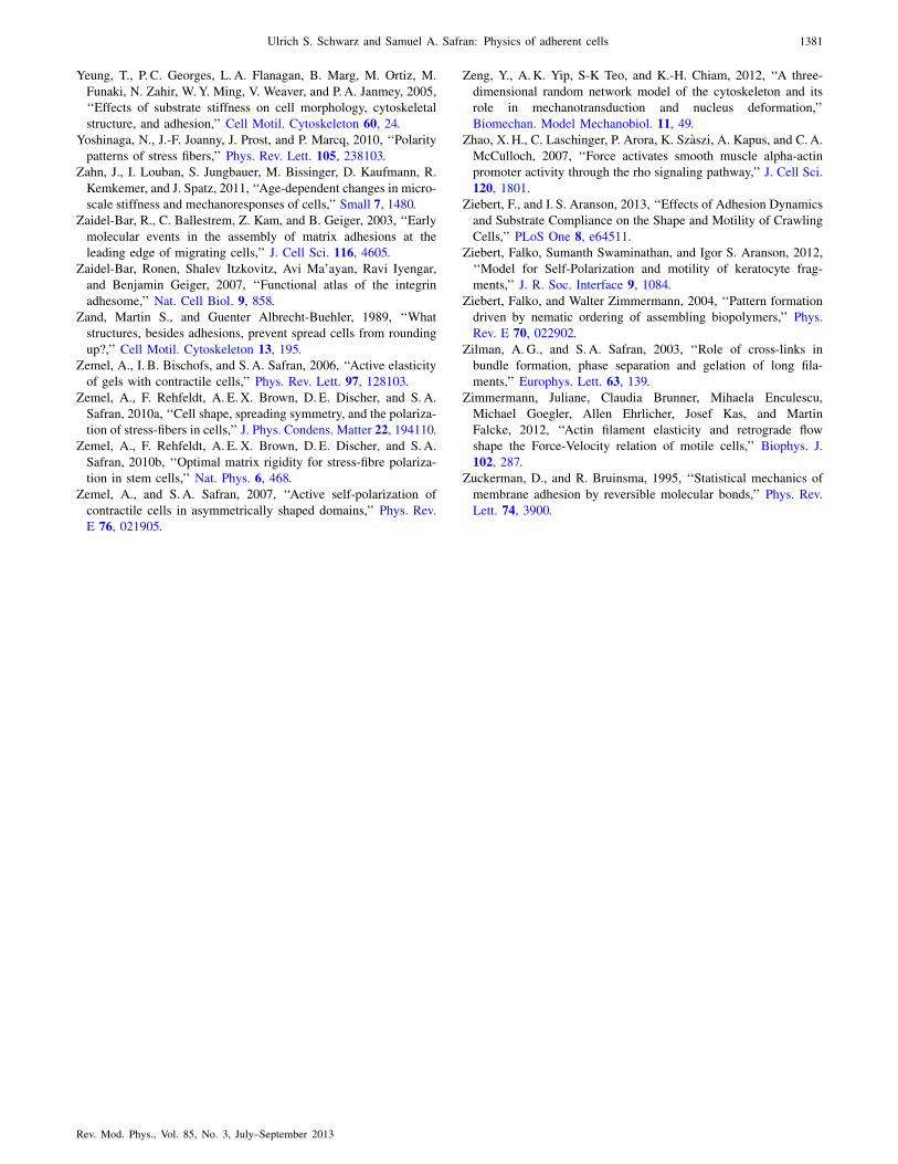
Yeung, T., P. C. Georges, L. A. Flanagan, B. Marg, M. Ortiz, M.
Funaki, N. Zahir, W.Y. Ming, V. Weaver, and P.A. Janmey, 2005,
‘‘Effects of substrate stiffness on cell morphology, cytoskeletal
structure, and adhesion,’’ Cell Motil. Cytoskeleton 60, 24.
Yoshinaga, N., J.-F. Joanny, J. Prost, and P. Marcq, 2010, ‘‘Polarity
patterns of stress fibers,’’ Phys. Rev. Lett. 105, 238103.
Zahn, J., I. Louban, S. Jungbauer, M. Bissinger, D. Kaufmann, R.
Kemkemer, and J. Spatz, 2011, ‘‘Age-dependent changes in micro-
scale stiffness and mechanoresponses of cells,’’ Small 7, 1480.
Zaidel-Bar, R., C. Ballestrem, Z. Kam, and B. Geiger, 2003, ‘‘Early
molecular events in the assembly of matrix adhesions at the
leading edge of migrating cells,’’ J. Cell Sci. 116, 4605.
Zaidel-Bar, Ronen, Shalev Itzkovitz, Avi Ma’ayan, Ravi Iyengar,
and Benjamin Geiger, 2007, ‘‘Functional atlas of the integrin
adhesome,’’ Nat. Cell Biol. 9, 858.
Zand, Martin S., and Guenter Albrecht-Buehler, 1989, ‘‘What
structures, besides adhesions, prevent spread cells from rounding
up?,’’ Cell Motil. Cytoskeleton 13, 195.
Zemel, A., I. B. Bischofs, and S. A. Safran, 2006, ‘‘Active elasticity
of gels with contractile cells,’’ Phys. Rev. Lett. 97, 128103.
Zemel, A., F. Rehfeldt, A. E. X. Brown, D. E. Discher, and S. A.
Safran, 2010a, ‘‘Cell shape, spreading symmetry, and the polariza-
tion of stress-fibers in cells,’’ J. Phys. Condens. Matter 22, 194110.
Zemel, A., F. Rehfeldt, A. E. X. Brown, D. E. Discher, and S. A.
Safran, 2010b, ‘‘Optimal matrix rigidity for stress-fibre polariza-
tion in stem cells,’’ Nat. Phys. 6, 468.
Zemel, A., and S. A. Safran, 2007, ‘‘Active self-polarization of
contractile cells in asymmetrically shaped domains,’’ Phys. Rev.
E 76, 021905.
Zeng, Y., A.K. Yip, S-K Teo, and K.-H. Chiam, 2012, ‘‘A three-
dimensional random network model of the cytoskeleton and its
role in mechanotransduction and nucleus deformation,’’
Biomechan. Model Mechanobiol. 11, 49.
Zhao, X.H., C. Laschinger, P. Arora, K. Szaszi, A. Kapus, and C.A.
McCulloch, 2007, ‘‘Force activates smooth muscle alpha-actin
promoter activity through the rho signaling pathway,’’ J. Cell Sci.
120, 1801.
Ziebert, F., and I. S. Aranson, 2013, ‘‘Effects of Adhesion Dynamics
and Substrate Compliance on the Shape and Motility of Crawling
Cells,’’ PLoS One 8, e64511.
Ziebert, Falko, Sumanth Swaminathan, and Igor S. Aranson, 2012,
‘‘Model for Self-Polarization and motility of keratocyte frag-
ments,’’ J. R. Soc. Interface 9, 1084.
Ziebert, Falko, and Walter Zimmermann, 2004, ‘‘Pattern formation
driven by nematic ordering of assembling biopolymers,’’ Phys.
Rev. E 70, 022902.
Zilman, A. G., and S. A. Safran, 2003, ‘‘Role of cross-links in
bundle formation, phase separation and gelation of long fila-
ments,’’ Europhys. Lett. 63, 139.
Zimmermann, Juliane, Claudia Brunner, Mihaela Enculescu,
Michael Goegler, Allen Ehrlicher, Josef Kas, and Martin
Falcke, 2012, ‘‘Actin filament elasticity and retrograde flow
shape the Force-Velocity relation of motile cells,’’ Biophys. J.
102, 287.
Zuckerman, D., and R. Bruinsma, 1995, ‘‘Statistical mechanics of
membrane adhesion by reversible molecular bonds,’’ Phys. Rev.
Lett. 74, 3900.
Ulrich S. Schwarz and Samuel A. Safran: Physics of adherent cells 1381
Rev. Mod. Phys., Vol. 85, No. 3, July–September 2013


![[XLS]navy-training-transformation2.wikispaces.com · Web view0 15 15 85 85 100 100 5 85 100 0.3 1 0.35 0.35 1 85 85 85 85 85 85 85 85 85 85 85 85 85 85 85 85 85 85 85 85 15 15 5 11.5](https://img.pdfslide.us/doc/110x75/5adf226a7f8b9a6e5c8bbbe3/xlsnavy-training-view0-15-15-85-85-100-100-5-85-100-03-1-035-035-1-85-85-85.jpg)