Embed Size (px)
Citation preview
Review
The utility of grimace scales for practical pain assessment in laboratory animals
Daniel Mota-Rojas1, Adriana Olmos-Hernández2, Antonio Verduzco-Mendoza2, Elein
Hernández3, Julio Martínez-Burnes4 and Alexandra L. Whittaker5*
1 Neurophysiology, behaviour and animal welfare assessment. DPAA. Universidad Autónoma
Metropolitana, Xochimilco, Mexico City. Mexico. 2 Division of Biotechnology - Bioterio and Experimental Surgery, Instituto Nacional de Rehabilitación-Luis
Guillermo Ibarra Ibarra (INR-LGII), Mexico City. Mexico. 3 Department of Clinical Studies and Surgery- Facultad de Estudios Superiores Cuautiltán UNAM,
Cuautitlán, Estado de México. 4 Graduate and Research Department, Facultad de Medicina Veterinaria y Zootecnia, Universidad
Autónoma de Tamaulipas, Victoria City, Tamaulipas, Mexico. 5 School of Animal and Veterinary Sciences, The University of Adelaide, Roseworthy Campus, Australia.
* Correspondence: [email protected]
Simple Summary: Grimace scales for laboratory animals were first reported ten years ago. Yet, in
spite of their promise as pain assessment tools it appears that they have not been implemented
widely in animal research establishments for clinical pain assessment. We discuss potential reasons
for this based on the knowledge gained to date on their use and suggest avenues for further research,
which might improve uptake of their use in laboratory animal medicine.
Abstract: Animals’ facial expressions have been widely used as a readout for emotion. Scientific
interest in the facial expressions of laboratory animals has centered primarily on negative
experiences, such as pain, experienced as a result of scientific research procedures. Recent attempts
to standardize evaluation of facial expressions associated with pain in laboratory animals has
culminated in the development of “grimace scales”. In the context of laboratory animals, these have
been developed and evaluated for mice, rats, rabbits, sheep, and ferrets. The prevention or relief of
pain in laboratory animals is a fundamental requirement for in vivo research to satisfy community
expectations. However, to date it appears that the grimace scales have not seen widespread
implementation as clinical pain assessment techniques in biomedical research. In this review, we
discuss some of the barriers to implementation of the scales in clinical laboratory animal medicine,
progress made in automation of collection, and suggest avenues for future research.
Keywords: facial expressions, pain, grimace scales, mice, rat, rabbit
1. Introduction
Animal welfare is an important societal concern1,2. The use of animals in biomedical scientific
research is widespread, and globally significant, with approximately 115 million animals used per
year3. Incontrovertibly, there is an ethical obligation to safeguard welfare of these animals through
employing strategies to minimize pain, fear and distress4-6, in addition to the promotion of positive
welfare states. However, to achieve this, validated methods for identification of animal emotional
state are required. In spite of significant research attention, ascertaining nature and strength of animal
emotion remains a challenging task7-11. This is especially so for prey species, such as rats and mice,
that are naturally more likely to mask emotional responses to increase their chances of survivals12.
Preprints (www.preprints.org) | NOT PEER-REVIEWED | Posted: 4 September 2020 doi:10.20944/preprints202009.0101.v1
© 2020 by the author(s). Distributed under a Creative Commons CC BY license.
The study of emotion in laboratory animals has typically focused on aversive states such as pain.
This area of study has been driven from two perspectives: a scientific and welfare standpoint. The
scientific viewpoint, based on the extrinsic value of the animal, relates to the robustness of results
acquired from animal models. There is an abundance of data on the impact of pain on a wide range
of metabolic, immunologic and other processes in the body. These alterations introduce variability or
confound interpretation of results13-15. The welfare viewpoint, considering the intrinsic value of the
animal, assumes that pain occurs frequently in animal models and should therefore be avoided or
minimized for the benefit of the animal. Notwithstanding, differences between these viewpoints in
terms of underlying motivation for study, the requirement for a reliable, practical method for
assessment of pain is shared by both.
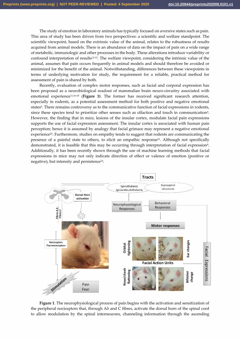
Recently, evaluation of complex motor responses, such as facial and corporal expression has
been proposed as a neurobiological readout of mammalian brain neuro-circuitry associated with
emotional experience11,16-18 (Figure 1). The former has received significant research attention,
especially in rodents, as a potential assessment method for both positive and negative emotional
states9. There remains controversy as to the communicative function of facial expressions in rodents,
since these species tend to prioritize other senses such as olfaction and touch in communication8.
However, the finding that in mice, lesions of the insular cortex, modulate facial pain expressions
supports the use of facial expression assessment. The insular cortex is associated with human pain
perception; hence it is assumed by analogy that facial grimace may represent a negative emotional
experience20. Furthermore, studies on empathy tends to suggest that rodents are communicating the
presence of a painful state to others, to elicit an empathic response21. Although not specifically
demonstrated, it is feasible that this may be occurring through interpretation of facial expression8.
Additionally, it has been recently shown through the use of machine learning methods that facial
expressions in mice may not only indicate direction of effect or valence of emotion (positive or
negative), but intensity and persistence22.
Figure 1. The neurophysiological process of pain begins with the activation and sensitization of
the peripheral nociceptors that, through Aδ and C fibres, activate the dorsal horn of the spinal cord
to allow modulation by the spinal interneurons, channeling information through the ascending
Preprints (www.preprints.org) | NOT PEER-REVIEWED | Posted: 4 September 2020 doi:10.20944/preprints202009.0101.v1
pathways (spinothalamic and spinoreticulothalamic). The seventh cranial nerve regulates the
spontaneous facial movements that produce facial expressions. The spinothalamic tract is considered
the principle nociceptive pathway responsible for the ascent of the afferent signals of pain from the
spinal cord to the cortex in rats23.
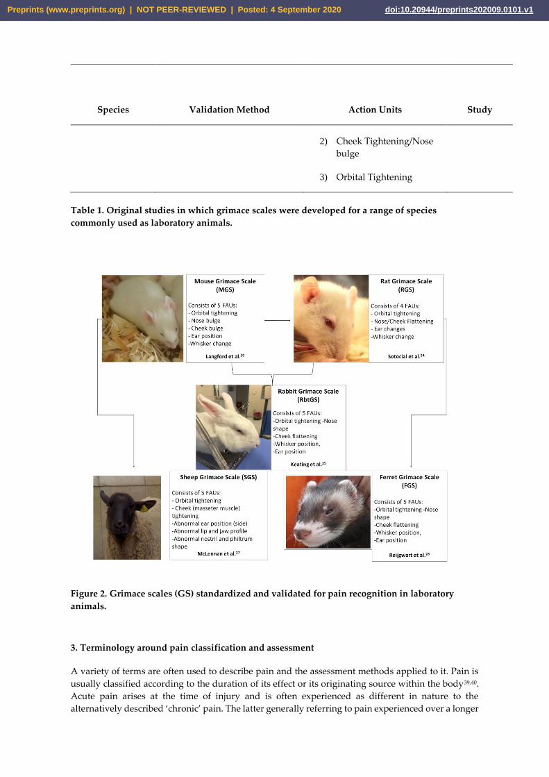
Attempts to standardize evaluation of facial expressions for pain assessment has culminated in
the development of the “grimace scales”. These were developed originally for mice20 and have been
adapted for use in rats24, rabbits25,26, sheep27, and ferrets28, amongst other species. Grimace scales are
simplified methods for evaluating facial expressions specifically related to pain based on the
assessment of action units focusing on the eyes, ears and cheeks. The utility of the scales has been
well-established across a range of laboratory animal species and animal model types. However, this
evaluation has typically focused on their use via retrospective video recording review, and as a
research tool to obtain data relevant to the animal model. There has been less dedicated study into
the scales as ‘bedside’ pain assessment tools for rapid evaluation of pain status in laboratory animals
in order to implement humane endpoints, or provide analgesia. Therefore, the focus of this review is
to discuss the practical utility of grimace scales in a range of laboratory animal species, identifying
barriers to their use and potential confounders. The focus will be on laboratory animal rodents as the
most common species used in biomedical research, but research from other species will be drawn
upon. It is anticipated that this review will guide biomedical researchers, animal technicians and
ethics committees when implementing pain assessment methods as part of research protocols.
2. History of Facial Expression Scoring for Pain in Laboratory Animals
In recognition of the poor translation of outcomes from animal pre-clinical studies on pain physiology
and analgesic development to humans29,30, there has been a recent focus on development of methods
for assessment of the affective pain response using non-evoked (spontaneous) responses31. Grimace
scales are one such response derived from human facial codification scales32,33. The Facial Action
Codification System (FACS) systematically catalogues all possible movements of the facial muscles,
or combinations of them, such as lowering the eyebrows, tightening and closing the eyelids,
wrinkling the nose and raising the upper lip. Categorization of changes in these muscle movements
or so-called “Units of Facial Action” (UFA) enables facial recognition and categorization of
emotions17,19,34. The finding that facial codification scales could quantify pain in humans with limited
or non-existent verbal communication35, provided the basis for using UFA in the development of
grimace scales (GS) for animals36.
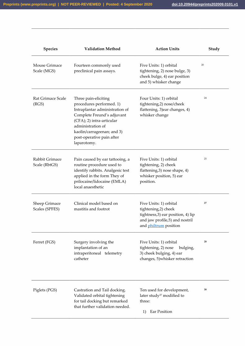
The mouse grimace scale (MGS) was the first to be developed. Langford et al.20 in 2010 applied a
nociceptive abdominal constriction test through administration of acetic acid, that allowed the
elucidation of facial action units that reliably detected pain. Validation was performed using a variety
of traditional preclinical pain assays20. Five action units were described: 1) orbital tightening, 2) nose
bulge, 3) cheek bulge, 4) ear position and 5) whisker change. A year later, Sotocinal et al.24 in 2011
published the rat grimace scale (RGS) comprising four action units, due to consolidation of nose and
cheek flattening into one unit. Utility of the RGS to detect pain was demonstrated in standard pre-
clinical nociceptive tests in addition to following a surgical laparotomy procedure. Furthermore, the
RGS was shown to be modified after analgesic administration indicating the specificity to pain24.
Further, development of grimace scales in other common laboratory animal species followed see
Table 1 and Figure 2.
Preprints (www.preprints.org) | NOT PEER-REVIEWED | Posted: 4 September 2020 doi:10.20944/preprints202009.0101.v1
Species
Validation Method
Action Units
Study
Mouse Grimace
Scale (MGS)
Fourteen commonly used
preclinical pain assays.
Five Units: 1) orbital
tightening, 2) nose bulge, 3)
cheek bulge, 4) ear position
and 5) whisker change
20
Rat Grimace Scale
(RGS)
Three pain-eliciting
procedures performed. 1)
Intraplantar administration of
Complete Freund’s adjuvant
(CFA); 2) intra-articular
administration of
kaolin/carrageenan; and 3)
post-operative pain after
laparotomy.
Four Units: 1) orbital
tightening,2) nose/cheek
flattening, 3)ear changes, 4)
whisker change
24
Rabbit Grimace
Scale (RbtGS)
Pain caused by ear tattooing, a
routine procedure used to
identify rabbits. Analgesic test
applied in the form They of
prilocaine/lidocaine (EMLA)
local anaesthetic
Five Units: 1) orbital
tightening, 2) cheek
flattening,3) nose shape, 4)
whisker position, 5) ear
position.
25
Sheep Grimace
Scales (SPFES)
Clinical model based on
mastitis and footrot
Five Units: 1) orbital
tightening,2) cheek
tightness,3) ear position, 4) lip
and jaw profile,5) and nostril
and philtrum position
27
Ferret (FGS)
Surgery involving the
implantation of an
intraperitoneal telemetry
catheter
Five Units: 1) orbital
tightening, 2) nose bulging,
3) cheek bulging, 4) ear
changes, 5)whisker retraction
28
Piglets (PGS) Castration and Tail docking.
Validated orbital tightening
for tail docking but remarked
that further validation needed.
Ten used for development,
later study37 modified to
three:
1) Ear Position
38
Preprints (www.preprints.org) | NOT PEER-REVIEWED | Posted: 4 September 2020 doi:10.20944/preprints202009.0101.v1
Species
Validation Method
Action Units
Study
2) Cheek Tightening/Nose
bulge
3) Orbital Tightening
Table 1. Original studies in which grimace scales were developed for a range of species
commonly used as laboratory animals.
Figure 2. Grimace scales (GS) standardized and validated for pain recognition in laboratory
animals.
3. Terminology around pain classification and assessment
A variety of terms are often used to describe pain and the assessment methods applied to it. Pain is
usually classified according to the duration of its effect or its originating source within the body39,40.
Acute pain arises at the time of injury and is often experienced as different in nature to the
alternatively described ‘chronic’ pain. The latter generally referring to pain experienced over a longer
Preprints (www.preprints.org) | NOT PEER-REVIEWED | Posted: 4 September 2020 doi:10.20944/preprints202009.0101.v1
duration, although there appears to be no accepted duration marking the transition from acute to
chronic pain41. An alternative distinction between the two time-course descriptors has been suggested
by scientists; that related to functionality. Acute pain is argued to be adaptive, provoking a learned
response by the animal to avoid a similar painful insult in the future39. Chronic pain on the other
hand is said to be maladaptive42. However, this latter point is controversial with a variety of studies
(see43 for review) suggesting that pain-related hypervigilance may influence estimation of risk,
subsequent behavior, and thus enhance survival.
Pain scales themselves are often described in terms of their validity, reliability, sensitivity41. Validity
describes the extent to which the scale measures its intended outcome i.e. pain. There are a number
of sub-categories describing validity. The most commonly referred to in the context of grimace scales
are face validity and construct validity. Face validity describes what the test appears to be measuring
i.e. pain. Construct validity relates to the extent to which the scales measure that specific construct.
Therefore, the test needs to be both sensitive and specific to pain44,45. In pain studies construct validity
is often determined using an applied analgesic test, since this is assumed to reduce pain and thereby
reduce grimace scores if the test is truly pain-related44. External validity refers to how generalizable
the measure is to other settings. In the context of grimace scales this is relevant in taking the scales
from research scenarios to the clinical setting. This relates to practicability to perform during the
working day, simplicity of the task, as well as the need for equipment and training. To date, this is
the area that has received the least attention with regard to grimace scales.
Reliability refers to the scale producing the same result each time it is used both within, and between
animals, and time-points46. In the context of grimace scales, this is determined by the variability
resulting in a single observer’s measurements (intra-observer variability), the variation between
different observers’ measurements (inter-observer), and variability between laboratories or research
centers44. Sensitivity describes the ability of the scale to accurately identify changes in the degree of
pain such that subtle changes are recognized45. In the context of pain scales this is often indicated
when scale changes that occur correlate in direction, and proportion with other measures45. It is
common in assessment of pain in veterinary species to achieve measurement accuracy in pain scoring
by utilizing a smaller number of broad category groups, such as mild, moderate and severe, rather
than expecting sensitivity when small differences in scores are considered. The following will
consider how all of these measurement characteristics may influence the clinical applicability of
grimace scales for use in biomedical research.
4. Clinical Applicability of Grimace Scales in Biomedical Research
4.1. Development of Real-Time Grimace Scores
There is now an extensive body of literature on the application of grimace scales in a range of animal
models used commonly in biomedical research. The majority of this validation work has occurred in
rodent models. It is beyond the scope of this review to describe all of the models used but the range
includes oncology (see e.g.47-50), infectious disease51, pain models48,52,53, neurological conditions33,54,55,
genetic conditions56, and maxillofacial interventions 49,50,57. However, the vast majority of research to
date has performed grimace scoring retrospectively from captured video footage.
Retrospective scoring is likely superior when using grimace scores to inform research outcomes, for
example determining efficacy of analgesics or success of model induction. These methods allow for
the possibility of replication, by multiple observers where appropriate, with an increased time
available for scoring at the researcher’s leisure. A cage-side or ‘real time’ method on the other hand
would ideally provide instant assessment allowing interventions to support welfare, for example by
implementing humane endpoints or administering analgesics. Development of the latter is clearly of
more interest to ethical review committees and animal carers needing to make rapid clinical
Preprints (www.preprints.org) | NOT PEER-REVIEWED | Posted: 4 September 2020 doi:10.20944/preprints202009.0101.v1
decisions. To date there has been substantially less focus on development and validation of real time
methods.
Miller and Leach58 in 2015 performed the first comprehensive evaluation of a real-time method
applied in mice. In this study, both retrospective and real-time scoring were compared. Real-time
scoring was performed by observing mice three times over a 10 min period, whilst animals were
being filmed for the retrospective analysis. Grimace scores were calculated by summation of each
action unit as described by Langford et al.20, and totals were then averaged across the observation
points. Live scores were always found to be significantly lower than corresponding retrospective
video scoring. The authors posed that this could have resulted from the activity levels and changing
nature of the face during live scoring. Blinking for instance, resulting in a score of 0 for orbital
tightening, will likely be selected at least some of the time as a result of random chance selection of
photographs for scoring. In a real time scenario, the rapid nature of blinking will likely preclude its
scoring. Similarly, Chartier et al.47 in 2020 also found consistently lower scores from live scoring
compared to retrospective scoring in a mouse model of colitis-associated colo-rectal cancer. One
potential explanation for this trend is that the presence of a human observer influences performance
of the facial action units, for example, an increased alertness to the human (predator) could lead to
wider eyes and ‘pricked’ ears, lowering the grimace score. On the contrary, intriguing findings from
Sorge et al.59 demonstrated that not all observers are equal, with no impact of a female observer on
scores in rats and mice (obtained retrospectively), but a reduction of scores in the presence of a male59.
In the first investigation of real-time scoring in rats, Leung et al.60 in 2016 found that interval
observations (15s of observation) were able to discriminate between control and analgesic–treated
groups whereas point observations (conducted several times over a period) showed poor group
discrimination. In this study substantial variability was seen between single observations of either
point or interval. Limits of agreement, with a retrospective scoring system were however fairly large
with a 0.5 score range either side of the bias meaning there is was a substantial risk of both over or
underestimating the score. Furthermore, point scoring became generally unreliable at discriminating
groups when done for less than 2 minutes, assumed to be due to a loss of power due to fewer
observations. A later rat study by the same research group61, investigated the interval method
compared to a retrospective method in a colitis model showing the former to be reliable in predicting
pain, with scores similar to the standard method.
The implications of these findings for clinical pain assessment are several. Firstly, it needs to be
considered that although good discriminant ability was generally found in these studies, results were
obtained by statistical combination of multiple scores. In a clinical scenario, an observer is likely to
take one score, and not have the means or time to mathematically manipulate the values to arrive at
a reliable score. Secondly, the Leung et al.60 study suggests that variability across the observation
period is likely and that at least 2 minutes of observation is needed. It is unlikely to be practical for a
caregiver to spend 2 minutes per animal performing pain assessment across a study. In this case,
some other more general method of distress measurement is likely to be needed to ‘triage’ animals
for secondary grimace assessment. There has been no investigation of the effect of movement to the
clear cages, in isolation, as typically occurs in grimace studies as opposed to scoring occurring in the
home cage environment. A number of factors may influence the grimace scoring between these two
scenarios. The novelty of the scoring box may trigger a state of alert influencing grimace scores in a
similar vein to that suggested for the presence of a human observer. This novelty may indeed
contribute to the variability seen between scores over time since habituation will eventually occur.
Alternately, if scoring in the home cage, the presence of cage furniture, a potential more relaxed state
of the animal in its familiar environment, or even the influence of circadian rhythms (see later) may
all variously influence the action units or ability to see them accurately. A further consideration with
real-time scoring is that there may be an inherent observer bias as the animal’s overall demeanor, or
presence of other pain behaviors such as twitching may be noted leading the observer to err on the
side of higher action unit scores when unsure. This is not necessarily an issue per se in a clinical
scenario since the goal is to recognize sick animals for further evaluation and treatment. However,
Preprints (www.preprints.org) | NOT PEER-REVIEWED | Posted: 4 September 2020 doi:10.20944/preprints202009.0101.v1
these other behaviors may not be unique to pain but represent general sickness behavior that may
not be able to be rectified by analgesic administration, and hence inappropriate medication
administration may occur. If such biasing were occurring it would be expected that there may be
differences in grimace scoring between observers experienced with working with the species in
question versus more naïve observers47.
Notwithstanding, these findings some research groups do appear to have been able to use the MGS
or RGS in a point observation, real-time scenario to obtain predicted results. For example, in
chemotherapy-induced toxicity models in mice62, and rats63 single grimace scores allowed
distinguishment between groups and followed the progression of the disease course as expected,
after induction of chemotherapy-induced gut toxicity. Alternately, Hsi et al.64 in 2020 were unable to
use point mouse grimace scores to distinguish between groups either supplemented or not with
dextrose following bariatric surgery. However, in this experimental design there was no sham group
so it is unknown whether the MGS can reliably determine pain in this model64.
There is clearly a need for further validation of real-time observation methods with a particular focus
on one-off observations versus a series of observations, correlation with other established measures
of pain assessment, inter-observer variability and home-cage versus novel area.
4.2 Impact of Biology and the Environment
4.2.1. Strain and sex differences
There is some evidence that features of biology, performance of routine procedures, or aspects of the
environment may influence grimace scores. This has implications for setting of intervention scores
(see later), and should be a consideration in driving further research or recommendations for
application to clinical practice.
Aspects of biology have perhaps been the most researched with regard to their impact on grimace
scores. The greatest implication of such changes likely relates to any differences between rodents
strains or stocks given the wide range typically used in research. In mice, strain differences in MGS
scores in animals not exposed to any painful interventions has been demonstrated. Miller and Leach
in 201558 found that C3H/He mice showed significantly higher scores than CD-1 and C57BL/6
animals, although the order of effect for the latter two strains was different between males and
females. In female BALB/c mice the grimace score was even higher than C3H/He (males were not
investigated in this study). Cho et al.65 in 2019 similarly demonstrated a difference in MGS scores
post-craniotomy, with C57BL/6 mice having lower scores than CD-1 animals65. However, in pairwise
comparisons of the CBA and DBA/2 strains in two further studies, no differences were found66,67. It
has been suggested by some authors that detection of facial features in dark animals may be more
difficult65,68. Improving the image quality and providing a contrasting background colour when
recording appear to mitigate the effects20, hence this may not be a feature of animal pigmentation per
se. It should however be noted that in the Miller and Leach58 in 2015 study, female C57BL/6 animals
were not scored the lowest; that place being taken by the white CD-1 animals. Brown C3H animals
also occupied an intermediate position. In a clinical scenario where real time scoring is likely to take
place the issue of poor background contrast on videos if not of concern. However, some investigation
of the effects of colour on live grimace scoring is warranted since it may be equally as difficult for a
human observer to distinguish features such as whiskers against a similar coat colour background,
especially when trying to observe at a distance so as not to influence the animal’s behavior.
Differences between sexes have also been uncovered in research to date on the MGS, but results are
complex and suggest there may be strain interactions. For example, Miller and Leach58 observed no
differences in MGS scores between male and female C57BL/6 mice58. However in the same study,
both CD-1 and C3H/He males had greater scores than their female counterparts58. Similarly, male
Preprints (www.preprints.org) | NOT PEER-REVIEWED | Posted: 4 September 2020 doi:10.20944/preprints202009.0101.v1
BALB/c mice had higher grimace scores than females69. Alternately, Cho et al.65 found no sex
differences in CD-1 mice, although differences in response to analgesic were noted with females
appearing to respond to carprofen with a reduction in grimace score more readily than males65. In
rats limited study has been carried out into sex differences but no differences were found in the
original validation study24, or in a later study70. Unfortunately, it appears that most grimace studies
in rats and mice appear to have been conducted in one sex, with a large proportion using male
animals see eg.52,71-73. This bias in study design towards males, coupled with the enhanced
understanding of the existence of different pathways and immune-cell types for pain processing
between male and female rodents74, renders extrapolation of findings to female rodents problematic.
4.2.2. Impact of routine procedures
It is clear that procedures occurring fairly often as part of vivarium routines may influence responses
and should be taken into consideration when considering practical implementation of the grimace
scales. For example, a number of studies have evaluated the impact of anesthetics on rodent grimace
scales. In general, both inhalational and injectable anesthetics lead to a short-term increase in grimace
scores in both rats73 and mice66,75,76, although strain differences in the presence of this response have
been reported66,75,76. Whilst, this response is generally short-lived, repeated exposures lead to
enhanced duration of the increase73,68. This is a particular consideration since grimace assessment
would typically occur post-operatively to allow rescue analgesia administration and there is
suggestion that the score increase may persist for up to a few hours post anesthesia75,76.
There is a growing body of evidence that non-aversive handling of mice leads to reduced anxiety and
improved resilience in the face of accompanying pain77-79. Cupping or tunnel handling are proposed
as alternatives to the traditional method of picking up by the tail78. Perhaps somewhat surprisingly
given the reported specificity of the MGS for pain there is some evidence that method of routine
handling influences MGS with increased scores in mice handled by the tail compared to those that
were tunnel handled69. This contradicts the findings of a previous study where no differences
between the two methods were reported67. This is an area that should be a priority for further
investigation for a number of reasons. Firstly, since non-aversive methods have not been widely
incorporated into laboratory animal practice, especially amongst researchers80, it is quite likely that
mice even within one study will be subject to different handling techniques. Any effect of handling
method on grimace score could therefore confound interpretation of grimace scores used to
determine research protocol effects on pain. Secondly, whilst there appears to have been no dedicated
study on whether tail handling induces pain, there is suggestion that it is non-painful, yet aversive78.
If the method is actually non-painful this calls into question the specificity of the MGS for pain, and
therefore whether it has construct validity.
Ear-tagging or ear-notching are routine handling procedures used to permanently identify laboratory
animals81. These procedures are known to cause acute pain as reflected by alterations in physiological
indices such as heart rate and blood pressure82. However, the results obtained by Miller and Leach81
in mice did not reveal any change to MGS scores as a result of ear notching81. In a later mouse study,
with a factorial study design evaluating handling method with ear tagging or tattooing, MGS was
increased following ear tagging but tattooing or restraint had no impact on scores69. Alternately,
Keating et al.25 in 2012 showed that ear-tattooing in rabbits led to increases in rabbit grimace scale
scores, that were ameliorated by the application of a local topical anaesthetic (lidocaine/prilocaine)25.
Corticosterone measures in this study suggest that the pain response was short-lived and had
resolved by 1-hour post procedure. Given that only three studies, performed in different species,
have evaluated these common procedures, it would be unwise to draw firm conclusions. However,
the lack of grimace score increase in the Miller and Leach81 study does imply that the scale may not
be sensitive to pain of a mild and short-lived nature either intrinsically, or as a result of practical
features whereby the pain is missed due to the scoring process required. Conversely, this finding
provides some evidence that routine procedures may have minimal effect on grimace scales, reducing
Preprints (www.preprints.org) | NOT PEER-REVIEWED | Posted: 4 September 2020 doi:10.20944/preprints202009.0101.v1
the risk of confounding arising when using the scales for humane endpoint implementation in animal
models. When reconciling the difference in findings between this81, and the later study, Roughan and
Sevenoaks69 in 2019 speculated that ear tagging may be perceived as more painful than notching due
to the prolonged irritation by the tag69.
4.2.3. Environmental Impacts
If the grimace scales are to be utilized as a practical tool they need to be repeatable across time and
conditions, and not subject to extraneous influences. This requirement also relates to their face
validity as reliable indicators of pain. In common with the other factors that may influence the scales,
there has been limited research in this sphere.
Miller and Leach in 201558 performed a comprehensive evaluation of some of the factors that might
be predicted to influence grimace responses. One factor that may have an impact is the circadian cycle
and whether differences in score occur across the day. For both live scoring and retrospective scoring,
there were largely no differences seen between scores dependent on whether scoring took place in
the morning, lunchtime or at the end of the day. There were some exceptions to this with BALB/c
mice showing a greater live MGS score at noon compared to am and C57BL/6 mice showing higher
retrospective MGS scores in both afternoon time points in comparison to the morning. It should be
noted that in this, as in the majority of the studies examining grimace scores, animals were scored
during the light phase of the circadian cycle when they would be expected to be inactive. There is
some evidence that grimace scores may not be comparable between dark and light conditions with
the finding that MGS was higher in the dark than in bright light in CD1 mice treated with a peptide
believed to induce pain and migraine symptoms83. Analysis of the actions units showed that the
transition to light caused a significant decrease in orbital tightening and nose bulge83. Given that this
finding was also observed in vehicle controls it appears unrelated to the migraine symptoms, and
may be an aspect of normal biology needing consideration. Alternately, Matsumiya et al. 2012 found
no difference in baseline MGS scores between morning and evening but did find that in operated
animals scores were higher in the dark cycle, implying that pain was greater in the mice active
phase84. However, in consideration of the use of the scales as a practical tool the reality is that most
scoring will occur during the working day, in the light cycle, and therefore the findings of Miller and
Leach in 201558 provide confidence that time of scoring should not influence the score. A further
important outcome from Miller and Leach58 was that there was no effect of repeatedly being placed
in the photography boxes on grimace score i.e. a habituation effect over the 3 occasions used58. The
later study by Jirkof et al.85 in 2020 supports this finding. This provides assurance that longitudinal
monitoring post-procedure could occur throughout the day without the need to account for time of
day or habituation to the box. However, as discussed earlier the need to remove animals to a separate
box does impede the practical application of the test. Further study should consider time of day
effects in the non-stimulated home environment.
There is further evidence of an impact of the external environment on grimace scores. Sorge et al.59
compared grimace responses of mice and rats recorded after a painful insult, in the presence of a
male compared to a female. Significant decreases in grimace response were recorded compared to
the situation with no observer in the room. Females did not induce such a change. The findings
therefore suggest that olfactory cues from human males lead to a physiological stress response, and
associated stress-induced analgesia.
There is also some evidence of inter-laboratory variation in the outputs obtained from behavioural
testing, to include MGS scores. In a multicenter study, Jirkof et al.85 in 2020 demonstrated some
quantitative differences in scores, although they were qualitatively comparable (direction of effect).
However, variability between research centers in the MGS, especially when presented as a median
score, was less pronounced than in burrowing behavior readouts84. This inter-lab variability has been
recognized across the spectrum of preclinical research pursuits, arising as a result of environmental
Preprints (www.preprints.org) | NOT PEER-REVIEWED | Posted: 4 September 2020 doi:10.20944/preprints202009.0101.v1
variables leading to stress. 86 Whilst this issue may be a concern when considering basic-to –clinical
translation and reproducibility, it is less likely to be of concern for clinical application of the grimace
scales. As a clinical tool, provided good inter and intra-observer, and thus intra-site agreement is
obtained, grimace scores may be relied on locally for welfare determination subject to some of the
other caveats discussed in this paper.
4.3 Validity
If grimace scales are to be implemented as a routine clinical assessment tool in biomedical research
facilities, there needs to be a clear understanding of whether they are specific to pain, and can reliably
measure pain in the models being used. This is important because it influences the animal caretaker’s
decision as regard to treatment options, for example, whether analgesics will be effective in
mitigating clinical signs. It can be seen from the above discussion that there are a range of external
factors that affect grimace scores, speaking to their validity as a pain assessment tool; anesthetics are
a prime example. Setting aside the lack of study into their application in a real-time scenario, which
influences their generalizability, another key concern is whether they are valid for all pain types.
Results of the original Langford et al.20 in 2010 study suggested that the technique was only applicable
for acute pain states20, since changes were not recorded after the application of traditional models of
chronic pain, such as chronic constriction injury (CCI). However, there have now been a range of
studies, largely performed in mice, which suggest that the grimace scale may be applicable for pain
that is chronic or neuropathic in nature, or of a non-surgical origin.
The study findings of Akintola et al.52 in 2017 contradict the previous results of Langford et al 2010
with both RGS and MGS increasing after application of the CCI model in these species. Pain arising
from cancer has also been shown to cause an elevation of the MGS, for example in colo-rectal cancer47
and in a metastatic breast cancer model49,50. The MGS has been successfully used in models expected
to produce pain of a neuropathic nature, for example in headache and migraine55,87 and craniotomy65.
There is also suggestion that pain of a visceral nature elevates scores based on studies evaluating
colonic nociception88, pelvic pain89, colitis61, and alimentary mucositis62,63. Hereditary sickle cell
disease frequently leads to painful episodes in human patients. Cold treatment of transgenic sickle
mice led to increased grimace scores which were alleviated using a known analgesic agent.
Furthermore, body changes of decreased length and increased back curvature were also correlated
with the change in grimace scores56. These findings lend support to the proposition that the grimace
scales have good construct validity for non-acute pain.
In spite of these findings, results from other studies implies that further evaluation of the grimace
techniques are necessary to ascertain validity. For example, in contradiction to later work62,63
demonstrating elevations in scores in rats and mice with mucositis, Whittaker et al.90 in 2015 found
no change in grimace scores in a rat model, albeit using retrospective rather than real time scoring.
However, this study did find increases in frequency of established behavioral indicators of pain such
as back arching and twitching90. Alternately, Leung et al.61 in a rat DSS- colitis model found grimace
score increases in the absence of an increase in composite behavioral score.
Other studies also raise questions of whether the grimace scales are truly unique to pain. Caecal
ligation and puncture models are commonly used to study sepsis91. Whilst sepsis is undoubtedly a
painful condition based on human reports92, there is also an overwhelming cytokine response causing
sickness behavior. Studies to date on this model51,93 have not teased apart the possible contribution of
this sickness response to the facial expression changes. There is a study that lends support to this
idea; the work of Yamamoto et al. in 201694, whilst not employing the published rat grimace scale,
provides evidence that nausea influences the eye action unit. Toxin administration, which might also
be expected to cause dual symptoms of pain and sickness, similarly elevated the MGS95. Furthermore,
analgesic administration was not always successful in reducing the scores implying an alternate
Preprints (www.preprints.org) | NOT PEER-REVIEWED | Posted: 4 September 2020 doi:10.20944/preprints202009.0101.v1
cause of the facial action unit response. Finally, head injury may alter the animal’s ability to influence
the facial action units via neural mechanisms and render grimace scores unreliable44.
4.4 Automation of Techniques
One of the main current barriers to widespread clinical application of the grimace scales is the lack
of understanding as to their validity and reliability when used for live scoring. However, as
illustrated, there is now a wealth of literature on the validity and application of retrospective
techniques using video or photo footage. In a clinical scenario these methods have limited application
due to the time taken to extract the images, perform the scoring and potentially combine scores using
statistical methods. However, there has been investigation of a range of technologies which minimize
the time taken for various aspects of this process. At the simplest level use of freeware video to JPG
converter software can reduce the time associated with manual searching and capture of images from
recorded video footage by automating the capture process48. However, this still requires manual
viewing of the selected images to obtain unobstructed head shots. Sotocinal et al.24 in 2011 developed
Rodent Face Finder® which is able to detect rodent eyes and ears to generate stills of rodent faces24.
This software has been used in a range of studies measuring grimace scores in both rats and mice see
eg. 52,84,44,96. Recently, another research group generated an algorithm to generate repeatable, non-
observer biased, standardised and randomised pictures in one step. The authors suggest that their
system offers benefits in scoring animals with dark fur and allowing several animals to be filmed and
generate images simultaneously97. They further went on to show that the system was robust across a
number of facilities potentially minimising issues around inter-laboraotry variability as discussed
previously98.
This process of semi-automation makes grimace scoring somwehat more applicable to a clinical
environment but the time taken to manually score images is still likely to be a barrier to
implementation. In recent years there has been some progress on further automation of facial
expression recognition using machine learning techniques. Deep learning methods allow
classification and predictions on the data without previous feature design99. Using combined
methods, Andersen et al.99 in 2020 created a software which locates and extracts the mouse face in an
image, as well as scoring expression using a deep neural network. Based on assessment of a binary
outcome (pain versus no pain) this system achieved an accuracy of 99%. Other groups have similarly
demonstrated the promise of deep learning methods for use with the MGS when based on binary
outputs100,101.
These automation methods are in their infancy and no doubt there will be further development of
these techniques over the next few years. A key issue at the moment is that they lack sensitivity- being
only able to distinguish a painful from a non-painful state. This renders their current use for welfare
assessment and endpoint implementation limited. However, given the success and practical
implementation of machine learning methods in recognition of human facial expression, it is likely
to be only a matter of time before a similar level of sensitivity of scoring will be possible in animal –
focused methods102.
5. Practical Considerations
The above discussion highlights some areas in need of future research particularly in regard to
practical usage of the grimace scales in laboratory animal medicine. A key issue is what to do with
the data when it is acquired, and what it means for the animal. In research use of the grimace scales,
statistically significant differences in grimace scores in comparison with controls are typically
reported. However, in a clinical scenario, a mass of data or control animals’ results may not be
available to make this comparison on-the-spot. Moreover, statistical significance may not always
equate with clinical significance. There needs to be ascertainment of the level of grimace score at
which pain is actually occurring, since the evidence suggests that grimace scores in healthy animals
Preprints (www.preprints.org) | NOT PEER-REVIEWED | Posted: 4 September 2020 doi:10.20944/preprints202009.0101.v1
are rarely zero58. Some attempts have been made to address this issue with the development of an
intervention threshold. Scores that are above this level signify that the animal is in pain, and
consideration should be given to providing rescue analgesia103. These thresholds would need to be
derived based on the method of combining individual action unit scores used, for example
summation of scores leads to a maximum of 10, whereas averaging leads to a maximum of 2. Oliver
et al.103 in 2014 determined for rats, that 0.67/2 was a suitable intervention threshold. Intervention
thresholds have not yet been developed in other species. Since individuals experience pain
differently, and there are associated sex differences in both pain experience and response to
analgesics, work is needed to tailor intervention thresholds considering these factors.
Given, the lack of established intervention thresholds perhaps the best current advice would be to
use a holistic approach in pain assessment and consider grimace scores alongside other measures of
well-being such as standard clinical scoring, and where possible look for trends in score progression
within the same animal to guide decision-making. Animal carers also need to consider the potential
impacts of inter and intra-observer variability on scoring which may be significant when statistical
methods on group data are not used to smooth out variability. A prudent approach, where possible,
would be to use the same scorer in a clinical case. This concern also brings up the issue of training of
scorers which has received minimal research attention. Some studies have implied that minimal
training, such as the provision of online instructions, is all that is necessary to achieve consistent
results between expert and novice scorers69,104. However, another study has shown that more in-depth
training, utilizing practice scoring associated with structured opportunities for discussion, enhanced
scoring ability105.
6. Conclusions and Future Directions
In spite of 10 years of investigation, widespread uptake of grimace scoring in biomedical research has
not occurred. The grimace scales offer enormous potential for clinical use in biomedical research.
They are simple, require no equipment and have been shown through research study to have good
construct validity for most conditions. However, the methodology used in research on grimace scales
is unlikely to lend to practical implementation due to its time intensive and retrospective nature. To
date, few studies have investigated the validity of grimace scales in scenarios requiring on the spot
pain assessment and clinical decision-making. Key areas for focus are on grimace score validity in
animals housed in home cages, the reliability of using a limited number of real-time observation
points, the impact of observers on scores, and the need for observer training. This is an area in urgent
need of future research to realise the potential value of grimace scales.
One area that has received attention is the automation of scales using machine learning and
algorithmic methods. This is a welcome development and will enhance the practical potential of
grimace scales. It is hoped that in future years, grimace scale scoring may just be one of a number of
outcome measures acquired routinely through facility-automated systems. This scenario is most
likely to address the practical issues inherent when dealing with large numbers of animals, going
some way towards addressing public concern around ethical decision-making in biomedical
research.
Author Contributions:
Funding: This research received no external funding. A.W. is supported by an Australian Government, NHMRC
Peter Doherty Biomedical Research Fellowship (APP1140072).
Conflicts of Interest: The authors declare that they have no conflict of interest.
References
Preprints (www.preprints.org) | NOT PEER-REVIEWED | Posted: 4 September 2020 doi:10.20944/preprints202009.0101.v1
1 Mota-Rojas, D., Velarde, A., Maris-Huertas, S., Cajiao, M. N. Editors. In: Animal welfare, a
global vision in Ibero-America. 3rd ed. Barcelona, Spain. 1-516. (Elsevier, 2016).
2 Lewejohann, L., Schwabe, K., Häger, C. & Jirkof, P. Impulse for animal welfare outside the
experiment. Lab. Anim. 54, 150-158 (2020).
3 Taylor, K., Gordon, N., Langley, G. & Higgins, W. Estimates for worldwide laboratory animal
use in 2005. Altern. Lab. Anim. 36, 327-342 (2008).
4 Mota-Rojas, D. et al. Neurological modulation of facial expressions in pigs and implications for
production. J. Anim. Behav. Biometeorol. 8, 232–243 (2020).
5 Mota-Rojas D. et al. Infrared thermal imaging associated with pain in laboratory animals. Exp.
Anim. 70, Accepted (2021).
6 Baumans, V. Science-based assessment of animal welfare: laboratory animals. Rev. sci. tech. Off.
int. Epiz. 24 (2005)
7 Lezama-García, K. et al. Facial expressions and emotions in domestic animals. CAB Rev.
Perspect. Agric. Vet. Sci. Nutr. Nat. Resour. 14, (2019).
8 Finlayson, K., Lampe, J. F., Hintze, S., Würbel, H. & Melotti, L. Facial indicators of positive
emotions in rats. PloS One 11 (2016).
9 Whittaker, A. L. & Marsh, L. E. The role of behavioural assessment in determining 'positive'
affective states in animals. CAB Rev. Perspect. Agric. Vet. Sci. Nutr. Nat. Resour. 14, 1-13 (2019).
10 Boissy, A. et al. Assessment of positive emotions in animals to improve their welfare. Physiol.
Behav. 92, 375–397 (2007)
11 Panksepp, J. Affective consciousness: Core emotional feelings in animals and humans.
Conscious. Cogn. 14, 30-80 (2005).
12 Stasiak, K. L., Maul, D., French, E., Hellyer, P. W. & VandeWoude, S. Species-specific
assessment of pain in laboratory animals. Contemp. Top Lab. Anim. Sci. 42, 13-20 (2003).
13 Carbone, L. & Austin, J. Pain and Laboratory Animals: Publication Practices for Better Data
Reproducibility and Better Animal Welfare. PLoS One 11, e0155001 (2016).
14 Peterson, N. C., Nunamaker, E. A. & Turner, P. V. To Treat or Not to Treat: The Effects of Pain
on Experimental Parameters. Comp. Med. 67, 469-482 (2017).
15 Zurlo, J. & Hutchinson, E. Refinement. ALTEX 31, 4-10 (2014).
16 Bennett, V., Gourkow, N. & Mills, D. S. Facial correlates of emotional behaviour in the
domestic cat (Felis catus). Behav. Processes 141, 342-350 (2017).
17 Ekman, P. Are there basic emotions?. Psychol. Rev. 99, 550-553 (1992).
18 Mota-Rojas, D. et al. Teaching animal welfare in veterinary schools in Latin America. Int. J. Vet.
Sci. Med. 6, 131-140 (2018).
19 Descovich, K. A. et al. Facial Expression: An Under-Utilized Tool for the Assessent of Welfare
in Mammals. ALTEX 34, 409-429 (2017).
20 Langford, D. J. et al. Coding of facial expressions of pain in the laboratory mouse. Nat. Methods
7, 447 (2010).
21 Langford, D. J. et al. Social modulation of pain as evidence for empathy in mice. Science 312,
1967-1970 (2006).
22 Dolensek, N., Gehrlach, D. A., Klein, A. S. & Gogolla, N. Facial expressions of emotion states
and their neuronal correlates in mice. Science 368, 89-94 (2020).
23 Palecek, J., Paleckova, V. & Willis, W. D. Postsynaptic dorsal column neurons express NK1
receptors following colon inflammation. Neuroscience 116, 565-572 (2003).
24 Sotocinal, S. G. et al. The Rat Grimace Scale: A partially automated method for quantifying pain
in the laboratory rat via facial expressions. Mol. Pain 7 (2011).
25 Keating, S. C., Thomas, A. A., Flecknell, P. A. & Leach, M. C. Evaluation of EMLA cream for
preventing pain during tattooing of rabbits: changes in physiological, behavioural and facial
expression responses. PLoS One 7, e44437 (2012).
26 Hampshire, V. & Robertson, S. Using the facial grimace scale to evaluate rabbit wellness in
post-procedural monitoring. Lab. Anim. (NY) 44, 259-260 (2015).
27 McLennan, K. M. et al. Development of a facial expression scale using footrot and mastitis as
models of pain in sheep. Appl. Anim. Behav. Sci. 176, 19-26 2016.
Preprints (www.preprints.org) | NOT PEER-REVIEWED | Posted: 4 September 2020 doi:10.20944/preprints202009.0101.v1
28 Reijgwart, M. L. et al. The composition and initial evaluation of a grimace scale in ferrets after
surgical implantation of a telemetry probe. PLoS One 12, e0187986 (2017).
29 Apkarian, A. V., Hashmi, J. A. & Baliki, M. N. Pain and the brain: specificity and plasticity of
the brain in clinical chronic pain. Pain 152, S49-64 (2011).
30 Blackburn-Munro, G. Pain-like behaviours in animals - how human are they? Trends Pharmacol.
Sci. 25, 299-305 (2004).
31 Hernandez-Avalos, I. et al. Review of different methods used for clinical recognition and
assessment of pain in dogs and cats. Int. J. Vet. Sci. Med. 7, 43-54 (2019).
32 Nagakura, Y. et al. Spontaneous pain-associated facial expression and efficacy of clinically used
drugs in the reserpine-induced rat model of fibromyalgia. Eur. J. Pharmacol. 864 (2019).
33 Serizawa, K., Tomizawa-Shinohara, H., Yasuno, H., Yogo, K. & Matsumoto, Y. Anti-IL-6
Receptor Antibody Inhibits Spontaneous Pain at the Pre-onset of Experimental Autoimmune
Encephalomyelitis in Mice. Front. Neurol. 10, 341 (2019).
34 LeResche, L. Facial expression in pain: A study of candid photographs. J. Nonverbal Behav. 7,
46-56 (1982).
35 Williams, A. Facial expression of pain: An evolutionary account. Behav. Brain Sci. 25, 439-455
(2002).
36 Schanz, L., Krueger, K. & Hintze, S. Sex and Age Don't Matter, but Breed Type Does-Factors
Influencing Eye Wrinkle Expression in Horses. Front. Vet. Sci. 6, 154 (2019).
37 Viscardi, A. V., Hunniford, M., Lawlis, P., Leach, M. & Turner, P. V. Development of a Piglet
Grimace Scale to Evaluate Piglet Pain Using Facial Expressions Following Castration and Tail
Docking: A Pilot Study. Front. Vet. Sci. 4, 51 (2017).
38 Di Giminiani, P. et al. The assessment of facial expressions in piglets undergoing tail docking
and castration: toward the development of the piglet grimace scale. Front. Vet. Sci. 3, 100 (2016).
39 Bateson, P. Assessment of pain in animals. Anim. Behav. 42, 827-839 (1991).
40 Whittaker, A. L. & Howarth, G. S. Use of spontaneous behaviour measures to assess pain in
laboratory rats and mice: How are we progressing? Appl. Anim. Behav. Sci. 151, 1-12 (2014).
41 Rutherford, K. Assessing pain in animals. Anim. Welfare 11, 31-53 (2002).
42 Williams, A. C. d. C. Persistence of pain in humans and other mammals. Philos. Trans. R. Soc.
Lond. B Biol. Sci. 374, 20190276 (2019).
43 Walters, E. T. & Williams, A. C. De C. Evolution of mechanisms and behaviour important for
pain. Philos. Trans. R. Soc. Lond. B Biol. Sci 374, 20190275 (2019).
44 McLennan, K. M. et al. Conceptual and methodological issues relating to pain assessment in
mammals: The development and utilisation of pain facial expression scales. Appl. Anim. Behav 217,
1-15 (2019).
45 Bendinger, T. & Plunkett, N. Measurement in pain medicine. BJA Educ. 16, 310-315 (2016).
46 Good, M. et al. Sensation and distress of pain scales: reliability, validity, and sensitivity. J. Nurs.
Meas. 9, 219-238 (2001).
47 Chartier, L. C., Hebart, M. L., Howarth, G. S., Whittaker, A. L. & Mashtoub, S. Affective state
determination in a mouse model of colitis-associated colorectal cancer. PLoS One 15, e0228413
(2020).
48 George, R. P., Howarth, G. S. & Whittaker, A. L. Use of the Rat Grimace Scale to Evaluate
Visceral Pain in a Model of Chemotherapy-Induced Mucositis. Animals 9, 678 (2019).
49 de Almeida, A. S. et al. Characterization of Cancer-Induced Nociception in a Murine Model of
Breast Carcinoma. Cell. Mol. Neurobiol. 39, 605–617 (2019).
50 de Almeida, A. S. et al. Role of transient receptor potential ankyrin 1 (TRPA1) on nociception
caused by a murine model of breast carcinoma. Pharmacol. Res. 152, 104576 (2020).
51 Mai, S. H. C. et al. Body temperature and mouse scoring systems as surrogate markers of death
in cecal ligation and puncture sepsis. Intensive Care Med. Exp. 6, 20 (2018).
52 Akintola, T. et al. The grimace scale reliably assesses chronic pain in a rodent model of
trigeminal neuropathic pain. Neurobiol. Pain 2, 13-17 (2017).
53 Akintola, T. et al. In search of a rodent model of placebo analgesia in chronic orofacial
neuropathic pain. Neurobiol. Pain 6, 100033 (2019).
Preprints (www.preprints.org) | NOT PEER-REVIEWED | Posted: 4 September 2020 doi:10.20944/preprints202009.0101.v1
54 Duffy, S. S. et al. Peripheral and Central Neuroinflammatory Changes and Pain Behaviors in an
Animal Model of Multiple Sclerosis. Front. Immuno.l 7, (2016).
55 Hassler, S. N. et al. Protease activated receptor 2 (PAR2) activation causes migraine-like pain
behaviors in mice. Cephalalgia 39, 111-122 (2019).
56 Mittal, A., Gupta, M., Lamarre, Y., Jahagirdar, B. & Gupta, K. Quantification of pain in sickle
mice using facial expressions and body measurements. Blood Cells, Mol. Dis. 57, 58-66 (2016).
57 Gao, M. et al. The role of periodontal ASIC3 in orofacial pain induced by experimental tooth
movement in rats. Eur. J. Orthod. 38, 577-583 (2016).
58 Miller, A. L. & Leach, M. C. The Mouse Grimace Scale: A Clinically Useful Tool? PLoS One 10,
e0136000 (2015).
59 Sorge, R. E. et al. Olfactory exposure to males, including men, causes stress and related
analgesia in rodents. Nat. Methods 11, 629-632 (2014).
60 Leung, V., Zhang, E. & Pang, D. S. J. Real-time application of the Rat Grimace Scale as a
welfare refinement in laboratory rats. Sci. Rep. 6, 31667 (2016).
61 Leung, V. S. Y., Benoit-Biancamano, M. O. & Pang, D. S. J. Performance of behavioral assays:
the Rat Grimace Scale, burrowing activity and a composite behavior score to identify visceral pain
in an acute and chronic colitis model. PAIN Rep. 4, e718 (2019).
62 Wardill, H. R. et al. Irinotecan-Induced Gastrointestinal Dysfunction and Pain Are Mediated by
Common TLR4-Dependent Mechanisms. Mol. Cancer Ther. 15, 1376-1386 (2016).
63 Gibson, R. J. et al. Chemotherapy-induced gut toxicity and pain: involvement of TLRs. Support.
Care Cancer 24, 2251-2258 (2016).
64 Hsi, Z. Y., Stewart, L. A., Lloyd, K. C. K. & Grimsrud, K. N. Hypoglycemia after Bariatric
Surgery in Mice and Optimal Dosage and Efficacy of Glucose Supplementation. Comp. Med. 70, 111-
118 (2020).
65 Cho, C. et al. Evaluating analgesic efficacy and administration route following craniotomy in
mice using the grimace scale. Sci. Rep. 9, 359 (2019).
66 Miller, A., Kitson, G., Skalkoyannis, B. & Leach, M. The effect of isoflurane anaesthesia and
buprenorphine on the mouse grimace scale and behaviour in CBA and DBA/2 mice. Appl. Anim.
Behav. Sci. 172, 58-62 (2015).
67 Miller, A. L. & Leach, M. C. The effect of handling method on the mouse grimace scale in two
strains of laboratory mice. Lab. Anim. 50, 305-307 (2016).
68 Dalla Costa, E. et al. Can grimace scales estimate the pain status in horses and mice? A
statistical approach to identify a classifier. PLoS One 13, e0200339 (2018).
69 Roughan, J. V. & Sevenoaks, T. Welfare and Scientific Considerations of Tattooing and Ear
Tagging for Mouse Identification. J. Am. Assoc. Lab. Anim. Sci.58, 142-153 (2019).
70 Waite, M. E. et al. Efficacy of Common Analgesics for Postsurgical Pain in Rats. J. Am. Assoc.
Lab. Anim. Sci. 54, 420-425 (2015).
71 Wang, S. et al. TRPV1 and TRPV1-Expressing Nociceptors Mediate Orofacial Pain Behaviors in
a Mouse Model of Orthodontic Tooth Movement. Front. Physiol. 10 (2019).
72 Zhu, Y. et al. Effect of static magnetic field on pain level and expression of P2X3 receptors in
the trigeminal ganglion in mice following experimental tooth movement. Bioelectromagnetics 38, 22-
30 (2017).
73 Miller, A. L., Golledge, H. D. R. & Leach, M. C. The Influence of Isoflurane Anaesthesia on the
Rat Grimace Scale. Plos One 11, e0166652 (2016).
74 Sorge, R. et al. & Mogil, JS (2015). Different immune cells mediate mechanical pain
hypersensitivity in male and female mice. Nat. Neurosci. 18,1081-1083 (2015)
75 Hohlbaum, K. et al. Severity classification of repeated isoflurane anesthesia in C57BL/6JRj mice-
Assessing the degree of distress. PLoS One 12, e0179588 (2017).
76 Hohlbaum, K. et al. Impact of repeated anesthesia with ketamine and xylazine on the well-
being of C57BL/6JRj mice. PLoS One 13, e0203559 (2018).
77 Gouveia, K. & Hurst, J. L. Optimising reliability of mouse performance in behavioural testing:
the major role of non-aversive handling. Sci. Rep. 7, 44999 (2017).
78 Hurst, J. L. & West, R. S. Taming anxiety in laboratory mice. Nat. Methods 7, 825-826 (2010).
Preprints (www.preprints.org) | NOT PEER-REVIEWED | Posted: 4 September 2020 doi:10.20944/preprints202009.0101.v1
79 Gouveia, K. & Hurst, J. L. Reducing Mouse Anxiety during Handling: Effect of Experience
with Handling Tunnels. PLoS One 8, e66401 (2013).
80 Henderson, L. J., Smulders, T. V. & Roughan, J. V. Identifying obstacles preventing the uptake
of tunnel handling methods for laboratory mice: An international thematic survey. PLoS One 15,
e0231454 (2020).
81 Miller, A. & Leach, M. Using the mouse grimace scale to assess pain associated with routine
ear notching and the effect of analgesia in laboratory mice. Lab. Anim. 49, 117-120 (2015).
82 Kasanen, I. H. E., Voipio, H. M., Leskinen, H., Luodonpää, M. & Nevalainen, T. O. Comparison
of ear tattoo, ear notching and microtattoo in rats undergoing cardiovascular telemetry. Lab. Anim.
45, 154-159 (2011).
83 Rea, B. J. et al. Peripherally administered calcitonin gene-related peptide induces spontaneous
pain in mice: implications for migraine. Pain 159, 2306-2317 (2018).
84 Matsumiya, L. C. et al. Using the Mouse Grimace Scale to reevaluate the efficacy of
postoperative analgesics in laboratory mice. J. Am. Assoc. Lab. Anim. Sci. 51, 42-49 (2012).
85 Jirkof, P. et al. A safe bet? Inter-laboratory variability in behaviour-based severity assessment.
Lab. Anim. 54, 73-82 (2020).
86 Mogil, J. S. Laboratory environmental factors and pain behavior: the relevance of unknown
unknowns to reproducibility and translation. Lab. Anim. (NY). 46, 136-141 (2017).
87 Burgos-Vega, C. C. et al. Non-invasive dural stimulation in mice: A novel preclinical model of
migraine. Cephalalgia 39, 123-134 (2019).
88 Hassan, A. M. et al. Visceral hyperalgesia caused by peptide YY deletion and Y2 receptor
antagonism. Sci. Re.p 7, 40968 (2017).
89 Bu, X., Liu, Y., Lu, Q. & Jin, Z. Effects of "Danzhi Decoction" on Chronic Pelvic Pain,
Hemodynamics, and Proinflammatory Factors in the Murine Model of Sequelae of Pelvic
Inflammatory Disease. Evid. Based Complement. Alternat. Med. 2015, 547251, doi:10.1155/2015/547251
(2015).
90 Whittaker, A. L., Leach, M. C., Preston, F. L., Lymn, K. A. & Howarth, G. S. Effects of acute
chemotherapy-induced mucositis on spontaneous behaviour and the grimace scale in laboratory
rats. Lab. Anim. 50, 108-118 (2015).
91 Toscano, M. G., Ganea, D. & Gamero, A. M. Cecal ligation puncture procedure. J. Vis. Exp.
doi:10.3791/2860 (2011).
92 Nguyen, H. B. et al. Severe Sepsis and Septic Shock: Review of the Literature and Emergency
Department Management Guidelines. Ann. Emerg. Med. 48, 54.e51 (2006).
93 Dwivedi, D. J. et al. Differential expression of PCSK9 modulates infection, inflammation, and
coagulation in a murine model of sepsis. Shock 46, 672-680, (2016).
94 Yamamoto, K., Tatsutani, S. & Ishida, T. Detection of Nausea-Like Response in Rats by
Monitoring Facial Expression. Front. Pharmacol. 7, 534 (2017).
95 Herrera, C., Bolton, F., Arias, A. S., Harrison, R. A. & Gutierrez, J. M. Analgesic effect of
morphine and tramadol in standard toxicity assays in mice injected with venom of the snake
Bothrops asper. Toxicon 154, 35-41 (2018).
96 Wong S.M.et al. The Rat Face Finder and improved assessment of visceral pain. 9th SALAS Annual
Regional Conference -Neuroscience: A New Frontier (2013).
97 Ernst, L. et al. Improvement of the Mouse Grimace Scale set-up for implementing a semi-
automated Mouse Grimace Scale scoring (Part 1). Lab Anim 54, 83-91, doi:10.1177/0023677219881655
(2020).
98 Ernst, L. et al. Semi-automated generation of pictures for the Mouse Grimace Scale: A multi-
laboratory analysis (Part 2). Lab. Anim. 54, 92-98 (2020).
99 Andresen, N. et al. Towards a fully automated surveillance of well-being status in laboratory
mice using deep learning: Starting with facial expression analysis. PLoS One 15, e0228059 (2020).
100 Tuttle, A. H. et al. A deep neural network to assess spontaneous pain from mouse facial
expressions. Mol. Pain 14, 1744806918763658 (2018).
Preprints (www.preprints.org) | NOT PEER-REVIEWED | Posted: 4 September 2020 doi:10.20944/preprints202009.0101.v1

101 Eral, M., Aktas, C. C., Kocak, E. E., Dalkara, T. & Halici, U. Assessment of pain in mouse facial
images. in 2016 20th National Biomedical Engineering Meeting (BIYOMUT) 1–4 (IEEE, 2016).
doi:10.1109/BIYOMUT.2016.7849416.
102 Littlewort, G., Frank, M., Lainscsek, C., Fasel, I. & Movellan, J. Recognizing Facial Expression:
Machine Learning and Application to Spontaneous Behavior. 2012 IEEE Conference on Computer
Vision and Pattern Recognition 2, 568-573, doi:10.1109/CVPR.2005.297 (2005).
103 Oliver, V. et al. Psychometric Assessment of the Rat Grimace Scale and Development of an
Analgesic Intervention Score. PLOS ONE 9, e97882 (2014).
104 Roughan, J. V., Bertrand, H. & Isles, H. M. Meloxicam prevents COX-2-mediated post-surgical
inflammation but not pain following laparotomy in mice. Eur. J. Pain 20, 231-240(2016).
105 Zhang, E. Q., Leung, V. S. & Pang, D. S. Influence of Rater Training on Inter- and Intrarater
Reliability When Using the Rat Grimace Scale. J. Am. Assoc. Lab. Anim. Sci. 58, 178-183, (2019).
Preprints (www.preprints.org) | NOT PEER-REVIEWED | Posted: 4 September 2020 doi:10.20944/preprints202009.0101.v1


![HANGING SCALES/CRANE SCALES - Aviga HFO 159 page 166 1020,-from € Hanging scales/Crane scales Lisa Mayer Product specialist Hanging scales/Crane scales Tel. +49 [0] 7433 9933 - 219](https://img.pdfslide.us/doc/110x75/5afd22507f8b9a68498c727e/hanging-scalescrane-scales-hfo-159-page-166-1020-from-hanging-scalescrane.jpg)