Embed Size (px)
Citation preview

Review
The tubulin code in mitosis and cancer
Danilo Lopes1,2 and Helder Maiato1,2,3,*
1 Chromosome Instability & Dynamics Group, i3S - Instituto de Investigação e Inovação em Saúde,
Universidade do Porto, Rua Alfredo Allen 208, 4200-135 Porto, Portugal 2 Instituto de Biologia Molecular e Celular, Universidade do Porto, Rua Alfredo Allen 208, 4200-135 Porto,
Portugal 3 Cell Division Group, Experimental Biology Unit, Department of Biomedicine, Faculdade de Medicina,
Universidade do Porto, Alameda Prof. Hernâni Monteiro, 4200-319 Porto, Portugal
* Correspondence to: [email protected]
Abstract: The “tubulin code” combines different α/β-tubulin isotypes with several post-
translational modifications (PTMs) to generate microtubule diversity in cells. During cell division,
specific microtubule populations in the mitotic spindle are differentially modified, but only recently
the functional significance of the tubulin code, with particular emphasis on the role specified by
tubulin PTMs, started to be elucidated. This is the case of α-tubulin detyrosination, which was
shown to guide chromosomes during congression to the metaphase plate and allow the
discrimination of mitotic errors, whose correction is required to prevent chromosomal instability -
a hallmark of human cancers implicated in tumor evolution and metastasis. Although alterations in
the expression of certain tubulin isotypes and associated PTMs have been reported in human
cancers, it remains unclear whether and how the tubulin code has any functional implications for
cancer cell properties. Here we review the role of the tubulin code in chromosome segregation
during mitosis and how it impacts cancer cell properties. In this context, we discuss the existence of
an emerging “cancer tubulin code” and the respective implications for diagnostic, prognostic and
therapeutic purposes.
Keywords: cancer; chromosomal instability; microtubule; mitosis; tubulin code; tubulin post-
translational modifications.
I. The tubulin code
Microtubules are dynamic, hollow cylindrical structures typically formed by thirteen laterally
associated protofilaments of α/β-tubulin heterodimers that interact head-to-tail [1]. α- and β-tubulin
proteins are encoded by several different genes (also known as tubulin isotypes) that diverge in their
C-terminal tail regarding length and amino acid composition [2,3]. In eukaryotes, the expression and
distribution of different tubulin isotypes is cell- and tissue-specific [2]. In addition, α- and β-tubulin
isotypes may undergo multiple post-translational modifications (PTMs). As α/β-tubulin
heterodimers polymerize into microtubules, the combination of isotype expression with PTMs
generate microtubule diversity or a “tubulin code” (Figure 1), which has been implicated in the
regulation of microtubule properties and functions underlying fundamental cellular processes [4,5].
Acetylation, detyrosination, polyglutamylation and polyglycylation are amongst the best
characterized tubulin PTMs (Figure 1). Acetylation occurs in both α- and β-tubulins, more specifically
at the luminal-side Lysine-40 (K40) of α-tubulin [6,7] and Lysine 252 (K252) of β-tubulin [8]. While
K252 is modified by the acetyltransferase San [8], K40 is acetylated by the acetyltransferase MEC-
17/αTAT1 [9,10] and deacetylated by histone deacetylase 6 (HDAC6) and sirtuin2 (SIRT2) [11,12].
When incorporated into microtubules, α-tubulin can also be detyrosinated, which consists on the
catalytic removal of the last tyrosine present at the C-terminal tail of most isoforms by tubulin
carboxypeptidases (TCPs), including the recently identified Vasohibin 1 (VASH1) and Vasohibin 2
Preprints (www.preprints.org) | NOT PEER-REVIEWED | Posted: 21 October 2020 doi:10.20944/preprints202010.0433.v1
© 2020 by the author(s). Distributed under a Creative Commons CC BY license.

2 of 19
(VASH2) complexes with their associated Small Vasohibin-Binding Protein (SVBP) [13-19]. As
microtubules depolymerize, soluble detyrosinated α-tubulin can be retyrosinated by a highly specific
tubulin tyrosine ligase (TTL) that closes the cycle [20,21]. Noteworthy, additional TCPs remain to be
identified, as substantial α-tubulin detyrosination still occurs in human cells in which both
Vasohibin-encoding genes were knocked out by CRISPR-Cas9 [14]. After detyrosination, α-tubulin
C-terminal tails may also be subject to the removal of the penultimate and antepenultimate
glutamates by cytosolic carboxypeptidases (CCPs) [22,23], leading to formation of the non-
tyrosinatable Δ2- and Δ3-tubulin, respectively [24,25]. Additionally, C-terminal tails of both α- and
β-tubulins undergo side-chain polyglutamylation and polyglycylation [26,27]. The single or
consecutive addition of glutamate residues to the -carboxyl group of C-terminal tails is performed
by several TTL-like (TTLL) (poly)glutamylases [5,28,29] and is/are removed by a set of CCPs known
as deglutamylases [5,22,23,30]. Similarly, the addition of glycine residues relies on the
(poly)glycylases TTLL3, TTLL8 and TTLL10 [31,32], but the identity of tubulin deglycylases remains
unknown. Lastly, several other tubulin PTMs, such as methylation, polyamination, phosphorylation,
ubiquitinylation, sumoylation, palmitoylation (reviewed in [5]) and O-GlcNAcylation [33] occur in
the tubulin core structure adjacent to the C-terminal tails. These PTMs remain poorly characterized
at the functional level but are likely to be implicated in microtubule assembly and dynamics [5,34,35].
Figure 1. The tubulin code combines different tubulin isotypes and PTMs to generate microtubule diversity.
Only the best characterized isotypes and PTMs (+ respective enzymes) are depicted. See main text for details.
II. The tubulin code in mitosis
Preprints (www.preprints.org) | NOT PEER-REVIEWED | Posted: 21 October 2020 doi:10.20944/preprints202010.0433.v1

3 of 19
Mitosis relies on the critical contribution of microtubules, as well as several microtubule-
associated proteins (MAPs) and motors, to regulate several key mechanisms underlying the faithful
segregation of the genetic material during cell division. It involves the assembly of a specialized
microtubule-based structure known as the mitotic spindle. Due to their intrinsic dynamic nature,
mitotic spindle microtubules are vastly tyrosinated, i.e. remain essentially non-modified (note that
most gene-encoded α-tubulin isoforms carry a last Tyrosine residue at their C-terminal tails; see
Figure 1). As some spindle microtubules become gradually stabilized due to the establishment of
chromosome attachments at the kinetochore, as well as possible interactions between some interpolar
microtubules, they become increasingly detyrosinated [19,36-40] (Figure 2). Likewise, kinetochore
microtubules are highly acetylated on K40 of α-tubulin [36,41], polyglutamylated [42], and
accumulate Δ2-tubulin [43]. The action of spindle microtubules during mitosis is regulated by several
MAPs [44] and assisted by several motor proteins [45]. For instance, the initial capture and transport
of peripheral chromosomes by microtubules is mediated by dynein/dynactin [46-49], a minus-end-
directed motor localized at unattached kinetochores [50,51], whereas the subsequent congression to
the spindle equator is mediated by another kinetochore-associated motor, Centromere Protein E
(CENP-E)/kinesin-7, with microtubule plus-end-directed activity [52,53]. Other mitotic motors
include kinesin-5, which slides antiparallel microtubules to ensure proper centrosome separation,
spindle bipolarity and spindle elongation during anaphase, as well as kinesin-13s, which lack motor
activity but promote microtubule depolymerization to control spindle length and mediate mitotic
error correction [54-59]. Thus, the mitotic spindle is an anisotropic and highly heterogeneous
structure, with dynamic astral microtubules essentially tyrosinated, in contrast with more stable
microtubule subpopulations, such as kinetochore and a fraction of interpolar microtubules, which
accumulate detyrosinated, Δ2, acetylated and polyglutamylated tubulin. How these modifications
impact the action of the different mitotic motors that assist chromosome segregation remains poorly
understood.
• A navigation system guides chromosomes to the spindle equator
Although tubulin diversity in the mitotic spindle has been recognized for several decades, the
respective functional relevance for mitosis remained unclear until recently. One crucial implication
of the tubulin code hypothesis is the regulation of MAPs and motors by specific tubulin isotypes and
PTMs [4]. Original work in neurons revealed that classic kinesin motors, such as Kinesin-1, are able
to recognize and have a preference for microtubules with particular tubulin PTMs, namely
detyrosination and acetylation [60,61]. Subsequently, -tubulin detyrosination was shown to regulate
mitotic chromosome congression to the metaphase plate by guiding the microtubule plus-end-
directed motor CENP-E/kinesin-7 at kinetochores in human cells [36]. In contrast, the microtubule
minus-end-directed motor dynein/dynactin that is also localized at unattached kinetochores [51,62],
preferentially associates with tyrosinated microtubules [40,63-65], which favors the initiation of
motion, but is dispensable for subsequent dynein/dynactin processivity [64,65]. Thus,
detyrosinated/tyrosinated -tubulin regulates the activity of opposing kinetochore motors,
establishing a navigation system for chromosomes that assists their congression to the spindle
equator [66] (Figure 2). Accordingly, during the initial capture of chromosomes, dynein/dynactin
counteracts the action of chromokinesins on chromosome arms to move peripheral chromosomes
along tyrosinated astral microtubules towards the vicinity of the poles [67]. By transporting
peripheral chromosomes to the poles where the microtubule destabilizing activity of Aurora A kinase
is higher [68,69], dynein/dynactin prevents the formation of stable end-on kinetochore–microtubule
attachments that would otherwise cause the random ejection of polar chromosomes by
chromokinesins [66,67]. Once at the poles, Aurora A-mediated phosphorylation activates CENP-E at
kinetochores of polar chromosomes [70], thus allowing their transport specifically along
detyrosinated spindle microtubules towards the equator. In agreement, recent super-resolution
Coherent-Hybrid Stimulated Emission Depletion microscopy [71] of CENP-E-GFP revealed its
exclusive association with stable kinetochore- and interpolar microtubule bundles, but not with
tyrosinated astral microtubules [72]. Curiously, -tubulin acetylation on K40, which is also enriched
Preprints (www.preprints.org) | NOT PEER-REVIEWED | Posted: 21 October 2020 doi:10.20944/preprints202010.0433.v1
4 of 19
on stable spindle microtubules [41], does not interfere with polar chromosome congression [36].
While the potential contribution of other tubulin PTMs to chromosome congression remains
unknown, these findings support a robust working model in which tyrosinated/detyrosinated
microtubules guide peripheral chromosomes towards the spindle equator.
• A mitotic error code
Regulation of kinetochore microtubule dynamics is essential for error correction and the
maintenance of genome stability since it allows the establishment of amphitelic kinetochore-MT
attachments that lead to chromosome bi-orientation relative to the spindle poles. Kinesin-13s, such
as Kinesin superfamily 2b (Kif2b) and Mitotic Centromere Associated Kinesin (MCAK), promote
kinetochore microtubule dynamics, thus playing a key role in the correction of mal-oriented
chromosomes with erroneous kinetochore-microtubule attachments (e.g. syntelic in which both sister
kinetochores are oriented towards a single spindle pole, and merotelic where a single kinetochore is
attached with microtubules oriented to both poles) and ultimately in the prevention of chromosome
missegregation [55,73] (Figure 2). In agreement, stimulation of kinetochore microtubule dynamics in
otherwise chromosomally unstable cancer cells by increasing Kinesin-13 depolymerase activity
reestablished chromosomal stability [55,74]. Building on the previous finding that MCAK´s
microtubule depolymerizing activity is reduced four fold in the presence of detyrosinated
microtubules in vitro [75,76], it was recently shown that the mitotic error correction activity of MCAK
and Kif2b is regulated by α-tubulin detyrosination [37]. Accordingly, experimental depletion of TTL
or overexpression of VASH1-SVBP, which caused a constitutive increase of α-tubulin detyrosination
in the vicinity of the kinetochores, compromised error correction, leading to chromosome segregation
errors. Importantly, α-tubulin detyrosination specifically impaired the MCAK-based error correction
machinery located on centromeres/kinetochores and it did so without affecting global kinetochore
microtubule dynamics, suggesting that mitotic error correction is exquisitely sensitive to the
detyrosinated state of α-tubulin that likely occurs at the individual microtubule level. These data
support the existence of a “mitotic error code” in which α-tubulin detyrosination/tyrosination signals
and regulates MCAK activity at centromeres/kinetochores to discriminate between correct and
incorrect kinetochore-MT attachments during mitosis (Figure 2).
Complete centrosome separation before nuclear envelope breakdown prevents subsequent
segregation errors and ensures mitotic fidelity [77]. This relies on several elements, including the
microtubule motors kinesin-5, required for centrosome separation, and dynein/dynactin, which
promotes both centrosome separation and positioning [78,79]. Similar to dynein/dynactin, kinesin-5
appears to have increased affinity to tyrosinated dendritic microtubules in neurons [80], but direct
evidence from in vitro reconstitution assays is still lacking. Nonetheless, recent work in which
centrosome positioning in human mitotic cells was tracked in 3D indicated that centrosome
separation at nuclear envelope breakdown is insensitive to the tyrosinated state of -tubulin [37].
This reinforces the idea that the observed increase in mitotic errors associated with excessive α-
tubulin detyrosination is due to the incapacity to correct, rather than an increased propensity to make
errors.
• Role in mitotic spindle orientation and positioning
Mitotic spindle orientation and positioning in the cell center is essential for accurate cell division
and relies on the action of pulling forces on astral microtubules [81]. In particular, dynein/dynactin
anchored to cortical proteins or cytoplasmic organelles was shown to play a significant role in spindle
orientation/positioning [82-84], possibly through its increased affinity to tyrosinated astral
microtubules (Figure 1). Indeed, modulation of α-tubulin tyrosination state, either through TTL
knockout [40] or CRISPR/Cas9-mediated editing of the C-terminal tyrosine [84], caused spindle
orientation defects. In contrast, experimental decrease of α-tubulin detyrosination after VASH1/2
silencing increased the depolymerase activity of MCAK, resulting in disoriented spindles, with
shorter astral microtubules [19]. Taken together, these observations indicate that the mechanisms
Preprints (www.preprints.org) | NOT PEER-REVIEWED | Posted: 21 October 2020 doi:10.20944/preprints202010.0433.v1
5 of 19
behind spindle orientation/positioning rely on the intrinsic nature (i.e. non-modified) of tyrosinated
α-tubulin to allow astral microtubules to establish a correct cell division plane (Figure 2).
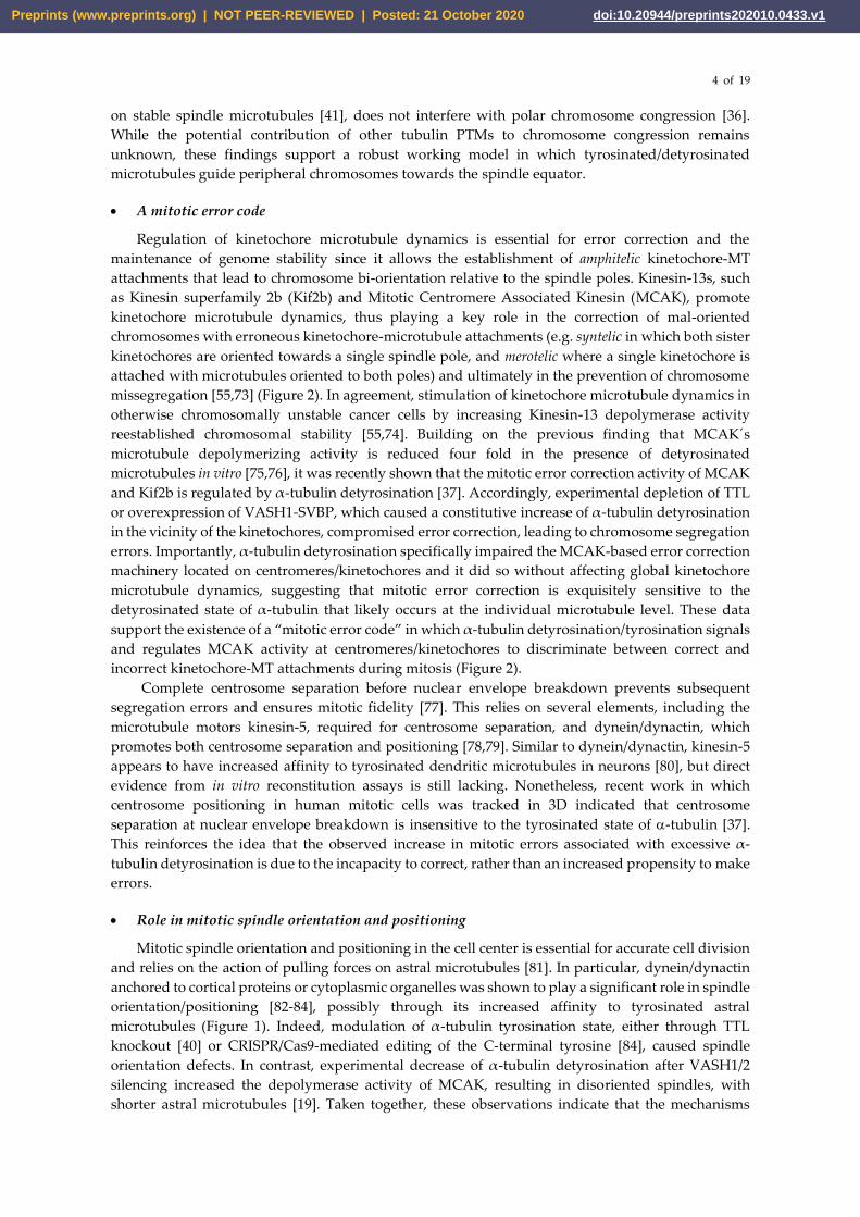
Figure 2. Summary of the established roles of the tubulin code in mitosis. The initial capture of peripheral
chromosomes is mediated by Dynein/Dynactin at kinetochores, upon which the chromosome is brought to the
vicinity of the centrosome by lateral transport along tyrosinated astral microtubules. This prevents the random
ejection of the chromosome by the action of Chromokinesins on chromosome arms. Once at the pole, high
Aurora A activity prevents stabilization of end-on kinetochore-microtubule attachments, which otherwise
would favor the action of Chromokinesins on chromosome arms. In parallel, Aurora A-mediated
phosphorylation activates CENP-E at kinetochores. This initiates transport towards the spindle equator
(congression) along stable detyrosinated microtubules. MCAK and Kif2b (not depicted) at centromeres and
kinetochores are also inhibited by tubulin detyrosination on kinetochore microtubules, allowing the correction
of syntelic and merotelic attachments, while preserving correct amphitelic attachments on bi-oriented
chromosomes. MCAK at microtubule plus ends also regulates astral microtubule length to allow interaction
with Dynein/Dynactin at the cortex or cytoplasmic organelles (not depicted), which exerts pulling forces
necessary for spindle orientation and positioning. See main text for details.
• Roles in centrosome structure and cytokinesis
Tubulin polyglutamylation is highly enriched on centriole microtubules [42,85] and has been
proposed to contribute to normal mitosis by maintaining centrosome structure [85,86]. Indeed, recent
Preprints (www.preprints.org) | NOT PEER-REVIEWED | Posted: 21 October 2020 doi:10.20944/preprints202010.0433.v1

6 of 19
super-resolution imaging of centriole structure revealed the specific distribution of
polyglutamylation on centriole MTs and suggested a key role for this PTM in ultrastructural
organization of specific centriolar proteins [87]. Furthermore, tubulin polyglutamylation promotes
the activity of the microtubule severing enzymes spastin and katanin [88-91], which are also
implicated in cell division. Indeed, their activities regulate several cellular processes that likely
impact chromosome segregation fidelity, such as microtubule poleward flux, spindle orientation and
length [92-94]. Spastin and katanin are also required for the abscission step and completion of
cytokinesis [95-97]. Like spastin [95] and katanin [97], polyglutamylated tubulin is enriched at the
midbody [88], and a tubulin mutation that compromises polyglutamylation (and possibly also
polyglycylation) in cilia was shown to cause cytokinesis defects [98]. These results suggest that the
completion of cytokinesis relies on the regulation of spastin and katanin activities by tubulin
polyglutamylation.
III. The cancer tubulin code
• (De)regulation of tubulin isotypes and PTMs in cancer
Several works have reported an emerging link between alterations of tubulin isotypes and PTMs
and/or associated modifying enzymes with certain cancers, most noticeable those occurring in breast,
colon, prostate, liver, brain, bile duct and pancreas (Table 1). These alterations often correlate with
specific cancer properties, including poor outcome/prognosis [99-101] and metastatic ability [99],
supporting the potential use of cancer tubulin isotypes and/or PTM signatures as useful biomarkers,
as well as for therapeutic purposes. However, a comprehensive and definitive view on the real
potential is still lacking, especially concerning causality, since the available data is still limited and
often contradictory.
Table 1. Tubulin isotypes, post-translational modifications and modifying enzymes in cancer
Tubulin PTM (and/or
enzymes)/ Isotype Cancer Regulation References
Detyrosination
Prostate Cancer Cells Up-regulated [102]
Poor Prognosis Breast
Tumors Up-regulated [100]
Invasive Ductal Carcinoma
(Breast) Up-regulated [103]
TTL
Prostate Cancer Cells Down-regulated [102]
Poor Prognosis
Neuroblastomas Down-regulated [101]
VASH2 Hepatocellular carcinoma
Tissues and Cell Lines Up-regulated [104]
Δ2-Tubulin Prostate Cancer Cells Down-regulated [102]
Acetylation Metastatic Breast Tumors
and Cell Lines Up-regulated [99]
HDAC6
Pancreatic Tumors Up-regulated [105]
Glioblastoma Tissues and
Cell Lines Up-regulated [106]
Cholangiocarcinoma Cell
Lines Up-regulated [107]
Glutamylation/
Polyglutamylation Prostate Cancer Cells Up-regulated [102]
Preprints (www.preprints.org) | NOT PEER-REVIEWED | Posted: 21 October 2020 doi:10.20944/preprints202010.0433.v1
7 of 19
TTLL4 Pancreatic Ductal
Adenocarcinoma Cells Up-regulated [108]
Glycylation
TTLL3
Colon Tumors and Cell
Lines Down-regulated [109]
β3-tubulin
Pancreatic Tumors and Cell
Lines Up-regulated [110]
Pancreatic Ductal
Adenocarcinoma Tissues Up-regulated [111]
Breast Cancer Brain
Metastases Up-regulated [112]
• Functional implications of the cancer tubulin code
The differential regulation of specific tubulin isotypes and/or PTMs in cancer might reflect their
role in key mechanisms underlying cell transformation (Figure 3). β3-tubulin (TUBB3) is the most
frequent tubulin isotype associated with specific cancer features. Its expression was proposed to be
important for tumor development [110,113] and metastatic ability [110,112], correlating with poor
outcomes [112,114]. Expression of other isotypes such as β2-tubulin, and its altered cellular
localization in colorectal cancer, also correlate with poor outcomes [115]. Differential expression of
tubulin isotypes have been extensively associated with response to microtubule-targeting drugs, such
as taxanes, commonly used in chemotherapy (reviewed in [116]). The origin of this link may be on
the known regulation of microtubule dynamics by specific tubulin isotypes, with microtubules
containing β3-tubulin being more dynamic compared to other β-tubulin isotypes [117-119].
In addition, the regulation of cell proliferation, which is essential for cancer development, was
proposed to be mediated by certain tubulin PTMs. In this regard, the tubulin glycylase TTLL3 was
proposed to restrict cell proliferation in the colon and is down-regulated in colon cancer [109],
whereas the tubulin glutamylase TTLL4 was suggested to promote cell proliferation in pancreatic
cancer cells [108]. However, whether this was specifically due to a role of TTLL4 in tubulin
glutamylation remains controversial, since an additional activity towards non-tubulin substrates has
been reported [120,121]. The tubulin acetyltransferase αTat1 was also shown to be required for
contact inhibition of cell proliferation in vitro [122]. In agreement, the tubulin deacetylase HDAC6
seems to promote cell proliferation in several cancer cell lines [106,107,123-125], consistent with its
upregulation in some cancers (Table 1). Nevertheless, specificity remains to be demonstrated, since
HDAC6 is also known to modulate acetylation of other substrates besides tubulin [126]. Interestingly,
the activity of TTLL3 and HDAC6 was also proposed to impact tumorigenesis. Accordingly,
experimental loss of TTLL3 in a mouse model of tumorigenesis resulted in the development of cancer,
thus validating its downregulation in colon cancer and suggesting a cancer suppressing role for
tubulin glycylation [109]. In contrast, the expression of HDAC6 promoted colony and spheroid
formation of cancer cells, as well as tumor growth in mice [106,107,123,125]. The activity of other
tubulin modifying enzymes, such as TTL, is also decreased during tumorigenesis in mouse models,
resulting in increased detyrosinated- and Δ2-tubulin levels [127]. This is consistent with the
association between -tubulin detyrosination and tumor aggressiveness [100], as well as with the
frequent downregulation of TTL and consequent upregulation of -tubulin detyrosination in several
cancers (Table 1).
The recent discovery of Vasohibins (VASH1 and VASH2) as TCPs [13,14] revitalized the
discussion about the role of tubulin detyrosination in cancer. Vasohibins and their associated SVBP
were originally identified as secreted proteins implicated in angiogenesis [128]. While VASH2
promotes vascularity by accumulating at the sprouting zone, VASH1 expression is increased in
endothelial cells of the termination zone, where it inhibits vascularity [129]. During tumor
development in mice xenograft models, experiments involving administration of ectopic VASH1
indicated that it inhibits tumor lymphangiogenesis [130], angiogenesis and growth [131]. On the other
hand, VASH2, which appears to play an important role in cancer cell proliferation [104], promotes
tumor angiogenesis and growth [104,132-134]. Noteworthy, none of these studies demonstrate that
the observed impact in cancer was due to defective tubulin detyrosination. However, a recent work
Preprints (www.preprints.org) | NOT PEER-REVIEWED | Posted: 21 October 2020 doi:10.20944/preprints202010.0433.v1
8 of 19
reported that human patients suffering from a broad range of carcinomas had mutations in VASH1
and VASH2 that compromised their tubulin detyrosination activity [17]. Taken together, these
findings suggest that in addition to downregulation of TTL [127], the link between tubulin
detyrosination and tumorigenesis may be attributed to the increased expression of Vasohibins. The
availability of VASH1/2-SVBP knockout mice [129,135] will be instrumental to clarify the apparently
opposite roles of VASH1 and VASH2 in cancer and whether this is due to their secreted and/or
tubulin detyrosinating activities.
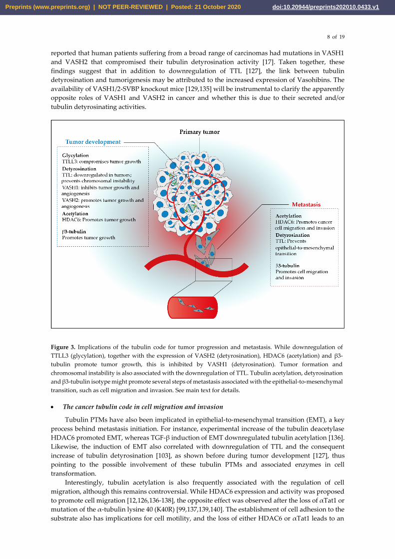
Figure 3. Implications of the tubulin code for tumor progression and metastasis. While downregulation of
TTLL3 (glycylation), together with the expression of VASH2 (detyrosination), HDAC6 (acetylation) and β3-
tubulin promote tumor growth, this is inhibited by VASH1 (detyrosination). Tumor formation and
chromosomal instability is also associated with the downregulation of TTL. Tubulin acetylation, detyrosination
and β3-tubulin isotype might promote several steps of metastasis associated with the epithelial-to-mesenchymal
transition, such as cell migration and invasion. See main text for details.
• The cancer tubulin code in cell migration and invasion
Tubulin PTMs have also been implicated in epithelial-to-mesenchymal transition (EMT), a key
process behind metastasis initiation. For instance, experimental increase of the tubulin deacetylase
HDAC6 promoted EMT, whereas TGF-β induction of EMT downregulated tubulin acetylation [136].
Likewise, the induction of EMT also correlated with downregulation of TTL and the consequent
increase of tubulin detyrosination [103], as shown before during tumor development [127], thus
pointing to the possible involvement of these tubulin PTMs and associated enzymes in cell
transformation.
Interestingly, tubulin acetylation is also frequently associated with the regulation of cell
migration, although this remains controversial. While HDAC6 expression and activity was proposed
to promote cell migration [12,126,136-138], the opposite effect was observed after the loss of αTat1 or
mutation of the α-tubulin lysine 40 (K40R) [99,137,139,140]. The establishment of cell adhesion to the
substrate also has implications for cell motility, and the loss of either HDAC6 or αTat1 leads to an
Preprints (www.preprints.org) | NOT PEER-REVIEWED | Posted: 21 October 2020 doi:10.20944/preprints202010.0433.v1

9 of 19
increased focal adhesion area and number, respectively, as well as decreased dynamics [137,141].
However, other works reported that loss of αTat1 leads to a decrease in focal adhesion number [122].
The basis for this discrepancy remains unclear, but it is likely associated with different experimental
set ups: one study investigated the role of αTat1 in wound-induced migrating cells [137], while the
other used normally growing cells were used [122], raising the possibility that αTat1 promotes focal
adhesion dynamics specifically during cell migration.
The upregulation of tubulin acetylation in metastatic breast tumors and cell lines [99] is
consistent with its association with cancer cell invasiveness. RNAi-mediated depletion of either αTat1
or HDAC6 indicated that their expression induced breast cancer cell invasion [139,140,142].
Additionally, the increased tubulin acetylation of these metastatic breast cancer cells promoted
microtentacle generation and cell reattachment ability, essential for metastasis [99]. Likewise, a high
frequency of microtentacles and cell reattachment was also associated with tubulin detyrosination
[103,143]. Collectively, these data favor a potential role of tubulin acetylation in metastasis
progression. While HDAC6 indiscriminately acts upon multiple protein targets, the direct
modulation of tubulin acetylation by K40R mutation experiments suggest that the upregulation of
tubulin acetylation is a metastasis-promoting factor, supporting the αTat1-related findings. This
would explain the link between upregulation of tubulin acetylation and poor prognosis in breast
cancer patients [99], but unspecific effects due to overexpression of GFP-tagged K40R mutant α-
tubulin cannot be excluded.
IV. How alterations of the tubulin code in mitosis might be implicated in cancer
Chromosomal instability, a hallmark of cancers, has been shown to promote the metastatic
process [74]. Indeed, the overexpression of Kif2b or MCAK, in addition to reestablishing the stability
of chromosomally unstable cancer cells [55,74], inhibits metastasis in vitro and in vivo, with a
consequent increase in survival [74]. Given that excessive tubulin detyrosination might lead to
chromosomal instability by suppressing the error correction activity of MCAK and Kif2b [37],
together with the observed upregulation of tubulin detyrosination in invasive cancer and with poor
prognosis (Table 1), it raises the exciting possibility that an increase in tubulin detyrosination might
promote cancer progression through inhibition of the mitotic error correction machinery. However,
an extensive analysis of tubulin detyrosination in chromosomal instability-prone cancers, together
with the elucidation of its implications for cancer metastasis, is necessary for its establishment as
potential diagnostic and prognostic biomarkers. In addition, tubulin detyrosination represents a
promising therapeutic target for cancer suppression, for example by using TCP inhibitors, such as
epoY [13] or parthenolide [144].
Deregulation of tubulin detyrosination in cancers might also be involved in other mitotic-related
cancer features. Firstly, the cell cycle delay observed upon VASH1/2 [19] and VASH2 [104] deletion
might unveil the importance of VASH2 for proper cancer cell proliferation and tumor development
[104,132-134]. Furthermore, both experimental upregulation and downregulation of tubulin
detyrosination led to congression defects, causing alterations in CENP-E-mediated transport of
chromosomes to the spindle equator [36]. Additionally, the decrease of CENP-E expression is well
established to promote mild chromosomal instability and aneuploidy, as well as tumorigenesis in
mice [145-148]. Therefore, deregulation of tubulin detyrosination in cancers (Table 1) may also
account for cancer promotion under conditions of moderate chromosomal instability, such as those
associated with mild problems in chromosome congression. Further investigation is required to fully
understand the potential implications of tubulin detyrosination and other PTMs for tumorigenesis
and the respective link with chromosomal instability.
V. Conclusions and outlook
Since the initial observations implicating tubulin PTMs in cell division, recent works have
allowed a deeper understanding of their involvement, in particular detyrosination/tyrosination, in
the coordination of several mechanisms underlying faithful chromosome segregation during mitosis.
However, considerable knowledge is still lacking in order to establish a complete picture of the roles
Preprints (www.preprints.org) | NOT PEER-REVIEWED | Posted: 21 October 2020 doi:10.20944/preprints202010.0433.v1
10 of 19
played by the tubulin code in mitosis. The scenario is no different regarding the emerging cancer
tubulin code, in which a considerable amount of disconnected data dominates. Nevertheless, there
are already some promising links between deregulation of certain tubulin isotypes and PTMs
(notoriously acetylation, detyrosination and glycylation) and several cancers. A more systematic
investigation of these links will be of high priority to the field and might prove important for
diagnostic and prognostic purposes. This is likely to have a major impact in understanding and
mitigating acquired resistance to microtubule-targeting drugs, the biggest threat in current cancer
chemotherapy. Future work is also necessary to establish clear functional links beyond correlations
by taking advantage of emerging molecular tools and model systems for modulation and analysis of
tubulin isotypes and PTMs (both in vitro and in vivo) that will strengthen and clarify their potential
therapeutic value for the treatment of human cancers.
Funding: Work in the laboratory of H.M. is funded by the European Research Council (ERC) under the European
Union’s Horizon 2020 research and innovation programme (grant agreement No 681443).
Acknowledgements: We thank Liam Cheeseman for comments and critical reading of this manuscript. Danilo
Lopes is a student of Programa de Pós-Graduação Ciência para o Desenvolvimento (PGCD) from Instituto
Gulbenkian de Ciência (IGC), Oeiras, Portugal and recipient of a studentship (SFRH/BD/135077/2017) from
Fundação para a Ciência e a Tecnologia of Portugal.
Conflicts of Interest: The authors declare no conflict of interest.
References
1. Desai, A.; Mitchison, T.J. Microtubule polymerization dynamics. Annu Rev Cell Dev Biol 1997, 13, 83-
117.
2. Ludueña, R.F.; Banerjee, A. The isotypes of tubulin. In The role of microtubules in cell biology, neurobiology,
and oncology, Springer: 2008; pp 123-175.
3. Janke, C. The tubulin code: Molecular components, readout mechanisms, and functions. J Cell Biol 2014,
206, 461-472.
4. Verhey, K.J.; Gaertig, J. The tubulin code. Cell Cycle 2007, 6, 2152-2160.
5. Janke, C.; Magiera, M.M. The tubulin code and its role in controlling microtubule properties and
functions. Nat Rev Mol Cell Biol 2020, 21, 307-326.
6. L'Hernault, S.W.; Rosenbaum, J.L. Chlamydomonas alpha-tubulin is posttranslationally modified by
acetylation on the epsilon-amino group of a lysine. Biochemistry 1985, 24, 473-478.
7. Soppina, V.; Herbstman, J.F.; Skiniotis, G.; Verhey, K.J. Luminal localization of α-tubulin k40
acetylation by cryo-em analysis of fab-labeled microtubules. PloS one 2012, 7.
8. Chu, C.W.; Hou, F.; Zhang, J.; Phu, L.; Loktev, A.V.; Kirkpatrick, D.S.; Jackson, P.K.; Zhao, Y.; Zou, H.
A novel acetylation of beta-tubulin by san modulates microtubule polymerization via down-regulating
tubulin incorporation. Mol Biol Cell 2011, 22, 448-456.
9. Akella, J.S.; Wloga, D.; Kim, J.; Starostina, N.G.; Lyons-Abbott, S.; Morrissette, N.S.; Dougan, S.T.;
Kipreos, E.T.; Gaertig, J. Mec-17 is an alpha-tubulin acetyltransferase. Nature 2010, 467, 218-222.
10. Shida, T.; Cueva, J.G.; Xu, Z.; Goodman, M.B.; Nachury, M.V. The major alpha-tubulin k40
acetyltransferase alphatat1 promotes rapid ciliogenesis and efficient mechanosensation. Proceedings of
the National Academy of Sciences of the United States of America 2010, 107, 21517-21522.
11. North, B.J.; Marshall, B.L.; Borra, M.T.; Denu, J.M.; Verdin, E. The human sir2 ortholog, sirt2, is an nad+-
dependent tubulin deacetylase. Molecular cell 2003, 11, 437-444.
12. Hubbert, C.; Guardiola, A.; Shao, R.; Kawaguchi, Y.; Ito, A.; Nixon, A.; Yoshida, M.; Wang, X.F.; Yao,
T.P. Hdac6 is a microtubule-associated deacetylase. Nature 2002, 417, 455-458.
Preprints (www.preprints.org) | NOT PEER-REVIEWED | Posted: 21 October 2020 doi:10.20944/preprints202010.0433.v1

11 of 19
13. Aillaud, C.; Bosc, C.; Peris, L.; Bosson, A.; Heemeryck, P.; Van Dijk, J.; Le Friec, J.; Boulan, B.; Vossier,
F.; Sanman, L.E., et al. Vasohibins/svbp are tubulin carboxypeptidases (tcps) that regulate neuron
differentiation. Science 2017, 358, 1448-1453.
14. Nieuwenhuis, J.; Adamopoulos, A.; Bleijerveld, O.B.; Mazouzi, A.; Stickel, E.; Celie, P.; Altelaar, M.;
Knipscheer, P.; Perrakis, A.; Blomen, V.A., et al. Vasohibins encode tubulin detyrosinating activity.
Science (New York, N.Y 2017, 358, 1453-1456.
15. Li, F.; Li, Y.; Ye, X.; Gao, H.; Shi, Z.; Luo, X.; Rice, L.M.; Yu, H. Cryo-em structure of vash1-svbp bound
to microtubules. Elife 2020, 9.
16. Liu, X.; Wang, H.; Zhu, J.; Xie, Y.; Liang, X.; Chen, Z.; Feng, Y.; Zhang, Y. Structural insights into tubulin
detyrosination by vasohibins-svbp complex. Cell discovery 2019, 5, 65.
17. Wang, N.; Bosc, C.; Ryul Choi, S.; Boulan, B.; Peris, L.; Olieric, N.; Bao, H.; Krichen, F.; Chen, L.;
Andrieux, A., et al. Structural basis of tubulin detyrosination by the vasohibin-svbp enzyme complex.
Nat Struct Mol Biol 2019, 26, 571-582.
18. Li, F.; Hu, Y.; Qi, S.; Luo, X.; Yu, H. Structural basis of tubulin detyrosination by vasohibins. Nat Struct
Mol Biol 2019, 26, 583-591.
19. Liao, S.; Rajendraprasad, G.; Wang, N.; Eibes, S.; Gao, J.; Yu, H.; Wu, G.; Tu, X.; Huang, H.; Barisic, M.,
et al. Molecular basis of vasohibins-mediated detyrosination and its impact on spindle function and
mitosis. Cell Res 2019, 29, 533-547.
20. Ersfeld, K.; Wehland, J.; Plessmann, U.; Dodemont, H.; Gerke, V.; Weber, K. Characterization of the
tubulin-tyrosine ligase. J Cell Biol 1993, 120, 725-732.
21. Schroder, H.C.; Wehland, J.; Weber, K. Purification of brain tubulin-tyrosine ligase by biochemical and
immunological methods. The Journal of cell biology 1985, 100, 276-281.
22. Rogowski, K.; van Dijk, J.; Magiera, M.M.; Bosc, C.; Deloulme, J.C.; Bosson, A.; Peris, L.; Gold, N.D.;
Lacroix, B.; Bosch Grau, M., et al. A family of protein-deglutamylating enzymes associated with
neurodegeneration. Cell 2010, 143, 564-578.
23. Tort, O.; Tanco, S.; Rocha, C.; Bieche, I.; Seixas, C.; Bosc, C.; Andrieux, A.; Moutin, M.J.; Aviles, F.X.;
Lorenzo, J., et al. The cytosolic carboxypeptidases ccp2 and ccp3 catalyze posttranslational removal of
acidic amino acids. Mol Biol Cell 2014, 25, 3017-3027.
24. Paturle-Lafanechere, L.; Edde, B.; Denoulet, P.; Van Dorsselaer, A.; Mazarguil, H.; Le Caer, J.P.;
Wehland, J.; Job, D. Characterization of a major brain tubulin variant which cannot be tyrosinated.
Biochemistry 1991, 30, 10523-10528.
25. Aillaud, C.; Bosc, C.; Saoudi, Y.; Denarier, E.; Peris, L.; Sago, L.; Taulet, N.; Cieren, A.; Tort, O.; Magiera,
M.M., et al. Evidence for new c-terminally truncated variants of alpha- and beta-tubulins. Molecular
biology of the cell 2016, 27, 640-653.
26. Edde, B.; Rossier, J.; Le Caer, J.P.; Desbruyeres, E.; Gros, F.; Denoulet, P. Posttranslational glutamylation
of alpha-tubulin. Science 1990, 247, 83-85.
27. Redeker, V.; Levilliers, N.; Schmitter, J.M.; Le Caer, J.P.; Rossier, J.; Adoutte, A.; Bre, M.H.
Polyglycylation of tubulin: A posttranslational modification in axonemal microtubules. Science 1994,
266, 1688-1691.
28. Janke, C.; Rogowski, K.; Wloga, D.; Regnard, C.; Kajava, A.V.; Strub, J.M.; Temurak, N.; van Dijk, J.;
Boucher, D.; van Dorsselaer, A., et al. Tubulin polyglutamylase enzymes are members of the ttl domain
protein family. Science 2005, 308, 1758-1762.
Preprints (www.preprints.org) | NOT PEER-REVIEWED | Posted: 21 October 2020 doi:10.20944/preprints202010.0433.v1

12 of 19
29. van Dijk, J.; Rogowski, K.; Miro, J.; Lacroix, B.; Edde, B.; Janke, C. A targeted multienzyme mechanism
for selective microtubule polyglutamylation. Mol Cell 2007, 26, 437-448.
30. Kimura, Y.; Kurabe, N.; Ikegami, K.; Tsutsumi, K.; Konishi, Y.; Kaplan, O.I.; Kunitomo, H.; Iino, Y.;
Blacque, O.E.; Setou, M. Identification of tubulin deglutamylase among caenorhabditis elegans and
mammalian cytosolic carboxypeptidases (ccps). The Journal of biological chemistry 2010, 285, 22936-22941.
31. Rogowski, K.; Juge, F.; van Dijk, J.; Wloga, D.; Strub, J.M.; Levilliers, N.; Thomas, D.; Bre, M.H.; Van
Dorsselaer, A.; Gaertig, J., et al. Evolutionary divergence of enzymatic mechanisms for posttranslational
polyglycylation. Cell 2009, 137, 1076-1087.
32. Wloga, D.; Webster, D.M.; Rogowski, K.; Bre, M.H.; Levilliers, N.; Jerka-Dziadosz, M.; Janke, C.;
Dougan, S.T.; Gaertig, J. Ttll3 is a tubulin glycine ligase that regulates the assembly of cilia.
Developmental cell 2009, 16, 867-876.
33. Walgren, J.L.; Vincent, T.S.; Schey, K.L.; Buse, M.G. High glucose and insulin promote o-glcnac
modification of proteins, including alpha-tubulin. American journal of physiology. Endocrinology and
metabolism 2003, 284, E424-434.
34. Tian, J.L.; Qin, H. O-glcnacylation regulates primary ciliary length by promoting microtubule
disassembly. iScience 2019, 12, 379-391.
35. Ji, S.; Kang, J.G.; Park, S.Y.; Lee, J.; Oh, Y.J.; Cho, J.W. O-glcnacylation of tubulin inhibits its
polymerization. Amino acids 2011, 40, 809-818.
36. Barisic, M.; Silva e Sousa, R.; Tripathy, S.K.; Magiera, M.M.; Zaytsev, A.V.; Pereira, A.L.; Janke, C.;
Grishchuk, E.L.; Maiato, H. Mitosis. Microtubule detyrosination guides chromosomes during mitosis.
Science 2015, 348, 799-803.
37. Ferreira, L.T.; Orr, B.; Rajendraprasad, G.; Pereira, A.J.; Lemos, C.; Lima, J.T.; Guasch Boldu, C.; Ferreira,
J.G.; Barisic, M.; Maiato, H. Alpha-tubulin detyrosination impairs mitotic error correction by
suppressing mcak centromeric activity. The Journal of cell biology 2020, 219.
38. Gundersen, G.G.; Bulinski, J.C. Distribution of tyrosinated and nontyrosinated alpha-tubulin during
mitosis. J Cell Biol 1986, 102, 1118-1126.
39. Gundersen, G.G.; Kalnoski, M.H.; Bulinski, J.C. Distinct populations of microtubules: Tyrosinated and
nontyrosinated alpha tubulin are distributed differently in vivo. Cell 1984, 38, 779-789.
40. Peris, L.; Thery, M.; Faure, J.; Saoudi, Y.; Lafanechere, L.; Chilton, J.K.; Gordon-Weeks, P.; Galjart, N.;
Bornens, M.; Wordeman, L., et al. Tubulin tyrosination is a major factor affecting the recruitment of cap-
gly proteins at microtubule plus ends. J Cell Biol 2006, 174, 839-849.
41. Wilson, P.J.; Forer, A. Acetylated alpha-tubulin in spermatogenic cells of the crane fly nephrotoma-
suturalis - kinetochore microtubules are selectively acetylated. Cell Motility and the Cytoskeleton 1989, 14,
237-250.
42. Bobinnec, Y.; Moudjou, M.; Fouquet, J.P.; Desbruyeres, E.; Edde, B.; Bornens, M. Glutamylation of
centriole and cytoplasmic tubulin in proliferating non-neuronal cells. Cell Motil Cytoskeleton 1998, 39,
223-232.
43. Ferreira, L.T.; Figueiredo, A.C.; Orr, B.; Lopes, D.; Maiato, H. Dissecting the role of the tubulin code in
mitosis. In Methods in cell biology, Elsevier: 2018; Vol. 144, pp 33-74.
44. Maiato, H.; Sampaio, P.; Sunkel, C.E. Microtubule-associated proteins and their essential roles during
mitosis. Int Rev Cytol 2004, 241, 53-153.
45. Cross, R.A.; McAinsh, A. Prime movers: The mechanochemistry of mitotic kinesins. Nature reviews 2014,
15, 257-271.
Preprints (www.preprints.org) | NOT PEER-REVIEWED | Posted: 21 October 2020 doi:10.20944/preprints202010.0433.v1

13 of 19
46. Yang, Z.; Tulu, U.S.; Wadsworth, P.; Rieder, C.L. Kinetochore dynein is required for chromosome
motion and congression independent of the spindle checkpoint. Curr Biol 2007, 17, 973-980.
47. Hayden, J.H.; Bowser, S.S.; Rieder, C.L. Kinetochores capture astral microtubules during chromosome
attachment to the mitotic spindle: Direct visualization in live newt lung cells. The Journal of cell biology
1990, 111, 1039-1045.
48. Li, Y.; Yu, W.; Liang, Y.; Zhu, X. Kinetochore dynein generates a poleward pulling force to facilitate
congression and full chromosome alignment. Cell Res 2007, 17, 701-712.
49. Vorozhko, V.V.; Emanuele, M.J.; Kallio, M.J.; Stukenberg, P.T.; Gorbsky, G.J. Multiple mechanisms of
chromosome movement in vertebrate cells mediated through the ndc80 complex and dynein/dynactin.
Chromosoma 2008, 117, 169-179.
50. Pfarr, C.M.; Coue, M.; Grissom, P.M.; Hays, T.S.; Porter, M.E.; McIntosh, J.R. Cytoplasmic dynein is
localized to kinetochores during mitosis. Nature 1990, 345, 263-265.
51. Steuer, E.R.; Wordeman, L.; Schroer, T.A.; Sheetz, M.P. Localization of cytoplasmic dynein to mitotic
spindles and kinetochores. Nature 1990, 345, 266-268.
52. Kapoor, T.M.; Lampson, M.A.; Hergert, P.; Cameron, L.; Cimini, D.; Salmon, E.D.; McEwen, B.F.;
Khodjakov, A. Chromosomes can congress to the metaphase plate before biorientation. Science (New
York, N.Y 2006, 311, 388-391.
53. Wood, K.W.; Sakowicz, R.; Goldstein, L.S.; Cleveland, D.W. Cenp-e is a plus end-directed kinetochore
motor required for metaphase chromosome alignment. Cell 1997, 91, 357-366.
54. Mann, B.J.; Wadsworth, P. Kinesin-5 regulation and function in mitosis. Trends in cell biology 2019, 29,
66-79.
55. Bakhoum, S.F.; Thompson, S.L.; Manning, A.L.; Compton, D.A. Genome stability is ensured by
temporal control of kinetochore-microtubule dynamics. Nat Cell Biol 2009, 11, 27-35.
56. Kline-Smith, S.L.; Khodjakov, A.; Hergert, P.; Walczak, C.E. Depletion of centromeric mcak leads to
chromosome congression and segregation defects due to improper kinetochore attachments. Molecular
biology of the cell 2004, 15, 1146-1159.
57. Lan, W.; Zhang, X.; Kline-Smith, S.L.; Rosasco, S.E.; Barrett-Wilt, G.A.; Shabanowitz, J.; Hunt, D.F.;
Walczak, C.E.; Stukenberg, P.T. Aurora b phosphorylates centromeric mcak and regulates its
localization and microtubule depolymerization activity. Curr Biol 2004, 14, 273-286.
58. Andrews, P.D.; Ovechkina, Y.; Morrice, N.; Wagenbach, M.; Duncan, K.; Wordeman, L.; Swedlow, J.R.
Aurora b regulates mcak at the mitotic centromere. Developmental cell 2004, 6, 253-268.
59. Domnitz, S.B.; Wagenbach, M.; Decarreau, J.; Wordeman, L. Mcak activity at microtubule tips regulates
spindle microtubule length to promote robust kinetochore attachment. The Journal of cell biology 2012,
197, 231-237.
60. Reed, N.A.; Cai, D.; Blasius, T.L.; Jih, G.T.; Meyhofer, E.; Gaertig, J.; Verhey, K.J. Microtubule acetylation
promotes kinesin-1 binding and transport. Curr Biol 2006, 16, 2166-2172.
61. Konishi, Y.; Setou, M. Tubulin tyrosination navigates the kinesin-1 motor domain to axons. Nature
Neuroscience 2009, 12, 559-567.
62. Pfarr, C.M.; Coue, M.; Grissom, P.M.; Hays, T.S.; Porter, M.E.; Mcintosh, J.R. Cytoplasmic dynein is
localized to kinetochores during mitosis. Nature 1990, 345, 263-265.
63. McKenney, R.J.; Huynh, W.; Tanenbaum, M.E.; Bhabha, G.; Vale, R.D. Activation of cytoplasmic dynein
motility by dynactin-cargo adapter complexes. Science 2014, 345, 337-341.
Preprints (www.preprints.org) | NOT PEER-REVIEWED | Posted: 21 October 2020 doi:10.20944/preprints202010.0433.v1

14 of 19
64. McKenney, R.J.; Huynh, W.; Vale, R.D.; Sirajuddin, M. Tyrosination of alpha-tubulin controls the
initiation of processive dynein-dynactin motility. Embo J 2016, 35, 1175-1185.
65. Nirschl, J.J.; Magiera, M.M.; Lazarus, J.E.; Janke, C.; Holzbaur, E.L. Alpha-tubulin tyrosination and clip-
170 phosphorylation regulate the initiation of dynein-driven transport in neurons. Cell Rep 2016, 14,
2637-2652.
66. Barisic, M.; Maiato, H. The tubulin code: A navigation system for chromosomes during mitosis. Trends
Cell Biol 2016, 26, 766-775.
67. Barisic, M.; Aguiar, P.; Geley, S.; Maiato, H. Kinetochore motors drive congression of peripheral polar
chromosomes by overcoming random arm-ejection forces. Nat Cell Biol 2014, 16, 1249-1256.
68. Chmatal, L.; Yang, K.; Schultz, R.M.; Lampson, M.A. Spatial regulation of kinetochore microtubule
attachments by destabilization at spindle poles in meiosis i. Curr Biol 2015, 25, 1835-1841.
69. Ye, A.A.; Deretic, J.; Hole, C.M.; Hinman, A.W.; Cimini, D.; Welburn, J.P.; Maresca, T.J. Aurora a kinase
contributes to a pole-based error correction pathway. Mol Biol Cell 2015, 26.
70. Kim, Y.; Holland, A.J.; Lan, W.; Cleveland, D.W. Aurora kinases and protein phosphatase 1 mediate
chromosome congression through regulation of cenp-e. Cell 2010, 142, 444-455.
71. Pereira, A.; Sousa, M.; Almeida, A.C.; Ferreira, L.T.; Costa, A.R.; Novais-Cruz, M.; Ferras, C.; Sousa,
M.M.; Sampaio, P.; Belsley, M., et al. Coherent-hybrid sted: High contrast sub-diffraction imaging using
a bi-vortex depletion beam. Optics express 2019, 27, 8092-8111.
72. Steblyanko, Y., Rajendraprasad, G., Osswald, M., Eibes, S., Jacome, A., Geley, S., Pereira, A.J., Maiato,
H. and Barisic, M. . Microtubule poleward flux in human cells is driven by the coordinated action of
four kinesins. . The EMBO journal 2020, e105432.
73. Bakhoum, S.F.; Genovese, G.; Compton, D.A. Deviant kinetochore microtubule dynamics underlie
chromosomal instability. Curr Biol 2009, 19, 1937-1942.
74. Bakhoum, S.F.; Ngo, B.; Laughney, A.M.; Cavallo, J.A.; Murphy, C.J.; Ly, P.; Shah, P.; Sriram, R.K.;
Watkins, T.B.K.; Taunk, N.K., et al. Chromosomal instability drives metastasis through a cytosolic DNA
response. Nature 2018, 553, 467-472.
75. Peris, L.; Wagenbach, M.; Lafanechere, L.; Brocard, J.; Moore, A.T.; Kozielski, F.; Job, D.; Wordeman, L.;
Andrieux, A. Motor-dependent microtubule disassembly driven by tubulin tyrosination. The Journal of
cell biology 2009, 185, 1159-1166.
76. Sirajuddin, M.; Rice, L.M.; Vale, R.D. Regulation of microtubule motors by tubulin isotypes and post-
translational modifications. Nat Cell Biol 2014, 16, 335-344.
77. Silkworth, W.T.; Nardi, I.K.; Paul, R.; Mogilner, A.; Cimini, D. Timing of centrosome separation is
important for accurate chromosome segregation. Mol Biol Cell 2012, 23, 401-411.
78. Nunes, V.; Dantas, M.; Castro, D.; Vitiello, E.; Wang, I.; Carpi, N.; Balland, M.; Piel, M.; Aguiar, P.;
Maiato, H., et al. Centrosome-nuclear axis repositioning drives the assembly of a bipolar spindle
scaffold to ensure mitotic fidelity. Mol Biol Cell 2020, mbcE20010047.
79. Raaijmakers, J.A.; van Heesbeen, R.G.H.P.; Meaders, J.L.; Geers, E.F.; Fernandez-Garcia, B.; Medema,
R.H.; Tanenbaum, M.E. Nuclear envelope-associated dynein drives prophase centrosome separation
and enables eg5-independent bipolar spindle formation. Embo J 2012, 31, 4179-4190.
80. Kahn, O.I.; Sharma, V.; Gonzalez-Billault, C.; Baas, P.W. Effects of kinesin-5 inhibition on dendritic
architecture and microtubule organization. Mol Biol Cell 2015, 26, 66-77.
81. Siller, K.H.; Doe, C.Q. Spindle orientation during asymmetric cell division. Nat Cell Biol 2009, 11, 365-
374.
Preprints (www.preprints.org) | NOT PEER-REVIEWED | Posted: 21 October 2020 doi:10.20944/preprints202010.0433.v1

15 of 19
82. Kotak, S.; Busso, C.; Gonczy, P. Cortical dynein is critical for proper spindle positioning in human cells.
The Journal of cell biology 2012, 199, 97-110.
83. Nguyen-Ngoc, T.; Afshar, K.; Gonczy, P. Coupling of cortical dynein and g alpha proteins mediates
spindle positioning in caenorhabditis elegans. Nature cell biology 2007, 9, 1294-1302.
84. Barbosa, D.J.; Duro, J.; Prevo, B.; Cheerambathur, D.K.; Carvalho, A.X.; Gassmann, R. Dynactin binding
to tyrosinated microtubules promotes centrosome centration in c. Elegans by enhancing dynein-
mediated organelle transport. PLoS Genet 2017, 13, e1006941.
85. Bobinnec, Y.; Khodjakov, A.; Mir, L.M.; Rieder, C.L.; Edde, B.; Bornens, M. Centriole disassembly in
vivo and its effect on centrosome structure and function in vertebrate cells. Journal of Cell Biology 1998,
143, 1575-1589.
86. Abal, M.; Keryer, G.; Bornens, M. Centrioles resist forces applied on centrosomes during g2/m
transition. Biology of the Cell 2005, 97, 425-434.
87. Mahecic, D.; Gambarotto, D.; Douglass, K.M.; Fortun, D.; Banterle, N.; Ibrahim, K.A.; Le Guennec, M.;
Gonczy, P.; Hamel, V.; Guichard, P., et al. Homogeneous multifocal excitation for high-throughput
super-resolution imaging. Nat Methods 2020, 17, 726-733.
88. Lacroix, B.; van Dijk, J.; Gold, N.D.; Guizetti, J.; Aldrian-Herrada, G.; Rogowski, K.; Gerlich, D.W.;
Janke, C. Tubulin polyglutamylation stimulates spastin-mediated microtubule severing. Journal of Cell
Biology 2010, 189, 945-954.
89. Sharma, N.; Bryant, J.; Wloga, D.; Donaldson, R.; Davis, R.C.; Jerka-Dziadosz, M.; Gaertig, J. Katanin
regulates dynamics of microtubules and biogenesis of motile cilia. Journal of Cell Biology 2007, 178, 1065-
1079.
90. Shin, S.C.; Im, S.K.; Jang, E.H.; Jin, K.S.; Hur, E.M.; Kim, E.E. Structural and molecular basis for katanin-
mediated severing of glutamylated microtubules. Cell Reports 2019, 26, 1357-+.
91. Valenstein, M.L.; Roll-Mecak, A. Graded control of microtubule severing by tubulin glutamylation. Cell
2016, 164, 911-921.
92. Jiang, K.; Rezabkova, L.; Hua, S.S.; Liu, Q.Y.; Capitani, G.; Altelaar, A.F.M.; Heck, A.J.R.; Kammerer,
R.A.; Steinmetz, M.O.; Akhmanova, A. Microtubule minus-end regulation at spindle poles by an aspm-
katanin complex (vol 19, pg 480, 2017). Nature Cell Biology 2017, 19, 873-873.
93. McNally, K.; Audhya, A.; Oegema, K.; McNally, F.J. Katanin controls mitotic and meiotic spindle
length. Journal of Cell Biology 2006, 175, 881-891.
94. Zhang, D.; Rogers, G.C.; Buster, D.W.; Sharp, D.J. Three microtubule severing enzymes contribute to
the "pacman-flux" machinery that moves chromosomes. Journal of Cell Biology 2007, 177, 231-242.
95. Connell, J.W.; Lindon, C.; Luzio, J.P.; Reid, E. Spastin couples microtubule severing to membrane traffic
in completion of cytokinesis and secretion. Traffic 2009, 10, 42-56.
96. Guizetti, J.; Schermelleh, L.; Mantler, J.; Maar, S.; Poser, I.; Leonhardt, H.; Muller-Reichert, T.; Gerlich,
D.W. Cortical constriction during abscission involves helices of escrt-iii-dependent filaments. Science
2011, 331, 1616-1620.
97. Matsuo, M.; Shimodaira, T.; Kasama, T.; Hata, Y.; Echigo, A.; Okabe, M.; Arai, K.; Makino, Y.; Niwa,
S.I.; Saya, H., et al. Katanin p60 contributes to microtubule instability around the midbody and
facilitates cytokinesis in rat cells. Plos One 2013, 8.
98. Thazhath, R.; Liu, C.B.; Gaertig, J. Polyglycylation domain of beta-tubulin maintains axonemal
architecture and affects cytokinesis in tetrahymena. Nature Cell Biology 2002, 4, 256-259.
Preprints (www.preprints.org) | NOT PEER-REVIEWED | Posted: 21 October 2020 doi:10.20944/preprints202010.0433.v1

16 of 19
99. Boggs, A.E.; Vitolo, M.I.; Whipple, R.A.; Charpentier, M.S.; Goloubeva, O.G.; Ioffe, O.B.; Tuttle, K.C.;
Slovic, J.; Lu, Y.; Mills, G.B., et al. Alpha-tubulin acetylation elevated in metastatic and basal-like breast
cancer cells promotes microtentacle formation, adhesion, and invasive migration. Cancer Res 2015, 75,
203-215.
100. Mialhe, A.; Lafanechere, L.; Treilleux, I.; Peloux, N.; Dumontet, C.; Bremond, A.; Panh, M.H.; Payan, R.;
Wehland, J.; Margolis, R.L., et al. Tubulin detyrosination is a frequent occurrence in breast cancers of
poor prognosis. Cancer Res 2001, 61, 5024-5027.
101. Kato, C.; Miyazaki, K.; Nakagawa, A.; Ohira, M.; Nakamura, Y.; Ozaki, T.; Imai, T.; Nakagawara, A.
Low expression of human tubulin tyrosine ligase and suppressed tubulin tyrosination/detyrosination
cycle are associated with impaired neuronal differentiation in neuroblastomas with poor prognosis.
International Journal of Cancer 2004, 112, 365-375.
102. Soucek, K.; Kamaid, A.; Phung, A.D.; Kubala, L.; Bulinski, J.C.; Harper, R.W.; Eiserich, J.P. Normal and
prostate cancer cells display distinct molecular profiles of alpha-tubulin posttranslational
modifications. Prostate 2006, 66, 954-965.
103. Whipple, R.A.; Matrone, M.A.; Cho, E.H.; Balzer, E.M.; Vitolo, M.I.; Yoon, J.R.; Ioffe, O.B.; Tuttle, K.C.;
Yang, J.; Martin, S.S. Epithelial-to-mesenchymal transition promotes tubulin detyrosination and
microtentacles that enhance endothelial engagement. Cancer Res 2010, 70, 8127-8137.
104. Xue, X.; Gao, W.; Sun, B.; Xu, Y.; Han, B.; Wang, F.; Zhang, Y.; Sun, J.; Wei, J.; Lu, Z., et al. Vasohibin 2
is transcriptionally activated and promotes angiogenesis in hepatocellular carcinoma. Oncogene 2013,
32, 1724-1734.
105. Li, D.W.; Sun, X.D.; Zhang, L.L.; Yan, B.; Xie, S.B.; Liu, R.M.; Liu, M.; Zhou, J. Histone deacetylase 6 and
cytoplasmic linker protein 170 function together to regulate the motility of pancreatic cancer cells.
Protein Cell 2014, 5, 214-223.
106. Wang, Z.; Hu, P.; Tang, F.; Lian, H.; Chen, X.; Zhang, Y.; He, X.; Liu, W.; Xie, C. Hdac6 promotes cell
proliferation and confers resistance to temozolomide in glioblastoma. Cancer Lett 2016, 379, 134-142.
107. Gradilone, S.A.; Radtke, B.N.; Bogert, P.S.; Huang, B.Q.; Gajdos, G.B.; LaRusso, N.F. Hdac6 inhibition
restores ciliary expression and decreases tumor growth. Cancer Res 2013, 73, 2259-2270.
108. Kashiwaya, K.; Nakagawa, H.; Hosokawa, M.; Mochizuki, Y.; Ueda, K.; Piao, L.; Chung, S.; Hamamoto,
R.; Eguchi, H.; Ohigashi, H., et al. Involvement of the tubulin tyrosine ligase-like family member 4
polyglutamylase in pelp1 polyglutamylation and chromatin remodeling in pancreatic cancer cells.
Cancer Res 2010, 70, 4024-4033.
109. Rocha, C.; Papon, L.; Cacheux, W.; Marques Sousa, P.; Lascano, V.; Tort, O.; Giordano, T.; Vacher, S.;
Lemmers, B.; Mariani, P., et al. Tubulin glycylases are required for primary cilia, control of cell
proliferation and tumor development in colon. The EMBO journal 2014, 33, 2247-2260.
110. McCarroll, J.A.; Sharbeen, G.; Liu, J.; Youkhana, J.; Goldstein, D.; McCarthy, N.; Limbri, L.F.; Dischl, D.;
Ceyhan, G.O.; Erkan, M., et al. Betaiii-tubulin: A novel mediator of chemoresistance and metastases in
pancreatic cancer. Oncotarget 2015, 6, 2235-2249.
111. Lee, K.M.; Cao, D.; Itami, A.; Pour, P.M.; Hruban, R.H.; Maitra, A.; Ouellette, M.M. Class iii beta-
tubulin, a marker of resistance to paclitaxel, is overexpressed in pancreatic ductal adenocarcinoma and
intraepithelial neoplasia. Histopathology 2007, 51, 539-546.
112. Kanojia, D.; Morshed, R.A.; Zhang, L.; Miska, J.M.; Qiao, J.; Kim, J.W.; Pytel, P.; Balyasnikova, I.V.;
Lesniak, M.S.; Ahmed, A.U. Betaiii-tubulin regulates breast cancer metastases to the brain. Mol Cancer
Ther 2015, 14, 1152-1161.
Preprints (www.preprints.org) | NOT PEER-REVIEWED | Posted: 21 October 2020 doi:10.20944/preprints202010.0433.v1

17 of 19
113. McCarroll, J.A.; Gan, P.P.; Liu, M.; Kavallaris, M. Betaiii-tubulin is a multifunctional protein involved
in drug sensitivity and tumorigenesis in non-small cell lung cancer. Cancer Res 2010, 70, 4995-5003.
114. Ferrandina, G.; Zannoni, G.F.; Martinelli, E.; Paglia, A.; Gallotta, V.; Mozzetti, S.; Scambia, G.; Ferlini,
C. Class iii beta-tubulin overexpression is a marker of poor clinical outcome in advanced ovarian cancer
patients. Clin Cancer Res 2006, 12, 2774-2779.
115. Ruksha, K.; Mezheyeuski, A.; Nerovnya, A.; Bich, T.; Tur, G.; Gorgun, J.; Luduena, R.; Portyanko, A.
Over-expression of betaii-tubulin and especially its localization in cell nuclei correlates with poorer
outcomes in colorectal cancer. Cells 2019, 8.
116. Parker, A.L.; Teo, W.S.; McCarroll, J.A.; Kavallaris, M. An emerging role for tubulin isotypes in
modulating cancer biology and chemotherapy resistance. International journal of molecular sciences 2017,
18.
117. Panda, D.; Miller, H.P.; Banerjee, A.; Luduena, R.F.; Wilson, L. Microtubule dynamics in vitro are
regulated by the tubulin isotype composition. Proceedings of the National Academy of Sciences of the United
States of America 1994, 91, 11358-11362.
118. Pamula, M.C.; Ti, S.C.; Kapoor, T.M. The structured core of human beta tubulin confers isotype-specific
polymerization properties. The Journal of cell biology 2016, 213, 425-433.
119. Ti, S.C.; Alushin, G.M.; Kapoor, T.M. Human beta-tubulin isotypes can regulate microtubule
protofilament number and stability. Developmental cell 2018, 47, 175-190 e175.
120. van Dijk, J.; Miro, J.; Strub, J.M.; Lacroix, B.; van Dorsselaer, A.; Edde, B.; Janke, C. Polyglutamylation
is a post-translational modification with a broad range of substrates. The Journal of biological chemistry
2008, 283, 3915-3922.
121. Regnard, C.; Desbruyeres, E.; Huet, J.C.; Beauvallet, C.; Pernollet, J.C.; Edde, B. Polyglutamylation of
nucleosome assembly proteins. The Journal of biological chemistry 2000, 275, 15969-15976.
122. Aguilar, A.; Becker, L.; Tedeschi, T.; Heller, S.; Iomini, C.; Nachury, M.V. Alpha-tubulin k40 acetylation
is required for contact inhibition of proliferation and cell-substrate adhesion. Mol Biol Cell 2014, 25,
1854-1866.
123. Lee, Y.S.; Lim, K.H.; Guo, X.; Kawaguchi, Y.; Gao, Y.; Barrientos, T.; Ordentlich, P.; Wang, X.F.; Counter,
C.M.; Yao, T.P. The cytoplasmic deacetylase hdac6 is required for efficient oncogenic tumorigenesis.
Cancer Res 2008, 68, 7561-7569.
124. Putcha, P.; Yu, J.; Rodriguez-Barrueco, R.; Saucedo-Cuevas, L.; Villagrasa, P.; Murga-Penas, E.; Quayle,
S.N.; Yang, M.; Castro, V.; Llobet-Navas, D., et al. Hdac6 activity is a non-oncogene addiction hub for
inflammatory breast cancers. Breast Cancer Res 2015, 17, 149.
125. Woan, K.V.; Lienlaf, M.; Perez-Villaroel, P.; Lee, C.; Cheng, F.; Knox, T.; Woods, D.M.; Barrios, K.;
Powers, J.; Sahakian, E., et al. Targeting histone deacetylase 6 mediates a dual anti-melanoma effect:
Enhanced antitumor immunity and impaired cell proliferation. Mol Oncol 2015, 9, 1447-1457.
126. Zhang, X.; Yuan, Z.; Zhang, Y.; Yong, S.; Salas-Burgos, A.; Koomen, J.; Olashaw, N.; Parsons, J.T.; Yang,
X.J.; Dent, S.R., et al. Hdac6 modulates cell motility by altering the acetylation level of cortactin. Mol Cell
2007, 27, 197-213.
127. Lafanechere, L.; Courtay-Cahen, C.; Kawakami, T.; Jacrot, M.; Rudiger, M.; Wehland, J.; Job, D.;
Margolis, R.L. Suppression of tubulin tyrosine ligase during tumor growth. Journal of cell science 1998,
111 ( Pt 2), 171-181.
128. Sato, Y. The vasohibin family: A novel family for angiogenesis regulation. J Biochem 2013, 153, 5-11.
Preprints (www.preprints.org) | NOT PEER-REVIEWED | Posted: 21 October 2020 doi:10.20944/preprints202010.0433.v1

18 of 19
129. Kimura, H.; Miyashita, H.; Suzuki, Y.; Kobayashi, M.; Watanabe, K.; Sonoda, H.; Ohta, H.; Fujiwara, T.;
Shimosegawa, T.; Sato, Y. Distinctive localization and opposed roles of vasohibin-1 and vasohibin-2 in
the regulation of angiogenesis. Blood 2009, 113, 4810-4818.
130. Heishi, T.; Hosaka, T.; Suzuki, Y.; Miyashita, H.; Oike, Y.; Takahashi, T.; Nakamura, T.; Arioka, S.;
Mitsuda, Y.; Takakura, T., et al. Endogenous angiogenesis inhibitor vasohibin1 exhibits broad-spectrum
antilymphangiogenic activity and suppresses lymph node metastasis. Am J Pathol 2010, 176, 1950-1958.
131. Hosaka, T.; Kimura, H.; Heishi, T.; Suzuki, Y.; Miyashita, H.; Ohta, H.; Sonoda, H.; Moriya, T.; Suzuki,
S.; Kondo, T., et al. Vasohibin-1 expression in endothelium of tumor blood vessels regulates
angiogenesis. Am J Pathol 2009, 175, 430-439.
132. Kitahara, S.; Suzuki, Y.; Morishima, M.; Yoshii, A.; Kikuta, S.; Shimizu, K.; Morikawa, S.; Sato, Y.; Ezaki,
T. Vasohibin-2 modulates tumor onset in the gastrointestinal tract by normalizing tumor angiogenesis.
Mol Cancer 2014, 13, 99.
133. Koyanagi, T.; Suzuki, Y.; Saga, Y.; Machida, S.; Takei, Y.; Fujiwara, H.; Suzuki, M.; Sato, Y. In vivo
delivery of sirna targeting vasohibin-2 decreases tumor angiogenesis and suppresses tumor growth in
ovarian cancer. Cancer Sci 2013, 104, 1705-1710.
134. Takahashi, Y.; Koyanagi, T.; Suzuki, Y.; Saga, Y.; Kanomata, N.; Moriya, T.; Suzuki, M.; Sato, Y.
Vasohibin-2 expressed in human serous ovarian adenocarcinoma accelerates tumor growth by
promoting angiogenesis. Mol Cancer Res 2012, 10, 1135-1146.
135. Pagnamenta, A.T.; Heemeryck, P.; Martin, H.C.; Bosc, C.; Peris, L.; Uszynski, I.; Gory-Faure, S.; Couly,
S.; Deshpande, C.; Siddiqui, A., et al. Defective tubulin detyrosination causes structural brain
abnormalities with cognitive deficiency in humans and mice. Hum Mol Genet 2019, 28, 3391-3405.
136. Gu, S.; Liu, Y.; Zhu, B.; Ding, K.; Yao, T.P.; Chen, F.; Zhan, L.; Xu, P.; Ehrlich, M.; Liang, T., et al. Loss of
alpha-tubulin acetylation is associated with tgf-beta-induced epithelial-mesenchymal transition. J Biol
Chem 2016, 291, 5396-5405.
137. Bance, B.; Seetharaman, S.; Leduc, C.; Boeda, B.; Etienne-Manneville, S. Microtubule acetylation but not
detyrosination promotes focal adhesion dynamics and astrocyte migration. Journal of cell science 2019,
132.
138. Haggarty, S.J.; Koeller, K.M.; Wong, J.C.; Grozinger, C.M.; Schreiber, S.L. Domain-selective small-
molecule inhibitor of histone deacetylase 6 (hdac6)-mediated tubulin deacetylation. Proc Natl Acad Sci
U S A 2003, 100, 4389-4394.
139. Castro-Castro, A.; Janke, C.; Montagnac, G.; Paul-Gilloteaux, P.; Chavrier, P. Atat1/mec-17
acetyltransferase and hdac6 deacetylase control a balance of acetylation of alpha-tubulin and cortactin
and regulate mt1-mmp trafficking and breast tumor cell invasion. Eur J Cell Biol 2012, 91, 950-960.
140. Montagnac, G.; Meas-Yedid, V.; Irondelle, M.; Castro-Castro, A.; Franco, M.; Shida, T.; Nachury, M.V.;
Benmerah, A.; Olivo-Marin, J.C.; Chavrier, P. Alphatat1 catalyses microtubule acetylation at clathrin-
coated pits. Nature 2013, 502, 567-570.
141. Tran, A.D.; Marmo, T.P.; Salam, A.A.; Che, S.; Finkelstein, E.; Kabarriti, R.; Xenias, H.S.; Mazitschek, R.;
Hubbert, C.; Kawaguchi, Y., et al. Hdac6 deacetylation of tubulin modulates dynamics of cellular
adhesions. J Cell Sci 2007, 120, 1469-1479.
142. Rey, M.; Irondelle, M.; Waharte, F.; Lizarraga, F.; Chavrier, P. Hdac6 is required for invadopodia
activity and invasion by breast tumor cells. Eur J Cell Biol 2011, 90, 128-135.
Preprints (www.preprints.org) | NOT PEER-REVIEWED | Posted: 21 October 2020 doi:10.20944/preprints202010.0433.v1
19 of 19
143. Whipple, R.A.; Vitolo, M.I.; Boggs, A.E.; Charpentier, M.S.; Thompson, K.; Martin, S.S. Parthenolide
and costunolide reduce microtentacles and tumor cell attachment by selectively targeting detyrosinated
tubulin independent from nf-kappab inhibition. Breast Cancer Res 2013, 15, R83.
144. Fonrose, X.; Ausseil, F.; Soleilhac, E.; Masson, V.; David, B.; Pouny, I.; Cintrat, J.C.; Rousseau, B.; Barette,
C.; Massiot, G., et al. Parthenolide inhibits tubulin carboxypeptidase activity. Cancer Res 2007, 67, 3371-
3378.
145. Clemente-Ruiz, M.; Muzzopappa, M.; Milan, M. Tumor suppressor roles of cenp-e and nsl1 in
drosophila epithelial tissues. Cell Cycle 2014, 13, 1450-1455.
146. Silk, A.D.; Zasadil, L.M.; Holland, A.J.; Vitre, B.; Cleveland, D.W.; Weaver, B.A. Chromosome
missegregation rate predicts whether aneuploidy will promote or suppress tumors. Proceedings of the
National Academy of Sciences of the United States of America 2013, 110, E4134-4141.
147. Weaver, B.A.; Silk, A.D.; Montagna, C.; Verdier-Pinard, P.; Cleveland, D.W. Aneuploidy acts both
oncogenically and as a tumor suppressor. Cancer Cell 2007, 11, 25-36.
148. Zasadil, L.M.; Britigan, E.M.; Ryan, S.D.; Kaur, C.; Guckenberger, D.J.; Beebe, D.J.; Moser, A.R.; Weaver,
B.A. High rates of chromosome missegregation suppress tumor progression but do not inhibit tumor
initiation. Mol Biol Cell 2016, 27, 1981-1989.
Preprints (www.preprints.org) | NOT PEER-REVIEWED | Posted: 21 October 2020 doi:10.20944/preprints202010.0433.v1