Embed Size (px)
Citation preview

Review
The ‘ORC cycle’: a novel pathway for regulating eukaryotic DNA
replication
Melvin L. DePamphilis*
National Institute of Child Health and Human Development, Building 6/416, 9000 Rockville Pike, National Institutes of Health, Bethesda,
MD 20892-2753, USA
Received 20 December 2002; received in revised form 26 February 2003; accepted 12 March 2003
Received by A.J. van Wijnen
Abstract
The function of the ‘origin recognition complex’ (ORC) in eukaryotic cells is to select genomic sites where pre-replication complexes
(pre-RCs) can be assembled. Subsequent activation of these pre-RCs results in bi-directional DNA replication that originates at or close to the
ORC DNA binding sites. Recent results have revealed that one or more of the six ORC subunits is modified during the G1 to S-phase
transition in such a way that ORC activity is inhibited until mitosis is complete and a nuclear membrane is assembled. In yeast, Cdk1/Clb
phosphorylates ORC. In frog eggs, pre-RC assembly destabilizes ORC/chromatin sites, and ORC is eventually hyperphosphorylated and
released. In mammals, the affinity of Orc1 for chromatin is selectively reduced during S-phase and restored during early G1-phase. Unbound
Orc1 is ubiquitinated during S-phase and in some cases degraded. Thus, most, perhaps all, eukaryotes exhibit some manifestation of an ‘ORC
cycle’ that restricts the ability of ORC to initiate pre-RC assembly to the early G1-phase of the cell cycle, making the ‘ORC cycle’ the
premier step in determining when replication begins.
q 2003 Published by Elsevier Science B.V.
Keywords: Origin recognition complexes; Replication origins; Orc1; Cell division cycle; Ubiquitination; Nuclear structure
1. DNA replication and the cell division cycle
In normal cells, the cell division cycle is highly regulated
and consists of four easily recognized phases: G1-phase
(preparation for DNA replication), S-phase (DNA replica-
tion), G2 phase (preparation for mitosis) and M-phase
(mitosis). Following mitosis, each cell must decide whether
to enter G1-phase, which is a commitment to cell division,
or to enter a quiescent state (G0-phase). Once cells are
committed to undergoing DNA replication, they must
complete the process of cell division or die. Thus,
commitment to DNA replication is the primary point at
which cells regulate their division cycle. In contrast, cells in
G0-phase remain capable of proliferating for long periods of
time (e.g. hepatocytes, lymphocytes). Alternatively, cells
can undergo terminal differentiation (e.g. neurons, germ
cells), a state from which they do not reenter the cell
division cycle. Unregulated cell proliferation is the primary
characteristic of the pathological state known as cancer.
One critical feature of all eukaryotic cell cycles,
regardless of species or developmental stage, is that
initiation of DNA replication is limited to once per
replication origin per cell division cycle. Not all origins
are used during every cell cycle, but if they are, they cannot
be used a second time until after cell division has occurred.
Presumably, this mechanism evolved to avoid the genomic
instability that would invariably result if some sequences
were duplicated more than others and replication forks
remained during mitosis. Therefore, it is not surprising that
the proteins and sequence of events that comprise
eukaryotic DNA replication are highly conserved (Bogan
et al., 2000; Bell and Dutta, 2002).
0378-1119/03/$ - see front matter q 2003 Published by Elsevier Science B.V.
doi:10.1016/S0378-1119(03)00546-8
Gene 310 (2003) 1–15
www.elsevier.com/locate/gene
* Fax: þ1-301-480-9354.
E-mail address: [email protected] (M.L. DePamphilis).
Abbreviations: ORC, origin recognition complex; Orc1–Orc6, ORC
subunits; Mcm, 2–10, minichromosome maintenance proteins; Mcm(2–7),
heterohexameric complex of Mcm proteins 2–7; Cdc, cell division cycle
protein; Cdt1, protein encoded by Cdc10 dependent transcript 1 in
Schizosaccharomyces pombe; RLF-B, Cdt1 and replication licensing
factor B are the same protein; Cdk, cyclin dependent protein kinase; Pre-
RCs, pre-replication complexes consisting of ORC, Cdc6, Cdt1 and
Mcm(2–7); Sc, Saccharomyces cerevisiae; Sp, Schizosaccharomyces
pombe; Xl, Xenopus laevis; Dm, Drosophila melanogaster.

Eukaryotic DNA replication begins with the binding of a
six subunit ‘origin recognition complex’ (ORC) to DNA
(reviewed in Bell and Dutta, 2002; outlined in Fig. 1).
Proteins Cdc6 and Cdt1(RLF-B) are then required to load
Mcm proteins 2–7 onto the ORC/chromatin site to form a
pre-replication complex (pre-RC). Mcm2–Mcm7 exist as a
hexameric complex [Mcm(2–7)] that appears to be the
helicase responsible for unwinding parental DNA strands.
Presumably, there is at least one Mcm(2–7) complex per
replication fork, although more are possible (Edwards et al.,
2002). Pre-RCs are activated upon binding of Mcm10
protein (Wohlschlegel et al., 2002). Cdc6 is then released by
the cyclin dependent protein kinase, Cdk2/Cyclin A, and
replaced by Cdc45 with the help of the protein kinases
Cdc7/Dbf4 and Cdk2/cyclin E. Cdc45 associates with DNA
polymerase-a: DNA primase and guides it to the complex to
initiate RNA-primed DNA synthesis.
Several mechanisms have been identified that limit
initiation of eukaryotic DNA replication to once-per-origin-
per-cell division. In yeast, Cdc6 is phosphorylated when
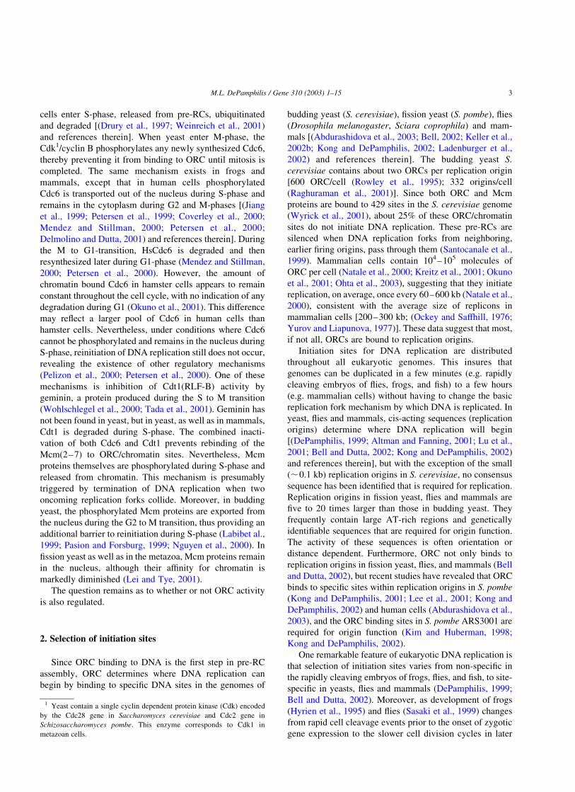
Fig. 1. Sequence of events during the initiation of DNA replication in mammalian chromatin. Upper panel: Metaphase chromatin from cultured, somatic
mammalian cells incubated in a Xenopus egg extract. Bottom panel: DNA replication in cultured, somatic mammalian cells. In general, ORC binds to the DNA
component of chromatin, but XlORC exists as a stable complex in egg extracts, whereas HsORC exists as a stable complex of Orc(2–5) to which human Orc1
and Orc6 bind only weakly. Thus, in Xenopus egg extract, the entire XlORC binds to chromatin, but in mammalian cells only the Orc1 subunit is selectively
bound during the M to G1 transition (concurrent with cyclin B degradation); the remaining ORC subunits remain stably bound to chromatin throughout the cell
cycle. Furthermore, in Xenopus, Cdc6 facilitates binding of ORC to hamster chromatin, but not to Xenopus sperm chromatin. In mammals, Cdc6 associates
with Orc1, and both Orc1 and Cdc6 are present in mitotic cells, suggesting that an Orc1/Cdc6 complex binds either to Orc(2–5) or to Orc(2–6) complexes that
are bound to chromatin. Cdt1 then brings in at least one Mcm(2–7) hexamer per replication fork. In Xenopus, ORC is released from somatic cell chromatin
following stable binding of Mcm proteins. In mammals, only Orc1 is released concurrent with DNA replication. In mammals, Orc1 is also associated with some
component of nuclear structure during G1-phase. Following pre-replication complex (pre-RC) assembly, DNA replication is initiated by the action of Mcm10.
This allows Cdk2/cyclin A to phosphorylate and release Cdc6 and for Cdc7/Dbf4 and Cdk2/cyclin E to modify one or more proteins and allow Cdc45 to bring
to the replication origin DNA polymerase-a: DNA primase, the enzyme that initiates synthesis of the first RNA-primed nascent DNA strands. This event marks
the beginning of S-phase. Concomitant with DNA synthesis is the inactivation of Cdt1 by geminin, and the phosphorylation of Mcm proteins. Cdc6 and Cdt1
are released from chromatin and eventually degraded. Mcm proteins remain in the nucleus where they are weakly associated with chromatin.
M.L. DePamphilis / Gene 310 (2003) 1–152
cells enter S-phase, released from pre-RCs, ubiquitinated
and degraded [(Drury et al., 1997; Weinreich et al., 2001)
and references therein]. When yeast enter M-phase, the
Cdk1/cyclin B phosphorylates any newly synthesized Cdc6,
thereby preventing it from binding to ORC until mitosis is
completed. The same mechanism exists in frogs and
mammals, except that in human cells phosphorylated
Cdc6 is transported out of the nucleus during S-phase and
remains in the cytoplasm during G2 and M-phases [(Jiang
et al., 1999; Petersen et al., 1999; Coverley et al., 2000;
Mendez and Stillman, 2000; Petersen et al., 2000;
Delmolino and Dutta, 2001) and references therein]. During
the M to G1-transition, HsCdc6 is degraded and then
resynthesized later during G1-phase (Mendez and Stillman,
2000; Petersen et al., 2000). However, the amount of
chromatin bound Cdc6 in hamster cells appears to remain
constant throughout the cell cycle, with no indication of any
degradation during G1 (Okuno et al., 2001). This difference
may reflect a larger pool of Cdc6 in human cells than
hamster cells. Nevertheless, under conditions where Cdc6
cannot be phosphorylated and remains in the nucleus during
S-phase, reinitiation of DNA replication still does not occur,
revealing the existence of other regulatory mechanisms
(Pelizon et al., 2000; Petersen et al., 2000). One of these
mechanisms is inhibition of Cdt1(RLF-B) activity by
geminin, a protein produced during the S to M transition
(Wohlschlegel et al., 2000; Tada et al., 2001). Geminin has
not been found in yeast, but in yeast, as well as in mammals,
Cdt1 is degraded during S-phase. The combined inacti-
vation of both Cdc6 and Cdt1 prevents rebinding of the
Mcm(2–7) to ORC/chromatin sites. Nevertheless, Mcm
proteins themselves are phosphorylated during S-phase and
released from chromatin. This mechanism is presumably
triggered by termination of DNA replication when two
oncoming replication forks collide. Moreover, in budding
yeast, the phosphorylated Mcm proteins are exported from
the nucleus during the G2 to M transition, thus providing an
additional barrier to reinitiation during S-phase (Labibet al.,
1999; Pasion and Forsburg, 1999; Nguyen et al., 2000). In
fission yeast as well as in the metazoa, Mcm proteins remain
in the nucleus, although their affinity for chromatin is
markedly diminished (Lei and Tye, 2001).
The question remains as to whether or not ORC activity
is also regulated.
2. Selection of initiation sites
Since ORC binding to DNA is the first step in pre-RC
assembly, ORC determines where DNA replication can
begin by binding to specific DNA sites in the genomes of
budding yeast (S. cerevisiae), fission yeast (S. pombe), flies
(Drosophila melanogaster, Sciara coprophila) and mam-
mals [(Abdurashidova et al., 2003; Bell, 2002; Keller et al.,
2002b; Kong and DePamphilis, 2002; Ladenburger et al.,
2002) and references therein]. The budding yeast S.
cerevisiae contains about two ORCs per replication origin
[600 ORC/cell (Rowley et al., 1995); 332 origins/cell
(Raghuraman et al., 2001)]. Since both ORC and Mcm
proteins are bound to 429 sites in the S. cerevisiae genome
(Wyrick et al., 2001), about 25% of these ORC/chromatin
sites do not initiate DNA replication. These pre-RCs are
silenced when DNA replication forks from neighboring,
earlier firing origins, pass through them (Santocanale et al.,
1999). Mammalian cells contain 104–105 molecules of
ORC per cell (Natale et al., 2000; Kreitz et al., 2001; Okuno
et al., 2001; Ohta et al., 2003), suggesting that they initiate
replication, on average, once every 60–600 kb (Natale et al.,
2000), consistent with the average size of replicons in
mammalian cells [200–300 kb; (Ockey and Saffhill, 1976;
Yurov and Liapunova, 1977)]. These data suggest that most,
if not all, ORCs are bound to replication origins.
Initiation sites for DNA replication are distributed
throughout all eukaryotic genomes. This insures that
genomes can be duplicated in a few minutes (e.g. rapidly
cleaving embryos of flies, frogs, and fish) to a few hours
(e.g. mammalian cells) without having to change the basic
replication fork mechanism by which DNA is replicated. In
yeast, flies and mammals, cis-acting sequences (replication
origins) determine where DNA replication will begin
[(DePamphilis, 1999; Altman and Fanning, 2001; Lu et al.,
2001; Bell and Dutta, 2002; Kong and DePamphilis, 2002)
and references therein], but with the exception of the small
(,0.1 kb) replication origins in S. cerevisiae, no consensus
sequence has been identified that is required for replication.
Replication origins in fission yeast, flies and mammals are
five to 20 times larger than those in budding yeast. They
frequently contain large AT-rich regions and genetically
identifiable sequences that are required for origin function.
The activity of these sequences is often orientation or
distance dependent. Furthermore, ORC not only binds to
replication origins in fission yeast, flies, and mammals (Bell
and Dutta, 2002), but recent studies have revealed that ORC
binds to specific sites within replication origins in S. pombe
(Kong and DePamphilis, 2001; Lee et al., 2001; Kong and
DePamphilis, 2002) and human cells (Abdurashidova et al.,
2003), and the ORC binding sites in S. pombe ARS3001 are
required for origin function (Kim and Huberman, 1998;
Kong and DePamphilis, 2002).
One remarkable feature of eukaryotic DNA replication is
that selection of initiation sites varies from non-specific in
the rapidly cleaving embryos of frogs, flies, and fish, to site-
specific in yeasts, flies and mammals (DePamphilis, 1999;
Bell and Dutta, 2002). Moreover, as development of frogs
(Hyrien et al., 1995) and flies (Sasaki et al., 1999) changes
from rapid cell cleavage events prior to the onset of zygotic
gene expression to the slower cell division cycles in later
1 Yeast contain a single cyclin dependent protein kinase (Cdk) encoded
by the Cdc28 gene in Saccharomyces cerevisiae and Cdc2 gene in
Schizosaccharomyces pombe. This enzyme corresponds to Cdk1 in
metazoan cells.
M.L. DePamphilis / Gene 310 (2003) 1–15 3

embryonic stages and adult animals, initiation events
changes from non-specific to site-specific. Since only one
set of Orc genes has been discovered in frogs and flies, site-
specific initiation must be developmentally acquired
epigenetically. Epigenetic changes that can affect site
specificity include the ratio of initiation factors to DNA,
chromatin or nuclear composition and structure, DNA
methylation, and the acquisition of ORC accessory proteins
that facilitate binding of ORC to specific sequences
(DePamphilis, 1999). Recent studies have suggested that
transcription factors may also facilitate binding of ORC to
specific DNA sites (Bosco et al., 2001; Beall et al., 2002;
Saitoh et al., 2002).
3. The ‘ORC cycle” in summary
Reinitiation of DNA replication at the same replication
origins during a single cell division cycle is prevented in
eukaryotic cells by regulating at least three steps in the
assembly of pre-RCs – association of Cdc6, Cdt1 and
Mcm(2–7) proteins with chromatin (see Section 1). Recent
results have revealed a fourth regulatory step in which ORC
activity is inhibited during the G1 to S-phase transition and
not reestablished until mitosis is complete and a nuclear
membrane has reassembled. Therefore, regulation of ORC
activity becomes the premier step in determining when
DNA replication begins. Cell cycle dependent changes in
ORC activity, and in some cases the affinity of one or
more ORC subunits for chromatin, is hereafter referred to
as ‘the ORC cycle’. However, the manifestation of the ORC
cycle can vary among species and during development
(summarized in Fig. 2).
The ORC cycle was first recognized in mammalian cells
(Natale et al., 2000; Kreitz et al., 2001) where the Orc1
subunit is selectively destabilized during S-phase, ubiqui-
tinated and in some cases degraded, and then rebinds stably
to chromatin during the M to G1 transition to establish pre-
RCs at specific genomic sites (see Section 4). Since all six
ORC subunits are required for ORC activity in yeast (Bell,
2002), release of Orc1 should prevent ORC function, and in
fact, mammalian metaphase chromatin lacks functional
ORCs. Cell cycle dependent loss of ORC function is even
more obvious when mammalian somatic cell chromatin is
incubated in a Xenopus laevis (Xl) egg extract (see Section
7). In this case, XlORC binds to the cell chromatin, initiates
pre-RC assembly, and the entire XlORC is then released
upon completion of pre-RC assembly. The timing of this
release, however, appears to be dependent on chromatin
structure, because when Xenopus sperm chromatin is
incubated in the same extract, the affinity of XlORC for
sperm chromatin is reduced, but ORC remains bound to the
chromatin throughout DNA replication. In this case, XlORC
appears to be released during G2/M phase as a result of
hyperphosphorylation by Cdk1/cyclin A. In fact, phos-
phorylation has been shown to play a role in regulating ORC
activity in yeast even though all six ORC subunits remain
stably bound to chromatin throughout the cell cycle (see
Section 8). Whether or not an ORC cycle also exists in other
eukaryotes, such as flies, remains to be demonstrated,
although some evidence suggests that it does (see Section
9). Release of ORC subunits from chromatin after pre-RC
assembly, either as a programmed event during DNA
replication (Sun et al., 2002), or by selective elution using
salt or cyclin-dependent protein kinase activity (Hua and
Newport, 1998; Jares and Blow, 2000), does not interfere
with assembly of active replication forks. Therefore,
regulating ORC association with chromatin is a feasible
mechanism for restricting reinitiation events during S-phase
without interfering with DNA replication. Whether or not
ORC or an ORC subcomplex binds to newly synthesized
replication origins during S-phase is unknown, but in
Xenopus egg extracts (discussed below) ORC cannot rebind
to somatic cell chromatin until the next cell cycle begins.
It should not be surprising that species-specific variations
exist in the regulation of ORC activity, because although
ORC proteins are highly conserved within a single
taxonomic family, conservation among all species is
modest. For example, human, hamster and mouse Orc1
proteins are ,81% identical and 84% similar in their total
amino acid sequence, and 93% identical and 95% similar in
their C-terminal portion (Bogan et al., 2000). The C-
terminal portion contains a homology with Cdc6 protein and
an ATP binding domain that appears to be required for site-
specific DNA binding (Bell, 2002). Among all species,
however, total amino acid sequence conservation drops to
24% identity and 35% similarity.
4. The ORC cycle in mammals
4.1. Selective release Of Orc1
In mammals, both the cellular concentrations of Orc
proteins 2–6 and the amount of each protein bound to
chromatin appear constant throughout the cell division cycle
[Orc2 (Ritzi et al., 1998; Saha et al., 1998; Mendez and
Stillman, 2000; Natale et al., 2000; Mendez et al., 2002;
Ohta et al., 2003), Orc3 (Mendez et al., 2002; Ohta et al.,
2003), Orc4 (Okuno et al., 2001; Ohta et al., 2002), Orc5
(Ohta et al., 2003), Orc6 (Dhar and Dutta, 2000; Mendez
et al., 2002). Nevertheless, there are changes in the
distribution of ORC subunits that suggest they have
activities independent of their role in DNA replication.
For example, both the cellular concentration (Dhar and
Dutta, 2000) and the amount of chromatin bound HsOrc6
(Mendez et al., 2002) remain constant throughout the cell
cycle, but some Orc6 is found at non-chromosomal sites in
mitotic cells (Prasanth et al., 2002). This appears to be due
to a fraction of Orc6 that is not associated with ORC.
Furthermore, the abundance of individual ORC subunits
relative to each other can vary among tissues, and some
M.L. DePamphilis / Gene 310 (2003) 1–154

subunits are expressed in non-proliferating tissues (Thome
et al., 2000). Orc1, however, is unique in that it appears to
regulate ORC activity in a cell cycle dependent manner
(Fig. 1, ‘Mammals’; Fig. 2, ‘Mammalian cells’).
In hamster cells, the cellular concentration of Orc1 is
constant throughout the cell cycle (Natale et al., 2000;
Okuno et al., 2001; Li and DePamphilis, 2002), but Orc1 is
selectively released from chromatin during the S and M-
phases while the remaining Orc proteins remain chromatin
bound when cells are lysed with a non-ionic detergent in the
presence of 0.1–0.15 M salt and ATP (Natale et al., 2000; Li
and DePamphilis, 2002). The same is true in human cells,
except that the cellular concentration of Orc1 oscillates
during the cell cycle, because Orc1 is degraded during S-
phase (Kreitz et al., 2001; Fujita et al., 2002; Mendez et al.,
2002; Ohta et al., 2003); J. Teer and A. Dutta, unpublished
data]. Previous reports that HsOrc1 levels did not decrease
during S-phase (Saha et al., 1998; Tatsumi et al., 2000)
appear to have resulted from differences in the specificity of
the antibodies used to detect HsOrc1 in various experiments
(A. Dutta; C. Obuse, personal communications).
In contrast to the reports described above, release of
chromatin bound Orc1 during S-phase in hamster cells was
not detected by Okuno et al. (2001) who concluded that both
the amount of chromatin-bound Orc1 and Orc4 as well as
their cellular concentrations remained constant throughout
the cell cycle. However, although it is not clear why they did
not observe a release of Orc1 during S-phase, it does appear
that the amount of Orc1 bound to chromatin in their
experiments was markedly diminished during M-phase,
suggesting that Orc1 is selectively destabilized sometime
during the cell cycle. The absence of Orc1 in their
chromatin-unbound fraction from M-phase cells (Okuno
et al., 2001) probably resulted from the fact that unbound
Orc1 is easily degraded, even when cell extracts are on ice,
whereas chromatin bound Orc1 is stable (Li and DePam-
philis, 2002). In addition, the amount of chromatin bound
Orc1 in M-phase arrested cells can vary depending on the
length of time cells are held in nocodazole. For example,
chromatin bound Orc1 was not detected in M-phase cells
that were not collected in nocodazole (Natale et al., 2000; Li
and DePamphilis, 2002), whereas cells arrested in nocoda-
zole could contain up to 20% of the amount of chromatin
bound Orc1 observed in G1-phase cells, depending on how
long the cells were held in nocodazole (Li and DePamphilis,
2002). Nocodazole blocks anaphase by preventing micro-
tubule assembly, which does not necessarily prevent other
cell cycle related events.
The selective release of Orc1 during S-phase accounts
for the fact that one or more ORC subunits are both
functionally and physically absent from metaphase chro-
matin. The absence of ORC activity is revealed by the fact
that hamster M-phase chromatin will replicate in a complete
Xenopus egg extract, but not in one that has been depleted of
Xenopus Orc proteins (Yu et al., 1998; Li et al., 2000; Natale
et al., 2000). In contrast, nuclei isolated in the same manner
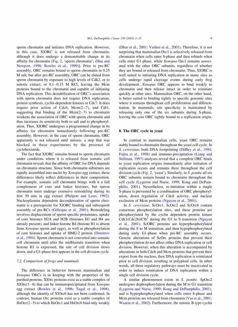
Fig. 2. Four manifestations of the ORC cycle in eukaryotes. Yeast cells: ORC (six gray cylinders) remains bound to replication origins throughout the cell
cycle, but ORC is phosphorylated (-P) during the S to M periods, and this phosphorylation inhibits its ability to assemble a pre-RC. Xenopus egg extract: ORC
binds to sperm chromatin, but the stability of ORC/chromatin sites is reduced (red boxes) following pre-RC assembly. ORC is phosphorylated by Cdk1/cyclin
A (yellow ball) during G2/M and released from chromatin. If somatic cell chromatin instead of sperm chromatin is incubated in the extract, then ORC is
released from chromatin following pre-RC assembly. Mammalian cells: The Orc1 subunit is selectively destabilized (red box) and released from chromatin
when cells enter S-phase. Orc1 is then mono-ubiquitinated (Ub) and in some cases polyubiquitinated ([Ub]n) and degraded. Orc1 in mitotic cells appears
hyperphosphorylated. Thus, in yeast, frogs and mammals, stable ORC/chromatin sites that can initiate assembly of a pre-RC are not present until mitosis is
complete and a nuclear membrane is present.
M.L. DePamphilis / Gene 310 (2003) 1–15 5
from G1-phase cells will initiate DNA replication de novo
under both conditions, revealing that hamster pre-RCs are
assembled during the M to G1 transition. Loss of ORC
activity can be accounted for by the fact that Orc1 protein is
not associated with metaphase chromatin under conditions
where other Orc proteins remain stably bound (Natale et al.,
2000; Tatsumi et al., 2000; Okuno et al., 2001; Mendez
et al., 2002) and mammalian Cdc6 protein is abundant
[(Mendez and Stillman, 2000; Petersen et al., 2000). This
observation is consistent with the cell cycle-dependent
changes observed in a footprint at the human lamin B2
origin (Abdurashidova et al., 1998) that encompasses the
start sites for leading strand DNA replication (Abdurashi-
dova et al., 2000). A large G1-phase footprint changed into a
smaller S and G2 phase footprint that disappeared during M-
phase. Recent UV-induced cross-linking experiments reveal
that the large G1-phase footprint could be accounted for by
the presence of a pre-RC containing at least Orc1, Orc2,
Cdc6, and Mcm3 proteins (Abdurashidova et al., 2003).
During S-phase, only the Orc2 protein can be cross-linked to
chromatin, consistent with the disappearance of the pre-RC
and the release of Orc1. By M-phase, not even Orc2 could
be cross-linked to DNA, suggesting additional changes
occur in the affinity of ORC for chromatin. Similarly, both
Orc1 and Orc2 can be cross-linked to specific replication
origins in vivo by treating human G1-phase cells with
formaldehyde, but only Orc2 can be cross-linked during S-
phase (Ladenburger et al., 2002). These observations in
mammalian cells are consistent with earlier reports that both
Orc1 and Orc2 are present on chromatin in Xenopus
interphase cells but not in metaphase cells (Romanowski
et al., 1996). Moreover, Orc proteins in Xenopus eggs
competent for DNA replication will bind to sperm
chromatin, whereas Orc proteins in Xenopus eggs in
metaphase II will not (Coleman et al., 1996; Hua and
Newport, 1998; Findeisen et al., 1999; Rowles et al., 1999).
Whether Orc1 is associated with chromatin in vivo in an
unstable state or is actually released from chromatin in vivo
as well as in cell extracts is difficult to assess. What is clear
is that Orc1 is not stably bound to chromatin in comparison
with other Orc proteins, and that metaphase cells do not
contain functional ORCs. M-phase cells permeabilized with
digitonin retain Orc1 in the chromatin pellet (Natale et al.,
2000), but this M-phase chromatin does not initiate DNA
replication in an ORC-depleted Xenopus egg extract (Yu
et al., 1998; Li et al., 2000; Natale et al., 2000). Expression
of a GFP-tagged Orc1 protein in hamster cells resulted in
GFP-labeled metaphase chromosomes (Okuno et al., 2001),
but there is no evidence that the GFP-Orc1 protein is
assembled into ORCs and if so, that the GFP-ORCs are
active.
4.2. Ubiquitination Of Orc1
Orc1 in S-phase mammalian cells is selectively ubiqui-
tinated and, under some conditions, degraded. In hamster
cells, two forms of Orc1 are released concurrently during
S-phase, one that is not ubiquitinated and one that is
mono-ubiquitinated (Li and DePamphilis, 2002). Orc1 and
mono-ubiquitinated Orc1 also were detected in the non-
chromatin bound fraction from human S-phase cells
(Mendez et al., 2002). However, in human cells, Orc1
became polyubiquitinated and was degraded via the 26S
proteasome pathway (Fujita et al., 2002; Mendez et al.,
2002), a reaction mediated by the SCFskp2 ubiquitin-ligase
complex (Mendez et al., 2002). In contrast, only trace
amounts of polyubiquitinated Orc1 were detected in
hamster cells. Hamster Orc1 was polyubiquitinated and
degraded only when it was released from nuclei into cellular
extracts. Thus, the chromatin unbound fraction of Orc1 in
non-ionic detergent extracts of hamster mitotic cells can be
lost, even when cell lysates are held in ice (Okuno et al.,
2001; Li and DePamphilis, 2002).
In contrast to Orc1, Orc2 not only remains bound to
chromatin throughout the cell cycle, but Orc2 is not a
substrate for ubiquitination, even as a soluble protein (Li
and DePamphilis, 2002). Interestingly, the half-life of Orc1
in both hamster and human exponentially proliferating cells
is ,3 h, the same as observed for Orc2 [(Li and
DePamphilis, 2002); J. Mendez, personal communication].
Therefore, it may not be necessary to deubiquitinate Ub-
Orc1 in order to reestablish functional ORC/chromatin sites
in early G1-phase, because in both hamster and human cells,
most of the Orc1 in M-phase cells will have been
synthesized during the previous 6 h.
5. Orc1 and the assembly of pre-replication complexes
Several observations suggest that the stable binding of
Orc1 to chromatin is the premier regulatory step in the
assembly of pre-RCs and thereby the commitment to cell
proliferation. First, both HsOrc1 (but not HsOrc2) and
DmOrc1 expression are regulated by E2F (Ohtani et al.,
1996; Asano and Wharton, 1999), a transcription factor that
plays an important role in driving cells from G1 into S-
phase. E2F also regulates expression of Cdc6 and Mcm
genes (Leone et al., 1998; Ohtani et al., 1998; Yan et al.,
1998), suggesting that assembly of pre-RCs depends on the
E2F/Rb pathway with ORC activity regulated by expression
of Orc1. Second, the Orc1 gene is the only ORC gene whose
expression is correlated with cell proliferation: Orc1 is
expressed only in proliferating mammalian cells whereas
Orc2–Orc5 are expressed to varying extents in many non-
proliferating cells (Thome et al., 2000). Third, stable
binding of Orc1 to chromatin immediately precedes
assembly of pre-RCs at specific chromosomal loci.
During the M to G1 transition, Orc1 rebinds stably to
chromatin with the concomitant appearance of functional
ORCs. M-phase hamster cells contain as much Orc1 as G1-
phase cells, but most, if not all, of the Orc1 in M-phase
hamster cells is not bound to chromatin and only a minor
M.L. DePamphilis / Gene 310 (2003) 1–156

fraction is ubiquitinated (Li and DePamphilis, 2002). M-
phase human cells generally contain much less Orc1 than
G1-phase human cells, as a result of Orc1 degradation
during S-phase (Mendez et al., 2002). Therefore, human
Orc1 must be resynthesized during the M to G1 transition,
whereas hamster Orc1 does not (Okuno et al., 2001). ORC
activity is restored during the first 2 h after either hamster or
human M-phase cells (arrested in nocodazole) are released
into G1-phase (Yu et al., 1998; Natale et al., 2000). This
corresponds to the time period (1–3 h post-M) when Orc1
(Natale et al., 2000; Li and DePamphilis, 2002; Mendez
et al., 2002; Ohta et al., 2003), Mcm2 and Mcm3 reassociate
stably with chromatin (Dimitrova et al., 1999; Natale et al.,
2000; Dimitrova et al., 2002; Ohta et al., 2003). Nuclei
isolated from these early G1-cells can initiate de novo DNA
replication in a Xenopus egg extract depleted either of ORC
or of Mcm proteins. The appearance of chromatin bound
Orc1 is inversely related to the lost of cyclin B (Mendez and
Stillman, 2000), which is required to drive cells into mitosis,
and directly linked to formation of an intact nuclear
envelope (Dimitrova and Gilbert, 1998; Natale et al.,
2000), which is required for initiation of DNA replication.
Thus, the bulk of Orc1 and Mcm binding occurs after
mitosis is complete and a nuclear membrane has reformed
around the genome, a time referred to either as late
telophase or early G1-phase. Consistent with this view is
the fact that HsCdc6 protein, like cyclin B, is degraded
during the M to G1 transition and then resynthesized during
G1 (Petersen et al., 2000; Mendez et al., 2002), just in time
to be incorporated into the newly reconstituted active ORC/
chromatin sites. Therefore, stable rebinding of Orc1 to
chromatin appears to be the rate-limiting step for assembly
of pre-RCs.
One experiment, however, suggests that pre-RC assem-
bly is completed about 2 h before the stable binding of
hamster Orc1, Mcm2 and Mcm3 is completed. Geminin, a
specific inhibitor of Cdt1, prevents stable binding of
Mcm(2–7) to chromatin. Once pre-RCs are assembled,
DNA replication is no longer sensitive to geminin. DNA
replication in hamster metaphase chromatin incubated in
Xenopus egg extract is sensitive to geminin, but DNA
replication in chromatin isolated 1 h after metaphase is not
(Okuno et al., 2001; Dimitrova et al., 2002). In these
experiments, Orc1 appears to bind to chromatin within the
1st h after release of cells from the nocodazole block,
consistent with the execution point for geminin. Interest-
ingly, Mcm proteins continued to bind to chromatin until
mid to late G1-phase (Dimitrova et al., 1999; Natale et al.,
2000; Dimitrova et al., 2002), suggesting that more Mcm
proteins bind to chromatin than are actually assembled into
pre-RCs. Apparent differences in the published time courses
for protein binding and the geminin execution point likely
reflect differences in experimental protocols that affect the
amount of protein recovered in different fractions and the
time required for cells to progress from M to G1 when the
synchronizing agent, nocodazole, is removed.
6. ORC and the ‘origin decision point’
Xenopus egg extract can initiate DNA replication de
novo in virtually any DNA, chromatin or nuclei provided,
but these initiation events are generally distributed non-
specifically throughout the DNA substrate. The only
exception is when nuclei are isolated from mammalian
cells under conditions that preserve their impermeability to
macromolecules (Gilbert et al., 1995). Under these con-
ditions, initiation events are distributed non-specifically
throughout the genome when ‘nuclei’ are isolated from M-
phase or early G1-phase cells, but at specific sites when
nuclei are taken from late G1-phase cells (Wu and Gilbert,
1996; Wu and Gilbert, 1997; Li et al., 2000; Okuno et al.,
2001). DNA replication in late G1-nuclei is independent of
XlORC proteins, begins in vitro at the same site-specific
origins of bi-directional DNA replication used by the cells
in vivo, and is sensitive to protein kinase inhibitors.
Therefore, it appears that pre-RCs in late G1-phase nuclei
are present at specific genomic sites waiting to be activated
by Cdk2/cyclin A, E and Cdc7/Dbf4 (Fig. 1). This cell cycle
dependent transition that occurs in mammalian chromatin,
from a substrate for non-specific initiation events to a
substrate for site-specific initiation events, is referred to as
the ‘origin decision point’ (ODP).
Two views of the ODP have emerged. In one view, the
ODP marks the transition from Xenopus ORC directed
initiation events to mammalian ORC directed initiation
events. Prior to the ODP in hamster cells (midpoint ,2 h
post-M), initiation of DNA replication in hamster chromatin
requires the presence of Xenopus ORC in the egg extract
(Natale et al., 2000). XlORC present in these experiments
rapidly binds to hamster metaphase chromatin within the
first few minutes of incubation (Sun et al., 2002) and is
responsible for most of the DNA replication that occurs
during the first 1–2 h post-M (Yu et al., 1998; Natale et al.,
2000) where it initiates replication non-specifically through-
out the genome (Yu et al., 1998; Dimitrova and Gilbert,
1999; Li et al., 2000; Natale et al., 2000). After the ODP in
hamster cells, Xenopus egg extract simply activates hamster
pre-RCs that were assembled in vivo. Initiation sites are
established in hamster cells upon the stable binding of Orc1
(midpoint ,1.5 h post-M) to Orc2–6/chromatin complexes
located at specific sites along the genome (Natale et al.,
2000; Li and DePamphilis, 2002). This view is supported by
the studies in human cells where Orc1 binding to chromatin
follows a similar time course to that in hamster cells
(Mendez et al., 2002) and where cross-linking studies have
revealed that Orc2 is bound to specific genomic sites, some
of which correspond to origins of bi-directional DNA
replication (Abdurashidova et al., 2003; Keller et al., 2002b;
Ladenburger et al., 2002). This view also accounts for the
observation that the normal temporal order for initiation is
absent in chromatin isolated from cells in M-phase to 1 h
post-M phase but present in chromatin isolated from late
G1-phase cells (Dimitrova and Gilbert, 1999; Li et al.,
M.L. DePamphilis / Gene 310 (2003) 1–15 7

2001). XlORC initiates replication non-specifically in
chromatin from M-phase and telophase cells.
An alternative view of the ODP is that pre-RCs are
initially assembled ‘randomly’ along the genome during the
M to G1-transition in mammalian cells, and then reorgan-
ized, by some as yet unidentified mechanism, into specific
sites [(Okuno et al., 2001) and references therein]. One
suggested mechanism is that some pre-RCs are inactivated
at the ODP, leaving those pre-RCs at specific genome sites
to initiate replication at the start of S-phase. However, this
implies the existence of a larger number of ORC/chromatin
sites in mammalian cells that the cellular concentration of
ORC would permit. This view of the ODP is based on the
conclusion that all six mammalian ORC subunits are stably
bound to chromatin throughout the cell cycle, that pre-RCs
are assembled in hamster cells within 1 h after metaphase,
and that pre-RC assembly precedes the ODP in hamster
cells (midpoint ,3 h post-M) by 2–2.5 h. Various
experimental artifacts that are innumerated in the papers
cited above may well explain some of the differences
between these two views. Nevertheless, the possibility
remains that there exists a novel, as yet undiscovered,
mechanism for determining where initiation events occur in
mammalian genomes.
6.1. Association of Orc1 with nuclear structure
The fact that assembly of pre-RCs is delayed until
mitosis is complete and a nuclear membrane is assembled
implies that nuclear structure plays a role in the initiation of
DNA replication. The nucleus appears to regulate the
concentration of proteins required to initiate DNA replica-
tion (Walter et al., 1998), to facilitate the assembly or
activity of DNA replication forks, and to determine where in
the genome initiation of DNA replication occurs (reviewed
in DePamphilis, 2000; Keller et al., 2002a). Replication
origins have been reported to be associated with ‘nuclear
matrix’ preparations (Djeliova et al., 2001), although not
with ‘nucleoskeleton’ preparations (Ortega and DePamphi-
lis, 1998), revealing that interactions between replication
origins and nuclear structure are sensitive to experimental
conditions.
Recent observations reveal that Orc1 (and to a lesser
extent the other ORC subunits) is associated with a fraction
of chromatin that is not solubilized by digestion with
endonucleases (Kreitz et al., 2001; Fujita et al., 2002), and
that this fraction accumulates during G1 phase and then
decreases during S-phase (Ohta et al., 2003). In HeLa cells,
the appearance of Orc1 following release of cells from a
metaphase block occurs concurrently with the appearance of
Orc1 and other ORC proteins in an insoluble nuclease-
resistant fraction, suggesting that newly assembled ORC/
chromatin complexes are tethered to some component of
nuclear structure and then released as the Orc1 subunit is
destabilized, ubiquitinated, and in the case of human cells,
degraded (Fig. 1, ‘Mammals’). Mcm proteins, which appear
to bind to chromatin flanking the ORC binding site, appear
in the soluble nuclease-sensitive fraction. This transition of
ORC/chromatin complexes from untethered to tethered may
contribute to the phenomenon referred to as the ‘origin
decision point’.
7. The ORC cycle in frogs
Metaphase chromatin lacks functional ORC complexes,
and therefore when metaphase chromatin from hamster cells
is incubated in a Xenopus egg extract, XlORC rapidly binds
to the chromatin and initiates DNA replication. However, in
contrast to mammalian ORC, where only the Orc1 subunit is
destabilized upon DNA replication, the entire XlORC is
released from the somatic cell chromatin into the replication
extract (50 mM KCl) as soon as pre-RCs have been
assembled [Fig. 1, ‘Xenopus’; Fig. 2, ‘somatic cell
chromatin’; (Sun et al., 2002)]. This programmed release
of XlORC was insensitive to aphidicolin (specific inhibitor
of replicative DNA polymerases) and olomoucine (specific
inhibitor of cyclin dependent protein kinases), but sensitive
to geminin (specific inhibitor of Cdt1), and prevented by
immuno-depletion of XlMcm proteins from the egg extract.
Therefore, assembly of pre-RCs triggers release of ORC
from cell chromatin. Alternatively, ORC release may be
triggered by some event that occurs between completion of
pre-RC assembly and the action of Cdk2. For example,
Mcm10 binds to chromatin after pre-RC assembly and
before Cdk2 activity where it is required to initiate DNA
replication (Wohlschlegel et al., 2002).
Once pre-RCs were assembled in egg extract, XlORC
was no longer present on somatic cell chromatin, despite the
fact that egg extract contained a great excess of soluble
XlORC. Furthermore, XlORC could not bind to chromatin
in nuclei isolated from hamster cells that had progressed
past their ‘origin decision point’ (Sun et al., 2002). Cdk/
cyclins were not involved in these events, since neither
binding nor release of XlORC to somatic cell chromatin was
affected by olomoucine. Moreover, XlORC in meiotic eggs
(metaphase II) does not bind to sperm chromatin (Coleman
et al., 1996; Hua and Newport, 1998; Findeisen et al., 1999;
Rowles et al., 1999), and XlORC is not bound to metaphase
chromatin in Xenopus somatic cells (Romanowski et al.,
1996). This inability of XlORC in cells undergoing meiosis
or mitosis to bind to chromatin likely reflects the fact that
XlORC is hyperphosphorylated in these cells where it
associates with Cdk1/cyclin A (Romanowski et al., 2000).
These data strongly suggest that XlORC cannot rebind to
either somatic cell chromatin or sperm chromatin until cell
division is completed.
7.1. Chromatin structure and the affinity of ORC for DNA
Xenopus sperm chromatin, like mammalian metaphase
chromatin, lacks ORC proteins, and XlORC rapidly binds to
M.L. DePamphilis / Gene 310 (2003) 1–158
sperm chromatin and initiates DNA replication. However,
in this case, XlORC is not released from chromatin,
although it does undergo a demonstrable change in its
affinity for chromatin [Fig. 2, ‘sperm chromatin’; (Hua and
Newport, 1998; Rowles et al., 1999)]. Prior to pre-RC
assembly, ORC remains bound to sperm chromatin in 0.25
M salt, but after pre-RC assembly, ORC can be eluted from
sperm chromatin by exposure to high levels of Cdk2, or to
mitotic extract, or 0.1–0.15 M KCl, leaving the Mcm
proteins bound to the chromatin and capable of initiating
DNA replication. This destabilization of ORC’s association
with sperm chromatin does not require DNA replication,
protein synthesis, cyclin-dependent kinases or Cdc7. It does
require prior action of Cdc6, Mcm(2–7), and Cdt1,
suggesting that binding of the Mcm(2–7) to chromatin
weakens the association of ORC with sperm chromatin and
thus increases its sensitivity both to salt and to phosphoryl-
ation. Thus, XlORC undergoes a programmed change in its
affinity for chromatin immediately following pre-RC
assembly. However, in the case of sperm chromatin, ORC
apparently is not released until mitosis, a step that was
blocked in these experiments by the presence of
cycloheximide.
The fact that XlORC remains bound to sperm chromatin
under conditions where it is released from somatic cell
chromatin reveals that the affinity of ORC for DNA depends
on chromatin structure. Since both chromatin substrates are
rapidly assembled into nuclei by Xenopus egg extract, these
differences likely reflect differences in their composition.
For example, somatic cell chromatin brings with it a full
complement of core and linker histones, but sperm
chromatin must undergo extensive remodeling during its
first 30 min in egg extract (Philpott and Leno, 1992).
Nucleoplasmin dependent decondensation of sperm chro-
matin is a prerequisite for XlORC binding and subsequent
assembly of pre-RCs (Gillespie et al., 2001). Remodeling
involves displacement of sperm specific protamines, uptake
of core histones H2A and H2B (histones H3 and H4 are
already present), and linker histone B4 (histone H1 is absent
from Xenopus sperm and eggs), as well as phosphorylation
of core histones and uptake of HMG-2 protein (Dimitrov
et al., 1994). Sperm chromatin is not converted into somatic
cell chromatin until after the midblastula transition when
histone H1 is expressed, the rate of cell division slows
down, and a G1-phase first appears in the cell division cycle.
7.2. Comparison of frogs and mammals
The difference in behavior between mammalian and
Xenopus ORCs is in keeping with the properties of the
purified proteins. XlOrc proteins exist as a stable complex of
XlOrc(1–6) that can be immunoprecipitated from Xenopus
egg extract (Rowles et al., 1996; Tugal et al., 1998),
although the identity of XlOrc6 has not been confirmed. In
contrast, human Orc proteins exist as a stable complex of
HsOrc(2–5) to which HsOrc1 and HsOrc6 bind only weakly
(Dhar et al., 2001; Vashee et al., 2001). Therefore, it is not
surprising that mammalian Orc1 is selectively released from
chromatin when cells enter S-phase and then rebinds when
cells enter G1-phase, while Xenopus Orc1 remains associ-
ated with the other ORC subunits, regardless of whether
they are bound or released from chromatin. Thus, XlORC is
well suited to initiating DNA replication at many sites as
cells undergo rapid cleavage events during early frog
development. Xenopus ORC appears to bind weakly to
chromatin and then release intact in order to reinitiate
quickly at other sites. Mammalian ORC, on the other hand,
is better suited to binding tightly to specific genomic sites
where it remains throughout cell proliferation and differen-
tiation. In mammals, site specificity is maintained by
releasing only one of the six subunits during S-phase,
leaving the core ORC tightly bound to a replication origin.
8. The ORC cycle in yeast
In contrast to mammalian cells, yeast ORC remains
stably bound to chromatin throughout the yeast cell cycle. In
S. cerevisiae, both DNA footprinting (Diffley et al., 1994;
Fujita et al., 1998) and immuno-precipitation (Liang and
Stillman, 1997) analyses reveal that a complete ORC binds
to yeast replication origins immediately after initiation of
replication occurs and remains there throughout the cell
division cycle (Fig. 2, ‘yeast’). Similarly, in S. pombe all six
ORC subunits remain bound to chromatin throughout the
cell cycle (Lygerou and Nurse, 1999; Kong and DePam-
philis, 2001). Nevertheless, re-initiation within a single
S-phase is prevented by a combination of ORC phosphoryl-
ation, down regulation of Cdc6 activity, and nuclear
exclusion of Mcm proteins (Nguyen et al., 2001).
In S. cerevisiae, ScOrc1, ScOrc2 and ScOrc6 contain
consensus phosphorylation sites, and these proteins are
phosphorylated by the cyclin dependent protein kinase
Cdk1(Cdc28)/Clb1 during the G1 to S transition (Nguyen
et al., 2001). ScORC proteins are hyperphosphorylated
during the S to M transition, and then hypophosphorylated
during early G1-phase when pre-RC assembly occurs.
Genetic alterations of ScOrc proteins that prevent their
phosphorylation do not affect either DNA replication or cell
division. However, when this alteration is accompanied by
alterations in both Cdc6 and Mcm proteins that prevent their
export from the nucleus, then DNA replication is reinitiated
prior to cell division, resulting in polyploid cells. In other
words, all three regulatory pathways must be inactivated in
order to induce reinitiation of DNA replication within a
single cell division cycle.
A similar phenomenon exists in S. pombe. SpOrc2
undergoes dephosphorylation during the M to G1 transition
(Lygerou and Nurse, 1999; Kong and DePamphilis, 2001),
and is hyperphosphorylated when cells enter S-phase and
Mcm proteins are released from chromatin (Vas et al., 2001;
Wuarin et al., 2002). Furthermore, the mitotic B type cyclin
M.L. DePamphilis / Gene 310 (2003) 1–15 9
Cdc13 complexed with the Cdk1(Cdc2)1 protein kinase
associates with replication origins during S-phase and
remains there during G2 and early M-phases (Wuarin
et al., 2002). This association is ORC-dependent, apparently
by association of Cdk1(Cdc2) with the Orc2 subunit, and
prevents reinitiation of DNA replication before mitosis has
been completed. These data strongly suggest that the
phosphorylated state of SpOrc2 determines ORC activity.
8.1. Comparison of yeast and frogs
The ORC cycle in yeast resembles that in Xenopus egg
extract when sperm chromatin in the substrate (Fig. 2). ORC
proteins in both species are hyperphosphorylated during
metaphase by Cdk1, and phosphorylation inhibits its ability
to initiate pre-RC assembly. Phosphorylated XlORC binds
poorly to chromatin (Romanowski et al., 2000), while pre-
RC assembly is inhibited by Cdk1 dependent phosphoryl-
ation of ScORC (Nguyen et al., 2001) or by association of
SpORC with Cdk1 (Wuarin et al., 2002). Whether or not
phosphorylation of XlORC subunits, like those in yeast, also
occurs during DNA replication is not clear. Whether or not
yeast ORC, like XlORC, can be selectively eluted at lower
salt concentrations following pre-RC assembly or DNA
replication also is not clear. Dephosphorylation of the Orc2
subunit occurs during the M to G1 transition in Xenopus
(Sun et al., 2002) and in yeast (Nguyen et al., 2001; Vas
et al., 2001), presumably as an activation step in pre-RC
assembly. Phosphorylation of ORC may alter its affinity
either for chromatin or for other pre-RC proteins during the
G1 to S transition, while chromatin composition may
determine whether the modified ORC subunits are remain
bound or are released. Interestingly, yeast chromatin, like
Xenopus sperm chromatin, lacks a classical histone H1
linker (Landsman, 1996), suggesting that reduced chromatin
condensation may facilitate binding of ORC during S-phase.
9. The ORC cycle in flies
Direct analyses of cell cycle dependent changes in the
activity, post-translational modifications or chromatin
affinity of DmORC are not yet available. However, several
observations are consistent with a cell cycle dependent,
differential association of DmOrc proteins with chromatin.
In Drosophila, Orc2 is distributed fairly homogeneously in
interphase nuclei, while it only remains bound to the
heterochromatic region of chromosomes through mitosis in
embryos and larval neuroblasts (Pak et al., 1997; Loupart
et al., 2000). Mutations in Orc2 exhibited delayed entry into
S-phase as well as delayed exit from metaphase with some
euchromatic regions replicating even later than heterochro-
matin (Loupart et al., 2000). In addition, mitotic chromo-
somes were irregularly condensed, suggesting a novel role
for ORC in chromosome architecture as well as DNA
replication. In contrast to Orc2, Orc1 levels change
dramatically throughout Drosophila development, and its
accumulation is regulated by E2F-dependent transcription
(Asano and Wharton, 1999). In embryos, Orc1 accumulates
preferentially in proliferating cells, and in the eye imaginal
disc, Orc1 accumulation is cell cycle regulated, with high
levels in late G1 and S phase. Moreover, overexpression of
Orc1 altered the pattern of DNA synthesis, implicating Orc1
in regulating initiation of DNA replication.
The DNA binding activity of purified DmORC is largely
non-specific and ATP-independent, although DmORC, like
ScORC, requires only the ORC1 component of the complex
to bind ATP for tight DNA interactions (Chesnokov et al.,
2001). In vivo, DmORC does bind to specific genomic sites
(Austin et al., 1999; Bielinsky et al., 2001). Such site-
specific DNA binding may require association with other
proteins (Bosco et al., 2001; Beall et al., 2002), and these
associations may regulate ORC activity as well.
10. Mechanisms that regulate ORC activity
The results described above reveal at least five
mechanisms by which ORC activity is regulated during
cell proliferation.
10.1. Ubiquitination
Since both Orc1 and Ub-Orc1 are rapidly released from
chromatin when hamster cells enter S-phase and Ub-Orc1 is
largely absent from mitotic cells, ubiquitination per se is not
likely the cause of Orc1 release, but a mechanism to prevent
its reassociation with ORC/chromatin sites during S-phase.
Two phenomena suggest that Ub-Orc1 may become
sequestered in the nuclear membrane. First, several
examples have been reported where monoubiquitination is
required for endosomal sorting of proteins into membrane
vesicles (Hicke, 2001; Haglund et al., 2002). Second, a
protein called SUMO that is very similar in structure to
ubiquitin conjugates proteins with a single adduct and this
modification generally results in the protein’s relocation to
the nucleus (Wilson and Rangasamy, 2001). Some cells,
such as HeLa and other cancer cells, may contain larger
pools of Orc1 that cause it to be exported to the cytoplasm
where it is polyubiquitinated and degraded. In fact,
differences in pool sizes could account for the observation
that Cdc6 is exported from the nucleus and degraded in
human cells but not in hamster cells (Okuno et al., 2001).
10.2. Phosphorylation
Phosphorylation of Orc proteins can affect both their
affinity for chromatin and their activity. In yeast, Cdk1/cy-
clin B prevents pre-RC assembly until mitosis has been
completed by phosphorylating one or more of the ORC
subunits and Cdc6. In frogs, both Orc1 and Orc2 are
hyperphosphorylated in metaphase-arrested eggs relative to
M.L. DePamphilis / Gene 310 (2003) 1–1510

activated eggs (Carpenter and Dunphy, 1998; Tugal et al.,
1998). Moreover, XlORC can be selectively released from
chromatin by incubating chromatin either in a metaphase
extract (Rowles et al., 1999) or with Cdk1(Cdc2)/cyclin A
(Hua and Newport, 1998; Findeisen et al., 1999). In
mammals, hyperphosphorylation of Orc1 has been detected
in metaphase cells (Tatsumi et al., 2000; Thome et al.,
2000), and over-expression of cyclin A results in export of
Orc1 to the cytoplasm, a process that is dependent on the
phosphorylation status of Cdk target sites in Orc1 (Laman
et al., 2001). Given the fact that Cdk2/cyclin A interacts
with HsOrc1 (Mendez et al., 2002) and Cdk1/cyclin A
interacts with XlORC (Romanowski et al., 2000), cyclin
dependent phosphorylation may be the mechanism that
releases ORC proteins from chromatin during the G1 to S
transition, and the released protein then may be exported to
the cytoplasm where it undergoes polyubiquitination and
degradation.
10.3. DNA replication
DNA replication appears to be required to destabilize
Orc1 in mammalian cells, because significant amounts Orc1
are not released from chromatin in cells arrested at their
G1/S interphase (Li and DePamphilis, 2002). Therefore,
Orc1 release in mammals may be triggered by DNA
synthesis through the origin region. This hypothesis is
supported by data from yeast and flies.
Orc1, Orc4, and Orc5 from yeast and flies can each bind
ATP, and the same ORC subunits in all other eukaryotes
contain potential ATP binding sites [reviewed in (Bell,
2002)]. Moreover, the affinity of both DmORC (Chesnokov
et al., 2001) and ScORC (Klemm et al., 1997) for DNA
increases by binding of ATP to the Orc1 subunit. ATP
binding to ScOrc1 is required for site-specific binding of
ScORC to origin DNA. The rate of ATP hydrolysis is then
reduced in response to origin binding, suggesting that ATP
is not hydrolyzed until a subsequent step in replication. That
step appears to be the generation of single stranded DNA at
replication origins. ScORC binds to ssDNA, but its affinity
for ssDNA is dependent only on ssDNA length, not on either
DNA sequence or ATP binding. In fact, association of
ScORC with ssDNA stimulates ATP hydrolysis, and
stabilizes an altered ORC structure (Lee et al., 2000). In
yeast, this altered ORC structure may be locked in place by
phosphorylation of ORC subunits and thereby prevent
subsequent pre-RC assembly. In mammals, this altered
ORC structure may cause selective release of Orc1.
10.4. Pre-replication complex assembly
In Xenopus egg extract, assembly of pre-RCs triggers a
change in the affinity of ORC for chromatin that is
manifested differently, depending on the chromatin sub-
strate. In the case of somatic cell chromatin, the entire ORC
is released under DNA replication conditions. In the case of
sperm chromatin, the entire ORC becomes salt-sensitive.
10.5. Auxiliary protein interactions
Interactions between ORC and other proteins can
facilitate pre-RC assembly (Fig. 1). For example, HsOrc1
binds Cdc6 (Saha et al., 1998), and Cdc6 facilitates binding
of XlORC to somatic cell chromatin (Sun et al., 2002) and
ScORC to its cognate replication origin (Mizushima et al.,
2000). Moreover, ScCdc6 specifically recognizes the ATP-
bound state of ScOrc1 (Klemm and Bell, 2001). Therefore,
Orc1 may recruit Cdc6, which is present during M-phase,
and this Orc1/Cdc6 complex may then target specifically
one of the many Orc2–6 complexes that are bound to
various sites along the genome (the ‘Jesuit Model’ for site
selection, (DePamphilis, 1993, 1996, 1999).
Interactions between ORC and other proteins can also
prevent pre-RC assembly. For example, Skp2, a component
of the mammalian ubiquitination system, binds to Orc1 and
is required for Orc1 ubiquitination during S-phase (Mendez
et al., 2002). Association of ORC/chromatin sites with
Cdk1/cyclin B during mitosis in S. pombe prevents
assembly of pre-RCs (Wuarin et al., 2002). Sic1, a specific
inhibitor of Cdks, prevents the S-phase-specific Cdk1/Clb5
protein kinase in S. cerevisiae from interacting with ORC,
but does not prevent the G1–phase specific Cdk1/Cln2
kinase from binding to ORC (Weinreich et al., 2001).
Therefore, Sic1 can prevent Cdk1 activation of pre-RCs
during the G1 to S-phase transition, without also preventing
subsequent inactivation of pre-RC assembly by the same
protein kinase during the S to M-phase transition.
11. Regulating initiation of DNA replication by
regulating ORC activity
One can envisage at least four advantages to regulating
ORC activity. The first is to prevent assembly of pre-RCs
during S-phase by inactivating the premier step in their
assembly. By applying this strategy to newly assembled
ORC/chromatin sites as well as to those sites where
initiation has already occurred, initiation of DNA replica-
tion is restricted to once-per-origin-per-cell division cycle.
Moreover, the assembly of pre-RCs cannot begin again until
mitosis is complete and a nuclear membrane has been
reassembled. To this end, mammals have taken the more
sophisticated approach. Since Cdc6 binds specifically to
Orc1, selective inactivation of Orc1 would directly prevent
recruitment of Cdc6 to ORC/chromatin sites without
disturbing the genomic locations of the remaining chroma-
tin bound Orc proteins.
A second advantage is to provide the metazoa with a
mechanism for selecting which of the many potential
initiation sites along the genome will be activated [‘Jesuit
Model’, (DePamphilis, 1993, 1996)]. A core complex
M.L. DePamphilis / Gene 310 (2003) 1–15 11

consisting of Orc(2–5) or Orc(2–6) could be assembled at
many potential initiation sites, but which of these sites is
activated during each round of cell proliferation would be
determined by the binding either of Orc1 or of an Orc1/Cdc6
complex. This would help to resolve the problem of
preventing DNA replication from interfering with DNA
transcription as the pattern of gene expression and
chromatin organization changes during animal develop-
ment. The ORC cycle would allow some replication origins
to be inactivated while activating others. A dramatic
example of this is the transition from non-specific selection
of initiation sites in embryos undergoing rapid cell
cleavages to site-specific initiation in cells of the same
animal undergoing a normal mitotic cell cycle (Hyrien et al.,
1995; Sasaki et al., 1999).
Third, the ORC cycle offers an excellent opportunity for
checkpoint control mechanisms. For example, cells that
have sustained DNA damage, or that have entered a
quiescent or terminally differentiated state could prevent
activation of the pathway leading to S-phase by inactivating
one or more ORC subunits. Human cells treated with
adriamycin, a DNA damage-inducing agent, do, in fact,
selectively degrade Orc1 (Mendez et al., 2002).
Finally, the existence of multiple mechanisms for
regulating initiation of DNA replication serves to prevent
terminally differentiated cells from reentering their cell
proliferation cycle. Cells entering a quiescent state have
been shown to lose their ability to establish pre-replication
complexes at specific genomic sites (Wu and Gilbert, 1997)
and their ability to bind Cdc6 and Mcm proteins to their
chromatin (Madine et al., 2000; Sun et al., 2001). Moreover,
terminally differentiated cells lack critical proteins required
for assembly of pre-replication complexes (Lu et al., 1999).
While the mechanisms involved have not yet been
elucidated, destabilization of one or more ORC subunits,
followed by the sequestration of Ub-Orc or the destruction
of [Ub]n-Orc1 may well trigger a cascade of events that
shuts down cell proliferation pathways.
Acknowledgements
The author wishes to thank Anindya Dutta, Arturo
Falaschi, Juan Mendez and Chikashi Obuse for sharing
unpublished results with him. In addition, the author is
grateful to Anindya Dutta, Margarete Heck and the three
reviewers for their thoughtful suggestions.
References
Abdurashidova, G., Riva, S., Biamonti, G., Giacca, M., Falaschi, A., 1998.
Cell cycle modulation of protein-DNA interactions at a human
replication origin. Embo J. 17, 2961–2969.
Abdurashidova, G., Deganuto, M., Klima, R., Riva, S., Biamonti, G.,
Giacca, M., Falaschi, A., 2000. Start sites of bidirectional DNA
synthesis at the human lamin B2 origin. Science 287, 2023–2026.
Abdurashidova, G., Danailov, M.B., Ochem, A., Triolo, G., Vindigni, A.,
Riva, S., Falaschi, A., 2003. Localization of proteins bound to a
replication origin of human DNA along the cell cycle, submitted.
Altman, A.L., Fanning, E., 2001. The Chinese hamster dihydrofolate
reductase replication origin beta is active at multiple ectopic
chromosomal locations and requires specific DNA sequence elements
for activity. Mol. Cell. Biol. 21, 1098–1110.
Asano, M., Wharton, R.P., 1999. E2F mediates developmental and cell
cycle regulation of ORC1 in Drosophila. Embo J. 18, 2435–2448.
Austin, R.J., Orr-Weaver, T.L., Bell, S.P., 1999. Drosophila ORC
specifically binds to ACE3, an origin of DNA replication control
element. Genes Dev. 13, 2639–2649.
Beall, E.L., Manak, J.R., Zhou, S., Bell, M., Lipsick, J.S., Botchan, M.R.,
2002. Role for a Drosophila Myb-containing protein complex in site-
specific DNA replication. Nature 420, 833–837.
Bell, S.P., 2002. The origin recognition complex: from simple origins to
complex functions. Genes Dev. 16, 659–672.
Bell, S.P., Dutta, A., 2002. DNA replication in eukaryotic cells. Annu. Rev.
Biochem. 71, 333–374.
Bielinsky, A.K., Blitzblau, H., Beall, E.L., Ezrokhi, M., Smith, H.S.,
Botchan, M.R., Gerbi, S.A., 2001. Origin recognition complex binding
to a metazoan replication origin. Curr. Biol. 11, 1427–1431.
Bogan, J.A., Natale, D.A., DePamphilis, M.L., 2000. Initiation of
eukaryotic DNA replication: conservative or liberal? J. Cell. Physiol.
184, 139–150.
Bosco, G., Du, W., Orr-Weaver, T.L., 2001. DNA replication control
through interaction of E2F-RB and the origin recognition complex. Nat.
Cell. Biol. 3, 289–295.
Carpenter, P.B., Dunphy, W.G., 1998. Identification of a novel 81-kDa
component of the Xenopus origin recognition complex. J. Biol. Chem.
273, 24891–24897.
Chesnokov, I., Remus, D., Botchan, M., 2001. Functional analysis of
mutant and wild-type Drosophila origin recognition complex. Proc.
Natl. Acad. Sci. USA 98, 11997–12002.
Coleman, T.R., Carpenter, P.B., Dunphy, W.G., 1996. The Xenopus Cdc6
protein is essential for the initiation of a single round of DNA
replication in cell-free extracts. Cell 87, 53–63.
Coverley, D., Pelizon, C., Trewick, S., Laskey, R.A., 2000. Chromatin-
bound Cdc6 persists in S and G2 phases in human cells, while soluble
Cdc6 is destroyed in a cyclin A-cdk2 dependent process. J. Cell Sci.
113, 1929–1938.
Delmolino, L., Dutta, A., 2001. Multiple mechanisms regulate subcellular
localization of human CDC6. J. Biol. Chem. 276, 26947–26954.
DePamphilis, M.L., 1993. Eukaryotic DNA replication: anatomy of an
origin. Annu. Rev. Biochem. 62, 29–63.
DePamphilis, M.L., 1996. Origins of DNA replication. In: DePamphilis,
M.L., (Ed.), DNA Replication in Eukaryotic cells, Cold Spring Harbor
Laboratory Press, New York, pp. 45–86.
DePamphilis, M.L., 1999. Replication origins in metazoan chromosomes:
fact or fiction? Bioessays 21, 5–16.
DePamphilis, M.L., 2000. Review: nuclear structure and DNA replication.
J. Struct. Biol. 129, 186–197.
Dhar, S.K., Dutta, A., 2000. Identification and characterization of the
human ORC6 homolog. J. Biol. Chem. 275, 34983–34988.
Dhar, S.K., Delmolino, L., Dutta, A., 2001. Architecture of the human
origin recognition complex. J. Biol. Chem. 276, 29067–29071.
Diffley, J.F., Cocker, J.H., Dowell, S.J., Rowley, A., 1994. Two steps in the
assembly of complexes at yeast replication origins in vivo. Cell 78,
303–316.
Dimitrov, S., Dasso, M.C., Wolffe, A.P., 1994. Remodeling sperm
chromatin in Xenopus laevis egg extracts: the role of core histone
phosphorylation and linker histone B4 in chromatin assembly. J. Cell
Biol. 126, 591–601.
Dimitrova, D.S., Gilbert, D.M., 1998. Regulation of mammalian replication
origin usage in Xenopus egg extract. J. Cell Sci. 111, 2989–2998.
M.L. DePamphilis / Gene 310 (2003) 1–1512

Dimitrova, D.S., Gilbert, D.M., 1999. The spatial position and replication
timing of chromosomal domains are both established in early G1 phase.
Mol. Cell 4, 983–993.
Dimitrova, D.S., Todorov, I.T., Melendy, T., Gilbert, D.M., 1999. Mcm2,
but not RPA, is a component of the mammalian early G1-phase
prereplication complex. J. Cell Biol. 146, 709–722.
Dimitrova, D.S., Prokhorova, T.A., Blow, J.J., Todorov, I.T., Gilbert, D.M.,
2002. Mammalian nuclei become licensed for DNA replication during
late telophase. J. Cell Sci. 115, 51–59.
Djeliova, V., Russev, G., Anachkova, B., 2001. Dynamics of association of
origins of DNA replicton with the nuclear matrix during the cell cycle.
Nucleic Acids Res. 29, 3181–3187.
Drury, L.S., Perkins, G., Diffley, J.F., 1997. The Cdc4/34/53 pathway
targets Cdc6p for proteolysis in budding yeast. Embo J. 16, 5966–5976.
Edwards, M.C., Tutter, A.V., Cvetic, C., Gilbert, C.H., Prokhorova, T.A.,
Walter, J.C., 2002. MCM2–7 complexes bind chromatin in a
distributed pattern surrounding the origin recognition complex in
Xenopus egg extracts. J. Biol. Chem. 277, 33049–33057.
Findeisen, M., El-Denary, M., Kapitza, T., Graf, R., Strausfeld, U., 1999.
Cyclin A-dependent kinase activity affects chromatin binding of ORC,
Cdc6, and MCM in egg extracts of Xenopus laevis. Eur. J. Biochem.
264, 415–426.
Fujita, M., Hori, Y., Shirahige, K., Tsurimoto, T., Yoshikawa, H., Obuse,
C., 1998. Cell cycle dependent topological changes of chromosomal
replication origins in Saccharomyces cerevisiae. Genes Cells 3,
737–749.
Fujita, M., Ishimi, Y., Nakamura, H., Kiyono, T., Tsurumi, T., 2002.
Nuclear organization of DNA replication initiation proteins in
mammalian cells. J. Biol. Chem. 277, 10354–10361.
Gilbert, D.M., Miyazawa, H., DePamphilis, M.L., 1995. Site-specific
initiation of DNA replication in Xenopus egg extract requires nuclear
structure. Mol. Cell. Biol. 15, 2942–2954.
Gillespie, P.J., Li, A., Blow, J.J., 2001. Reconstitution of licensed
replication origins on Xenopus sperm nuclei using purified proteins.
BMC Biochem. 2, 15.
Haglund, K., Shimokawa, N., Szymkiewicz, I., Dikic, I., 2002. Cbl-directed
monoubiquitination of CIN85 is involved in regulation of ligand-
induced degradation of EGF receptors. Proc. Natl. Acad. Sci. USA 99,
12191–12196.
Hicke, L., 2001. A new ticket for entry into budding vesicles-ubiquitin. Cell
106, 527–530.
Hua, X.H., Newport, J., 1998. Identification of a preinitiation step in DNA
replication that is independent of origin recognition complex and cdc6,
but dependent on cdk2. J. Cell Biol. 140, 271–281.
Hyrien, O., Maric, C., Mechali, M., 1995. Transition in specification of
embryonic metazoan DNA replication origins. Science 270, 994–997.
Jares, P., Blow, J.J., 2000. Xenopus cdc7 function is dependent on licensing
but not on XORC, XCdc6, or CDK activity and is required for XCdc45
loading. Genes Dev. 14, 1528–1540.
Jiang, W., Wells, N.J., Hunter, T., 1999. Multistep regulation of DNA
replication by Cdk phosphorylation of HsCdc6. Proc. Natl. Acad. Sci.
USA 96, 6193–6198.
Keller, C., Hyrien, O., Knippers, R., Krude, T., 2002a. Site-specific and
temporally controlled initiation of DNA replication in a human cell-free
system. Nucleic Acids Res. 30, 2114–2123.
Keller, C., Ladenburger, E.M., Kremer, M., Knippers, R., 2002b. The origin
recognition complex marks a replication origin in the human TOP1
gene promoter. J. Biol. Chem. 277, 31430–31440.
Kim, S.M., Huberman, J.A., 1998. Multiple orientation-dependent,
synergistically interacting, similar domains in the ribosomal DNA
replication origin of the fission yeast, Schizosaccharomyces pombe.
Mol. Cell. Biol. 18, 7294–7303.
Klemm, R.D., Bell, S.P., 2001. ATP bound to the origin recognition
complex is important for preRC formation. Proc. Natl. Acad. Sci. USA
98, 8361–8367.
Klemm, R.D., Austin, R.J., Bell, S.P., 1997. Coordinate binding of ATP and
origin DNA regulates the ATPase activity of the origin recognition
complex. Cell 88, 493–502.
Kong, D., DePamphilis, M.L., 2001. Site-specific DNA binding of the
Schizosaccharomyces pombe origin recognition complex is determined
by the Orc4 subunit. Mol. Cell. Biol. 21, 8095–8103.
Kong, D., DePamphilis, M.L., 2002. Site-specific ORC binding, pre-
replication complex assembly and DNA synthesis at Schizosacchar-
omyces pombe replication origins. Embo J. 21, 5567–5576.
Kreitz, S., Ritzi, M., Baack, M., Knippers, R., 2001. The human origin
recognition complex protein 1 dissociates from chromatin during S
phase in HeLa cells. J. Biol. Chem. 276, 6337–6342.
Labib, K., Diffley, J.F., Kearsey, S.E., 1999. G1-phase and B-type cyclins
exclude the DNA-replication factor Mcm4 from the nucleus. Nat. Cell.
Biol. 1, 415–422.
Ladenburger, E.M., Keller, C., Knippers, R., 2002. Identification of a
binding region for human origin recognition complex proteins 1 and 2
that coincides with an origin of DNA replication. Mol. Cell. Biol. 22,
1036–1048.
Laman, H., Peters, G., Jones, N., 2001. Cyclin-mediated export of human
Orc1. Exp. Cell Res. 271, 230–237.
Landsman, D., 1996. Histone H1 in Saccharomyces cerevisiae: a double
mystery solved? Trends Biochem. Sci. 21, 287–288.
Lee, D.G., Makhov, A.M., Klemm, R.D., Griffith, J.D., Bell, S.P., 2000.
Regulation of origin recognition complex conformation and ATPase
activity: differential effects of single-stranded and double-stranded
DNA binding. Embo J. 19, 4774–4782.
Lee, J.K., Moon, K.Y., Jiang, Y., Hurwitz, J., 2001. The Schizosacchar-
omyces pombe origin recognition complex interacts with multiple AT-
rich regions of the replication origin DNA by means of the AT-hook
domains of the spOrc4 protein. Proc. Natl. Acad. Sci. USA 98,
13589–13594.
Lei, M., Tye, B.K., 2001. Initiating DNA synthesis: from recruiting to
activating the MCM complex. J. Cell Sci. 114, 1447–1454.
Leone, G., DeGregori, J., Yan, Z., Jakoi, L., Ishida, S., Williams, R.S.,
Nevins, J.R., 1998. E2F3 activity is regulated during the cell cycle and
is required for the induction of S phase. Genes Dev. 12, 2120–2130.
Li, C.J., DePamphilis, M.L., 2002. Mammalian Orc1 protein is selectively
released from chromatin and ubiquitinated during the S-to-M transition
in the cell division cycle. Mol. Cell. Biol. 22, 105–116.
Li, C.J., Bogan, J.A., Natale, D.A., DePamphilis, M.L., 2000. Selective
activation of pre-replication complexes in vitro at specific sites in
mammalian nuclei. J. Cell Sci. 113, 887–898.
Li, F., Chen, J., Izumi, M., Butler, M.C., Keezer, S.M., Gilbert, D.M., 2001.
The replication timing program of the Chinese hamster beta-globin
locus is established coincident with its repositioning near peripheral
heterochromatin in early G1 phase. J. Cell Biol. 154, 283–292.
Liang, C., Stillman, B., 1997. Persistent initiation of DNA replication and
chromatin-bound MCM proteins during the cell cycle in cdc6 mutants.
Genes Dev. 11, 3375–3386.
Loupart, M.L., Krause, S.A., Heck, M.S., 2000. Aberrant replication timing
induces defective chromosome condensation in Drosophila ORC2
mutants. Curr. Biol. 10, 1547–1556.
Lu, Z.H., Xu, H., Leno, G.H., 1999. DNA replication in quiescent cell
nuclei: regulation by the nuclear envelope and chromatin structure.
Mol. Biol. Cell 10, 4091–4106.
Lu, L., Zhang, H., Tower, J., 2001. Functionally distinct, sequence-specific
replicator and origin elements are required for Drosophila chorion gene
amplification. Genes Dev. 15, 134–146.
Lygerou, Z., Nurse, P., 1999. The fission yeast origin recognition complex
is constitutively associated with chromatin and is differentially
modified through the cell cycle. J. Cell Sci. 112, 3703–3712.
Madine, M.A., Swietlik, M., Pelizon, C., Romanowski, P., Mills, A.D.,
Laskey, R.A., 2000. The roles of the MCM, ORC, and Cdc6 proteins in
determining the replication competence of chromatin in quiescent cells.
J. Struct. Biol. 129, 198–210.
Mendez, J., Stillman, B., 2000. Chromatin association of human origin
recognition complex, cdc6, and minichromosome maintenance proteins
M.L. DePamphilis / Gene 310 (2003) 1–15 13

during the cell cycle: assembly of prereplication complexes in late
mitosis [In Process Citation]. Mol. Cell. Biol. 20, 8602–8612.
Mendez, J., Zou-Yang, X.H., Kim, S.Y., Hidaka, M., Tansey, W.P.,
Stillman, B., 2002. Human origin recognition complex large subunit is
degraded by ubiquitin-mediated proteolysis after initiation of DNA
replication. Mol. Cell 9, 481–491.
Mizushima, T., Takahashi, N., Stillman, B., 2000. Cdc6p modulates the
structure and DNA binding activity of the origin recognition complex in
vitro. Genes Dev. 14, 1631–1641.
Natale, D.A., Li, C.J., Sun, W.H., DePamphilis, M.L., 2000. Selective
instability of Orc1 protein accounts for the absence of functional origin
recognition complexes during the M-G(1) transition in mammals. Embo
J. 19, 2728–2738.
Nguyen, V.Q., Co, C., Irie, K., Li, J.J., 2000. Clb/Cdc28 kinases promote
nuclear export of the replication initiator proteins Mcm2–7. Curr. Biol.
10, 195–205.
Nguyen, V.Q., Co, C., Li, J.J., 2001. Cyclin-dependent kinases prevent
DNA re-replication through multiple mechanisms. Nature 411,
1068–1073.
Ockey, C.H., Saffhill, R., 1976. The comparative effects of short-term DNA
Inhibition on replicon synthesis in mammalian cells. Exp. Cell Res.
103, 361–373.
Ohta, S., Tatsumi, Y., Fujita, M., Tsurimoto, T., Obuse, C., 2003. Cell cycle
dependent oscillation of ORC1 determines the subnuclear localization
of the origin recognition complex in human cells, submitted.
Ohtani, K., DeGregori, J., Leone, G., Herendeen, D.R., Kelly, T.J., Nevins,
J.R., 1996. Expression of the HsOrc1 gene, a human ORC1 homolog, is
regulated by cell proliferation via the E2F transcription factor. Mol.
Cell. Biol. 16, 6977–6984.
Ohtani, K., Tsujimoto, A., Ikeda, M., Nakamura, M., 1998. Regulation of
cell growth-dependent expression of mammalian CDC6 gene by the cell
cycle transcription factor E2F. Oncogene 17, 1777–1785.
Okuno, Y., McNairn, A.J., den Elzen, N., Pines, J., Gilbert, D.M., 2001.
Stability, chromatin association and functional activity of mammalian
pre-replication complex proteins during the cell cycle. Embo J. 20,
4263–4277.
Ortega, J.M., DePamphilis, M.L., 1998. Nucleoskeleton and initiation of
DNA replication in metazoan cells. J. Cell Sci. 111, 3663–3673.
Pak, D.T., Pflumm, M., Chesnokov, I., Huang, D.W., Kellum, R., Marr, J.,
Romanowski, P., Botchan, M.R., 1997. Association of the origin
recognition complex with heterochromatin and HP1 in higher
eukaryotes. Cell 91, 311–323.
Pasion, S.G., Forsburg, S.L., 1999. Nuclear localization of Schizosacchar-
omyces pombe Mcm2/Cdc19p requires MCM complex assembly. Mol.
Biol. Cell 10, 4043–4057.
Pelizon, C., Madine, M.A., Romanowski, P., Laskey, R.A., 2000.
Unphosphorylatable mutants of cdc6 disrupt its nuclear export but
still support DNA replication once per cell cycle. Genes Dev. 14,
2526–2533.
Petersen, B.O., Lukas, J., Sorensen, C.S., Bartek, J., Helin, K., 1999.
Phosphorylation of mammalian CDC6 by cyclin A/CDK2 regulates its
subcellular localization. Embo J. 18, 396–410.
Petersen, B.O., Wagener, C., Marinoni, F., Kramer, E.R., Melixetian, M.,
Denchi, E.L., Gieffers, C., Matteucci, C., Peters, J.M., Helin, K., 2000.
Cell cycle- and cell growth-regulated proteolysis of mammalian CDC6
is dependent on APC-CDH1. Genes Dev. 14, 2330–2343.
Philpott, A., Leno, G.H., 1992. Nucleoplasmin remodels sperm chromatin
in Xenopus egg extracts. Cell 69, 759–767.
Prasanth, S.G., Prasanth, K.V., Stillman, B., 2002. Orc6 involved in DNA
replication, chromosome segregation, and cytokinesis. Science 297,
1026–1031.
Raghuraman, M.K., Winzeler, E.A., Collingwood, D., Hunt, S., Wodicka,
L., Conway, A., Lockhart, D.J., Davis, R.W., Brewer, B.J., Fangman,
W.L., 2001. Replication dynamics of the yeast genome. Science 294,
115–121.
Ritzi, M., Baack, M., Musahl, C., Romanowski, P., Laskey, R.A., Knippers,
R., 1998. Human minichromosome maintenance proteins and human
origin recognition complex 2 protein on chromatin. J. Biol. Chem. 273,
24543–24549.
Romanowski, P., Madine, M.A., Rowles, A., Blow, J.J., Laskey, R.A.,
1996. The Xenopus origin recognition complex is essential for DNA
replication and MCM binding to chromatin. Curr. Biol. 6, 1416–1425.
Romanowski, P., Marr, J., Madine, M.A., Rowles, A., Blow, J.J., Gautier,
J., Laskey, R.A., 2000. Interaction of Xenopus Cdc2 x cyclin A1 with
the origin recognition complex. J. Biol. Chem. 275, 4239–4243.
Rowles, A., Chong, J.P., Brown, L., Howell, M., Evan, G.I., Blow, J.J.,
1996. Interaction between the origin recognition complex and the
replication licensing system in Xenopus. Cell 87, 287–296.
Rowles, A., Tada, S., Blow, J.J., 1999. Changes in association of the
Xenopus origin recognition complex with chromatin on licensing of
replication origins. J. Cell Sci. 112, 2011–2018.
Rowley, A., Cocker, J.H., Harwood, J., Diffley, J.F., 1995. Initiation
complex assembly at budding yeast replication origins begins with the
recognition of a bipartite sequence by limiting amounts of the initiator,
ORC. Embo J. 14, 2631–2641.
Saha, P., Chen, J., Thome, K.C., Lawlis, S.J., Hou, Z.H., Hendricks, M.,
Parvin, J.D., Dutta, A., 1998. Human CDC6/Cdc18 associates with
Orc1 and cyclin-cdk and is selectively eliminated from the nucleus at
the onset of S phase. Mol. Cell. Biol. 18, 2758–2767.
Saitoh, Y., Miyagi, S., Ariga, H., Tsutsumi, K., 2002. Functional domains
involved in the interaction between Orc1 and transcriptional repressor
AlF-C that bind to an origin/promoter of the rat aldolase B gene.
Nucleic Acids Res. 30, 5205–5212.
Santocanale, C., Sharma, K., Diffley, J.F., 1999. Activation of dormant
origins of DNA replication in budding yeast. Genes Dev. 13,
2360–2364.
Sasaki, T., Sawado, T., Yamaguchi, M., Shinomiya, T., 1999. Specification
of regions of DNA replication initiation during embryogenesis in the
65-kilobase DNApolalpha-dE2F locus of Drosophila melanogaster.
Mol. Cell. Biol. 19, 547–555.
Sun, W.H., Hola, M., Baldwin, N., Pedley, K., Brooks, R.F., 2001.
Heterogeneity in nuclear transport does not affect the timing of DNA
synthesis in quiescent mammalian nuclei induced to replicate in
Xenopus egg extracts. Cell Prolif. 34, 55–67.
Sun, W.H., Coleman, T.R., DePamphilis, M.L., 2002. Cell cycle-dependent
regulation of the association between origin recognition proteins and
somatic cell chromatin. Embo J. 21, 1437–1446.
Tada, S., Li, A., Maiorano, D., Mechali, M., Blow, J.J., 2001. Repression of
origin assembly in metaphase depends on inhibition of RLF-B/Cdt1 by
geminin. Nat. Cell. Biol. 3, 107–113.
Tatsumi, Y., Tsurimoto, T., Shirahige, K., Yoshikawa, H., Obuse, C., 2000.
Association of human origin recognition complex 1 with chromatin
DNA and nuclease-resistant nuclear structures. J. Biol. Chem. 275,
5904–5910.
Thome, K.C., Dhar, S.K., Quintana, D.G., Delmolino, L., Shahsafaei, A.,
Dutta, A., 2000. Subsets of human origin recognition complex (ORC)
subunits are expressed in non-proliferating cells and associate with non-
ORC proteins. J. Biol. Chem. 275, 35233–35241.
Tugal, T., Zou-Yang, X.H., Gavin, K., Pappin, D., Canas, B., Kobayashi,
R., Hunt, T., Stillman, B., 1998. The Orc4p and Orc5p subunits of the
Xenopus and human origin recognition complex are related to Orc1p
and Cdc6p. J. Biol. Chem. 273, 32421–32429.
Vas, A., Mok, W., Leatherwood, J., 2001. Control of DNA rereplication via
Cdc2 phosphorylation sites in the origin recognition complex. Mol.
Cell. Biol. 21, 5767–5777.
Vashee, S., Simancek, P., Challberg, M.D., Kelly, T.J., 2001. Assembly of
the human origin recognition complex. J. Biol. Chem. 276,
26666–26673.
Walter, J., Sun, L., Newport, J., 1998. Regulated chromosomal DNA
replication in the absence of a nucleus. Mol. Cell 1, 519–529.
Weinreich, M., Liang, C., Chen, H.H., Stillman, B., 2001. Binding of
cyclin-dependent kinases to ORC and Cdc6p regulates the chromosome
replication cycle. Proc. Natl. Acad. Sci. USA 98, 11211–11217.
M.L. DePamphilis / Gene 310 (2003) 1–1514
Wilson, V.G., Rangasamy, D., 2001. Intracellular targeting of proteins by
sumoylation. Exp. Cell Res. 271, 57–65.
Wohlschlegel, J.A., Dwyer, B.T., Dhar, S.K., Cvetic, C., Walter, J.C.,
Dutta, A., 2000. Inhibition of eukaryotic DNA replication by geminin
binding to cdt1. Science 290, 2309–2312.
Wohlschlegel, J.A., Dhar, S.K., Prokhorova, T.A., Dutta, A., Walter, J.C.,
2002. Xenopus Mcm10 binds to origins of DNA replication after
Mcm2–7 and stimulates origin binding of Cdc45. Mol. Cell 9,
233–240.
Wu, J.R., Gilbert, D.M., 1996. A distinct G1 step required to specify the
Chinese hamster DHFR replication origin. Science 271, 1270–1272.
Wu, J.R., Gilbert, D.M., 1997. The replication origin decision point is
a mitogen-independent, 2-aminopurine-sensitive, G1-phase event
that precedes restriction point control. Mol. Cell. Biol. 17,
4312–4321.
Wuarin, J., Buck, V., Nurse, P., Millar, J.B., 2002. Stable association of
mitotic cyclin B/Cdc2 to replication origins prevents endoreduplication.
Cell 111, 419–431.
Wyrick, J.J., Aparicio, J.G., Chen, T., Barnett, J.D., Jennings, E.G., Young,
R.A., Bell, S.P., Aparicio, O.M., 2001. Genome-wide distribution of
ORC and MCM proteins in S. cerevisiae: high-resolution mapping of
replication origins. Science 294, 2357–2360.
Yan, Z., DeGregori, J., Shohet, R., Leone, G., Stillman, B., Nevins, J.R.,
Williams, R.S., 1998. Cdc6 is regulated by E2F and is essential for
DNA replication in mammalian cells. Proc. Natl. Acad. Sci. USA 95,
3603–3608.
Yu, G., Wu, J.R., Gilbert, D.M., 1998. Analysis of mammalian origin
specification in ORC-depleted Xenopus egg extracts. Genes Cells 3,
709–720.
Yurov, Y.B., Liapunova, N.A., 1977. The units of DNA replication in the
mammalian chromosomes: evidence for a large size of replication units.
Chromosoma 60, 253–267.
M.L. DePamphilis / Gene 310 (2003) 1–15 15


![Role of Mitochondrial Retrograde Pathway in Regulating Ethanol … · 2017. 4. 13. · RAS-cAMP, Snf1, Rpd3(L), and Rim101] but required the mitochondrial retrograde (RTG) pathway,](https://img.pdfslide.us/doc/110x75/60de9948da4cc905903cb41a/role-of-mitochondrial-retrograde-pathway-in-regulating-ethanol-2017-4-13-ras-camp.jpg)