Embed Size (px)
Citation preview
TOXICOLOGICAL SCIENCES 114(2), 162–182 (2010)
doi:10.1093/toxsci/kfp265
Advance Access publication November 9, 2009
REVIEW
The Biological Mechanisms and Physicochemical CharacteristicsResponsible for Driving Fullerene Toxicity
Helinor J. Johnston,*,1,2 Gary R. Hutchison,* Frans M. Christensen,† Karin Aschberger,† and Vicki Stone*
*Centre for Nano Safety, School of Life Sciences, Edinburgh Napier University, Edinburgh EH10 5DT, UK; and †Nanobiosciences Unit, Institute for Health and
Consumer Protection, European Commission—DG Joint Research Centre, Ispra I-21027 (VA), Italy
1To whom correspondence should be addressed. Fax: þ44 (0)131-455-2291. E-mail: [email protected] address: Department for Environment, Food and Rural affairs, Chemicals and Nanotechnologies Division, Area 2a Nobel House,
17 Smith Square, London SW1P 3JR.
Received July 23, 2009; accepted October 13, 2009
This review provides a comprehensive critical review of the
available literature purporting to assess the toxicity of carbon
fullerenes. This is required as prior to thewidespread utilization and
production of fullerenes, it is necessary to consider the implications
of exposure for human health. Traditionally, fullerenes are formed
from 60 carbon atoms, arranged in a spherical cage-like structure.
However, manipulation of surface chemistry and molecular
makeup has created a diverse population of fullerenes, which
exhibit drastically different behaviors. The cellular processes that
underlie observed fullerene toxicity will be discussed and include
oxidative, genotoxic, and cytotoxic responses. The antioxidant/
cytoprotective properties of fullerenes (and the attributes re-
sponsible for driving these phenomena) have been considered and
encourage their utilization within the treatment of oxidant-
mediated disease. A number of studies have focused on improving
thewater solubility of fullerenes in order to enable their exploitation
within biological systems. Manipulating fullerene water solubility
has included the use of surface modifications, solvents, extended
stirring, and mechanical processes. However, the ability of these
processes to also impact on fullerene toxicity requires assessment,
especially when considering the use of solvents, which par-
ticularly appear to enhance fullerene toxicity. A number of the
discussed investigations were not conducted to reveal if fullerene
behavior was due to their nanoparticle dimensions but instead
addressed the biocompatibility and toxicity of fullerenes. The
hazards to human health, associated with fullerene exposure, are
uncertain at this time, and further investigations are required to
decipher such effects before an effective risk assessment can be
conducted.
Key Words: fullerene; carbon; nanoparticle; nanotoxicology;
tetrahydrofuran.
The realization that the field of nanotechnology has the ability
to provide many benefits to society, as well as financial gains,
has prompted the rapid growth of the types and quantities of
available nanomaterials (defined as having one dimension less
than 100 nm; British Standards Institution [BSI] Report, 2007)
that are anticipated to be exploited in numerous diverse
applications. A number of reports (see, e.g., Maynard et al.,2006) have called for an improved understanding of the potential
detrimental implications for human health that are associated with
the rapid development of this field. Therefore, revealing the risks
(comprising of exposure and hazard assessments) of such
nanomaterials for humans is of paramount importance. In
particular, as nanomaterials are a diverse population of materials
that vary with regards to their size, shape, and composition,
identification of nanomaterial attributes that may be associated
with adverse health effects is essential. This will enable the safe
development and integration of nanomaterials into products. Such
knowledge will also be useful in managing risk in the future by
allowing control measures to be introduced for minimizing
exposure to hazardous nanomaterials, perhaps through the
development of protective measures, or by the use of alternative
materials. This would therefore allow safety to be built into the
design of nanomaterials and their applications and demonstrates
that the field utilizes a responsible research approach. This hazard
review, relating to the toxicity of fullerenes, was adapted from
a series of reviews conducted as part of the Engineered
Nanoparticles: Review of Health and Environmental Safety
(ENRHES) project, funded by the European Commission FP7
funding program (http://nmi.jrc.ec.europa.eu/project/ENRHES.
htm). The project aimed to conduct a comprehensive and critical
review of the available health and environmental safety data.
Studies that are commonly employed to determine the human
toxicity of a wide variety of nanomaterials include in vivo (within
mice and rats) and in vitro (using cell lines and primary cells)
models. Such investigations relating to fullerene toxicity will be
discussed within this review. The information used in this review
� The Author 2009. Published by Oxford University Press on behalf of the Society of Toxicology. All rights reserved.For permissions, please email: [email protected]
by guest on May 6, 2012
http://toxsci.oxfordjournals.org/D
ownloaded from
is to be combined with a review of the human and environmental
published exposure data and an evaluation of industrial activity in
this area in order to provide the basis for a risk assessment based
on current understanding.
Carbon fullerenes (also termed C60, Buckminsterfullerene,
or buckyballs) were first discovered by Kroto et al. (1985) and
describe 60 linked carbon atoms in a highly stable icosahedron,
consisting of 60 vertices and 32 (12 pentagonal and 20
hexagonal) faces (see Nielson et al., 2008 for more details).
Consequently, carbon fullerenes have a spherical cage-like
structure and have a diameter of about 1 nm and thus can be
defined as a nanomaterial. Fullerene production can occur
naturally as they can be released from combustion processes
such as forest fires (Powell and Kanarek, 2006). Alternatively,
there has been an increase in the intentional production of
fullerenes due to the realization that novel exploitable prop-
erties are exhibited by materials that contain ‘‘nano’’ dimen-
sions (< 100 nm), and as a result, the use of C60 is being
considered for drug delivery and recently within a number of
cosmetic products, such as face creams (Halford, 2006).
Fullerenes therefore conform to the definition of a nanomaterial,
whereby they contain a dimension of less than 100 nm (BSI
Report, 2007), which gives rise to unique novel properties to
enable their exploitation within numerous applications. There-
fore, assessing the toxicity of fullerenes is necessary as,
although beneficial properties are exhibited by nanomaterials as
a consequence of their small size, unpredictable toxic effects
may also transpire that require identification. However, it is
relevant that a number of the described studies are relatively
old, and so their focus was not on the nano dimensions of
fullerenes, and instead were preliminary investigations into
fullerene toxicity and biocompatibility. A particular focus of
the more recent studies has been to determine the antioxidant
properties exhibited by fullerenes due to the fact that this
phenomenon can be exploited within the treatment of oxidant-
mediated disease. Studies have also investigated methods to
improve fullerene dispersion within aqueous suspensions to
enable their utilization within biological systems.
Although C60 is depicted as particles consisting of a cage of 60
or more carbon atoms, in reality, fullerene molecules crystallize
into larger structures. Therefore, exposure is often to clusters of
crystals termed nano or colloidal fullerenes. In order to enhance
dispersion, and to minimize the cluster/crystal size, it is common
to use chemical or physical means of particle dispersion. Due to
the importance of fullerene solubility to its exploitation, fullerene
derivatives have been generated that exhibit greater water
solubility than their ‘‘pristine’’ (i.e., unmodified) counterparts.
For example, fullerols (also termed fullerenols) are often
produced, whereby the surface of the fullerene molecule is
polyhydroxylated to render fullerenes more water soluble.
Therefore, numerous fullerene types exist, and their impact
on fullerene properties and toxicity requires identification.
However, despite the use of chemical or physical procedures to
aid in particle dispersal, the stability of such suspensions is
uncertain and is likely to impact on the toxicity exerted by
fullerenes.
Ascertaining which attributes of fullerenes drive the observed
toxic responses, as well as identifying the mechanisms un-
derlying any observed toxicity, is of vital importance.
Experimental exposure conditions are described within the
review and include preparation techniques utilized, dispersion
methods employed, model used, concentration of particles
administered, and duration of exposure. However, such in-
formation is not always clearly stipulated by investigators,
which is a disadvantage when assessing the contribution of
experimental design to the observed toxicity of fullerenes. In
addition, identifying hazards related to fullerene exposure is
complicated by the fact that there are a variety of fullerene
derivatives available, which stems from the number of carbon
atoms used to generate fullerenes, the diverse array of moieties
that can be attached to the fullerene surface, and the different
preparation processes utilized to render fullerenes water soluble.
Therefore, thorough physicochemical characterization of full-
erenes is required to accompany the toxicological observations
made (Warheit, 2008) in order to assess what attributes of
fullerenes are responsible for driving the observed toxic
responses. Specifically, determining particle characteristics that
are repeatedly associated with toxic responses would facilitate
the design of nanomaterials, which avoid the incorporation of
such attributes and thus allow their safer integration into
products to allow the benefits of nanotechnology to be realized.
IN VIVO ASSESSMENT OF FULLERENE TOXICITY
As a consequence of the number of expected applications that
contain fullerenes, it is anticipated that exposure could occur via
oral, dermal, pulmonary, or injection routes. Therefore, toxicity
at the site of exposure is of particular interest (namely the skin,
lungs, and gastrointestinal tract), but it is also relevant that
fullerenes may distribute throughout the body and accumulate
within sites distal to their portal of entry, such as the liver and
spleen. The in vivo studies identified have either administered
fullerenes via the lungs, via injection (iv or ip administration), or
dermally. These studies are few in number, and so the available
data relating to in vivo toxicity are rather limited. It is necessary
to highlight that a particular focus on pulmonary and dermal
exposures to fullerenes has been used in this review, with
injection studies used to address the toxicokinetics and
biocompatibility of fullerenes.
Pulmonary Exposure to Fullerenes
Previous studies have highlighted that particle size is an
important determinant of particle toxicity, specifically that
particles with nano dimensions (< 100 nm) are more toxic than
their larger equivalents (see, e.g., Ferin et al., 1992), and the
relevance of this to fullerenes is worthy of consideration due to
their definition as nanoparticles (NPs). Accordingly, Baker et al.(2008) exposed rats to NP (55 nm diameter, 2.22 mg/m3) and
FULLERENE TOXICITY 163
by guest on May 6, 2012
http://toxsci.oxfordjournals.org/D
ownloaded from
microparticle (0.93 lm diameter, 2.35 mg/m3) forms of C60 via
nasal inhalation. Briefly, aerosol generation involved the milling
of bulk fullerenes, which were transferred into a nitrogen gas
stream, then a particle attrition chamber was used to reduce
particle size, NPs only were then heated and flash vaporized and
oxygen levels were increased prior to their exposure of animals.
The exposures were conducted for 3 h/day for 10 consecutive
days, and toxicological assessments were conducted up to 7-day
postexposure. The lung burden of particles was also assessed,
which was generally greater for the NP-exposure group.
Specifically, the pulmonary deposition fraction of C60 NPs
was 14.1% and for C60 microparticles was 9.3%. However, the
half-life for C60 within both treatment groups was similar, being
26 days for NPs and 29 days for microparticles, thus suggesting
that similar elimination processes were involved during their
removal from the lungs. The exposures did not result in
detectable gross or microscopic lesions at necropsy, and minimal
hematology and serum chemistry changes were observed.
Within the lungs, no cellular infiltration (indicative of an
inflammatory response) was observed, although C60 was
internalized by alveolar macrophages. Therefore, the study did
not reveal any inflammation or toxicity for C60 in the lungs of
rats nor did it reveal any differences in toxicity when generated
in the NP or microparticle forms.
Fujita et al. (2009) treated rats with C60 via whole-body
inhalation for 6 h/day, 5 days/week, for a total of 4 weeks. The
exposure system consisted of a pressurized nebulizer and a mist
dryer, connected to the exposure chamber. Observations
continued for a period of up to 1-month postexposure, during
which time the authors observed, using DNA microarrays, an
upregulation in a small number of genes involved with the
stimulation of inflammation, oxidative stress, apoptosis, and
metalloendopeptidase activity. C60 was also observed within
alveolar macrophages and epithelial cells. However, the
authors concluded that the inflammatory response and tissue
injury induced were not severe in magnitude, despite the fact
that only gene changes were measured. Similarly, Sayes et al.(2007) reported that no pulmonary toxicity was associated with
intratracheal exposure to C60 or C60(OH)24 of rats (up to 3 mg/
kg for a period of up to 3 months following exposure), which is
in contrast to the response induced by a-quartz that was
proinflammatory and profibrotic.
Many studies have demonstrated that a range of NPs induce
proinflammatory effects in the lung (see, e.g., Donaldson and
Stone, 2003 for a review), but a study by Roursgaard et al.(2008) assessed the anti-inflammatory potential of fullerols at
doses of 0.02–200 lg per mouse (equivalent to 0.001–10 mg/
kg in saline). This was achieved by evaluating their ability to
attenuate the pulmonary inflammatory response elicited by
a-quartz in mice. In fact, intratracheal exposure to fullerols at
a dose of 200 lg (equivalent to 10 mg/kg) elicited a neutrophil-
driven pulmonary inflammatory response, which was associ-
ated with increased macrophage inflammatory protein-2
production. This inflammatory response was however less
pronounced than that for quartz. Mice pretreated with fullerol
(< 20 lg [equivalent to 1 mg/kg]) demonstrated an attenuation
of the subsequent inflammatory response elicited by quartz.
This was proposed by the authors to be due to the ability of
fullerols to reduce reactive oxygen species (ROS)-mediated
inflammation, but this finding was only relevant with the lower
doses of fullerols studied. Therefore, the results implied that at
low concentrations, fullerols may have protective anti-
inflammatory properties but at higher concentrations they
exhibit a proinflammatory response.
Therefore, the findings from these small number of available
studies demonstrate that following exposure via the pulmonary
route, fullerenes are capable of eliciting localized responses that
are generally not inflammatory or toxic but in contrast are anti-
inflammatory in nature, with the type of response initiated likely
to be reliant on the fullerene in question, exposure method, and
the dose used. Consequently, insufficient evidence is currently
available to make definitive conclusions about what drives the
pro- and anti-inflammatory responses, associated with fullerene
pulmonary exposure. No studies were identified that addressed
uptake of fullerenes from the lungs to the cardiovascular system.
Intraperitoneal Exposure
Chen et al. (1998a) generated fullerene-protein conjugates,
using bovine thyroglobulin, bovine or rabbit serum albumin, or
derivatives of lysine, and investigated their antigenicity. Mice
exposed to the particles ip (in Freund’s adjuvant) generated
antibodies against the C60 derivatives, suggesting that they
exhibited antigenic behavior. The findings were expanded upon
by Erlanger et al. (2001) who demonstrated that anti-C60
antibodies were able to interact with single walled carbon
nanotubes, which was imaged using atomic force microscopy.
The findings insinuated that C60 derivatives may act as
sensitizing agents and thus have the potential to modulate
subsequent immune responses.
Chen et al. (1998b) administered rats with water-soluble
polyalkylsulfonated C60 via ip injection (in water) in an acute
(up to 1000 mg/kg for 24 h) or a subacute (up to 60 mg/kg, with
daily exposures for 12 consecutive days) setting. Specifically,
within 24 h, five of six rats died when administered a dose of
750 mg/kg of fullerene, and 100% of exposed rats died in the
1000 mg/kg treatment group. The fullerene was found to have an
LD50 of 600 mg/kg. The kidney was recognized as a primary site
of fullerene elimination and toxicity within the acute study, which
was reproduced within the subacute study. In addition, macro-
phages within the liver and spleen were observed to be laden with
particles in the subacute group. Within preliminary studies, liver
cytochrome P450 activity was also observed to be suppressed. It
is necessary to highlight that exceptionally high doses were
utilized within this study (in order to attain an LD50 value) that
could explain the pathology and mortality that transpired.
In the very limited number of studies that have been
conducted, the ip injection of fullerenes has been used to assess
fullerene biocompatibility and tissue distribution. It would
164 JOHNSTON ET AL.
by guest on May 6, 2012
http://toxsci.oxfordjournals.org/D
ownloaded from
appear that fullerenes are able to elicit an antigenic response
due to its potential to modulate inflammatory responses, but the
applicability of this to other fullerene derivatives requires
assessment. In addition, the kidney, liver, and spleen have been
demonstrated to be a target of fullerene toxicity, and so their
transport within the blood is anticipated following ip injection,
but this requires further investigation to determine how
universal this finding is to all fullerenes.
Dermal Exposure
Only one investigation studying the potential dermal effects
of fullerenes was found. Specifically, Huczko et al. (1999) used
patch testing to assess the skin irritant potential of fullerene
soot within 30 volunteers (who reported irritation and allergic
susceptibilities) for a 96-h exposure time. No skin irritation was
found.
Studies that purport to study the consequences of dermal
exposure to fullerenes are lacking, with the only available
investigation suggesting that no detrimental outcome on the
skin is apparent, but this requires more extensive investigation,
especially due to the exploitation of fullerenes within cosmetic
products.
Oral Administration
Yamago et al. (1995) investigated the distribution of 14C-
labeled water-soluble C60 (in saline containing 0.2% Tween 80)
within rats, following oral administration, for a period of up to
160-h postexposure. Subsequent to oral exposure, C60 was not
effectively absorbed, but instead, the majority was excreted in the
feces within 48 h. However, it is of interest that trace amounts of
fullerene were observed within urine, therefore implying that
some fullerenes were able to pass through the gut wall.
Mori et al. (2006) used fullerite, a mixture of C60 and C70 (in
0.5% sodium carboxymethyl cellulose aqueous solution in-
cluding 0.1% Tween 80), to evaluate the acute toxicity (up to
14 days) of fullerenes, subsequent to the oral exposure of rats,
at a dose of 2000 mg/kg. No lethality or other signs of toxicity
in terms of behavior or body weight were evident during the
observation period, despite the high dose that was adminis-
tered, with fullerene elimination within feces evident.
Chen et al. (1998b) demonstrated that polyalkylsulfonated
(water-soluble) C60 (in water) was not lethal, subsequent to the
oral exposure of rats in acute (50 mg/kg, single administration)
or subacute (50 mg/kg daily for 12 days) exposure setups, and
as a consequence was considered to be nontoxic. These
findings are in contrast with the lethality associated with ip
exposure, as mentioned previously. However, perhaps sub-
lethal toxicity should be a focus of future studies.
The limited number of investigations that evaluated the
consequences of oral administration suggested that fullerenes
are primarily eliminated within feces. However, it has also been
suggested that a small but unspecified proportion of the
fullerene dose is able to pass through the gut wall and thereby
enter the circulation. Such studies are inadequate in number to
make definitive conclusions regarding the transfer of fullerenes
into the circulation and therefore their systemic availability
following oral exposure.
absorption, distribution metabolism excretion (ADME)Profile of Fullerenes
Determining the kinetics of fullerenes within the body,
subsequent to exposure (via the lungs, gut, and skin), is
necessary to identify potential targets of fullerene toxicity and
thereby direct relevant in vitro assessments of their toxicity at
particular target sites. This is necessary as the delivery of
fullerenes to target organs, such as the liver or kidneys, requires
their transfer into blood from their exposure site, and so their
likelihood of accessing different sites within the body is of
relevance. Accordingly, a number of barriers (at the exposure
site and those apparent within secondary targets) are in place to
prevent against uptake, and it is necessary to determine if this is
surmounted by fullerenes to determine their systemic uptake
and therefore availability.
Studies that provide evidence for the absorption of fullerenes
into the blood from their exposure site are few in number, and
as such, this question should be a focus of future investigations.
Baker et al. (2008) did not detect fullerenes within the blood,
following inhalation by rats, suggesting that they do not
translocate from their exposure site. However, this was sug-
gested to occur due to their potential biotransformation within
the lung and insensitivity of the detection method. Particles
were presumably eliminated due to the action of alveolar
macrophages and mucociliary escalator, but this requires fur-
ther consideration. Perhaps radioisotope or fluorescent labeling
could allow for the better detection of fullerenes when eval-
uating fullerene kinetics. In contrast, Yamago et al. (1995)
suggested that fullerenes were able to pass into the blood from
the gut. Chen et al. (1998b) also illustrated that the kidney,
liver, and spleen were associated with the toxicity or accu-
mulation of fullerenes following ip injection, which is sug-
gestive of their transport within blood. Targeting of these
organs is likely to be driven by the resident macrophage
populations that sequester foreign particles.
The metabolism of fullerenes has been suggested to occur
following their accumulation within the liver (Gharbi et al.,2005). As yet, the metabolites formed are unspecified, so that
their identification requires investigation in the future.
The elimination of fullerenes within urine (Yamago et al.,1995) and feces (Mori et al., 2006; Yamago et al., 1995) has
been demonstrated, suggesting that they may be eliminated, in
part, from the body following exposure via a number of routes.
Information regarding the absorption, distribution metabo-
lism excretion (ADME) profile of fullerenes is generally lacking
and therefore warrants further investigation in future studies. In
the small number of studies described here, it would appear that
the majority of fullerenes remain at the deposition site
(specifically within the lungs and gut) but that it is also possible
for fullerenes to cross cell barriers and to be transported within
FULLERENE TOXICITY 165
by guest on May 6, 2012
http://toxsci.oxfordjournals.org/D
ownloaded from
the blood. Accumulation appears to be predominant within the
liver, kidneys, and spleen, with evidence of toxicity also
manifesting at these sites of accumulation. Metabolism of
fullerenes has also been suggested, and the consequences of this
require consideration. Elimination of fullerenes within the feces
and urine has also been demonstrated, which may reduce their
propensity for distribution and toxicity. However, it is relevant to
note that the representative nature of the limited number of
findings for all fullerene derivatives is unknown at this time.
Distribution of C60 Following Injection
Fullerene distribution following injection into blood has
been studied both due to their potential use as carriers for
drugs and to assess their distribution and localization sites
should they enter the blood via other routes (e.g., following
inhalation).
Yamago et al. (1995) investigated the distribution of 14C-
labeled water-soluble C60 within rats after iv injection. Sub-
sequent to exposure, the fullerenes were rapidly removed from
the blood (only 1.6% of the administered dose remained in the
blood after an hour) and accumulated within the liver, which was
the primary site of localization, although some localization was
also evident within, e.g., the kidney, lungs, spleen, heart, and
brain. In a similar study, Bullard-Dillard et al. (1996) also
exposed rats via iv exposure to radiolabeled C60 (0.2lM,
equivalent to 0.144 lg/ml, in PBS). Clearance of C60 from the
blood was again rapid, with only 1% of the administered dose of
pristine C60 remaining within the circulation after 1 min.
However, the clearance of quaternary ammonium salt–derivat-
ized C60 was slower, with 9% of the dose remaining at 1-min
postexposure, which was attributed to its more hydrophilic
water-soluble character. Again, the majority of the unmodified
particles were contained within the liver (more than 90%) at
120-min postexposure, with minimal accumulation within the
spleen, lung, and muscle. The water-soluble C60 had a wider
tissue distribution, with only 50% of the administered dose
evident within the liver and the remaining dose contained in the
spleen, lungs, muscle, and cellular component of blood. After
120 h, it was apparent that the majority (95%) of unmodified C60
still remained within the liver, with no evidence of elimination
within urine or feces, highlighting that the liver is a potential
target for fullerene accumulation and toxicity.
In line with these findings, Gharbi et al. (2005) demonstrated
that C60 (dispersed using mechanical milling in an aqueous
media) was able to accumulate within the liver following the ip
exposure of rats, which was also indicated by a color change (to
dark brown) of the liver. However, C60 localization within the
liver decreased with time (nearly all were eliminated by day 13),
and so it was suggested that the liver was capable of either
eliminating C60 (within the feces) or biochemically transforming
C60 as C60 metabolites were identified within the liver.
Histological analysis revealed that no inflammation or fibrosis
was associated with the hepatic accumulation of particles, which
was primarily accounted for by their uptake by Kupffer cells.
The findings from the different studies therefore share the
commonality that subsequent to injection, fullerenes preferen-
tially accumulate within the liver. Therefore, it is of high
relevance to evaluate the impact of fullerene accumulation on
liver function and to assess the contribution of the liver to the
metabolism of fullerenes, in addition to considering the ability
of the liver to facilitate the removal of fullerenes from the body
within bile and therefore the feces.
IN VITRO INVESTIGATIONS OF C60 TOXICITY
As for the in vivo assessment of fullerene toxicity, there are
a limited number of investigations that describe the toxic
potential of fullerenes in vitro, which have concentrated on the
dermal and cardiovascular toxicity of fullerenes.
Dermal Models
Scrivens et al. (1994) demonstrated that 14C-labeled
C60 (1.3lM, equivalent to 0.936 lg/ml, with particles prepared
using tetrahydrofuran [THF] and dispersed in serum-free
cell culture medium) was internalized by immortalized human
keratinocytes so that after a 6-h exposure time, 50% of
the applied concentration was contained within the cells.
Despite the internalization, C60 exposure (20nM–2lM) did not
impact on cell proliferation. A similar effect was observed by
Bullard-Dillard et al. (1996) who observed that C60 and
quaternary ammonium salt–derivatized C60 (up to 2lM,
equivalent to 1.44 lg/ml) were internalized by keratinocytes,
with the process being slower for derivatized particles.
However, it was apparent that C60 elicited a decrease in cell
proliferation that was evident at high concentrations (2lM) and
over an extended period of time of 8 days. Rouse et al. (2006)
found that phenylalanine-derivatized C60 (up to 0.4 mg/ml,
dispersed in serum-free culture medium, with vortexing and
sonication used to break up aggregates) was internalized by
HEK keratinocytes and elicited an inflammatory response,
indicated by an increase in interleukin (IL)-6, IL-8, and IL-1bproduction. The fullerene ultimately initiated dose-dependent
cytotoxicity via a necrotic mechanism. These results were
expanded upon by Rouse et al. (2007) who illustrated that there
was a relationship between C60 penetration and skin flexing
within an ex vivo pig skin preparation. Specifically, a fullerene-
peptide conjugate (dispersed in PBS) was internalized into
epidermal and dermal layers (not reaching microvasculature or
blood), and this effect was more pronounced within flexed skin
(experienced, e.g., when walking barefoot) than unflexed skin. It
is also of interest that the penetration of the particles did not occur
via their direct transport through cells but indirectly between skin
cells via intercellular spaces. Sayes et al. (2004) found that the
cytotoxic potential (mediated by lipid peroxidation) of different
forms of derivatized fullerenes to human dermal fibroblasts,
HepG2 hepatocytes, and normal human astrocytes was de-
pendent on the type and level of functionalization (see below).
166 JOHNSTON ET AL.
by guest on May 6, 2012
http://toxsci.oxfordjournals.org/D
ownloaded from
The findings from the discussed studies suggest that the
fullerene type, skin condition, and experimental protocol (cell
type, concentration, and duration) are able to influence the
inflammogenic and cytotoxic potential of fullerenes to the skin
in vitro. No clear conclusion regarding uptake potential or toxicity
can be generated for skin at this time, and it is possible that
different fullerenes will behave differently in this target organ.
Models of Cardiovascular Effects
As fullerenes may have the potential to translocate from
their site of exposure into the circulation, or be directly
administered into the blood through injection, they are likely
to encounter the endothelial cells that line blood vessels to
potentially cause vascular injury. As a result, Yamawaki and
Iwai (2006) investigated the ability of C60(OH)24 (1–100 lg/ml,
dispersed in serum-containing cell culture medium) to induce
endothelial damage within the HUVEC cell line. Following an
acute exposure (24 h), fullerenes were internalized by cells and
elicited a dose-dependent decrease in cell viability, which was
suggested to be autophagic (and was demonstrated to be
nonapoptotic). Subsequent to a chronic exposure (10 days),
fullerenes detrimentally affected cell attachment and slowed cell
growth. It was therefore speculated (by the authors) that
exposure to fullerenes is a potential risk for cardiovascular
disease initiation or progression. However, further investigations
in vivo would be required to confirm such a suggestion.
Radomski et al. (2005) demonstrated that a number of
engineered particles and urban particulate matter (0.2–300 lg/ml,
dispersed in Tyrode’s solution, with sonication used to minimize
aggregation) were able to stimulate the aggregation of platelets
(to varying extents) after an 8-min exposure. However, C60 was
not effective in this assay, suggesting that they are relatively less
thrombogenic than other NPs.
The limited number of available investigations provided
conflicting results regarding the prothrombogenic potential of
fullerenes.
Additional Targets
Interest in investigating the ocular toxicity of fullerenes
derives from the potential to exploit fullerenes as drug carriers
that bypass blood-ocular barriers to enable their delivery to the
blood (Roberts et al., 2008). Fullerols (C60(OH)22–26) have been
observed to accumulate within human HLE-B3 lens epithelial
cells in in vitro and ex vivo models, and this accumulation was
associated with cytotoxicity (Roberts et al., 2008). The
cytotoxicity of fullerols was observed to be enhanced with
ultraviolet A (UVA) and visible light exposure during
treatment, illustrating that there is a photosensitive aspect to
fullerol toxicity. The endogenous antioxidant lutein was able to
offer some protection against the photo-oxidative cytotoxicity
induced by fullerol, thus suggesting an ROS component to the
response. However, this was not conclusive since neither
ascorbic acid nor N-acetylcysteine (NAC) antioxidants could
achieve the same effect. It was also observed that fullerol was
able to bind to the lens protein a-crystalline (which is likely to
increase its retention within cells) so that interactions with
biological molecules is a realistic possibility. Consequently, the
potential for fullerene internalization, enhanced ROS pro-
duction, and interactions with cellular components were
highlighted within this study.
However, it is of interest that Huczko et al. (1999) used the
Draize rabbit eye irritation test to reveal the potential toxicity of
fullerenes to the eye. Instillation of a fullerene soot suspension
(for up to 72 h) was observed to have no toxicity within the eye.
THE BIOLOGICAL MECHANISMS DRIVING
FULLERENE TOXICITY
A number of investigators have demonstrated that fullerenes
are capable of eliciting toxicity that is mediated via the
stimulation of an inflammatory response and the involvement
of oxidative stress. Therefore, it may be possible to generate
broad conclusions regarding the mechanisms underlying
fullerene toxicity. Processes underlying fullerene toxicity will
therefore be discussed. In addition, the uptake of fullerenes by
cells will be addressed as this has the ability to promote not
only their clearance but also their toxicity. The genotoxic and
reproductive toxicology of fullerenes will also be considered.
Fullerene-Mediated Inflammatory Responses
NPs, of a variety of types, have been demonstrated to induce
inflammation, and so it is often believed to be a common
response associated with exposure (for reviews, see, e.g.,
Donaldson and Stone, 2003; Donaldson et al, 2005; Kagan
et al., 2005). As such, the proinflammatory potential of fullerenes
requires consideration due to their definition as NPs. There are
in vitro investigations that indicate that an inflammatory response
may be instrumental to the toxicity of fullerenes, as demonstrated
by the enhanced production of proinflammatory mediators, such
as IL-8 and tumor necrosis factor a (TNFa) (see, e.g., Rouse
et al., 2006). However, there is a lack of information available
regarding in vivo inflammatory-mediated responses, which
should be a focus of future experiments. Furthermore, a concen-
tration-dependent effect is likely as Roursgaard et al. (2008)
demonstrated that fullerol has an anti-inflammatory effect within
the mouse lung at lower doses but a proinflammatory effect at
higher concentrations within mice.
Some studies suggest that fullerene derivatives may in fact
be capable of suppressing inflammatory responses. Huang
et al. (2008) generated C60-based fulleropyrrolidine-xanthine
molecules (dispersed in cell culture medium containing 1%
dimethyl sulfoxide [DMSO]). It was anticipated that the
fullerene component would act as a free radical scavenger
and the xanthine attachment would be capable of suppressing
inflammatory reactions. Pretreatment of lipopolysaccharide
(LPS)-stimulated J774 macrophage–like cells with the fuller-
ene was effective at scavenging LPS-induced nitric oxide and
TNFa production. These findings therefore suggest that
FULLERENE TOXICITY 167
by guest on May 6, 2012
http://toxsci.oxfordjournals.org/D
ownloaded from
fullerene derivatives could be exploited as anti-inflammatory
agents. However, more work is required to investigate this
hypothesis further.
Additionally, Tsao et al. (1999) demonstrated that carbox-
yfullerene (2 mg/ml) pre- or posttreatment (up to 40 mg/kg in
PBS) was able to attenuate Escherichia coli–mediated
meningitis within mice following ip injection. It was suggested
that E. coli-induced inflammation increased permeability of the
blood-brain barrier, thus permitting the access of fullerenes into
the brain in order to enable their protective behavior to emerge.
Carboxyfullerenes may therefore have a protective effect
against bacterial meningitis, which was more effective than
dexamethasone (anti-inflammatory steroid) treatment. How-
ever, it is worth highlighting that the concentrations of ful-
lerenes used within this experiment were high.
Harhaji et al. (2008) investigated the impact of fullerene
treatment on TNFa-mediated cell death. It was observed that
C60/C70 (prepared using THF and dispersed in serum-containing
cell culture medium) and polyhydroxylated fullerene prepara-
tions (up to 250 lg/ml for 24 h, dispersed in serum-containing
cell culture medium) were cytotoxic to the mouse L929 fi-
broblast cell line but that C60/C70 was more potent. Furthermore,
a combined treatment of C60/C70 with TNFa was more toxic
than observed for each treatment alone, thus suggesting a
synergistic interaction. Paradoxically, it was evident that a co-
treatment of polyhydroxylated fullerene with TNFa was able to
reduce the cytotoxic effect of TNFa, thus insinuating that
functionalized fullerenes acquired a protective activity. This
finding was further supported by the observation that C60/C70
exposure enhanced, and polyhydroxylated fullerenes prevented,
TNFa-mediated ROS production and mitochondrial depolariza-
tion. It was speculated that the capacity of fullerenes to
modulate TNFa-mediated toxicity was dictated by their ability
to modulate TNFa-mediated ROS production. Specifically, C60/
C70 was suggested to enhance ROS production, increasing the
cytotoxic response associated with TNFa exposure, whereas
polyhydroxylated preparations attenuated ROS production, and
thereby had a cytoprotective effect, by antagonizing TNFa-
mediated cytotoxicity. The study therefore highlighted that two
fullerene preparations can behave very differently, which is
a logical conclusion but means that it is difficult to make
generalizations about fullerene behavior and therefore predict
their behavior. However, it is relevant that the inclusion of THF
within the preparation of the C60/C70 sample may contribute to
the greater toxicity of this sample (see below).
It is often assumed that NPs stimulate a response that is
inflammatory, and this has been demonstrated, to a very
limited extent, for fullerenes. However, in contrast, the anti-
inflammatory behavior exhibited by fullerenes has been
a focus of investigations due to opportunity to exploit this
phenomenon within therapeutic interventions. The findings
indicate that the concentration of fullerene, the fullerene
derivative in question (which, on occasions, were purposefully
altered to integrate an anti-inflammatory aspect), and experi-
mental model used are able to impact on the inflammatory
potential of fullerenes.
Fullerene-Mediated Oxidative Responses
The ability of NPs to enhance ROS production within cells,
and thereby stimulate the development of oxidative stress (see,
e.g., Stone et al., 1998), has prompted investigations to
determine whether fullerenes have the same pro-oxidant
potential due to their classification as NPs.
Sayes et al. (2005) demonstrated that nanoC60 (0.24–2400
ppb, equivalent to 0.00024–2.4 lg/ml, prepared using THF
and then dispersed in serum-containing cell culture medium)
exerted cytotoxicity that was mediated through enhanced ROS
production, lipid peroxidation, and membrane damage in
a variety of cell lines (dermal fibroblasts, hepatocytes, and
astrocytes). The damage to cell membrane integrity was
confirmed by evidence that lactate dehydrogenase (LDH) was
released from cells and that fullerene-exposed cells were more
permeable to dextran. The involvement of ROS was confirmed
by the observation that the administration of the antioxidant
ascorbic acid could prevent against the appearance of
fullerene-mediated cytotoxicity. Similar observations were
made by Kamat et al. (2000) who observed that C60 and
C60(OH)18 could elicit membrane damage under photosensi-
tive conditions, which was accounted for by the appearance of
lipid peroxidation within isolated rat liver microsomes.
Furthermore, the oxidation of proteins indicated by the
formation of protein carbonyls, depletion of membrane
enzymes, and attenuation of the toxic response using
antioxidants all provided further confirmation of an oxidant-
driven response. Although both fullerene species were capable
of eliciting a pro-oxidant response, the toxicity was greater for
C60(OH)18 than for C60.
Paradoxically, Xia et al. (2006) illustrated that fullerol
(C60(OH)22–26, dispersed in serum-containing cell culture
medium) exposure was incapable of stimulating ROS pro-
duction, depletion of glutathione (GSH), or stimulation of
heme oxygenase 1 expression within RAW 264.7 macro-
phages, despite the fact that it was a powerful ROS producer in
cell-free conditions and that the fullerols were internalized by
cells. No TNFa production was associated with fullerol
exposure, but an increase in mitochondrial calcium levels
was observed, which implied that mitochondrial damage
occurred, although no changes in mitochondrial membrane
potential were realized. A panel of ambient and manufactured
NPs were tested within the same study, and importantly, it was
recognized that they differed in their ability to be internalized,
stimulate ROS production, deplete cellular antioxidants, and
induce mitochondrial toxicity and cytotoxicity.
The findings outlined are often contradictory (summarized in
Table 1) and suggest that in some conditions, fullerenes may
induce pro-oxidant effects, but in others they do not, and this is
likely to be dictated by the fullerene in question, the cell type
being investigated, and the experimental setup.
168 JOHNSTON ET AL.
by guest on May 6, 2012
http://toxsci.oxfordjournals.org/D
ownloaded from
Antioxidant Properties of Fullerenes
There has been a focus on investigating the potential free
radical–scavenging activity of C60, which has prompted some
fullerene derivatives to be described as ‘‘radical’’ sponges
(Xiao et al., 2006). This is driven by the knowledge that
fullerene administration may be exploited to protect against
radical-mediated damage that is associated with toxicant
exposure or a number of disease states. A summary of the
antioxidant behavior of fullerenes can be observed in Table 2.
Wang et al. (1999) demonstrated that lipid-soluble and
water-soluble C60 derivatives prevented superoxide- and
hydroxyl radical–initiated lipid peroxidation within rats to
a greater extent than the natural antioxidant vitamin E. While
Dugan et al. (1996) observed that fullerols were potent
antioxidants and were able to decrease excitotoxic-mediated
neuronal cell death exerted by N-methyl-D-aspartic acid or
a-amino-3-hydroxy-5-methyl-4-isoxazole-propionate.
Gharbi et al. (2005) observed that the pretreatment of rats
with C60 (0.5–2 g/kg) via ip injection protected the liver from
carbon tetrafluoride–mediated liver damage, with the free
radical–scavenging activity of C60 assumed to drive the
protective effect observed. It was also suggested that the
antioxidant potential of C60 is dependent on its degree of
dispersion. The authors suggested that aggregates of C60 will not
exhibit antioxidant properties due to the lack of availability of
the unsaturated bonds contained within the molecule’s structure
so that the antioxidant behavior of fullerenes is improved within
water-soluble forms. The authors did not discuss the potential for
C60 to bind to carbon tetrafluoride, therefore reducing its
bioavailability. A decrease in aggregate size would also increase
particle surface area, which would make this rather nonspecific
mechanism even more potent. In addition, the authors
highlighted that it was necessary to inject exceptionally high
doses of C60 in order to obtain a reproducible level of
accumulation within the liver and thereby allow the exertion
of its protective effects. However, as no toxicity was associated
with this exposure, the use of such high doses was justified
ethically but not perhaps in terms of relevancy.
In a different study, Yin et al. (2009) investigated the free
radical–scavenging activity and therefore cytoprotective prop-
erties of a number of fullerene derivatives (dispersed in serum-
containing cell culture medium). It was observed that
a gadolinium-based metallofullerol (fullerene cage encapsu-
lates metal) Gd@C82(OH)22, C60(OH)22, and C60(C(COOH)2)2
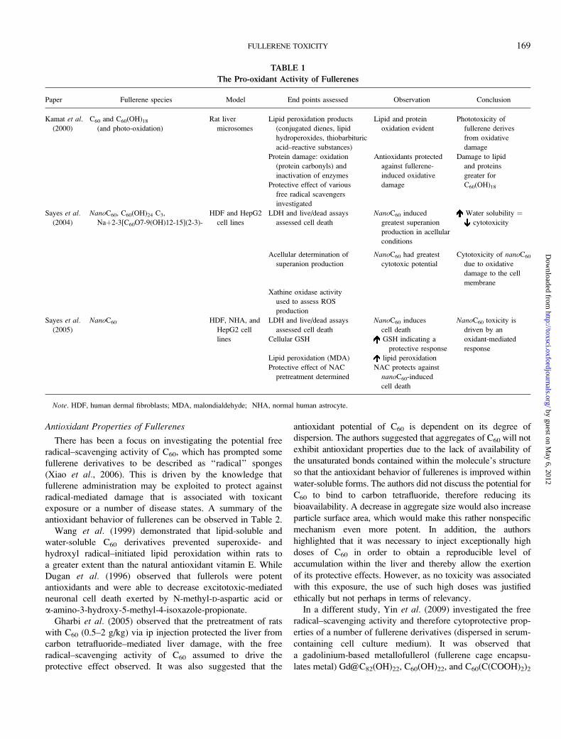
TABLE 1
The Pro-oxidant Activity of Fullerenes
Paper Fullerene species Model End points assessed Observation Conclusion
Kamat et al.
(2000)
C60 and C60(OH)18
(and photo-oxidation)
Rat liver
microsomes
Lipid peroxidation products
(conjugated dienes, lipid
hydroperoxides, thiobarbituric
acid–reactive substances)
Lipid and protein
oxidation evident
Phototoxicity of
fullerene derives
from oxidative
damage
Protein damage: oxidation
(protein carbonyls) and
inactivation of enzymes
Antioxidants protected
against fullerene-
induced oxidative
damage
Damage to lipid
and proteins
greater for
C60(OH)18Protective effect of various
free radical scavengers
investigated
Sayes et al.(2004)
NanoC60, C60(OH)24 C3,
Naþ2-3[C60O7-9(OH)12-15](2-3)-
HDF and HepG2
cell lines
LDH and live/dead assays
assessed cell death
NanoC60 induced
greatest superanion
production in acellular
conditions
Water solubility ¼cytotoxicity
Acellular determination of
superanion production
NanoC60 had greatest
cytotoxic potential
Cytotoxicity of nanoC60
due to oxidative
damage to the cell
membrane
Xathine oxidase activity
used to assess ROS
production
Sayes et al.
(2005)
NanoC60 HDF, NHA, and
HepG2 cell
lines
LDH and live/dead assays
assessed cell death
NanoC60 induces
cell death
NanoC60 toxicity is
driven by an
oxidant-mediated
response
Cellular GSH GSH indicating a
protective response
Lipid peroxidation (MDA) lipid peroxidation
Protective effect of NAC
pretreatment determined
NAC protects against
nanoC60-induced
cell death
Note. HDF, human dermal fibroblasts; MDA, malondialdehyde; NHA, normal human astrocyte.
FULLERENE TOXICITY 169
by guest on May 6, 2012
http://toxsci.oxfordjournals.org/D
ownloaded from
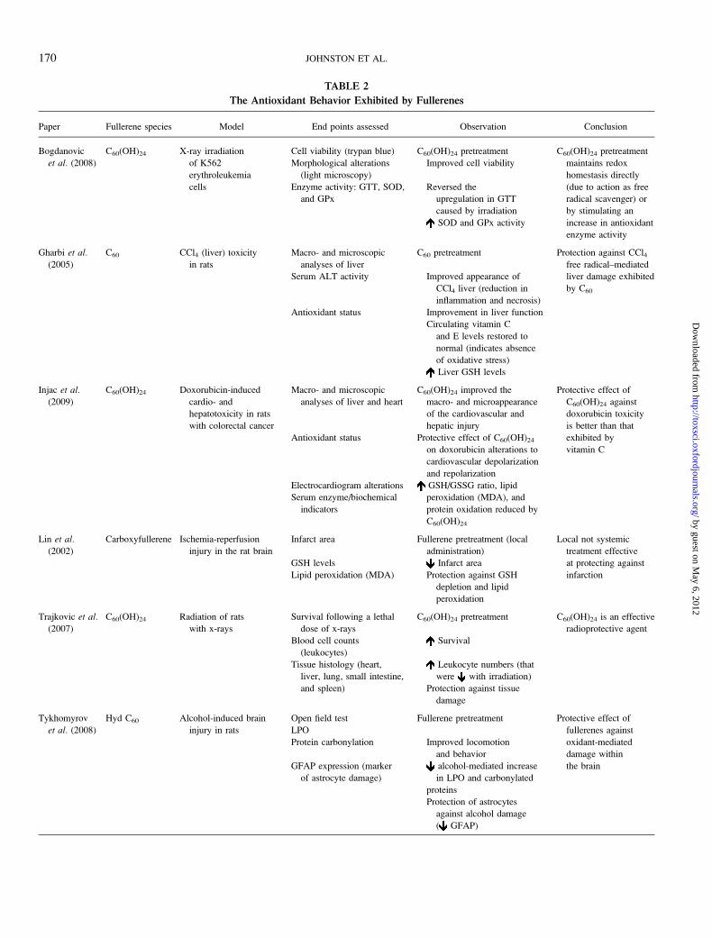
TABLE 2
The Antioxidant Behavior Exhibited by Fullerenes
Paper Fullerene species Model End points assessed Observation Conclusion
Bogdanovic
et al. (2008)
C60(OH)24 X-ray irradiation
of K562
erythroleukemia
cells
Cell viability (trypan blue) C60(OH)24 pretreatment C60(OH)24 pretreatment
maintains redox
homestasis directly
(due to action as free
radical scavenger) or
by stimulating an
increase in antioxidant
enzyme activity
Morphological alterations
(light microscopy)
Improved cell viability
Enzyme activity: GTT, SOD,
and GPx
Reversed the
upregulation in GTT
caused by irradiation
SOD and GPx activity
Gharbi et al.
(2005)
C60 CCl4 (liver) toxicity
in rats
Macro- and microscopic
analyses of liver
C60 pretreatment Protection against CCl4free radical–mediated
liver damage exhibited
by C60
Serum ALT activity Improved appearance of
CCl4 liver (reduction in
inflammation and necrosis)
Antioxidant status Improvement in liver function
Circulating vitamin C
and E levels restored to
normal (indicates absence
of oxidative stress)
Liver GSH levels
Injac et al.
(2009)
C60(OH)24 Doxorubicin-induced
cardio- and
hepatotoxicity in rats
with colorectal cancer
Macro- and microscopic
analyses of liver and heart
C60(OH)24 improved the
macro- and microappearance
of the cardiovascular and
hepatic injury
Protective effect of
C60(OH)24 against
doxorubicin toxicity
is better than that
exhibited by
vitamin C
Antioxidant status Protective effect of C60(OH)24
on doxorubicin alterations to
cardiovascular depolarization
and repolarization
Electrocardiogram alterations GSH/GSSG ratio, lipid
peroxidation (MDA), and
protein oxidation reduced by
C60(OH)24
Serum enzyme/biochemical
indicators
Lin et al.(2002)
Carboxyfullerene Ischemia-reperfusion
injury in the rat brain
Infarct area Fullerene pretreatment (local
administration)
Local not systemic
treatment effective
at protecting against
infarction
GSH levels Infarct area
Lipid peroxidation (MDA) Protection against GSH
depletion and lipid
peroxidation
Trajkovic et al.
(2007)
C60(OH)24 Radiation of rats
with x-rays
Survival following a lethal
dose of x-rays
C60(OH)24 pretreatment C60(OH)24 is an effective
radioprotective agent
Blood cell counts
(leukocytes)
Survival
Tissue histology (heart,
liver, lung, small intestine,
and spleen)
Leukocyte numbers (that
were with irradiation)
Protection against tissue
damage
Tykhomyrov
et al. (2008)
Hyd C60 Alcohol-induced brain
injury in rats
Open field test Fullerene pretreatment Protective effect of
fullerenes against
oxidant-mediated
damage within
the brain
LPO
Protein carbonylation Improved locomotion
and behavior
GFAP expression (marker
of astrocyte damage)
alcohol-mediated increase
in LPO and carbonylated
proteins
Protection of astrocytes
against alcohol damage
( GFAP)
170 JOHNSTON ET AL.
by guest on May 6, 2012
http://toxsci.oxfordjournals.org/D
ownloaded from
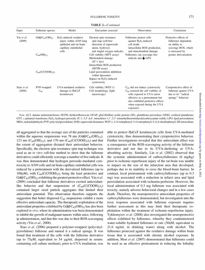
all aggregated so that the average size of the particles contained
within the aqueous suspensions was 78 nm (Gd@C82(OH)22),
123 nm (C60(OH)22), and 170 nm (C60(C(COOH)2)2) and that
the extent of aggregation dictated their antioxidant behavior.
Specifically, the electron spin resonance spin trap technique was
used as an in vitro cell-free method to show that the fullerene
derivatives could efficiently scavenge a number of free radicals. It
was then demonstrated that hydrogen peroxide–mediated cyto-
toxicity to A549 cells and rat brain capillary endothelial cells was
reduced by a pretreatment with the derivatized fullerenes (up to
100lM), with C60(C(COOH)2)2 being the least protective and
Gd@C82(OH)22 exhibiting the greatest protective effect. Yin et al.(2009) concluded that fullerene derivatives exerted antioxidant-
like behavior and that suspensions of (C60(C(COOH)2)2)
contained larger sized particle aggregates that limited their
antioxidant potential. This observation therefore supports the
suggestion that better dispersed C60 suspensions exhibit a more
effective antioxidant capacity. The therapeutic exploitation of the
antioxidant properties exhibited by Gd@C82(OH)22 has also been
explored in vivo, where its administration was been demonstrated
to inhibit the growth of malignant tumors within mice, following
ip administration, and that this was due to their ROS-scavenging
activity (Yin et al., 2008).
Xiao et al. (2006) prepared a polymer-wrapped (polyvinyl-
pyrrolidone) fullerene and named it a radical sponge. It was
found that treatment of the cells with the fullerene derivative
(up to 75lM, equivalent to 54 lg/ml, dispersed in serum-
containing cell culture medium), prior to UVA irradiation, was
able to protect HaCaT keratinocyte cells from UVA-mediated
cytotoxicity, thus demonstrating their cytoprotective behavior.
Further investigations revealed that this antioxidant effect was
a consequence of the ROS-scavenging activity of the fullerene
derivative and not due to its UVA-sheltering or UVA-
absorbing activity. Similarly, Lin et al. (2002) observed that
the systemic administration of carboxyfullerenes (6 mg/kg)
prior to ischemic-reperfusion injury of the rat brain was unable
to impact on the size of the infarction area that developed,
perhaps due to its inability to cross the blood-brain barrier. In
contrast, local pretreatment with carboxyfullerenes (up to 0.3
mg) was associated with a reduction in infarct area and lipid
peroxidation associated with ischemia-perfusion. However, the
local administration of 0.3 mg fullerene was associated with
toxicity, namely adverse behavioral changes and in a few cases
death. Therefore, the neuroprotective antioxidant properties of
carboxyfullerenes were demonstrated, but investigation into the
toxic response associated with fullerene exposure requires
further assessment as this may limit their therapeutic ex-
ploitation within the treatment of ischemic-reperfusion injury.
Tykhomyrov et al. (2008) also investigated the neuroprotective
effects exhibited by fullerenes, whereby they coadministered
water-soluble hydrated fullerenes to rats (30nM, equivalent to
21.6 ng/ml, in drinking water) along with alcohol. The
fullerenes protected against the oxidative damage within brain
tissue that is associated with chronic alcohol exposure. In
addition, Mori et al. (2007) demonstrated that fullerenes could
be used as an effective pretreatment in reducing the lethality
TABLE 2—Continued
Paper Fullerene species Model End points assessed Observation Conclusion
Yin et al.(2009)
Gd@C82(OH)22 H202-induced oxidative
injury within A549 lung
epithelial and rat brain
capillary endothelial
cells
Electron spin resonance
spin trap (cell-free
detection of superoxide
anion, hydroxyl,
and singlet oxygen radicals)
Fullerenes protect cells
against H202-induced
cell death,
intracellular ROS production,
and mitochondrial damage
Protective effects of
fullerenes dependent
on ability to
scavenge ROS, which
is increased by
greater derivatizationC60(OH)22 Cell viability (MTT assay) Fullerenes can scavenge free
radicals and LPOMitchondrial damage
(JC-1 dye)
Intracellular ROS production
(DCFH assay)
C60(C(COOH)2)2 Lipid peroxidation inhibition
within liposomes
Impact on H2O2-induced
cytotoxicity
Xiao et al.(2006)
PVP-wrapped
C60
UVA-mediated oxidative
damage to HaCaT
keratinocyte
Cell viability (WST-1) C60 did not induce cytotoxicity Cytoprotective effect of
fullerene against UVA
due to its ‘‘radical
sponge’’ behavior
Cell morphology (light
microscopy)
C60 restored the cell viability of
cells exposed to UVA (most
effective as a pretreatment but
also exhibited protective effects
when exposed during the UVA
exposure)
Note. ALT, alanine aminotransferase; DCFH, dichlorofluorescin; GFAP, glial fibrillary acidic protein; GPx, glutathione peroxidase; GSSG, oxidised glutathione;
GTT, c glutamyl transferase; H202, hydrogen peroxide; JC-1, 5,5#,6,6#-tetrachloro-1,1#,3,3#-tetraethylbenzimidazolylcarbocyanine iodide; LPO, lipid peroxidation;
MDA, malondialdehyde; PVP, polyvinylpyrrolidone; SOD, superoxide dismutase; WST-1, 2-(4-iodophenyl)-3-(4-nitrophenyl)-5-(2,4-disulfophenyl)-2H-tetrazolium.
FULLERENE TOXICITY 171
by guest on May 6, 2012
http://toxsci.oxfordjournals.org/D
ownloaded from
associated with methamphetamine and morphine co-exposure
of mice, which is also known to occur via oxidative stress. This
protective effect was equivalent or superior to that of more
traditional treatments, including cooling and the administration
of phospholipase 2 inhibitors.
The use of fullerenes as therapeutic agents has also been
explored in relation to their antioxidant-driven cytoprotective
behavior. Injac et al. (2009) investigated the protective effect of
fullerol (C60(OH)24) pretreatment (25, 50, and 100 mg/kg in
saline with DMSO following ip injection) on doxorubicin
toxicity (which is oxidant mediated) within the heart and
liver of rats with colorectal cancer. The protective effect of
fullerol was witnessed within macro- and microscopic
observations, electrocardiogram evaluation, serum biomarkers
for myocardial or hepatic damage, and oxidative stress
development. In general, an improvement in doxorubicin-
associated toxicity was observed within the heart and liver with
fullerol pre-exposure. However, lower doses of fullerols
exhibited a greater protective effect, and this may be accounted
for by the fact that higher doses of fullerols were less well
absorbed from the gut or that the high doses administered
contributed to the toxicity that was apparent. Therefore, it was
again demonstrated that fullerols are able to exhibit protective
effects against oxidative-mediated injury, thereby promoting
their exploitation as antioxidants, and that this was a dose-
dependent phenomenon. Trajkovic et al. (2007) investigated
the protective effects of fullerol (10 and 100 mg/kg, dispersed
in distilled water, for a period of up to 30 days), administered
via ip injection, against ionizing radiation within rats and
compared this to the traditional radioprotector amifostine.
Fullerol pretreatment was able to improve survival rates within
rats. The radioprotective effect exerted by fullerols was most
pronounced at a dose of 100 mg/kg and was comparable to that
of amifostine. As the harmful effects of radiotherapy are known
to be mediated by ROS, fullerols were anticipated to be
effective radioprotectors by a mechanism that was antioxidant
driven. The potential therapeutic exploitation of fullerols was
addressed further by Bogdanovic et al. (2008) who assessed the
ability of C60(OH)24 to protect against ionizing radiation–
mediated ROS production in vitro. The protective effects of
fullerol (10lM, equivalent to 11.28 lg/ml, dispersed in serum-
containing cell culture medium) were investigated within
irradiated, malignant cultured K562 erythroleukemia cells.
The survival rate of irradiated cells was improved by fullerol
pretreatment, which was suggested to occur due to increased
antioxidant defenses within irradiated cells that counteracted
the oxidative damage associated with radiation that acted to
preserve cell viability.
Although the free scavenging activity of fullerenes is
accepted as being potentially beneficial, in certain circum-
stances, it can be problematic. For example, Ueng et al. (1997)
observed that a single ip injection of fullerol, at concentrations
of 0.1, 0.5, and 1 g/kg (in water), induced mortality of 10, 22,
and 54%, respectively, within 3 days of the treatment. Fullerol
administration, of greater than 0.5 g/kg, also elicited a decrease
in cytochrome P450 content and activity within liver micro-
somes (isolated from fullerol-exposed animals). The mecha-
nism of this decrease is not known but could be due to the
electron-scavenging behavior of polyhydroxylated C60 or
binding of fullerol to the enzyme thus promoting enzyme
destruction or prevention of enzyme synthesis as a result of
cell injury. In addition, mitochondrial function was observed
to be diminished by fullerol exposure, as indicated by the
suppression of oxidative phosphorylation, which is likely to
derive from a reduction in the transfer of electrons. However,
the high doses used are likely to account for the mortality and
cell injury observed and are unlikely to be encountered by
humans.
Overall, the studies relating to antioxidant properties of
fullerenes suggest that contrary from being toxic, C60 and its
derivatives could actually exhibit beneficial health effects.
However, it appears that the antioxidant properties exhibited by
fullerenes are restricted to particular fullerene forms and
therefore a number of conditions being met in order to allow its
manifestation. Water solubility is likely to impact on their
antioxidant/cytoprotective potential so that the better dispersed
the fullerene is, the more likely it is that it will exert free
radical–scavenging activity, which is likely to derive from their
derivatization as they are specifically generated to improve
fullerene water solubility. The concentration administered is
therefore also key to dictating the free radical activity as
fullerenes exposed at high concentrations are more likely to
interact to form larger structures, which is known to
detrimentally impact on its antioxidant behavior. Many of the
studies reported have used exceptionally high exposure
concentrations and are therefore difficult to interpret in terms
of relevance unless large doses are used in such applications.
Uptake of Fullerenes into Cells
Uptake studies have investigated the behavior of both
professional phagocytes, such as macrophages, and nonphago-
cytic cells. Determining the uptake of fullerenes by cells is of
relevance as phagocytic cells, located at exposure sites, are
responsible for the clearance of particles. Second, the uptake of
fullerenes by cells has the potential to impact on normal cell
physiology and function, which requires assessment. When
addressing the uptake of fullerenes, a variety of cell types have
been considered. In addition, computer simulations have been
conducted that act to predict the interactions of fullerenes with
cell membranes and their subsequent penetration.
It has been observed that subsequent to pulmonary exposure,
fullerenes are evident within alveolar macrophages (Fujita
et al., 2009; Xia et al., 2006). Furthermore, subsequent to ip
administration, fullerenes enter the circulation and have been
observed to accumulate within Kupffer cells in the liver
(Gharbi et al., 2005). Macrophages therefore appear to be
capable of taking up particles to thereby fulfill their role within
host defense. However, the consequences of fullerene uptake
172 JOHNSTON ET AL.
by guest on May 6, 2012
http://toxsci.oxfordjournals.org/D
ownloaded from
require attention as oxidative or inflammatory events may be
stimulated. In addition, a number of other cell types have been
demonstrated to internalize fullerenes, such as keratinocytes
(Rouse et al. 2006), epithelial cells (Fujita et al., 2009), and eye
lens cells (Roberts et al., 2008) often with oxidative and lethal
consequences.
Computer simulation has been exploited to reveal the
mechanism of fullerene permeation through cell membranes
(Wong-Ekkabut et al., 2008). It was identified that small
fullerene clusters (< 10 molecules) were able to localize within
the membrane lipid bilayer, where they disaggregated, that this
process was passive and spontaneous, and that even at high
concentrations, no mechanical damage to the membrane was
observed. However, although the computer simulations provide
insight into the possible behavior of fullerenes, caution is
required when extrapolating the results as the interactions of
fullerenes with other membrane components, such as other
lipids, carbohydrates, and proteins, were not considered within
the model, and it is therefore necessary to verify the results.
However, the tendency for fullerenes to interact with the lipid
tails within the model parallels the finding that lipid per-
oxidation is a prominent feature of their exposure to cells.
In addition, Bedrov et al. (2008) used molecular dynamic
models to investigate the interaction and transport of C60
within a plasma membrane model. C60 was observed to interact
with the lipid head groups and lipid core of the membrane.
Consequently, fullerenes were predicted to have a high
permeability within the simulations conducted. For this reason,
the authors suggested that fullerenes could be exploited as
efficient carriers to enable drug entry into cells. Similarly, Qiao
et al. (2007) used molecular dynamics to study the trans-
location of fullerenes across a model cell membrane. Pristine
C60 was observed to readily translocate across the lipid
membrane. This was achieved due to the ability of C60 to
create a cavity (termed transient micropores) within the mem-
brane. C60 molecules were then speculated to ‘‘jump’’ into the
membrane, which enabled the penetration of the molecule
through the membrane. In contrast, C60(OH)20 derivatives
barely penetrated the membrane, which was explained by their
absorption onto the membrane surface, due to its hydrophilic
nature. This interaction discouraged C60(OH)20 interaction with
the lipid core of the membrane so that it did not enter the
membrane, but instead, the strong interactions with the
membrane surface head groups caused a ‘‘pinch’’ to form in
the plasma membrane. Therefore, pristine fullerenes and
fullerene derivatives exhibit differences, with regards to their
translocation through the model membrane, and perhaps this
can explain differences in their toxicity and why fullerene
derivatives exhibit reduced toxicity than their pristine counter-
parts (see, e.g., Sayes et al., 2004). However, mainly, the
simulations did not consider the aggregation state of fullerenes
and depict the consequences of membrane exposure to
individual molecules, which would be anticipated to more
easily enter cells due to their small size (< 1 nm).
Porter et al. (2006) found that no cytotoxicity to human-
derived macrophages was elicited by C60 (0.16–10 lg/ml,
prepared using THF and dispersed in serum-free cell culture
medium) in vitro, despite the fact that they were internalized
and contained within the cytoplasm, nucleus, and lysosomes. It
was observed that C60 crystals and aggregates were contained
within lysosomes (perhaps as a consequence of their in-
ternalization by phagocytosis or endocytosis) in which C60 was
degraded into smaller structures. C60 aggregates were also
apparent along the plasma membrane, which was suggested by
the authors to promote the development of lipid peroxidation
observed by other investigators. It was highlighted that there
was a difficulty in imaging particles, due to the fact that it is
difficult to distinguish them from artifact presence, and so the
results require further verification.
The uptake of fullerenes has been demonstrated on numerous
occasions within a variety of cell types. The implications of
uptake are relatively unknown, and therefore worthy of
consideration in the future, but are likely to involve oxidative
or cytotoxic responses. Computer simulations have also been
used to predict the penetration of fullerenes within the plasma
membrane and attributes of particles that encourage such an
interaction, but their relevance requires confirmation.
Genotoxicity of Fullerenes
Genotoxicity tests are conducted to reveal damage to DNA
elicited by fullerene exposure by, e.g., detecting mutations and
changes in chromosome structure or number. A number of
assays can be adopted to detect genotoxicity, including the
comet assay, Ames test, and determining tumor development
within animals.
Dhawan et al. (2006) investigated whether C60 was able to
inflict DNA damage within human lymphocytes and was
detected using the comet assay when exposed at concentrations
ranging from 0.42 to 2100 lg/l (dispersed in serum-free cell
culture medium) for up to 6 h. To ensure that residual solvents
used to prepare C60 suspensions were not responsible for any
observed toxicity (see below), the experiments were conducted
using preparation methods that were free of organic solvents.
These included prolonged mixing of C60 in water (aquC60)
or the ‘‘solvent to water exchange’’ method using ethanol
(EtOHC60). It was demonstrated that solvents were more ef-
fective at dispersing C60, as demonstrated by the fact that the
size of C60 clusters was smaller (122 nm diameter) than those
produced within aquC60 suspensions (178 nm diameter). Both
samples were able to cause DNA damage within lymphocytes,
with aquC60 being more effective. The results therefore
highlight that the dispersion method is able to impact on the
toxicity of C60, whereby fullerenes prepared by mixing in water
were more capable of eliciting a genotoxic response than those
produced using the solvent to water exchange method. In
addition, Sera et al. (1996) investigated the mutagenic effect of
fullerene exposure (up to 30 lg/plate for 48 h) on Salmonellatyphimurium in light and dark conditions using the Ames test. If
FULLERENE TOXICITY 173
by guest on May 6, 2012
http://toxsci.oxfordjournals.org/D
ownloaded from
exposure occurred within the dark, no mutagenic responses
were evident. In contrast, a mutagenic effect was observed when
exposure occurred in the presence of visible light due to the
production of ROS, which interact with DNA to elicit damage,
and was typified by the formation of 8-hydroxydeoxyguanosine.
Lipid peroxidation was also increased by fullerene exposure in
light, further highlighting the oxidative consequences associated
with light irradiation. The study therefore illustrated the
phototoxic and mutagenic properties of fullerenes.
Contrary to the ability of C60 to induce genotoxic events
within cells, fullerene derivatives have been demonstrated to
have potential therapeutic properties for the treatment of
cancer. Chen et al. (2005) sc implanted H22 hepatoma cells
into mice, and tumor growth was monitored until the tumor that
developed reached a size of 2 or 2.2 cm. Tumor-bearing mice
were then treated, once a day, with Gd@C82(OH)22 (dispersed
in saline), via ip injection, at a concentration of 114 or 228
lg/ml, and the antitumor effect was evaluated by determining
the impact of treatment on tumor size. It was observed that
Gd@C82(OH)22 inhibited skin tumor growth within hepatoma-
implanted mice, with this inhibitory effect apparent on
treatment of mice for 6 days or more. The antitumor efficiency
of Gd@C82(OH)22 was greater than that of the conventional
antineoplastic agents cyclophosphamide and cisplatin. The
administration of C60 was associated with very low toxicity
in vivo, and no cytotoxicity was associated with hepatocyte cell
line exposure in vitro. Furthermore, Tabata et al. (1997) aimed
to target radiolabeled 125I C60-polyethylene glycol (PEG)
conjugates (424 lg/kg, dispersed in PBS) to tumors following
iv injection within tumor-bearing mice. The distribution of the
fullerene was determined following exposure, and 78% of the
administered dose was eliminated from the body within 24 h.
No marked accumulation within a particular organ was
observed (probably as a consequence of its derivatization),
although localization within the liver and gastrointestinal tract
was observed, with no toxicity associated with exposure. The
fullerene was able to accumulate within tumors due to its large
size (which also relied on the hyperpermeability of tumor
vasculature). Light irradiation (to achieve photoactivation of
the fullerene) at the tumor site allowed the specific destruction
of the tumor by the fullerenes, with no damage to the overlying
normal skin. Tumor destruction was not apparent with C60-
PEG administration alone; therefore, light irradiation was
essential for the tumor-destructive effect to manifest. Accord-
ingly, the phototoxic property of fullerenes was exploited
within the destruction of tumors, and fullerenes may therefore
be considered for photodynamic therapy of tumors. However,
its exploitation is reliant on the utilization of particular C60
derivatives; accordingly, PEG conjugation to C60 was neces-
sary to increase the size of the molecule and to increase its
water solubility. In addition, Zhu et al. (2008) evaluated the
tumor inhibitory effect of fullerols (0.2 and 1 mg/kg, dispersed
in deionized water, administered daily via ip injection, for up to
17 days) within the mouse H22 hepatocarcinoma model.
C60(OH)x exhibited antitumor activity, with treatment more
effective if administered from the time of tumor inoculation
onward, as opposed to when treatment was initiated after tumor
growth reached 1 cm.
Furthermore, genotoxicity has not been associated with
fullerene exposure in a number of models. Mori et al. (2006)
investigated the mutagenicity of a C60/C70 mixture. It was
illustrated that no mutagenic responses were evident within
a variety of S. typhimurium and E. coli strains using the Ames
test (up to 5000 lg/plate). In addition, within the chromosomal
aberration test (in CHL/IU hamster lung cells), no aberrations
within the structure or number of chromosomes were apparent.
The separate tests therefore reached the same conclusion that
the fullerene investigated was not mutagenic. Furthermore,
Jacobsen et al. (2008) investigated the mutagenicity associated
with a number of carbon-based NPs, including C60 within the
mouse FE1-Muta epithelial cell line. It was demonstrated that
exposure of cells to C60 (dispersed in serum-containing cell
culture medium, with sonication used to minimize particle
aggregation) was associated with a slight increase in ROS
production in cells, but no impact on cell viability was
observed. This was demonstrated following a short term
exposure scenario (24 h exposure, at concentrations of 20 to
200 lg/ml), and in the long term (following 9 rounds of particle
exposures of 72 h for each round (total duration of 576 h), at
a concentration of 100 lg/ml). An increase of ROS production
by C60 was also observed in cell-free conditions.
The genotoxicity of fullerenes is therefore difficult to
interpret from the studies conducted so far, with contradicting
results reported. Again, these are likely to be influenced by the
dose, dispersion, model, and end point measured. An important
component of the genotoxic response exhibited by fullerenes
is anticipated to be their photoactivity, which is able to pro-
mote such a response. However, the opportunity to exploit
fullerenes as antitumor agents is of interest and warrants further
investigation.
Reproductive Toxicology of Fullerenes
Evaluation of fullerene effects on the reproductive system is
limited to a small number of in vivo and in vitro studies. Most
of these have focused on how treatment can affect the
developing embryo. A study by Tsuchiya et al. (1996) ip
administered polyvinylpyrrolidone-solubilized C60 (up to 137
mg/kg in distilled water) to pregnant mice on day 10 (vaginal
plug: day 0). After 18 h of exposure, all the embryos were
examined and found to be dead. A variety of doses were
examined with C60 apparently distributed into the embryos at
50 mg/kg resulting in head region and tail abnormalities. The
yolk sac appeared with shrunken membrane and narrow blood
vessels, which may suggest insufficient blood supply to the
embryos. At 25 mg/kg, one embryo had abnormal enlargement
of the head, whereas all other embryos appeared normal. The
no-observed-adverse-effect level was 16.7 mg/kg. The study
recommends further work due to the low number of animals
174 JOHNSTON ET AL.
by guest on May 6, 2012
http://toxsci.oxfordjournals.org/D
ownloaded from
per exposure group. This is an unusual route of administration
using a relatively high exposure dose, and it covers only
a small part of the pregnancy period.
A study by Zhu et al. (2008) assessed embryonic
development using the zebrafish model. The group exposed
newly fertilized eggs to 50 mg/l fullerol (C60(OH)16–18), nano
aggregates of C60 (1.5 mg/l, particle size: ~100 nm), or
a combined treatment of nanoC60 (1.5 mg/l) and GSH (30 mg/l)
for 96 h. Observed end points were survival, hatching rate,
heart beat rate, and pericardial edema. The fullerol solution had
no adverse effect on the end points, whereas the C60 suspension
had a conspicuous adverse effect on all parameters that was
lessened by the addition of GSH. Zhu et al. (2008) concluded
that the adverse effects of C60 were due, at least partly, to free
radical–induced mechanism or another form of oxidative stress.
Similarly, Usenko et al. (2008) also used zebrafish embryos to
examine the potential of C60 to elicit oxidative stress responses.
The group’s findings showed reduced light, and therefore,
reduced photocatalytic activity during C60 exposure signifi-
cantly decreased mortality and the incidence of fin malforma-
tions and pericardial edema at 200 and 300 ppb (equivalent to
0.2 and 0.3 lg/ml, respectively) C60 24-h postfertilization.
Embryos co-exposed to the glutathione precursor, NAC, also
showed reduced mortality and pericardial edema; however, fin
malformations were not reduced.
The reviewed literature examines the effects of fullerenes
during pregnancy, highlight effects on developing embryos;
however, studies are extremely limited in number and in
sample size. No specific in vitro or in vivo studies were found
examining fullerene effects in male reproductive system.
Interactions of Fullerenes with Biological Molecules
The ability of fullerenes to interact with biological
molecules, such as proteins, is of concern as it not only has
the propensity to alter the normal structure and function of
these biological moieties and enable fullerene transport within
the body (as interactions of fullerenes with serum proteins are
evident) but also is potentially able to modify the behavior of
the particles.
Belgorodsky et al. (2006) demonstrated that cyclodextrin-
capped fullerenes were able to form stable complexes with
bovine serum albumin (BSA). The implications of fullerene
binding are uncertain, but normal protein conformation and
function were retained, and therefore, such an interaction is
likely to facilitate fullerene transport within the body.
Benyamini et al. (2006) used computational models to
understand the interactions between fullerenes (in a pristine
and carboxylated form) and proteins (namely human serum
albumin, BSA, human immunodeficiency virus [HIV] protease,
and a fullerene-specific antibody) that had all been previously
demonstrated to interact with fullerene molecules. The binding
sites for fullerenes within the proteins were identified and
demonstrated to have similarities between the different proteins
(although not for the fullerene antibody) investigated.
The ability of fullerenes to interact with proteins may be
beneficial within the treatment of disease. Schinazi et al. (1993)
illustrated that water-soluble fullerene derivatives exhibited
antiviral activity against HIV within infected lymphocytes
(with no cytotoxicity associated with the response). This
antiviral activity is anticipated to derive from a direct in-
teraction between the fullerene and the active site of the HIV
protease, which causes the subsequent inhibition of enzyme
activity (Friedman et al., 1993). However, the design of the
fullerene molecule is of vital importance to its interactions with
proteins as within the described studies, the fullerene deriva-
tives used were designed in such a way to encourage such
interactions, and so the applicability of the response to
fullerenes as a whole requires investigation.
The described studies highlight that fullerenes are able to
interact with proteins, which may have detrimental implica-
tions for protein function. This property can be considered
beneficial within their exploitation within antiviral therapy.
However, the specificity of the response requires consideration
as unregulated enzyme inhibition would be detrimental and
likely to be associated with adverse responses. The binding of
fullerenes to serum proteins is of interest as it is likely to
facilitate their transport within the body to enable them to reach
target sites distal to their exposure site.
LINKING THE PHYSICOCHEMICAL ATTRIBUTES OF
FULLERENES TO PATHOGENICITY OR TOXICITY
As for other particles, the physicochemical properties of
fullerenes are likely to impact on their toxicity. Such char-
acteristics include composition, crystal, or aggregate size, water
solubility, and surface modifications/functionalization.
Surface Modifications
Surface modification is also termed derivatization or
functionalization and is used to achieve a specific change in
particle properties. Common examples of particle functional-
ization include hydroxylation (–OH) and carboxylation
(–COOH). In general, the surface of fullerenes is completed
for a specific purpose, such as improving water solubility. In
addition, it is recognized that such changes may also influence
toxicity as, e.g., fullerene functionalization has been observed
to promote the appearance of beneficial properties, such as
antioxidant or anti-inflammatory activity (see above).
Accordingly, pristine (unmodified) C60 has been demon-
strated to be more toxic than the functionalized water-soluble
counterparts (including C60(OH)24) when examined in vitrowith keratinocyte and hepatocyte cell lines (Sayes et al., 2004).
In fact, as the fullerene cage became more derivatized, and
therefore more water soluble, the corresponding toxicity was
observed to decrease. However, in a separate study, Sayes
et al. (2007) exposed rats via intratracheal instillation (0.2–3
mg/kg, dispersed in MilliQ ultrapure water) to C60 or
FULLERENE TOXICITY 175
by guest on May 6, 2012
http://toxsci.oxfordjournals.org/D
ownloaded from
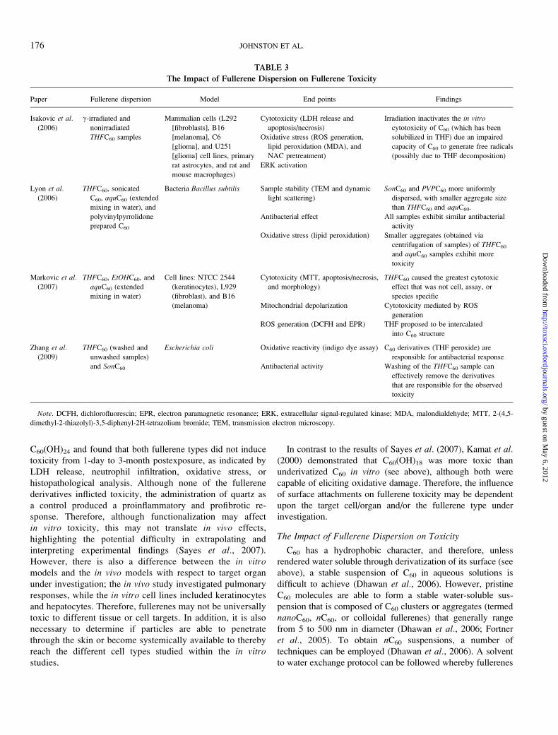
C60(OH)24 and found that both fullerene types did not induce
toxicity from 1-day to 3-month postexposure, as indicated by
LDH release, neutrophil infiltration, oxidative stress, or
histopathological analysis. Although none of the fullerene
derivatives inflicted toxicity, the administration of quartz as
a control produced a proinflammatory and profibrotic re-
sponse. Therefore, although functionalization may affect
in vitro toxicity, this may not translate in vivo effects,
highlighting the potential difficulty in extrapolating and
interpreting experimental findings (Sayes et al., 2007).
However, there is also a difference between the in vitromodels and the in vivo models with respect to target organ
under investigation; the in vivo study investigated pulmonary
responses, while the in vitro cell lines included keratinocytes
and hepatocytes. Therefore, fullerenes may not be universally
toxic to different tissue or cell targets. In addition, it is also
necessary to determine if particles are able to penetrate
through the skin or become systemically available to thereby
reach the different cell types studied within the in vitrostudies.
In contrast to the results of Sayes et al. (2007), Kamat et al.(2000) demonstrated that C60(OH)18 was more toxic than
underivatized C60 in vitro (see above), although both were
capable of eliciting oxidative damage. Therefore, the influence
of surface attachments on fullerene toxicity may be dependent
upon the target cell/organ and/or the fullerene type under
investigation.
The Impact of Fullerene Dispersion on Toxicity
C60 has a hydrophobic character, and therefore, unless
rendered water soluble through derivatization of its surface (see
above), a stable suspension of C60 in aqueous solutions is
difficult to achieve (Dhawan et al., 2006). However, pristine
C60 molecules are able to form a stable water-soluble sus-
pension that is composed of C60 clusters or aggregates (termed
nanoC60, nC60, or colloidal fullerenes) that generally range
from 5 to 500 nm in diameter (Dhawan et al., 2006; Fortner
et al., 2005). To obtain nC60 suspensions, a number of
techniques can be employed (Dhawan et al., 2006). A solvent
to water exchange protocol can be followed whereby fullerenes
TABLE 3
The Impact of Fullerene Dispersion on Fullerene Toxicity
Paper Fullerene dispersion Model End points Findings
Isakovic et al.
(2006)
c-irradiated and
nonirradiated
THFC60 samples
Mammalian cells (L292
[fibroblasts], B16
[melanoma], C6
[glioma], and U251
[glioma] cell lines, primary
rat astrocytes, and rat and
mouse macrophages)
Cytotoxicity (LDH release and
apoptosis/necrosis)
Irradiation inactivates the in vitro
cytotoxicity of C60 (which has been
solubilized in THF) due an impaired
capacity of C60 to generate free radicals
(possibly due to THF decomposition)
Oxidative stress (ROS generation,
lipid peroxidation (MDA), and
NAC pretreatment)
ERK activation
Lyon et al.
(2006)
THFC60, sonicated
C60, aquC60 (extended
mixing in water), and
polyvinylpyrrolidone
prepared C60
Bacteria Bacillus subtilis Sample stability (TEM and dynamic
light scattering)
SonC60 and PVPC60 more uniformly
dispersed, with smaller aggregate size
than THFC60 and aquC60.
Antibacterial effect All samples exhibit similar antibacterial
activity
Oxidative stress (lipid peroxidation) Smaller aggregates (obtained via
centrifugation of samples) of THFC60
and aquC60 samples exhibit more
toxicity
Markovic et al.(2007)
THFC60, EtOHC60, and
aquC60 (extended
mixing in water)
Cell lines: NTCC 2544
(keratinocytes), L929
(fibroblast), and B16
(melanoma)
Cytotoxicity (MTT, apoptosis/necrosis,
and morphology)
THFC60 caused the greatest cytotoxic
effect that was not cell, assay, or
species specific
Mitochondrial depolarization Cytotoxicity mediated by ROS
generation
ROS generation (DCFH and EPR) THF proposed to be intercalated
into C60 structure
Zhang et al.(2009)
THFC60 (washed and
unwashed samples)
and SonC60
Escherichia coli Oxidative reactivity (indigo dye assay) C60 derivatives (THF peroxide) are
responsible for antibacterial response
Antibacterial activity Washing of the THFC60 sample can
effectively remove the derivatives
that are responsible for the observed
toxicity
Note. DCFH, dichlorofluorescin; EPR, electron paramagnetic resonance; ERK, extracellular signal-regulated kinase; MDA, malondialdehyde; MTT, 2-(4,5-
dimethyl-2-thiazolyl)-3,5-diphenyl-2H-tetrazolium bromide; TEM, transmission electron microscopy.
176 JOHNSTON ET AL.
by guest on May 6, 2012
http://toxsci.oxfordjournals.org/D
ownloaded from
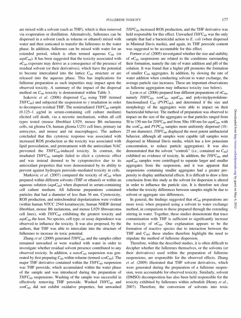
are mixed with a solvent (such as THF), which is then removed
via evaporation or distillation. Alternatively, fullerenes can be
dispersed in a solvent (such as toluene or ethanol) mixed with
water and then sonicated to transfer the fullerenes to the water
phase. In addition, fullerenes can be mixed with water for an
extended period, which is often termed aqueous C60 (or
aquC60). It has been suggested that the toxicity associated with
nC60 exposure may derive as a consequence of the presence of
residual solvent (or their derivatives), which have the potential
to become intercalated into the lattice C60 structure or are
released into the aqueous phase. This has implications for
fullerene preparation as such impurities may impact upon the
observed toxicity. A summary of the impact of the dispersal
method on C60 toxicity is demonstrated within Table 3.
Isakovic et al. (2006) dispersed C60 using THF (termed
THFC60) and subjected the suspension to c irradiation in order
to decompose residual THF. The nonirradiated THFC60 sample
(0.125–1 lg/ml in serum-containing cell culture medium)
elicited cell death, via a necrotic mechanism, within all cell
types tested (mouse fibroblast L929, mouse B6 melanoma
cells, rat glioma C6, human glioma U251 cell lines, primary rat
astrocytes, and mouse and rat macrophages). The authors
concluded that this cytotoxic response was associated with
increased ROS production as the toxicity was associated with
lipid peroxidation, and pretreatment with the antioxidant NAC
prevented the THFC60-induced toxicity. In contrast, the
irradiated THFC60 sample failed to elicit a cytotoxic effect
and was instead deemed to be cytoprotective due to its
antioxidant properties that were demonstrated by its ability to
prevent against hydrogen peroxide–mediated toxicity in cells.
Markovic et al. (2007) compared the toxicity of nC60 when
prepared within different solvents (THF or ethanol) or within an
aqueous solution (aquC60) when dispersed in serum-containing
cell culture medium. All fullerene preparations contained
particles that had a diameter of less than 36 nm. Cytotoxicity,
ROS production, and mitochondrial depolarization were evident
(within human NTCC 2544 keratinocyte, human NHDF dermal
fibroblast, mouse B6 melanoma, and mouse L929 fibrosarcoma
cell lines), with THFC60 exhibiting the greatest toxicity and
aquC60 the least. No species, cell type, or assay dependence was
observed to influence the toxicity. It was also proposed, by the
authors, that THF was able to intercalate into the structure of
fullerenes to increase its toxic potential.
Zhang et al. (2009) generated THFC60, and the samples either
remained unwashed or were washed with water in order to
investigate whether residual solvent presence contributed to any
observed toxicity. In addition, a nanoC60 suspension was gen-
erated by first preparing C60 within toluene (termed sonC60). The
major THF derivative contained within the THFC60 suspension
was THF peroxide, which accumulated within the water phase
of the sample and was introduced during the preparation of
THFC60 suspensions. Washing of the sample was successful in
effectively removing THF peroxide. Washed THFC60 and
sonC60 did not exhibit oxidative properties, but unwashed
THFC60 increased ROS production, and the THF derivative was
held responsible for this effect. Unwashed THFC60 was the only
sample that had a bactericidal action to E. coli (when dispersed
in Minimal Davis media), and again, its THF peroxide content
was suggested to be accountable for this effect.
Fortner et al. (2005) investigated whether the size and stability
of nC60 suspensions are related to the conditions surrounding
their formation, namely the rate of water addition and pH of the
solution. It was found that a higher pH promotes the formation
of smaller C60 aggregates. In addition, by slowing the rate of
water addition when conducting solvent to water exchange, the
average particle size increases. These are important observations
as fullerene aggregation may influence toxicity (see below).
Lyon et al. (2006) prepared four different preparations of nC60,
namely THFC60, sonC60, aquC60, and polyvinylpyrrolidone-
functionalized C60 (PVPC60), and determined if the size and
morphology of the aggregates were able to impact on their
bactericidal behavior. The method of preparation was observed to
impact on the size of the aggregates so that particles ranged from
50 to 150 nm for THFC60 and from 30to 100 nm for aquC60, with
the sonC60 and PVPC60 samples more uniformly dispersed (10–
25 nm diameter). THFC60 displayed the most potent antibacterial
behavior, although all samples were capable (all samples were
dispersed in Minimal Davis media, which has a low potassium
concentration, to reduce particle aggregation). It was also
demonstrated that the solvent ‘‘controls’’ (i.e., contained no C60)
exhibited no evidence of toxicity. In addition, the THFC60, and
aquC60 samples were centrifuged to separate larger and smaller
aggregates from the suspension, and it was apparent that
suspensions containing smaller aggregates had a greater pro-
pensity to display antibacterial effects. It is difficult to draw a firm
conclusion from this study as the solvent for dispersion is altered
in order to influence the particle size. It is therefore not clear
whether the toxicity differences between samples might be due to
solvent or aggregate size differences.
In general, the findings suggested that nC60 preparations are
more toxic when prepared using a solvent to water exchange
method, in comparison to those prepared through the extending
stirring in water. Together, these studies demonstrate that trace
contamination with THF is sufficient to significantly increase
the toxicity of nC60. One explanation appears to be the
formation of reactive species due to interaction between the
THF and C60; these studies therefore highlight the need to
stipulate the method of fullerene dispersion.
Therefore, within the described studies, it is often difficult to
decipher whether the fullerenes themselves, or the solvents (or
their derivatives) used within the preparation of fullerene
suspensions, are responsible for the observed effects. Zhang
et al. (2009) illustrated that THF solvent derivatives, which
were generated during the preparation of a fullerene suspen-
sion, were accountable for observed toxicity. Similarly, solvent
(DMSO) decomposition has also been held responsible for the
toxicity exhibited by fullerenes within zebrafish (Henry et al.,2007). Therefore, the conversion of solvents into toxic
FULLERENE TOXICITY 177
by guest on May 6, 2012
http://toxsci.oxfordjournals.org/D
ownloaded from
byproducts during the particle suspension preparation is
possible and potentially responsible for driving the observed
toxicity of fullerenes. As such, it would be relevant to detect
the formation of such solvent decomposition products within
particle suspensions or include appropriate controls (such as
solvent degradation products) within investigations. Therefore,
it is plausible that fullerenes may be mistakenly identified as
driving any observed toxicity when it is really the use of
solvents within the preparation of particle suspensions that are
responsible for the response observed. As such, it is necessary
for investigators to consider how the preparation of fullerenes
into a suspension impacts on the observed toxicity. In addition,
it is of importance to consider the relevance of the preparation
method used to generate particle suspensions and in particular
if humans will actually encounter fullerenes in this form.
Contribution of Photocatalytic Behavior of Fullerenes toTheir Toxicity
It is acknowledged that fullerenes are able to absorb ultraviolet
or visible light to stimulate the generation of ROS and to
ultimately increase their toxic potency (Nielsen et al., 2008)
through the promotion of oxidative-mediated damage. The
contribution of photocatalytic reactions to fullerene toxicity has
been a focus of a number of previously discussed studies
(Dhawan et al., 2006; Kamat et al., 2000; Roberts et al., 2008;
Usenko et al., 2008). This phenomenon has also been exploited
within the destruction of tumors in mice (Chen et al., 2005), but
more work is required to determine the specificity of the response
and how this may be exploited within the treatment of humans.
COMMENTS ON THE EXPERIMENTAL DESIGNS
Relevant fullerene exposure concentrations to be used within
in vitro and in vivo experiments cannot be obtained without
information regarding the human exposure levels, which is
information that is currently lacking. However, some concen-
trations used have been exceptionally high and unlikely to be
encountered by humans; for example, Gharbi et al. (2005)
exposed rats to fullerenes at a dose of up to 2 g/kg via ip
exposure. In addition, it is often the case that a single dose has
been administered to animals or cells and toxicity assessed at
a number of postexposure time points, but it is likely that the
utilization of fullerenes within occupational or consumer settings
will involve the exposure of humans for extended periods, and
this should perhaps be considered within future studies. The
uncertainty regarding human exposure levels to fullerenes also
makes interpretation of the observed hazards difficult.
In addition, the most relevant way of expressing fullerene
concentrations is debatable and within the studies outlined has
been expressed as parts per million, micromolars, milligrams
per kilogram, milligrams per milliliter, etc (which in some
circumstances is likely to derive from the exposure scenario).
In order to counteract this problem, molar concentrations
stipulated within the described studies have been converted to
a standardized dose descriptor (of mass weight of particles
administered per animal, milliliters [of particle suspension], or
kilograms [of the exposed animal]) in order to allow for easier
comparisons between investigations. Importantly, when the
molecular formula of the fullerene in question was not
explicitly stated by investigators, calculations were based on
the use of C60. The toxicity of NPs has been demonstrated in
a number of studies to be related to their small size and
therefore high surface area (Brown et al., 2001; Duffin et al.,2002, 2007; Stoeger et al., 2006). The high propensity for
fullerenes to aggregate means that not all the potential
calculated surface will be available at the surface, and
therefore, expressing the concentration as the surface area of
particles administered is perhaps not the best dose descriptor to
use for fullerenes. However, introducing a standard way of
expressing fullerene concentration would also allow the
opportunity to more easily compare the findings of different
investigators. In line with this, it would be of benefit for
investigators to provide as much information as possible,
relating to the exposure conditions and particle dimensions
used (including mass of particles administered, in what volume
they were administered [if relevant to the experiment], the size
and surface area of particles used, and the molecular formula of
the fullerene) so that the concentration used can be more easily
converted by others to allow comparisons between different
investigations to be made, until such a decision on how to best
standardize the concentration is agreed on.
There are a diverse number of fullerenes available, which
derives from the number of carbon atoms contained within the
cage structure, the surface modification, and the preparation
processes utilized to promote their water solubility. Therefore,
fullerenes used by different investigators are likely to behave
differently, even if described as THFC60, aquC60, etc as the
processes underlying their generation by different groups will
be distinct. Full physicochemical characterization is therefore
essential to allow comparisons between fullerene toxicity to be
made. This is an important issue as the physicochemical
characterization of fullerenes (and nanomaterials as a whole) is
often lacking within the published literature. It is relevant to
highlight that a series of reviews relating to the toxicity of four
nanomaterial types, namely carbon nanotubes, metal oxides,
metals, and fullerenes, were considered within the ENRHES
project and that fullerenes were the least extensively charac-
terized material studied. As such, it is recommended that the
particle size, surface area, aggregation tendency, composition,
surface chemistry, charge, crystal structure, and solubility are
determined by investigators when identifying the hazards
associated with nanomaterial exposure (see Fig. 1). This is in
line with the recommendations of Warheit (2008) who
highlighted which parameters should be assessed with the
highest priority when conducting the physicochemical analysis
of nanomaterials prior to the initiation of toxicological studies.
However, a number of different methods can be utilized to
178 JOHNSTON ET AL.
by guest on May 6, 2012
http://toxsci.oxfordjournals.org/D
ownloaded from
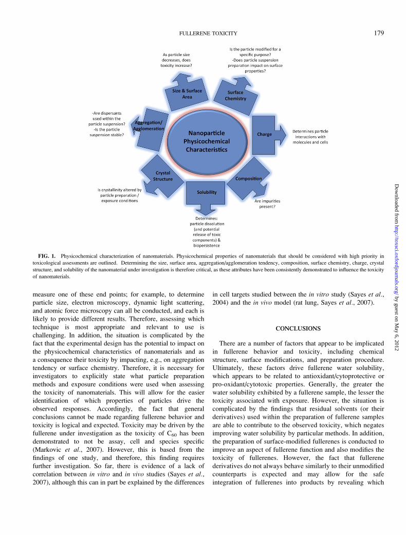
measure one of these end points; for example, to determine
particle size, electron microscopy, dynamic light scattering,
and atomic force microscopy can all be conducted, and each is
likely to provide different results. Therefore, assessing which
technique is most appropriate and relevant to use is
challenging. In addition, the situation is complicated by the
fact that the experimental design has the potential to impact on
the physicochemical characteristics of nanomaterials and as
a consequence their toxicity by impacting, e.g., on aggregation
tendency or surface chemistry. Therefore, it is necessary for
investigators to explicitly state what particle preparation
methods and exposure conditions were used when assessing
the toxicity of nanomaterials. This will allow for the easier
identification of which properties of particles drive the
observed responses. Accordingly, the fact that general
conclusions cannot be made regarding fullerene behavior and
toxicity is logical and expected. Toxicity may be driven by the
fullerene under investigation as the toxicity of C60 has been
demonstrated to not be assay, cell and species specific
(Markovic et al., 2007). However, this is based from the
findings of one study, and therefore, this finding requires
further investigation. So far, there is evidence of a lack of
correlation between in vitro and in vivo studies (Sayes et al.,2007), although this can in part be explained by the differences
in cell targets studied between the in vitro study (Sayes et al.,2004) and the in vivo model (rat lung, Sayes et al., 2007).
CONCLUSIONS
There are a number of factors that appear to be implicated
in fullerene behavior and toxicity, including chemical
structure, surface modifications, and preparation procedure.
Ultimately, these factors drive fullerene water solubility,
which appears to be related to antioxidant/cytoprotective or
pro-oxidant/cytotoxic properties. Generally, the greater the
water solubility exhibited by a fullerene sample, the lesser the
toxicity associated with exposure. However, the situation is
complicated by the findings that residual solvents (or their
derivatives) used within the preparation of fullerene samples
are able to contribute to the observed toxicity, which negates
improving water solubility by particular methods. In addition,
the preparation of surface-modified fullerenes is conducted to
improve an aspect of fullerene function and also modifies the
toxicity of fullerenes. However, the fact that fullerene
derivatives do not always behave similarly to their unmodified
counterparts is expected and may allow for the safe
integration of fullerenes into products by revealing which
FIG. 1. Physicochemical characterization of nanomaterials. Physicochemical properties of nanomaterials that should be considered with high priority in
toxicological assessments are outlined. Determining the size, surface area, aggregation/agglomeration tendency, composition, surface chemistry, charge, crystal
structure, and solubility of the nanomaterial under investigation is therefore critical, as these attributes have been consistently demonstrated to influence the toxicity
of nanomaterials.
FULLERENE TOXICITY 179
by guest on May 6, 2012
http://toxsci.oxfordjournals.org/D
ownloaded from
attributes of particles are most influential in driving toxico-
logical findings.
The studies conducted so far suggest that fullerene toxicity
involves an oxidant-driven response, suggesting that toxicity
evaluations should evaluate the potential of fullerenes to cause
oxidative stress and related consequences, such as inflamma-
tion or genotoxicity. The studies conducted with fullerenes thus
far are rather limited in terms of models used, targets
investigated, and mechanisms of toxicity. Much more work
is required to generate sufficient knowledge to inform a risk
assessment. Accordingly, it is unrealistic to make general-
izations about the behavior of fullerenes from the limited
number of studies that have been conducted as investigations
into the toxicity of fullerenes via specific routes of delivery, or
at particular cell and organ targets, are often too few in number
to make definite conclusions about fullerene behavior. In
addition, the quality (including the concentrations used and
experimental model) of conducted experiments is relevant to
consider, which is of vital importance when considering the
risk associated with fullerene exposure.
FUNDING
European Commission FP7 funded project: Engineered
Nanoparticles: Review of Health and Environmental Safety
(218433).
ACKNOWLEDGMENTS
The authors would like to thank all participants of the
ENRHES project for useful discussions. The authors would
especially like to thank Steve Hankin who was responsible for
the management of the project and Sheona Peters for helping
with the literature search.
REFERENCES
Baker, G. L., Gupta, A., Clark, M. L., Valenzuela, B. R., Staska, L. M.,
Harbo, S. J., Pierce, J. T., and Dill, J. A. (2008). Inhalation toxicity and lung
toxicokinetics of C60 fullerene nanoparticles and microparticles. Toxicol. Sci.
101, 122–131.
Bedrov, D., Smith, G. D., Davande, H., and Li, L. (2008). Passive transport of
C60 fullerenes through a lipid membrane: a molecular dynamics simulation
study. J. Phys. Chem. B 112, 2078–2084.
Belgorodsky, B., Fadeev, L., Kolsenik, J., and Gozin, M. (2006). Formation of
a soluble stable complex between pristine C60-fullerene and a native blood
protein. Chembiochem. 7, 1783–1789.
Benyamini, H., Shulman-Peleg, A., Wolfson, H. J., Belgorodsky, B.,
Fadeev, L., and Gozin, M. (2006). Interaction of C60-fullerene and
carboxyfullerene with proteins: docking and binding site alignment.
Bioconjug. Chem. 17, 378–386.
Bogdanovic, V., Stankov, K., Icevic, I., Zikic, D., Nikolic, A., Solajic, S.,
Djordjevic, A., and Bogdanovic, G. (2008). Fullerenol C60(OH)24 effects on
antioxidative enzymes activity in irradiated human erythroleukemia cell line.
J. Radiat. Res. 49, 321–327.
Brown, D. M., Wilson, M. R., MacNee, W., Stone, V., and Donaldson, K.
(2001). Size-dependent proinflammatory effects of ultrafine polystyrene
particles: a role for surface area and oxidative stress in the enhanced activity
of ultrafines. Toxicol. Appl. Pharmacol. 175, 191–199.
BSI Report. (2007). PAS 136 Terminology for Nanomaterials. Available at: http://
shop.bsigroup.com/en/Browse-by-Subject/Nanotechnology/Terminologies-
for-nanotechnologies/. Accessed November 24, 2009.
Bullard-Dillard, R., Creek, K. E., Scrivens, W. A., and Tour, J. M. (1996).
Tissue sites of uptake of 14C labelled C60. Bioorg. Chem. 24, 376–385.
Chen, B. X., Wilson, S. R., Das, M., Coughlin, D. J., and Erlanger, B. F.
(1998a). Antigenicity of fullerenes: antibodies specific for fullerenes and
their characteristics. Proc. Natl. Acad. Sci. U.S.A. 95, 10809–10813.
Chen, C., Xing, G., Wang, J., Zhao, Y., Li, B., Tang, J., Jia, G., Wang, T.,
Sun, J., Xing, L., et al. (2005). Multihydroxylated [Gd@C82(OH)22]n
nanoparticles: antineoplastic activity of high efficiency and low toxicity.
Nano Lett. 5, 2050–2057.
Chen, H. H., Yu, C., Ueng, T. H., Chen, S., Chen, B. J., Huang, K. J., and
Chiang, L. Y. (1998b). Acute and subacute toxicity study of water-soluble
polyalkylsulfonated C60 in rats. Toxicol. Pathol. 26, 143–151.
Dhawan, A., Taurozzi, J. S., Pandey, A. K., Shan, W., Miller, S. M.,
Hashsham, S. A., and Tarabara, V. V. (2006). Stable colloidal dispersions of
C60 fullerenes in water: evidence for genotoxicity. Environ. Sci. Technol. 40,
7394–7401.
Donaldson, K., and Stone, V. (2003). Current hypotheses on the mechanisms of
toxicity of ultrafine particles. Ann. Ist. Super. Sanita. 39, 405–410.
Donaldson, K., Tran, C. L., Jimenez, L. A., Duffin, R., Newby, D., Mills, N.,
MacNee, W., and Stone, V. (2005). Combustion-derived nanoparticles:
a critical review of their toxicology following inhalation exposure. Part.
Fibre Toxicol. 2, 1–14.
Duffin, R., Tran, L., Brown, D., Stone, V., and Donaldson, K. (2007).
Proinflammogenic effects of low-toxicity and metal nanoparticles in vivo and
in vitro: highlighting the role of particle surface area and surface reactivity.
Inhal. Toxicol. 19, 849–856.
Duffin, R., Tran, C. L., Clouter, A., Brown, D. M., MacNee, W., Stone, V., and
Donaldson, K. (2002). The importance of surface area and specific reactivity
in the acute pulmonary inflammatory response to particles. Ann. Occup. Hyg.
46, 242–245.
Dugan, L. L., Gabrielsen, J. K., Yu, S. P., Lin, T. S., and Choi, D. W.
(1996). Buckminsterfullerenol free radical scavengers reduce excitotoxic
and apoptotic death of cultured cortical neurons. Neurobiol. Dis. 3,
129–135.
Erlanger, B. F., Chen, B. X., Zhu, M., and Brus, L. (2001). Binding of an anti-
fullerene IgG monoclonal antibody to single wall carbon nanotubes. Nano
Lett. 1, 465–467.
Ferin, J., Oberdorster, G., and Penney, D. P. (1992). Pulmonary retention
of ultrafine and fine particles in rats. Am. J. Respir. Cell Mol. Biol. 6,
535–542.
Fortner, J. D., Lyon, D. Y., Sayes, C. M., Boyd, A. M., Falkner, J. C.,
Hotze, E. M., Alemany, L. B., Tao, Y. J., Guo, W., Ausman, K. D., et al.
(2005). C60 in water: nanocrystal formation and microbial response. Environ.
Sci. Technol. 39, 4307–4316.
Friedman, S. H., DeCamp, D. L., Sijbesma, R., Srdanov, G., Wudl, F., and
Kenyon, G. L. (1993). Inhibition of the HIV-1 protease by fullerene
derivatives: model building studies and experimental verification. J. Am.
Chem. Soc. 115, 6506–6509.
Fujita, K., Morimoto, Y., Ogami, A., Myojyo, T., Tanaka, I., Shimada, M.,
Wang, W. N., Endoh, S., Uchida, K., Nakazato, T., et al. (2009). Gene
expression profiles in rat lung after inhalation exposure to C(60) fullerene
particles. Toxicology 258, 47–55.
180 JOHNSTON ET AL.
by guest on May 6, 2012
http://toxsci.oxfordjournals.org/D
ownloaded from
Gharbi, N., Pressac, M., Hadchouel, M., Szwarc, H., Wilson, S. R., and
Moussa, F. (2005). [60]Fullerene is a powerful antioxidant in vivo with no
acute or subacute toxicity. Nano Lett. 5, 2578–2585.
Halford, B. (2006). Fullerene for the face. Chem. Eng. News 84, 47.
Harhaji, L., Isakovic, A., Vucicevic, L., Janjetovic, K., Misirkic, M.,
Markovic, Z., Todorovic-Markovic, B., Nikolic, N., Vranjes-Djuric, S.,
Nikolic, Z., et al. (2008). Modulation of tumor necrosis factor-mediated cell
death by fullerenes. Pharm. Res. 25, 1365–1376.
Henry, T. B., Menn, F. M., Fleming, J. T., Wilgus, J., Compton, R. N., and
Sayler, G. S. (2007). Attributing effects of aqueous C60 nano-aggregates to
tetrahydrofuran decomposition products in larval zebrafish by assessment of
gene expression. Environ. Health. Perspect. 115, 1059–1065.
Huang, S. T., Liao, J. S., Fang, H. W., and Lin, C. M. (2008). Synthesis and
anti-inflammation evaluation of new C60 fulleropyrrolidines bearing bi-
ologically active xanthine. Bioorg. Med. Chem. Lett. 18, 99–103.
Huczko, A., Lange, H., and Calko, E. (1999). Fullerenes: experimental
evidence for a null risk of skin irritation and allergy. Fullerene Sci. Technol.
7, 935–939.
Injac, R., Perse, M., Cerne, M., Potocnik, N., Radic, N., Govedarica, B.,
Djordjevic, A., Cerar, A., and Strukelj, B. (2009). Protective effects
of fullerenol C60(OH)24 against doxorubicin-induced cardiotoxicity and
hepatotoxicity in rats with colorectal cancer. Biomaterials 30, 1184–1196.
Isakovic, A., Markovic, Z., Nikolic, N., Todorovic-Markovic, B., Vranjes-
Djuric, S., Harhaji, L., Raicevic, N., Romcevic, N., Vasiljevic-Radovic, D.,
Dramicanin, M., et al. (2006). Inactivation of nanocrystalline C60
cytotoxicity by gamma-irradiation. Biomaterials 27, 5049–5058.
Jacobsen, N. R., Pojana, G., White, P., Moller, P., Cohn, C. A.,
Korsholm, K. S., Vogel, U., Marcomini, A., Loft, S., and Wallin, H.
(2008). Genotoxicity, cytotoxicity, and reactive oxygen species induced
by single-walled carbon nanotubes and C(60) fullerenes in the FE1-
mutatrade mark mouse lung epithelial cells. Environ. Mol. Mutagen. 49,
476–487.
Kagan, V. E., Bayir, H., and Shvedova, A. A. (2005). Nanomedicine and
nanotoxicology: two sides of the same coin. Nanomedicine 1, 313–316.
Kamat, J. P., Devasagayam, T. P. A., Priyadarsini, K. I., and Mohan, H. (2000).
Reactive oxygen species mediated membrane damage induced by ful-
lerene derivatives and its possible biological implications. Toxicology 155,
55–61.
Kroto, H. W., Heath, J. R., Obrien, S. C., Curl, R. F., and Smalley, R. E.
(1985). C60: buckminsterfullerene. Nature, 318, 162–163.
Lin, A. M., Fang, S. F., Lin, S. Z., Chou, C. K., Luh, T. Y., and Ho, L. T.
(2002). Local carboxyfullerene protects cortical infarction in rat brain.
Neurosci. Res. 43, 317–321.
Lyon, D. Y., Adams, L. K., Falkner, J. C., and Alvarezt, P. J. (2006).
Antibacterial activity of fullerene water suspensions: effects of preparation
method and particle size. Environ. Sci. Technol. 40, 4360–4366.
Markovic, Z., Todorovic-Markovic, B., Kleut, D., Nikolic, N., Vranjes-
Djuric, S., Misirkic, M., Vucicevic, L., Janjetovic, K., Isakovic, A.,
Harhaji, L., et al. (2007). The mechanism of cell-damaging reactive oxygen
generation by colloidal fullerenes. Biomaterials 28, 5437–5448.
Maynard, A. D., Aitken, R. J., Butz, T., Colvin, V., Donaldson, K.,
Oberdorster, G., Philbert, M. A., Ryan, J., Seaton, A., Stone, V., et al.
(2006). Safe handling in nanotechnology. Nature 444, 267–269.
Mori, T., Ito, S., Matsubayashi, K., and Sawaguchi, T. (2007). Comparison of
nitric oxide synthase inhibitors, phospholipase A2 inhibitor and free radical
scavengers as attenuators of opioid withdrawal syndrome. Behav. Pharma-
col. 18, 725–729.
Mori, T., Takada, H., Ito, S., Matsubayashi, K., Miwa, N., and
Sawaguchi, T. (2006). Preclinical studies on safety of fullerene upon
acute oral administration and evaluation for no mutagenesis. Toxicology
225, 48–54.
Nielsen, G. D., Roursgaard, M., Jensen, K. A., Poulsen, S. S., and Larsen, S. T.
(2008). In vivo biology and toxicology of fullerenes and their derivatives.
Basic Clin. Pharmacol. Toxicol. 103, 197–208.
Porter, A. E., Muller, K., Skepper, J., Midgley, P., and Welland, M. (2006).
Uptake of C60 by human monocyte macrophages, its localization and
implications for toxicity: studied by high resolution electron microscopy and
electron tomography. Acta. Biomater. 2, 409–419.
Powell, M. C., and Kanarek, M. S. (2006). Nanomaterial health effects–Part
one, Background and current knowledge. Wisconsin Medical journal. 105,
16–20.
Qiao, R., Roberts, A. P., Mount, A. S., Klaine, S. J., and Ke, P. C. (2007).
Translocation of C60 and its derivatives across a lipid bilayer. Nano Lett. 7,
614–619.
Radomski, A., Jurasz, P., Alonso-Escolano, D., Drews, M., Morandi, M.,
Malinski, T., and Radomski, M. W. (2005). Nanoparticle-induced platelet
aggregation and vascular thrombosis. Br. J. Pharmacol. 146, 882–893.
Roberts, J. E., Wielgus, A. R., Boyes, W. K., Andley, U., and Chignell, C. F.
(2008). Phototoxicity and cytotoxicity of fullerol in human lens epithelial
cells. Toxicol. Appl. Pharmacol. 228, 49–58.
Roursgaard, M., Poulsen, S. S., Kepley, C. L., Hammer, M., Nielsen, G. D.,
and Larsen, S. T. (2008). Polyhydroxylated C60 fullerene (fullerenol)
attenuates neutrophilic lung inflammation in mice. Basic Clin. Pharmacol.
Toxicol. 103, 386–388.
Rouse, J. G., Yang, J., Barron, A. R., and Monteiro-Riviere, N. A. (2006).
Fullerene-based amino acid nanoparticle interactions with human epidermal
keratinocytes. Toxicol. In Vitro 20, 1313–1320.
Rouse, J. G., Yang, J., Ryman-Rasmussen, J. P., Barron, A. R., and Monteiro-
Riviere, N. A. (2007). Effects of mechanical flexion on the penetration of
fullerene amino acid-derivatized peptide nanoparticles through skin. Nano
Lett. 7, 155–160.
Sayes, C. M., Fortner, J. D., Guo, W., Lyon, D. Y., Boyd, A. M., Ausman, K.,
Tao, Y. J., Sitharaman, B., Wilson, L. J., Hughes, J. B., et al. (2004). The
differential cytotoxicity of water-soluble fullerenes. Nano Lett. 4,
1881–1887.
Sayes, C. M., Gobin, A. M., Ausman, K. D., Mendez, J., West, J. L., and
Colvin, V. L. (2005). Nano-C60 cytotoxicity is due to lipid peroxidation.
Biomaterials 26, 7587–7595.
Sayes, C. M., Marchione, A. A., Reed, K. L., and Warheit, D. B. (2007).
Comparative pulmonary toxicity assessments of C60 water suspensions in
rats: few differences in fullerene toxicity in vivo in contrast to in vitro
profiles. Nano Lett. 7, 2399–2406.
Schinazi, R. F., Sijbesma, R., Srdanov, G., Hill, C. L., and Wudl, F. (1993).
Synthesis and virucidal activity of a water-soluble, configurationally stable,
derivatised C60 fullerene. Antimicrob. Agents Chemother. 37, 1707–1710.
Scrivens, W. A., Tour, J. M., Creek, K. E., and Pirisi, L. (1994). Synthesis of14C-labelled C60, its suspension in water, and its uptake by human
keratinocytes. J. Am. Chem. Soc. 116, 4517–4518.
Sera, N., Tokiwa, H., and Miyata, N. (1996). Mutagenicity of the fullerene C60-
generated singlet oxygen dependent formation of lipid peroxides. Carcino-
genesis 17, 2163–2169.
Stoeger, T., Reinhard, C., Takenaka, S., Schroeppel, A., Karg, E., Ritter, B.,
Heyder, J., and Schulz, H. (2006). Instillation of six different ultrafine carbon
particles indicates a surface area threshold dose for acute lung inflammation
in mice. Environ. Health Perspect. 114, 328–333.
Stone, V., Shaw, J., Brown, D. M., MacNee, W., Faux, S. P., and
Donaldson, K. (1998). The role of oxidative stress in the prolonged
inhibitory effect of ultrafine carbon black on epithelial cell function. Toxicol.
In Vitro 12, 649–659.
Tabata, Y., Murakami, Y., and Ikada, Y. (1997). Photodynamic effect of
polyethylene glycol-modified fullerene on tumour. Jpn J. Cancer Res. 88,
1108–1116.
FULLERENE TOXICITY 181
by guest on May 6, 2012
http://toxsci.oxfordjournals.org/D
ownloaded from
Trajkovic, S., Dobric, S., Jacevic, V., Dragojevic-Simic, V., Milovanovic, Z.,
and Dordevic, A. (2007). Tissue-protective effects of fullerenol
C60(OH)24 and amifostine in irradiated rats. Colloids Surf. B Biointer-
faces 58, 39–43.
Tsao, N., Kanakamma, P. P., Luh, T. Y., Chou, C. K., and Lei, H. Y. (1999).
Inhibition of Escherichia coli-induced meningitis by carboxyfullerene.
Antimicrob. Agents Chemother. 43, 2273–2277.
Tsuchiya, T., Oguri, I., Yamakoshi, Y. N., and Miyata, N. (1996). Novel
harmful effects of [60]fullerene on mouse embryos in vitro and in vivo. FEBS
Lett. 393, 139–145.
Tykhomyrov, A. A., Nedzvetsky, V. S., Klochkov, V. K., and
Andrievsky, G. V. (2008). Nanostructures of hydrated C60 fullerene
(C60HyFn) protect rat brain against alcohol impact and attenuate behavioural
impairments of alcoholized animals. Toxicology 246, 158–165.
Ueng, T. H., Kang, J. J., Wang, H. W., Cheng, Y. W., and Chiang, L. Y.
(1997). Suppression of microsomal cytochrome P450-dependent monoox-
ygenases and mitochondrial oxidative phosphorylation by fullerenol,
a polyhydroxylated fullerene C60. Toxicol. Lett. 93, 29–37.
Usenko, C. Y., Harper, S. L., and Tanguay, R. L. (2008). Fullerene C60
exposure elicits an oxidative stress response in embryonic zebrafish. Toxicol.
Appl. Pharmacol. 229, 44–55.
Wang, I. C., Tai, L. A., Lee, D. D., Kanakamma, P. P., Shen, C. K., Luh, T. Y.,
Cheng, C. H., and Hwang, K. C. (1999). C(60) and water-soluble fullerene
derivatives as antioxidants against radical-initiated lipid peroxidation. J.
Med. Chem. 42, 4614–4620.
Warheit, D. B. (2008). How meaningful are the results of nanotoxicity studies
in the absence of adequate material characterization? Toxicol. Sci. 101,
183–185.
Wong-Ekkabut, J., Baoukina, S., Triampo, W., Tang, I. M., Tieleman, D. P.,
and Monticelli, L. (2008). Computer simulation study of fullerene
translocation through lipid membranes. Nat. Nanotechnol. 3, 363–368.
Xia, T., Kovochich, M., Brant, J., Hotze, M., Sempf, J., Oberley, T.,
Sioutas, C., Yeh, J. I., Wiesner, M. R., and Nel, A. E. (2006). Comparison
of the abilities of ambient and manufactured nanoparticles to induce cel-
lular toxicity according to an oxidative stress paradigm. Nano Lett. 6,
1794–1807.
Xiao, L., Takada, H., Gan, X., and Miwa, N. (2006). The water-soluble
fullerene derivative ‘‘radical sponge’’ exerts cytoprotective action against
UVA irradiation but not visible-light-catalyzed cytotoxicity in human skin
keratinocytes. Bioorg. Med. Chem. Lett. 16, 1590–1595.
Yamago, S., Tokuyama, H., Nakamura, E., Kikuchi, K., Kananishi, S.,
Sueki, K., Nakahara, H., Enomoto, S., and Ambe, F. (1995). In vivo
biological behaviour of a water-miscible fullerene: 14C labelling, absorption,
distribution, excretion and acute toxicity. Chem. Biol. 2, 385–389.
Yamawaki, H., and Iwai, N. (2006). Cytotoxicity of water-soluble fullerene in
vascular endothelial cells. Am. J. Physiol. 290, C1495–C1502.
Yin, J. J., Lao, F., Fu, P. P., Wamer, W. G., Zhao, Y., Wang, P. C., Qiu, Y.,
Sun, B., Xing, G., Dong, J., et al. (2009). The scavenging of reactive oxygen
species and the potential for cell protection by functionalised fullerene
materials. Biomaterials 30, 611–612.
Yin, J. J., Lao, F., Meng, J., Fu, P. P., Zhao, Y., Xing, G., Gao, X., Sun, B.,
Wang, P. C., Chen, C., et al. (2008). Inhibition of tumor growth by
endohedral metallofullerenol nanoparticles optimized as reactive oxygen
species scavenger. Mol. Pharmacol. 74, 1132–1140.
Zhang, B., Cho, M., Fortner, J. D., Lee, J., Huang, C. H., Hughes, J. B., and
Kim, J. H. (2009). Delineating oxidative processes of aqueous
C60 preparations: role of THF peroxide. Environ. Sci. Technol. 43,
108–113.
Zhu, J. T., He, J., Chen, J. Y., Lu, D. R., and Zhou, L. W. (2008). Fast
differential interference contrast imaging combined with autocorrelation
treatments to measure the heart rate of embryonic fish. J. Biomed. Opt. 13,
020503.
182 JOHNSTON ET AL.
by guest on May 6, 2012
http://toxsci.oxfordjournals.org/D
ownloaded from


![13C NMR and Raman Studies of Fullerene-Based Poly ... · multi-addition of organocopper reagents to fullerenes [17] were also carried out. The literature survey shows suffi- cient](https://img.pdfslide.us/doc/110x75/5f32af9eb832ae3bf8127a84/13c-nmr-and-raman-studies-of-fullerene-based-poly-multi-addition-of-organocopper.jpg)


![1 Carbon Nanostructures: Covalent and Macromolecular Chemistry · 1.2 Fullerene-Containing Polymers Since the achievement of [60]fullerenes in ponderable amounts [1], its combination](https://img.pdfslide.us/doc/110x75/5afe37407f8b9a944d8e86aa/1-carbon-nanostructures-covalent-and-macromolecular-chemistry-fullerene-containing.jpg)

![DESIGNING MINI-FULLERENES AND THEIR RELATIVES ON …mp.ipme.ru/e-journals/MPM/no_12014/MPM120_03_melker.pdf · 1. Introduction . In [1] the term “ fullerene ” was taken in a broad](https://img.pdfslide.us/doc/110x75/603d4dcafc3b9135a749ac7e/designing-mini-fullerenes-and-their-relatives-on-mpipmerue-journalsmpmno12014mpm12003.jpg)
![Fullerene (Version4.4 ...ctcp.massey.ac.nz/data/UserManualv4.4.pdf · number as defined in the book “Atlas of Fullerenes” by Fowler and Manolopoulos [8, 34] (e.g. input file](https://img.pdfslide.us/doc/110x75/5fca1eaff997976f7415ff60/fullerene-version44-ctcp-number-as-deined-in-the-book-aoeatlas-of-fullerenesa.jpg)