Embed Size (px)
Citation preview
Review
Stereotactic Body Radiotherapy Immunological Planning – Road to Abscopal
Effect by Design
Kumara Swamy1
1Consultant and Head Radiation Oncology, Aster CMI,
#43/2, New Airport Road, Sahakar Nagar, Bangalore – 560 092, India
E-mail: [email protected]
Correspondence e-mail: [email protected]
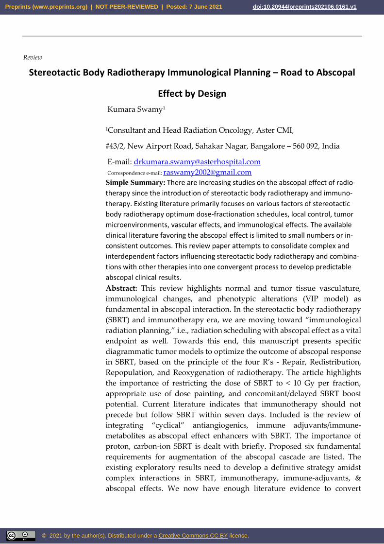
Simple Summary: There are increasing studies on the abscopal effect of radio-
therapy since the introduction of stereotactic body radiotherapy and immuno-
therapy. Existing literature primarily focuses on various factors of stereotactic
body radiotherapy optimum dose-fractionation schedules, local control, tumor
microenvironments, vascular effects, and immunological effects. The available
clinical literature favoring the abscopal effect is limited to small numbers or in-
consistent outcomes. This review paper attempts to consolidate complex and
interdependent factors influencing stereotactic body radiotherapy and combina-
tions with other therapies into one convergent process to develop predictable
abscopal clinical results.
Abstract: This review highlights normal and tumor tissue vasculature,
immunological changes, and phenotypic alterations (VIP model) as
fundamental in abscopal interaction. In the stereotactic body radiotherapy
(SBRT) and immunotherapy era, we are moving toward “immunological
radiation planning,” i.e., radiation scheduling with abscopal effect as a vital
endpoint as well. Towards this end, this manuscript presents specific
diagrammatic tumor models to optimize the outcome of abscopal response
in SBRT, based on the principle of the four R’s - Repair, Redistribution,
Repopulation, and Reoxygenation of radiotherapy. The article highlights
the importance of restricting the dose of SBRT to < 10 Gy per fraction,
appropriate use of dose painting, and concomitant/delayed SBRT boost
potential. Current literature indicates that immunotherapy should not
precede but follow SBRT within seven days. Included is the review of
integrating “cyclical” antiangiogenics, immune adjuvants/immune-
metabolites as abscopal effect enhancers with SBRT. The importance of
proton, carbon-ion SBRT is dealt with briefly. Proposed six fundamental
requirements for augmentation of the abscopal cascade are listed. The
existing exploratory results need to develop a definitive strategy amidst
complex interactions in SBRT, immunotherapy, immune-adjuvants, &
abscopal effects. We now have enough literature evidence to convert
Preprints (www.preprints.org) | NOT PEER-REVIEWED | Posted: 7 June 2021 doi:10.20944/preprints202106.0161.v1
© 2021 by the author(s). Distributed under a Creative Commons CC BY license.
“abscopal by chance” to “abscopal by design” by harmonized
combinatorial approach.
.
Keywords: SBRT, SABR, Abscopal, vascular-normalization, immunotherapy,
phenotypic, antiangiogenics, immunoadjuvants, VIP-model, High-LET
1. Introduction
Presently, several significant developments have changed
radiotherapy’s (RT) scope regarding its planning and outcomes. First is
the advent of Stereotactic Body Radiotherapy (SBRT). Technological
evolution in stereotactic localization of cancer and its movement has led to
very high precision therapy, limiting the dose to the normal tissue. This
precision has helped to deliver extreme hypofractionation to the
macroscopic disease safely.
The second has been the evolution of the concept of oligometastases.
In stage IV, a subset of patients has limited metastases
termed oligometastases. These patients are still amenable to treatment by
radical approach. A seminal review article by Timmerman et al. in 2009
emphasized the role of SBRT in the treatment of oligometastases. With
SBRT, in as much as 25% of patients with oligometastases long-term control
of the disease was achieved [1].
There has been a gradual evolution in selecting patients with
oligometastases for SBRT over the years. Initially, it was a single metastasis
of limited size in an organ, which then later included a few metastases in a
single organ. Presently, in general, a few metastases in multiple organs also
come within the ambit of the oligometastases status, as long as the total
volume does not exceed a particular level depending on the site of
metastases. Indications for oligometastases are still evolving in clinical
situations, with a few metastases persisting after chemo-immunotherapy
(oligo-persistence) or a few coming back (oligo-recurrence) or progressing
in a limited number of metastases (oligo-progression). Recently, the interest
is amplified in results of SBRT by increasing reports of outside the field and
distant (abscopal) response effects.
The third development is the arrival of several newer immunotherapy
agents to target cancer cells and enhance the immune response against
cancer at the same time. There has been a potential breakthrough with the
combined use of SBRT with these immunogenic drugs.
For a long time, RT has been deemed locally immune-suppressive with
its cytotoxic effects on leukocytes. This immunosuppressive action is
widely used in total body irradiation (TBI) as a bone marrow
transplantation-conditioning regimen. However, in recent years, the RT-
induced immune-stimulatory effect has been increasingly recognized,
Preprints (www.preprints.org) | NOT PEER-REVIEWED | Posted: 7 June 2021 doi:10.20944/preprints202106.0161.v1
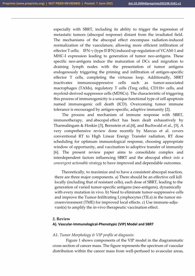
especially with SBRT, including its ability to trigger the regression of
metastatic tumors (abscopal response) distant from the irradiated field.
The mechanisms of the abscopal effect encompass radiation-induced
normalization of the vasculature, allowing more efficient infiltration of
effector T cells; IFN-γ (type II IFN) induced up-regulation of VCAM-1 and
MHC-I expression leading to generation of tumor neo-antigens. These
specific neo-antigens induce the maturation of DCs and migration to
draining lymph nodes with the presentation of tumor antigens
endogenously triggering the priming and infiltration of antigen-specific
effector T cells, completing the virtuous loop. Additionally, SBRT
inactivates immunosuppressive cells such as tumor-associated
macrophages (TAMs), regulatory T cells (Treg cells), CD11b+ cells, and
myeloid-derived suppressor cells (MDSCs). The characteristic of triggering
this process of immunogenicity is a unique functional type of cell apoptosis
named immunogenic cell death (ICD). Overcoming tumor immune
tolerance is encouraged by antigen-specific, adaptive immunity [2].
The process and mechanism of immune response with SBRT,
immunotherapy, and abscopal effect has been dealt exhaustively by
Tharmalingam & Hoskin [3], Bernstein et al.[4], and Buchwald et al., [5]. A
very comprehensive review done recently by Marcus et al. covers
conventional RT to High Linear Energy Transfer radiation, RT dose
scheduling for optimum immunological response, choosing appropriate
window of opportunity, and vaccination to adoptive transfer of immunity
[6]. The present review paper aims to consolidate complex and
interdependent factors influencing SBRT and the abscopal effect into a
convergent actionable strategy to have improved and dependable outcomes.
Theoretically, to maximize and to have a consistent abscopal reaction,
there are three major components. a) There should be an effective cell kill
locally (including that of resistant cells), each dose of SBRT, leading to the
generation of varied tumor-specific antigens (neo-antigens), dynamically
with every mutation in vivo. b) Need to eliminate tumor-suppressive cells
and improve the Tumor-Infiltrating Lymphocytes (TILs) in the tumor mi-
croenvironment (TME) for improved local effects. c) Use immune-adju-
vant(s) to amplify the in-vivo therapeutic vaccination effect.
2. Review
A). Vascular-Immunological-Phenotypic (VIP) Model and SBRT
A1. Tumor Morphology & VIP profile at diagnosis:
Figure 1 shows components of the VIP model in the diagrammatic
cross-section of cancer mass. The figure represents the spectrum of vascular
distribution within the cancer mass from well-perfused to avascular areas,
Preprints (www.preprints.org) | NOT PEER-REVIEWED | Posted: 7 June 2021 doi:10.20944/preprints202106.0161.v1
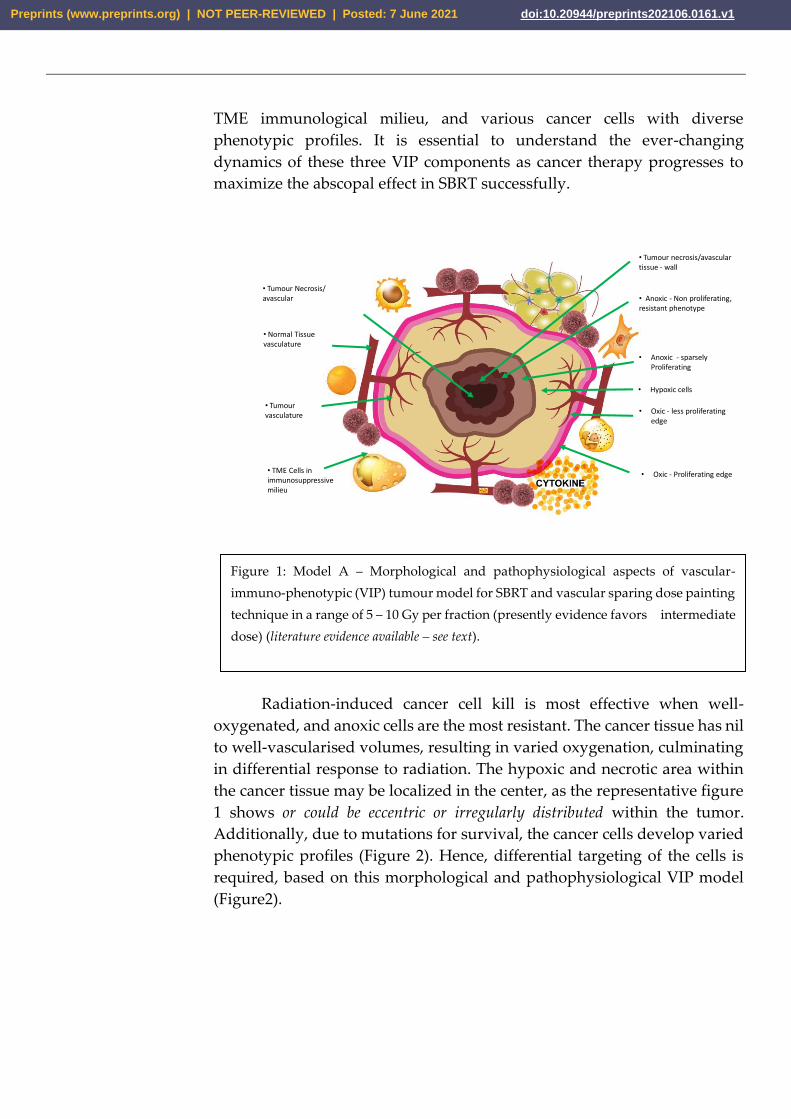
TME immunological milieu, and various cancer cells with diverse
phenotypic profiles. It is essential to understand the ever-changing
dynamics of these three VIP components as cancer therapy progresses to
maximize the abscopal effect in SBRT successfully.
• Oxic - Proliferating edge
• Tumour Necrosis/avascular • Anoxic - Non proliferating,
resistant phenotype
• Anoxic - sparsely Proliferating
• Hypoxic cells
• Oxic - less proliferating edge
• Normal Tissue vasculature
• TME Cells in immunosuppressive milieu
• Tumour vasculature
• Tumour necrosis/avasculartissue - wall
Radiation-induced cancer cell kill is most effective when well-
oxygenated, and anoxic cells are the most resistant. The cancer tissue has nil
to well-vascularised volumes, resulting in varied oxygenation, culminating
in differential response to radiation. The hypoxic and necrotic area within
the cancer tissue may be localized in the center, as the representative figure
1 shows or could be eccentric or irregularly distributed within the tumor.
Additionally, due to mutations for survival, the cancer cells develop varied
phenotypic profiles (Figure 2). Hence, differential targeting of the cells is
required, based on this morphological and pathophysiological VIP model
(Figure2).
Figure 1: Model A – Morphological and pathophysiological aspects of vascular-
immuno-phenotypic (VIP) tumour model for SBRT and vascular sparing dose painting
technique in a range of 5 – 10 Gy per fraction (presently evidence favors intermediate
dose) (literature evidence available – see text).
Preprints (www.preprints.org) | NOT PEER-REVIEWED | Posted: 7 June 2021 doi:10.20944/preprints202106.0161.v1
Tumor Cell
Tumor cells with resistant phenotype
In practical terms, three strategic aspects emerge. First, with
improvements in functional imaging techniques, it would be possible to
have three-dimensional models of oxygenation. The importance of figure
1lies in the fact that with SBRT, we have the technology to titrate the dose
accordingly by creating controlled hot spots (dose-painting). The dose-
painting has dual benefits with SBRT technology. One, a higher required
gradient dose could be delivered efficiently, if needed. Two, normal tissue
(including the vasculature) sparing will be more effective. Tubin et al.
demonstrated the feasibility of this concept by contouring and treating only
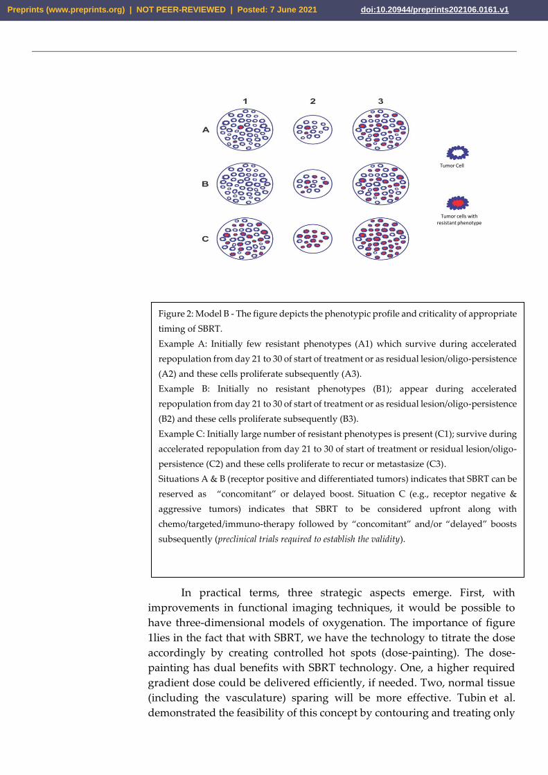
Figure 2: Model B - The figure depicts the phenotypic profile and criticality of appropriate
timing of SBRT.
Example A: Initially few resistant phenotypes (A1) which survive during accelerated
repopulation from day 21 to 30 of start of treatment or as residual lesion/oligo-persistence
(A2) and these cells proliferate subsequently (A3).
Example B: Initially no resistant phenotypes (B1); appear during accelerated
repopulation from day 21 to 30 of start of treatment or as residual lesion/oligo-persistence
(B2) and these cells proliferate subsequently (B3).
Example C: Initially large number of resistant phenotypes is present (C1); survive during
accelerated repopulation from day 21 to 30 of start of treatment or residual lesion/oligo-
persistence (C2) and these cells proliferate to recur or metastasize (C3).
Situations A & B (receptor positive and differentiated tumors) indicates that SBRT can be
reserved as “concomitant” or delayed boost. Situation C (e.g., receptor negative &
aggressive tumors) indicates that SBRT to be considered upfront along with
chemo/targeted/immuno-therapy followed by “concomitant” and/or “delayed” boosts
subsequently (preclinical trials required to establish the validity).
Preprints (www.preprints.org) | NOT PEER-REVIEWED | Posted: 7 June 2021 doi:10.20944/preprints202106.0161.v1
the hypoxic tumor segment. He delivered 10-12 Gy 1 to 3 fractions to a hypo-
vascularized and hypo-metabolic junctional zone between the central
necrotic and peripheral hyper-vascularized-hypermetabolic tumor segment
as a palliative approach and observed abscopal effect in non-irradiated
segments and nodes [7].
Second, some cancer cells are more sensitive and than others,
requiring different strategies. Primarily, two strategies are target cells with
some receptors/markers that are susceptible to cell kill or cells that have
reached the level of undifferentiation requiring the aggressive approach.
The strategy requires systematic combinations of treatment with appropriate
intensity and timing, where SBRT would be invaluable.
Third, the genomic landscape of cancer is dynamic and ever-
changing in response to the fluctuating tumor microenvironment and
cancer-directed treatment. This change requires an adoptive personalized
approach in the SBRT delivery. The major categories of cells with different
phenotypic profiles are stem cells, especially in a vascular niche or growing
edge; anoxic clonogenic cells in the wall of necrotic areas; hypoxic/anoxic
clonogenic cells in G0 phase of cell cycle; cells with differential SUV uptake
and cells with varying mutation burden. With improving imaging
technology, it would be possible to get three-dimensional information
facilitating matched approach and dose painting with SBRT.
A2. VIP profile of Responding and Residual lesions:
It is crucial to study the tumor profile during the treatment and later
residual tissue. Figure 3 enumerate the changing profile of cancer with the
treatment. During therapy with either fractionated RT or chemotherapy,
decreased interstitial pressure happens due to tumor size reduction
following initial cancer cell kill and improved vasculature, leading to
additional cell kill of oxygenated cells. TME also evolves, resulting in
improved immunological reaction (Figure 3). However, there could be
accelerated repopulation (AR) of surviving relatively resistant cancer cells
at this time.
Preprints (www.preprints.org) | NOT PEER-REVIEWED | Posted: 7 June 2021 doi:10.20944/preprints202106.0161.v1
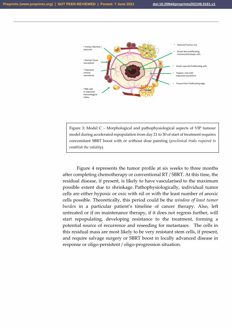
• Reduced Tumour size
• Anoxic Non proliferating, resistant phenotypic cells
• Anoxic sparsely Proliferating cells
• Hypoxic, now with improved vasculature
• Present Oxic Proliferating edge
Necrosis• Normal Tissue vasculature
•TME cellsin improved immunological milieu
• Improved tumour vasculature
• Tumour Necrosis /avascular
Figure 4 represents the tumor profile at six weeks to three months
after completing chemotherapy or conventional RT / SBRT. At this time, the
residual disease, if present, is likely to have vascularised to the maximum
possible extent due to shrinkage. Pathophysiologically, individual tumor
cells are either hypoxic or oxic with nil or with the least number of anoxic
cells possible. Theoretically, this period could be the window of least tumor
burden in a particular patient’s timeline of cancer therapy. Also, left
untreated or if on maintenance therapy, if it does not regress further, will
start repopulating, developing resistance to the treatment, forming a
potential source of recurrence and reseeding for metastases. The cells in
this residual mass are most likely to be very resistant stem cells, if present,
and require salvage surgery or SBRT boost in locally advanced disease in
response or oligo-persistent / oligo-progression situation.
Figure 3: Model C – Morphological and pathophysiological aspects of VIP tumour
model during accelerated repopulation from day 21 to 30 of start of treatment requires
concomitant SBRT boost with or without dose painting (preclinical trials required to
establish the validity).
Preprints (www.preprints.org) | NOT PEER-REVIEWED | Posted: 7 June 2021 doi:10.20944/preprints202106.0161.v1
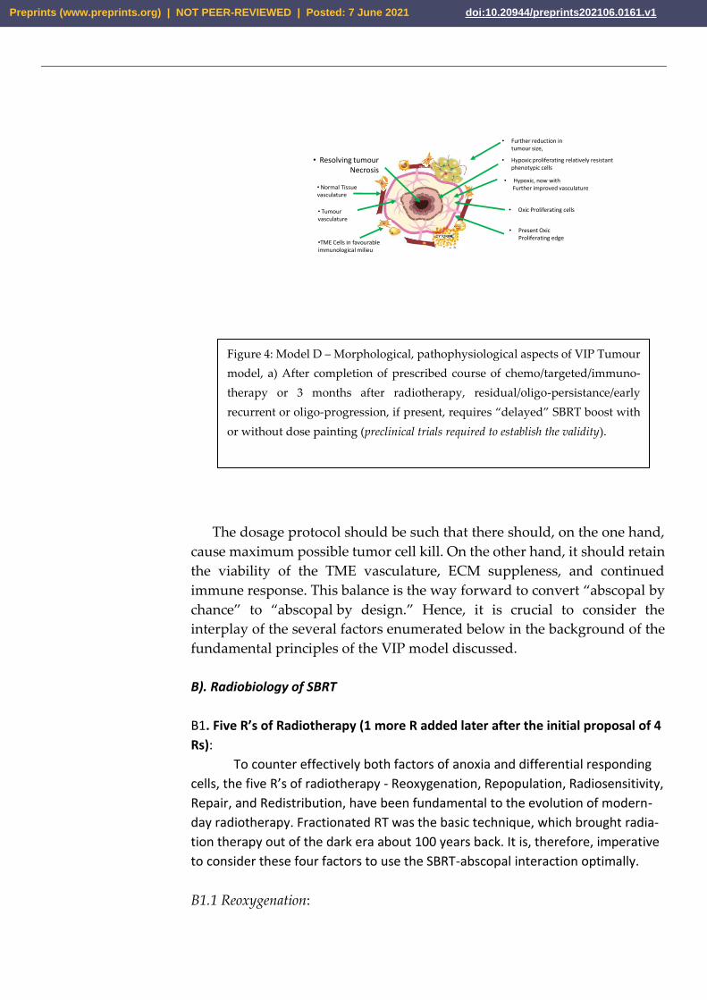
• Further reduction in tumour size,
• Hypoxic proliferating relatively resistant phenotypic cells
• Oxic Proliferating cells
• Hypoxic, now with Further improved vasculature
• Present OxicProliferating edge
Necrosis
• Normal Tissue vasculature
•TME Cells in favourable immunological milieu
• Tumour vasculature
• Resolving tumour Necrosis
The dosage protocol should be such that there should, on the one hand,
cause maximum possible tumor cell kill. On the other hand, it should retain
the viability of the TME vasculature, ECM suppleness, and continued
immune response. This balance is the way forward to convert “abscopal by
chance” to “abscopal by design.” Hence, it is crucial to consider the
interplay of the several factors enumerated below in the background of the
fundamental principles of the VIP model discussed.
B). Radiobiology of SBRT
B1. Five R’s of Radiotherapy (1 more R added later after the initial proposal of 4
Rs):
To counter effectively both factors of anoxia and differential responding
cells, the five R’s of radiotherapy - Reoxygenation, Repopulation, Radiosensitivity,
Repair, and Redistribution, have been fundamental to the evolution of modern-
day radiotherapy. Fractionated RT was the basic technique, which brought radia-
tion therapy out of the dark era about 100 years back. It is, therefore, imperative
to consider these four factors to use the SBRT-abscopal interaction optimally.
B1.1 Reoxygenation:
Figure 4: Model D – Morphological, pathophysiological aspects of VIP Tumour
model, a) After completion of prescribed course of chemo/targeted/immuno-
therapy or 3 months after radiotherapy, residual/oligo-persistance/early
recurrent or oligo-progression, if present, requires “delayed” SBRT boost with
or without dose painting (preclinical trials required to establish the validity).
Preprints (www.preprints.org) | NOT PEER-REVIEWED | Posted: 7 June 2021 doi:10.20944/preprints202106.0161.v1
In SBRT, Kim et al. elucidated that oxygen consumption would
drastically diminish after a massive death of tumor cells, and thus the
surviving hypoxic cells may be reoxygenated [8]. Shibamoto et al. proposed
the concept of ‘reoxygenation utilization rate’ (RUR) in SRT [9]. With two,
four, six, eight and thirty fractions were 50%, 75%, 83%, 87.5%, 97%
respectively. Therefore, the authors theorized that, unlike single fractions,
six to eight fractions of SBRT treatments might be sufficient to utilize
the reoxygenation phenomenon [9]. This study indicates that in fractionated
SBRT, unlike single fraction, reoxygenation does contribute to increased
cancer cell kill like in conventional RT.
B1.2 Repopulation and Radiosensitivity:
Withers et al. initially described the phenomenon of clonogenic
repopulation in squamous cell carcinoma of the head and neck, accelerating
after a lag period of four weeks(± one week) after the initiation of RT [10].
Although the onset of accelerated repopulation is not explicitly known for a
particular type of cancer in the temporal timeline, one to two weeks of
treatment with fractionated SBRT may be advantageous in reducing the
acceleration in repopulation. This phenomenon particularly holds good for
rapidly proliferating cells and may contribute to the improvement of local
control [11] in aggressive disease. The delayed acceleration can happen in
slow responding tumors like prostate cancer, as late as 69 days, depending
on the stage. Since relatively radioresistant cells are in the proliferation
phase (with apoptosis of sensitive cells), intensifying therapy with SBRT
dose schedule, with planned SBRT boost at 3-4thweek(concomitant boost)
and 10-12thweek (delayed boost) of initial SBRT, may be critical. The basis
for this proposed plan is on the literature analysis by Garau MM et al. [11],
about enhanced repopulation period varying from 19 days to 69 days,
depending on the type & stage of cancer.
B1.3 Redistribution and Repair:
The less explored biological significance of Redistribution and Repair
for SBRT is the limitation of these two factors. However, it is logical to
presume that the more partial repairs& faster redistribution of cancer cells
that are likely to occur with SBRT make them more susceptible to cell kill
and dysregulated repair with a higher probability of double-strand
deoxyribonucleic acid (DNA) breaks. This dysregulation of repair might be
much more effective with the combined effect of SBRT and chemo-
immunotherapy.
B2. Radiobiology of Normalization versus Vasculature disruption:
Microvascular damage due to extensive endothelial apoptosis and
consequent disruption of vasculature was the initially proposed SBRT
Preprints (www.preprints.org) | NOT PEER-REVIEWED | Posted: 7 June 2021 doi:10.20944/preprints202106.0161.v1
mechanism of action. Reports by Garcia-Barros et al. indicated that
microvascular disorder and death of the tissue regulates tumor cell response
to radiation in the clinically relevant dose range [12].
Fuks &Kolesnick showed that endothelial apoptosis becomes significant
above a dose of 10 Gy [13] in addition to a direct effect on the cancer cells
[14]. Additionally, the apoptosis switches from DNA double-strand breaks
intrinsic pathway changes over to extrinsic or membrane-stress-ceramide
pathway at a high dose [15]. Genetic data indicate an acute wave of
ceramide-mediated endothelial apoptosis, initiated by acid
sphingomyelinase (ASMase), which regulates tumor stem cell response to
single-dose RT of >10 Gy [16]. With the present-day technology of SBRT, it
is possible to deliver such vascular disruption doses of a high order, at least
to resistant sub-volumes, and one could expect total tumor elimination. In
addition, anti-vascular endothelial growth factor 2 (Anti-VEGFR2) induces
ASMase activation and resets ceramide rheostat apoptosis if high dose RT
is delivered immediately (within 24 hours), sensitizing the vasculature to
SBRT further. In contrast, anti-VEGFR2microvessel normalization requires
at least 24 h to manifest [16]. Therefore, a concurrent combination of
antiangiogenics and immediate single-dose SBRT as a combined ceramide
pathway vascular disruption strategy should have been a standard
operative therapy approach by now.
However, the work of Moding et al. is contrary to vascular disruption
as an optimum strategy for the SBRT dose schedule. His and his colleague’s
studies have generated a fresh look at maximizing the endothelial apoptosis
mechanism of cancer control. They utilized Flp recombinase to initiate
primary sarcomas and Cre recombinase to delete Ataxia-telangiectasia
mutated (Atm) or Bax nuclear-encoded protein selectively in the endothelial
cells of mice vasculature. With this dual recombinase technology (DRT), the
endothelial cells could be either sensitized or protected from the proposed
membrane damage-triggered apoptosis. Deleting Bax from the vasculature
did not affect radiation-induced endothelial cell death or tumor response to
doses of radiation commonly used in SBRT. In contrast, deletion of Atm in
endothelial cells successfully increased endothelial cell death 24 hours after
radiation treatment. In most of this group, the tumor recurred despite
extensive radiosensitizer effect on endothelial damage after a single dose of
50 Gy (where tumor cells were not radiosensitized), signifying endothelial
cell death just prolonged the control rate, but did not contribute to sarcoma
eradication. When Atm is deleted specifically within tumor cells, which
substantially sensitized tumor cells, it increased tumor eradication through
radiation therapy. These results in a primary cancer model system suggest
that the increased long-term tumor control observed with SBRT for many
tumors is not due to increased endothelial cell death.
Preprints (www.preprints.org) | NOT PEER-REVIEWED | Posted: 7 June 2021 doi:10.20944/preprints202106.0161.v1
Additionally, tumors can re-establish their vasculature. Finally, the
authors do not exclude the vasculature as a possible target for
radiosensitizers used in combination with SBRT [17, 18, 19]. However,
their results clearly show that endothelial cells cannot be a critical target for
cancer treatment with RT.
Drawing upon these findings of DRT, it is reasonable to propose that
the ideal dose would be the one that causes maximum tumor cell lysis and
preserves or normalizes and enhances the tumor and surrounding normal
tissue vasculature (vascular normalization). Therefore, there is a robust case
for using dual recombinase technology of Moding et al. in future preclinical
trials to identify/escalate the optimum dose per fraction of SBRT (to
eliminate the cancer cells selectively) with any of the combination
therapies. To reiterate, genetic modulation protecting the endothelial cells
might permit delivery higher dose per fraction with better cell lysis, and
abscopal cure rate than that is possible now.
B3. Which is the best choice? Apoptosis or Ablation/Necrosis:
The teaching from the onset of modern radiotherapy is that the
apoptosis approach is curative and necrosis of >3% is not acceptable in the
curative treatment of cancer. The introduction of SBRT and the acceptance
of inhomogeneous dose distribution within the tumor requires an update
with these basic concepts. Based on this, although SBRT is used
synonymously with Stereotactic Ablative Body Radiotherapy (SABR)
presently, a distinction can be made where the predominant action of
former is apoptosis (with <=10 Gy per fraction), and that of latter is an
intentional vascular disruption consequently accepting the impact of tumor
necrosis (with >10 Gy per fraction).
B3.1. Radio Frequency Ablation (RFA) or Microwave Ablation (MWA):
RFA has several similarities with SABR/SBRT. Both induce
immunological changes in the TME. A single amino-acid substituted
macrophage inflammatory protein-1 alpha (MIP-1α) enhanced tumor
growth inhibition and the abscopal effect following local antitumor therapy
such as radiation, radiofrequency ablation (RFA), or hyperthermia
treatment [20]. Bäcklunda et al., in a case report, showed how
a stereotactically navigated microwave ablation (MWA) of multiple lung
metastases showed possible immunological benefits
with pembrolizumab with a complete response in a recurrent setting [21].
Slovak et al. give a detailed review of immune-thermal radiation to boost
anticancer immune response, especially in patients who have had a
recurrence and underwent multiple therapies [22].
B3.2. Intratumoral Approach:
Preprints (www.preprints.org) | NOT PEER-REVIEWED | Posted: 7 June 2021 doi:10.20944/preprints202106.0161.v1
The other technique of differential dosing is the intratumoral,
invasive approach. Confino et al. developed an innovative method of tumor
ablation using diffusing intratumoral alpha-irradiation [23]. They used
alpha-radiation wires in combination with a myeloid-derived suppressor
cells (MDSC) inhibitor sildenafil, Treg inhibitor (low dose
cyclophosphamide), and an immune-stimulant in mice bearing mammary
adenocarcinoma with metastases. The combination of all four therapies led
to a complete rejection of primary tumors in tumor-bearing mice along with
the elimination of lung metastases. This study indicates the potential
beneficial effect of localized ablative radiation with immune-adjuvants [23].
Ablative methods like alpha therapy cause intense localized necrosis.
In summary, abscopal events do happen with intense ablative
approaches. Presently, results like these are available primarily in recurrent
cases and patients who undergo multiple therapies sequentially. Compiling
observational study outcomes in these recurrent patients who undergo a
combination of SBRT/SABR, RFA/MWA, or alpha therapy in an
opportunistic sequence with immunotherapy would help generate
hypotheses for the optimization of SBRT abscopal ramifications with
ablative procedures. Nonetheless, according to components of the VIP model,
these approaches, even with abscopal effect, at best could have prolonged palliative
benefit in locally advanced/oligometastatic (>3 cm) malignancies. Ablative
therapies like radiofrequency ablation (RFA) or microwave ablation (MWA)
require at least two centimeters of normal tissue around the tumor. Else,
there were increased chances of local recurrence [24]. Therefore, preclinical
trials are required to evaluate the cure rate rather than the local control rate
with ablative versus multiple factions incorporating the DRT principles.
However, in animal trials, one caveat is that proper evaluation of long-term
survival may not be possible with their limited life span.
C). Optimizing Tumor/stem-cell lysis and immunogenicity
SBRT has the potential to be a powerful clinical & immunological
weapon. There are indications that sudden disintegration of a significant
number of cells in SBRT (unlike conventional RT) will lead to a massive
release of tumor antigens, stimulating antitumor immunity [8]. The ideal
SBRT dose schedule strategy should have a high degree of spatial accuracy,
maximum immunogenic cell death with minimum possible disruption of
endothelial cells, and an appropriate dose per fraction to handle the varied
phenotypes. A combination of therapies along with SBRT should have the
ability to generate tumor-specific neo-antigens to prime DCs for in-vivo
vaccination effect. The technique of SBRT should induce maximum
bystander effects with the inactivation of immunosuppressive Tregs and
MDSCs. The following literature shows the way for optimization of these
factors.
Preprints (www.preprints.org) | NOT PEER-REVIEWED | Posted: 7 June 2021 doi:10.20944/preprints202106.0161.v1
C1. Dose per Fraction in SBRT - Single High Dose versus Multiple fractions:
C 1.1 Immunological effects:
a). Dose per fraction >10 Gy per fraction as vasculature disruptive, immu-
nogenic dose: Single fraction 20–24 Gy causes the massive release of antigens,
death-associated molecular patterns (DAMP) ligands, and Toll-like receptors (TLR)
and stimulates antigen-presenting cells (APC) [25]. In an animal model, fraction-
ated radiotherapy with 5 x 2 Gy or 5 x 5 Gy combined with the immunocytokine
L19–IL2 resulted in control of all primaries and delayed the growth in distant tu-
mors. When compared to the medium doses, a single dose of 15 Gy resulted in
complete remission of 20% of the non-irradiated tumors in addition to local con-
trol in all tumors in both arms [6], indicating immediate immunogenicity is higher
for disruptive doses. With the technique of increase in tolerance of endothelial
cells selectively as described by Moding et al., [17] use of a higher dose per frac-
tion may become practically applicable.
b). Dose per fraction < = 10 Gy as balanced Immunogenic dose: There are
several critical advantages of choosing of <=10 Gy compared to >10 Gy dose per
fraction, presently.
• High dose RT (15–20 Gy) may permanently reduce blood flow, limiting further
infiltration of immune cells and aggravating hypoxic immunosuppressive mi-
croenvironment.
• When a dose of >10 Gy led to activated M-2 macrophages polarization
through T helper type 2 (Th2) pathway, on the other hand, 1 to 10 Gy dose
per fraction reprogrammed from macrophage type 2 (M-2) towards an M-1
like antitumor phenotype through T helper type 1 (Th1) pathway [26]. Doses
of 5–10 Gy have increased nitric oxide synthetase, which repolarizes macro-
phages to the pro-immunogenic M1-phenotype [27].
• Along with an anti-CD40 agonistic antibody, 6 Gy showed equal or better ab-
scopal responses than 10 Gy and 15 Gy.
• Dendritic cell activation: Cytosolic DNA has a crucial impact on the activation
of antitumor immunity by enhancing DNA sensor cyclic GMP-AMP (cGAMP)
synthase (cGAS) and its downstream effector, STimulator of Interferon Genes
(STING). This cascade results in interferon-beta secretion, which in turn
causes dendritic cell recruitment-activation, an essential element for priming
CD8 T cells antitumor immunity. The doses above 12–18 Gy per fraction cause
activation of DNA exonuclease Trex-1resulting in degradation of cytosolic
DNA attenuating the immunologic response. These studies indicate that this
delicate balance between cytosolic DNA and activated Trex1 is optimal at RT
for 8 Gy x 3 fr for the emergence of the abscopal effect when combined with
immunotherapy [26]. A Clear limit emerges for the induction of TREX1 upreg-
ulation is by the single radiation dose and not determined by the total dose
delivered [28]. Although in vitro studies suggest that radiation compromises
the stimulatory activities of DCs, in vivo models demonstrate that radiation at
Preprints (www.preprints.org) | NOT PEER-REVIEWED | Posted: 7 June 2021 doi:10.20944/preprints202106.0161.v1
intermediate radiation doses 5 × 8.5 Gy enhances the ability of DCs to capture
tumor antigens and promotes DCs migration to lymph nodes in a toll-like re-
ceptor-dependent manner. In a murine melanoma study testing intratumoral
DC vaccination, 5 × 8.5 Gy enhanced the ability of DCs to capture tumor anti-
gens without inducing enhanced DC maturation but improving cross-priming
of T-cells.
• In a glioma model, high-dose radiation 1 × 15 Gy induced more marked re-
cruitment of immunosuppressive CD11b+ myeloid cells than lower doses 1 ×
8 Gy [27].
• In another study 8 Gy three times enhanced the upregulation of IFN-I [28].
• In a mouse tumor model, fractionated radiotherapy and not single-dose RT
induced an immune-mediated abscopal effect when combined with anti-
CTLA-4 antibody [6].
• Results published by Schaue et al. about maximizing tumor immunity with
fractionated radiation in the murine melanoma mouse model showed that
7.5 Gy in two fractions and 5 Gy in 3 fractions affected regulatory T
cells (Tregs) representation [29].
• Therefore, literature favors a window of 5 Gy to 10 Gy per fraction regarding
immunological response [2], and a dose per fraction of >12 Gy appears to be
counterproductive.
C 1.2 Dose per fraction effect on endothelial cells (ECs) and vascular permeability:
EC integrity is a surrogate of vascular normalization. With doses
above 10 Gy per fraction, there will be extensive endothelial damage,
causing reduced vascular flow, increased interstitial pressure, vascular
collapse, hypoxia, and late extensive fibrosis [27]. Ten Gy in a single fraction
is the threshold for induction of apoptosis in ECs, and doses of 4–10 Gy per
fraction may induce tumor vessel normalization, with dilation, reduced
leakage, and consequently increased tumor oxygenation. A single dose of 8
Gy post-RT 4 hours causes minimal damage to microvessels and the ECs,
with a modest <5% reduction in perfusion. In another study, irradiation of
bovine aortic ECs with gradient dose from 5 to 15 Gy, there were two-fold
increases in flattened senescent-like cells at a higher dose of 15 Gy when
compared to 10 Gy. At 15 Gy, massive endothelial cell death manifested at
2–5 weeks compared to transient morphological alterations with 5 Gy [27].
C1.3 The importance of extracellular matrix (ECM) and dose per fraction:
Generally, increased tissue stiffness and tensile strength happen due
to augmented collagen deposition in solid tumors. This stiffness of ECM
interferes with the motility of antitumor T-cells, antigen-antibody
interaction, and delivery of immune-chemotherapeutic drugs. This feature
of tumors dramatically weakens the immune surveillance and response to
immunotherapy, as it is. Added to this 1 x 15 Gy increased collagen-I
Preprints (www.preprints.org) | NOT PEER-REVIEWED | Posted: 7 June 2021 doi:10.20944/preprints202106.0161.v1
staining in xenograft tumors preclinical study when excised 17 days post-
RT, but not with doses 2 Gy and 5 Gy. Essentially master switch for the
fibrotic program is TGF-ß, which stimulates collagen production and
facilitates its functions. In lung tissue in a mice study with a single dose of
12 Gy triggered TGF-β release, which peaked after 12 hours but had an
insignificant rise with 6 Gy dose [27]. Therefore, given increasing stiffness
of ECM along with disruption of the vasculature with high dose per
fraction >10 Gy, it can induce sanctuary for the persisting resistant cells by
debilitating immune-surveillance, immunological interactions, and hamper
subsequent delivery of drugs.
C1.4. Dose per fraction and outcomes:
• Preclinical studies: Poleszczuk et al., with their mathematical models,
show that to maximize the immune response, the dose per fraction
needs to be between 10 Gy and 13 Gy [30]. In the mouse breast
carcinoma model, Dewan et al. found that in the mouse tumor
model, a dose of 20 Gy in 1 fraction did not significantly improve
the response. The different schedules tested found that 8 Gy in three
fractions was superior to 6 Gy in five fractions in inducing abscopal
outcome and tumor-specific T cells [31]. These results suggest a
specific therapeutic dose window between 6 Gy and 10 Gy for SBRT
in combination with cytotoxic T-lymphocyte-associated protein
(CTLA) blockade.
• Clinical studies: A trial by Videtic et al. indicated that fractionated
SBRT might give better clinical results. In a randomized phase II
study, they compared two schedules of SBRT for medically
inoperable patients with early peripheral Non-Small Cell Lung
Cancer. Compared 34 Gy in a single fraction to 48 Gy in four
fractions showed better two-year overall survival (OS) and disease-
free survival (DFS) 61.3% vs. 77.7% and 56.4% vs. 71.7%
respectively, with lower and favorable >= Grade III toxicity for 48
Gy arm. Although the trend of OS favored the 48 Gy arm, their
study was not powered to address survival differences [32].
C2. VIP model and innovative SBRT schedule harmonization
C2.1. Possible role of concomitant SBRT Boost during accelerated repopulation at
Day 21 of RT (Figure 2& 3):
As discussed earlier, accelerated repopulation occurs around the third to
fourth week of the first dose of RT. This period might be a suitable window pe-
riod for SBRT to improve the cell kills in newly oxygenated hypoxic cells or evolv-
ing resistant phenotypes (Figure. 2 & 3). The study of the history of conventional
RT indicates that of all the accelerated and/or hyperfractionated techniques
tried, the accelerated hyper-fractionated concomitant boost technique, second
Preprints (www.preprints.org) | NOT PEER-REVIEWED | Posted: 7 June 2021 doi:10.20944/preprints202106.0161.v1
fraction of the day delivered after > 8 hours starting from day 21, was the one
that encouraging results in the pre-cisplatin era (cf.- in concurrent technique ad-
ditional dose/fraction starts from day 1). Overall, in a meta-analysis of six clinical
trials, having 988 patients, Matuschek et al. concluded that accelerated RT tech-
niques did not improve loco-regional control or overall survival in high-risk pa-
tients. Additionally, acute if not late radiation toxicity was more frequent [33].
Nevertheless, when looking at the results of Ang et al., in their multi-insti-
tutional, prospective, randomized trial, the comparison between conventional
radiation and concomitant boost in high-risk post-operative patients showed that
the concomitant arm had significantly better loco-regional control and overall
survival without increasing the toxicities. This improvement in outcomes is after
considering the post-operative period and radiation overall-time together [34].
Therefore, the institution of preclinical trials about the feasibility of giving SBRT
boost to the gross tumor volume (GTV) anywhere between days 21 and 30 after
the initial course of SBRT not only to increase the intensity of treatment but also
to make use of the potential enhanced cell kill during proliferation phase is re-
quired.
C2.2. Possible role of delayed SBRT Boost with chemo-immunotherapy (Figure 4):
Paik et al. published the results of 23 patients with 29 oligo metastases,
treated with a split course technique [35]. They delivered one to three sessions of
SBRT course initially, and a second course at around four weeks with a range of
18-60 days, to reduce the dose to critical Organs based on the observation of
faster rates of tumor regression with SBRT compared to that of conventional RT.
Their data showed a partial response in 55% of the patients before the second
course of SBRT [35]. Triple therapy of anti-PD-1, a checkpoint inhibitor, indoxi-
mod an immune-metabolic adjuvant, together with 2 x 12 Gy RT induced rapid
tumor regression in mice bearing melanoma. In this trial, eventual tumor recur-
rence was associated with increased apoptosis of intratumoral T cells. Re-irradia-
tion with 2 x 10 Gy at a late tumor regression phase or after relapse cured the
majority, which correlated with more memory T cells in the tumor-draining
lymph nodes and spleen. Also, re-irradiation effectively delayed the relapse in
mice having poorly immunogenic mammary carcinoma [6]. This finding could sig-
nify that the delayed boost schedule fits in with synchronizing the SBRT boost
during the phase of least tumor burden and possibly having the most resistant
residual cells. In addition, treating selected patients who have residual lesions
with reduced irradiation volume would be a plus point in reducing the overall
side effects and is worth exploring.
C3. Optimal Sequencing of SBRT with chemo-immunotherapy
The optimum scheduling would be to deliver the maximum permissible
dose per fraction and total dose of SBRT without vascular disruption during the
Preprints (www.preprints.org) | NOT PEER-REVIEWED | Posted: 7 June 2021 doi:10.20944/preprints202106.0161.v1
window period, which would enhance the uniform delivery of immune-chemo-
therapy drugs within the tumor and augment the immune stimulation.
C3.1 Evidence against immunotherapy before RT:
There is a theoretical concern that SBRT may come in the way of
immune response if immunotherapy precedes SBRT. The mechanism
presumed is the obliteration of the recently infiltrated and reinvigorated T-
cell response in checkpoint inhibitor immunotherapy [5]. RT of 10 Gy single
dose before starting immunotherapy with L19–IL2 was not beneficial in a
murine F9 terato-carcinoma model, and anti-OX40 agonist antibody was
optimal when given a day following radiation during the window period of
amplified antigen presentation [6].
C3.2 Evidence for concurrent or <=7 days of SBRT:
• Vascular permeability would be a surrogate indicator for
improvement in the drug delivery to the cancer cell. In the skin of
C3H-mice exposed to local irradiation 2, 15, or 50 Gy vascular permeability
peaked 24 h post-radiation, followed by a steady decline to baseline over 3–10
days. A colon adenocarcinoma xenograft study showed 1 × 4 Gy RT
increased vascular permeability at 24 h post-RT, but no difference at
72 h. In another study with a radiation dose 5 or 15 Gy to mammary
adenocarcinoma xenografts, drug administration before and after
RT showed 1.2- to the 3.3-fold enhancement of probe accumulation
in tumors lasting the first two days post-RT. These results show that
intermediate to high doses of radiation, even if not optimal to
achieve tumor control, are sufficient to enhance drug delivery [27].
• Immune cell infiltration & outcomes: Several published clinical studies
of radioimmunotherapy combinations report abscopal effects when
used concurrently or immediately afterward, depending on the type
of immunotherapy. Immune infiltration started within 2–4 days
after irradiation with 2 x 5 Gy, in a CT26 colorectal mouse model.
RT of 2 Gy X 5 fraction increased OS when used in schedules with
anti-PD-L1 day 1 to 5 and not in the schedule given on day 7.AB16
melanoma model demonstrated infiltration of CD8+ T cells five
days after irradiation with 2 x 12 Gy[6]. In a mouse study, decreased
PD-L1 expression and anergy of tumor-reactive T-cells were
reported seven days after the last dose of RT by Dovedi et al. [36, 37].
Buchwald et al. propose that anti-PD-1/L1 and RT should be
concurrent [5]. In PEMBRO-RT randomized study, patients in the
SBRT arm received a dose of 24 Gy in 3 fractions along with
standard pembrolizumab within seven days of the last dose of RT to
a single site of metastatic NSCLC. In addition to the improved
response rate from 20% to 50%, patients with SBRT arm had
Preprints (www.preprints.org) | NOT PEER-REVIEWED | Posted: 7 June 2021 doi:10.20944/preprints202106.0161.v1
improvement in both median PFS and OS, although non-
significant. The finding of importance was that 22% and 4% of
patients with 0% PD-L1 staining (immunologically cold tumor) had
a response, respectively, in the SBRT group vs. pembrolizumab
alone arm. Despite several limitations to the study, it was a well-
designed randomized clinical trial and was the first of its kind [38].
C3.3 Evidence for SBRT with immunotherapy with or without chemotherapy
beyond 7 days:
• In a preclinical study of colon cancer as a model, MHC-II positive
DC recruitment into tumors was observed only between days after
first radiation dose 5 and 10 [27].
• In the landmark PACIFIC trial, durvalumab delivered after chemo-
radiotherapy led to improved survival for patients with
unresectable stage III lung cancer. Initiating durvalumab within 14
days of completing fractionated RT experienced a better survival
benefit than those who started on durvalumab from 14 days to 42
days [38].
C3.4 Evidence for any time after SBRT:
With 2-5 Gy, observation of upregulation of the immunosuppressive
M2-gene signature to the pro-immunogenic M1-phenotype in vitro & in
vivo in few days of irradiation lasts long for several weeks [27]. KEYNOTE-
01 clinical trial of non-small cell lung cancer, the analysis determined that
group who received immunotherapy even at a median of 9.5 months after
RT had longer OS and PFS compared to those who did not receive RT. This
study shows the lingering synergistic benefit of radiation given before,
although this study has limitations being a retrospective review of a single-
arm trial [38]. These studies indicate that immunological interaction will
continue for a long time after the initial 2 – 7 days, even if it is not the optimal
response.
C3.5. SBRT with multiple Combination Immunotherapies:
• RT in combination with dual immune checkpoint blockade by anti-CTLA-4
and anti-PD-L1 or anti-PD-1 resulted in the long-term survival of the mice.
This improved action is due to triple action of broadening of the T cell rep-
ertoire by RT, depletion of intratumoral regulatory T cells by anti-CTLA-4,
and reinvigoration of the exhausted T cells by anti-PD-L1.
• In another study, PD-L1 upregulation resulting from a concurrent block-
ade of TGFβ along with 6 Gy X 5 fraction radiation when nullified by anti-
PD-1delayed the tumor recurrence and extended mice survival.
Preprints (www.preprints.org) | NOT PEER-REVIEWED | Posted: 7 June 2021 doi:10.20944/preprints202106.0161.v1
• Radiation of 5 x 5 Gy combined with a bifunctional fusion protein (M7824)
blocking both TGFβ & PD-L1 led to increased tumor-specific CD8 T cells,
resulting in rejection of irradiated and abscopal tumors.
• Triple therapy-induced better tumor regression in mice bearing tumors
following local radiation to one tumor along with an agonistic anti-CD137
(4-IBB) and a neutralizing PD-1 antibody.
• An oligonucleotide aptamer enhanced tumor response by simultaneously
targeting vascular endothelial growth factor (VEGF) and 4-1BB ligand, up-
regulated VEGF for tumor-targeted radiation dose of 12Gy X 1 fraction
[28]. These studies indicate the critical place for trials of combination of
immunotherapies with SBRT with overall <10 Gy per fraction.
C3.6 Synchronization with Treg cells targeting:
Immune tolerance associated with cancer is responsible for a poor prog-
nosis. Increased Treg cells, a particular type of CD4+ T cells, play a crucial role in
immune tolerance and tumor progression [2]. In a mouse model, the combined
RT and anti-CD25/ CTLA4monoclonal antibody decreased Tregs, PD1+CD8+, and
PD1+CD4+T cells resulted in suppression of locally irradiated and distal unirradi-
ated tumor growth, improved OS, and reduced liver metastasis [39]. Minimum 5
Gy is required to set in motion the inflammatory response with immunotherapy,
and in a study, 2 x 7.5 Gy schedule resulted in similar tumor growth inhibition as
15 Gy in a single dose. Additionally, lower Treg cell numbers were present in the
spleens than in a single dose [6].
C4. Optimization of Dose Painting: Biological target volume
One technique of differential dose delivery is dose painting. The
cancer cells in the infiltrating edge of the gross disease are oxic and
proliferating. They are likely to be the most sensitive cells (except possibly
resistant stem cells) in the entire tumor compared to those within. These
cells are likely to respond initially and maximally. Varied hypoxia manifests
in cancer cells well within this infiltrating edge, either as a concentric
gradient (Figure 1) or eccentric /diffuse irregular fashion. Hypoxic and
anoxic regions require relatively higher doses per fraction of RT for
comparable cell kill. Since we have not yet found a clinically applicable
effective hypoxic cell sensitizer, optimization of delivering differential
doses to these varied areas by the technique of dose painting may be worth
exploring diligently.
Even with the strategy of increasing the intra-tumoral dosage with
dose painting, which is technically easy, there is a limit of vascular
endothelial cell tolerance beyond a particular dose-per-fraction level, as
discussed above. Within these limitations, one strategy worth exploring is
to deliver controlled hot spots within the gross tumor volume to target the
resistant cells, especially cells in the “necrotic wall.” The other strategy
Preprints (www.preprints.org) | NOT PEER-REVIEWED | Posted: 7 June 2021 doi:10.20944/preprints202106.0161.v1
would be to gradually increase the dose gradient in the hypoxic/necrotic
area of the tumor from the periphery, assisted by 3D functional imaging
dose ranging from 6 Gy to 10 Gy per fraction. Implementation of this dose
gradient is easily achievable with modern-day SBRT technology.
Crane et al. adopted dose-painting techniques in large HCC tumors,
with photon as well as proton therapy [40]. They safely used a very high
dose (up to 140 Gy BED), simultaneous integrated protector volumes, longer
fractionation, and boost to the sub-volume within gross tumor volume.
Even though not conclusive, results were encouraging with local control
rates of 85%-90% and without significant toxicity [40]. In a
rat rhabdomyosarcoma model, by delivering a sub-volume boost of 40%
and a 60% dose gradient to the high fluorodeoxyglucose (FDG) uptake area,
Trani et al. did not find any improvement in tumor control, and in certain
conditions, the tumor growth accelerated [41]. This study indicates critical
optimization of boost uptake-based sub-volumes within positron emission
tomography with computerized tomography (PETCT) scan delineated
gross disease is open for SBRT biological targeting. Since PET CT scan
reflects cell activity, maximization of PETCT scan information for biological
planning can be rewarding.
C5. Improving the Tumor Vasculature
Following SBRT, the indirect effect of radiation and fixation damage
by free radicals in the presence of oxygen persists for six to twelve weeks
with continued cell death. It leads to the hypothesis that, during
the post SBRT potential lethal damage fixation period, cells would continue
to be susceptible to cell kill with the local immune response and abscopal
response. For this local immune response to occur, at least a skeletal tumor
vasculature needs to be present post-SBRT.
The first step is maintaining the vascular integrity with an appropriate
SBRT dose schedule. As discussed above, >10 Gy per fraction likely to
induce reduced perfusion, EC apoptosis-cell death & increase hypoxia with
ensuing worsening of immunosuppression. Less than 10 Gy per fraction
doses promotes the dilation, normalization, vascular integrity of existing
vessels, pericyte recruitment, and maturation of surviving vessels [27],
along with limited/recoverable damage in ECs.
The second step is the improvement of the vasculature with
combination therapies. The exploitation of normalization action of
antiangiogenics is by optimally in combining with SBRT. Usually, after
commencement of antiangiogenics, starting in 1–2 days, normalization of
tumor vasculature results in a reduction in tumor hypoxia, a drop in
interstitial tumor pressure, improved tumor perfusion, decrease in the
peritumoral edema, and the majority of evidence comes from preclinical
studies, in mice subjected to continuous antiangiogenics therapy. These
Preprints (www.preprints.org) | NOT PEER-REVIEWED | Posted: 7 June 2021 doi:10.20944/preprints202106.0161.v1
vascular normalization features were eventually lost and replaced by
pronounced vascular regression in mice subjected to continued
antiangiogenics therapy. These temporal changes demonstrated the
existence of a “normalization window.” Usually, this vascular
normalization “time window” persisted for at least 28 days. There was
“uncoupling” of the timing of different aspects of vessel normalization (i.e.,
vessel size and permeability) in clinical studies, not observed in preclinical
studies due to shorter observation time. Clinical MRI studies also showed
changes in patients on toxicity-related “drug holidays,” the normalization
phenotype reversed while patients were off the drug. The normalization
window opens in human patients with GBM as early as 24 h after cediranib
therapy commences and lasts at least 28 days [42]. Antiangiogenics beyond
the window period carries the risk of increased tumor hypoxia, in turn
aggravating immunosuppression in a dose-dependent manner [43]. These
findings have several implications.
• Typically, in clinical practice, antiangiogenics are administered on
the same day as chemotherapy. A delay of chemotherapy drug
delivery by a specified time after antiangiogenic administration
allowing normalization to set in can enhance the response.
• Proper synchronization of antiangiogenics can enhance the efficacy
of immunotherapy, independent of other effects of VEGF
suppression [42].
• Need to investigate with preclinical studies cyclical administration
of antiangiogenics in combination therapies, including SBRT, by
“switching on” the “switched off” normalization window, giving
planned antiangiogenics “drug holidays.”
• SBRT effects will improve if the vasculature becomes normal or
enhanced due to a decrease in hypoxia. Utilize normalization effect
by delaying SBRT for a minimum 2 days after the administration of
antiangiogenics. Preclinical trials to find out the harmonized cyclical
combination of antiangiogenics and SBRT, matching with
normalization window “on,” with or without chemo-
immunotherapy, likely to improve the abscopal response.
Other than antiangiogenics, several molecules presently used in cancer
therapy have the component of vascular normalization. A review article
by Karar and Maity innovatively illustrates that a specific class of drugs,
human immunodeficiency virus protease inhibitors (HPIs) (nelfinavir,
amprenavir, and saquinavir), blocks the PI3K-Akt signaling axis.
Nelfinavir decreases hypoxia-inducible factors-1α and vascular endothelial
growth factor (VEGF) expression in vitro and in vivo [44]. Pore et
al. noted that nelfinavir improves tumor oxygenation in A549 lung
carcinoma xenografts [45]. Qayum et al. found that nelfinavir treatment
Preprints (www.preprints.org) | NOT PEER-REVIEWED | Posted: 7 June 2021 doi:10.20944/preprints202106.0161.v1
normalized the tumor vessels and observed that they were more regular
with increased inter-branch length and reduced tortuosity [46]. Results
with nelfinavir are very similar to the one with erlotinib. Erlotinib, followed
by radiation, inhibited tumor regrowth to a greater degree than radiation
alone [47]. These reports open up new avenues in improving the tumor
vasculature, in turn possibly influencing the abscopal response, and can be
used in SABR combination therapies.
C6. Immune Metabolism
The other important dimension in TME is immune-metabolism, which
needs exploration in combination with SBRT. Activation of an
interconnected complex series of processes involving
inflammation, immunomodulation, revascularisation, cycling hypoxia
(which directly affects radio-sensitivity), immune metabolites, and
radiation-induced fibrosis is observed in TME [48]. The immune cells also
have to compete with the cancer cells for nutrients, essential metabolites,
and oxygen [49]. All immune cells need to adapt to navigate a punitive
metabolic environment created by the cancer cells. Hypoxia results in the
generation of adenosine, a metabolite that is highly suppressive
of cytotoxicity by natural killer (NK) cells. mTOR is a critical driver of NK
cell metabolic reprogramming [50]. Therefore, exploitation of this pathway
can enhance natural killer cell activity. Vanherwegen et al. enunciated the
importance of Vitamin D’s ability to control the human dendritic cell
activity to induce functional regulatory T cells by regulating glucose
metabolism [51].
D). Immunoadjuvants and abscopal effect enhancers (AEEs)
Several constituents can enhance the primary abscopal interaction
between SBRT and immunotherapy agents (abscopal effect enhancers).
Communicable diseases are primarily under control due to several vaccines
with adjuvants contributing to their efficacy. Like conventional vaccines, if
we can incorporate an effective adjuvant that can enhance this immune
reaction to SBRT in-vivo/in-situ, it would answer the therapeutic cancer
vaccines need.
The objective of a combination of RT with different immunotherapeutic
modalities is to induce action at independent levels using dendritic cells,
natural killer cells, conjugated antibodies, and immune checkpoint
inhibitors. Radiation of 2 x 8 Gy boosted immunogenicity of un-methylated
cytosine-guanine with oligonucleotides even in poorly immunogenic mouse
breast carcinoma [6].
Concept of patient-specific neo-antigens:
Preprints (www.preprints.org) | NOT PEER-REVIEWED | Posted: 7 June 2021 doi:10.20944/preprints202106.0161.v1
After the initial enthusiasm, SBRT has not shown abscopal effects to
the expected level compared to molecularly defined vaccines. The most
important reason could be that the flooding of non-mutant peptides will
dilute the neoantigens released coming in the way of organized specific
mutation-oriented antigen presentation. Recent technological innovations
have made it possible to dissect the immune response to patient-specific
neoantigens that arise because of tumor-specific mutations. Recognition of
such neoantigens is now critical [52]. Augmentation of such specific
neoantigen response or inactivation of non-mutant peptides along with
SBRT would be a valuable area of trials.
Compared with traditional RT, a single dose of 20-24 Gy SABR
generates more DNA double-strand breaks, less DNA damage repair,
massive release of antigens. It releases more death-associated molecular
patterns ligands and induces Toll-like receptors activating immune cell
responses [25]. In contrast, multiple fractions of radiation can produce an
increased number and diversity of tumor neo-antigens, unlike in a single
fraction [38].
E). High Linear Energy Transfer (LET) SBRT
High LET radiation expected to have more significant immunogenic
potential than photon radiotherapy due to Bragg peak effect, higher
ionization density, RBE of 1.1 (proton) to 3 (carbon ion), higher unrepaired
damage leading to more complex clustered DNA lesions with genomic
instability ending up in micronuclei and neo-antigens with greater diversity,
and less irradiated leukocytes. HIF-1 stabilization, photon radiotherapy
(PRT) feature, contrarily, Carbon Ion Radiotherapy (CIRT) attenuated HIF-
1 signaling. CIRT is more effective against cancer stem cells residing in the
hypoxic niche than photon radiotherapy. Largely, high LET radiation will
be expected to be more effective in combination with immunotherapies in
hypoxic tumors. CIRT formed less distant metastases in the mouse
osteosarcoma model than photons after exposure to iso-effective dose
single dose of 10 Gy (5 GyE). With greater efficacy against the primary
tumor, CIRT dose might be facilitating the development of the protective
immunological memory [6].
In an osteosarcoma mouse model, CIRT alone reduced the number of
lung metastases more efficiently than PRT, and in combination with IT, both
radiation types suppressed metastasis outgrowth, but with greater
efficiency for carbon ions. However, using the same physical dose of 10 Gy
(not biological equivalent dose) in both groups might have introduced bias
to the study. Results are awaited from the majority of the ongoing trials [6].
According to the present author, making use of better normal tissue sparing,
higher immunogenic potential with high LET radiation appears to be
encouraging; yet, preclinical studies are required to identify the optimum
Preprints (www.preprints.org) | NOT PEER-REVIEWED | Posted: 7 June 2021 doi:10.20944/preprints202106.0161.v1
dose with attention to the integrity of vasculature. The same principles hold
good for FLASH radiotherapy, an additional advantage being its ability to
spare the vasculature better.
F). Dose versus toxicities: Newer drugs
In the PEMBRO-RT trial, in the SBRT plus immunotherapy arm, 12/35
(34%) had grade 3+ toxicities. On another phase II trial, 4/29 (14%) patients with
advanced lung cancer treated with SBRT followed by maintenance chemotherapy
grade 3+ toxicities). SBRT with immunotherapy showed grade 3+ toxicity rates of
7–31% in any extracranial disease treatment, as shown by the review studies
[38]. The effective use dose per fraction (presently <10 Gy per fraction), appropri-
ate total dose, and technique of dose painting reduces the potential lethal toxici-
ties with SBRT, keeping in mind unknown toxicities of newer drug combinations.
3. Discussion
Moving away from anatomical and biological planning, we may be
approaching an era of immunological planning in the field of radiation
oncology by decoding “abscopal by chance” to “abscopal by design,”
resulting in a statistically predictable and consistent effect.
3.1 SBRT dose schedule:
The objective is to kill the cancer cells and optimally enable and
enhance the immunity against the cancer cells in combination with chemo-
immunotherapy. The key components in the stratagem of improving tumor
lysis and immunogenicity are: choosing an appropriate dose per fraction in
improving cancer cell kill while simultaneously reducing the EC damage
by genetic modulation (e.g., DRT), improving the vessel permeability,
reducing hypoxia, accelerating deactivation of cancer immune-suppressive
pathways, proper synchronization of chemo-targeted-immunotherapy,
and improving the presentation of specific neo-antigens. For a robust
immunological response and to have an amplifying abscopal effect, along
with tumor lysis, intact vasculature is required.
Even though an increasing dose of a single fraction has the accelerated
potential to induce tumor lysis, in the absence of an effective vascular
conduit, the subsequent antigen-antibody reaction may be mitigated, thus
affecting the abscopal response. The number of fractions and total dosage
would depend on the clinical situation and tolerance of surrounding
tissues. Buchwald et al. concluded that optimal radiation dose appears to
be somewhere between 8 Gy and 10 Gy per fraction (intermediate dose)
in one to three fractions [5]. An intermediate dose range in multiple
fractions seems optimal in terms of effective cell kill for the first and
subsequent doses (with continued tumor cell kill and generation of tumor
antigens) and local and abscopal response compared to a single high dose.
Preprints (www.preprints.org) | NOT PEER-REVIEWED | Posted: 7 June 2021 doi:10.20944/preprints202106.0161.v1
More balanced in stimulating pro-inflammatory and inhibiting anti-
inflammatory signals appears to be happening with intermediate-dose [6].
3.2. Optimal Sequencing of SBRT with immunotherapy:
For treatment-naive patients, initiating immunotherapy within one
week of completing SBRT may lead to improved responses until the
availability of more data represents a potential standard practice [38]. Based
on permeability and preclinical outcome studies enumerated above, the
optimal time maybe the second day after SBRT. However, one has to
consider the overall potential toxicity for the total dose planned.
3.3. The way forward:
• Present literature favors SBRT with a gradient dose of 6-10 Gy per frac-
tion (10 Gy sub-volume boost), which may be the trade-off between max-
imum tumor lysis and minimal vascular disruption, followed by immedi-
ate immunotherapy. After the initial course of SBRT, 8-10 Gy per frac-
tion concomitant boost dose SBRT at 3-4 weeks and a delayed SBRT
boost dose of 8 -10 Gy per fraction at 6-12 weeks with “shrinking vol-
ume” dose painting technique is worth exploring. Studies are required to
identify the optimum immunogenic dose between 6 to 10 Gy. The total
dose of initial, concomitant, and delayed boost put together depends on
the size of the lesion (treatment volume), surrounding critical structures
(organs at risk), and response, respecting the permissible dose con-
straints. This strategy appears to satisfy the requirements of the VIP
model, e.g., integrity of the vasculature, handling of accelerated repopu-
lation at 3-4 weeks, and residual resistant phenotypic stem cells at 6-12
weeks resulting in the maximum generation of varied evolving immuno-
genic tumor antigens (Figures 1-4). Table 1 shows the optimum utilization
of the VIP model for the SBRT harmonized combinatorial schedule. This
approach also fulfills the requirement of delivering the maximum possi-
ble tolerable dose with acceptable side effects.
• Future trials should incorporate DRT technology enunciated by Moding et
al. [18] to increase the cancer cell kill, simultaneously protecting the en-
dothelial cells to rework the optimum dose of ICD.
• The other convergent action trials are the following. a). Cyclical SBRT be-
fore each dose/cycle of immunotherapy. b). Cyclical administration of
antiangiogenics making use of the window of normalization “off and on”
with the delivery of SBRT with each normalization “on” after planned
antiangiogenic “drug holidays.” c). Enhancing the vasculature during can-
cer-directed therapy. d). Immunoadjuvants for in vivo vaccination ef-
fect, and immune-metabolites as abscopal effect enhancers. Consolidat-
ing all the above perspectives, concepts, and clinical results, the author
proposes five important fundamental requirements optimization of SBRT
(TABLE 2). The author has proposed and discussed the hypothetical foun-
dation, principles, and analysis of the vascular-immuno-phenotypic (VIP)
Preprints (www.preprints.org) | NOT PEER-REVIEWED | Posted: 7 June 2021 doi:10.20944/preprints202106.0161.v1
model in general aspects of cancer therapy of locally advanced and oli-
gometastases [53].
4. Conclusions:
These basic concepts enunciated above should be part of systematic
clinical trials and artificial intelligence-based analysis. There is a need to
collate and analyze varied multi-institutional SBRT data through
observational studies to develop an effective model given AE response
being multi-factorial. SBRT is a powerful immunological weapon that
requires proper dose schedule, immaculate timing of window of
opportunity, deactivating immunosuppressive factors in TME, and
enhancing tumor vasculature and tumor-specific neoantigens to induce in-
situ/in-vivo therapeutic vaccination in a strategy of harmonized
combination therapies.
Table 1: SBRT dose schedules with or without harmonized combination therapies matching tumor
profile of vascular-immuno-phenotypic (VIP) model
SBRT Dose Schedule / Strategy Tumor Profile Targeting Vis-a-vis normal tissue effects
1. High dose, disruptive, Single
fraction immunogenic dose (>
12 Gy dose) as per the present
literature
Vascular disruption with subsequently increased hypoxia;
Higher dose may not be adequate to kill resistant cells and
hence “wasted radiation” component (?); a one-time flood of
antigen generation and presentation; dose modification not
possible for concurrent side effects; DRT clinical trials required.
2. Intermediate Immunogenic
dose (< 10 Gy/fraction), multi-
ple fractions with optimum cell
kill vis-a-vis vascular disruption
One of the fundamental 5 R’s of RT “Re-oxygenation” is
accounted for to an extent resulting in increased levels of
hypoxic cells lysis; vascular & ECM integrity maintained;
multiple time, scalable neo-antigenic presentation; leeway for
optimization of total tolerable dose of SBRT based on
concurrent side effects; DRT clinical trials required to identify
optimum dose between 6 & 10 Gy.
3. Intermediate Immunogenic
dose (<10 Gy/fraction), Multi-
ple fractions with Boost during
AR (concomitant boost), and
residual lesion (delayed boost)
Targets proliferating resistant cells and stem cells; vascular
and ECM integrity maintained with better oxygenation & drug
delivery; continued scalable neo-antigen presentation;
optimization of volume and total tolerable dose of SBRT based
on response with limitable “titratable” acute side effects;
clinical trials required.
4. “Cyclical” SBRT: Immunogenic
dose (<10 Gy/ fraction), Multi-
ple fractions before each
SBRT as sensitizer secondary to primary therapy i.e., chemo-
immunotherapy; Optimum reoxygenation; repeated scalable
neo-antigenic presentation; vascular and ECM integrity
Preprints (www.preprints.org) | NOT PEER-REVIEWED | Posted: 7 June 2021 doi:10.20944/preprints202106.0161.v1
immunotherapy dose and/or
cyclical antiangiogenics
maintained; optimization of total tolerable dose of SBRT with
limitable and titratable acute side effects; clinical trials required.
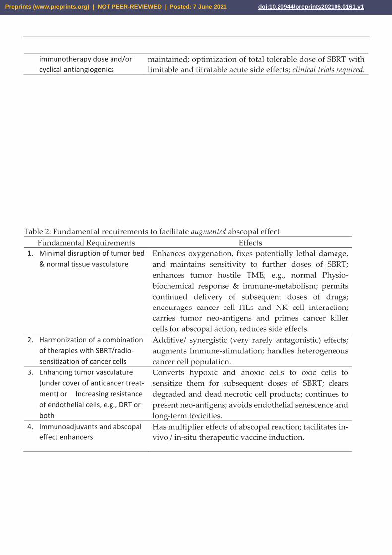
Table 2: Fundamental requirements to facilitate augmented abscopal effect
Fundamental Requirements Effects
1. Minimal disruption of tumor bed
& normal tissue vasculature
Enhances oxygenation, fixes potentially lethal damage,
and maintains sensitivity to further doses of SBRT;
enhances tumor hostile TME, e.g., normal Physio-
biochemical response & immune-metabolism; permits
continued delivery of subsequent doses of drugs;
encourages cancer cell-TILs and NK cell interaction;
carries tumor neo-antigens and primes cancer killer
cells for abscopal action, reduces side effects.
2. Harmonization of a combination
of therapies with SBRT/radio-
sensitization of cancer cells
Additive/ synergistic (very rarely antagonistic) effects;
augments Immune-stimulation; handles heterogeneous
cancer cell population.
3. Enhancing tumor vasculature
(under cover of anticancer treat-
ment) or Increasing resistance
of endothelial cells, e.g., DRT or
both
Converts hypoxic and anoxic cells to oxic cells to
sensitize them for subsequent doses of SBRT; clears
degraded and dead necrotic cell products; continues to
present neo-antigens; avoids endothelial senescence and
long-term toxicities.
4. Immunoadjuvants and abscopal
effect enhancers
Has multiplier effects of abscopal reaction; facilitates in-
vivo / in-situ therapeutic vaccine induction.
Preprints (www.preprints.org) | NOT PEER-REVIEWED | Posted: 7 June 2021 doi:10.20944/preprints202106.0161.v1
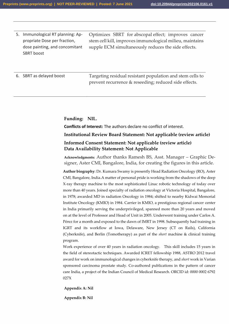
5. Immunological RT planning: Ap-
propriate Dose per fraction,
dose painting, and concomitant
SBRT boost
Optimizes SBRT for abscopal effect; improves cancer
stem cell kill, improves immunological milieu, maintains
supple ECM simultaneously reduces the side effects.
6. SBRT as delayed boost Targeting residual resistant population and stem cells to
prevent recurrence & reseeding; reduced side effects.
Funding: NIL.
Conflicts of Interest: The authors declare no conflict of interest.
Institutional Review Board Statement: Not applicable (review article)
Informed Consent Statement: Not applicable (review article)
Data Availability Statement: Not Applicable
Acknowledgments: Author thanks Ramesh BS, Asst. Manager – Graphic De-
signer, Aster CMI, Bangalore, India, for creating the figures in this article.
Author biography: Dr. Kumara Swamy is presently Head Radiation Oncology (RO), Aster
CMI, Bangalore, India.A matter of personal pride is working from the shadows of the deep
X-ray therapy machine to the most sophisticated Linac robotic technology of today over
more than 40 years. Joined specialty of radiation oncology at Victoria Hospital, Bangalore,
in 1978; awarded MD in radiation Oncology in 1984; shifted to nearby Kidwai Memorial
Institute Oncology (KMIO) in 1984. Carrier in KMIO, a prestigious regional cancer center
in India primarily serving the underprivileged, spanned more than 20 years and moved
on at the level of Professor and Head of Unit in 2005. Underwent training under Carlos A.
Pérez for a month and exposed to the dawn of IMRT in 1998. Subsequently had training in
IGRT and its workflow at Iowa, Delaware, New Jersey (CT on Rails), California
(Cyberknife), and Berlin (Tomotherapy) as part of the short machine & clinical training
program.
Work experience of over 40 years in radiation oncology. This skill includes 15 years in
the field of stereotactic techniques. Awarded ICRET fellowship 1988, ASTRO 2012 travel
award for work on immunological changes in cyberknife therapy, and short work in Varian
sponsored carcinoma prostate study. Co-authored publications in the pattern of cancer
care India, a project of the Indian Council of Medical Research. ORCID id: 0000 0002 6792
027X
Appendix A: Nil
Appendix B: Nil
Preprints (www.preprints.org) | NOT PEER-REVIEWED | Posted: 7 June 2021 doi:10.20944/preprints202106.0161.v1
References
1. Timmerman, R. D.; Bizekis, C. S.; Pass, H. I.; Fong, Y.; Dupuy, D. E.; Dawson, L. A.; Lu, D. Lo-
cal Surgical, Ablative, and Radiation Treatment of Metastases. CA: a cancer journal for clinicians
2009, 59 (3), 145–170. DOI: 10.3322/caac.20013
https://doi.org/10.3322/caac.20013
2. Liu, Y.; Dong, Y.; Kong, L.; Shi, F.; Zhu, H.; Yu, J. Abscopal Effect of Radiotherapy Combined
with Immune Checkpoint Inhibitors. Journal of hematology & oncology 2018, 11 (1), 1–15. DOI:
10.1186/s13045-018-0647-8
https://doi.org/10.1186/s13045-018-0647-8
3. Tharmalingam, H.; Hoskin, P. J. The Optimism Surrounding Stereotactic Body Radiation Ther-
apy and Immunomodulation. Chin Clin Oncol 2017, 6, S9. DOI: 10.21037/cco.2017.05.01
https://doi.org/10.21037/cco.2017.05.01
4. Bernstein, M. B.; Krishnan, S.; Hodge, J. W.; Chang, J. Y. Immunotherapy and Stereotactic Ab-
lative Radiotherapy (ISABR): A Curative Approach? Nature reviews Clinical oncology 2016, 13
(8), 516. DOI: 10.1038/nrclinonc.2016.30
https://doi.org/10.1038/nrclinonc.2016.30
5. Buchwald, Z. S.; Wynne, J.; Nasti, T. H.; Zhu, S.; Mourad, W. F.; Yan, W.; Gupta, S.; Khleif, S.
N.; Khan, M. K. Radiation, Immune Checkpoint Blockade and the Abscopal Effect: A Critical
Review on Timing, Dose and Fractionation. Frontiers in oncology 2018, 8, 612. DOI:
10.3389/fonc.2018.00612
https://doi.org/10.3389/fonc.2018.00612
6. Marcus, D.; Lieverse, R. I.; Klein, C.; Abdollahi, A.; Lambin, P.; Dubois, L. J.; Yaromina, A.
Charged Particle and Conventional Radiotherapy: Current Implications as Partner for Immu-
notherapy. Cancers 2021, 13 (6), 1468. DOI: 10.3390/cancers13061468
https://doi.org/10.3390/cancers13061468
7. Tubin, S.; Popper, H. H.; Brcic, L. Novel Stereotactic Body Radiation Therapy (SBRT)-Based
Partial Tumor Irradiation Targeting Hypoxic Segment of Bulky Tumors (SBRT-PATHY):
Preprints (www.preprints.org) | NOT PEER-REVIEWED | Posted: 7 June 2021 doi:10.20944/preprints202106.0161.v1
Improvement of the Radiotherapy Outcome by Exploiting the Bystander and Abscopal Effects.
Radiation Oncology 2019, 14 (1), 1–11. DOI: 10.1186/s13014-019-1227-y
https://doi.org/10.1186/s13014-019-1227-y
8. Kim, M.-S.; Kim, W.; Park, I. H.; Kim, H. J.; Lee, E.; Jung, J.-H.; Cho, L. C.; Song, C. W. Radiobi-
ological Mechanisms of Stereotactic Body Radiation Therapy and Stereotactic Radiation Sur-
gery. Radiation oncology journal 2015, 33 (4), 265. DOI: 10.3857/roj.2015.33.4.265
https://doi.org/10.3857/roj.2015.33.4.265
9. Shibamoto, Y.; Miyakawa, A.; Otsuka, S.; Iwata, H. Radiobiology of Hypofractionated Stereo-
tactic Radiotherapy: What Are the Optimal Fractionation Schedules? Journal of radiation research
2016, 57 (S1), i76–i82.DOI: 10.1093/jrr/rrw015
https://doi.org/10.1093/jrr/rrw015
10. Withers, H. R. The Four R’s of Radiotherapy. In Advances in radiation biology; Elsevier, 1975;
Vol. 5, pp 241–271. DOI: 10.1016/B978-0-12-035405-4.50012-8
https://doi.org/10.1016/B978-0-12-035405-4.50012-8
11. Garau, M. M. Radiobiology of Stereotactic Body Radiation Therapy (SBRT). Reports of Practical
Oncology & Radiotherapy 2017, 22 (2), 86–95. DOI: 10.1016/j.rpor.2017.02.010
https://doi.org/10.1016/j.rpor.2017.02.010
12. Garcia-Barros, M.; Paris, F.; Cordon-Cardo, C.; Lyden, D.; Rafii, S.; Haimovitz-Friedman, A.;
Fuks, Z.; Kolesnick, R. Tumor Response to Radiotherapy Regulated by Endothelial Cell Apop-
tosis. Science 2003, 300 (5622), 1155–1159. DOI: 10.1126/science.1082504
https://doi.org/10.1126/science.1082504
13. Fuks, Z.; Kolesnick, R. Engaging the Vascular Component of the Tumor Response. Cancer cell
2005, 8 (2), 89–91. DOI: 10.1016/j.ccr.2005.07.014
https://doi.org/10.1016/j.ccr.2005.07.014
14. Park, H. J.; Griffin, R. J.; Hui, S.; Levitt, S. H.; Song, C. W. Radiation-Induced Vascular Damage
in Tumors: Implications of Vascular Damage in Ablative Hypofractionated Radiotherapy
(SBRT and SRS). Radiation research 2012, 177 (3), 311–327. DOI: 10.1667/rr2773.1
https://doi.org/10.1667/rr2773.1
Preprints (www.preprints.org) | NOT PEER-REVIEWED | Posted: 7 June 2021 doi:10.20944/preprints202106.0161.v1
15. Maier, P.; Hartmann, L.; Wenz, F.; Herskind, C. Cellular Pathways in Response to Ionizing
Radiation and Their Targetability for Tumor Radiosensitization. International journal of
molecular sciences 2016, 17 (1), 102. DOI: 10.3390/ijms17010102
https://doi.org/10.3390/ijms17010102
16. Truman, J.-P.; García-Barros, M.; Kaag, M.; Hambardzumyan, D.; Stancevic, B.; Chan, M.; Fuks,
Z.; Kolesnick, R.; Haimovitz-Friedman, A. Endothelial Membrane Remodeling Is Obligate for
Anti-Angiogenic Radiosensitization during Tumor Radiosurgery. PloS one 2010, 5 (8), e12310.
DOI: 10.1371/annotation/6e222ad5-b175-4a00-9d04-4d120568a897
https://doi.org/10.1371/annotation/6e222ad5-b175-4a00-9d04-4d120568a897
17. Moding, E. J.; Lee, C.-L.; Castle, K. D.; Oh, P.; Mao, L.; Zha, S.; Min, H. D.; Ma, Y.; Das, S.;
Kirsch, D. G. Atm Deletion with Dual Recombinase Technology Preferentially Radiosensitizes
Tumor Endothelium. The Journal of clinical investigation 2014, 124 (8), 3325–3338. DOI:
10.1172/JCI73932
https://doi.org/10.1172/JCI73932
18. Moding, E. J.; Mowery, Y. M.; Kirsch, D. G. Opportunities for Radiosensitization in the SBRT
Era. Cancer journal (Sudbury, Mass.) 2016, 22 (4), 267. DOI: 10.1097/PPO.0000000000000203
https://doi.org/10.1097/PPO.0000000000000203
19. Moding, E. J.; Castle, K. D.; Perez, B. A.; Oh, P.; Min, H. D.; Norris, H.; Ma, Y.; Cardona, D. M.;
Lee, C.-L.; Kirsch, D. G. Tumor Cells, but Not Endothelial Cells, Mediate Eradication of Pri-
mary Sarcomas by Stereotactic Body Radiation Therapy. Science translational medicine 2015, 7
(278), 278ra34–278ra34. DOI: 10.1126/scitranslmed.aaa4214
https://doi.org/10.1126/scitranslmed.aaa4214
20. Kanegasaki, S.; Tsuchiya, T. Alarmins Released during Local Antitumor Treatments Play an
Essential Role in Enhancing Tumor Growth Inhibition at Treated and Non-Treated Sites via a
Derivative of CCL3. Oncoimmunology 2014, 3 (10), e958956. DOI: 10.4161/21624011.2014.958956
https://doi.org/10.4161/21624011.2014.958956
21. Bäcklund, M.; Freedman, J. Microwave Ablation and Immune Activation in the Treatment of
Recurrent Colorectal Lung Metastases: A Case Report. Case reports in oncology 2017, 10 (1), 383–
387. DOI: 10.1159/000468982
https://doi.org/10.1159/000468982
Preprints (www.preprints.org) | NOT PEER-REVIEWED | Posted: 7 June 2021 doi:10.20944/preprints202106.0161.v1
22. Slovak, R.; Ludwig, J. M.; Gettinger, S. N.; Herbst, R. S.; Kim, H. S. Immuno-Thermal Abla-
tions–Boosting the Anticancer Immune Response. Journal for immunotherapy of cancer 2017, 5 (1),
1–15. DOI: 10.1186/s40425-017-0284-8
https://doi.org/10.1186/s40425-017-0284-8
23. Confino, H.; Schmidt, M.; Efrati, M.; Hochman, I.; Umansky, V.; Kelson, I.; Keisari, Y. Inhibi-
tion of Mouse Breast Adenocarcinoma Growth by Ablation with Intratumoral Alpha-Irradia-
tion Combined with Inhibitors of Immunosuppression and CpG. Cancer Immunology, Immuno-
therapy 2016, 65 (10), 1149–1158. DOI: 10.1007/s00262-016-1878-6
https://doi.org/10.1007/s00262-016-1878-6
24. Kim, Y.; Lee, W. J.; Rhim, H.; Lim, H. K.; Choi, D.; Lee, J. Y. The Minimal Ablative Margin of
Radiofrequency Ablation of Hepatocellular Carcinoma (> 2 And< 5 Cm) Needed to Prevent Lo-
cal Tumor Progression: 3D Quantitative Assessment Using CT Image Fusion. American Journal
of Roentgenology 2010, 195 (3), 758–765. DOI: 10.2214/AJR.09.2954
https://doi.org/10.2214/AJR.09.2954
25. Jarosz-Biej, M.; Smolarczyk, R.; Cichoń, T.; Ku\lach, N. Tumor Microenvironment as a “Game
Changer” in Cancer Radiotherapy. International journal of molecular sciences 2019, 20 (13), 3212.
DOI: 10.3390/ijms20133212
https://doi.org/10.3390/ijms20133212
26. Yilmaz, M. T.; Elmali, A.; Yazici, G. Abscopal Effect, from Myth to Reality: From Radiation On-
cologists’ Perspective. Cureus 2019, 11 (1). DOI: 10.7759/cureus.3860
https://doi.org/10.7759/cureus.3860
27. Martinez-Zubiaurre, I.; Chalmers, A. J.; Hellevik, T. Radiation-Induced Transformation of Im-
munoregulatory Networks in the Tumor Stroma. Frontiers in immunology 2018, 9, 1679. DOI:
10.3389/fimmu.2018.01679
https://doi.org/10.3389/fimmu.2018.01679
28. Rodríguez-Ruiz, M. E.; Vanpouille-Box, C.; Melero, I.; Formenti, S. C.; Demaria, S. Immunolog-
ical Mechanisms Responsible for Radiation-Induced Abscopal Effect. Trends in immunology
2018, 39 (8), 644–655. DOI: 10.1016/j.it.2018.06.001
https://doi.org/10.1016/j.it.2018.06.001
29. Schaue, D.; Ratikan, J. A.; Iwamoto, K. S.; McBride, W. H. Maximizing Tumor Immunity with
Fractionated Radiation. International Journal of Radiation Oncology* Biology* Physics 2012, 83 (4),
1306–1310. DOI: 10.1016/j.ijrobp.2011.09.049
https://doi.org/10.1016/j.ijrobp.2011.09.049
Preprints (www.preprints.org) | NOT PEER-REVIEWED | Posted: 7 June 2021 doi:10.20944/preprints202106.0161.v1
30. Poleszczuk, J.; Enderling, H. The Optimal Radiation Dose to Induce Robust Systemic Anti-
tumor Immunity. International journal of molecular sciences 2018, 19 (11), 3377. DOI:
10.3390/ijms19113377
https://doi.org/10.3390/ijms19113377
31. Dewan, M. Z.; Galloway, A. E.; Kawashima, N.; Dewyngaert, J. K.; Babb, J. S.; Formenti, S. C.;
Demaria, S. Fractionated but Not Single-Dose Radiotherapy Induces an Immune-Mediated Ab-
scopal Effect When Combined with Anti–CTLA-4 Antibody. Clinical Cancer Research 2009, 15
(17), 5379–5388. DOI: 10.1158/1078-0432.CCR-09-0265
https://doi.org/10.1158/1078-0432.CCR-09-0265
32. Videtic, G. M.; Hu, C.; Singh, A. K.; Chang, J. Y.; Parker, W.; Olivier, K. R.; Schild, S. E.; Ko-
maki, R.; Urbanic, J. J.; Choy, H. A Randomized Phase 2 Study Comparing 2 Stereotactic Body
Radiation Therapy Schedules for Medically Inoperable Patients with Stage I Peripheral Non-
Small Cell Lung Cancer: NRG Oncology RTOG 0915 (NCCTG N0927). International Journal of
Radiation Oncology* Biology* Physics 2015, 93 (4), 757–764. DOI: 10.1016/j.ijrobp.2018.11.051
https://doi.org/10.1016/j.ijrobp.2018.11.051
33. Matuschek, C.; Haussmann, J.; Bölke, E.; Gripp, S.; Schuler, P. J.; Tamaskovics, B.; Gerber, P. A.;
Djiepmo-Njanang, F.-J.; Kammers, K.; Plettenberg, C. Accelerated vs. Conventionally Fraction-
ated Adjuvant Radiotherapy in High-Risk Head and Neck Cancer: A Meta-Analysis. Radiation
Oncology 2018, 13 (1), 1–12. DOI: 10.1186/s13014-018-1133-8
https://doi.org/10.1186/s13014-018-1133-8
34. Ang, K. K.; Trotti, A.; Brown, B. W.; Garden, A. S.; Foote, R. L.; Morrison, W. H.; Geara, F. B.;
Klotch, D. W.; Goepfert, H.; Peters, L. J. Randomized Trial Addressing Risk Features and Time
Factors of Surgery plus Radiotherapy in Advanced Head-and-Neck Cancer. International Jour-
nal of Radiation Oncology* Biology* Physics 2001, 51 (3), 571–578. DOI: 10.1016/s0360-
3016(01)01690-x
https://doi.org/10.1016/s0360-3016(01)01690-x
35. Paik, E. K.; Kim, M.-S.; Seo, Y.-S.; Jang, W. I.; Kang, J.-K.; Cho, C.-K.; Yoo, H. J. Feasibility of
Split-Course Stereotactic Ablative Radiotherapy for Oligometastases. Japanese journal of clinical
oncology 2018, 48 (6), 548–554. DOI: 10.1093/jjco/hyy062
https://doi.org/10.1093/jjco/hyy062
36. Dovedi, S. J.; Melis, M. H.; Wilkinson, R. W.; Adlard, A. L.; Stratford, I. J.; Honeychurch, J.;
Illidge, T. M. Systemic Delivery of a TLR7 Agonist in Combination with Radiation Primes Du-
rable Antitumor Immune Responses in Mouse Models of Lymphoma. Blood, The Journal of the
American Society of Hematology 2013, 121 (2), 251–259. DOI: 10.1182/blood-2012-05-432393
Preprints (www.preprints.org) | NOT PEER-REVIEWED | Posted: 7 June 2021 doi:10.20944/preprints202106.0161.v1
https://doi.org/10.1182/blood-2012-05-432393
37. Dovedi, S. J.; Adlard, A. L.; Lipowska-Bhalla, G.; McKenna, C.; Jones, S.; Cheadle, E. J.;
Stratford, I. J.; Poon, E.; Morrow, M.; Stewart, R. Acquired Resistance to Fractionated
Radiotherapy Can Be Overcome by Concurrent PD-L1 Blockade. Cancer research 2014, 74 (19),
5458–5468. DOI: 10.1158/0008-5472.CAN-14-1258
https://doi.org/10.1158/0008-5472.CAN-14-1258
38. Breen, W. G.; Leventakos, K.; Dong, H.; Merrell, K. W. Radiation and Immunotherapy: Emerg-
ing Mechanisms of Synergy. Journal of Thoracic Disease 2020, 12 (11), 7011. DOI: 10.21037/jtd-
2019-cptn-07
https://doi.org/10.21037/jtd-2019-cptn-07
39. Ji, D.; Song, C.; Li, Y.; Xia, J.; Wu, Y.; Jia, J.; Cui, X.; Yu, S.; Gu, J. Combination of Radiotherapy
and Suppression of Tregs Enhances Abscopal Antitumor Effect and Inhibits Metastasis in Rec-
tal Cancer. Journal for immunotherapy of cancer 2020, 8 (2). DOI: 10.1136/jitc-2020-000826
https://doi.org/10.1136/jitc-2020-000826
40. Crane, C. H.; Koay, E. J. Solutions That Enable Ablative Radiotherapy for Large Liver Tumors:
Fractionated Dose Painting, Simultaneous Integrated Protection, Motion Management, and
Computed Tomography Image Guidance. Cancer 2016, 122 (13), 1974–1986. DOI:
10.1002/cncr.29878
https://doi.org/10.1002/cncr.29878
41. Trani, D.; Yaromina, A.; Dubois, L.; Granzier, M.; Peeters, S. G.; Biemans, R.; Nalbantov, G.;
Lieuwes, N.; Reniers, B.; Troost, E. E. Preclinical Assessment of Efficacy of Radiation Dose
Painting Based on Intratumoral FDG-PET Uptake. Clinical Cancer Research 2015, 21 (24), 5511–
5518. DOI: 10.1158/1078-0432.CCR-15-0290
https://doi.org/10.1158/1078-0432.CCR-15-0290
42. Goel, S.; Duda, D. G.; Xu, L.; Munn, L. L.; Boucher, Y.; Fukumura, D.; Jain, R. K. Normalization
of the Vasculature for Treatment of Cancer and Other Diseases. Physiological reviews 2011, 91
(3), 1071–1121. DOI: 10.1152/physrev.00038.2010
https://doi.org/10.1152/physrev.00038.2010
43. Ramjiawan, R. R.; Griffioen, A. W.; Duda, D. G. Anti-Angiogenesis for Cancer Revisited: Is
There a Role for Combinations with Immunotherapy? Angiogenesis 2017, 20 (2), 185–204. DOI:
10.1007/s10456-017-9552-y
https://doi.org/10.1007/s10456-017-9552-y
Preprints (www.preprints.org) | NOT PEER-REVIEWED | Posted: 7 June 2021 doi:10.20944/preprints202106.0161.v1

44. Karar, J.; Maity, A. Modulating the Tumor Microenvironment to Increase Radiation Respon-
siveness. Cancer biology & therapy 2009, 8 (21), 1994–2001. DOI: 10.4161/cbt.8.21.9988
https://doi.org/10.4161/cbt.8.21.9988
45. Pore, N.; Gupta, A. K.; Cerniglia, G. J.; Jiang, Z.; Bernhard, E. J.; Evans, S. M.; Koch, C. J.; Hahn,
S. M.; Maity, A. Nelfinavir Down-Regulates Hypoxia-Inducible Factor 1α and VEGF Expres-
sion and Increases Tumor Oxygenation: Implications for Radiotherapy. Cancer research 2006, 66
(18), 9252–9259. DOI: 10.1158/0008-5472.CAN-06-1239
https://doi.org/10.1158/0008-5472.CAN-06-1239
46. Qayum, N.; Muschel, R. J.; Im, J. H.; Balathasan, L.; Koch, C. J.; Patel, S.; McKenna, W. G.; Bern-
hard, E. J. Tumor Vascular Changes Mediated by Inhibition of Oncogenic Signaling. Cancer re-
search 2009, 69 (15), 6347–6354. DOI: 10.1158/0008-5472.CAN-09-0657
https://doi.org/10.1158/0008-5472.CAN-09-0657
47. Cerniglia, G. J.; Pore, N.; Tsai, J. H.; Schultz, S.; Mick, R.; Choe, R.; Xing, X.; Durduran, T.;
Yodh, A. G.; Evans, S. M. Epidermal Growth Factor Receptor Inhibition Modulates the Micro-
environment by Vascular Normalization to Improve Chemotherapy and Radiotherapy Effi-
cacy. PloS one 2009, 4 (8), e6539. DOI: 10.1371/journal.pone.0006539
https://doi.org/10.1371/journal.pone.0006539
48. Barker, H. E.; Paget, J. T.; Khan, A. A.; Harrington, K. J. The Tumour Microenvironment after
Radiotherapy: Mechanisms of Resistance and Recurrence. Nature Reviews Cancer 2015, 15 (7),
409–425. DOI: 10.1038/nrc3958
https://doi.org/10.1038/nrc3958
49. Buck, M. D.; Sowell, R. T.; Kaech, S. M.; Pearce, E. L. Metabolic Instruction of Immunity. Cell
2017, 169 (4), 570–586. DOI: 10.1016/j.cell.2017.04.004
https://doi.org/10.1016/j.cell.2017.04.004
50. Chambers, A. M.; Lupo, K. B.; Matosevic, S. Tumor Microenvironment-Induced Immunometa-
bolic Reprogramming of Natural Killer Cells. Frontiers in immunology 2018, 9, 2517. DOI:
10.3389/fimmu.2018.02517
https://doi.org/10.3389/fimmu.2018.02517
51. Vanherwegen, A.-S.; Eelen, G.; Ferreira, G. B.; Ghesquière, B.; Cook, D. P.; Nikolic, T.; Roep, B.;
Carmeliet, P.; Telang, S.; Mathieu, C. Vitamin D Controls the Capacity of Human Dendritic
Cells to Induce Functional Regulatory T Cells by Regulation of Glucose Metabolism. The
Preprints (www.preprints.org) | NOT PEER-REVIEWED | Posted: 7 June 2021 doi:10.20944/preprints202106.0161.v1
Journal of steroid biochemistry and molecular biology 2019, 187, 134–145. DOI:
10.1016/j.jsbmb.2018.11.011
https://doi.org/10.1016/j.jsbmb.2018.11.011
52. Schumacher, T. N.; Schreiber, R. D. Neoantigens in Cancer Immunotherapy. Science 2015, 348
(6230), 69–74. DOI: 10.1126/science.aaa4971
https://doi.org/10.1126/science.aaa4971
53. K. Swamy, Vascular-Immuno-Phenotypic (VIP) Model for locally advanced and oligo-metastatic can-
cer: A Hypothesis, Medical Hypotheses 2021, in press. doi:10.1016/j.mehy.2021.110618
https://doi.org/10.1016/j.mehy.2021.110618
Preprints (www.preprints.org) | NOT PEER-REVIEWED | Posted: 7 June 2021 doi:10.20944/preprints202106.0161.v1