Embed Size (px)
Citation preview
406
ABSTRACT
Dyslipidemia refers to an abnormal amount of lipid in the blood, and the total cholesterol level is defined as the sum of high-density lipoprotein cholesterol, low-density lipoprotein (LDL) cholesterol, and very-LDL cholesterol concentrations. In Korea, the westernization of lifestyle habits in recent years has caused an increase in the incidence of dyslipidemia, which is an important risk factor of cardiovascular disease (CVD). Several studies have been conducted on how dyslipidemia affects not only CVD, but also chorioretinal diseases such as age-related macular degeneration (AMD) and diabetic retinopathy. Recently, a pathological model of AMD was proposed under the assumption that AMD proceeds through a mechanism similar to that of atherosclerotic CVD. However, controversy remains regarding the relationship between chorioretinal diseases and lipid levels in the blood, and the effects of lipid-lowering agents. Herein, we summarize the role of lipids in chorioretinal diseases. In addition, the effects of lipid-lowering agents on the prevention and progression of chorioretinal diseases are presented.
Keywords: Age-related macular degeneration; Cholesterol; Diabetic retinopathy; Lipids; Hypolipidemic agents
INTRODUCTION
Since 2015, Korean Society of Lipid and Atherosclerosis has published “Dyslipidemia Fact Sheets in Korea” based on the Korea National Health and Nutrition Examination Survey conducted by the Ministry of Health and Welfare and the Korean Centers for Disease Control and Prevention. According to the “Dyslipidemia Fact Sheets in Korea, 2018,” the prevalence of elevated levels of low-density lipoprotein (LDL) cholesterol, hypertriglyceridemia, and reduced levels of high-density lipoprotein (HDL) cholesterol among adults aged 30 years or older is 17.6%, 17.5%, and 19.4%, respectively.1 The above results show a decrease in hypertriglyceridemia, a decrease in hypo-HDL-cholesterolemia, and an increase in hyper-LDL-cholesterolemia compared to a report published in 2015. Because dyslipidemia is recognized as a prominent risk factor for cardiovascular disease (CVD),2 current guidelines focus on lowering LDL cholesterol levels through statins in both primary and secondary intervention settings.3-5
J Lipid Atheroscler. 2020 Sep;9(3):406-418https://doi.org/10.12997/jla.2020.9.3.406pISSN 2287-2892·eISSN 2288-2561
Review
Received: Aug 27, 2020Revised: Sep 11, 2020Accepted: Sep 13, 2020
Correspondence toDong Ho ParkDepartment of Ophthalmology, School of Medicine, Kyungpook National University, Kyungpook National University Hospital, 130 Dongdeok-ro, Jung-gu, Daegu 41944, Korea.E-mail: [email protected]
Copyright © 2020 The Korean Society of Lipid and Atherosclerosis.This is an Open Access article distributed under the terms of the Creative Commons Attribution Non-Commercial License (https://creativecommons.org/licenses/by-nc/4.0/) which permits unrestricted non-commercial use, distribution, and reproduction in any medium, provided the original work is properly cited.
ORCID iDsSu Jin Park https://orcid.org/0000-0001-8313-7640Dong Ho Park https://orcid.org/0000-0001-6911-3765
FundingDHP is financially supported by the Basic Science Research Program of the National Research Foundation of Korea (NRF), funded by the Korean government (Ministry of Science and ICT) (2019R1A2C1084371), and the Korea Health Technology R&D Project of the Korea Health Industry Development Institute (KHIDI), funded by the Ministry of Health & Welfare, Republic of Korea (HI16C1501).
Su Jin Park , Dong Ho Park
Department of Ophthalmology, School of Medicine, Kyungpook National University, Kyungpook National University Hospital, Daegu, Korea
REvisiting Lipids in REtinal Diseases: A Focused Review on Age-related Macular Degeneration and Diabetic Retinopathy
https://e-jla.org
Journal of Lipid and Atherosclerosis
Conflict of InterestThe authors have no conflicts of interest to declare.
Author ContributionsConceptualization: Park DH; Data curation: Park DH; Formal analysis: Park DH; Methodology: Park DH; Resources: Park SJ, Park DH; Supervision: Park DH; Validation: Park DH; Writing - original draft: Park SJ, Park DH; Writing - review & editing: Park SJ, Park DH.
The retina, a structure composed of the neurosensory layers in the eyes, is a highly metabolic tissue, and lipid metabolism plays a critical role in maintaining the homeostasis of various kinds of cells. Previous studies have reported of the role of lipids in various chorioretinal diseases, including diabetic retinopathy (DR) and age-related macular degeneration (AMD), although some controversies remain.
In this review paper, we discuss the associations between lipids and various chorioretinal diseases and present an evaluation of whether lipid-lowering agents could affect disease progression or prevention.
BACKGROUND
1. Cholesterol and lipoproteinsCholesterol plays many diverse roles in the body, including serving as a contributor to the structure of cell membranes, a precursor for steroid hormones, a regulator of gene transcription, and a component involved in the formation of neuronal synapses.6-9 For lipid transportation through the systemic circulation, lipoproteins are required. Lipoproteins are complex plasma particles that include a core composed of cholesterol esters and triglycerides (TG) and a surface composed of apolipoproteins, phospholipids, and unesterified cholesterol. According to their size, structure, and apolipoprotein content, lipoproteins are classified as chylomicrons, very-LDLs, intermediate-density lipoproteins, LDLs, HDLs, and lipoprotein (a).10
2. Distribution of cholesterol in the retinaThe retina consists of 10 layers, from its internal limiting membrane to the retinal pigment epithelium (RPE). In addition to retinal vessels, the choroidal vasculature, which is connected to the systemic circulation, supplies the blood to the retina, especially outer the retina.11 To achieve physiological structure and function, the retina receives cholesterol from endogenous biosynthesis12,13 and the systemic circulation across the RPE.14,15
For lipid transportation, the RPE rapidly takes up lipoproteins from the systemic circulation, and it contains receptors for LDL (e.g., LDL-R) and HDL (e.g., SR-BI and SRBII) on the basolateral side, which contacts the choroid.16
AMD
1. AMD specific lesions: drusen and basal linear deposits (BLinDs)AMD is the most common cause of blindness in developed countries and accounts for 8.7% of all cases of blindness worldwide.17,18 In AMD, central vision is gradually reduced by changes in the macular region of the retina.19 According to the Beckman classification, AMD is classified into early, intermediate, and late AMD. Early AMD is diagnosed based on the presence of medium-sized drusen (>63 and ≤125 μm) and no retinal pigmentary abnormalities (hyperpigmentation or hypopigmentation). Intermediate AMD is defined as the presence of large drusen (>125 μm) and/or any pigmentary abnormalities. Late AMD is defined by geographic atrophy (GA) or neovascular AMD.20 Neovascular AMD is characterized by choroidal neovascularization (CNV), and GA is characterized by a sharply defined area of RPE degeneration in which the choroidal blood vessels are visible.21 Drusen are focal, dome-
407https://doi.org/10.12997/jla.2020.9.3.406
Lipids and Chorioretinal Disease
https://e-jla.org
Journal of Lipid and Atherosclerosis
shaped lesions between the RPE basal lamina and the inner collagenous layer of Bruch's membrane (BrM) and are observed as yellow-white deposits on a fundus examination.22
BrM, which is the basement membrane of the RPE, has unique characteristics in terms of cholesterol content and is relevant to AMD. As aging progresses, BrM thickens and develops basal deposits. Depending on its location, a basal deposit is classified as a basal laminar deposit (BLamD) or a BLinD. BLamD is considered as a stereotypically thickened area of the RPE basal lamina. BLinD is located in the sub-RPE space, bounded internally by the basal lamina and externally by the inner collagenous layer of BrM. Drusen and BLinD are 2 forms of an AMD-specific lesion, in the shape of a lump and thin layer, respectively.23-30 BLinD and soft drusen are considered to be alternative forms of the same entity because these lesions are located in the same plane and contains the same materials.31,32 Curcio and Millican32 reported that eyes with AMD were 24 times more likely to have BLinD and large drusen (>125 μm) than age-matched controls.
These lesions contain lipoprotein-derived particles that have the physical forms of cholesterol seen in the core of atherosclerotic plaques,33-35 a mark of atheroma maturity. The formation of AMD lesions has thus been considered to share mechanisms with the formation of atherosclerotic plaques.36,37 In atherosclerosis, trapped apolipoprotein B100 lipoproteins within the arterial wall initiate a cascade of pathological events, including innate immune system-mediated inflammation.38 This launches various downstream deleterious events, including macrophage recruitment, cytokine release, and neovascularization.
Similarly, in AMD, lipoprotein particles from multiple resources accumulate during aging. Those particles could fuse to form lipoprotein-derived debris such as soft drusen and BLinD. These processes may be accompanied by oxidative stress and inflammation, which contribute to the progression from non-neovascular AMD to neovascular AMD.39
2. Serum lipids, cholesterol-lowering medication, and AMDSince the early 1960s, the correlations of AMD with concentrations of plasma cholesterol or apolipoproteins have been evaluated.40 Several clinical studies of the associations between lipids and AMD are summarized in Table 1. Klein et al.41 reported that carotid artery intima-media thickness and carotid plaques had a weak relationship with the incidence of late AMD. The Eye Disease Case Control Study reported a 4-fold increased risk of exudative AMD in patients with high total cholesterol levels (≥6.749 mmol/L).42 Reynolds et al.43 reported lower mean HDL cholesterol levels and higher LDL cholesterol levels in patients with advanced AMD than in controls. In addition, high total cholesterol and LDL cholesterol levels were found to be related to an increased risk of AMD after adjusting for environmental and genetic covariates.
Several studies have investigated the relationship between the risk of AMD and a specific lipid profile, such as HDL cholesterol. van Leeuwen et al.44 reported that elevated HDL cholesterol levels increased the risk of AMD. Klein et al.45 reported that high serum HDL cholesterol levels were associated with GA, and that a high total cholesterol/HDL ratio was associated with the incidence of RPE depigmentation and GA. The Pathologies Oculaires Liées à l'Age study reported that high HDL cholesterol and apolipoprotein A1 were associated with an increased risk of soft drusen.46 A large clinical study with 1,235 patients in North America showed an interesting finding that only neovascular AMD was associated with high HDL cholesterol levels, while non-neovascular AMD was unrelated to serum lipid levels.47
408https://doi.org/10.12997/jla.2020.9.3.406
Lipids and Chorioretinal Disease
https://e-jla.org
Journal of Lipid and Atherosclerosis
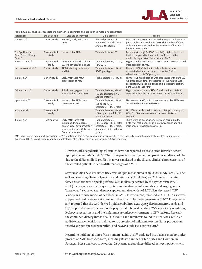
However, other epidemiological studies have not reported an association between serum lipid profile and AMD risk.48,49 The discrepancies in results among previous studies could be due to the different lipid profiles that were analyzed or the diverse clinical characteristics of the enrolled patients, such as different stages of AMD.
Several studies have evaluated the effect of lipid metabolites in an in vivo model of CNV. The ω-3 and ω-6 long-chain polyunsaturated fatty acids (LCPUFAs) are 2 classes of essential fatty acids that have opposing effects. Metabolites generated by the cytochrome P450 (CYP)—epoxygenase pathway are potent modulators of inflammation and angiogenesis. Yanai et al.50 reported that dietary supplementation with ω-3 LCPUFAs decreased CNV lesions in a mouse model of neovascular AMD. Furthermore, mice fed ω-3 LCPUFAs showed suppressed leukocyte recruitment and adhesion molecule expression in CNV.50 Hasegawa et al.51 reported that the CYP-derived lipid metabolites 17,18-epoxyeicosatetraenoic acids and 19,20-epoxydocosapentaenoic acids play a vital role in alleviating CNV severity by regulating leukocyte recruitment and the inflammatory microenvironment in CNV lesions. Recently, the combined dietary intake of ω-3 LCPUFAs and lutein was found to attenuate CNV in an additive manner, which was related to suppression of inflammatory mediator production, reactive oxygen species generation, and NADPH oxidase 4 expression.52
Regarding lipid metabolites from humans, Lains et al.53 evaluated the plasma metabolomics profiles of AMD from 2 cohorts, including Boston in the United States and Coimbra in Portugal. Meta-analyses showed that 28 plasma metabolites differed between patients with
409https://doi.org/10.12997/jla.2020.9.3.406
Lipids and Chorioretinal Disease
https://e-jla.org
Journal of Lipid and Atherosclerosis
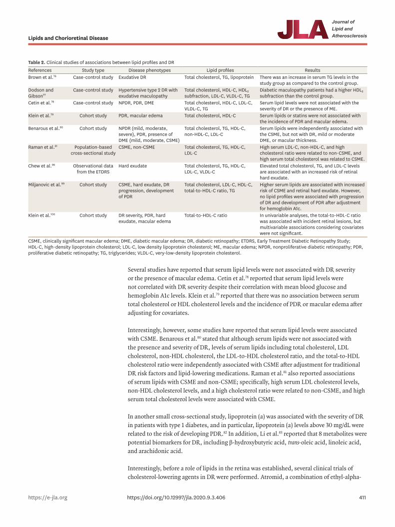
Table 1. Clinical studies of associations between lipid profiles and age-related macular degenerationReferences Study design Disease phenotypes Lipid profiles ResultsKlein et al.41 Cohort study No AMD, early AMD, late
AMDIMT and presence of plaque of carotid artery
Mean IMT was associated with the 10-year incidence of pure GA, but not exudative AMD. The number of sites with plaque was related to the incidence of late AMD, but not to early AMD.
Angina, MI, stroke
The Eye Disease Case-Control Study Group42
Case-control study
Neovascular AMD Total cholesterol, TG Patients with high (> 6.749 mmol/L) total cholesterol levels, compared to those with low levels, had a markedly higher risk of neovascular AMD.
Reynolds et al.43 Case-control study
Advanced AMD with either GA or neovascular disease
Total cholesterol, LDL-C, HDL-C, TG
Higher total cholesterol and LDL-C were associated with increased risk of AMD.
van Leeuwen et al.44 Cohort study AMD including both early and late
Total cholesterol, HDL-C, APOE genotype
Elevated HDL-C, but not total cholesterol, was associated with an increased risk of AMD after adjustment for APOE genotype.
Klein et al.45 Cohort study Early AMD, late AMD, progression of AMD
Total cholesterol, HDL-C Higher HDL-C at baseline was associated with pure GA. A higher serum total cholesterol-to-HDL-C ratio was associated with the incidence of RPE depigmentation, pure GA, and late AMD.
Delcourt et al.46 Cohort study Soft drusen, pigmentary abnormalities, late AMD
Total cholesterol, HDL-C High concentrations of HDL-C and apolipoprotein A1 were associated with an increased risk of soft drusen.TG, apolipoprotein A1,
apolipoprotein BHyman et al.47 Case-control
studyNeovascular AMD, non-neovascular AMD
Total cholesterol, HDL-C Neovascular AMD, but not non-neovascular AMD, was associated with elevated HDL-C.LDL-C, TG, total
cholesterol/HDL-C ratioAbalain et al.48 Case-control
studyTotal cholesterol, HDL-C, LDL-C, phospholipid, TG, apolipoproteins
No differences in total cholesterol, TG, phospholipids, HDL-C, LDL-C were observed between AMD and controls.
Klein et al.65 Meta-analysis Early AMD, large soft indistinct drusen, large drusen area, any pigmentary abnormality, late AMD, pure GA, exudative AMD
Total cholesterol, HDL-C, non-HDL-C, total cholesterol/HDL-C ratio, Statin use, lipid pathway genes
There were no associations between serum lipids, history of statin use, or lipid pathway genes and the incidence or progression of AMD.
AMD, age-related macular degeneration; APOE, apolipoprotein E; GA, geographic atrophy; HDL-C, high-density lipoprotein cholesterol; IMT, intima-media thickness; LDL-C, low density lipoprotein cholesterol; RPE, retinal pigment epithelium; TG, triglycerides.
AMD and controls and most of the significant metabolites were lipids. In particular, the metabolites mapping to glycerophospholipid pathways were altered in AMD subjects. Li et al.54 also stated that the glycerophospholipid pathway was one of the most significant pathways in their study group of polypoidal choroidal vasculopathy, a subtype of neovascular AMD.
Statins, which are 3-hydroxy-3-methylglutaryl coenzyme A reductase inhibitors, have been used to control blood cholesterol levels.55 In AMD, statins have several potential effects such as preserving the blood supply to the outer retina, downregulation of LDL cholesterol and peroxidized lipids, and anti-inflammatory properties.56-59 Previous studies evaluated whether cholesterol-lowering agents have a therapeutic effect on AMD, although the results are inconsistent. Vavvas et al.60 reported that high-dose statins (80 mg of atorvastatin daily) may result in the regression of drusenoid pigment epithelial detachments and a vision gain of 3.3 letters, without progression to advanced AMD such as GA or CNV. Barbosa et al.61 suggested a possible beneficial effect of statin intake for the prevention of AMD in individuals 68 years or older. Guymer et al.62 also reported a decrease in the risk of progression in the simvastatin group, and the most prominent effect was observed in individuals who were homozygous for the at risk C allele at Y402H of the complement factor H gene.
However, the effects of statins remain controversial. van Leeuwen et al.63 and Klein et al.64 did not find an association between statin use and the risk of AMD. In addition, a meta-analysis of 3 clinical trials showed no correlation between statin use and the incidence or progression of AMD.65
DR
1. Serum lipids, cholesterol-lowering medication, and DRDR, which is one of the most common microvascular complications of diabetes, is characterized by retinal hemorrhage and yellowish retinal exudates.66 DR is broadly classified into nonproliferative DR and proliferative DR (PDR) according to the presence of neovascularization of the disc or elsewhere, or vitreous hemorrhage.67 Diabetic macular edema is the most common cause of vision loss in patients with diabetes,68 and of particular note, the Early Treatment of Diabetic Retinopathy Study introduced the term “clinically significant macular edema” (CSME), which is defined upon slit lamp biomicroscopy as “1) thickening of the retina at or within 500 μm of the center of the macula; 2) hard exudate at or within 500 μm of the center of the macula associated with thickening of adjacent retina; or 3) a zone of retinal thickening 1 disc area or larger, any part of which is within 1 disc diameter of the center of the macula.69”
Table 2 summarizes several studies that have been conducted on the associations between lipids and DR. Many studies reported risk factors for the development of DR, including the duration of diabetes, glucose control, and the presence of hypertension.70-74 The effect of fat intake on exudative maculopathy has been studied. Ernest et al.75 reported that retinal exudates decreased in 8 diabetic patients after consumption of a low-fat diet for 2–3 years. Several studies have suggested that serum lipids may cause the development of retinal exudates. Patients with diabetes who had severe exudative maculopathy demonstrated higher levels of serum TG than those with nonexudative retinopathy, although serum cholesterol levels did not differ between the 2 groups.76 A case-control study reported that patients with maculopathy tended to have higher mean serum HDL cholesterol and total cholesterol levels over 7 years, but the difference was not significant.77
410https://doi.org/10.12997/jla.2020.9.3.406
Lipids and Chorioretinal Disease
https://e-jla.org
Journal of Lipid and Atherosclerosis
Several studies have reported that serum lipid levels were not associated with DR severity or the presence of macular edema. Cetin et al.78 reported that serum lipid levels were not correlated with DR severity despite their correlation with mean blood glucose and hemoglobin A1c levels. Klein et al.79 reported that there was no association between serum total cholesterol or HDL cholesterol levels and the incidence of PDR or macular edema after adjusting for covariates.
Interestingly, however, some studies have reported that serum lipid levels were associated with CSME. Benarous et al.80 stated that although serum lipids were not associated with the presence and severity of DR, levels of serum lipids including total cholesterol, LDL cholesterol, non-HDL cholesterol, the LDL-to-HDL cholesterol ratio, and the total-to-HDL cholesterol ratio were independently associated with CSME after adjustment for traditional DR risk factors and lipid-lowering medications. Raman et al.81 also reported associations of serum lipids with CSME and non-CSME; specifically, high serum LDL cholesterol levels, non-HDL cholesterol levels, and a high cholesterol ratio were related to non-CSME, and high serum total cholesterol levels were associated with CSME.
In another small cross-sectional study, lipoprotein (a) was associated with the severity of DR in patients with type 1 diabetes, and in particular, lipoprotein (a) levels above 30 mg/dL were related to the risk of developing PDR.82 In addition, Li et al.83 reported that 8 metabolites were potential biomarkers for DR, including β-hydroxybutyric acid, trans-oleic acid, linoleic acid, and arachidonic acid.
Interestingly, before a role of lipids in the retina was established, several clinical trials of cholesterol-lowering agents in DR were performed. Atromid, a combination of ethyl-alpha-
411https://doi.org/10.12997/jla.2020.9.3.406
Lipids and Chorioretinal Disease
https://e-jla.org
Journal of Lipid and Atherosclerosis
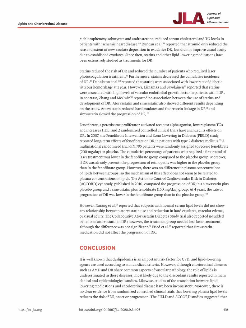
Table 2. Clinical studies of associations between lipid profiles and DRReferences Study type Disease phenotypes Lipid profiles ResultsBrown et al.76 Case-control study Exudative DR Total cholesterol, TG, lipoprotein There was an increase in serum TG levels in the
study group as compared to the control group.Dodson and Gibson77
Case-control study Hypertensive type 2 DR with exudative maculopathy
Total cholesterol, HDL-C, HDL2 subfraction, LDL-C, VLDL-C, TG
Diabetic maculopathy patients had a higher HDL2 subfraction than the control group.
Cetin et al.78 Case-control study NPDR, PDR, DME Total cholesterol, HDL-C, LDL-C, VLDL-C, TG
Serum lipid levels were not associated with the severity of DR or the presence of ME.
Klein et al.79 Cohort study PDR, macular edema Total cholesterol, HDL-C Serum lipids or statins were not associated with the incidence of PDR and macular edema.
Benarous et al.80 Cohort study NPDR (mild, moderate, severe), PDR, presence of DME (mild, moderate, CSME)
Total cholesterol, TG, HDL-C, non-HDL-C, LDL-C
Serum lipids were independently associated with the CSME, but not with DR, mild or moderate DME, or macular thickness.
Raman et al.81 Population-based cross-sectional study
CSME, non-CSME Total cholesterol, TG, HDL-C, LDL-C
High serum LDL-C, non-HDL-C, and high cholesterol ratio were related to non-CSME, and high serum total cholesterol was related to CSME.
Chew et al.98 Observational data from the ETDRS
Hard exudate Total cholesterol, TG, HDL-C, LDL-C, VLDL-C
Elevated total cholesterol, TG, and LDL-C levels are associated with an increased risk of retinal hard exudate.
Miljanovic et al.99 Cohort study CSME, hard exudate, DR progression, development of PDR
Total cholesterol, LDL-C, HDL-C, total-to-HDL-C ratio, TG
Higher serum lipids are associated with increased risk of CSME and retinal hard exudate. However, no lipid profiles were associated with progression of DR and development of PDR after adjustment for hemoglobin A1c.
Klein et al.100 Cohort study DR severity, PDR, hard exudate, macular edema
Total-to-HDL-C ratio In univariable analyses, the total-to-HDL-C ratio was associated with incident retinal lesions, but multivariable associations considering covariates were not significant.
CSME, clinically significant macular edema; DME, diabetic macular edema; DR, diabetic retinopathy; ETDRS, Early Treatment Diabetic Retinopathy Study; HDL-C, high-density lipoprotein cholesterol; LDL-C, low density lipoprotein cholesterol; ME, macular edema; NPDR, nonproliferative diabetic retinopathy; PDR, proliferative diabetic retinopathy; TG, triglycerides; VLDL-C, very-low-density lipoprotein cholesterol.
p-chlorophenoxyisobutyrate and androsterone, reduced serum cholesterol and TG levels in patients with ischemic heart disease.84 Duncan et al.85 reported that atromid only reduced the rate and extent of new exudate deposition in exudative DR, but did not improve visual acuity due to established exudates. Since then, statins and other lipid-lowering medications have been extensively studied as treatments for DR.
Statins reduced the risk of DR and reduced the number of patients who required laser photocoagulation treatment.86 Furthermore, statins decreased the cumulative incidence of DR.87 Denniston et al.88 reported that statins were associated with lower rate of diabetic vitreous hemorrhage at 1 year. However, Liinamaa and Savolainen89 reported that statins were associated with high levels of vascular endothelial growth factor in patients with PDR. In contrast, Zhang and McGwin90 reported no association between the use of statins and development of DR. Atorvastatin and simvastatin also showed different results depending on the study. Atorvastatin reduced hard exudates and fluorescein leakage in DR91 and simvastatin slowed the progression of DR.92
Fenofibrate, a peroxisome proliferator-activated receptor alpha agonist, lowers plasma TGs and increases HDL, and 2 randomized controlled clinical trials have analyzed its effects on DR. In 2007, the Fenofibrate Intervention and Event Lowering in Diabetes (FIELD) study reported long-term effects of fenofibrate on DR in patients with type 2 diabetes mellitus.93 A multinational randomized trial of 9,795 patients were randomly assigned to receive fenofibrate (200 mg/day) or placebo. The cumulative percentage of patients who required a first round of laser treatment was lower in the fenofibrate group compared to the placebo group. Moreover, if DR was already present, the progression of retinopathy was higher in the placebo group than in the fenofibrate group. However, there was no difference in plasma concentrations of lipids between groups, so the mechanism of this effect does not seem to be related to plasma concentrations of lipids. The Action to Control Cardiovascular Risk in Diabetes (ACCORD) eye study, published in 2010, compared the progression of DR in a simvastatin plus placebo group and a simvastatin plus fenofibrate (160 mg/day) group. At 4 years, the rate of progression of DR was lower in the fenofibrate group than in the placebo group.94
However, Narang et al.95 reported that subjects with normal serum lipid levels did not show any relationship between atorvastatin use and reduction in hard exudates, macular edema, or visual acuity. The Collaborative Atorvastatin Diabetes Study trial also reported no added benefits of atorvastatin in DR; however, the treatment group needed less laser treatment, although the difference was not significant.96 Fried et al.97 reported that simvastatin medication did not affect the progression of DR.
CONCLUSION
It is well known that dyslipidemia is an important risk factor for CVD, and lipid-lowering agents are used according to standardized criteria. However, although chorioretinal diseases such as AMD and DR share common aspects of vascular pathology, the role of lipids is underestimated in these diseases, most likely due to the discordant results reported in many clinical and epidemiological studies. Likewise, studies of the association between lipid-lowering medications and chorioretinal disease have been inconsistent. Moreover, there is no clear evidence from randomized controlled clinical trials that lowering plasma lipid levels reduces the risk of DR onset or progression. The FIELD and ACCORD studies suggested that
412https://doi.org/10.12997/jla.2020.9.3.406
Lipids and Chorioretinal Disease
https://e-jla.org
Journal of Lipid and Atherosclerosis
fenofibrate may play a potential role in reducing the risk of DR progression, but it is not clear whether the process of plasma lipid changes is involved in its mechanism.
However, some repeated findings could be observed among the inconsistent data, namely the relationship between serum lipid levels and the incidence of hard exudates and CSME and the relationship between serum lipid metabolites and AMD. Although a clear relationship between lipids and chorioretinal disease has not yet been identified, more large-scale clinical studies are expected to provide additional benefits to patients. These studies will contribute to the understanding of pathophysiology of AMD and DR and can serve as a basis for identifying future biomarkers and precision medicine for these conditions that cause blindness.
REFERENCES
1. The Korean Society of Lipid and Atherosclerosis. Dyslipidemia fact sheets in Korea 2018. Seoul: The Korean Society of Lipid and Atherosclerosis; 2018.
2. Yusuf S, Hawken S, Ôunpuu S, Dans T, Avezum A, Lanas F, et al. Effect of potentially modifiable risk factors associated with myocardial infarction in 52 countries (the INTERHEART study): case-control study. Lancet 2004;364:937-952. PUBMED | CROSSREF
3. National Cholesterol Education Program (NCEP) Expert Panel on Detection, Evaluation, and Treatment of High Blood Cholesterol in Adults (Adult Treatment Panel III). Third report of the national cholesterol education program (NCEP) expert panel on detection, evaluation, and treatment of high blood cholesterol in adults (adult treatment panel III) final report. Circulation 2002;106:3143-3421. PUBMED | CROSSREF
4. Grundy SM, Cleeman JI, Merz CNB, Brewer HB Jr, Clark LT, Hunninghake DB, et al. Implications of recent clinical trials for the national cholesterol education program adult treatment panel III guidelines. J Am Coll Cardiol 2004;44:720-732. PUBMED | CROSSREF
5. Brunzell JD, Davidson M, Furberg CD, Goldberg RB, Howard BV, Stein JH, et al. Lipoprotein management in patients with cardiometabolic risk: consensus statement from the American Diabetes Association and the American College of Cardiology Foundation. Diabetes Care 2008;31:811-822. PUBMED | CROSSREF
6. Lee JW, Fuda H, Javitt NB, Strott CA, Rodriguez IR. Expression and localization of sterol 27-hydroxylase (CYP27A1) in monkey retina. Exp Eye Res 2006;83:465-469. PUBMED | CROSSREF
7. Mauch DH, Nägler K, Schumacher S, Göritz C, Müller EC, Otto A, et al. CNS synaptogenesis promoted by glia-derived cholesterol. Science 2001;294:1354-1357. PUBMED | CROSSREF
8. Saher G, Quintes S, Nave KA. Cholesterol: a novel regulatory role in myelin formation. Neuroscientist 2011;17:79-93. PUBMED | CROSSREF
9. Brown MS, Goldstein JL. Cholesterol feedback: from Schoenheimer's bottle to Scap's MELADL. J Lipid Res 2009;50:S15-S27. PUBMED | CROSSREF
10. Wengrofsky P, Lee J, Makaryus AN. Dyslipidemia and its role in the pathogenesis of atherosclerotic cardiovascular disease: implications for evaluation and targets for treatment of dyslipidemia based on recent guidelines. London: IntechOpen; 2019.
11. Hayreh SS. Segmental nature of the choroidal vasculature. Br J Ophthalmol 1975;59:631-648. PUBMED | CROSSREF
12. Fliesler SJ. Retinal degeneration in a rat model of smith-lemli-opitz syndrome: thinking beyond cholesterol deficiency. In: Anderson RE, Hollyfield JG, LaVail MM, editors. Retinal degenerative diseases: laboratory and therapeutic investigations. New York (NY): Springer New York; 2010. p.481-489.
13. Fliesler SJ, Bretillon L. The ins and outs of cholesterol in the vertebrate retina. J Lipid Res 2010;51:3399-3413. PUBMED | CROSSREF
413https://doi.org/10.12997/jla.2020.9.3.406
Lipids and Chorioretinal Disease
https://e-jla.org
Journal of Lipid and Atherosclerosis
14. Elner VM. Retinal pigment epithelial acid lipase activity and lipoprotein receptors: effects of dietary omega-3 fatty acids. Trans Am Ophthalmol Soc 2002;100:301-338.PUBMED
15. Tserentsoodol N, Sztein J, Campos M, Gordiyenko NV, Fariss RN, Lee JW, et al. Uptake of cholesterol by the retina occurs primarily via a low density lipoprotein receptor-mediated process. Mol Vis 2006;12:1306-1318.PUBMED
16. Duncan KG, Hosseini K, Bailey KR, Yang H, Lowe RJ, Matthes MT, et al. Expression of reverse cholesterol transport proteins ATP-binding cassette A1 (ABCA1) and scavenger receptor BI (SR-BI) in the retina and retinal pigment epithelium. Br J Ophthalmol 2009;93:1116-1120. PUBMED | CROSSREF
17. Wong WL, Su X, Li X, Cheung CM, Klein R, Cheng CY, et al. Global prevalence of age-related macular degeneration and disease burden projection for 2020 and 2040: a systematic review and meta-analysis. Lancet Glob Health 2014;2:e106-e116.PUBMED
18. Resnikoff S, Pascolini D, Etya'ale D, Kocur I, Pararajasegaram R, Pokharel GP, et al. Global data on visual impairment in the year 2002. Bull World Health Organ 2004;82:844-851.PUBMED
19. Coleman HR, Chan CC, Ferris FL 3rd, Chew EY. Age-related macular degeneration. Lancet 2008;372:1835-1845.PUBMED
20. Ferris FL 3rd, Wilkinson CP, Bird A, Chakravarthy U, Chew E, Csaky K, et al. Clinical classification of age-related macular degeneration. Ophthalmology 2013;120:844-851. PUBMED | CROSSREF
21. Klein R, Davis MD, Magli YL, Segal P, Klein BE, Hubbard L. The Wisconsin age-related maculopathy grading system. Ophthalmology 1991;98:1128-1134. PUBMED | CROSSREF
22. Sarks SH, Arnold JJ, Killingsworth MC, Sarks JP. Early drusen formation in the normal and aging eye and their relation to age related maculopathy: a clinicopathological study. Br J Ophthalmol 1999;83:358-368. PUBMED | CROSSREF
23. Feeney-Burns L, Burns RP, Gao CL. Age-related macular changes in humans over 90 years old. Am J Ophthalmol 1990;109:265-278. PUBMED | CROSSREF
24. Green WR, Enger C. Age-related macular degeneration histopathologic studies: the 1992 Lorenz E. Zimmerman Lecture. 1992. Retina 2005;25:1519-1535. PUBMED | CROSSREF
25. Löffler KU, Lee WR. Basal linear deposit in the human macula. Graefes Arch Clin Exp Ophthalmol 1986;224:493-501. PUBMED | CROSSREF
26. Rosa RH, Thomas MA, Green WR. Clinicopathologic correlation of submacular membranectomy with retention of good vision in a patient with age-related macular degeneration. Arch Ophthalmol 1996;114:480-487. PUBMED | CROSSREF
27. Sarks SH. Ageing and degeneration in the macular region: a clinico-pathological study. Br J Ophthalmol 1976;60:324-341. PUBMED | CROSSREF
28. Sarks SH. Council lecture. Drusen and their relationship to senile macular degeneration. Aust J Ophthalmol 1980;8:117-130. PUBMED | CROSSREF
29. van der Schaft TL, de Bruijn WC, Mooy CM, Ketelaars DA, de Jong PT. Is basal laminar deposit unique for age-related macular degeneration? Arch Ophthalmol 1991;109:420-425. PUBMED | CROSSREF
30. van der Schaft TL, Mooy CM, de Bruijn WC, Oron FG, Mulder PG, de Jong PT. Histologic features of the early stages of age-related macular degeneration. A statistical analysis. Ophthalmology 1992;99:278-286. PUBMED | CROSSREF
31. Bressler NM, Silva JC, Bressler SB, Fine SL, Green WR. Clinicopathologic correlation of drusen and retinal pigment epithelial abnormalities in age-related macular degeneration. Retina 1994;14:130-142. PUBMED | CROSSREF
32. Curcio CA, Millican CL. Basal linear deposit and large drusen are specific for early age-related maculopathy. Arch Ophthalmol 1999;117:329-339. PUBMED | CROSSREF
414https://doi.org/10.12997/jla.2020.9.3.406
Lipids and Chorioretinal Disease
https://e-jla.org
Journal of Lipid and Atherosclerosis
33. Curcio CA, Presley JB, Millican CL, Medeiros NE. Basal deposits and drusen in eyes with age-related maculopathy: evidence for solid lipid particles. Exp Eye Res 2005;80:761-775. PUBMED | CROSSREF
34. Guyton JR, Klemp KF. The lipid-rich core region of human atherosclerotic fibrous plaques. Prevalence of small lipid droplets and vesicles by electron microscopy. Am J Pathol 1989;134:705-717.PUBMED
35. Small DM. George Lyman Duff memorial lecture. Progression and regression of atherosclerotic lesions. Insights from lipid physical biochemistry. Arteriosclerosis 1988;8:103-129. PUBMED | CROSSREF
36. Williams KJ, Tabas I. The response-to-retention hypothesis of early atherogenesis. Arterioscler Thromb Vasc Biol 1995;15:551-561. PUBMED | CROSSREF
37. Curcio CA, Johnson M, Huang JD, Rudolf M. Aging, age-related macular degeneration, and the response-to-retention of apolipoprotein B-containing lipoproteins. Prog Retin Eye Res 2009;28:393-422. PUBMED | CROSSREF
38. Olofsson SO, Borèn J. Apolipoprotein B: a clinically important apolipoprotein which assembles atherogenic lipoproteins and promotes the development of atherosclerosis. J Intern Med 2005;258:395-410. PUBMED | CROSSREF
39. Curcio CA, Johnson M, Rudolf M, Huang JD. The oil spill in ageing Bruch membrane. Br J Ophthalmol 2011;95:1638-1645. PUBMED | CROSSREF
40. Dashti N, McGwin G, Owsley C, Curcio CA. Plasma apolipoproteins and risk for age related maculopathy. Br J Ophthalmol 2006;90:1028-1033. PUBMED | CROSSREF
41. Klein R, Cruickshanks KJ, Myers CE, Sivakumaran TA, Iyengar SK, Meuer SM, et al. The relationship of atherosclerosis to the 10-year cumulative incidence of age-related macular degeneration: the Beaver Dam studies. Ophthalmology 2013;120:1012-1019. PUBMED | CROSSREF
42. Risk factors for neovascular age-related macular degeneration. The Eye Disease Case-Control Study Group. Arch Ophthalmol 1992;110:1701-1708. PUBMED | CROSSREF
43. Reynolds R, Rosner B, Seddon JM. Serum lipid biomarkers and hepatic lipase gene associations with age-related macular degeneration. Ophthalmology 2010;117:1989-1995. PUBMED | CROSSREF
44. van Leeuwen R, Klaver CC, Vingerling JR, Hofman A, van Duijn CM, Stricker BH, et al. Cholesterol and age-related macular degeneration: is there a link? Am J Ophthalmol 2004;137:750-752. PUBMED | CROSSREF
45. Klein R, Klein BEK, Tomany SC, Cruickshanks KJ. The association of cardiovascular disease with the long-term incidence of age-related maculopathy: the Beaver Dam eye study. Ophthalmology 2003;110:636-643. PUBMED | CROSSREF
46. Delcourt C, Michel F, Colvez A, Lacroux A, Delage M, Vernet MH, et al. Associations of cardiovascular disease and its risk factors with age-related macular degeneration: the POLA study. Ophthalmic Epidemiol 2001;8:237-249. PUBMED | CROSSREF
47. Hyman L, Schachat AP, He Q, Leske MC. Hypertension, cardiovascular disease, and age-related macular degeneration. Age-Related Macular Degeneration Risk Factors Study Group. Arch Ophthalmol 2000;118:351-358. PUBMED | CROSSREF
48. Abalain JH, Carre JL, Leglise D, Robinet A, Legall F, Meskar A, et al. Is age-related macular degeneration associated with serum lipoprotein and lipoparticle levels? Clin Chim Acta 2002;326:97-104. PUBMED | CROSSREF
49. Nowak M, Swietochowska E, Marek B, Szapska B, Wielkoszynski T, Kos-Kudla B, et al. Changes in lipid metabolism in women with age-related macular degeneration. Clin Exp Med 2005;4:183-187. PUBMED | CROSSREF
50. Yanai R, Mulki L, Hasegawa E, Takeuchi K, Sweigard H, Suzuki J, et al. Cytochrome P450-generated metabolites derived from ω-3 fatty acids attenuate neovascularization. Proc Natl Acad Sci U S A 2014;111:9603-9608. PUBMED | CROSSREF
415https://doi.org/10.12997/jla.2020.9.3.406
Lipids and Chorioretinal Disease
https://e-jla.org
Journal of Lipid and Atherosclerosis
51. Hasegawa E, Inafuku S, Mulki L, Okunuki Y, Yanai R, Smith KE, et al. Cytochrome P450 monooxygenase lipid metabolites are significant second messengers in the resolution of choroidal neovascularization. Proc Natl Acad Sci U S A 2017;114:E7545-E7553. PUBMED | CROSSREF
52. Yanai R, Chen S, Uchi SH, Nanri T, Connor KM, Kimura K. Attenuation of choroidal neovascularization by dietary intake of ω-3 long-chain polyunsaturated fatty acids and lutein in mice. PLoS One 2018;13:e0196037. PUBMED | CROSSREF
53. Laíns I, Chung W, Kelly RS, Gil J, Marques M, Barreto P, et al. Human plasma metabolomics in age-related macular degeneration: meta-analysis of two cohorts. Metabolites 2019;9:127. PUBMED | CROSSREF
54. Li M, Zhang X, Liao N, Ye B, Peng Y, Ji Y, et al. Analysis of the serum lipid profile in polypoidal choroidal vasculopathy. Sci Rep 2016;6:38342. PUBMED | CROSSREF
55. Jain MK, Ridker PM. Anti-inflammatory effects of statins: clinical evidence and basic mechanisms. Nat Rev Drug Discov 2005;4:977-987. PUBMED | CROSSREF
56. Guymer RH, Chiu AW, Lim L, Baird PN. HMG CoA reductase inhibitors (statins): do they have a role in age-related macular degeneration? Surv Ophthalmol 2005;50:194-206. PUBMED | CROSSREF
57. Friedman E. Update of the vascular model of AMD. Br J Ophthalmol 2004;88:161-163. PUBMED | CROSSREF
58. Penfold PL, Madigan MC, Gillies MC, Provis JM. Immunological and aetiological aspects of macular degeneration. Prog Retin Eye Res 2001;20:385-414. PUBMED | CROSSREF
59. Curcio CA, Messinger JD, Sloan KR, McGwin G, Medeiros NE, Spaide RF. Subretinal drusenoid deposits in non-neovascular age-related macular degeneration: morphology, prevalence, topography, and biogenesis model. Retina 2013;33:265-276. PUBMED | CROSSREF
60. Vavvas DG, Daniels AB, Kapsala ZG, Goldfarb JW, Ganotakis E, Loewenstein JI, et al. Regression of some high-risk features of age-related macular degeneration (AMD) in patients receiving intensive statin treatment. EBioMedicine 2016;5:198-203. PUBMED | CROSSREF
61. Barbosa DT, Mendes TS, Cíntron-Colon HR, Wang SY, Bhisitkul RB, Singh K, et al. Age-related macular degeneration and protective effect of HMG Co-A reductase inhibitors (statins): results from the National Health and Nutrition Examination Survey 2005–2008. Eye (Lond) 2014;28:472-480. PUBMED | CROSSREF
62. Guymer RH, Baird PN, Varsamidis M, Busija L, Dimitrov PN, Aung KZ, et al. Proof of concept, randomized, placebo-controlled study of the effect of simvastatin on the course of age-related macular degeneration. PLoS One 2013;8:e83759. PUBMED | CROSSREF
63. van Leeuwen R, Tomany SC, Wang JJ, Klein R, Mitchell P, Hofman A, et al. Is medication use associated with the incidence of early age-related maculopathy? Pooled findings from 3 continents. Ophthalmology 2004;111:1169-1175. PUBMED | CROSSREF
64. Klein R, Klein BEK, Tomany SC, Danforth LG, Cruickshanks KJ. Relation of statin use to the 5-year incidence and progression of age-related maculopathy. Arch Ophthalmol 2003;121:1151-1155. PUBMED | CROSSREF
65. Klein R, Myers CE, Buitendijk GH, Rochtchina E, Gao X, de Jong PT, et al. Lipids, lipid genes, and incident age-related macular degeneration: the three continent age-related macular degeneration consortium. Am J Ophthalmol 2014;158:513-24.e3. PUBMED | CROSSREF
66. Cheung N, Mitchell P, Wong TY. Diabetic retinopathy. Lancet 2010;376:124-136. PUBMED | CROSSREF
67. Wilkinson CP, Ferris FL 3rd, Klein RE, Lee PP, Agardh CD, Davis M, et al. Proposed international clinical diabetic retinopathy and diabetic macular edema disease severity scales. Ophthalmology 2003;110:1677-1682. PUBMED | CROSSREF
68. Mitchell P, Annemans L, Gallagher M, Hasan R, Thomas S, Gairy K, et al. Cost-effectiveness of ranibizumab in treatment of diabetic macular oedema (DME) causing visual impairment: evidence from the RESTORE trial. Br J Ophthalmol 2012;96:688-693. PUBMED | CROSSREF
416https://doi.org/10.12997/jla.2020.9.3.406
Lipids and Chorioretinal Disease
https://e-jla.org
Journal of Lipid and Atherosclerosis
69. Grading diabetic retinopathy from stereoscopic color fundus photographs--an extension of the modified Airlie House classification. ETDRS report number 10. Early Treatment Diabetic Retinopathy Study Research Group. Ophthalmology 1991;98:786-806. PUBMED | CROSSREF
70. Zander E, Herfurth S, Bohl B, Heinke P, Herrmann U, Kohnert KD, et al. Maculopathy in patients with diabetes mellitus type 1 and type 2: associations with risk factors. Br J Ophthalmol 2000;84:871-876. PUBMED | CROSSREF
71. Diabetes Control and Complications Trial Research GroupNathan DM, Genuth S, Lachin J, Cleary P, Crofford O, et al. The effect of intensive treatment of diabetes on the development and progression of long-term complications in insulin-dependent diabetes mellitus. N Engl J Med 1993;329:977-986. PUBMED | CROSSREF
72. Intensive blood-glucose control with sulphonylureas or insulin compared with conventional treatment and risk of complications in patients with type 2 diabetes (UKPDS 33). UK Prospective Diabetes Study (UKPDS) Group. Lancet 1998;352:837-853. PUBMED | CROSSREF
73. UK Prospective Diabetes Study Group. Tight blood pressure control and risk of macrovascular and microvascular complications in type 2 diabetes: UKPDS 38. UK Prospective Diabetes Study Group. BMJ 1998;317:703-713. PUBMED | CROSSREF
74. Esmann V, Lundbaek K, Madsen PH. Types of exudates in diabetic retinopathy. Acta Med Scand 1963;174:375-384. PUBMED | CROSSREF
75. Ernest I, Linnér E, Svanborg A. Carbohydrate-rich, fat-poor diet in diabetes. Am J Med 1965;39:594-600. PUBMED | CROSSREF
76. Brown GC, Ridley M, Haas D, Lucier AC, Sarin LK. Lipemic diabetic retinopathy. Ophthalmology 1984;91:1490-1495. PUBMED | CROSSREF
77. Dodson PM, Gibson JM. Long-term follow-up of and underlying medical conditions in patients with diabetic exudative maculopathy. Eye (Lond) 1991;5:699-703. PUBMED | CROSSREF
78. Cetin EN, Bulgu Y, Ozdemir S, Topsakal S, Akın F, Aybek H, et al. Association of serum lipid levels with diabetic retinopathy. Int J Ophthalmol 2013;6:346-349.PUBMED
79. Klein BE, Myers CE, Howard KP, Klein R. Serum lipids and proliferative diabetic retinopathy and macular edema in persons with long-term type 1 diabetes mellitus: the Wisconsin epidemiologic study of diabetic retinopathy. JAMA Ophthalmol 2015;133:503-510. PUBMED | CROSSREF
80. Benarous R, Sasongko MB, Qureshi S, Fenwick E, Dirani M, Wong TY, et al. Differential association of serum lipids with diabetic retinopathy and diabetic macular edema. Invest Ophthalmol Vis Sci 2011;52:7464-7469. PUBMED | CROSSREF
81. Raman R, Rani PK, Kulothungan V, Rachepalle SR, Kumaramanickavel G, Sharma T. Influence of serum lipids on clinically significant versus nonclinically significant macular edema: SN-DREAMS report number 13. Ophthalmology 2010;117:766-772. PUBMED | CROSSREF
82. Guerci B, Meyer L, Sommer S, George JL, Ziegler O, Drouin P, et al. Severity of diabetic retinopathy is linked to lipoprotein (a) in type 1 diabetic patients. Diabetes Metab 1999;25:412-418.PUBMED
83. Li X, Luo X, Lu X, Duan J, Xu G. Metabolomics study of diabetic retinopathy using gas chromatography-mass spectrometry: a comparison of stages and subtypes diagnosed by Western and Chinese medicine. Mol Biosyst 2011;7:2228-2237. PUBMED | CROSSREF
84. Oliver MF. Reduction of serum-lipid and uric-acid levels by an orally active androsterone. Lancet 1962;1:1321-1323. PUBMED | CROSSREF
85. Duncan LJ, Cullen JF, Ireland JT, Nolan J, Clarke BF, Oliver MF. A three-year trial of atromid therapy in exudative diabetic retinopathy. Diabetes 1968;17:458-467. PUBMED | CROSSREF
86. Gaede P, Lund-Andersen H, Parving HH, Pedersen O. Effect of a multifactorial intervention on mortality in type 2 diabetes. N Engl J Med 2008;358:580-591. PUBMED | CROSSREF
417https://doi.org/10.12997/jla.2020.9.3.406
Lipids and Chorioretinal Disease
https://e-jla.org
Journal of Lipid and Atherosclerosis

87. Nielsen SF, Nordestgaard BG. Statin use before diabetes diagnosis and risk of microvascular disease: a nationwide nested matched study. Lancet Diabetes Endocrinol 2014;2:894-900. PUBMED | CROSSREF
88. Denniston AK, Banerjee S, Gibson JM, Dodson PM. Cardiovascular therapies and their role in diabetic eye disease. Diabet Med 2005;22:665-666. PUBMED | CROSSREF
89. Liinamaa MJ, Savolainen MJ. High vitreous concentration of vascular endothelial growth factor in diabetic patients with proliferative retinopathy using statins. Ann Med 2008;40:209-214. PUBMED | CROSSREF
90. Zhang J, McGwin G Jr. Association of statin use with the risk of developing diabetic retinopathy. Arch Ophthalmol 2007;125:1096-1099. PUBMED | CROSSREF
91. Panagiotoglou TD, Ganotakis ES, Kymionis GD, Moschandreas JA, Fanti GN, Charisis SK, et al. Atorvastatin for diabetic macular edema in patients with diabetes mellitus and elevated serum cholesterol. Ophthalmic Surg Lasers Imaging 2010;41:316-322. PUBMED | CROSSREF
92. Sen K, Misra A, Kumar A, Pandey RM. Simvastatin retards progression of retinopathy in diabetic patients with hypercholesterolemia. Diabetes Res Clin Pract 2002;56:1-11. PUBMED | CROSSREF
93. Keech AC, Mitchell P, Summanen PA, O'Day J, Davis TME, Moffitt MS, et al. Effect of fenofibrate on the need for laser treatment for diabetic retinopathy (FIELD study): a randomised controlled trial. Lancet 2007;370:1687-1697. PUBMED | CROSSREF
94. Chew EY, Ambrosius WT, Davis MD, Danis RP, Gangaputra S, Greven CM, et al. Effects of medical therapies on retinopathy progression in type 2 diabetes. N Engl J Med 2010;363:233-244. PUBMED | CROSSREF
95. Narang S, Sood S, Kaur B, Singh R, Mallik A, Kaur J. Atorvastatin in clinically-significant macular edema in diabetics with a normal lipid profile. Nepal J Ophthalmol 2012;4:23-28. PUBMED | CROSSREF
96. Colhoun HM, Betteridge DJ, Durrington PN, Hitman GA, Neil HA, Livingstone SJ, et al. Primary prevention of cardiovascular disease with atorvastatin in type 2 diabetes in the Collaborative Atorvastatin Diabetes Study (CARDS): multicentre randomised placebo-controlled trial. Lancet 2004;364:685-696. PUBMED | CROSSREF
97. Fried LF, Forrest KY, Ellis D, Chang Y, Silvers N, Orchard TJ. Lipid modulation in insulin-dependent diabetes mellitus: effect on microvascular outcomes. J Diabetes Complications 2001;15:113-119. PUBMED | CROSSREF
98. Chew EY, Klein ML, Ferris FL 3rd, Remaley NA, Murphy RP, Chantry K, et al. Association of elevated serum lipid levels with retinal hard exudate in diabetic retinopathy. Early Treatment Diabetic Retinopathy Study (ETDRS) report 22. Arch Ophthalmol 1996;114:1079-1084. PUBMED | CROSSREF
99. Miljanovic B, Glynn RJ, Nathan DM, Manson JE, Schaumberg DA. A prospective study of serum lipids and risk of diabetic macular edema in type 1 diabetes. Diabetes 2004;53:2883-2892. PUBMED | CROSSREF
100. Klein BE, Klein R, Moss SE. Is serum cholesterol associated with progression of diabetic retinopathy or macular edema in persons with younger-onset diabetes of long duration? Am J Ophthalmol 1999;128:652-654. PUBMED | CROSSREF
418https://doi.org/10.12997/jla.2020.9.3.406
Lipids and Chorioretinal Disease
https://e-jla.org
Journal of Lipid and Atherosclerosis