Embed Size (px)
Citation preview

Nguyen et al. Cell Communication and Signaling 2013, 11:52http://www.biosignaling.com/content/11/1/52
REVIEW Open Access
When ubiquitination meets phosphorylation:a systems biology perspective ofEGFR/MAPK signallingLan K Nguyen1*, Walter Kolch1,2,3 and Boris N Kholodenko1,2,3*
Abstract
Ubiquitination, the covalent attachment of ubiquitin to target proteins, has emerged as a ubiquitous post-translational modification (PTM) whose function extends far beyond its original role as a tag for protein degradationidentified three decades ago. Although sharing parallel properties with phosphorylation, ubiquitination distinguishesitself in important ways. Nevertheless, the interplay and crosstalk between ubiquitination and phosphorylation eventshave become a recurrent theme in cell signalling regulation. Understanding how these two major PTMs intersect toregulate signal transduction is an important research question. In this review, we first discuss the involvement ofubiquitination in the regulation of the EGF-mediated ERK signalling pathway via the EGF receptor, highlighting theinterplay between ubiquitination and phosphorylation in this cancer-implicated system and addressing open questions.The roles of ubiquitination in pathways crosstalking to EGFR/MAPK signalling will then be discussed. In the final part ofthe review, we demonstrate the rich and versatile dynamics of crosstalk between ubiquitination and phosphorylationby using quantitative modelling and analysis of network motifs commonly observed in cellular processes. We arguethat given the overwhelming complexity arising from inter-connected PTMs, a quantitative framework based onsystems biology and mathematical modelling is needed to efficiently understand their roles in cell signalling.
Keywords: Ubiquitination, Ubiquitination-phosphorylation crosstalk, Quantitative modelling, Phosphorylation-inducedubiquitination, MAPK signalling
IntroductionCell signalling crucially depends on a repertoire of post-translational modification (PTM) mechanisms for its regu-lation. Protein ubiquitination, the covalent attachment ofthe short protein modifier ubiquitin to target proteins, hasemerged as a prevalent modification utilised by signallingprocesses to regulate a range of functional behaviours. Firstrecognised as a targeting signal to send proteins to theproteosomal degradation pathway [1], ubiquitination hassince been implicated in the non-degradative regulation ofa plethora of cellular processes, including signal transduc-tion [2], enzymatic activation [2,3], endocytosis and traffick-ing [4], chromatin rearrangement [5] and DNA repair [6].Unlike phosphorylation where the addition of the
phosphate group to the modified targets is a rather
* Correspondence: [email protected]; [email protected] Biology Ireland, University College Dublin, Belfield, Dublin 4, Ireland2Conway Institute, University College Dublin, Belfield, Dublin 4, IrelandFull list of author information is available at the end of the article
© 2013 Nguyen et al.; licensee BioMed CentraCommons Attribution License (http://creativecreproduction in any medium, provided the or
straightforward single step, ubiquitination occurs in athree-step reaction requiring three different enzymes: anubiquitin-activating enzyme (E1), an ubiquitin conjugat-ing enzyme (E2), and an ubiquitin ligase enzyme (E3).Ubiquitin is first activated by E1, followed by conjuga-tion to an E2 before finally ligated to the lysine residuesof target proteins by the E3 ligase (Figure 1a,b) [1].While phosphorylation can occur on several differentamino acids, primarily serine, threonine, tyrosine andhistidine, only a single phosphate group can be added toa particular residue. In contrast, ubiquitination can onlytarget a single amino acid, i.e. lysine, but can attach mul-tiple ubiquitin residues which can be linked via differenttypes of bonds through any one of the seven lysine resi-dues of the ubiquitin molecule., e.g. monoubiquitination,multi-monoubiquitination, and polyubiquitin chains(Figure 1a,b). The versatile diversity of signalling associ-ated with ubiquitination further stems from the myriad
l Ltd. This is an Open Access article distributed under the terms of the Creativeommons.org/licenses/by/2.0), which permits unrestricted use, distribution, andiginal work is properly cited.

Figure 1 Illustration of phosphorylation, ubiquitination as PTMs on a substrate, and domain structures of the Cbl protein family.(a, b) Phosphorylation versus ubiquitination as post-translational modifying mechanisms of a protein substrate. (c) Mammalian Cbl protein familydomain structures. The Cbl proteins contain, from N to C terminus, a TKB domain, a linker region (L), RING finger domain (RF), Pro-rich regions,poly-Pro-Arg motif (PR) and a UBA domain. The TKB domain consists of a four-helix bundle (4H), an EF hand, and a variant Src homology region2 (SH2) domain. Cbl-3 lacks the PR and UBA domain.
Nguyen et al. Cell Communication and Signaling 2013, 11:52 Page 2 of 15http://www.biosignaling.com/content/11/1/52
ways in which the polyubiquitin chains can be formed,either as uniform (e.g. containing only Lysine 48 or 63linkages) or as recently discovered atypical branchedchains with mixed linkages (e.g. Lysine 6/27/48-linkedchains [7]), which seem to serve distinct context-specificfunctions. Thus like phosphorylation, ubiquitination is adynamic modification that not only targets proteins fordegradation, but can change the conformation and activ-ity of the target proteins. Furthermore, similar to proteinphosphorylation, ubiquitination is regulated by pairs ofopposing modifying enzymes: E3 ligases and de-ubiquitinating enzymes (DUBs). These regulating pro-teins, in an analogous manner to kinases and phospha-tases, serve to fine-tune the levels of the target proteinubiquitination. An extra level of analogy comes from theobservation that, just as the phosphorylation network inwhich the kinases and phosphatases are often (de)acti-vated by phosphorylation, ubiquitinating enzymes appearto be regulated by ubiquitination events.Over the past few years, the interplay between
ubiquitination and phosphorylation has emerged as a
prominent posttranslational crosstalk and a key principlein eukaryotic cell signalling [8]. Phosphorylation oftenserves as a marker that triggers subsequent ubiquitination,in particular where ubiquitination leads to degradation[9-11]. In many cases, phosphorylation of substrate E3 li-gases acts as a signal that can dramatically influence theiractivity. In other cases, ubiquitination provide a switchingmechanism that can turn on/off the kinase activity of cer-tain proteins [12]. Understanding of how these two majorPTMs interact to regulate signal transduction is an im-portant topic in cell signalling. In this review, we discussthe involvement of ubiquitination in the regulation of theepidermal growth factor (EGF)-mediated extracellularsignal-regulated kinase (ERK) signalling pathway via theEGF receptor (EGFR), and highlight the interplay betweenubiquitination and phosphorylation in this system, whichbeyond its many physiological functions is also a majorplayer in human cancer. The review contains two parts. Inthe first part we survey recent biological findings relatedto ubiquitination and crosstalk with phosphorylation asmeans for the functional control of the components of the

Nguyen et al. Cell Communication and Signaling 2013, 11:52 Page 3 of 15http://www.biosignaling.com/content/11/1/52
EGFR-mediated ERK pathway, and highlight someremaining open questions. In the second part, we demon-strate the rich and versatile dynamics of crosstalk betweenubiquitination and phosphorylation by using quantitativemodelling and analysis of various network motifs wheresuch crosstalk is often observed. Multiple lines of evidencefrom both theoretical and experimental studies haveshown that intricate dynamics including bistable switches,mutistability and sustained oscillation can be broughtabout as a result of the interplay between feedback regula-tions and nonlinear post-translational modification cas-cades, such as phosphorylation [13-16], ubiquitination [3]and GTPase cascades [17]. Oscillations in GTPase cas-cades drive periodic protrusion and retraction of la-mellipodia during cell migration [18,19]. In addition,short-period (20 min) and long-period (4–5 hrs) ERKoscillations have been experimentally reported [15,16].It is likely that these complex dynamics may also emergefrom crosstalk between phosphorylation and ubiquitina-tion. Our aim here is to illuminate non-trivial dynamicsarising from these generic crosstalk mechanisms thatwould apply not only to the EGFR pathway but to manyother pathways. We argue that given the overwhelmingcomplexity originating from interconnected PTMs, aquantitative framework based on systems biology andmathematical modelling is needed to efficiently under-stand their regulatory roles in cell signalling [20].
Involvement of ubiquitination in EGFR-mediated MAPKsignalling pathwayUbiquitin-mediated regulation of EGFR, adaptor proteinsand roles in endocytosisThe function of ubiquitination as a regulatory mechanismin Receptor Tyrosine Kinases (RTKs) endocytosis was oneof the early findings of the non-proteolytic roles of thisPTM in cell signalling [21,22]. Ubiquitination of the recep-tor and endocytic adaptor proteins was found criticallyimportant in mediating EGFR internalisation and down-stream signal transduction. The proteins of the Cbl family,consisting of three mammalian homologs c-Cbl, Cbl-band Cbl-3, are the best characterized E3 ligases that regu-late the EGFR endocytosis pathway. Located next to theRING finger domain, which is responsible for transferringubiquitin to substrates, the Cbl N-terminal region is com-posed of three conserved domains: a 4 helix bundle do-main (4H), an EF hand-like domain, and a SH2-likedomain (Figure 1c). Together, these conserved regionsform the TKB (tyrosine kinase binding) domain that en-ables Cbl to recognise phosphotyrosine residues and inter-act with phosphotyrosine-containing proteins. Followingligand binding and activation of EGFR by autophosphoryl-ation, Clb directly binds to activated EGFR via the TKBdomain [23-25]. Cbl can also be recruited to activatedRTKs through its constitutive binding partner Grb2 which
directly binds to RTK phosphotyrosines via its SH2 do-main [26-28]. Recent structural studies suggested thatonce bound, Cbl becomes phosphorylated on a criticaltyrosine (371 in c-Cbl and 363 in Cbl-b) due to theopening-up of the compact structure within Cbl that pre-viously hides the E2 binding site [29,30]. This phosphoryl-ation enables full rotation of the Cbl linker region whichexposes the RING domain enabling binding of theubiquitin-loaded E2 complex. This then triggers allostericactivation of the E2 and stimulates Cbl E3 ligase activityresulting in the subsequent multi-monoubiquitination andpolyubiquitination of the EGFR [29,30].Ubiquitination-related mechanisms regulating the
adaptor proteins also play crucial roles in the function-ing of the endocytotic pathway, including cargo recogni-tion and delivering. These adaptors include proteins atthe plasma membrane including the clathrin coat, theEGFR substrate 15 (EPS15), a member of the EPS15-interacting protein family (EPSIN1–EPSIN3), and hep-atocyte growth factor-regulated Tyr kinase substrate(HRS) at the endosomes. Adaptor proteins, which con-tain ubiquitin binding domains (UBD) such as theubiquitin-interacting motif (UIM) can recognise the ubi-quitin molecules on the ubiquitinated EGFR. This leadsto the assembly of active receptors in clathrin-coatedpits of the plasma membrane, endosomes and themultivesicular bodies (MVBs) [31]. Adaptor proteins alsoundergo ubiquitination upon ligand stimulation througha process known as coupled monoubiquitination, whichrequires the presence of an intact UBD [32]. For in-stance, upon EGF stimulation EPS15 interacts directlywith NEDD4 via its UBD and is ubiquitinated byNEDD4, a homologous to the E6AP carboxyl terminus(HECT) E3 ligase. NEDD4 then transfers the thiolester-conjugated ubiquitin from its catalytic cysteine residueto the adaptor protein, inducing monoubiquitination[32]. This directs progression of the ubiquitinated recep-tors toward lysosomal degradation through the ESCRTcomplexes [31,33].Ubiquitin-mediated EGFR endocytosis affects the sig-
nalling dynamics of the downstream pathways, therebymodulating the cellular decisions. Cells have evolvedways to reverse ubiquitination events through de-ubiquitinating enzymes [34]. The STAM-binding protein(STAMBP, also known as AMSH) is a DUB specificallycleaving the lysine 63 and 48-linked ubiquitin chains an-chored at the endosome via interaction with the clathrincoat [35]. Thus, STAMBP counteracts the ubiquitin-dependent sorting of receptors to lysosomes [36]. An-other DUB which can abrogate the endocytosis of EGFRreceptors is USP8 [37]. Before being incorporated intointernal vesicles of MVBs, the ubiquitinated EGFR canundergo USP8-induced deubiquitination which movesthe EGFR into the recycling pathway back to the plasma

Nguyen et al. Cell Communication and Signaling 2013, 11:52 Page 4 of 15http://www.biosignaling.com/content/11/1/52
membrane [38]. Interestingly, USP8 can be tyrosine andserine phosphorylated in an EGFR- and Src-kinasedependent manner [39]. Since decreased USP8 tyrosinephosphorylation is associated with enhanced endosomalrecycling of EGFR when cells are stimulated by TGFα, itis likely that USP8 phosphorylation may regulate itsDUB activity. Further research is required to shed morelight on this issue.
Ubiquitin-mediated regulation of Ras as a major EGFR effectorRas is a small GTPase that connects RTK activation tothe triggering of many downstream effector pathways in-cluding MAP kinase cascades. Ras exists in threeisoforms: H-Ras, N-Ras and K-Ras which, despite shar-ing some regulators and effectors due to similar inter-action domains, exhibit divergent functional propertiesand involvement in carcinogenesis. In certain cell types,K-Ras is the most potent activator of Raf-1 [40,41],whereas H-Ras most efficiently activates PI3K [40]. K-Ras is frequently activated by mutations in cancers ofthe lung, colon, pancreas and biliary tract, whereas acti-vated mutations of H-Ras and N-Ras are much rarer andmainly confined to urinary tract tumours in the case ofH-Ras, and leukemia, melanoma and neuroblastoma inthe case of N-Ras [42]. These observations beg the ques-tion which biological mechanisms govern the functionaldifferences among the Ras isoforms. A major contributorto functional diversification seems to stem from the dif-ferential localisation of the Ras isoforms. Ras subcellular
Figure 2 Schematic representation of ubiquitination-mediated actionRabex-5 promotes their endosomal association, leading to the attenuationcatalysed by a yet unknown E3 ligase inhibits its GAP-mediated hydrolysis,Ras-ERK signalling. Both Rin1 and Rabex-5 are GEFs for the GTPase Rab5, hoH/N-Ras, while Rin1 GEF activity is necessary for enhancing Rabex5-mediatecatalysis, black arrows indicate transformation and red blunt arrows indicat
localisation is mainly determined by the fatty acid(farnesylation and palmitoylation) modifications of theC-terminus and the amino acid sequence of the adjacenthypervariable region. However, ubiquitination is an im-portant dynamic modifier of localisation. In a seminalstudy, Jura et al. showed that H-Ras (and N-Ras), but notK-Ras, are subject to ubiquitination in the Chinese hamsterovary CHOK1 cells. Ubiquitination subsequently promotesthe association of H- and N-Ras with the endosomes,thereby modulating the capacity to activate the Raf/ERKpathway (Figure 2) [43]. An H-Ras mutant incapable of be-ing ubiquitinated is a 4-time stronger activator of ERK thanthe wild-type, suggesting that H-Ras ubiquitination impairsERK signalling. Ubiquitin conjugation of H-Ras was foundto occur mainly by mono- and di-ubiquitination on Lysine63, with diubiquitin conjugates being the more predomin-ant species [43]. Interestingly, H-Ras ubiquitination wasconstitutive and not affected by EGF treatment or H-Rasactivity state, but seems to depend on the H-Ras hyper-variable region located at the C-terminus [43,44]. Consist-ent with these results, maintaining a certain level of Rasubiquitination is vital to prevent inappropriate Ras/ERKactivation in Drosophila [45].In an effort to identify the molecular mechanism by
which Ras ubiquitination is regulated, Xu et al. foundthat Rabex-5 (Rab5 GDP/GTP exchange factor), knownpreviously as a GEF for Rab5 [46,47], is also an E3 ligasefor H- and N-Ras [48]. This discovery was supported bythe earlier knowledge that Rabex-5 possesses a zinc
of Ras isoforms. H/N-Ras mono- and di-ubiquitination controlled byof Ras-ERK signalling. On the other hand, K-Ras mono-ubiquitinationleading to an increase in its GTP-bound active form and strengtheningwever the GEF activity of Rabex-5 is not required for ubiquitinatingd ubiquitination of the H/N-Ras isoforms. Gray arrows indicatee inhibition.

Nguyen et al. Cell Communication and Signaling 2013, 11:52 Page 5 of 15http://www.biosignaling.com/content/11/1/52
finger (ZnF) domain similar to that of A20 with E3 ligaseactivity [49-51], and that Rabex-5 interacts with Ras[52,53]. Using in vivo and in vitro ubiquitination assaysalong with RNAi technology, the authors showed thatRabex-5 is necessary and sufficient to catalyse H/N-Rasubiquitination, promoting their endosomal localisationand resulting in suppressed ERK activation (Figure 2) [48].Overexpression of Rabex-5 did not induce K-Rasubiquitination, suggesting Rabex-5 is specific to H/N-Ras.Importantly, a mutation in the ZnF domain but not theGEF domain blocked Rabex-5’s ability to ubiquitinate Ras,indicating that Rabex-5 GEF activity is not required forubiquitination. Interestingly, this is not the case for Rin1,which is a Rab5-directed GEF, where the GEF function isrequired for enhancing Rabex-5-dependent Ras ubiquiti-nation (Figure 2) [48]. Because Rin1 is a Ras effector [54],this constitutes a negative feedback which serves to at-tenuate Ras-mediated ERK signalling. This mechanism isconsistent with earlier observations that Rin1 competeswith Raf-1 for binding to Ras [54,55]. What remains un-clear is how these distinct mechanisms of diminishingERK signalling interplay at specific cell locations. Addingto the already complex picture, Rabex-5 was known toundergo coupled monoubiquitination [56], determined byits ability to bind ubiquitin through two independent ubi-quitin binding domains (UBDs) [49,51]. However, what isthe function of this autoubiquitination and how it is in-volved in Ras ubiquitination are open questions.Although the studies by the Bar-Sagi group [48,57,58]
did not find ubiquitination of K-Ras, it has beenreported that K-Ras could be monoubiquitinated inHEK293T cells, preferably at lysine 147 [59]. These dis-crepancies are most likely due to the usage of differentcell types, which may differ in the expression of E3 li-gases or the DUBs which determine the detectable levelsof K-Ras ubiquitination. Interestingly, the ubiquitinationof K-Ras strongly enhances ERK signalling as opposed toH-Ras ubiquitination, indicating dramatic isoform-specific functional difference. Monoubiquitination of K-Ras results in its enhanced GTP loading, whereas for theoncogenic G12V-K-Ras mutant, monoubiquitination in-creases Ras binding to its main downstream effectors in-cluding Raf-1 and PI3K [59]. In identifying the molecularmechanism responsible for the monoubiquitination-mediated activation of K-Ras, Baker et al. recently showedthat monoubiquitination at lysine 147 does not alter K-Ras’s intrinsic biochemical properties, but strongly inhibitsGAPs-mediated hydrolysis resulting in increased GTP-bound population of monoubiquitinated Ras in vivo [60].Combined, these findings illuminate a novel role for ubi-quitin in controlling Ras activity, in addition to regulatingits spatial location. It however remains to be discoveredwhether a similar regulatory mechanism exists for otherRas isoforms under other cellular contexts. It is also
noteworthy that all Ras isoforms are subject topolyubiquitination mediated by the F-box protein b-TrCP(b-transducin repeat–containing protein), leading toproteasome-dependent degradation of Ras [61]. In conclu-sion, the above studies suggest that ubiquitination is an es-sential mechanism controlling Ras compartmentalisationand its signalling output.
Ubiquitin-mediated regulation of components of the Raf/MEK/ERK MAPK cascadeThe transduction of a cellular signal as it propagatesthrough the MAPK cascades, exemplified by the Raf/MEK/ERK module, is predominantly controlled by phos-phorylation events where typically, each kinase in the cas-cade is activated by an upstream kinase and inactivated byrelevant phosphatases. However, accumulating evidencehas revealed that components of this cascade also canundergo ubiquitination, which not only leads to the deg-radation of the substrate proteins but also appears to regu-late their activity and/or localisation [62].Raf proteins are the main effectors of Ras [63,64] and
direct activators of MEK [65,66], serving as essentialconnectors linking Ras to the MEK-ERK pathway. Exten-sive work focusing on Raf regulation have revealed acomplex, yet still incomplete, picture of the Raf activa-tion/inactivation cycle where phosphorylation eventsplay major regulatory roles (reviewed in [67]). In con-trast, the involvement of ubiquitination in the modula-tion of Raf has received far less attention and remainslargely elusive. Raf-1 exists in a complex with the heatshock protein HSP90 and this association is essential forRaf-1 stability [68]. Using NIH3T3 cells treated with GA(the benzoquinone ansamycin Geldanamycin) to disruptthe Raf-1-HSP90 complex which induces rapid Raf-1degradation, Schulte et al. [69] then used different inhib-itors for various proteolytic systems to investigate themechanisms responsible for the degradation of Raf-1. In-hibition of the proteosome, rather than of the lysosomeor other proteases, prevented the observed enhan-ced Raf-1 degradation. Moreover, the Raf-1 fractionprotected from GA-induced degradation showed asmearing pattern typical of polyubiquitinated proteins[69]. These data indicate that Raf degradation involvesubiquitination and the proteosome-mediated pathway.The next important question emerges as to how Raf ’sproteosomal degradation is regulated. Investigating if thekinase activity of Raf-1 is regulating its degradation,Noble et al. argued that that Raf-1 kinase activity is re-quired to induce an (in cis) autophosphorylation of thesite S621 which helps stabilise Raf-1 [70]. Interestingly,autophosphorylation does not appear to regulate B-Rafstability, since the equivalent S729 site is notautophosphorylated in B-Raf, and B-Raf activity has noeffect on its expression level [70]. Clearly, additional

Nguyen et al. Cell Communication and Signaling 2013, 11:52 Page 6 of 15http://www.biosignaling.com/content/11/1/52
work must be done to further elucidate the Rafubiquitination-related regulation.Although evidence pointing to an ubiquitination-
related mechanism involving MEK in mammalian cellsis sparse, the yeast MEK protein Ste7 has been shown bymultiple studies to undergo ubiquitination and regulateMAPK specificity [71-73]. The terminal kinases of thecascade, ERK1 and ERK2 have been shown to beubiquitinated by MEKK1, a MAP kinase kinase of theSTE11 family [74]. MEKK1 phosphorylates severalMEKs, and its major targets are MKK3 and MKK4,which in turn activates JNK [75,76]. In addition to acti-vating JNK, MEKK1 is also known to regulate ERK sig-nalling [77]. Lu et al. showed that MEKK1 has a dualrole as a kinase that also has E3 ligase activity due to aseparate kinase domain and a RING-finger like structurecontaining the PHD domain [74]. Under stress stimula-tion induced by sorbitol, MEKK1 directly interacts withand poly-ubiquitinates ERK1/2, sending it for degrad-ation which subsequently leads to down-regulation ofERK activity. This however is not the case for serum orEGF stimulation [74]. The dual role of MEKK1 appearsto provide opposing controls over ERK, with activatingfunction and also inhibiting function as a direct de-stabiliser. It is important though to note that the exist-ence of multiple regulatory mechanisms does not neces-sarily imply that they are simultaneously active, but onemay be favoured over another under certain physio-logical conditions. Interestingly, the MEKK1 kinase ac-tivity was found to be involved in ERK1/2 ubiquitination[74]. Furthermore, MEKK1 undergoes non-proteolyticself-ubiquitination which inhibits its catalytic activity asa kinase, attenuating MEKK1-mediated phosphorylationof MKK3/4 and resulting in inhibition of ERK1/2 signal-ling [12]. This represents a rather interesting case whereubiquitination modifies the kinase activity rather thanligase activity of the modified protein. A recent studyfurther reported that under hyperosmotic stress, anotherMAPK kinase kinase, MEKK2, mediates the transient ac-tivation of ERK [78]. However, unlike MEKK1, MEKK2is instead controlled by an external E3 ligase, the carb-oxyl terminus of Hsc70-interacting protein (CHIP).CHIP depletion attenuates the degradation of MEKK2and prolongs ERK activity.
Roles of ubiquitination in crosstalked pathways
Functional roles of Itch in the EGFR/ERK signallingpathway ITCH is the HECT E3 ubiquitin ligase belong-ing to the NEDD4 protein family. It is characterised bythe N-terminal C2 domain responsible for membrane lo-calisation, 2 to 4 WW domains involved in substrate rec-ognition, and the C-terminal catalytic HECT ligasedomain [79]. Although ITCH is better known for its role
in the immune system development [80,81] where itsdeficiency causes syndromic multisystem autoimmunedisease [82], increasing evidence implicates ITCH in-volvement in EGF signalling and EGF-mediated anti-apoptosis.
ITCH self-ubiquitination increases its activity ITCHcan catalyse its own ubiquitination. However, the self-ubiquitinated conjugates of ITCH do not have K48-linked polyubiquitin chains, which would target theprotein for degradation like most other E3 ligases. In-stead they have K63 linkages, which serve to promoteITCH ligase activity [83], establishing a non-degradativerole for ITCH self-ubiquitination (Figure 3). Importantly,ITCH self-ubiquitination follows an intermolecular inter-action mechanism rather than intramolecular reactions[83]. It has been recognised that intermolecular self-modification (including phosphorylation and ubiquitina-tion) can induce complex dynamic behaviours includingbistability, multistability, sustained oscillations and excit-ability [3,13]. Subsequent reports further identified JNK asthe upstream kinase of ITCH. JNK-mediated phosphoryl-ation promotes ITCH self-ubiquitination and greatly stim-ulates ITCH activity [84,85] (Figure 3). Phosphorylation ofthree sites, S199, S232 and T222, located within a proline-rich region of ITCH is necessary and sufficient to disruptan inhibitory interaction between the WW and HECTdomains of ITCH, triggering a conformational changethat boosts the catalytic activity of its ligase function[84]. Furthermore, treatment of cells with EGF leads toJNK-dependent phosphorylation of ITCH, stimulatingits activity [85].
ITCH connects EGF signalling and apoptotic pathwayITCH was demonstrated to interact with the truncatedform of the proapototic protein Bid (tBid), ubiqui-tinate tBid and induce its proteosomal degradation[86] (Figure 3). tBid is a truncated form of Bid arisingthrough caspase mediated cleavage during apoptosis. tBidamplifies the mitochondrial apoptosis pathway by bindingto and inactivating Bcl2 family proteins promoting mito-chondrial permeability transition and apoptosis [87]. Incontrast, the full-length form of Bid does not interact withITCH and is not subject to proteosomal degradation re-gardless of whether or not ITCH is present [88]. Import-antly, the ITCH-mediated down-regulation of tBidincreased following EGF treatment [86]. Furthermore,ITCH expression can significantly reduce cell apoptosisinduced by tBid and influences the balance between cellsurvival and apoptosis in normal cell culture conditions[86]. Taken together, these studies suggest a sequence ofevents involving ITCH that is initiated from the cell sur-face following EGF treatment: EGF triggers receptor acti-vation which stimulates ITCH auto-ubiquitination partly

Figure 3 Schematic representation of ITCH self-ubiquitination and its involvement in crosstalk between Raf/MEK/ERK and Raf/MST2/LATS1/YAP signalling. Ubiquitin ligase activity of ITCH is negatively regulated by Fyn-mediated tyrosine phosphorylation but positively byJNK-mediated serine/threonine phosphorylation. The MST2/LATS1/YAP signalling cascade is triggered by RASSF1A as a result of a balancingact between RASSF1A-MST2 and MST2-Raf-1 complexes. Akt-mediated phosphorylation of YAP leads to its sequestration by 14-3-3. Active YAPtranslocated into the nucleus binds p73 to induce pro-apoptotic gene expression. Active Itch ubiquitinates and promotes proteosomaldegradation of tBid. Itch also poly-ubiquitinates LATS1 and p73 and targets these proteins for degradation. Gray arrows indicate catalysis, blackarrows indicate transformation and red blunt arrows indicate inhibition.
Nguyen et al. Cell Communication and Signaling 2013, 11:52 Page 7 of 15http://www.biosignaling.com/content/11/1/52
due to EGF-mediated JNK phosphorylation. This leads toincreased degradation of ITCH substrates, including trun-cated tBid, resulting in decreased apoptosis and thus pro-moting cell survival.
ITCH connects EGF signalling to apoptosis via theMST2 pathway Another route through which ITCHlinks EGFR/Raf/ERK signalling to apoptosis is via theMST2/LATS1 pathway (Figure 3). Our group has shownthat Raf-1 controls the proapoptotic kinase MST2 activ-ity and restrains cell apoptosis via the Raf-1-MST2 com-plex formation, which occurs in two ways [89,90]. First,Raf-1 binding interferes with MST2 dimerisation andsubsequent activating autophosphorylation [90]. Second,Raf-1 recruits a phosphatase that dephosphorylates theactivating sites on MST2, thereby limit its activation[89]. Furthermore, using a signalling pathway mappingstrategy based on tracking dynamically changing proteininteractions, we have mapped a multistep pathway fromthe cell membrane through MST2 activation to p73dependent transcription in the nucleus, in which MST2directly activates LATS1 [91]. Interestingly, ITCH has
been recently reported as an E3 ligase for LATS1 as wellas for p73, which targets these proteins for poly-ubiquitination and degradation [92,93]. Thus, the in-volvement of ITCH as a degradation regulator of keycomponents of the proapoptotic MST2/p73 pathwaymay link EGF signalling to apoptosis in a manner inde-pendent of the Raf-1-MST2 binding (Figure 3). It wouldbe interesting in future studies to explore the role ofITCH in regulating apoptosis in this direction. Further-more, since ITCH contains a consensus phosphorylationmotif for LATS1 substrates, ITCH may be a substrate ofLATS1 [92]. Understanding if LATS1 phosphorylatesand alters ITCH activity therefore would be an interest-ing research avenue.
Fyn phosphorylation negatively regulates ITCH func-tion JNK is not the only kinase identified so far to targetITCH. Previous studies has indicated that Src-familytyrosine kinases are targeted for degradation by HECT-domain E3 ligase. Yang et al. therefore set out to investi-gate whether the Src-family kinase Fyn is ubiquitinatedby ITCH in T cells, but instead discovered that ITCH is

Nguyen et al. Cell Communication and Signaling 2013, 11:52 Page 8 of 15http://www.biosignaling.com/content/11/1/52
a substrate for Fyn [94]. Fyn phosphorylates ITCH atY371 located in the third WW domain. Importantly, anITCH mutant where Y371 is replaced by phenylalaninecauses a substantial increase in association of ITCH andone of its major substrate, JunB [94]. Thus, the ubiquitinligase activity of ITCH is regulated negatively by Fyn-mediated tyrosine phosphorylation and positively by JNK-mediated serine/threonine phosphorylation (Figure 3).Furthermore, Yang et al. found that ITCH Y371 to Phemutation did not alter the self-ligase activity of ITCH in Tcells, and hypothesised that Y371 phosphorylation resultsin a structural hindrance for JunB interaction. However, itremains unclear whether this tyrosine phosphorylationwould affect the K63 self-ubiquitination of ITCH in othercell lines, such as HEK293, or whether it would affect Biddegradation and tBid-directed apoptosis. It is also openfor investigation as to what are the inputs upstream ofFyn which triggers ITCH tyrosine phosphorylation.Nevertheless, it is intriguing to observe a signalling para-digm where two functionally opposing kinases act on acommon E3 ligase to tune its activity. We anticipate thisparadigm will become more commonly seen as morestudies are carried out.
Quantitative modelling as a tool for analysis ofubiquitination-phosphorylation crosstalk networksThe last decade has witnessed an unprecedented explo-sion of biological knowledge and large data sets acquiredfor many signalling processes at the cellular level, largelydue to the development of sophisticated and high-throughput biochemical techniques in proteomics andother omics. As part of this trend, the studies reviewedin the previous section, although still limited, have re-vealed a rather complex picture of how ubiquitinationand phosphorylation interplay to regulate signal trans-duction pathways such as the EGFR. The huge complex-ity hampers our ability to interpret and predict theregulation of the network as a whole, which is essentialto better understand EGFR signalling and its role in dis-eases. To unravel this complexity and obtain a systems-level understanding of network signalling, systems biol-ogy approaches employing quantitative frameworks informs of mathematical and computational models areemerging as promising solutions. These mathematicalmodels provide a platform for the description, predictionand understanding of the various regulatory mechanismsin a quantitative and integrative way [95-98]. In this sec-tion, we describe the rich and versatile dynamics ofcrosstalks between ubiquitination and phosphorylationby using mathematical modelling to analyse a number ofnetwork motifs largely motivated by the biological find-ings discussed in previous sections, and are commonlyseen in other signalling processes besides the EGFRpathway.
Phosphorylation-mediated ubiquitinationA recurring theme in the interplay between phosphoryl-ation and ubiquitination is that phosphorylation often in-fluences the ubiquitination and thus degradation of themodified protein, such as in the case of c-Myc [99,100],androgen receptor [101] or the yeast transcriptional factorRpn4 [102]. We consider two motifs where phosphoryl-ation either promotes or inhibits ubiquitination-triggereddegradation (named motifs 1 and 2, respectively, and illus-trated in Figure 4a, b). Then, we compare these two motifsto a network motif where (de)ubiquitination is notinfluenced by phosphorylation events, and phosphoryl-ation is omitted (motif 3, in Figure 4c). As shown in theschematic interactions diagrams, a substrate protein S isassumed to be first activated by an input signal to becomeactive S*, which can be phosphorylated by a kinase (Kin)to form pS*, which is dephosphorylated by a phosphatase(Phos). Both S* and its phosphorylated form pS* areubiquitinated by an E3 ligase (E3) and subsequentlytargeted to proteosomal degradation. The rate of ubiquiti-nation is much greater for pS* compared to S* in thephosphorylation-promoted degradation motif 1 (Figure 4a),whereas it is much less in the phosphorylation-inhibiteddegradation motif 2 (Figure 4b). On the other hand, ifphosphorylation does not change the (de)ubiquitinationand degradation rates as in motif 3 (Figure 4c), it is suffi-cient to consider the (de)ubiquitination of S* only. In allthree motifs, S is constitutively synthesised to allow for anonzero steady state. For convenience, we assume thatboth S* and pS* have the same catalytic activities toward asubstrate O whose active state (O*) is used as an output ofthe systems.Despite the simplicity of these motifs, intuitive predic-
tions regarding dynamical behaviour of the network com-ponents at various abundances of the regulatory proteins(e.g. Kin, Phos or E3) would be a nontrivial task withoutthe employment of mathematical models. We thusconstructed models based on ordinary differential equa-tions (ODEs) and the law of enzyme kinetics for these mo-tifs, whose details are given in the Additional file 1 (SI).Using the constructed models, we can simulate time-course as well as steady-state dose–response simulationsunder various conditions. Figure 4d compares the time-course dynamics following a step-function input signal forthe three motifs. Using the parameters of motif 1 as thereference set, the output shows similar transient patternwith similar peak time but different peak values amongthe compared motifs, with highest peak in motif 3followed by motif 1 and then 2. This suggests that tuningdifferential ubiquitination between the unphosphorylatedand phosphorylated forms of S by varying the kinasewould be a way to modulate the peak of the output with-out affecting its dynamical form. Indeed, increasing thekinase abundance decreases the output in motif 1

Figure 4 Kinetic schemes and model simulations for motifs 1–3. (a-c) Schematic kinetic diagrams of the network motifs 1–3 described in thetext. (d) Comparative temporal dynamics of the active output level for the three motifs (e, f) Comparative temporal dynamics of the activeoutput at increasing Kinase abundance for motif 1 and 2, respectively. Parameter values used: high [Kinase] = 1000 nM, medium [Kinase] = 100 nM,low [Kinase] = 10 nM. Detailed description of the models is given in the Additional file 1 (SI) document, along with the remaining parameter values.
Nguyen et al. Cell Communication and Signaling 2013, 11:52 Page 9 of 15http://www.biosignaling.com/content/11/1/52
(Figure 4e) and increases the output in motif 2 (Figure 4f)but does not affect the peak time and the adaptive re-sponse of the output. Simulations further show that vary-ing the abundance of the E3 ligase strongly influence theoutput expectedly but does not alter the peak time inmotif 1 (Figure 5a), while this is not the case for motif 3(Figure 5b) where more abundant E3 effectively shifts theoutput peak time to the left. The models also allow predic-tions of the steady-state dose–response curves. Interest-ingly, we see that the steady-state level of the output ofmotif 1 decreases exponentially with increasing kinaseabundance, whereas this output linearly increases formotif 2 (Figure 5c). Thus, augmenting the kinase abun-dance has opposite regulatory outcomes over the steady-state output levels in these two motifs (Figure 5c). Increas-ing the E3 ligase abundance leads to a consistent decreaseof the output level in all three motifs (Figure 5d). Interest-ingly, the E3-output dependence curves are pushed lowerand become more nonlinear (Figure 5d, dashed lines)when the difference between the ubiquitination rates of S*and pS* becomes more significant due to the kinase,
i.e. phosphorylation is more pronounced in influencingubiquitination.
Phosphorylation-mediated ubiquitination motif withfeedbackFeedback loops controlling signalling pathways are com-monly seen in ubiquitination-phosphorylation networks.Here, we assume that the output target in motif 1 is anE3 ligase, which can ubiquitinate S* and pS* (displayedin Figure 6a, as motif 4). This creates a negative feed-back loop, because an increase in S* will increase theproduction of active E3 (E3*), which in turn will increasethe amount of ubiquitinated S*-Ub and pS*-Ub, whichsubsequently will decrease the amount of S* and pS*,and thus their output E3*. For protein modification cas-cades, such as MAPK cascades, it was theoretically pre-dicted [14] and subsequently shown experimentally[15,16,103] that a negative feedback loop can bringabout sustained oscillations in the protein activities.These oscillations are caused by the time delay withinthe negative feedback loop and they also require some
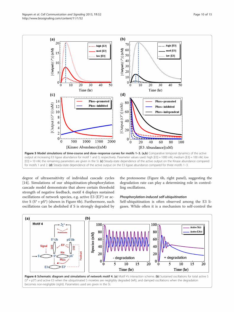
Figure 5 Model simulations of time-course and dose–response curves for motifs 1–3. (a,b) Comparative temporal dynamics of the activeoutput at increasing E3 ligase abundance for motif 1 and 3, respectively. Parameter values used: high [E3] = 1000 nM, medium [E3] = 100 nM, low[E3] = 10 nM, the remaining parameters are given in the SI. (c) Steady-state dependence of the active output on the Kinase abundance comparedfor motifs 1 and 2. (d) Steady-state dependence of the active output on the E3 ligase abundance compared for three motifs 1–3.
Nguyen et al. Cell Communication and Signaling 2013, 11:52 Page 10 of 15http://www.biosignaling.com/content/11/1/52
degree of ultrasensitivity of individual cascade cycles[14]. Simulations of our ubiquitination-phosphorylationcascade model demonstrate that above certain thresholdstrength of negative feedback, motif 4 displays sustainedoscillations of network species, e.g. active E3 (E3*) or ac-tive S (S* + pS*) (shown in Figure 6b). Furthermore, suchoscillations can be abolished if S is strongly degraded by
Figure 6 Schematic diagram and simulations of network motif 4. (a) M(S* + pS*) and active E3 when the ubiquitinated S moieties are negligibly dbecomes non-negligible (right). Parameters used are given in the SI.
the proteosome (Figure 6b, right panel), suggesting thedegradation rate can play a determining role in control-ling oscillations.
Phosphorylation-induced self-ubiquitinationSelf-ubiquitination is often observed among the E3 li-gases. While often it is a mechanism to self-control the
otif 4’s interaction scheme. (b) Sustained oscillations for total active Segraded (left), and damped oscillations when the degradation

Nguyen et al. Cell Communication and Signaling 2013, 11:52 Page 11 of 15http://www.biosignaling.com/content/11/1/52
ligase abundance, it can also serve non-proteolytic func-tions and can dramatically influence the ligase activity, asin the case of ITCH discussed earlier. Degradation ofITCH is independent of its self-ubiquitination, which oc-curs through K63 linkages and results in stronger catalyticactivity; whereas canonical K48-linked chains generated byother ligases target ITCH for degradation [83]. Likewise,self-ubiquitination of NEDD4 leads to better recognitionand higher rate of monoubiquitination of Eps15 in theEGFR internalisation and degradation pathway [32]. OtherE3 ligases with similar property include Ring1B (compo-nent of the human Polycomb transcriptional RepressiveComplex 1) whose self-ubiquitination generates atypical,branched K6/K27-linked chains and promotes itsmonoubiquitination activity toward histone H2A [3,7].Motivated by these examples, we next analyse a motifwhere kinase-mediated phosphorylation enhances the rateof self-ubiquitination of an E3 ligase on K63 linkages,which subsequently turns on its ligase activity towards asubstrate O, sending it to degradation (Figure 7a, motif 5).Note that, in some cases, the K63-ubiquitinated E3 candirectly or indirectly exert positive regulation over the kin-ase, providing a positive feedback to the system. We willfirst consider motif 5 with no feedback.
Figure 7 Schematic kinetic diagram of motif 5 and model simulationsthe phosphorylation of E3. (b) Reaction schemes depicting intra- and interresponses of relevant species against gradual increasing of the kinase abunlow and high branch of the hysteresis curves, the dashed lines indicates unwhen none, only self-ubiquitination, only positive feedback loop, or both m
Self-modification reactions can occur in either an intra-molecular or inter-molecular fashion, as depicted inFigure 7b. While our modelling analysis shows that theintra-molecular self-ubiquitination of the E3 ligase doesnot exhibit intricate dynamics, an inter-molecular form ofself-ubiquitination, such as of ITCH discussed above, canbring about bistable behaviour to the system, even withoutthe positive feedback loop [3,13]. Figures 7c,d showbistability and hysteresis for the ubiquitinated forms of theligase in response to the kinase abundance changes. Inter-estingly, E3-Ub and pE3-Ub have opposing off and onswitches with the increasing kinase level. Similarly, theoutput also shows a bistable response, with the hysteresiscurve being lower in the presence of high degradation rate(Figure 7e). Finally, we analyse motif 5 when the E3-to-Kinase positive feedback loop is also incorporated. Modelanalysis reveals that although self-ubiquitination or posi-tive feedback alone is sufficient to give rise to bistability,adding the positive feedback appears to enhance selfubiquitination-induced bistability while adding self-ubiquitination does not necessarily enhance bistabilityestablished by the positive feedback (comparing blue toblack curves, and blue to red in Figure 7f). Moreover,Figure 7f shows that the presence of both mechanisms
. (a) Dashed line indicates a positive feedback loop from pE3-Ub to-molecular self-activation mechanisms. (c-e) Steady-state bistabledance level. The vertical arrows (blue) indicate the jump between thestable state. (f) Comparison of bistable behaviour under four scenariosechanisms are operating. Parameters used are given in the SI.

Nguyen et al. Cell Communication and Signaling 2013, 11:52 Page 12 of 15http://www.biosignaling.com/content/11/1/52
brings the systems closer to irreversible hysteresis, indi-cated by the shift to the left of the corresponding hyster-esis curve (blue line).As discussed earlier, ubiquitination is a multi-step
process which depends not only on the abundance andproperties of the E3 ligase involved but also on otherfactors involving the preceding steps, including loadingof ubiquitin onto conjugating enzymes E2s and ubiquitintransferring to the substrate. Consideration of these fac-tors may be necessary for a detailed model of the controlof the EGFR pathway by ubiquitination. Such work how-ever would require comprehensive experimental effortto provide the missing kinetic data and other quantita-tive information to calibrate and validate the model.Our findings of potentially bistable and oscillatory be-
haviour of the ubiquitination-phosphorylation motifsawait experimental testing. In vitro experimental designbased on the model analysis results could be the firststep in confirming the predictions about the dynamics ofthe components of interest. An advantage of an in vitrosystem with purified forms of relevant E3 ligase, kinasesand phosphatases is that it can be used to explore wideranges of precisely set enzyme concentrations. To mimicthe in vivo situation, some of these proteins may be em-bedded into a phospholipid membrane bilayer or lipo-somes if required, which can also facilitate the formationof protein complexes and increase reaction rates [104].For instance to detect oscillations in motif 4, the systemcan be started by addition of the relevant input signal,followed by addition of ubiquitin, the E1/E2 enzymes, E3ligase, kinase and ATP to the reaction medium. At peri-odic selected time points, aliquots are taken, and thephosphorylated or ubiquitinated level of the substratecan be measured by immunoblotting using specific anti-bodies for phosphorylation or ubiquitination. It is how-ever worth mentioning that assembling an oscillatorynetwork in vitro is challenging due to a multitude of fac-tors at play, including the adequate level of ubiquitinand the essential participation of the relevant E1/E2 en-zymes. Therefore, direct in vivo approaches like imagingtechniques using microscopy-based binding assay can beexploited for high temporal resolution measurements ofcomponents kinetics and may be a more favourable op-tion [105]. On the other hand, detection of switchessuch as in motif 5 can be done by similar measurementtechniques in response to increasing titration of a dosecomponent, in this case the involved kinase protein(Figure 7).In summary, we have constructed mathematical models
and carried out analysis for a number of commonly seenmotifs of ubiquitination-phosphorylation crosstalk. Themotifs, although simplified, show diverse dynamics includ-ing sustained oscillations and bistability. More import-antly, the models have facilitated the identification of the
conditions under which these dynamics may realise, whichwould have been infeasible if such models are not used.Modelling therefore provides a useful and necessary toolfor efficient analysis of ubiquitination-phosphorylationcrosstalk, thereby potentially improving our systems-levelunderstanding of the integrated EGFR signalling.
ConclusionsSince the first discovery of protein ubiquitination morethan three decades ago, extensive work has revolution-ized our perception of its role in signalling networks.Not only protein ubiquitination serves as a main mech-anism for protein degradation, emerging evidence hasrevealed that different types of ubiquitin chains can in-duce a variety of non-proteolytic functions and can dra-matically alter the biological activities of a target protein.On top of that, ubiquitination is frequently observed tointerplay with other PTMs such as phosphorylation orsumoylation to coordinate regulation of signalling pro-cesses in intricate manners. Such complexity arisingfrom interconnected PTM networks poses enormouschallenges for the systems level analysis of signallingprocesses. Mathematical modelling is emerging as avaluable tool to provide insight into their dynamic be-haviour that would otherwise not be possible. Mathem-atical models help combine the mechanistic, molecularknowledge with rigorous analysis of the complex outputdynamics of the PTM networks.The expanding roles of ubiquitylation and phosphoryl-
ation in cell signalling, to large extent, have been uncov-ered thanks to recent advances in proteomics technologieswhich have enabled new ways for in-depth, unbiased andquantitative analysis of different PTMs on a global scale[106-110]. Techniques such as stable isotope labelling withamino acids in cell culture (SILAC) and label-free basedmass spectrometry can quantify changes in expression ofthousands of phosphoproteins and tens of thousandsphosphorylation events in a single experiment and havebecome well established [106,111]. Although proteome-wide analysis of endogenous ubiquitination has been morechallenging, recent developments on antibodies-based en-richment methods demonstrate the feasibilities of similarlarge-scale, quantitative and site-specific investigations ofthis PTM [112]. Moreover, novel methods that are aimedat identifying proteins comodified by both phosphoryl-ation and ubiquitination have revealed exciting global de-tails of the cross-regulation between these two PTMs[113]. A major limitation with current mass spectrometrybased methods however is the inability to distinguishamong modifications by ubiquitination, NEDD8 or ISG15,due to an identical di-Gly remnant generated by trypsinproteolysis of the modified proteins [112]. Nevertheless, itis likely that with the observed fast pace of technologicaladvance, sophisticated methods capable of resolving at

Nguyen et al. Cell Communication and Signaling 2013, 11:52 Page 13 of 15http://www.biosignaling.com/content/11/1/52
even higher quantitative resolution the extent of PTMscrosstalk and their distinct dynamics under different cellu-lar perturbations are within close reach. These data willundoubtedly be valuable inputs to the construction oflarge-scale, next-level quantitative models. A global, data-driven modelling-based understanding of PTMs networksand the ability to simulate their behaviour and form test-able predictions will open countless possibilities that candrive the frontiers of both biological and medical research.
Additional file
Additional file 1: Mathematical models for the investigated motifs.
AbbreviationsDUB: De-ubiquitinating enzyme; EGF: Epidermal growth factor;EGFR: Epidermal growth factor receptor; ERK: Extracellular signal-regulatedkinase; Cbl: Casitas b-lineage lymphoma; RTK: Receptor tyrosine kinase;EPS15: Epidermal growth factor receptor substrate 15; MVB: Multivesicularbody; UIM: Ubiquitin-interacting motif; HRS: Hepatocyte growth factor-regulated tyrosine kinase substrate; HECT: Homologous to the E6-AP carboxylterminus; STAMP: Signal transducing adaptor molecule; STAMBP: STAMbinding protein; USP8: Ubiquitin specific peptidase 8; Rab5: Ras-relatedprotein Rab5; GEF: Guanine nucleotide exchange factor; GA: Benzoquinoneansamycin Geldanamycin; JNK: c-Jun N-terminal kinase; MEKK1: MEK kinase 1;ITCH Itchy: E3 ubiquitin protein ligase; NEDD4: Neural precursor cellexpressed developmentally down-regulated protein 4; Rpn4: Regulatoryparticle non-ATPase; RING1B: Really interesting new gene 1 protein.
Competing interestsThe authors declare that they have no competing interests.
Authors’ contributionsLKN and BK designed the project. LKN carried out model simulations andanalysis. LKN, WK and BK wrote the paper. All authors read and approved thefinal manuscript.
AcknowledgementsThis work was supported by Science Foundation Ireland under Grant No. 06/CE/B1129 and the European Union Grant PRIMES No. FP7-HEALTH-2011-278568. We thank Alexander von Kriegsheim for critical reading of themanuscript.
Author details1Systems Biology Ireland, University College Dublin, Belfield, Dublin 4, Ireland.2Conway Institute, University College Dublin, Belfield, Dublin 4, Ireland.3School of Medicine and Medical Science, University College Dublin, Belfield,Dublin 4, Ireland.
Received: 4 April 2013 Accepted: 26 July 2013Published: 31 July 2013
References1. Ciechanover A: Proteolysis: from the lysosome to ubiquitin and the
proteasome. Nat Rev Mol Cell Biol 2005, 6:79–87.2. Zaaroor-Regev D, de Bie P, Scheffner M, Noy T, Shemer R, Heled M, Stein I,
Pikarsky E, Ciechanover A: Regulation of the polycomb protein Ring1B byself-ubiquitination or by E6-AP may have implications to thepathogenesis of Angelman syndrome. Proc Natl Acad Sci USA 2010,107:6788–6793.
3. Nguyen LK, Munoz-Garcia J, Maccario H, Ciechanover A, Kolch W,Kholodenko BN: Switches, excitable responses and oscillations in theRing1B/Bmi1 ubiquitination system. PLoS Comput Biol 2011, 7:e1002317.
4. Sorkin A, Goh LK: Endocytosis and intracellular trafficking of ErbBs. ExpCell Res 2009, 315:683–696.
5. Weake VM, Workman JL: Histone ubiquitination: triggering gene activity.Mol Cell 2008, 29:653–663.
6. Zhou W, Wang X, Rosenfeld MG: Histone H2A ubiquitination intranscriptional regulation and DNA damage repair. Int J Biochem Cell Biol2009, 41:12–15.
7. Ben-Saadon R, Zaaroor D, Ziv T, Ciechanover A: The polycomb proteinRing1B generates self atypical mixed ubiquitin chains required for itsin vitro histone H2A ligase activity. Mol Cell 2006, 24:701–711.
8. Hunter T: The age of crosstalk: phosphorylation, ubiquitination, andbeyond. Mol Cell 2007, 28:730–738.
9. Magnani M, Crinelli R, Bianchi M, Antonelli A: The ubiquitin-dependentproteolytic system and other potential targets for the modulation ofnuclear factor-kB (NF-kB). Curr Drug Targets 2000, 1:387–399.
10. Treier M, Staszewski LM, Bohmann D: Ubiquitin-dependent c-Jundegradation in vivo is mediated by the delta domain. Cell 1994, 78:787–798.
11. Fuchs SY, Dolan L, Davis RJ, Ronai Z: Phosphorylation-dependenttargeting of c-Jun ubiquitination by Jun N-kinase. Oncogene 1996,13:1531–1535.
12. Witowsky JA, Johnson GL: Ubiquitylation of MEKK1 inhibits itsphosphorylation of MKK1 and MKK4 and activation of the ERK1/2 andJNK pathways. J Biol Chem 2003, 278:1403–1406.
13. Kaimachnikov NP, Kholodenko BN: Toggle switches, pulses andoscillations are intrinsic properties of the Src activation/deactivationcycle. FEBS J 2009, 276:4102–4118.
14. Kholodenko BN: Negative feedback and ultrasensitivity can bring aboutoscillations in the mitogen-activated protein kinase cascades. Eur JBiochem 2000, 267:1583–1588.
15. Shankaran H, Ippolito DL, Chrisler WB, Resat H, Bollinger N, Opresko LK,Wiley HS: Rapid and sustained nuclear-cytoplasmic ERK oscillationsinduced by epidermal growth factor. Mol Syst Biol 2009, 5:332.
16. Nakayama K, Satoh T, Igari A, Kageyama R, Nishida E: FGF induces oscillationsof Hes1 expression and Ras/ERK activation. Curr Biol 2008, 18:R332–R334.
17. Tsyganov MA, Kolch W, Kholodenko BN: The topology design principlesthat determine the spatiotemporal dynamics of G-protein cascades. MolBiosyst 2012, 8:730–743.
18. Tkachenko E, Sabouri-Ghomi M, Pertz O, Kim C, Gutierrez E, Machacek M,Groisman A, Danuser G, Ginsberg MH: Protein kinase A governs a RhoA-RhoGDI protrusion-retraction pacemaker in migrating cells. Nat Cell Biol2011, 13:660–667.
19. Machacek M, Hodgson L, Welch C, Elliott H, Pertz O, Nalbant P, Abell A,Johnson GL, Hahn KM, Danuser G: Coordination of Rho GTPase activitiesduring cell protrusion. Nature 2009, 461:99–103.
20. Kholodenko BN: Cell-signalling dynamics in time and space. Nat Rev MolCell Biol 2006, 7:165–176.
21. Acconcia F, Sigismund S, Polo S: Ubiquitin in trafficking: the network atwork. Exp Cell Res 2009, 315:1610–1618.
22. Chen ZJ, Sun LJ: Nonproteolytic functions of ubiquitin in cell signaling.Mol Cell 2009, 33:275–286.
23. Levkowitz G, Waterman H, Ettenberg SA, Katz M, Tsygankov AY, Alroy I, LaviS, Iwai K, Reiss Y, Ciechanover A, et al: Ubiquitin ligase activity andtyrosine phosphorylation underlie suppression of growth factorsignaling by c-Cbl/Sli-1. Mol Cell 1999, 4:1029–1040.
24. Schmidt MH, Dikic I: The Cbl interactome and its functions. Nat Rev MolCell Biol 2005, 6:907–918.
25. Joazeiro CA, Wing SS, Huang H, Leverson JD, Hunter T, Liu YC: The tyrosinekinase negative regulator c-Cbl as a RING-type, E2-dependent ubiquitin-protein ligase. Science 1999, 286:309–312.
26. Huang F, Sorkin A: Growth factor receptor binding protein 2-mediatedrecruitment of the RING domain of Cbl to the epidermal growth factorreceptor is essential and sufficient to support receptor endocytosis. MolBiol Cell 2005, 16:1268–1281.
27. Waterman H, Katz M, Rubin C, Shtiegman K, Lavi S, Elson A, Jovin T, YardenY: A mutant EGF-receptor defective in ubiquitylation and endocytosisunveils a role for Grb2 in negative signaling. EMBO J 2002, 21:303–313.
28. Jiang X, Huang F, Marusyk A, Sorkin A: Grb2 regulates internalisation ofEGF receptors through clathrin-coated pits. Mol Biol Cell 2003, 14:858–870.
29. Dou H, Buetow L, Hock A, Sibbet GJ, Vousden KH, Huang DT: Structuralbasis for autoinhibition and phosphorylation-dependent activation of c-Cbl. Nat Struct Mol Biol 2012, 19:184–192.
30. Kobashigawa Y, Tomitaka A, Kumeta H, Noda NN, Yamaguchi M, Inagaki F:Autoinhibition and phosphorylation-induced activation mechanisms ofhuman cancer and autoimmune disease-related E3 protein Cbl-b. ProcNatl Acad Sci USA 2011, 108:20579–20584.

Nguyen et al. Cell Communication and Signaling 2013, 11:52 Page 14 of 15http://www.biosignaling.com/content/11/1/52
31. Katzmann DJ, Odorizzi G, Emr SD: Receptor downregulation andmultivesicular-body sorting. Nat Rev Mol Cell Biol 2002, 3:893–905.
32. Woelk T, Oldrini B, Maspero E, Confalonieri S, Cavallaro E, Di Fiore PP, Polo S:Molecular mechanisms of coupled monoubiquitination. Nat Cell Biol 2006,8:1246–1254.
33. Raiborg C, Bache KG, Gillooly DJ, Madshus IH, Stang E, Stenmark H: Hrssorts ubiquitinated proteins into clathrin-coated microdomains of earlyendosomes. Nat Cell Biol 2002, 4:394–398.
34. Clague MJ, Urbe S: Endocytosis: the DUB version. Trends Cell Biol 2006,16:551–559.
35. Nakamura M, Tanaka N, Kitamura N, Komada M: Clathrin anchorsdeubiquitinating enzymes, AMSH and AMSH-like protein, on earlyendosomes. Genes Cells 2006, 11:593–606.
36. McCullough J, Row PE, Lorenzo O, Doherty M, Beynon R, Clague MJ, Urbe S:Activation of the endosome-associated ubiquitin isopeptidase AMSH bySTAM, a component of the multivesicular body-sorting machinery. CurrBiol 2006, 16:160–165.
37. Niendorf S, Oksche A, Kisser A, Lohler J, Prinz M, Schorle H, Feller S, LewitzkyM, Horak I, Knobeloch KP: Essential role of ubiquitin-specific protease 8for receptor tyrosine kinase stability and endocytic trafficking in vivo.Mol Cell Biol 2007, 27:5029–5039.
38. Meijer IM, van Leeuwen JE: ERBB2 is a target for USP8-mediateddeubiquitination. Cell Signal 2011, 23:458–467.
39. Meijer IM, Kerperien J, Sotoca AM, van Zoelen EJ, van Leeuwen JE: TheUsp8 deubiquitination enzyme is post-translationally modified bytyrosine and serine phosphorylation. Cell Signal 2013, 25:919–930.
40. Yan J, Roy S, Apolloni A, Lane A, Hancock JF: Ras isoforms vary in theirability to activate Raf-1 and phosphoinositide 3-kinase. J Biol Chem 1998,273:24052–24056.
41. Voice JK, Klemke RL, Le A, Jackson JH: Four human ras homologs differ intheir abilities to activate Raf-1, induce transformation, and stimulate cellmotility. J Biol Chem 1999, 274:17164–17170.
42. Karnoub AE, Weinberg RA: Ras oncogenes: split personalities. Nat Rev MolCell Biol 2008, 9:517–531.
43. Jura N, Scotto-Lavino E, Sobczyk A, Bar-Sagi D: Differential modification ofRas proteins by ubiquitination. Mol Cell 2006, 21:679–687.
44. Hancock JF: Ras proteins: different signals from different locations. NatRev Mol Cell Biol 2003, 4:373–384.
45. Yan H, Chin ML, Horvath EA, Kane EA, Pfleger CM: Impairment ofubiquitylation by mutation in Drosophila E1 promotes both cell-autonomous and non-cell-autonomous Ras-ERK activation in vivo. J CellSci 2009, 122:1461–1470.
46. Horiuchi H, Lippe R, McBride HM, Rubino M, Woodman P, Stenmark H,Rybin V, Wilm M, Ashman K, Mann M, Zerial M: A novel Rab5 GDP/GTPexchange factor complexed to Rabaptin-5 links nucleotide exchange toeffector recruitment and function. Cell 1997, 90:1149–1159.
47. Delprato A, Merithew E, Lambright DG: Structure, exchange determinants,and family-wide rab specificity of the tandem helical bundle and Vps9domains of Rabex-5. Cell 2004, 118:607–617.
48. Xu L, Lubkov V, Taylor LJ, Bar-Sagi D: Feedback regulation of Ras signalingby Rabex-5-mediated ubiquitination. Curr Biol 2010, 20:1372–1377.
49. Penengo L, Mapelli M, Murachelli AG, Confalonieri S, Magri L, Musacchio A,Di Fiore PP, Polo S, Schneider TR: Crystal structure of the ubiquitinbinding domains of rabex-5 reveals two modes of interaction withubiquitin. Cell 2006, 124:1183–1195.
50. Mattera R, Tsai YC, Weissman AM, Bonifacino JS: The Rab5 guaninenucleotide exchange factor Rabex-5 binds ubiquitin (Ub) and functionsas a Ub ligase through an atypical Ub-interacting motif and a zinc fingerdomain. J Biol Chem 2006, 281:6874–6883.
51. Lee S, Tsai YC, Mattera R, Smith WJ, Kostelansky MS, Weissman AM,Bonifacino JS, Hurley JH: Structural basis for ubiquitin recognition andautoubiquitination by Rabex-5. Nat Struct Mol Biol 2006, 13:264–271.
52. Tam SY, Tsai M, Snouwaert JN, Kalesnikoff J, Scherrer D, Nakae S, ChatterjeaD, Bouley DM, Galli SJ: RabGEF1 is a negative regulator of mast cellactivation and skin inflammation. Nat Immunol 2004, 5:844–852.
53. Tam SY, Kalesnikoff J, Nakae S, Tsai M, Galli SJ: RabGEF1, a negativeregulator of Ras signalling, mast cell activation and skin inflammation.Novartis Found Symp 2005, 271:115–124. discussion 124–130, 145–151.
54. Wang Y, Waldron RT, Dhaka A, Patel A, Riley MM, Rozengurt E, Colicelli J:The RAS effector RIN1 directly competes with RAF and is regulated by14-3-3 proteins. Mol Cell Biol 2002, 22:916–926.
55. Han L, Colicelli J: A human protein selected for interference with Rasfunction interacts directly with Ras and competes with Raf1. Mol Cell Biol1995, 15:1318–1323.
56. Hicke L, Schubert HL, Hill CP: Ubiquitin-binding domains. Nat Rev Mol CellBiol 2005, 6:610–621.
57. Jura N, Bar-Sagi D: Mapping cellular routes of Ras: a ubiquitin trail. CellCycle 2006, 5:2744–2747.
58. Ahearn IM, Haigis K, Bar-Sagi D, Philips MR: Regulating the regulator: post-translational modification of RAS. Nat Rev Mol Cell Biol 2012, 13:39–51.
59. Sasaki AT, Carracedo A, Locasale JW, Anastasiou D, Takeuchi K, Kahoud ER,Haviv S, Asara JM, Pandolfi PP, Cantley LC: Ubiquitination of K-Rasenhances activation and facilitates binding to select downstreameffectors. Sci Signal 2011, 4:ra13.
60. Baker R, Lewis SM, Sasaki AT, Wilkerson EM, Locasale JW, Cantley LC,Kuhlman B, Dohlman HG, Campbell SL: Site-specific monoubiquitinationactivates Ras by impeding GTPase-activating protein function. Nat StructMol Biol 2013, 20:46–52.
61. Kim SE, Yoon JY, Jeong WJ, Jeon SH, Park Y, Yoon JB, Park YN, Kim H, ChoiKY: H-Ras is degraded by Wnt/beta-catenin signaling via beta-TrCP-mediated polyubiquitylation. J Cell Sci 2009, 122:842–848.
62. Laine A, Ronai Z: Ubiquitin chains in the ladder of MAPK signaling. SciSTKE 2005, 2005:re5.
63. Warne PH, Viciana PR, Downward J: Direct interaction of Ras and theamino-terminal region of Raf-1 in vitro. Nature 1993, 364:352–355.
64. Vojtek AB, Hollenberg SM, Cooper JA: Mammalian Ras interacts directlywith the serine/threonine kinase Raf. Cell 1993, 74:205–214.
65. Dent P, Haser W, Haystead TA, Vincent LA, Roberts TM, Sturgill TW:Activation of mitogen-activated protein kinase kinase by v-Raf in NIH3T3 cells and in vitro. Science 1992, 257:1404–1407.
66. Kyriakis JM, App H, Zhang XF, Banerjee P, Brautigan DL, Rapp UR, Avruch J:Raf-1 activates MAP kinase-kinase. Nature 1992, 358:417–421.
67. Matallanas D, Birtwistle M, Romano D, Zebisch A, Rauch J, von KriegsheimA, Kolch W: Raf family kinases: old dogs have learned new tricks. GenesCancer 2011, 2:232–260.
68. Schulte TW, Blagosklonny MV, Ingui C, Neckers L: Disruption of the Raf-1-Hsp90 molecular complex results in destabilisation of Raf-1 and loss ofRaf-1-Ras association. J Biol Chem 1995, 270:24585–24588.
69. Schulte TW, An WG, Neckers LM: Geldanamycin-induced destabilisation ofRaf-1 involves the proteasome. Biochem Biophys Res Commun 1997,239:655–659.
70. Noble C, Mercer K, Hussain J, Carragher L, Giblett S, Hayward R, Patterson C,Marais R, Pritchard CA: CRAF autophosphorylation of serine 621 isrequired to prevent its proteasome-mediated degradation. Mol Cell 2008,31:862–872.
71. Hurst JH, Dohlman HG: Dynamic Ubiquitination of the Mitogen-activatedProtein Kinase Kinase (MAPKK) Ste7 Determines Mitogen-activatedProtein Kinase (MAPK) Specificity. J Biol Chem 2013, 288:18660–18671.
72. Wang Y, Ge Q, Houston D, Thorner J, Errede B, Dohlman HG: Regulation ofSte7 ubiquitination by Ste11 phosphorylation and the Skp1-Cullin-F-boxcomplex. J Biol Chem 2003, 278:22284–22289.
73. Wang Y, Dohlman HG: Pheromone-dependent ubiquitination of themitogen-activated protein kinase kinase Ste7. J Biol Chem 2002,277:15766–15772.
74. Lu Z, Xu S, Joazeiro C, Cobb MH, Hunter T: The PHD domain of MEKK1acts as an E3 ubiquitin ligase and mediates ubiquitination anddegradation of ERK1/2. Mol Cell 2002, 9:945–956.
75. Yan M, Dai T, Deak JC, Kyriakis JM, Zon LI, Woodgett JR, Templeton DJ:Activation of stress-activated protein kinase by MEKK1 phosphorylationof its activator SEK1. Nature 1994, 372:798–800.
76. Johnson GL, Lapadat R: Mitogen-activated protein kinase pathwaysmediated by ERK, JNK, and p38 protein kinases. Science 2002,298:1911–1912.
77. Karandikar M, Xu S, Cobb MH: MEKK1 binds raf-1 and the ERK2 cascadecomponents. J Biol Chem 2000, 275:40120–40127.
78. Maruyama T, Kadowaki H, Okamoto N, Nagai A, Naguro I, Matsuzawa A,Shibuya H, Tanaka K, Murata S, Takeda K, et al: CHIP-dependenttermination of MEKK2 regulates temporal ERK activation required forproper hyperosmotic response. EMBO J 2010, 29:2501–2514.
79. Rotin D, Staub O, Haguenauer-Tsapis R: Ubiquitination and endocytosis ofplasma membrane proteins: role of Nedd4/Rsp5p family of ubiquitin-protein ligases. J Membr Biol 2000, 176:1–17.

Nguyen et al. Cell Communication and Signaling 2013, 11:52 Page 15 of 15http://www.biosignaling.com/content/11/1/52
80. Fang D, Elly C, Gao B, Fang N, Altman Y, Joazeiro C, Hunter T, Copeland N,Jenkins N, Liu YC: Dysregulation of T lymphocyte function in itchy mice: arole for Itch in TH2 differentiation. Nat Immunol 2002, 3:281–287.
81. Parravicini V, Field AC, Tomlinson PD, Basson MA, Zamoyska R: Itch−/−alphabeta and gammadelta T cells independently contribute toautoimmunity in Itchy mice. Blood 2008, 111:4273–7282.
82. Lohr NJ, Molleston JP, Strauss KA, Torres-Martinez W, Sherman EA,Squires RH, Rider NL, Chikwava KR, Cummings OW, Morton DH,Puffenberger EG: Human ITCH E3 ubiquitin ligase deficiency causessyndromic multisystem autoimmune disease. Am J Hum Genet 2010,86:447–453.
83. Scialpi F, Malatesta M, Peschiaroli A, Rossi M, Melino G, Bernassola F: Itchself-polyubiquitylation occurs through lysine-63 linkages. BiochemPharmacol 2008, 76:1515–1521.
84. Gallagher E, Gao M, Liu YC, Karin M: Activation of the E3 ubiquitin ligaseItch through a phosphorylation-induced conformational change. ProcNatl Acad Sci USA 2006, 103:1717–1722.
85. Azakir BA, Angers A: Reciprocal regulation of the ubiquitin ligase Itchand the epidermal growth factor receptor signaling. Cell Signal 2009,21:1326–1336.
86. Azakir BA, Desrochers G, Angers A: The ubiquitin ligase Itch mediates theantiapoptotic activity of epidermal growth factor by promoting theubiquitylation and degradation of the truncated C-terminal portion ofBid. FEBS J 2010, 277:1319–1330.
87. Wei MC, Lindsten T, Mootha VK, Weiler S, Gross A, Ashiya M, Thompson CB,Korsmeyer SJ: tBID, a membrane-targeted death ligand, oligomerizes BAKto release cytochrome c. Genes Dev 2000, 14:2060–2071.
88. Breitschopf K, Zeiher AM, Dimmeler S: Ubiquitin-mediated degradation ofthe proapoptotic active form of bid. A functional consequence onapoptosis induction. J Biol Chem 2000, 275:21648–21652.
89. O’Neill E, Kolch W: Taming the Hippo: Raf-1 controls apoptosis bysuppressing MST2/Hippo. Cell Cycle 2005, 4:365–367.
90. O'Neill E, Rushworth L, Baccarini M, Kolch W: Role of the kinase MST2 insuppression of apoptosis by the proto-oncogene product Raf-1. Science2004, 306:2267–2270.
91. Matallanas D, Romano D, Yee K, Meissl K, Kucerova L, Piazzolla D, BaccariniM, Vass JK, Kolch W, O'Neill E: RASSF1A elicits apoptosis through an MST2pathway directing proapoptotic transcription by the p73 tumorsuppressor protein. Mol Cell 2007, 27:962–975.
92. Ho KC, Zhou Z, She YM, Chun A, Cyr TD, Yang X: Itch E3 ubiquitin ligaseregulates large tumor suppressor 1 stability [corrected]. Proc Natl AcadSci USA 2011, 108:4870–4875.
93. Levy D, Adamovich Y, Reuven N, Shaul Y: The Yes-associated protein 1stabilizes p73 by preventing Itch-mediated ubiquitination of p73. CellDeath Differ 2007, 14:743–751.
94. Yang C, Zhou W, Jeon MS, Demydenko D, Harada Y, Zhou H, Liu YC:Negative regulation of the E3 ubiquitin ligase itch via Fyn-mediatedtyrosine phosphorylation. Mol Cell 2006, 21:135–141.
95. Kofahl B, Klipp E: Modelling the dynamics of the yeast pheromonepathway. Yeast 2004, 21:831–850.
96. Thomson TM, Benjamin KR, Bush A, Love T, Pincus D, Resnekov O, Yu RC,Gordon A, Colman-Lerner A, Endy D, Brent R: Scaffold number in yeastsignaling system sets tradeoff between system output and dynamicrange. Proc Natl Acad Sci USA 2011, 108:20265–20270.
97. Schaber J, Kofahl B, Kowald A, Klipp E: A modelling approach to quantifydynamic crosstalk between the pheromone and the starvation pathwayin baker’s yeast. FEBS J 2006, 273:3520–3533.
98. Paliwal S, Iglesias PA, Campbell K, Hilioti Z, Groisman A, Levchenko A:MAPK-mediated bimodal gene expression and adaptive gradientsensing in yeast. Nature 2007, 446:46–51.
99. Welcker M, Orian A, Jin J, Grim JE, Harper JW, Eisenman RN, Clurman BE:The Fbw7 tumor suppressor regulates glycogen synthase kinase 3phosphorylation-dependent c-Myc protein degradation. Proc Natl AcadSci USA 2004, 101:9085–9090.
100. Yada M, Hatakeyama S, Kamura T, Nishiyama M, Tsunematsu R, Imaki H,Ishida N, Okumura F, Nakayama K, Nakayama KI: Phosphorylation-dependent degradation of c-Myc is mediated by the F-box proteinFbw7. EMBO J 2004, 23:2116–2125.
101. Lin HK, Wang L, Hu YC, Altuwaijri S, Chang C: Phosphorylation-dependentubiquitylation and degradation of androgen receptor by Akt requireMdm2 E3 ligase. EMBO J 2002, 21:4037–4048.
102. Ju D, Xu H, Wang X, Xie Y: Ubiquitin-mediated degradation of Rpn4 iscontrolled by a phosphorylation-dependent ubiquitylation signal.Biochim Biophys Acta 2007, 1773:1672–1680.
103. Hu H, Goltsov A, Bown JL, Sims AH, Langdon SP, Harrison DJ, Faratian D:Feedforward and feedback regulation of the MAPK and PI3K oscillatorycircuit in breast cancer. Cell Signal 2013, 25:26–32.
104. Kholodenko BN, Hoek JB, Westerhoff HV: Why cytoplasmic signallingproteins should be recruited to cell membranes. Trends Cell Biol 2000,10:173–178.
105. Yang HW, Shin MG, Lee S, Kim JR, Park WS, Cho KH, Meyer T, Do Heo W:Cooperative activation of PI3K by Ras and Rho family small GTPases. MolCell 2012, 47:281–290.
106. Choudhary C, Mann M: Decoding signalling networks by massspectrometry-based proteomics. Nat Rev Mol Cell Biol 2010, 11:427–439.
107. Kim W, Bennett EJ, Huttlin EL, Guo A, Li J, Possemato A, Sowa ME, Rad R,Rush J, Comb MJ, et al: Systematic and quantitative assessment of theubiquitin-modified proteome. Mol Cell 2011, 44:325–340.
108. Olsen JV, Vermeulen M, Santamaria A, Kumar C, Miller ML, Jensen LJ, GnadF, Cox J, Jensen TS, Nigg EA, et al: Quantitative phosphoproteomicsreveals widespread full phosphorylation site occupancy during mitosis.Sci Signal 2010, 3:ra3.
109. Oppermann FS, Gnad F, Olsen JV, Hornberger R, Greff Z, Keri G, Mann M,Daub H: Large-scale proteomics analysis of the human kinome. Mol CellProteomics 2009, 8:1751–1764.
110. Mirzaei H, Rogers RS, Grimes B, Eng J, Aderem A, Aebersold R:Characterizing the connectivity of poly-ubiquitin chains by selectedreaction monitoring mass spectrometry. Mol Biosyst 2010, 6:2004–2014.
111. Mann M: Functional and quantitative proteomics using SILAC. Nat RevMol Cell Biol 2006, 7:952–958.
112. Wagner SA, Beli P, Weinert BT, Nielsen ML, Cox J, Mann M, Choudhary C: Aproteome-wide, quantitative survey of in vivo ubiquitylation sites revealswidespread regulatory roles. Mol Cell Proteomics 2011, 10:M111–M013284.
113. Swaney DL, Beltrao P, Starita L, Guo A, Rush J, Fields S, Krogan NJ, Villen J:Global analysis of phosphorylation and ubiquitylation cross-talk inprotein degradation. Nat Methods 2013, 10:676–682.
doi:10.1186/1478-811X-11-52Cite this article as: Nguyen et al.: When ubiquitination meetsphosphorylation: a systems biology perspective of EGFR/MAPK signalling.Cell Communication and Signaling 2013 11:52.
Submit your next manuscript to BioMed Centraland take full advantage of:
• Convenient online submission
• Thorough peer review
• No space constraints or color figure charges
• Immediate publication on acceptance
• Inclusion in PubMed, CAS, Scopus and Google Scholar
• Research which is freely available for redistribution
Submit your manuscript at www.biomedcentral.com/submit