Embed Size (px)
Citation preview
The Egyptian Journal of Hospital Medicine (October 2017) Vol. 69 (8), Page 3092-3103
3092
Received: 09/09/2017 DOI: 10.12816/0042860
Accepted: 19/09/2017
Review on Liver Cirrhosis Complications and Treatment Haider Issa Alaqaili
1, Ahmed Ibrahim AlJuraysan
2 , Razan Mansour A Hawsawi
3 , Fadia
Abdulelah Abuzaid 4 , Muath Abdullah Alharbi
5 , Abdulrhman Ebrahim A. Mughallis
6 , Yazeed
Abdullah H Alsubhi 7 , Mohammed Rraiy A Asiri
8 , Abdullah Saleh S Alamer
9 , Abdullah
Mohammed S Azab 7 , Abdullah Javed Khaleel
7 , Hanan Khalil Ibrahim Al-Rajeh
10
1- Dammam Medical Complex, 2- King Faisal University, 3- King sau-Hs , 4- King Abdulaziz
University,Medicine-Basic Medical Sciences-Anatomy,Research Assistant King Fahad Medical
Research Center , 5- Medical University of Lodz , 6- Jazan General Hospital , 7- King Abdulaziz
University ,8- Sharg Almjaredah PHCC , 9- Qassim University , 10- King Fahad Hospital
ABSTRACT
Background: cirrhosis is a late stage of scarring (fibrosis) of the liver caused by many forms of liver diseases and
conditions, such as hepatitis and chronic alcoholism. The liver carries out several necessary functions, including
detoxifying harmful substances in your body, cleaning your blood and making vital nutrients. Cirrhosis occurs in
response to damage to your liver. Each time your liver is injured, it tries to repair itself. In the process, scar tissue
forms. As cirrhosis progresses, more and more scar tissue forms, making it difficult for the liver to function.
Objective of the Study: review and evaluate the best practices in diagnosis, complications and management of
cirrhosis, and novel clinical and scientific developments.
Methods: electronic search in the scientific database from 1966 to 2017– (Medline, Embase, the Cochrane
Library as well as NHS center websites were searched for English Publications obtained from both reprint
requests and by searching the database. Data extracted included authors, country, year of publication, age and sex
of patients, epidemiology, geographical distribution, pathophysiology, risk factors, clinical manifestations,
investigations and types of surgical treatment.
Results: there is sufficient body of evidence suggesting that cirrhosis is a pathological diagnosis with no
laboratory cutoff values for the diagnosis of cirrhosis.
However, it can still be diagnosed clinically, by history, physical examination laboratory analyses and ancillary
testing such as ultrasonography. Early diagnosis has proven to give relevantly better case management results
while late detection can only hardly manage the symptoms accompanied with cirrhosis.
Conclusion: Screening for chronic liver disease is a key factor for early detection of signs for liver damage,
which can be performed inexpensively and easily with clinical history-taking, measurement of transaminase
concentrations, upper abdominal ultrasonography, and transient elastography (where available). Abnormal
findings should prompt specific diagnostic testing to determine the etiology of the underlying disease. In most
patients, the dynamic process of progressive fibrosis, which could ultimately lead to cirrhosis, can be interrupted
by the timely recognition of the risk, followed by appropriate treatment.
Keywords: cirrhosis, progressive liver disease, ascites, chronic disease.
INTRODUCTION
Cirrhosis results from different mechanisms of
liver injury that lead to necroinflammation and
fibrogenesis; histologically it is characterized by
diffuse nodular regeneration surrounded by dense
fibrotic septa with subsequent parenchymal
extinction and collapse of liver structures, together
causing pronounced distortion of hepatic vascular
architecture (1)
. This distortion results in increased
resistance to portal blood flow and hence in portal
hypertension and in hepatic synthetic dysfunction.
Clinically, cirrhosis has been regarded as an end-
stage disease that invariably leads to death, unless
liver transplantation is done, and the only preventive
strategies have been screening for esophageal
varices and hepatocellular carcinoma.
One-year mortality in cirrhosis varies widely,
from 1% to 57%, depending on the occurrence of
clinical decompensating events (2)
. Histopathologists
have proposed that the histological term cirrhosis
should be substituted by advanced liver disease, to
underline the dynamic processes and variable
prognosis of the disorder (3)
. Moreover, fibrosis,
even in the cirrhotic range, regresses with specific
therapy if available, such as antiviral treatment for
chronic hepatitis B or C (4)
.
Cirrhosis isn't curable, but it’s treatable. Doctors
have two main goals in treating this disease: stop the
damage to your liver, and prevent complications.
Alcohol abuse, hepatitis, and fatty liver disease are
some of the main causes. Your doctor will
Review on Liver Cirrhosis Complications and Treatment
3093
personalize your treatment based on what caused
your cirrhosis, and the amount of liver damage you
have (5)
. Cirrhosis can lead to a number of
complications, including liver cancer. In some
people, the symptoms of cirrhosis may be the first
signs of liver disease.
In the present article, we review the current
understanding of cirrhosis as a dynamic process and
outline current therapeutic options for prevention
and treatment of complications of cirrhosis, on the
basis of the sub-classification in clinical prognostic
stages.
The study was done according to the ethical
board of King Faisal university.
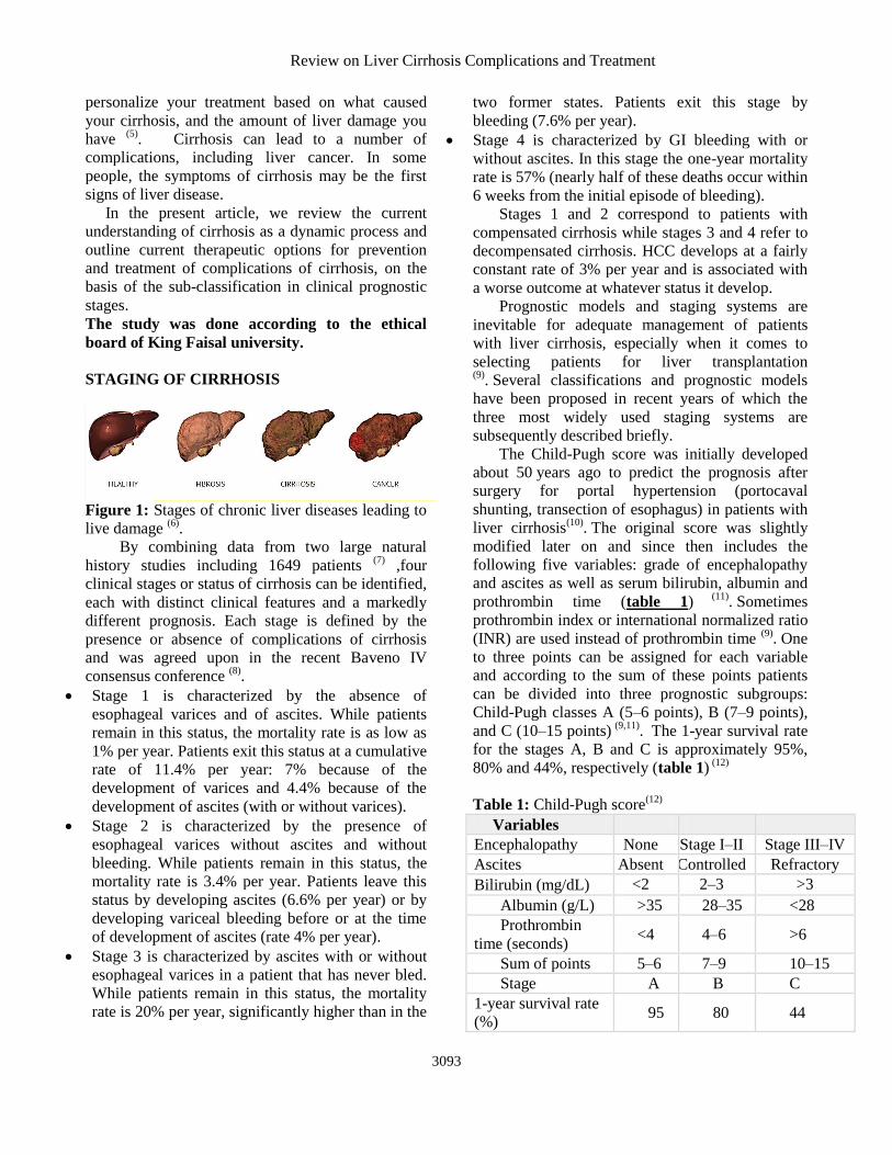
STAGING OF CIRRHOSIS
Figure 1: Stages of chronic liver diseases leading to
live damage (6)
.
By combining data from two large natural
history studies including 1649 patients (7)
,four
clinical stages or status of cirrhosis can be identified,
each with distinct clinical features and a markedly
different prognosis. Each stage is defined by the
presence or absence of complications of cirrhosis
and was agreed upon in the recent Baveno IV
consensus conference (8)
.
Stage 1 is characterized by the absence of
esophageal varices and of ascites. While patients
remain in this status, the mortality rate is as low as
1% per year. Patients exit this status at a cumulative
rate of 11.4% per year: 7% because of the
development of varices and 4.4% because of the
development of ascites (with or without varices).
Stage 2 is characterized by the presence of
esophageal varices without ascites and without
bleeding. While patients remain in this status, the
mortality rate is 3.4% per year. Patients leave this
status by developing ascites (6.6% per year) or by
developing variceal bleeding before or at the time
of development of ascites (rate 4% per year).
Stage 3 is characterized by ascites with or without
esophageal varices in a patient that has never bled.
While patients remain in this status, the mortality
rate is 20% per year, significantly higher than in the
two former states. Patients exit this stage by
bleeding (7.6% per year).
Stage 4 is characterized by GI bleeding with or
without ascites. In this stage the one-year mortality
rate is 57% (nearly half of these deaths occur within
6 weeks from the initial episode of bleeding).
Stages 1 and 2 correspond to patients with
compensated cirrhosis while stages 3 and 4 refer to
decompensated cirrhosis. HCC develops at a fairly
constant rate of 3% per year and is associated with
a worse outcome at whatever status it develop.
Prognostic models and staging systems are
inevitable for adequate management of patients
with liver cirrhosis, especially when it comes to
selecting patients for liver transplantation
(9). Several classifications and prognostic models
have been proposed in recent years of which the
three most widely used staging systems are
subsequently described briefly.
The Child-Pugh score was initially developed
about 50 years ago to predict the prognosis after
surgery for portal hypertension (portocaval
shunting, transection of esophagus) in patients with
liver cirrhosis(10)
. The original score was slightly
modified later on and since then includes the
following five variables: grade of encephalopathy
and ascites as well as serum bilirubin, albumin and
prothrombin time (table 1) (11)
. Sometimes
prothrombin index or international normalized ratio
(INR) are used instead of prothrombin time (9)
. One
to three points can be assigned for each variable
and according to the sum of these points patients
can be divided into three prognostic subgroups:
Child-Pugh classes A (5–6 points), B (7–9 points),
and C (10–15 points) (9,11)
. The 1-year survival rate
for the stages A, B and C is approximately 95%,
80% and 44%, respectively (table 1) (12)
Table 1: Child-Pugh score(12)
Variables 1 2 3
Encephalopathy None Stage I–II Stage III–IV
Ascites Absent Controlled Refractory
Bilirubin (mg/dL) <2 2–3 >3
Albumin (g/L) >35 28–35 <28
Prothrombin
time (seconds) <4 4–6 >6
Sum of points 5–6 7–9 10–15
Stage A B C
1-year survival rate
(%) 95 80 44
Haider Alaqaili et al.
3094
METHODS
Electronic search in the scientific database from
1960 to 2017.
Data source: Medline, Embase, the Cochrane Library
as well as NHS centre websites were searched for
English Publications were obtained from both reprint
requests and by searching the database.
Data extracted included authors, country, year of
publication, age and sex of patients, epidemiology,
geographical distribution, pathophysiology, risk
factors, clinical manifestations, investigations and
types of treatment.
ETIOLOGY OF CIRRHOSIS
Alcoholic liver disease and hepatitis C are the
most common causes in the Western world, while
hepatitis B prevails in most parts of Asia and sub-
Saharan Africa. After the identification of the
hepatitis C virus in 1989 and of nonalcoholic
steatohepatitis (NASH) in obese and diabetic
subjects, the diagnosis of cirrhosis without an
apparent cause (cryptogenic cirrhosis) is rarely
made.
Many cases of cryptogenic cirrhosis appear to
have resulted from nonalcoholic fatty liver disease
(NAFLD). When cases of cryptogenic cirrhosis are
reviewed, many patients have 1 or more of the
classic risk factors for NAFLD: obesity, diabetes,
and hypertriglyceridemia(13)
. It is postulated that
steatosis may regress in some patients as hepatic
fibrosis progresses, making the histologic diagnosis
of NAFLD difficult. Flavonoids have been reported
to have positive effects on key pathophysiologic
pathways in NAFLD (eg, lipid metabolism, insulin
resistance, inflammation, oxidative stress) and may
hold future potential for inclusion in NAFLD
treatment (13)
.
It is important to know the etiology of
cirrhosis, since it can predict complications and
direct treatment decisions. It also allows the
discussion of preventive measures, e.g., with family
members of patients with alcoholic cirrhosis or
chronic viral hepatitis, and consideration of (genetic)
testing and preventive advice for relatives of patients
with genetic diseases, such as hemochromatosis or
Wilson’s disease. Frequently multiple etiological
factors contribute to the development of cirrhosis, as
exemplified in epidemiological studies that
identified regular (moderate) alcohol consumption,
age above 50 years, and male gender as risk factors
in chronic hepatitis C (14)
, or older age obesity,
insulin resistance/type 2 diabetes, hypertension and
hyperlipidemia (all features of the metabolic
syndrome) in NASH
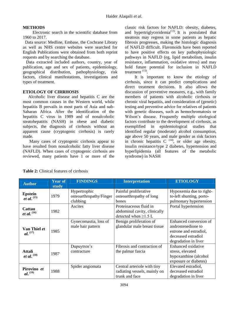
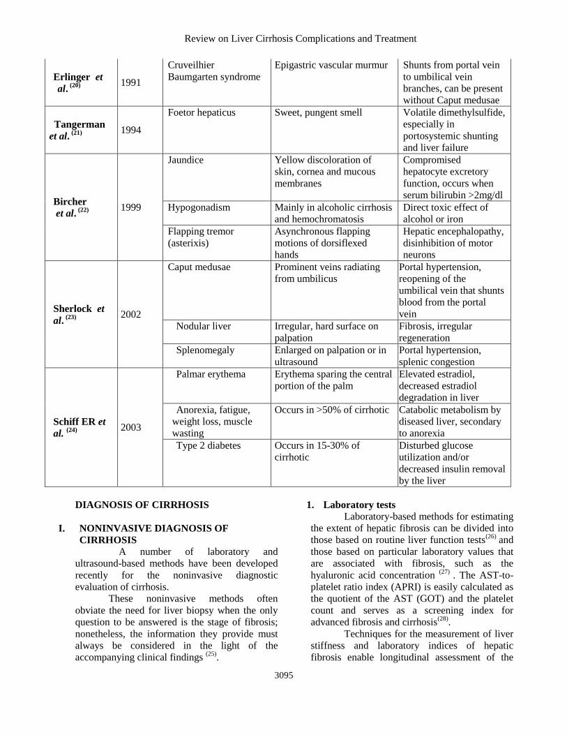
Table 2: Clinical features of cirrhosis
Author Year of
study
FINDINGS Interpretation ETIOLOGY
Epstein
et al. (15)
1979
Hypertrophic
osteoarthropathy/Finger
clubbing
Painful proliferative
osteoarthropathy of long
bones
Hypoxemia due to right-
to-left shunting, porto-
pulmonary hypertension
Cattau
et al. (16)
1982
Ascites Proteinaceous fluid in
abdominal cavity, clinically
detected when ≥1.5 L
Portal hypertension
Van Thiel et
al. (17)
1985
Gynecomastia, loss of
male hair pattern
Benign proliferation of
glandular male breast tissue
Enhanced conversion of
androstenedione to
estrone and estradiol,
decreased estradiol
degradation in liver
Attali
et al. (18)
1987
Dupuytren’s
contracture
Fibrosis and contraction of
the palmar fascia
Enhanced oxidative
stress, elevated
hypoxanthine (alcohol
exposure or diabetes)
Pirovino et
al. (19)
1988
Spider angiomata Central arteriole with tiny
radiating vessels, mainly on
trunk and face
Elevated estradiol,
decreased estradiol
degradation in liver
Review on Liver Cirrhosis Complications and Treatment
3095
Erlinger et
al. (20)
1991
Cruveilhier
Baumgarten syndrome
Epigastric vascular murmur Shunts from portal vein
to umbilical vein
branches, can be present
without Caput medusae
Tangerman
et al. (21)
1994
Foetor hepaticus Sweet, pungent smell Volatile dimethylsulfide,
especially in
portosystemic shunting
and liver failure
Bircher
et al. (22)
1999
Jaundice Yellow discoloration of
skin, cornea and mucous
membranes
Compromised
hepatocyte excretory
function, occurs when
serum bilirubin >2mg/dl
Hypogonadism Mainly in alcoholic cirrhosis
and hemochromatosis
Direct toxic effect of
alcohol or iron
Flapping tremor
(asterixis)
Asynchronous flapping
motions of dorsiflexed
hands
Hepatic encephalopathy,
disinhibition of motor
neurons
Sherlock et
al. (23)
2002
Caput medusae Prominent veins radiating
from umbilicus
Portal hypertension,
reopening of the
umbilical vein that shunts
blood from the portal
vein
Nodular liver Irregular, hard surface on
palpation
Fibrosis, irregular
regeneration
Splenomegaly Enlarged on palpation or in
ultrasound
Portal hypertension,
splenic congestion
Schiff ER et
al. (24)
2003
Palmar erythema Erythema sparing the central
portion of the palm
Elevated estradiol,
decreased estradiol
degradation in liver
Anorexia, fatigue,
weight loss, muscle
wasting
Occurs in >50% of cirrhotic Catabolic metabolism by
diseased liver, secondary
to anorexia
Type 2 diabetes Occurs in 15-30% of
cirrhotic
Disturbed glucose
utilization and/or
decreased insulin removal
by the liver
DIAGNOSIS OF CIRRHOSIS
I. NONINVASIVE DIAGNOSIS OF
CIRRHOSIS
A number of laboratory and
ultrasound-based methods have been developed
recently for the noninvasive diagnostic
evaluation of cirrhosis.
These noninvasive methods often
obviate the need for liver biopsy when the only
question to be answered is the stage of fibrosis;
nonetheless, the information they provide must
always be considered in the light of the
accompanying clinical findings (25)
.
1. Laboratory tests
Laboratory-based methods for estimating
the extent of hepatic fibrosis can be divided into
those based on routine liver function tests(26)
and
those based on particular laboratory values that
are associated with fibrosis, such as the
hyaluronic acid concentration (27)
. The AST-to-
platelet ratio index (APRI) is easily calculated as
the quotient of the AST (GOT) and the platelet
count and serves as a screening index for
advanced fibrosis and cirrhosis(28)
.
Techniques for the measurement of liver
stiffness and laboratory indices of hepatic
fibrosis enable longitudinal assessment of the
Haider Alaqaili et al.
3096
progression and regression of fibrosis in patients
with chronic liver disease.
ULTRASONOGRAPHY
Abdominal ultrasonography with Doppler
is a noninvasive, widely available modality that
provides valuable information regarding the
gross appearance of the liver and blood flow in
the portal and hepatic veins in patients suspected
to have cirrhosis. Ultrasonography should be the
first radiographic study performed in the
evaluation of cirrhosis because it is the least
expensive and does not pose a radiation exposure
risk or involve intravenous contrast with the
potential for nephrotoxicity as does computed
tomography (CT). Nodularity, irregularity,
increased echogenicity, and atrophy are
ultrasonographic hallmarks of cirrhosis. In
advanced disease, the gross liver appears small
and multinodular, ascites may be detected, and
Doppler flow can be significantly decreased in
the portal circulation. The discovery of hepatic
nodules via ultrasonography warrants further
evaluation because benign and malignant nodules
can have similar ultrasonographic appearances (29)
. A study using high-resolution
ultrasonography in patients with cirrhosis
confirmed with biopsy or laparoscopy found a
sensitivity and specificity for cirrhosis of 91.1
and 93.5 percent, respectively, and positive and
negative predictive values of 93.2 and 91.5
percent, respectively (30)
.
The diagnostic evaluation of cirrhosis
with ultrasonography is based on the direct
relation between the extent of fibrosis and the
ultrasonographically determined degree of liver
stiffness. Transient elastography and the acoustic
radiation force impulse (ARFI) technique are
now well-established methods for the staging of
fibrosis in various liver diseases(31)
. These two
techniques can be performed repeatedly on an
outpatient basis, and they can also be
combined(25)
.
Although ultrasonography can rule
cirrhosis in or out in over 90% of cases(31)
, its
findings are less than 100% specific because of
occasional incorrect measurements and false-
positive findings. There may be difficulty in
interpreting values that do not cross the
necessary thresholds for ruling advanced fibrosis,
or cirrhosis, in or out; in such situations, the
temporal course of the variable in question is its
clinically relevant feature. It should also be borne
in mind that the diagnostic threshold values vary
depending on the underlying etiology of liver
disease (32)
.
II. INVASIVE DIAGNOSIS OF CIRRHOSIS
1. CT AND MRI
CT and magnetic resonance imaging
(MRI) generally are poor at detecting
morphologic changes associated with early
cirrhosis, but they can accurately demonstrate
nodularity and lobar atrophic and hypertrophic
changes, as well as ascites and varices in
advanced disease. Although MRI sometimes
differentiates among regenerating or dysplastic
nodules and hepatocellular carcinoma, it is best
used as a follow-up study to determine whether
lesions have changed in appearance and size (33)
. CT portal phase imaging can be used to
assess portal vein patency, although flow volume
and direction cannot be determined accurately (34)
.
Although used rarely, magnetic resonance
angiography (MRA) can assess portal
hypertensive changes including flow volume and
direction, as well as portal vein thrombosis (34)
. One study reported that MRI can accurately
diagnose cirrhosis and provide correlation with
its severity. Despite the potential of MRI and
MRA in the diagnosis and evaluation of patients
with cirrhosis, their widespread use is limited by
their expense and by the ability of routine
ultrasonography with Doppler to obtain adequate
information for the diagnosis of cirrhosis and
presence of complications.
2. Liver biopsy
Referral for liver biopsy should be considered
after a thorough, noninvasive serologic and
radiographic evaluation has failed to confirm a
diagnosis of cirrhosis; the benefit of biopsy
outweighs the risk; and it is postulated that
biopsy will have a favorable impact on the
treatment of chronic liver disease. The sensitivity
and specificity for an accurate diagnosis of
cirrhosis and its etiology range from 80 to 100
percent, depending on the number and size of the
histologic samples and on the sampling method (35)
.
Liver biopsy is performed via percutaneous,
transjugular, laparoscopic, open operative, or
ultrasonography- or CT-guided fine-needle
approaches. Before the procedure, a CBC with
Review on Liver Cirrhosis Complications and Treatment
3097
platelets and prothrombin time measurement
should be obtained. Patients should be advised to
refrain from consumption of aspirin and
nonsteroidal anti-inflammatory drugs for seven
to 10 days before the biopsy to minimize the risk
of bleeding (35)
.
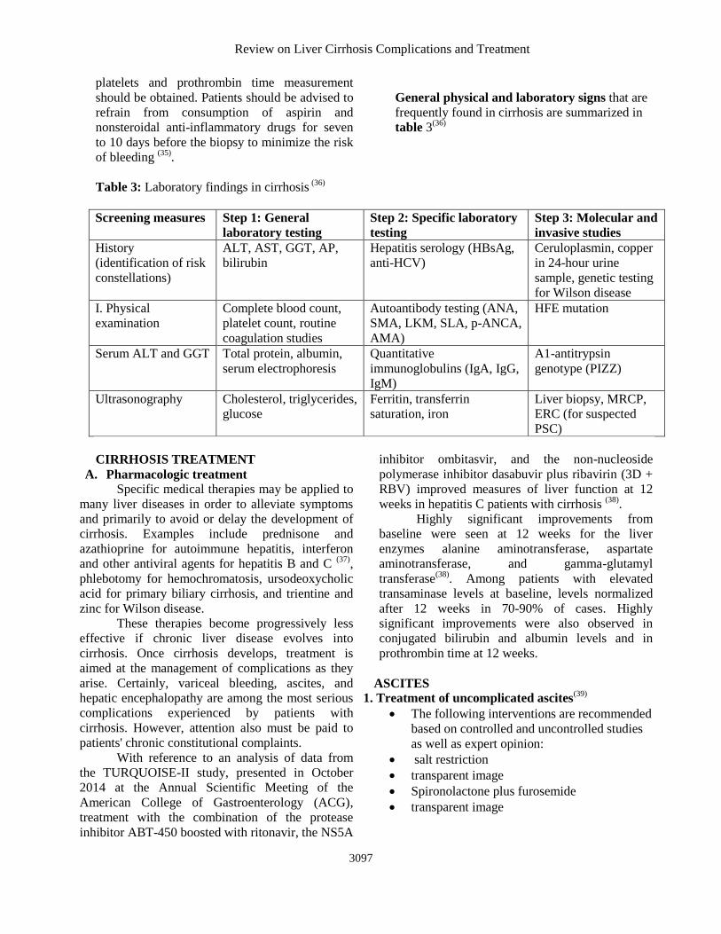
General physical and laboratory signs that are
frequently found in cirrhosis are summarized in
table 3(36)
Table 3: Laboratory findings in cirrhosis (36)
Screening measures Step 1: General
laboratory testing
Step 2: Specific laboratory
testing
Step 3: Molecular and
invasive studies
History
(identification of risk
constellations)
ALT, AST, GGT, AP,
bilirubin
Hepatitis serology (HBsAg,
anti-HCV)
Ceruloplasmin, copper
in 24-hour urine
sample, genetic testing
for Wilson disease
I. Physical
examination
Complete blood count,
platelet count, routine
coagulation studies
Autoantibody testing (ANA,
SMA, LKM, SLA, p-ANCA,
AMA)
HFE mutation
Serum ALT and GGT Total protein, albumin,
serum electrophoresis
Quantitative
immunoglobulins (IgA, IgG,
IgM)
A1-antitrypsin
genotype (PIZZ)
Ultrasonography Cholesterol, triglycerides,
glucose
Ferritin, transferrin
saturation, iron
Liver biopsy, MRCP,
ERC (for suspected
PSC)
CIRRHOSIS TREATMENT
A. Pharmacologic treatment
Specific medical therapies may be applied to
many liver diseases in order to alleviate symptoms
and primarily to avoid or delay the development of
cirrhosis. Examples include prednisone and
azathioprine for autoimmune hepatitis, interferon
and other antiviral agents for hepatitis B and C (37)
,
phlebotomy for hemochromatosis, ursodeoxycholic
acid for primary biliary cirrhosis, and trientine and
zinc for Wilson disease.
These therapies become progressively less
effective if chronic liver disease evolves into
cirrhosis. Once cirrhosis develops, treatment is
aimed at the management of complications as they
arise. Certainly, variceal bleeding, ascites, and
hepatic encephalopathy are among the most serious
complications experienced by patients with
cirrhosis. However, attention also must be paid to
patients' chronic constitutional complaints.
With reference to an analysis of data from
the TURQUOISE-II study, presented in October
2014 at the Annual Scientific Meeting of the
American College of Gastroenterology (ACG),
treatment with the combination of the protease
inhibitor ABT-450 boosted with ritonavir, the NS5A
inhibitor ombitasvir, and the non-nucleoside
polymerase inhibitor dasabuvir plus ribavirin (3D +
RBV) improved measures of liver function at 12
weeks in hepatitis C patients with cirrhosis (38)
.
Highly significant improvements from
baseline were seen at 12 weeks for the liver
enzymes alanine aminotransferase, aspartate
aminotransferase, and gamma-glutamyl
transferase(38)
. Among patients with elevated
transaminase levels at baseline, levels normalized
after 12 weeks in 70-90% of cases. Highly
significant improvements were also observed in
conjugated bilirubin and albumin levels and in
prothrombin time at 12 weeks.
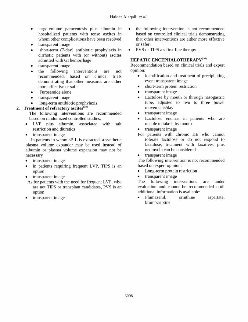
ASCITES
1. Treatment of uncomplicated ascites(39)
The following interventions are recommended
based on controlled and uncontrolled studies
as well as expert opinion:
salt restriction
transparent image
Spironolactone plus furosemide
transparent image
Haider Alaqaili et al.
3098
large-volume paracentesis plus albumin in
hospitalized patients with tense ascites in
whom other complications have been resolved
transparent image
short-term (7-day) antibiotic prophylaxis in
cirrhotic patients with (or without) ascites
admitted with GI hemorrhage
transparent image
the following interventions are not
recommended, based on clinical trials
demonstrating that other measures are either
more effective or safe:
Furosemide alone
transparent image
long-term antibiotic prophylaxis
2. Treatment of refractory ascites(39)
The following interventions are recommended
based on randomized controlled studies:
LVP plus albumin, associated with salt
restriction and diuretics
transparent image
In patients in whom <5 L is extracted, a synthetic
plasma volume expander may be used instead of
albumin or plasma volume expansion may not be
necessary
transparent image
in patients requiring frequent LVP, TIPS is an
option
transparent image
As for patients with the need for frequent LVP, who
are not TIPS or transplant candidates, PVS is an
option
transparent image
the following intervention is not recommended
based on controlled clinical trials demonstrating
that other interventions are either more effective
or safer:
PVS or TIPS a s first-line therapy
HEPATIC ENCEPHALOTHERAPY(40)
Recommendation based on clinical trials and expert
opinion:
identification and treatment of precipitating
event transparent image
short-term protein restriction
transparent image
Lactulose by mouth or through nasogastric
tube, adjusted to two to three bowel
movements/day
transparent image
Lactulose enemas in patients who are
unable to take it by mouth
transparent image
For patients with chronic HE who cannot
tolerate lactulose or do not respond to
lactulose, treatment with laxatives plus
neomycin can be considered
transparent image
The following intervention is not recommended
based on expert opinion:
Long-term protein restriction
transparent image
The following interventions are under
evaluation and cannot be recommended until
additional information is available:
Flumazenil, ornithine aspartate,
bromocriptine
Review on Liver Cirrhosis Complications and Treatment
3099
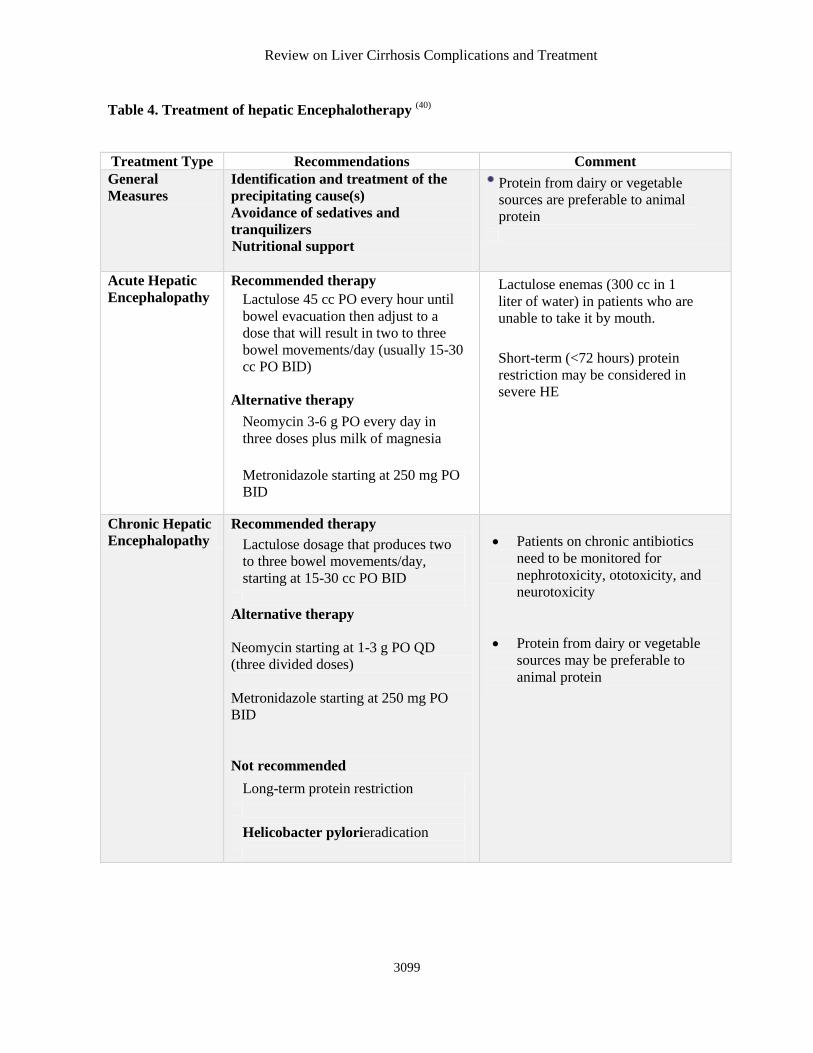
Table 4. Treatment of hepatic Encephalotherapy (40)
Treatment Type Recommendations Comment
General
Measures
Identification and treatment of the
precipitating cause(s)
Avoidance of sedatives and
tranquilizers
Nutritional support
Protein from dairy or vegetable
sources are preferable to animal
protein
Acute Hepatic
Encephalopathy
Recommended therapy Lactulose 45 cc PO every hour until
bowel evacuation then adjust to a
dose that will result in two to three
bowel movements/day (usually 15-30
cc PO BID)
Alternative therapy Neomycin 3-6 g PO every day in
three doses plus milk of magnesia
Metronidazole starting at 250 mg PO
BID
Lactulose enemas (300 cc in 1
liter of water) in patients who are
unable to take it by mouth.
Short-term (<72 hours) protein
restriction may be considered in
severe HE
Chronic Hepatic
Encephalopathy
Recommended therapy Lactulose dosage that produces two
to three bowel movements/day,
starting at 15-30 cc PO BID
Alternative therapy
Neomycin starting at 1-3 g PO QD
(three divided doses)
Metronidazole starting at 250 mg PO
BID
Not recommended Long-term protein restriction
Helicobacter pylorieradication
Patients on chronic antibiotics
need to be monitored for
nephrotoxicity, ototoxicity, and
neurotoxicity
Protein from dairy or vegetable
sources may be preferable to
animal protein
Haider Alaqaili et al.
3100
PRURITUS (41)
Pruritus is a common complaint in
cholestatic liver diseases (eg, primary biliary
cirrhosis) and in noncholestatic chronic liver
diseases (eg, hepatitis C). Although increased
serum bile acid levels once were thought to be the
cause of pruritus, endogenous opioids are more
likely to be the culprit pruritogen. Mild itching
complaints may respond to treatment with
antihistamines and topical ammonium lactate.
Cholestyramine is the mainstay of therapy
for the pruritus of liver disease. To avoid
compromising GI absorption, care should be taken
to avoid co-administration of this organic anion
binder with any other medication.
Other medications that may provide relief
against pruritus in addition to antihistamines (eg,
diphenhydramine, hydroxyzine) and ammonium
lactate 12% skin cream (Lac-Hydrin), include
ursodeoxycholic acid, doxepin, and rifampin.
Naltrexone may be effective but is often poorly
tolerated. Gabapentin is an unreliable therapy.
Patients with severe pruritus may require institution
of ultraviolet light therapy or plasmapheresis.
HYPOGONADISM(41)
Some male patients suffer from
hypogonadism. Patients with severe symptoms may
undergo therapy with topical testosterone
preparations, although their safety and efficacy is
not well studied. Similarly, the utility and safety of
growth hormone therapy remains unclear.
OSTEOPOROSIS(41)
Patients with cirrhosis may develop
osteoporosis. Supplementation with calcium and
vitamin D is important in patients at high risk for
osteoporosis, especially patients with chronic
cholestasis or primary biliary cirrhosis and patients
receiving corticosteroids for autoimmune hepatitis.
The discovery on bone densitometry studies of
decreased bone mineralization may prompt the
institution of therapy with an aminobisphosphonate
(eg, alendronate sodium).
Zinc deficiency(41)
Zinc deficiency commonly is observed in
patients with cirrhosis. Treatment with zinc sulfate
at 220 mg orally twice daily may improve
dysgeusia and can stimulate appetite. Furthermore,
zinc is effective in the treatment of muscle cramps
and is adjunctive therapy for hepatic
encephalopathy.
Analgesics(42)
The choice of appropriate analgesic agents
in patients with cirrhosis requires a thorough
knowledge of their pharmacokinetics and side
effect profiles.
Acetaminophen is an effective and safe
analgesic for patients with chronic liver disease
when used at low doses. For patients with ongoing
alcohol ingestion and cirrhosis, acetaminophen may
be used at a maximum of 2 grams per day, which is
one-half of the recommended daily dose. Although
some studies show 4 grams of acetaminophen per
day to be safe in patients with cirrhosis who are not
actively consuming alcohol, the authors
recommend no more than 2 grams per day to stay
well below toxicity levels. (42)
Many prescription and over the counter
remedies are offered as combination preparations.
Patients with cirrhosis need to be warned to read
medication labels carefully before starting any new
medicine to avoid accidental overdose.
NSAIDs are associated with an increased
risk of variceal hemorrhage, impaired renal
function, and the development of diuretic resistant
ascites. Thus, NSAIDs (including aspirin) should
generally be avoided in patients with cirrhosis.
Selective COX-2 inhibitors are effective
analgesics, which are associated with a decreased
incidence of gastrointestinal and renal toxicity and
an increased incidence of cardiovascular events.
Experience in patients with cirrhosis is limited. At
present, we advise against using these agents. (42)
Opioids should be used cautiously in
patients with cirrhosis. Fentanyl appears to be safe
in patients with modest hepatic dysfunction.
Morphine, oxycodone, and hydromorphone should
be used at reduced doses and prolonged intervals of
administration. Tramadol may be safe but
experience is limited. The effects of codeine are
difficult to predict and therefore alternatives should
be considered.
Strong consideration should be given to
referring patients who require long-term analgesics
to a pain management program(42)
Review on Liver Cirrhosis Complications and Treatment
3101
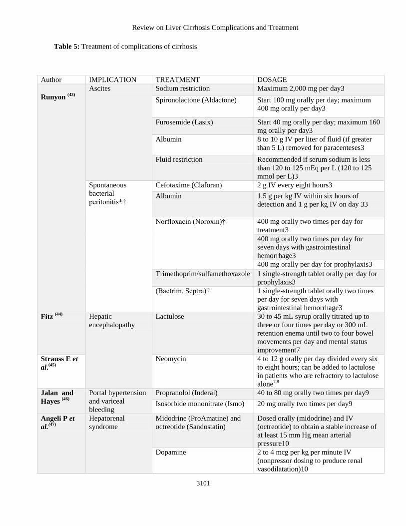
Table 5: Treatment of complications of cirrhosis
Author IMPLICATION TREATMENT DOSAGE
Runyon (43)
Ascites Sodium restriction Maximum 2,000 mg per day3
Spironolactone (Aldactone) Start 100 mg orally per day; maximum
400 mg orally per day3
Furosemide (Lasix) Start 40 mg orally per day; maximum 160
mg orally per day3
Albumin 8 to 10 g IV per liter of fluid (if greater
than 5 L) removed for paracenteses3
Fluid restriction Recommended if serum sodium is less
than 120 to 125 mEq per L (120 to 125
mmol per L)3
Spontaneous
bacterial
peritonitis*†
Cefotaxime (Claforan) 2 g IV every eight hours3
Albumin 1.5 g per kg IV within six hours of
detection and 1 g per kg IV on day 33
Norfloxacin (Noroxin)† 400 mg orally two times per day for
treatment3
400 mg orally two times per day for
seven days with gastrointestinal
hemorrhage3
400 mg orally per day for prophylaxis3
Trimethoprim/sulfamethoxazole 1 single-strength tablet orally per day for
prophylaxis3
(Bactrim, Septra)† 1 single-strength tablet orally two times
per day for seven days with
gastrointestinal hemorrhage3
Fitz (44)
Hepatic
encephalopathy
Lactulose 30 to 45 mL syrup orally titrated up to
three or four times per day or 300 mL
retention enema until two to four bowel
movements per day and mental status
improvement7
Strauss E et
al.(45)
Neomycin 4 to 12 g orally per day divided every six
to eight hours; can be added to lactulose
in patients who are refractory to lactulose
alone7,8
Jalan and
Hayes (46)
Portal hypertension
and variceal
bleeding
Propranolol (Inderal) 40 to 80 mg orally two times per day9
Isosorbide mononitrate (Ismo) 20 mg orally two times per day9
Angeli P et
al.(47)
Hepatorenal
syndrome
Midodrine (ProAmatine) and
octreotide (Sandostatin)
Dosed orally (midodrine) and IV
(octreotide) to obtain a stable increase of
at least 15 mm Hg mean arterial
pressure10
Dopamine 2 to 4 mcg per kg per minute IV
(nonpressor dosing to produce renal
vasodilatation)10
Haider Alaqaili et al.
3102
CONCLUSION
Screening for chronic liver disease is a key factor
for early detection of signs for liver damage, which
can be performed inexpensively and easily with
clinical history-taking, measurement of transaminase
concentrations, upper abdominal ultrasonography,
and transient elastography (where available).
Abnormal findings should prompt specific diagnostic
testing to determine the etiology of the underlying
disease. In most patients, the dynamic process of
progressive fibrosis, which could ultimately lead to
cirrhosis, can be interrupted by the timely recognition
of the risk, followed by appropriate treatment.
REFERENCES 1. Dooley J, Lok A, Burroughs AK, Heathcote
E(2011): Sherlock’s diseases of the liver and biliary
system, 12th edn. Oxford: Wiley-Blackwell.
2. D’Amico G, Garcia-Tsao G and Pagliaro L( 2006): Natural history and prognostic indicators of survival in
cirrhosis: a systematic review of 118 studies. J
Hepatol.,44: 217–31.
3. Hytiroglou P, Snover DC, Alves V et al.(2012): Beyond “cirrhosis”: a proposal from the International
Liver Pathology Study Group. Am J Clin Pathol., 137:
5–9.
4. Morgan TR, Ghany MG, Kim HY et al.(2010): and
the HALT-C Trial Group. Outcome of sustained
virological responders with histologically advanced
chronic hepatitis C. Hepatology,52: 833–44.
5. https://www.webmd.com/a-to-z-
guides/understanding-cirrhosis-treatment
6. https://www.liverfoundation.org/for-
patients/about-the-liver/the-progression-of-liver-
disease/#1503432933768-040e8645-d918
7. D’Amico G, Pasta L, Madonia S, Tarantino G,
Mancuso A, Malizia G et al.(2001): The incidence of
esophageal varices in cirrhosis.
Gastroenterology,120:A2.
8. de Franchis R(2005): Evolving consensus in portal
hypertension report of the baveno IV consensus
workshop on methodology of diagnosis and therapy in
portal hypertension. J Hepatol.,43:167–176.
9. Durand F, Valla D(2008): Assessment of prognosis
of cirrhosis. Semin Liver Dis., 28:110–22.
10. Child C(1964): Surgery and portal hypertension. In:
Child CG, ed. The liver and portal hypertension.
Philadelphia, PA: WB Saunders.
11. Pugh R, Murray‐Lyon IM, Dawson JL, Pietroni
MC, Williams R(1973): Transection of the
oesophagus for bleeding oesophageal varices. British
Journal of Surgery, 1;60(8):646-9.
12. D'Amico G, Garcia-Tsao G, Pagliaro L(2006): Natural history and prognostic indicators of survival in
cirrhosis: a systematic review of 118 studies. Journal
of hepatology,44(1):217-31.
13. Singh AK, Loscalzo J(2011): The Brigham Intensive
Review of Internal Medicine. Oxford University Press.
14. Bellentani S, Saccoccio G, Costa G et al.(1997): The
Dionysos Study Group Drinking habits as cofactors of
risk for alcohol induced liver damage. Gut,41:845–50.
15. Epstein O, Adukiewicz AB, Dick R, Sherlock
S(1979): Hypertrophic hepatic osteoarthropathy.
Clinical roentgenologic, biochemical hormonal and
cardiorespiratory studies, and review of the literature.
Am J Med.,67:88–97.
16. Cattau E, Benjamin SB, Knuff TE, Castell
DO(1982): The accuracy of the physical exam in the
diagnosis of suspected ascites. JAMA.,247:1164–66.
17. Van Thiel DH, Gavaler JS, Schade RR(1985): Liver
disease and the hypothalamic pituitary gonadal axis.
Semin Liver Dis.,5:35–45.
18. Attali P, Ink O, Pelletier G et al.(1987): Dupuytren’s
contracture, alcohol consumption, and chronic liver
disease. Arch Intern Med.,147:1065–67.
19. Pirovino M, Linder R, Boss C, Kochli HP, Mahler
F(1988): Cutaneous spider nevi in liver cirrhosis:
Capillary microscopical and hormonal investigations.
Klin Wochenschr., 66:298–302.
20. Erlinger S, Benhamou J(1991): Cirrhosis: Clinical
aspects. In: Mcintyre N, Benhamou J, Rizzetto M,
Rodes J, editors. Oxford Textbook of Clinical
Hepatology. University Press; Oxford.
21. Tangerman A, Meuwese-Arends MT, Jansen
JB(1999): Cause and composition of foetor hepaticus.
Lancet,343:483.
22. Bircher J, Benhamou JP, McIntyre N, Rizzetto M,
Rodes J(1999): Oxford Textbook of Clinical
Hepatology. 2nd Edition Oxford University Press.
23. Sherlock S, Dooley J(2002): Diseases of the Liver
and Biliary System. 11th Edition Blackwell Science;
Oxford, UK.
24. Schiff ER, Sorrell MF, Maddrey EC(2003): Schiff’s
Diseases of the Liver. 9th Edition Lippincott, Williams
& Wilkins; Philadelphia.
25. Castera L, Pinzani M(2010): Biopsy and non-
invasive methods for the diagnosis of liver fibrosis:
does it take two to tango? Gut,59:861–866.
26. Wai CT, Greenson JK, Fontana RJ et al.(2003): A
simple noninvasive index can predict both significant
fibrosis and cirrhosis in patients with chronic hepatitis
C. Hepatology,38:518–526.
27. Rosenberg WM, Voelker M, Thiel R et al.(2004): Serum markers detect the presence of liver fibrosis: a
cohort study. Gastroenterology,127:1704–1713
28. Snyder N, Gajula L, Xiao SY et al. (2006):APRI: an
easy and validated predictor of hepatic fibrosis in
chronic hepatitis C. J Clin Gastroenterol.,40:535–542
29. pubmedcentralcanada.ca/pmcc/articles/PMC56557
99/
Review on Liver Cirrhosis Complications and Treatment
3103
30. Simonovsky V(1999): The diagnosis of cirrhosis by
high resolution ultrasound of the liver surface. Br J
Radiol.,72:29–34.
31. Friedrich-Rust M, Nierhoff J, Lupsor M et
al.(2012): Performance of Acoustic Radiation Force
Impulse imaging for the staging of liver fibrosis: a
pooled meta-analysis. J Viral Hepat.,19:e212–e219.
32. Sebastiani G, Castera L, Halfon P et al.(2011): The
impact of liver disease aetiology and the stages of
hepatic fibrosis on the performance of non-invasive
fibrosis biomarkers: an international study of 2411
cases. Aliment Pharmacol Ther.,34:1202–1216
33. www.medsolutions.com/.../ABDOMEN%20IMAGI
NG%20GUIDELINES.pdf
34. Lomas DJ. The liver. In: Grainger RG, Allison
DJ(2001): Grainger and Allison’s Diagnostic
Radiology: A Textbook of Medical Imaging. 4th ed.
London, England: Churchill Livingstone, 201:1247–8
35. Heidelbaugh JJ, Bruderly M(2006): Cirrhosis and
chronic liver failure: part I. Diagnosis and evaluation.
Am Fam Physician,4(5):756-62.
36. Abdi W, Millan JC, Mezey E(1979): Sampling
variability on percutaneous liver biopsy. Arch Intern
Med.,139:667–9.
37. Vierling JM, Zeuzem S, Poordad F et al.(2014): Safety and efficacy of
boceprevir/peginterferon/ribavirin for HCV G1
compensated cirrhotics: meta-analysis of 5 trials. J
Hepatol.,61(2):200-9.
38. Harrison P(2014): 3D plus ribavirin improves liver
function in cirrhosis. Available at
http://www.medscape.com/viewarticle/833771.
39. Arroyo V, Gines P, Gerbes A L, Dudley F J,
Gentilini P, Laffi G, Reynolds T F, Ring-Larsen H,
Scholmerich J(1996): Definition and diagnostic
criteria of refractory ascites and hepatorenal syndrome
in cirrhosis. Hepatology, 23:164-176.
40. Goulenok C, Bernard B, Cadranel J F, Thabut D,
DiMartino V, Opolon P, Poynard T(2002): Flumazenil versus placebo in hepatic encephalopathy
in patients with cirrhosis: A meta-analysis. Aliment
Pharmacol Ther.,16:361-372.
41. Singh AK, Loscalzo J(2012): Internal Medicine: An
Intensive Review. Oxford University Press.
42. James P, Eric G, Sanjiv C (2011): Management of
pain in patients with cirrhosis.
http://cursoenarm.net/UPTODATE/contents/mobiprev
iew.htm?14/7/14463?source=HISTORY.
43. Runyon BA and the Practice Guidelines
Committee(2004): American Association for the
Study of Liver Diseases Management of adult patients
with ascites due to cirrhosis. Hepatology,39:841–56.
44. Fitz JG(2002): Hepatic encephalopathy,
hepatopulmonary syndromes, hepatorenal syndrome,
coagulopathy, and endocrine complications of liver
disease. In: Feldman M, Friedman LS, Sleisenger MH,
eds. Sleisenger and Fordtran’s Gastrointestinal and
Liver Disease: Pathophysiology, Diagnosis,
Management. 7th ed. Philadelphia, Pa.: Saunders.
45. Strauss E, Tramote R, Silva EP, Caly WR, Honain
NZ, Maffei RA et al.(1992): Double-blind
randomized clinical trial comparing neomycin and
placebo in the treatment of exogenous hepatic
encephalopathy. Hepatogastroenterology,39:542–5.
46. Jalan R, Hayes PC(2000): UK guidelines on the
management of variceal haemorrhage in cirrhotic
patients. British Society of
Gastroenterology. Gut,46(3–4):III1–III15.
47. Angeli P, Volpin R, Gerunda G, Craighero R,
Roner P, Merenda R et al.(1999): Reversal of type 1
hepatorenal syndrome with the administration of
midodrine and octreotide. Hepatology, 29:1690–7.