Embed Size (px)
Citation preview
Review
Myosins: a diverse superfamily
James R. Sellers *National Heart, Lung and Blood Institute, National Institutes of Health, Building 10, Room 8N202, Bethesda, MD 20892, USA
Received 15 October 1999; accepted 19 October 1999
Abstract
Myosins constitute a large superfamily of actin-dependent molecular motors. Phylogenetic analysis currently placesmyosins into 15 classes. The conventional myosins which form filaments in muscle and non-muscle cells form class II. Therehas been extensive characterization of these myosins and much is known about their function. With the exception of class Iand class V myosins, little is known about the structure, enzymatic properties, intracellular localization and physiology ofmost unconventional myosin classes. This review will focus on myosins from class IV, VI, VII, VIII, X, XI, XII, XIII, XIVand XV. In addition, the function of myosin II in non-muscle cells will also be discussed. ß 2000 Elsevier Science B.V. Allrights reserved.
Keywords: Myosin II; Myosin IV; Myosin VI^VIII; Myosin X^XV
1. Introduction
Myosins constitute a large superfamily of proteinsthat share a common domain which has been shownto interact with actin, hydrolyze ATP and producemovement in all cases examined to date (Fig. 1) [1,2].However, only a few members of the superfamilyhave been biochemically characterized and it is pos-sible that some myosins may have lost one or moreof these features. Myosins are typically constructedof three functional subdomains: (1) the motor do-main which interacts with actin and binds ATP, (2)the neck domain which binds light chains or calmod-ulin, and (3) the tail domain which serves to anchorand position the motor domain so that it can interactwith actin. The motor domains are relatively con-served with the exception of several surface loopsand the amino-terminus. Light chains and calmodu-
lin bind to a helical sequence termed the IQ motiffound in the neck which has a consensus sequence ofIQXXXRGXXXR [3]. The number of IQ motifspresent in the necks of di¡erent myosins can varybetween zero and six. The tail domains are themost diverse domains and vary widely in lengthand in sequence. Functional motifs, such as SH3domains, GAP domains, FERM domains, and pleck-strin homology (PH) domains are sometimes foundin the tails of myosins (Fig. 2). In addition, the tailsof many myosins contain coiled-coil forming sequen-ces which allow the molecules to dimerize and pro-duce two-headed molecules.
Phylogenetic analysis, usually of the motor do-main, groups myosins into 15 distinct classes,although there are a number of outliers which donot adequately align with any of the establishedclasses [1,2]. In addition, two of the de¢ned classes(IV and XII) are currently comprised of only a singlemember. Perhaps, in hindsight, they should also havebeen considered outliers. The nomenclature assigns
0167-4889 / 00 / $ ^ see front matter ß 2000 Elsevier Science B.V. All rights reserved.PII: S 0 1 6 7 - 4 8 8 9 ( 0 0 ) 0 0 0 0 5 - 7
* Fax: +1 (301) 402-1542; E-mail : [email protected]
BBAMCR 14595 6-3-00 Cyaan Magenta Geel Zwart
Biochimica et Biophysica Acta 1496 (2000) 3^22www.elsevier.com/locate/bba
BBAMCR 14595 6-3-00 Cyaan Magenta Geel Zwart
J.R. Sellers / Biochimica et Biophysica Acta 1496 (2000) 3^224
class number II to the large family of ¢lament form-ing conventional myosins which were discovered over60 years ago and are found in muscles and in thecytoplasm of animal cells. Class I myosins werenext discovered and the subsequent classes werenumbered in order of the discovery of the foundingmember of the class. While most family trees areconstructed by analysis of the motor domains, anal-ysis of the whole molecule or of the tail domainsalone generally gives the same relationships [4].
The number of myosin genes present in mammalsis conservatively estimated at 25^30 from classes I,II, III, V, VI, VII, IX, X and XV. The entire genomeof the budding yeast, Saccharomyces cerevisiae, isnow sequenced and contains only ¢ve myosin genes,two of class I, one of class II and two of class V [5].The slime mold, Dictyostelium discoideum, representsa higher level of organismal complexity. It can existas a single amoeboid cell or undergo a complex de-velopmental cycle to produce multicellular slugs andfruiting bodies. While the genome of Dictyosteliumhas not been fully sequenced, a large of number ofmyosins have been identi¢ed by a combination ofmethods. These include seven myosin I genes, a sin-gle myosin II gene and four other genes that do notstringently group with the existing classes [4]. One ofthese groups weakly with myosin V or XI while an-other groups weakly with myosin VII or X. The tworemaining genes do not classify well with any existingclass. The sequence of the Caenorhabditis elegans ge-nome is mostly complete. To date, there are fourgenes for muscle myosin II, two for non-muscle my-osin II, two for myosin I, one for myosin V, two formyosin VI, one for myosin VII, one for myosin IXand one for myosin XII [6] (M. Titus, personal com-munication). No class of myosin appears to be uni-versally expressed in all phyla. For example, no my-osin II class molecules have yet been found in plants,
and several myosins are, so far, found exclusively inplants (VIII, XI and XIII). However, all eukaryoticanimal cells examined contain at least one myosin IIgene and, usually, multiple myosin I genes. In addi-tion, myosin V genes are found widely, if not univer-sally (particularly if the criteria for relatedness arerelaxed somewhat).
The features of myosins from class I, III, V and IXare covered elsewhere in this special volume [7^9]and this article will concentrate on the remainingclasses. In addition, it will review the structure ofthe myosin motor domain as determined by crystal-lography of several myosin II myosin fragments andwhat the structure, combined with site-directed mu-tagenesis, can tell us about the function of the mol-ecule.
2. The myosin II motor domain and its use as a modelfor other myosins
The myosin II class is also referred to as `conven-tional' myosins since this was the only class of my-osin known for decades. Members of this class arehexameric enzymes composed of two heavy chainswith a molecular weight of 171^244 kDa and twopairs of light chains. The amino-terminal portion ofthe heavy chains (collectively referred to as the`head') contains the prototypical motor domain andtwo IQ motifs in the neck. The carboxyl-terminalhalf of the heavy chain consists of coiled-coil formingsequence which homodimerizes to form the long rod(or tail) which usually terminates in a short non-hel-ical segment [10^12]. Myosin II molecules have atwo-headed structure, due to the dimerization ofthe heavy chain in the tail. The tails of myosin self-associate to form ¢laments both in vivo and in vitroat low ionic strength. Such ¢laments complicate ki-
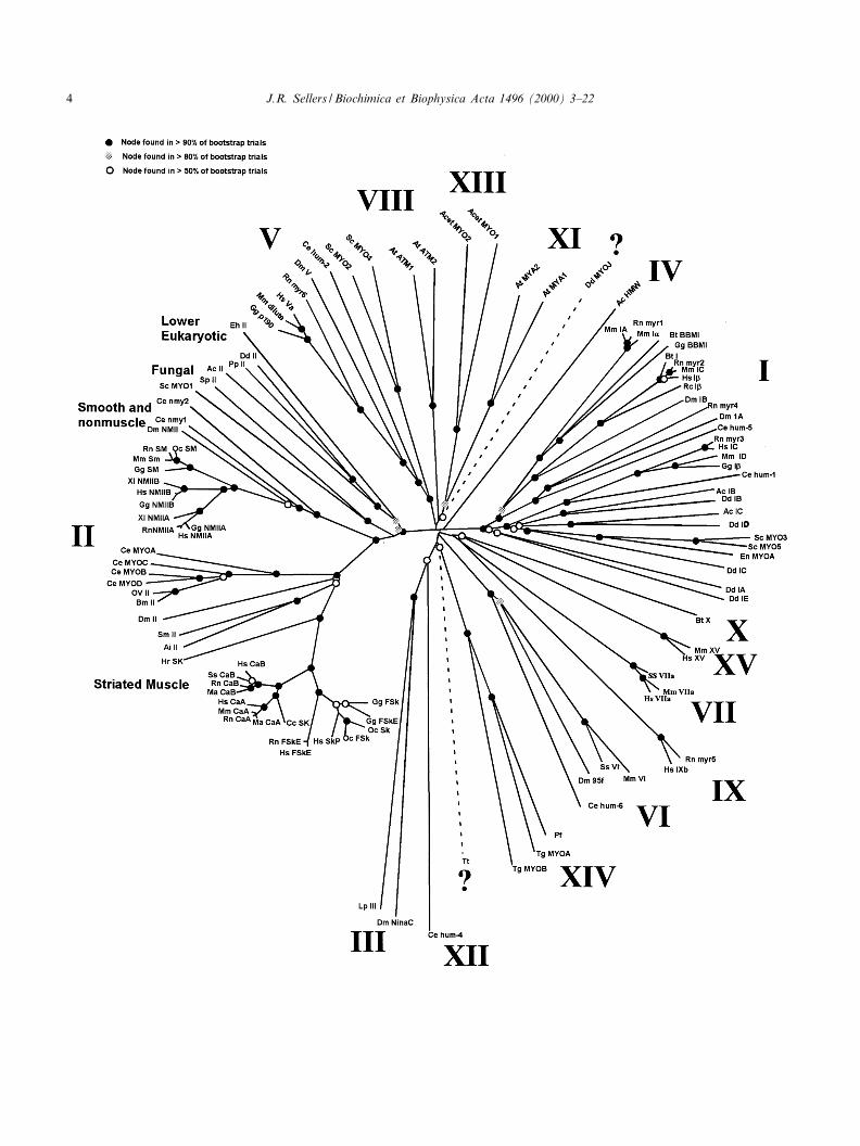
Fig. 1. Phylogenetic tree obtained from neighbor joining analysis of the myosin motor domain protein sequences as performed byClustalW. Branch lengths are drawn to scale in units of percent divergence. The tree is drawn unrooted. Bootstrap resampling (1000trials) was used to judge the robustness of nodes, and results are indicated on the ¢gure as explained in the key. Abbreviations are:Ac, Acanthamoeba castellani ; Acet, Acetabularia cliftonii ; At, Arabidopsis thaliana ; Ae, Aequipecten irradians ; Bt, Bos taurus ; Bm,Brugia malayi ; Ce, C. elegans ; Cc, Coturjix coturnix ; Dd, D. discoideum ; Dm, D. melanogaster ; En, Emericella nidulans ; Eh, Entamoe-ba histolytica ; Gg, Gallus gallus ; Hr, Halocynthia roretzi ; Hs, Homo sapiens ; Lp, L. polyphemus ; Ma, Mesocritus aureus ; Mm, Musmusculus ; Ov, Onchocerca volvulus ; Oc, Oryctolagus cuniculus ; Pp, Physarum polycephalum ; Pf, P. falciparum ; Rc, Rana catesbeiana ;Rt, Rattus norvegicus ; Sc, S. cerevisiae ; Sm, Schistosoma mansoni ; Sp, S. pombe ; Ss, Sus scrofa ;, Tt, Tetrahymena thermophilus ; Tg,T. gondii ; Xl, Xenopus laevis.6
BBAMCR 14595 6-3-00 Cyaan Magenta Geel Zwart
J.R. Sellers / Biochimica et Biophysica Acta 1496 (2000) 3^22 5

netic and biophysical characterization of myosin'senzymatic activity. This complication can be circum-vented by the use of proteolytically produced frag-ments which are soluble and enzymatically active.Myosin II molecules can be proteolytically cleavedinto discrete functional domains. One site of cleavageat the junction between the head and the tail produ-ces a soluble fragment termed subfragment one (S1)and the rod which remains a coiled-coil dimer andretains the solubility properties of the parent mole-
cule. S1 binds to actin and nucleotides and containsthe two light chains. Another cleavage site of myosinII occurs about 40 kDa into the rod structure andproduces two fragments, heavy meromyosin (HMM)and light meromyosin (LMM). HMM, which is solu-ble even at low ionic strength, contains the headregion and a portion of the coiled-coil forming se-quence (termed subfragment 2 or S2) which dimer-izes to produce a two-headed fragment. LMM re-tains the solubility properties of the parent
Fig. 2. Schematic representation of the domain structure of myosin superfamily members. The length of the molecules including theirvarious structural motifs is drawn roughly proportional to the number of amino acids. In some cases, members of a particular classmay have isoforms with alternatively spliced regions that change the length of the isoform.
BBAMCR 14595 6-3-00 Cyaan Magenta Geel Zwart
J.R. Sellers / Biochimica et Biophysica Acta 1496 (2000) 3^226
molecule. Both S1 and HMM have been critical tokinetic, biophysical and structural studies of the my-osin molecule.
The three-dimensional structure of fragments ofseveral myosin II molecules has been solved. The
fragments used for crystallography were producedby proteolysis or by expression of recombinant mol-ecules. These include the S1 of chicken fast skeletalmuscle myosin [13], the S1 of scallop myosin [14], arecombinant fragment of the smooth muscle myosin
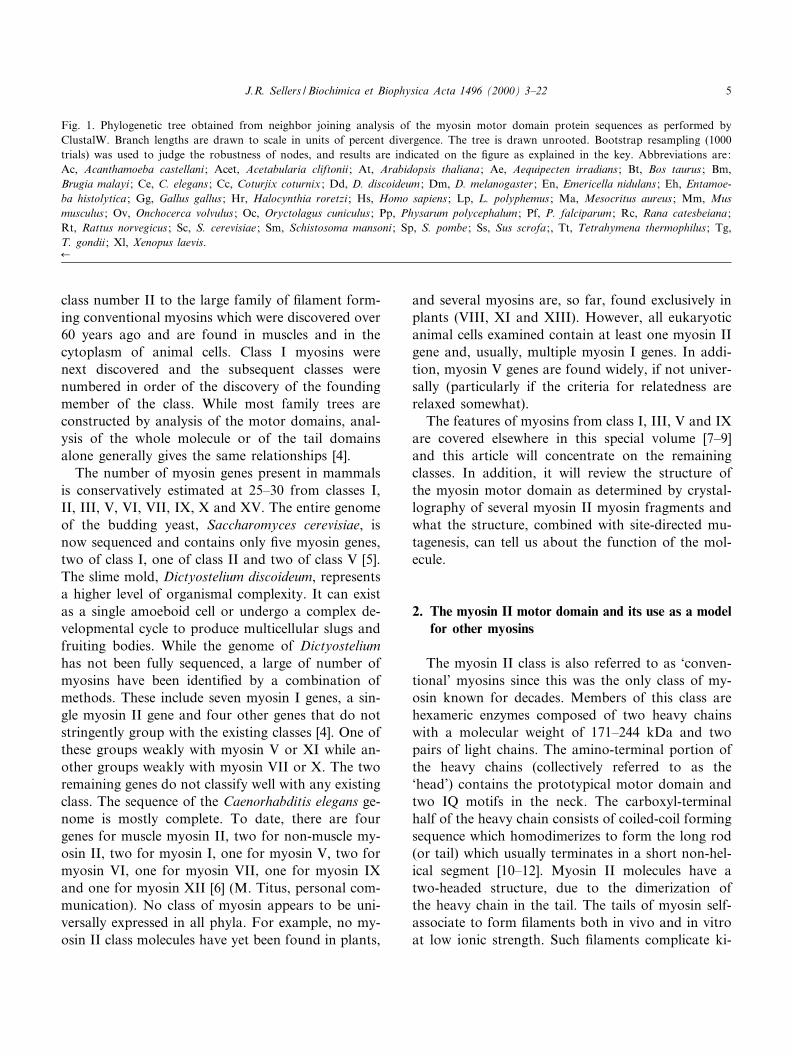
Fig. 3. Three-dimensional structure of chicken fast skeletal muscle myosin S1. The color scheme is as in Rayment et al. [13]. This ¢g-ure was made with MOLSCRIPT [172].
BBAMCR 14595 6-3-00 Cyaan Magenta Geel Zwart
J.R. Sellers / Biochimica et Biophysica Acta 1496 (2000) 3^22 7
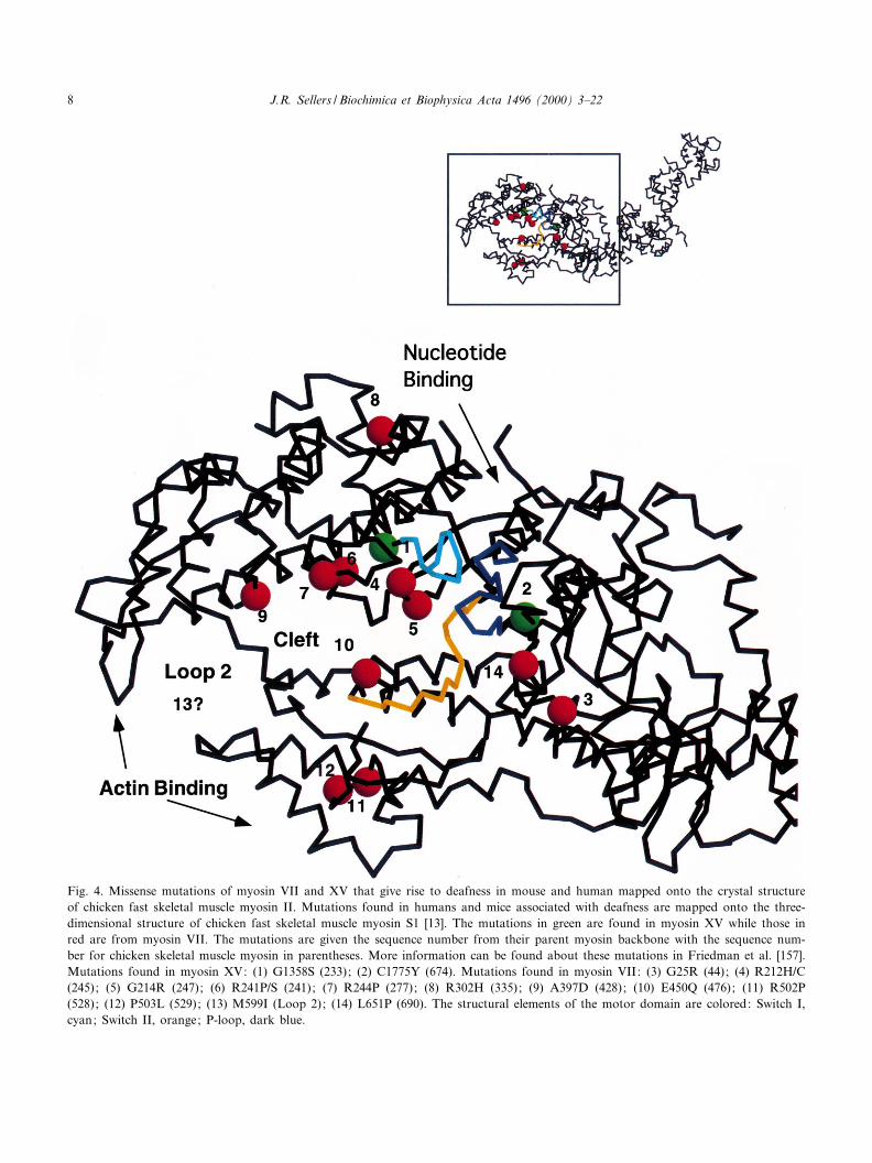
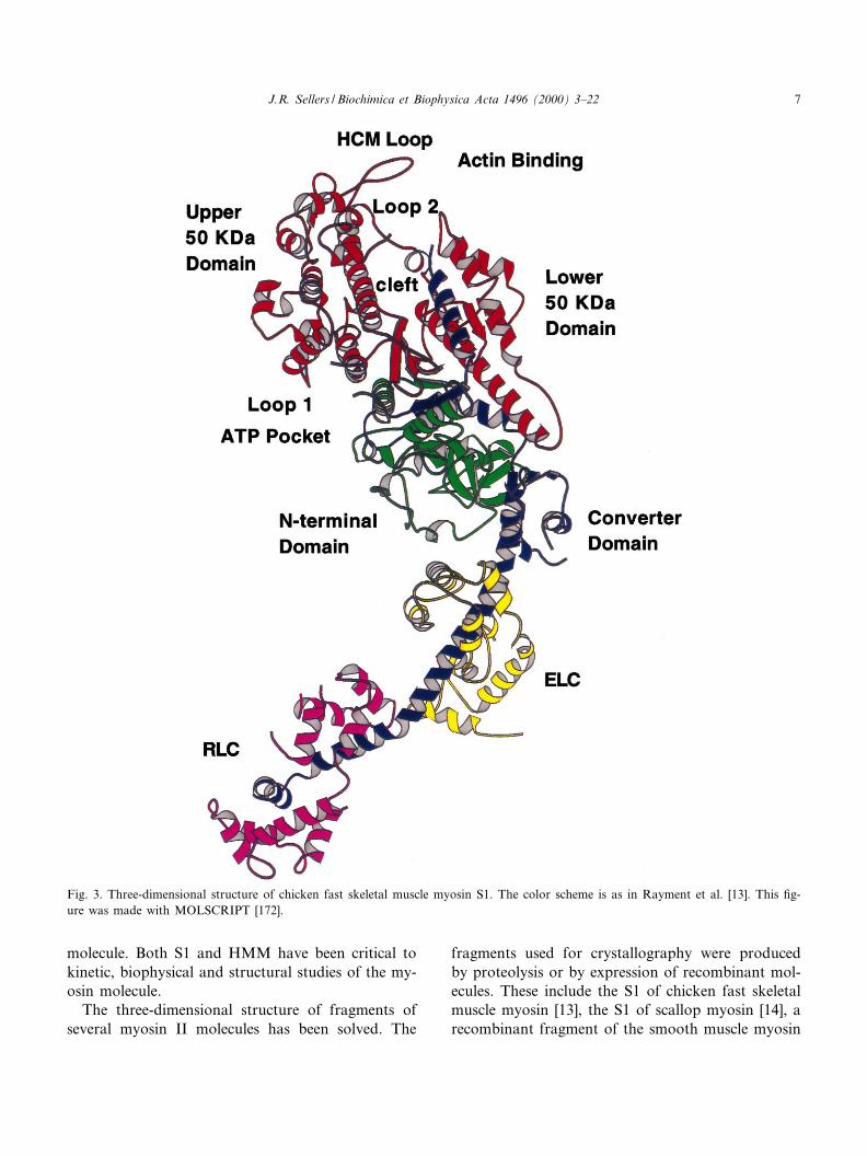
Fig. 4. Missense mutations of myosin VII and XV that give rise to deafness in mouse and human mapped onto the crystal structureof chicken fast skeletal muscle myosin II. Mutations found in humans and mice associated with deafness are mapped onto the three-dimensional structure of chicken fast skeletal muscle myosin S1 [13]. The mutations in green are found in myosin XV while those inred are from myosin VII. The mutations are given the sequence number from their parent myosin backbone with the sequence num-ber for chicken skeletal muscle myosin in parentheses. More information can be found about these mutations in Friedman et al. [157].Mutations found in myosin XV: (1) G1358S (233); (2) C1775Y (674). Mutations found in myosin VII: (3) G25R (44); (4) R212H/C(245); (5) G214R (247); (6) R241P/S (241); (7) R244P (277); (8) R302H (335); (9) A397D (428); (10) E450Q (476); (11) R502P(528); (12) P503L (529); (13) M599I (Loop 2); (14) L651P (690). The structural elements of the motor domain are colored: Switch I,cyan; Switch II, orange; P-loop, dark blue.
BBAMCR 14595 6-3-00 Cyaan Magenta Geel Zwart
J.R. Sellers / Biochimica et Biophysica Acta 1496 (2000) 3^228
motor domain containing the essential light chain(ELC) [15], a motor domain fragment of Dictyoste-lium myosin [16]. In addition, many of these struc-tures have been solved with one of several di¡erentnucleotides or nucleotide analogs bound at the activesite.
The myosin motor domain is essentially built offour subdomains connected by £exible linkers [14](Fig. 3). The amino-terminal subdomain forms anSH3-like motif in myosin II class molecules [15].This motif is not present in other myosin classes.The amino-terminal subdomain is connected to theupper 50 kDa subdomain which is, in turn, con-nected to the lower 50 kDa subdomain. The name(50 kDa) for these two subdomains is historical andrelates to the fact that there are two proteolyticallysensitive surface loops which connect them with therest of the myosin molecule which when cleaved givesrise to a band on sodium dodecyl sulfate^polyacryl-amide gels with a molecular weight of 50 kDa [17].The upper and lower 50 kDa domains are separatedby a cleft which is lined with many conserved resi-dues. This cleft closes slightly upon binding nucleo-tides and may close more dramatically upon bindingactin [14]. These two domains comprise most of theactin binding interface (see [1] for a review of theevidence). Of interest to later discussions of the my-osin superfamily is a loop that is found at the surfaceof the upper 50 kDa domain (residues 403^416 in thechicken skeletal muscle myosin sequence). This loopis the site of the ¢rst myosin mutation shown tocause a human disease, hypertrophic cardiomyo-pathy (HCM), and is sometimes referred to as theHCM loop [18]. The HCM loop is also the locationof a phosphorylatable serine (S) or threonine (T) incertain amoeboid myosin I molecules. (For an exten-sive sequence alignment of myosin motor domains,see [1]). The enzymatic activities of these myosins aredependent upon phosphorylation at this site (see [19]for review). The myosin VI class molecules also havea threonine at this residue [20,21]. Most other myosinsuperfamily members have a constitutively negativelycharged amino acid, either an aspartic acid (D) or aglutamic acid (E) residue, at this site. For this reason,Bement and Mooseker [22] named this the TEDSrule site after the one letter codes for the four aminoacids involved.
The nucleotide binding pocket is rather open and
does not substantially close upon binding of nucleo-tide (see [23] for review). It is formed primarily fromseven L-strands linked by loops [13]. There are manystructural similarities between the nucleotide bindingpocket of myosin and those of kinesin, a microtu-bule-dependent motor and of G-proteins. [24,25]. Ineach case, there are three conserved sequence ele-ments termed the P-loop, Switch I and Switch IIthat are involved in nucleotide binding and sensingof the hydrolysis state (ATP or ADP) of the nucleo-tide.
The fourth subdomain of the motor domain istermed the converter region. A long helix emergesfrom the converter domain which serves as the bind-ing site of both light chains. The ELC occupies thebinding site closest to the converter subdomain whilethe regulatory light chain (RLC) occupies the secondsite. The binding sites are highly speci¢c for theirrespective light chains. The elongated nature of theneck region has given rise to the suggestion that itacts as a lever arm to amplify small changes in thesequence of the motor domain into much larger dis-placements of actin [14]. In favor of this hypothesis,it was found that there is a correlation with speed ofin vitro motility and the length of the neck regionfrom myosin mutagenesis experiments in which bind-ing sites for light chains were either added or sub-tracted to vary the length of the neck of Dictyoste-lium myosin [26]. In addition, £uorescent probesattached to the RLC of skeletal muscle myosin inskinned ¢bers show a change in angle upon activa-tion of the muscle [27]. Structural evidence in favorof the lever arm hypothesis comes from crystal formsof a smooth muscle motor domain containing theELC and from the scallop muscle S1 with di¡erentbound nucleotides at the active site [14,15]. A portionof this movement may have been directly visualizedin three-dimensional reconstructions of electron mi-crographs of actin-myosin complexes in the presenceand absence of ADP [28,29]. These studies have ledto a model for force transduction that supposes thatthe amino-terminal subdomain and the upper andlower 50 kDa domains remain relatively constrainedas to their orientation with actin and that most of themotion is derived from a rigid body movement of theneck. The pivot point or joint for this movement is inthe converter subdomain and the movement is con-trolled by conformational changes in structural ele-
BBAMCR 14595 6-3-00 Cyaan Magenta Geel Zwart
J.R. Sellers / Biochimica et Biophysica Acta 1496 (2000) 3^22 9
ments that are in communication with Switch II ofthe nucleotide binding pocket [14].
Alignment of myosin sequences shows two mainhypervariable regions that occur at proteolyticallysensitive regions of myosin. Interestingly, these re-gions are also the site of alternative splicing insome myosin molecules (see below). The positionsof these hypervariable regions correspond to twoportions of the motor domain which were not re-solved in the crystal structure probably due to theirdisordered, £exible nature (Fig. 3). The ¢rst of these,termed loop 1, occurs about 25 kDa from the amino-terminus and is located near the nucleotide bindingregion. Loop 2 occurs about 75 kDa from the amino-terminus and is located in an actin binding interface.Interestingly, binding to actin protects this regionfrom proteolysis [17].
The similarity in the motor domains from thesephylogenetically diverse myosin II crystals is remark-able. In fact, alignment of the sequences of thesemyosins with those of other myosin classes stronglysuggests that the myosin II motor domain crystalsare a good template for analysis of all motor do-mains [30]. The alignment reveals that there is a con-served core domain that begins about 80 amino acidsfrom the amino-terminus of the myosin II class mol-ecules and continues to just before the beginning ofthe long light chain binding helix [31]. Several myo-sin classes have little or no sequence amino-terminalto the core, while others have large extensions (seeFig. 2 and below). There may also be large exten-sions at the positions of loop 1 or loop 2 comparedto myosin II class molecules. Myosin VI has a uniqueinsert of about 50 amino acids at the converter re-gion which will be discussed later. Finally, the lengthof the light chain binding helix varies considerablyamong myosins having anywhere from none to sevenIQ motifs. In myosin II molecules, there is usually a26 amino acid separation between the start of IQmotifs, but in unconventional myosins, the separa-tion can be between 23 and 26 residues [32]. Forexample, in myosin V molecules, the six IQ motifsare separated by an alternating pattern of 23 and25 residues. Since the heavy chain target forlight chain binding is a helix, the relative contactsbetween adjacent light chains could be very di¡erentdepending on the spacing [32]. This could a¡ect thesti¡ness of the neck, the relative disposition of the
heads and possibly the regulation of unconventionalmyosins.
3. Myosin II
The most unique characteristic of myosin II is theability to form ¢laments via the self-association ofthe rod-like coiled-coil K-helical tail. Bipolar ¢la-ments such as those found in sarcomeric musclesare formed by a combination of antiparallel tail in-teractions at the middle of the ¢lament followed byparallel interactions along most of the ¢lamentlength. These ¢laments have a central bare zonewhich is not populated by motor domains and aredesigned to pull actin ¢laments toward there center.Smooth muscle myosins can form side polar ¢la-ments which have no central bare zone [33]. These¢laments may allow for the extreme shortening thatis seen in smooth muscle tissues. The region of therod necessary for ¢lament assembly has been local-ized in several cases [34^36]. It is typically a shortsegment near the carboxyl-terminal one third of thetail. A point mutation at Arg-1889 of Dictyosteliummyosin is su¤cient to disrupt ¢lament assembly byinhibiting the nucleation step [36].
Myosin II molecules can be subclassi¢ed into dis-tinct classes based on sequence analysis of the motordomains (Fig. 1). The sarcomeric myosins from stri-ated and cardiac muscle fall into one group, whereasvertebrate smooth and non-muscle myosins fall intoanother. A third group is comprised of the myosinsfrom lower eukaryotic species such as Acanthamoeba,Dictyostelium and Physarum and the fourth is fromfungi. The fungal myosins are the most dissimilar.The coiled-coil forming sequences in their tails areinterrupted by numerous proline residues (see [37]for review). Since no fungal myosins have yet beenpuri¢ed, it is not clear whether they form ¢laments.One of the Schizosaccharomyces pombe myosins ap-pears to bind an EF-hand family protein, Cdc4p.This myosin has one well conserved consensus IQmotif and one that is very degenerate. It is not clearwhether it would bind two light chains [38]. Thisprotein, while not stringently grouping with eitherRLC, ELC or calmodulin, appears to be most similarto ELC. Interestingly, Cdc4p can be phosphorylatedin vivo at serine 2 or 6 [39]. Mutation of these resi-
BBAMCR 14595 6-3-00 Cyaan Magenta Geel Zwart
J.R. Sellers / Biochimica et Biophysica Acta 1496 (2000) 3^2210
dues to alanines or to aspartates did not a¡ectgrowth or cytokinesis [39]. There are no direct ho-mologs of the ELC and RLC in the S. cerevisiaegenome and, instead, a protein with 40% sequenceidentity to S. pombe Cdc4p is present as the bestcandidate for a light chain [40]. It interacts bothgenetically and directly by gel overlay assay withthe heavy chain of one of the myosin I genes,Myo2. It will be interesting to know if this is alsoa light chain for the myosin II protein of this species.
The MgATPase activity of vertebrate sarcomericmyosins is constitutively activated by actin. The reg-ulation in these muscles is via the troponin-tropo-myosin system [41]. In contrast, the MgATPase ac-tivity of most other myosins is regulated in one ofthree ways. The MgATPase activity of molluscanmuscle myosins is greatly activated by calcium bind-ing to the ELC [42,43]. Vertebrate smooth muscleand non-muscle myosins are regulated by phosphor-ylation of the RLCs by a calcium-calmodulin-depen-dent enzyme termed myosin light chain kinase (see[44,45] for review). Interestingly, the muscle myosinof Limulus polyphemus, the horseshoe crab, is alsoregulated by light chain phosphorylation [46]. Verte-brate sarcomeric myosins are also phosphorylated ontheir RLCs, but the e¡ect is only modulatory to-wards its enzymatic or mechanical properties [47].However, the phosphorylation of vertebrate striatedmuscle RLC does disrupt the packing order of themyosin heads against the backbone of the thick ¢la-ment [48]. The heavy chains of vertebrate smoothmuscle and non-muscle myosins are phosphorylatedby a variety of kinases. The consequences of thesephosphorylations are not well understood [49^51] ex-cept for the case of non-muscle myosin IIB, wherephosphorylation of the fragments of the heavy chainresults in an inhibition of ¢lament assembly [52]. Incontrast, the MgATPase activity of myosins fromAcanthamoeba and Dictyostelium myosins is inhibitedby heavy chain phosphorylation. The site of phos-phorylation di¡ers between the two myosins andthe mechanisms by which phosphorylation inhibitsthe activity of these two myosins are somewhat dif-ferent (see [19,53] for review). The RLC of Dictyo-stelium is also phosphorylated [54].
Much of our knowledge of the in vivo function ofnon-muscle myosin II comes from studies in threegenetic model systems, Dictyostelium discoideum, S.
cerevisiae and Drosophila melanogaster. The singlemyosin II gene of Dictyostelium was ablated by ho-mologous recombination [55]. Surprisingly, cellslacking non-muscle myosin II are viable if grownon a surface where they divide through a processtermed traction-mediated cyto¢ssion [55^57]. In sus-pension or on a non-adhesive hydrophobic surface,the myosin II null cells undergo karyokinesis, but notcytokinesis and large multi-nucleated cells areformed [55,58,59]. Under these conditions, the Dic-tyostelium myosin II null cells round up at the startof mitosis, but they do not elongate nor form acleavage furrow [60]. On an adhesive surface, how-ever, the traction-mediated cyto¢ssion events of themyosin null are coupled to mitosis [60,61]. The cellsround up, elongate, exhibit polar ru¥ing and form acleavage furrow which decreases in width to eventu-ally produce two daughter cells. However, the rate ofcleavage furrow constriction is only about half thatof wild-type cells [60]. These ¢ndings led to the pro-posal that cytokinesis is driven by two mechanisms[60]. Cytokinesis A is a myosin-dependent restrictionof the cleavage furrow and is the only event occur-ring in Dictyostelium cells when they are not grownon an adhesive surface. Cytokinesis B, which occursonly on adhesive surfaces, is myosin-independent andmay result from the traction forces generated by po-lar pseudopods exerting forces on the surface. Thestudies of cytokinesis in Dictyostelium myosin II nullcells prompted a reinvestigation of the spatial distri-bution of myosin II in mitotic cells [62]. This re-vealed that when grown on a surface, very little my-osin II gets targeted to the cleavage furrow.However, when Dictyostelium cells are £atten underagar, as is often used for imaging, the mechanicalstress results in a major recruitment of myosin tothe cleavage furrow [62]. This study concludes thatmyosin II is not essential for cytokinesis.
Dictyostelium myosin II null cells can migrate andchemotact towards a cAMP gradient, albeit some-what ine¤ciently, but do not complete the normaldevelopmental cycle [55,63^65]. They are unable tocap surface receptors or do so poorly [57,66] andthere is reduced cortical tension in null cells com-pared to wild-type [57]. The myosin null cells canalso serve as a host for reintroduction of mutatedmyosin II constructs which can be puri¢ed for invitro biochemical assays [67,68]. Alternatively, the
BBAMCR 14595 6-3-00 Cyaan Magenta Geel Zwart
J.R. Sellers / Biochimica et Biophysica Acta 1496 (2000) 3^22 11
mutated myosins can be assayed for their ability torestore wild-type phenotypes in such cell behaviors ascytokinesis, motility, chemotaxis and development[69^71]. The ability to perform both in vitro bio-chemical assays and in vivo studies of mutated my-osins constitutes a very powerful combination.
One example of this approach is the study of theregulation of Dictyostelium myosin's activity. Dic-tyostelium myosin is phosphorylated at severalclosely spaced threonine residues near the ¢lamentassembly domain in the tail and on the RLC[72,73]. Phosphorylation of the heavy chain sites in-activates the myosin by decreasing the propensity toform ¢laments in vitro [74]. The role of RLC phos-phorylation is not so clear. One study showed thatRLC phosphorylation is essential for in vitro motility[54], but subsequent studies showed that RLC phos-phorylation is more modulatory, producing onlyabout a 4-fold increase in the actin-activated MgAT-Pase activity [75^77]. To study the e¡ect of heavychain phosphorylation, a construct which deleted34 kDa of sequence around the phosphorylation sitesin the tail was expressed. This construct restoressome cellular function such as growth in suspension,development and capping of surface receptors, butthe cells have altered cytoskeletal dynamics [78]. Spe-ci¢cally, myosin does not properly disassemble oncerecruited to a speci¢c site. In vitro analysis revealedthat the mutant myosin forms stable ¢laments. Site-directed mutagenesis of the phosphorylation sites toalanines gives the overassembly defect [69]. Conver-sion of the phosphorylatable threonines to aspartatesresults in phenotypes very similar to those of nullcells. The mutated myosin shows lower incorporationinto Triton insoluble cytoskeletons and appears to beincapable of driving any contractile event within thecell. Biochemical analysis of ¢lament assembly dem-onstrates that the alanine mutants form stable ¢la-ments whereas the aspartate mutants do not [69]. Asubsequent experiment used GFP-tagged myosinsbearing the alanine and aspartate mutations. Imag-ing of living cells demonstrated that the amount ofaspartate mutant myosin that enters the cleavage fur-row is drastically reduced, whereas the amount of thealanine mutant myosin is much greater than wild-type GFP-labeled myosin [79]. Thus, it appears thatas long as myosin can form ¢laments, it can carryout cellular functions, but inhibition of ¢lament as-
sembly leads to a loss of myosin function and inabil-ity to rescue the null phenotype. The major problemwith mutants that cannot be phosphorylated is excessassembly in the recruited areas.
An RLC null line was created in Dictyosteliumwhich largely phenocopies the heavy chain mutants.However, the interpretation of this experiment isclouded by indications that the RLC-de¢cient myo-sin aggregates in the cells [80]. In vitro studies withchicken fast skeletal muscle myosin show that myo-sin aggregates if the RLC is not present and thatmyosin missing the RLC does not support in vitromotility of actin ¢laments even though the actin-ac-tivated MgATPase activity is not greatly impaired[81]. Another approach used site-directed mutagene-sis of the phosphorylation sites of RLC expressed inthe RLC null background. Replacement of the phos-phorylatable serine on the RLC with an alanine fullyrescues the RLC null phenotype [77]. In vitro studiesshowed that the non-phosphorylatable RLC mutantmyosin had a reduced actin-activated MgATPase ac-tivity that was similar to that of myosin with a de-phosphorylated RLC [77]. A separate experiment de-leted the heavy chain sequence that comprises thebinding site for RLC [76]. The mutant myosin func-tions in vitro (albeit with somewhat altered enzy-matic properties) and is capable of rescuing mostwild-type phenotypes in vivo. A more detailed studyexpressed a GFP-tagged myosin lacking both lightchain binding sites. The time course of cleavage fur-row constriction, of nuclear separation and of celledge advancement is similar in both wild-type andmutant myosin cells [60]. In combination, these stud-ies support an important regulatory role for myosinheavy chain phosphorylation in Dictyostelium, butsuggest that RLC phosphorylation may not be soimportant.
Similar `rescue' experiments have been used tostudy the recruitment of myosin to the cleavage fur-row during cytokinesis. A major question is howdoes myosin get to the cleavage furrow. Several mod-els have been proposed to explain myosin's wellorchestrated localization during mitosis: (1) cortical£ow generated by contraction of the cortex bringsthe myosin and actin to the cleavage furrow [82],(2) a gradient of kinases or phosphatases creates acorresponding gradient of active myosin ¢lamentsleading to the cleavage furrow, and (3) a myosin
BBAMCR 14595 6-3-00 Cyaan Magenta Geel Zwart
J.R. Sellers / Biochimica et Biophysica Acta 1496 (2000) 3^2212
binding protein targets myosin to the cleavage fur-row. The kinase model is weakened by the abovedescribed myosin heavy chain phosphorylation sitesmutagenesis experiments which show that myosinmutants which cannot be phosphorylated still be-come targeted to the cleavage furrow [69,76^79,83].Several recent experiments demonstrate that myo-sin's contractile activity per se is not necessary fortargeting to the cleavage furrow. A myosin lackingthe motor domain accumulates in the cleavage fur-row as does a myosin carrying a mutation in themotor domain that inactivates its actin-activatedMgATPase and in vitro motility [84,85]. These ex-periments eliminate a cortical £ow model requiringmyosin II activity [86], but do not rule out such amodel if other mechanisms such as unconventionalmyosins generate the driving force for the £ow. Themyosin binding protein model has been indirectlytested in the many truncation and deletion experi-ments that have been performed. Since headless my-osin targets to the cleavage furrow, the putativebinding site cannot be in the head [85]. Similarly,many of the tail deletions either rescue cytokinesisin suspension or localize in the cleavage furrow[78,84,87]. A recent study speci¢cally addressed thisquestion with a series of tail deletions as well as theproduction of chimeric myosins containing the Dic-tyostelium motor domain and tails from chicken skel-etal muscle myosin [84]. All these constructs localizeto cleavage furrow and partially or completely rescuecytokinesis in suspension. Thus, a putative myosinbinding protein targeted to a speci¢c area on myosinprobably does not exist, but rather the ability toform a normal ¢lamentous structure may be critical.In this regard, it is interesting that Dictyostelium my-osin mutations that do not form ¢laments do notrescue cytokinesis [55,69,88].
The Dictyostelium model system has also been use-ful in detecting mutations critical to myosin functionusing mutagenesis screens. Many of the mutations liewithin the motor domain although some are in thetail. One screen identi¢ed a number of mutationsthat line the cleft separating the upper and lower50 kDa domains [89]. Another phenotypic screenhas identi¢ed a series of conditional mutants withvery di¡erent molecular phenotypes, some of whicha¡ect the binding of ATP and others a¡ect the cou-pling between ATP hydrolysis and movement of ac-
tin [89]. Several are in the actin binding interface [90]while another is in the rod region where it greatlydisrupts ¢lament assembly by interfering with thenucleation event independently of altering phosphor-ylation of the heavy chain [88].
The role of the single S. cerevisiae myosin II inmitosis has been explored by ablation experimentswhich show cytokinesis defects, although the exactphenotype of the mutant was controversial [91,92].After the ¢rst study which showed a cytokinesis de-fect, a later study showed that the major defects inthe myosin II null cells were in chitin deposition tothe septum of the cell wall between the mother andbud. Two recent studies revisited cytokinesis in yeastusing GFP-tagged myosin II expressed in the nullbackground. Myosin localizes in a contractile ringat the site of the mother-bud junction and the diam-eter of the ring decreases with time until only a spotof myosin £uorescence is observed [93,94]. There aretwo myosin heavy chain genes, Myo2 and Myp2, inS. pombe [95,96]. Deletion of Myo2p is lethal due toan apparent cytokinesis defect [95]. Deletion ofMyp2p is not lethal, but does show cytokinesis de-fects under conditions of limiting nutrients [96]. GFPderivatives of both of these myosins localize to thecontractile ring [95^97].
Drosophila has a single non-muscle myosin IIheavy chain gene that can be alternatively splicedat the amino-terminus and at loop 1 [98,99]. It isthe product of the zipper locus which is embryoniclethal due to a failure to complete dorsal closure[100]. The Drosophila non-muscle RLC and ELCgenes have also been cloned [101,102]. The RLCgene is the locus of the spaghetti squash (sqh) genewhich is embryonic lethal due to extensive failure tocomplete cytokinesis [101]. The analysis of both thezipper and sqh mutation is complicated by the con-tribution of maternal protein which is su¤cient toallow the embryos to proceed through certain devel-opmental events [100,101]. In order to study the roleof non-muscle myosin II in later development, anRLC transgene with a heat shock promoter in thesqh background was created [103]. Flies will developinto normal adults provided they are heat-shockedperiodically to provide the RLC needed for function-al myosin II activity. Heat shock can be withheld atdi¡erent developmental stages to determine the re-quirement of myosin in various processes [103]. An-
BBAMCR 14595 6-3-00 Cyaan Magenta Geel Zwart
J.R. Sellers / Biochimica et Biophysica Acta 1496 (2000) 3^22 13
other study used a genetic trick in which RLC nullgerm line cystoblast ovarian cells could be createdwithin an otherwise RLC wild-type £y [104]. Thesetwo developmental studies, along with others thatanalyzed mutant embryos or used antibody injectiontechniques to disrupt myosin function, strongly sup-port a role for non-muscle myosin in oogenesis. Mul-tiple defects in this process can be detected includingdefects in ring canal structure, in migration of folliclecells and in dumping, the process late in oogenesiswhereby the nurse cells rapidly transfer their contentsto the oocyte [103,104]. Myosin II is required fornuclear migration, cellularization, dorsal closure,imaginal disc formation and cell sheet movement[100,103,105]. The Drosophila RLC contains twophosphorylation sites for myosin light chain kinase.Mutation of these sites to alanine in germline cysto-blasts does not rescue oogenesis, but conversion ofthe primary site to glutamate allows for successfulcompletion of oogenesis, supporting a regulatoryrole for RLC phosphorylation in this myosin [104].
There are two non-muscle myosin II genes in C.elegans. One of these, nmy-2, is important in estab-lishing embryonic polarity [106]. Injection of nmy-2antisense RNA into the ovaries of adult wormscauses embryonic partitioning defects and is associ-ated with mislocalization of PAR proteins which area family of proteins required for early asymmetricaldivisions. The PAR-1 protein, a putative Ser/Thrprotein kinase, immunoprecipitates with the tail ofNMY-2 from whole worm extracts. Antisense injec-tion that targets the nmy-1 gene expression does notgive an embryonic lethal phenotype [106].
Vertebrates have two non-muscle myosin heavychain genes, termed non-muscle myosin IIA andIIB [107]. Most cells express relatively equal amountsof both of these myosins, with a few exceptions suchas platelets and chicken intestinal epithelial cellswhich have only non-muscle myosin IIA and neuro-nal tissues which express predominantly non-musclemyosin IIB [108,109]. In tissue culture cell models,RBL2H3 cells express only IIA whereas Cos cellsexpress only IIB (Dr. Robert Adelstein, personalcommunication). Myosin IIA has a higher actin-ac-tivated MgATPase activity and moves actin faster invitro than does myosin IIB [110]. The non-musclemyosin IIB gene contains exons which are alterna-tively spliced in neuronal tissue [111,112]. The alter-
native splicing introduces longer sequences at loop 1or loop 2 or both. The signi¢cance of this splicing isunknown as the spliced and unspliced loop 1 iso-forms have relatively similar enzymatic activities[113]. The smooth muscle myosin gene is also alter-natively spliced at loop 1 which introduces sevenmore amino acids. In this case, the longer isoformtranslocates actin about 2.5 times faster than theshorter isoform in vitro [114].
Myosin IIA and IIB localization varies betweendi¡erent cell types. In general, both isoforms arefound in stress ¢bers in stationary cells, but myosinIIB is often also found in the cell cortex[110,115,116]. In several polarized migrating celltypes, myosin IIB is found at the leading edge[110,117,118]. The non-muscle myosin IIB gene hasbeen knocked-out in mice by homologous recombi-nation [119]. Heterozygotes are fully functional, buthomozygotes die in utero or within a few hours ofbirth as a result of profound developmental defectsin the heart and show hydrocephalus of the brain. Itis interesting that cardiomyocytes express only non-muscle myosin IIB and not IIA [109,120,121]. Theheart shows atrial-septal defects which mirror tetral-ogy of Fallot in humans. The fact that the mice getthis far along in development suggests that myosinIIB is not essential for cytokinesis, although it isfound at the cleavage furrow in dividing cells.
Other higher eukaryotic model systems suggestthat non-muscle myosin II is important in generatingforces within cells. The closure of puncture woundsin Xenopus oocytes involves an actomyosin-generatedpurse string [122]. Interestingly, myosin arrives at thesite of the wound before actin. A similar multicellu-lar purse string can be seen during dorsal [100]. My-osin ¢laments were directly observed by electron mi-croscopy in ¢sh epidermal keratocytes [123]. Theserapidly migrating cells develop broad leading lamel-lipodia that contain a dense meshwork of mostlyunipolar actin ¢laments. Short bipolar myosin ¢la-ments are observed in the lamellipodia with the high-est density seen near the cell body border. The dy-namics of this actomyosin network are not consistentwith a sarcomeric contraction model of keratocytemotility, but rather with a dynamic network contrac-tion model where the forces for motility are gener-ated in the zone between the cell body and the la-mellipodia [123].
BBAMCR 14595 6-3-00 Cyaan Magenta Geel Zwart
J.R. Sellers / Biochimica et Biophysica Acta 1496 (2000) 3^2214
Recently, non-muscle and smooth muscle myosinII has been shown to be phosphorylated by severalkinases in various signal transduction pathways. TheRLC of smooth muscle myosin is phosphorylated byRho kinase at the same site phosphorylated by my-osin light chain kinase, which may provide for acalcium-independent pathway for activation ofsmooth muscle [124]. Rho kinase also phosphorylatesmyosin phosphatase and inhibits its activity [125].This would also have the e¡ect of increasing the levelof activation of smooth muscle. However, the overallpicture is not so clear. Another kinase in the Rhopathway, P21-activated kinase, phosphorylates myo-sin light chain kinase and decreases its activity whichwould decrease the level of myosin activity [126].Recently, non-muscle myosin II has been shown tobe phosphorylated on the heavy chain in PC12 cellsfollowing stimulation of the Rac pathway [127]. Ifthe heavy chain phosphorylation destabilizes ¢la-ments [52] and is thus inhibitory, this would providea mechanism for the Rac opposition of the Rhopathway.
4. Myosin IV
Myosin IV has thus far only been found in Acan-thamoeba [128]. It links very loosely with myosin Iclass molecules and was ¢rst termed HMW myosin I(high molecular weight). The conserved motor do-main is followed by a single IQ motif and a taillacking any coiled-coil forming sequence. There is aMyTH4 domain in the tail which has homology tosimilar domains in the tails of myosin VII and XV. Apreliminary study showed that it binds to actin, hy-drolyzes ATP and moves actin ¢laments in vitro[129].
5. Myosin VI
Myosin VI is a two-headed myosin with a singleIQ domain [20,21]. Its tail has a short segment pre-dicted to form a coiled-coil, but is otherwise unre-markable. The motor domain is unique in two re-gions. There is a 25 amino acid insertion, which bycomparison with myosin II crystal structures shouldbe present in a loop at the surface of the upper 50
kDa domain, and another insertion of about 50 ami-no acids just before the IQ motif. Similar to the low-er eukaryotic myosin I molecules, all myosin VI mol-ecules have a threonine at the TEDS rule site (aminoacid 406 of the mouse sequence). The motor domainof myosin VI is phosphorylated in ¢broblasts andp21-activated kinase phosphorylates the protein invitro [130]. While myosin VI has not yet been puri-¢ed from tissue, a fragment corresponding to themotor domain and the IQ motif has been expressedin Sf9 cells via recombinant baculovirus infection[131]. Interestingly, the myosin VI fragment translo-cates actin ¢laments in the opposite direction thanthat shown for other myosins. This is a result of the50 amino acid insertion which probably alters theconverter subdomain. Cryo-electron microscopicthree-dimensional reconstructions of decorated acto-myosin VI complexes show that the light chain bind-ing region projects to the pointed end of the actin¢lament [131]. Myosin VI is the only myosin to havethis particular insert at the converter region and,thus, may be the only pointed end motor.
Myosin VI has been identi¢ed in pigs [21], mice[132], chickens [130,134], humans [133], Drosophila[20] and in C. elegans [6]. The Drosophila myosinVI gene is found at locus 95F [20]. The protein isassociated with particles in syncititial blastodermswhich undergo cell cycle-dependent movements inwhich transient membrane furrows are produced be-tween adjacent mitotic spindles [135]. Microinjectionexperiments with antibodies show that myosin VI isessential to early development [136]. The injected em-bryos show aberrant nuclear position and morphol-ogy. In addition, the normal rearrangement of theactin cytoskeleton during mitotic cycles is disrupted.Myosin VI may be required to produce normal actin-based transient membrane septa found in embryos.Drosophila myosin VI is also involved in transport ofparticles from nurse cells into oocytes during oogen-esis [137].
In mice, myosin VI is encoded by the Snell's walt-zer deafness gene [132]. The mutant mice are congen-itally deaf and exhibit waltzing and circling behavior[138]. There are two alleles of the Snell's waltzer lo-cus. One, termed (sv), is a result of a splice site dele-tion, which leads to a skipped exon and a frameshiftin the coding region. This results in premature ter-mination of the protein around the single IQ motif
BBAMCR 14595 6-3-00 Cyaan Magenta Geel Zwart
J.R. Sellers / Biochimica et Biophysica Acta 1496 (2000) 3^22 15
[132] and e¡ectively creates a null mutation as noprotein is detected in any tissues by Western blotting.The mutant mice show a progressive loss of cochleahair cells which begins at birth. By 6 weeks, very fewhair cells remain [66]. In addition, the membranes ofadjacent stereocilia fuse to form giant stereocilia[139].
Myosin VI is found at several locations in the haircells of the ear. It has a prominent localization at thecuticular plate at the base of the stereocilia whichserves to anchor the stereocilia to the soma [140].It is particularly enriched in the pericuticularnecklace region between the circumferential actinband and the cuticular plate. In addition, it is founddi¡usely in the cell body of hair cells. In frog innerear hair cells, it is also present at low levels in thestereocilia. Hasson et al. [140] have proposed thatmyosin VI may be responsible for the anchoring ofthe stereocilia rootlets. In this regards, it is interest-ing that myosin VI is also found in the terminal webof polarized intestinal epithelial cells which also haveactin-rich microvilli where it may serve a similarfunction [21,141].
Myosin VI is also expressed in most other tissuesand cell types [21]. It is localized to the Golgi com-plex and to the leading edge of ¢broblasts suggestingthat it plays a role in membrane tra¤cking [130].However, the sv mutant Snell's waltzer mice showno obvious phenotypes, besides those describedabove, which suggests that the membrane tra¤ckingroles of this myosin are either not essential or areredundant with other myosins or with microtubule-dependent motors. In this regard, it is interesting thatDrosophila myosin VI interacts with D-CLIP-190(cytoplasmic linker protein-190) [142]. This proteinis an orthologue of human CLIP-170 which linksendocytotic vesicles to microtubules [143].
6. Myosin VII
Myosin VII has been found in mouse [144], pig[145], human [146,147], Drosophila [148] and in C.elegans [6]. Two myosin VII genes, termed VIIAand VIIB, have been localized in mouse [146]. My-osin VIIA has a conserved motor domain followedby ¢ve IQ motifs. A short predicted coiled-coil motifin the tail probably allows for dimerization to form a
two-headed structure. The tail region contains twoFERM domains (formerly termed talin homologydomains) which have been implicated in cytoskeletalprotein interactions in other systems, two MyTH4domains and an SH3 domain. The function of theMyTH4 domains is unknown, but similar domainsare also found in myosin IV and myosin XV.
Mutations in myosin VIIA genes are responsiblefor hereditary deafness in both mouse and human[144,149^152]. In humans, myosin VIIA mutationsare associated with Usher syndrome type 1B(USH1B), an autosomal recessive disease with sen-sorineural hearing loss and retinitis pigmentosa thatgives rise to gradual blindness. It is the most com-mon form of deafness-blindness. In addition, twoforms of non-syndromic deafness, DFNB2 andDFNA11, are also caused by myosin VIIA muta-tions [150,152]. Myosin VIIA is the locus of the shak-er1 gene in mice [144]. Mice that are homozygous forany of the seven shaker1 alleles exhibit deafness, hy-peractivity and head tossing. Interestingly, the shak-er1 mice do not experience retinitis pigmentosus[144]. In this respect, the phenotype of these micemore closely parallels that of the human non-syn-dromic deafnesses which are not associated with ret-initis pigmentosus. A possible reason for the lack ofassociated blindness may be the short lifespan ofmice.
Myosin VIIA may be involved in stereocilia integ-rity and in membrane tra¤cking in the inner ear haircells. It is found in cross links of adjacent stereociliaand in the cuticular plate [139,140]. The mutationresults in progressively disorganized stereocilia inmice. The protein is also found in the pigmentedepithelium of the retina and in photoreceptor cellswhere it is postulated to play a role in phagocytosisand may function in the transport of opsin [153^156].
Nine missense mutations for human myosin VIIAand two for mouse myosin VIIA have been describedthat lie within the motor domain (see [157,158] forreview) (Fig. 4). The mutations can be mapped ontothe structure of chicken skeletal muscle myosin S1[13]. Examination of the localization and nature ofthe mutations reveal insights into possible molecularabnormalities. Mutations at three of these sites,R241P/S, R244P and A397D, are located on theupper surface of the cleft that separates the upperand lower domains at the tip of the myosin head
BBAMCR 14595 6-3-00 Cyaan Magenta Geel Zwart
J.R. Sellers / Biochimica et Biophysica Acta 1496 (2000) 3^2216
while a fourth mutation, E450Q, lies at the bottomsurface of the same cleft and is at the start of theSwitch II region. Three of these amino acids, R241,A397 and E450, are highly conserved among myosinsuperfamily members. Several myosin VIIA muta-tions lie in putative actin binding regions. TheM599I mutation lies within loop II at the tip endof myosin which is thought to interact with the neg-atively charged amino-terminus of actin. Recall thatthis loop was not seen in the crystal structure, prob-ably due to its £exible nature. In myosin II mole-cules, this loop is longer and more highly chargedthan it is in myosin VIIA, suggesting that the func-tion of the loop may be somewhat di¡erent in thelatter myosin. Two other mutations, R502P andP503L, lie on the outer surface of the lower domainof myosin in regions that are thought to interact withactin in myosin II. Mutations in two highly con-served residues, R212H/C and G214R, lie at the startof the Switch I segment. A ¢nal motor domain mu-tant, G25R, lies at the amino-terminus in a regionwith little conservation among myosin superfamilymembers. Myosin VIIA has yet to be puri¢ed fromtissue or expressed in Sf9 cells so there is no infor-mation on the enzymatic properties of the wild-typeprotein, nor on the e¡ect of these mutations on ac-tivity. Some of the mutations a¡ect the level of pro-tein expression in mice which may also contribute tothe abnormality of the tissue [140].
7. The plant myosins: classes VIII, XI and XIII
Myosin VIII, ¢rst described in the plant Arabidop-sis, has a predicted molecular weight of 131 000 Da[159]. There is a 90 residue amino-terminal extensionof the motor domain, four IQ motifs and a shortsegment of predicted coiled-coil forming sequence.Another partially sequenced Arabidopsis myosinVIII has been reported [160]. Virtually nothing isknown about its structure or function.
Myosin XI was also ¢rst identi¢ed in Arabidopsis[161]. This myosin has many structural features sim-ilar to myosin V, including six IQ motifs and a tailwith segments of coiled-coil interspersed with non-helical segments.
Two myosin XIII genes have been reported,termed MYO1 and MYO3 (accession numbers
O04146 and O04145). While their motor domainsare quite similar and they both have very short tails,they di¡er in the number of IQ motifs found in theneck region. MYO1 has 3^5 IQ motifs, whereasMYO3 has 5^7.
Some alga species, such as Nitella and Chara,show very rapid translocation of vesicles on orientedactin cables [162]. Several groups have reported par-tial puri¢cation of a myosin from Chara that trans-locates actin ¢laments at rates up to 60 Wm/s [163^165]. Interestingly, one of the groups rotary shad-owed this myosin and found that its head length issimilar to that of conventional myosin II, suggestingthat none of the plant myosins cloned to date en-codes this myosin [165].
8. Myosin X
Little is known about the function and localizationof myosin X. The protein was ¢rst identi¢ed in aPCR screen from a frog inner ear library, but thetranscript does not appear to be very abundant inthis tissue and there is no known role for myosinX in hearing [166]. The tail of myosin X has shortstretches of predicted coiled-coil forming sequenceand is dimeric. There is also a PH domain which isalso found in spectrin, dynamin, various GAPS andkinases that interact with signal transduction path-ways and the cytoskeleton. [167]. Three IQ motifsfollow the motor domain. Recently, a recombinantfragment of myosin X corresponding to an HMM-like fragment has been co-expressed with calmodulinin Sf9 cells using baculovirus [168]. The MgATPaseactivity is markedly activated by actin with a Vmax of10 s31 and a KATPase of 5 WM at 37³C. It moves actin¢laments at a rate of 0.18 Wm/s in the in vitro mo-tility assay. There is no activation of either activityby calcium.
9. Myosin XII
This myosin was identi¢ed in C. elegans by thegenome project [6]. Its motor domain sequence iseven more divergent from the consensus than thatof myosin III. The heavy chain is very large(300 000 kDa) with an amino-terminal extension of
BBAMCR 14595 6-3-00 Cyaan Magenta Geel Zwart
J.R. Sellers / Biochimica et Biophysica Acta 1496 (2000) 3^22 17
200 amino acids, two IQ domains and a very shortsequence predicted to form coiled-coil. However, un-like most myosin classes, the putative coiled-coilforming region is not juxtaposed to the last IQ, butis situated in the carboxyl-terminal one third of thetail. Whether myosin XII is a two-headed myosinwill await puri¢cation. There are two MyTH4 do-mains in the tail.
10. Myosin XIV
Two members of the myosin XIV class were foundin the parasites Toxoplasma gondii and Plasmodiumfalciparum [4]. Three transcripts were found in theformer which probably includes one case of an alter-natively spliced gene. The myosins are the simplest incomposition containing a motor domain with no IQmotifs and only a very short tail. It is not knownwhether these myosins participate in the infectiousprocess.
11. Myosin XV
This recently discovered class of myosins was dis-covered in a search for the human gene responsiblefor DFNB3, a recessive, non-syndromic profoundcongenital deafness [169]. The mouse ortholog isthe locus at the shaker2 gene in mice which is alsoassociated with deafness [170]. Myosin XV is thelargest myosin heavy chain found to date with itslongest transcript having a deduced size of 3530 ami-no acids with a calculated molecular weight of395 000 Da [171]. The gene encoding the proteinhas 66 exons. The second exon of both the mouseand human myosin XV encodes a proline-rich do-main (of about 1200 amino acids) found at the ami-no-terminus that has no obvious sequence homologyto other proteins. The tail of myosin XV does nothave any regions predicted to form coiled-coils.However, there are similarities to the tail of myosinVII in that there are two MyTH4 domains, twoFERM domains and an SH3 domain. Several ofthe exons appear to be alternatively spliced, includ-ing the large exon 2 and exon 8 which is 6 nucleo-tides in length and encodes two amino acids that arefound in loop 1 by homology with chicken skeletal
muscle myosin [171]. Two mutations in the motordomain of myosin XV have been described.C1775Y is located close in space to the P-loop andG1358S is located in the Switch I region [169].
The morphology of the shaker2 mice reveals someinsight into the function of myosin XV. Immuno-£uorescent staining shows that myosin XV is presentin the cell body and stereocilia. In the shaker2 mice,the stereocilia of the inner and outer hair cells areonly about one tenth of the normal length [170]. Theprotein is found in the cell body and stereocilia ofboth the inner and outer hair cells. The only othertissue which shows a signi¢cant amount of messagefor the protein is the pituitary [171].
12. Conclusions
Higher organisms clearly have a plethora of myo-sin genes. Only a few myosins have as of yet beenstudied in detail, but it is clear that they are involvedin many cellular processes where unique functionsare performed. One of the best studied cases todate in the auditory system, where myosins fromclass I, VI, VII and XV have been shown or postu-lated to play critical roles [140]. In addition, myosinV is certainly involved in neurotransmission in thistissue. It is also becoming increasing clear that my-osins provide an intimate link between signal trans-duction pathways and the cytoskeleton. Myosinsmay be a target for direct or indirect regulation oftheir motor activity by signal transduction kinases orthese pathways may a¡ect the localization of myosinor target association of myosin with other proteinsor factors within the cell.
Challenges remain to ¢nish the search for myosingenes in humans and in model genetic systems, topurify or express recombinant myosin moleculesthat have yet to be studied in vitro, to localize thetissue and intracellular distribution of myosins andto screen for involvement in human disease or foranimal model systems with which to study myosinfunction.
Acknowledgements
I thank Drs. Roxanne Yamashita and Robert
BBAMCR 14595 6-3-00 Cyaan Magenta Geel Zwart
J.R. Sellers / Biochimica et Biophysica Acta 1496 (2000) 3^2218
Adelstein for comments on the text, Meg Titus forhelpful discussions and Fei Wang for help in prepar-ing ¢gures.
References
[1] J.R. Sellers, Myosins, Oxford University Press, Oxford,1999.
[2] V. Mermall, P.L. Post, M.S. Mooseker, Science 279 (1998)527^533.
[3] R.E. Cheney, M.S. Mooseker, Curr. Opin. Cell Biol. 4(1992) 27^35.
[4] T. Soldati, H. Geissler, E.C. Schwarz, Cell Biochem. Bio-phys. 30 (1999) 389^411.
[5] S.S. Brown, Curr. Opin. Cell Biol. 9 (1997) 44^48.[6] J.P. Baker, M.A. Titus, J. Mol. Biol. 272 (1997) 523^535.[7] J.P. Albanesi, Biochim. Biophys. Acta (1999).[8] M. Ba«hler, Biochim. Biophys. Acta (1999) (in press).[9] S.L. Reck-Peterson, D.W. Provance, Jr., M.S. Mooseker and
J.A. Mercer, Biochim. Biophys. Acta (1999) (in press).[10] T.P. Hodge, R. Cross, J. Kendrick-Jones, J. Cell Biol. 118
(1992) 1085^1095.[11] C.A. Kelley, J.R. Sellers, P.K. Goldsmith, R.S. Adelstein,
J. Biol. Chem. 267 (1992) 2127^2130.[12] K. Maeda, A. Ro«sch, Y. Maeda, H.R. Kalbitzer, A. Wit-
tinghofer, FEBS Lett. 281 (1991) 23^26.[13] I. Rayment, W.R. Rypniewski, K. Schmidt-Ba«se, R. Smith,
D.R. Tomchick, M.M. Benning, D.A. Winkelmann, G. We-senberg, H.M. Holden, Science 261 (1993) 50^58.
[14] A. Houdusse, V.N. Kalabokis, D. Himmel, A.G. Szent-Gyo«rgyi, C. Cohen, Cell 97 (1999) 459^470.
[15] R. Dominguez, Y. Freyzon, K.M. Trybus, C. Cohen, Cell 94(1998) 559^571.
[16] C.A. Smith, I. Rayment, Biochemistry 35 (1996) 5404^5417.[17] D. Mornet, R.U. Bertrand, P. Pantel, E. Audemard, R. Kas-
sab, Biochemistry 20 (1981) 2110^2120.[18] A.A.T. Geisterfer-Lowrance, S. Kass, G. Tanigawa, H.-P.
Vosberg, W. McKenna, C.E. Seidman, J.G. Seidman, Cell62 (1990) 999^1006.
[19] H. Brzeska, E.D. Korn, J. Biol. Chem. 271 (1996) 16983^16986.
[20] K.A. Kellerman, K.G. Miller, J. Cell Biol. 119 (1992) 823^834.
[21] T. Hasson, M.S. Mooseker, J.Cell Biol. 127 (1994) 425^440.
[22] W.M. Bement, M.S. Mooseker, Cell Motil. Cytoskeleton 31(1995) 87^92.
[23] I. Rayment, C. Smith, R.G. Yount, Annu. Rev. Physiol. 58(1996) 671^702.
[24] F.J. Kull, E.P. Sablin, R. Lau, R.J. Fletterick, R.D. Vale,Nature 380 (1996) 550^555.
[25] C.A. Smith, I. Rayment, Biophys. J. 70 (1996) 1590^1602.[26] T.Q.P. Uyeda, P.D. Abramson, J.A. Spudich, Proc. Natl.
Acad. Sci. USA 93 (1996) 4459^4464.
[27] J.E.T. Corrie, B.D. Brandmeier, R.E. Ferguson, D.R. Tren-tham, J. Kendrick-Jones, S.C. Hopkins, U.A. van der Heide,C. Sabido-David, R.E. Dale, S. Criddle, M. Irving, Nature400 (1999) 425^430.
[28] M. Whittaker, E.M. Wilson-Kubalek, J.E. Smith, L. Faust,R.A. Milligan, H.L. Sweeney, Nature 378 (1995) 748^751.
[29] J.D. Jontes, E.M. Wilson-Kubalek, R.A. Milligan, Nature378 (1995) 751^753.
[30] J.R. Sellers, H.V. Goodson, F. Wang, J. Musc. Res. CellMotil. 17 (1996) 7^22.
[31] H.V. Goodson, J.A. Spudich, Proc. Natl. Acad. Sci. USA 90(1993) 659^663.
[32] A. Houdusse, M. Silver, C. Carolyn, Structure 4 (1996)1475^1490.
[33] J.Q. Xu, B.A. Harder, P. Uman, R. Craig, J. Cell Biol. 134(1996) 53^66.
[34] S.J. Atkinson, M. Stewart, J. Mol. Biol. 226 (1992) 7^13.[35] J.D. Sho¡ner, A. De Lozanne, Biochem. Biophys. Res.
Commun. 218 (1996) 860^864.[36] R.L. Sohn, K.L. Vikstrom, M. Strauss, C. Cohen, A.G.
Szent-Gyo«rgyi, L.A. Leinwand, J. Mol. Biol. 266 (1997)317^330.
[37] K.M. May, T.Z. Win, J.S. Hyams, Cell Motil. Cyoskeleton39 (1998) 195^200.
[38] D. McCollum, M. Balasubramanian, L.E. Pelcher, S.M.Hemmingsen, K.L. Gould, J. Cell Biol. 130 (1995) 651^660.
[39] D. McCollum, A. Feokisitova, K.L. Gould, J. Biol. Chem.274 (1999) 17691^17695.
[40] R.C. Stevens, T.N. Davis, J. Cell Biol. 142 (1998) 711^722.[41] A.M. Gordon, M. Regnier and E. Homsher, Physiol. Rev.
(2000) (in press).[42] A. Jancso, A.G. Szent-Gyo«rgyi, Proc. Natl. Acad. Sci. USA
91 (1994) 8762^8766.[43] X. Xie, D.H. Harrison, I. Schlichting, R.M. Sweet, V.N.
Kalabokis, A.G. Szent-Gyo«rgyi, C. Cohen, Nature 368(1994) 306^312.
[44] A.R. Bresnick, Curr. Opin. Cell Biol. 11 (1999) 26^33.[45] K.M. Trybus, in: M. Barany (Ed.), Biochemistry of Smooth
Muscle Contraction, Academic Press, San Diego, CA, 1996,pp. 37^46.
[46] F. Wang, B.M. Martin, J.R. Sellers, J. Biol. Chem. 268(1993) 3776^3780.
[47] H.L. Sweeney, J.T. Stull, Proc. Natl. Acad. Sci. USA 87(1990) 414^418.
[48] R.J.C. Levine, R.W. Kensler, Z.H. Yang, J.T. Stull, H.L.Sweeney, Biophys. J. 71 (1996) 898^907.
[49] C.A. Kelley, R.S. Adelstein, J. Biol. Chem. 265 (1990)17876^17882.
[50] M.A. Conti, J.R. Sellers, R.S. Adelstein, M. Elzinga, Bio-chemistry 30 (1991) 966^970.
[51] N. Murakami, G. Healy-Louie, M. Elzinga, J. Biol. Chem.265 (1990) 1041^1047.
[52] N. Murakami, S.S. Singh, V.P.S. Chauhan, M. Elzinga, Bio-chemistry 34 (1995) 16046^16055.
[53] J.L. Tan, S. Ravid, J.A. Spudich, Annu. Rev. Biochem. 61(1992) 721^759.
BBAMCR 14595 6-3-00 Cyaan Magenta Geel Zwart
J.R. Sellers / Biochimica et Biophysica Acta 1496 (2000) 3^22 19
[54] L.M. Gri¤th, S.M. Downs, J.A. Spudich, J. Cell Biol. 104(1987) 1309^1323.
[55] A. De Lozanne, J.A. Spudich, Science 236 (1987) 1086^1091.[56] J.A. Spudich, Cell Reg. 1 (1989) 1^11.[57] C. Pasternak, J.A. Spudich, E.L. Elson, Nature 341 (1989)
549^551.[58] D.A. Knecht, W.F. Loomis, Science 236 (1987) 1081^1086.[59] D.J. Manstein, M.A. Titus, A. De Lozanne, J.A. Spudich,
EMBO J. 8 (1989) 923^932.[60] J.-H. Zang, G. Cavet, J.H. Sabry, P. Wagner, S.L. Moores,
J.A. Spudich, Mol. Biol. Cell 8 (1997) 2617^2629.[61] R. Neujahr, C. Heizer, G. Gerisch, J. Cell Sci. 110 (1997)
123^137.[62] R. Neujahr, C. Heizer, R. Albrecht, M. Ecke, J.-M.
Schwartz, I. Weber, G. Gerisch, J. Cell Biol. 139 (1999)1793^1804.
[63] X.X.S. Xu, A. Kuspa, D. Fuller, W.F. Loomis, D.A.Knecht, Dev. Biol. 175 (1996) 218^226.
[64] D.A. Knecht, W.F. Loomis, Dev. Biol. 128 (1988) 178^184.[65] D.J. Peters, D.A. Knecht, W.F. Loomis, A. De Lozanne, J.
Spudich, P.J. Van Haastert, Dev. Biol. 128 (1988) 158^163.
[66] C. Aguado-Velasco, M.S. Bretscher, Proc. Natl. Acad. Sci.USA 94 (1997) 9684^9686.
[67] D.J. Manstein, K.M. Ruppel, J.A. Spudich, Science 246(1989) 656^658.
[68] D.J. Manstein, D.M. Hunt, J. Muscle Res. Cell Motil. 16(1995) 325^332.
[69] T.T. Egelho¡, R.J. Lee, J.A. Spudich, Cell 75 (1993) 363^371.
[70] E.W. Kubalek, T.Q.P. Uyeda, J.A. Spudich, Mol. Biol. Cell3 (1996) 1455^1462.
[71] S. Shu, R.J. Lee, T.Q.P. Uyeda, J. Cell Sci. 112 (1999) 2195^2201.
[72] J.P. Vaillancourt, C. Lyons, G.P. Cote, J. Biol. Chem. 263(1988) 10082^10087.
[73] D. Lu«ck-Vielmetter, M. Schleicher, B. Grabatin, J. Wippler,G. Gerisch, FEBS Lett. 269 (1990) 239^243.
[74] E.R. Kuczmarski, J.A. Spudich, Proc. Natl. Acad. Sci. USA77 (1980) 7292^7296.
[75] K.M. Ruppel, T.Q.P. Uyeda, J.A. Spudich, J. Biol. Chem.269 (1994) 18773^18780.
[76] T.Q.P. Uyeda, J.A. Spudich, Science 262 (1993) 1867^1870.[77] B.D. Ostrow, P. Chen, R.L. Chisholm, J. Cell Biol. 127
(1994) 1945^1955.[78] T.T. Egelho¡, S.S. Brown, J.A. Spudich, J. Cell Biol. 112
(1991) 677^688.[79] J.H. Sabry, S.L. Moores, S. Ryan, J.-H. Zang, J.A. Spudich,
Mol. Biol. Cell 8 (1997) 2605^2615.[80] P. Chen, B.D. Ostrow, S.R. Tafuri, R.L. Chisholm, J. Cell
Biol. 127 (1994) 1933^1944.[81] S. Lowey, G.S. Waller, K.M. Trybus, Nature 365 (1993)
454^456.[82] D. Bray, J.G. White, Science 239 (1988) 883^888.[83] S. Yumura, T.Q.P. Uyeda, Cell Motil. Cyoskeleton 36 (1997)
313^322.
[84] S. Yumura, T.Q.P. Uyeda, Mol. Biol. Cell 8 (1997) 2089^2099.
[85] J.-H. Zang, J.A. Spudich, Proc. Natl. Acad. Sci. USA 95(1998) 13652^13657.
[86] R.L. DeBiasio, L.-L. Wang, G.W. Fisher, D.L. Taylor,J. Cell Biol. 107 (1988) 2631^2645.
[87] R.J. Lee, T.T. Egelho¡, J.A. Spudich, J. Cell Sci. 107 (1994)2875^2886.
[88] S.L. Moores, J.A. Spudich, Mol. Cell 1 (1999) 1043^1050.[89] B. Patterson, K.M. Ruppel, Y. Wu, J.A. Spudich, J. Biol.
Chem. 272 (1997) 27612^27617.[90] K.C. Giese, J.A. Spudich, Biochemistry 36 (1997) 8465^
8473.[91] F.Z. Watts, G. Shiels, E. Orr, EMBO J. 6 (1987) 3499^
3505.[92] J.R. Rodriguez, B.M. Paterson, Cell Motil. Cytoskel. 17
(1990) 301^308.[93] J. Lippincott, R. Li, J. Cell Biol. 140 (1998) 355^366.[94] F. Bi, P. Maddox, D.J. Lew, E.D. Salmon, J.N. McMillan,
E. Yeh, J.R. Pringle, J. Cell Biol. 142 (1998) 1301^1312.[95] C. Kitayama, A. Sugimoto, M. Yamamoto, J. Cell Biol.
137 (1997) 1309^1319.[96] M. Bezanilla, S.L. Forsburg, T.D. Pollard, Mol. Biol. Cell
8 (1997) 2693^2705.[97] N.I. Naqvi, K. Eng, E.M. Gould, M.K. Balasubramanian,
EMBO J. 18 (1999) 854^862.[98] A.S. Ketchum, C.T. Stewart, M. Stewart, D.P. Kiehart,
Proc. Natl. Acad. Sci. USA 87 (1990) 6316^6320.[99] S.G. Mans¢eld, D.Y. Al-Shirawi, A.S. Ketchum, E.C.
Newbern, D.P. Kiehart, J. Mol. Biol. 255 (1996) 98^109.[100] P.E. Young, A.M. Richman, A.S. Ketchum, D.P. Kiehart,
Genes Dev. 7 (1993) 29^41.[101] R.E. Karess, X. Chang, K.A. Edwards, S. Kulkarni, I.
Aguilera, D.P. Kiehart, Cell 65 (1991) 1177^1189.[102] K.A. Edwards, X.J. Chang, D.P. Kiehart, J. Muscle Res.
Cell Motil. 16 (1995) 491^498.[103] K.A. Edwards, D.P. Kiehart, Development 122 (1996)
1499^1511.[104] S. Wheatley, S. Kulkarni, R. Karess, Development 121
(1995) 1937^1946.[105] P.E. Young, T.C. Pesacreta, D.P. Kiehart, Development
111 (1991) 1^14.[106] S. Guo, K.J. Kemphues, Nature 382 (1996) 455^458.[107] M. Simons, M. Wang, O.W. McBride, S. Kawamoto, K.
Yamakawa, D. Gdula, R.S. Adelstein, L. Weir, Circ. Res.69 (1991) 530^539.
[108] S. Kawamoto, R.S. Adelstein, J. Cell Biol. 112 (1991) 915^924.
[109] A.H. Conrad, T. Ja¡redo, G.W. Conrad, Cell Motil. Cy-toskel. 31 (1995) 93^112.
[110] C.A. Kelley, J.R. Sellers, D.L. Gard, D. Bui, R.S. Adel-stein, I.C. Baines, J. Cell Biol. 134 (1996) 675^687.
[111] K. Itoh, R.S. Adelstein, J. Biol. Chem. 270 (1995) 14533^14540.
[112] M. Takahashi, S. Kawamoto, R.S. Adelstein, J. Biol.Chem. 267 (1992) 17864^17871.
BBAMCR 14595 6-3-00 Cyaan Magenta Geel Zwart
J.R. Sellers / Biochimica et Biophysica Acta 1496 (2000) 3^2220
[113] M.D. Pato, J.R. Sellers, Y.A. Preston, E.V. Harvey, R.S.Adelstein, J. Biol. Chem. 271 (1996) 2689^2695.
[114] C.A. Kelley, M. Takahashi, J.H. Yu, R.S. Adelstein, J. Biol.Chem. 268 (1993) 12848^12854.
[115] P. Maupin, C.L. Phillips, R.S. Adelstein, T.D. Pollard,J. Cell Sci. 107 (1994) 3077^3090.
[116] M.W. Rochlin, K. Itoh, R.S. Adelstein, P.C. Bridgman,J. Cell Sci. 108 (1995) 3661^3670.
[117] T.P.O. Cheng, N. Murakami, M. Elzinga, FEBS Lett. 311(1992) 91^94.
[118] M. Miller, E. Bower, P. Levitt, D. Li, P.D. Chantler, Neu-ron 8 (1992) 25^44.
[119] A.N. Tullio, D. Accili, V.J. Ferrans, Z.X. Yu, K. Takeda,A. Grinberg, H. Westphal, Y.A. Preston, R.S. Adelstein,Proc. Natl. Acad. Sci. USA 94 (1997) 12407^12412.
[120] D. Rhee, J.M. Sanger, J.W. Sanger, Cell Motil. Cyoskele-ton 28 (1994) 1^24.
[121] N. Murakami, E. Trenkner, M. Elzinga, Dev. Biol. 157(1993) 19^27.
[122] W.M. Bement, C.A. Mandato, M.N. Kirsch, Curr. Biol. 9(1999) 579^587.
[123] T.M. Svitkina, A.B. Verkhovsky, K.M. McQuade, G.G.Borisy, J. Cell Biol. 139 (1997) 397^415.
[124] M. Amano, M. Ito, K. Kimura, Y. Fukata, K. Chihara, T.Nakano, Y. Matsuura, K. Kaibuchi, J. Biol. Chem. 271(1996) 20246^20249.
[125] K. Kimura, M. Ito, M. Amano, K. Chihara, Y. Fukata, M.Nakafuku, B. Yamamori, J.H. Feng, T. Nakano, K. Oka-wa, A. Iwamatsu, K. Kaibuchi, Science 273 (1996) 245^248.
[126] L.C. Sanders, F. Matsumura, G.M. Bokoch, P. de Laner-olle, Science 283 (1999) 2083^2085.
[127] F.N. van Leeuwen, S. Van Delft, H.E. Kain, R.A. van derDammen, J.G. Collard, Nat. Cell Biol. 1 (1999) 242^248.
[128] J.A. Horowitz, J.A. Hammer III, J. Biol. Chem. 265 (1990)20646^20652.
[129] M. Repezza, J.R. Sellers, R. Urrutia, Mol. Biol. Cell Suppl.5 (1994) 277a.
[130] F. Buss, J. Kendrick-Jones, C. Lionne, A.E. Knight, G.P.Coªte, J.P. Luzio, J. Cell Biol. 143 (1998) 1535^1545.
[131] A. Wells, A.W. Lin, L.-Q. Chen, D. Safer, S.M. Cain, T.Hasson, B.O. Carragher, R.A. Milligan, H.L. Sweeney, Na-ture 401 (1999) 505^508.
[132] K.B. Avraham, T. Hasson, K.P. Steel, D.M. Kingsley, Nat.Genet. 11 (1995) 369^375.
[133] K.B. Avraham, T. Hasson, T. Sobe, B. Balsara, J.R. Testa,A.B. Skvorak, C.C. Morton, N.G. Copeland, N.A. Jenkins,Hum. Mol. Genet. 6 (1997) 1225^1231.
[134] J. Breckler, K. Au, J. Cheng, T. Hasson, B. Burnside, Exp.Eye Res. 70 (2000) 121^134.
[135] V. Mermall, J.G. McNally, K.G. Miller, Nature 369 (1994)560^562.
[136] V. Mermall, K.G. Miller, J. Cell Biol. 129 (1995) 1575^1588.
[137] J. Bohrmann, Cell. Mol. Life Sci. 53 (1997) 652^662.[138] K.P. Steel, S.D.M. Brown, Trends Genet. 10 (1994) 428^
435.
[139] T. Self, T. Sobe, N.G. Copeland, N.A. Jenkins, K.B. Avra-ham, K.P. Steel, Dev. Biol. 214 (1999) 331^341.
[140] T. Hasson, P.G. Gillespie, J.A. Garcia, R.B. MacDonald,Y.D. Zhao, A.G. Yee, M.S. Mooseker, D.P. Corey, J. CellBiol. 137 (1997) 1287^1307.
[141] M.B. Heintzelman, T. Hasson, M.S. Mooseker, J. Cell Sci.107 (1994) 3535^3543.
[142] V.A. Lantz, K.G. Miller, J. Cell Biol. 140 (1998) 897^910.[143] P. Pierre, J. Scheel, J.E. Rickard, T.E. Kreis, Cell 70 (1992)
887^900.[144] F. Gibson, J. Walsh, P. Mburu, A. Varela, K.A. Brown, M.
Antonio, K.W. Beisel, K.P. Steel, S.D.M. Brown, Nature374 (1995) 62^64.
[145] W.M. Bement, T. Hasson, J.A. Wirth, R.E. Cheney, M.S.Mooseker, Proc. Natl. Acad. Sci. USA 91 (1994) 6549^6553.
[146] T. Hasson, J.F. Skowron, D.J. Gilbert, K.B. Avraham,W.L. Perry, W.M. Bement, B.L. Anderson, E.H. Sherr,Z.Y. Chen, L.A. Greene, D.C. Ward, D.P. Corey, M.S.Mooseker, N.G. Copeland, N.A. Jenkins, Genomics 36(1996) 431^439.
[147] D. Weil, G. Levy, I. Sahly, F. Levi-Acobas, S. Blanchard,A. El-Amraoui, F. Crozet, H. Philippe, M. Abitbol,C. Petit, Proc. Natl. Acad. Sci. USA 93 (1996) 3232^3237.
[148] T.I. Chen, K.A. Edwards, R.C. Lin, L.W. Coats, D.P. Kie-hart, J. Cell Biol. 115 (1991) 330a.
[149] D. Weil, S. Blanchard, J. Kaplan, P. Guilford, F. Gibson,J. Walsh, P. Mburu, A. Varela, J. Levilliers, M.D. Weston,P.M. Kelley, W.J. Kimberling, M. Wagenaar, F. Levi-Aco-bas, D. Larget-Piet, A. Munnich, K.P. Steel, S.D.M.Brown, C. Petit, Nature 374 (1995) 60^61.
[150] D. Weil, P. Ku«ssel, S. Blanchard, G. Levy, F. Levi-Acobas,M. Drira, H. Ayadi, C. Petit, Nat. Genet. 16 (1997) 191^193.
[151] X.Z. Liu, J. Walsh, Y. Tamagawa, K. Kitamura, M. Nish-izawa, K.P. Steel, S.D.M. Brown, Nat. Genet. 17 (1997)268^269.
[152] X.Z. Liu, J. Walsh, P. Mburu, J. Kendrick-Jones, M.J.T.V.Cope, K.P. Steel, S.D.M. Brown, Nat. Genet. 16 (1997)188^190.
[153] T. Hasson, M.B. Heintzelman, J. Santos-Sacchi, D.P. Cor-ey, M.S. Mooseker, Proc. Natl. Acad. Sci. USA 92 (1995)9815^9819.
[154] A. El-Amraoui, I. Sahly, S. Picaud, J. Sahel, M. Abitbol, C.Petit, Hum. Mol. Genet. 5 (1996) 1171^1178.
[155] X. Liu, B. Ondek, D.S. Williams, Nat. Genet. 19 (1998)117^118.
[156] X. Liu, I.P. Udovichenko, S.D.M. Brown, K.P. Steel, D.S.Williams, J. Neurosci. 19 (1999) 6267^6274.
[157] T.B. Friedman, J.R. Sellers and K.B. Avraham, Am.J. Hum. Genet. (1999) (in press).
[158] M.J. Redowicz, J. Musc. Res. Cell Motil. 20 (1999) 241^248.
[159] A.E. Knight, J. Kendrick-Jones, J. Mol. Biol. 231 (1993)148^154.
BBAMCR 14595 6-3-00 Cyaan Magenta Geel Zwart
J.R. Sellers / Biochimica et Biophysica Acta 1496 (2000) 3^22 21

[160] M. Kinkema, H. Wang, J. Schiefelbein, Plant Mol. Biol. 26(1994) 1139^1153.
[161] M. Kinkema, J. Schiefelbein, J. Mol. Biol. 239 (1994) 591^597.
[162] B. Kachar, Science 227 (1985) 1355^1357.[163] S. Higashi-Fujime, R. Ishikawa, H. Iwasawa, O. Kagami,
E. Kurimoto, K. Kohama, T. Hozumi, FEBS Lett. 375(1995) 151^154.
[164] M.N. Rivolta, R. Urrutia, B. Kachar, Biochim. Biophys.Acta Bio-Energ. 1232 (1995) 1^4.
[165] K. Yamamoto, M. Kikuyama, N. Sutoh-Yamamoto, E.Kamitsubo, E. Katayama, J. Mol. Biol. 254 (1995) 109^112.
[166] C.K. Solc, B.H. Der£er, G.M. Duyk, D.P. Corey, Audit.Neurosci. 1 (1994) 63^75.
[167] T.J. Gibons, M. Hyvonen, A. Musacchio and M. Saraste,TIBS 19 (1994).
[168] L. Chen, F. Wang, H. Meng, D.P. Corey and J.R. SellersMol. Biol. Cell 10 (1999) (164a).
[169] A. Wang, Y. Liang, R.A. Fridell, F.J. Probst, E.R. Wilcox,J.W. Touchman, C.C. Morton, R.J. Morell, K. Noben-Trauth, S.A. Camper, T.B. Friedman, Science 280 (1998)1447^1451.
[170] F.J. Probst, R.A. Fridell, Y. Raphael, T.L. Saunders, A.Wang, Y. Liang, R.J. Morell, J.W. Touchman, R.H.Lyons, K. Noben-Trauth, T.B. Friedman, S.A. Camper,Science 280 (1998) 1444^1447.
[171] Y. Liang, A. Wang, I.A. Belyantseva, D.W. Anderson, F.J.Probst, T.D. Barber, W. Miller, J.W. Touchman, L. Jin,S.L. Sullivan, J.R. Sellers, S.A. Camper, R.V. Lloyd, B.Bachar, T.B. Friedman, R.A. Fridell, Genomics 61 (1999)243^258.
[172] P.J. Kraulis, J. Appl. Cryst. 24 (1991) 946^950.
BBAMCR 14595 6-3-00 Cyaan Magenta Geel Zwart
J.R. Sellers / Biochimica et Biophysica Acta 1496 (2000) 3^2222