Embed Size (px)
Citation preview
Biochimica et Biophysica Acta 1764 (2006) 1647–1676www.elsevier.com/locate/bbapap
Review
Modeling electrostatic effects in proteins
Arieh Warshel a,⁎, Pankaz K. Sharma a, Mitsunori Kato a, William W. Parson b
a University of Southern California, 418 SGM Building, 3620 McClintock Avenue, Los Angeles, CA 90089-1062, USAb Department of Biochemistry, Box 357350, University of Washington, Seattle, WA 98195-7350, USA
Received 2 May 2006; received in revised form 17 August 2006; accepted 18 August 2006Available online 25 August 2006
Abstract
Electrostatic energies provide what is perhaps the most effective tool for structure–function correlation of biological molecules. This reviewconsiders the current state of simulations of electrostatic energies in macromolecules as well as the early developments of this field. We focus onthe relationship between microscopic and macroscopic models, considering the convergence problems of the microscopic models and the fact thatthe dielectric ‘constants’ in semimacroscopic models depend on the definition and the specific treatment. The advances and the challenges in thefield are illustrated considering a wide range of functional properties including pKa's, redox potentials, ion and proton channels, enzyme catalysis,ligand binding and protein stability. We conclude by pointing out that, despite the current problems and the significant misunderstandings in thefield, there is an overall progress that should lead eventually to quantitative descriptions of electrostatic effects in proteins and thus to quantitativedescriptions of the function of proteins.© 2006 Elsevier B.V. All rights reserved.
Keywords: Electrostatic effect; Enzyme catalysis; Ion channel; Proton channel; Generalized Born model; Macroscopic model; PDLD/S-LRA; Convergence;Long-range effect; Helix dipole; COmputer modeling; pKa
1. Introduction
Electrostatic effects play major roles in proteins [1–3], andthe ability to quantify electrostatic interactions is essential forany structure–function correlation in proteins. Accomplishingthis task, however, is far from simple. The foundation of theclassical theory of continuum electrostatics was laid in theeighteenth century with the landmark work of Charles AugustinCoulomb, which was later, in the nineteenth century, formulatedrigorously by James Clark Maxwell. However, this formalprogress and the analytical solutions that were obtained for afew simple cases did not provide adequate tools for calculatingelectrostatic energies in proteins. Here one faced electrostaticinteractions at microscopically small distances where theconcept of a dielectric constant is problematic, and the irregularshape of protein environments made the use of analyticalmodels impractical. Nevertheless, the realization that someaspects of protein action must involve electrostatic interactions
⁎ Corresponding author. Tel.: +1 213 740 4114; fax: +1 213 740 2701.E-mail address: [email protected] (A. Warshel).
1570-9639/$ - see front matter © 2006 Elsevier B.V. All rights reserved.doi:10.1016/j.bbapap.2006.08.007
led to the emergence of simplified macroscopic models [4,5]that could provide useful insights [3,6,7].
The availability of X-ray and NMR structures of manyproteins, coupled with the search for a more quantitativetheoretical understanding of protein structure and function, ledto the emergence of microscopically based studies of electro-static effects in proteins [8–11]. These studies and subsequentnumerical continuum studies led to the gradual realization thatelectrostatic energies provide by far the best structure–functioncorrelator for proteins and other macromolecules. Yet the appealof macroscopic concepts such as the dielectric constant stillleads to widespread confusion about the strengths andlimitations of various approaches to calculating electrostaticenergies.
This review will consider microscopic, macroscopic andsemimacroscopic calculations of electrostatic effects in pro-teins. We will try to lead the reader from concepts to models andapplications, pointing out some of the pitfalls and promisingdirections. Additional background material and varying per-spectives on the topic can be found in several other reviews[3,6–9,12–15].
1648 A. Warshel et al. / Biochimica et Biophysica Acta 1764 (2006) 1647–1676
2. The emergence of electrostatic modeling of proteins
All electrostatic theories start basically from the experimen-tal observations that led to Coulomb's law, which expresses thefree energy (the reversible work) of bringing two charges (Qi
and Qj) together in a vacuum by
DG ¼ 332QiQj=rij: ð1ÞHere the free energy (ΔG) is given in kcal/mol, the distance (rij)in Å, and the charges in atomic units. Manipulations of Eq. (1)lead to the general macroscopic equation for the macroscopicelectric field (E) [16]:
E rð Þ ¼Z ðr � r VÞqðr VÞ
jr � r Vjðr � r VÞ2 dr V; ð2Þ
where ρ is the charge density ðqðrÞ ¼ Pi Qidðr� riÞÞ. The
field E also can be expressed as the gradient (∇= (i∂/∂x, j∂/∂y,k∂/∂z)) of a scalar potential, U:
EðrÞ ¼ �jUðrÞ ð3Þit is also important to keep in mind that the above macroscopicfield can be obtained by averaging the field due to the micro-scopic dipoles (plus any external field) in a macroscopicallysmall region (see Ref. [12] for discussion).
Further manipulation of Eqs. (1) and (2) leads to the Poissonequation:
j2UðrÞ ¼ �4pqðrÞ ð4Þwhere ∇2 =∂2/∂x2 +∂2/∂y2 +∂2/∂z2. For a condensed mediumin which dielectric effects can be represented by a dielectricconstant, ε(r), Eq. (4) becomes
j½eðrÞjUðrÞ� ¼ �4pqðrÞ ð5Þif ions are present in the solution and are distributed accordingto a Boltzmann distribution we obtain, for weak fields, thelinearized Poisson–Boltzmann (PB) equation:
j½eðrÞjUðrÞ� ¼ �4pqðrÞ þ j2UðrÞ; ð6Þwhere κ2 is the Debye–Hückel screening parameter.
The problem with these seemingly rigorous expressions isthe weakness of their physical foundation. Not only are thecontinuum assumptions problematic on a molecular level, butalso the nature of the dielectric constant in Eq. (5) is unclearwhen applied to heterogeneous environments. Despite thisobvious difficulty, early studies of electrostatics effects inproteins were dominated by phenomenological models thatused a uniform dielectric constant for protein interiors. Perhapsthe most influential such model was the Tanford–Kirkwood(TK) model [5], which described the protein as a sphere with auniform and low dielectric constant.
Although the TK model helped to inspire other early studiesof electrostatic interactions in proteins, it reflected assumptionsthat were formulated before much was known about actual pro-tein structures. For example, Tanford and Kirkwood assumed
that all the ionized groups are located on the protein surface,which has turned out to be incorrect in many cases. Subsequentanalysis by Warshel and coworkers [10] indicated that the TKmodel also ignored one of the most important physical aspects ofprotein electrostatics, the self-energy of a charged group (theenergy of charging the group in its protein site when all the otherionizable groups are neutral). Nevertheless, the TKmodel is stilloften invoked as a physically reasonable model, with only theassumption of a spherical shape being regarded as a roughapproximation.
The work of Warshel and Levitt [11], which marked theemergence of modern studies of electrostatic effects in proteins,started from the realization that the only way to reach uniqueconclusions about the energetics of enzyme catalysis was toabandon the concept of a dielectric constant and to develop asimplified microscopic model that included all the significantcontributions. The solvent was modeled by using an explicitgrid of Langevin dipoles (LD) and the protein also was modeledexplicitly, taking into account its permanent and induceddipoles. Reorganization of the protein structure around chargedgroups was treated in a limited way by energy minimization.The resulting Protein-Dipoles–Langevin-Dipoles (PDLD)model was the first model to capture correctly the physics ofprotein electrostatics, and the basic microscopic perspective ofthis model continues to provide the best way of avoiding thetraps of continuum concepts.
Poisson–Boltzmann (PB) models [3,17,18] solve Eq. (6) orits nonlinear extension for stronger fields by using a finitedifference grid and treating the shape of the protein in detail,while continuing to use macroscopic dielectric constants forboth the protein and the solvent. This approach has beenwidely accepted, in part because of its relative simplicity, butperhaps also because of the lingering impact of the seeminglyauthoritative continuum Equations (Eqs. (2)–(6)). Early PBmodels used unjustified dielectric constants (see for discus-sion, Refs. [12,19]) and ignored some of the major conceptualproblems that can be resolved by using microscopic models[8,10,12,20], including the self-energies of charged groupsand the effects of the protein's permanent dipoles [10]. How-ever, these macroscopic models have evolved into more mi-croscopic forms and in many respects have converged to thePDLD model and its semi-macroscopic version, the PDLD/S-LRA model (see below).
Although we now have a wide range of reasonableelectrostatic models, there still appear to be major misunder-standings about some aspects of these models. Some of theseissues will be addressed in Sections 3 and 4. As we willdiscuss, almost any conceptual problem of a continuum modelcan be resolved and clarified by the use of microscopicconcepts. Perhaps the best example is the elucidation of thenature of the protein dielectric constant [12,19], which wediscuss in Section 4.
Early, insightful observations by Perutz [1] and others [21–23] indicated that electrostatic interactions play an importantrole in biology. However, it was the development of compu-tational methods for assessing these interactions reliably that ledto recognition of the power of using calculated electrostatic
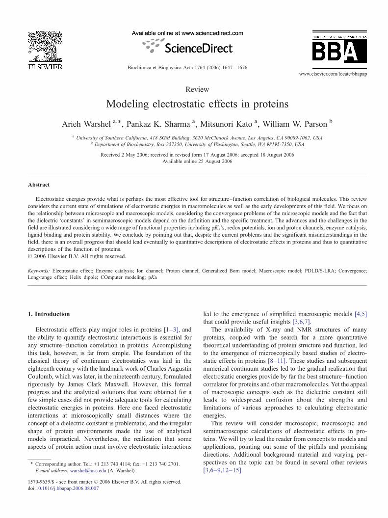
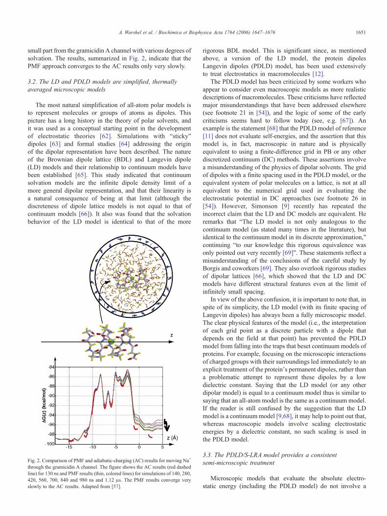
Fig. 1. The three main options for representing the solvent in computersimulation approaches. The microscopic model uses a detailed all-atomrepresentation and evaluates the average interaction between the solventresidual charges and the solute charges. Such calculations are comparativelyslow and expensive. The simplified microscopic model replaces the timeaverage dipole of each solvent molecule by a point dipole, while themacroscopic model uses a polarization vector to represent a collection ofsolvent dipoles in a large volume element.
1649A. Warshel et al. / Biochimica et Biophysica Acta 1764 (2006) 1647–1676
energies for structure–function correlation for proteins [2]. Thishas been demonstrated in studies of a wide range of propertiesthat will be considered in Sections 6–15.
3. The hierarchy of electrostatic models
Studies of electrostatic effects in proteins have involved awide range of modeling approaches that can be roughly dividedinto microscopic all-atom models, simplified dipolar models,and macroscopic models (see Fig. 1). Microscopic models canbe divided further into classical and quantum mechanicaltreatments. Each of these models has its own scope andlimitations. This includes, of course, the difficulty of interpret-ing continuum results without the use of microscopic models.Thus, it is important to understand the relationships betweenvarious approaches.
3.1. All-atom models, boundary conditions, long-rangetreatments, polarizability effects and convergence
An all-atom model describes a system as a collection ofparticles that interact via a quantum mechanical potentialsurface that can be approximated by a proper empirical forcefield. Although such approaches can be quite rigorous,adequate convergence may sometimes not be achieved evenwith nanosecond simulations, particularly for macromole-cules. One of the most significant “unresolved” issues inatomistic simulations is the proper representation of aninfinite system, which is critical for a proper treatment oflong-range electrostatic effects. Periodic boundary conditions(e.g., [24]), although appropriate in non-polar systems fornon-electrostatic problems or in periodic molecular crystals,cannot produce truly infinite non-crystalline systems withoutartifacts when long-range electrostatic interactions are signif-icant (e.g., see Fig. 5 in Ref. [25]). It is only now becomingcommonly appreciated that the Ewald method for periodicboundary conditions gives divergent solvation energies for thesame solute depending on the size of the simulated system(e.g., [26]). Correction formulas for particular charge dis-tributions [27] do not provide a general solution (see dis-cussion in [28] and [29]). In fact, Refs. [29–32] demonstratedthat the Ewald PME method, which has become more or lessa standard tool in molecular simulations, provides inadequatetreatment of electrostatic energies for non-periodic systems.The same is true for Ewald treatments with tinfoil boundariesand atom-based cutoffs. As demonstrated elsewhere (e.g.,[32–34]), this problem, which was anticipated long ago (e.g.,[25,35]), does not occur in simulations that use sphericalboundary conditions and utilize the local reaction field (LRF)approach [36]. Spherical boundary conditions including aspherical Ewald-type model [35] are physically more con-sistent than the periodic Ewald methods because there is nospurious periodicity; a spherical region of atomic representa-tion is just surrounded by a region that is treated more simply.Because the simplified outer region is far from the focal pointof the simulation, its effect can be made indistinguishablefrom that of an atomistic representation.
The issues involved in constructing proper boundaries forspherical models are logical and obvious. The most importantrequirement is that the system maintains proper polarization at
1650 A. Warshel et al. / Biochimica et Biophysica Acta 1764 (2006) 1647–1676
and around the boundaries [37]. This requirement is, in the caseof polar systems, much more important than the physicallytrivial requirement of maintaining proper heat flow by stochasticboundaries [38,39] and has been achieved by the surfaceconstraint all-atom solvent (SCAAS) model [40]. The problemis not the well-known bulk contribution from the surroundingsof the simulation sphere [41], which can be implemented easilyin simulation studies [42,43], but the polarization at themicroscopic boundaries of the simulation sphere [40].
Another important issue is the inclusion of the effect ofinduced dipoles in microscopic simulations of electrostaticenergies. The use of polarization terms in force fields dates backto the work of Warshel and Levitt, who introduced this approachas general way of capturing the effect of the electronicpolarization in protein modeling. This was done by bothiterative and non-iterative approaches. The use of a polarizableforce field became an integral part of simulations in the Warshelgroup [42,44], who analyzed its effect on electrostatic modelingin many studies [12,45,46]. The general realization that theeffects of induced dipoles are quite important has been quiteslow (some workers initially argued that these effects could notbe significant [47]), but is now widely appreciated. Severalgroups have started to develop different types of polarizableforce fields [48,49], which is clearly an important and overduestep. However, it is important to realize (in contrast to thepossible impression given by some of these recent works) thatthe use of polarizable force fields is not a new development andthat there is already a significant body of knowledge on caseswhere the effects of induced dipoles are important, and also onother cases where such a treatment is not crucial.
With reasonable boundary conditions, proper long-rangetreatment and induced dipoles, one can use all-atom models forevaluation of electrostatic free energies. The most appropriateapproach usually is the free energy perturbation (FEP) method[50,51]. This approach involves gradually changing the solutecharge from zero to its actual value Q=Q0 using a series ofmapping potentials of the form
Vm ¼ ð1� kmÞV ðQ ¼ 0Þ þ kmV ðQ0Þ; ð7Þwhere λm is changed gradually from 0 to 1 [50,52]. The corre-sponding free energy change then is evaluated by the expression
expf�DGðkmYkmþ1Þbg ¼ hexpf�ðVmþ1 � VmÞbgiVm; ð8Þ
where ⟨⟩Vmdesignates a molecular dynamics (MD) average over
Vm and β=1/kBT. The overall charging free energy is obtained bycollecting the ΔG(λm→λm+1). A very useful alternative is touse the linear response approximation (LRA), in which the freeenergy is given by [53]
DG Q ¼ 0YQ ¼ Q0ð Þ ¼ 12
hhV ðQ0Þ � V ðQ ¼ 0ÞiV ðQ0Þ
þ hV ðQ0Þ � V ðQ ¼ 0ÞiV ðQ¼0Þi:
ð9ÞAlthough the FEP and LRA approaches are both fundamentallysound, they can involve major convergence problems when
applied to charges in proteins, and they have been exploredsystematically in only a relatively small number of studies(e.g., [50,54,55]).
Another seemingly rigorous approach for studies ofelectrostatic energies in proteins and, in particular, for studyingthe free energy profile of ions in ion channels, is to evaluate theso-called potential of mean force (PMF). The PMF reflects thefree energy of moving a given charged group from the bulksolvent to a specific protein site, and is typically evaluated byusing umbrella sampling (US) or related approaches [52,57]. Inthe PMF approach, one commonly uses a mapping potential ofthe form
em ¼ ð1� kmÞe1 þ kme2 ð10aÞe1ðzÞ ¼ EgðzÞ þ Kðz� zð1Þ0 Þ2 ¼ EgðzÞ þ Eð1Þ
cons ð10bÞe2ðzÞ ¼ EgðzÞ þ Kðz� zð2Þ0 Þ2 ¼ EgðzÞ þ Eð2Þ
cons: ð10cÞHere z0
(1) and z0(2) are neighboring points on a reaction coordinate
(z) such as the position of solvated Na+ ion in a transmembranechannel, K is a quadratic constraint that holds the system at aspecified point, and Eg(z) is the total potential of the systemwithout the constraints. The free energy then can be obtained bythe FEP/US formula [57]
Dg zð Þ¼� 1bln expf�DGðkmÞbghdðz V� zÞexpfEm;consðzÞbgiem� �
:
ð11Þwhere, ΔG(λm) is the FEP energy associated with the change ofλ from zero to λm, and Em,cons(z) is Eg−Em.
Some workers replace theΔG(λm) term in Eq. (11) by a termobtained by the “weighted histograms analysis method”(WHAM), which is intended to provide the best overlap ofresults from simulations at different z′ [58]. In our experience,the WHAM approach does not improve the results [57]. This isworth noting, since the presumed advantages of the WHAMapproach and the seemingly rigorous formulation of PMFmethods (and related approaches such as Jarzynski's formula[59]) have drawn attention from the main question, which iswhether the PMF approach can provide reliable electrostaticenergies. Few of the benchmarks in PMF studies haveconsidered well-defined electrostatic problems that test themethod critically; most have considered less relevant systems,such as the alanine dipeptide [60,61]. In fact, PMF approachessuffer from major hysteresis problems, and suffer from theenormous challenge of calculating the reversible work ofmoving the ion from the bulk solvent to the protein interior.Furthermore, since PMF approaches do not provide absolutesolvation free energies, they generally do not give a clearwarning about major problems with the simulation method usedor with the specific treatment of long-range effects.
Although PMF calculations will be discussed further inSections 5, 7 and 14, it is pertinent to mention here a recent study[57] in which we compared FEP-based PMF to the adiabaticcharging (AC) approach, a non-standard PMF method thatinvolves pulling the ion using Eqs. (10) and (11). The com-parison was done for a well-defined benchmark composed of a
1651A. Warshel et al. / Biochimica et Biophysica Acta 1764 (2006) 1647–1676
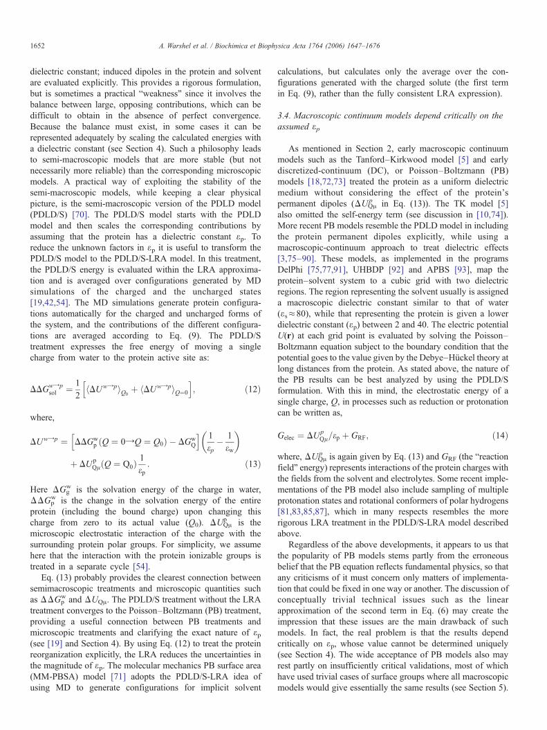
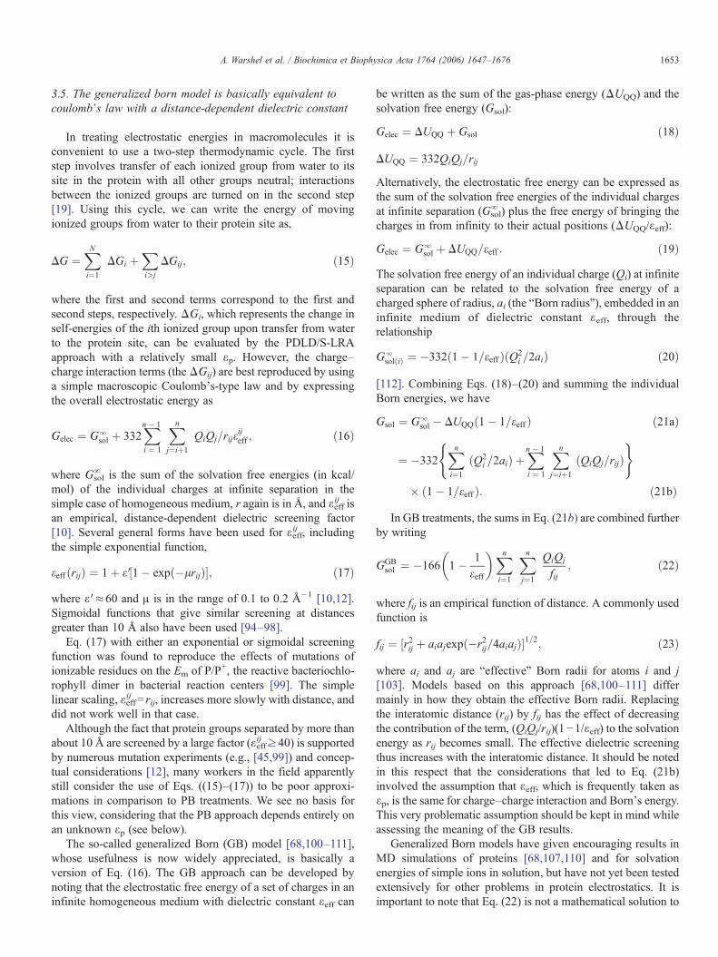
small part from the gramicidin A channel with various degrees ofsolvation. The results, summarized in Fig. 2, indicate that thePMF approach converges to the AC results only very slowly.
3.2. The LD and PDLD models are simplified, thermallyaveraged microscopic models
The most natural simplification of all-atom polar models isto represent molecules or groups of atoms as dipoles. Thispicture has a long history in the theory of polar solvents, andit was used as a conceptual starting point in the developmentof electrostatic theories [62]. Simulations with “sticky”dipoles [63] and formal studies [64] addressing the originof the dipolar representation have been described. The natureof the Brownian dipole lattice (BDL) and Langevin dipole(LD) models and their relationship to continuum models havebeen established [65]. This study indicated that continuumsolvation models are the infinite dipole density limit of amore general dipolar representation, and that their linearity isa natural consequence of being at that limit (although thediscreteness of dipole lattice models is not equal to that ofcontinuum models [66]). It also was found that the solvationbehavior of the LD model is identical to that of the more
Fig. 2. Comparison of PMF and adiabatic-charging (AC) results for moving Na+
through the gramicidin A channel. The figure shows the AC results (red dashedline) for 130 ns and PMF results (thin, colored lines) for simulations of 140, 280,420, 560, 700, 840 and 980 ns and 1.12 μs. The PMF results converge veryslowly to the AC results. Adapted from [57].
rigorous BDL model. This is significant since, as mentionedabove, a version of the LD model, the protein dipolesLangevin dipoles (PDLD) model, has been used extensivelyto treat electrostatics in macromolecules [12].
The PDLD model has been criticized by some workers whoappear to consider even macroscopic models as more realisticdescriptions of macromolecules. These criticisms have reflectedmajor misunderstandings that have been addressed elsewhere(see footnote 21 in [54]), and the logic of some of the earlycriticisms seems hard to follow today (see, e.g. [67]). Anexample is the statement [68] that the PDLD model of reference[11] does not evaluate self-energies, and the assertion that thismodel is, in fact, macroscopic in nature and is physicallyequivalent to using a finite-difference grid in PB or any otherdiscretized continuum (DC) methods. These assertions involvea misunderstanding of the physics of dipolar solvents. The gridof dipoles with a finite spacing used in the PDLD model, or theequivalent system of polar molecules on a lattice, is not at allequivalent to the numerical grid used in evaluating theelectrostatic potential in DC approaches (see footnote 26 in[54]). However, Simonson [9] recently has repeated theincorrect claim that the LD and DC models are equivalent. Heremarks that “The LD model is not only analogous to thecontinuum model (as stated many times in the literature), butidentical to the continuum model in its discrete approximation,”continuing “to our knowledge this rigorous equivalence wasonly pointed out very recently [69]”. These statements reflect amisunderstanding of the conclusions of the careful study byBorgis and coworkers [69]. They also overlook rigorous studiesof dipolar lattices [66], which showed that the LD and DCmodels have different structural features even at the limit ofinfinitely small spacing.
In view of the above confusion, it is important to note that, inspite of its simplicity, the LD model (with its finite spacing ofLangevin dipoles) has always been a fully microscopic model.The clear physical features of the model (i.e., the interpretationof each grid point as a discrete particle with a dipole thatdepends on the field at that point) has prevented the PDLDmodel from falling into the traps that beset continuum models ofproteins. For example, focusing on the microscopic interactionsof charged groups with their surroundings led immediately to anexplicit treatment of the protein's permanent dipoles, rather thana problematic attempt to represent these dipoles by a lowdielectric constant. Saying that the LD model (or any otherdipolar model) is equal to a continuum model thus is similar tosaying that an all-atom model is the same as a continuum model.If the reader is still confused by the suggestion that the LDmodel is a continuum model [9,68], it may help to point out that,whereas macroscopic models involve scaling electrostaticenergies by a dielectric constant, no such scaling is used inthe PDLD model.
3.3. The PDLD/S-LRA model provides a consistentsemi-microscopic treatment
Microscopic models that evaluate the absolute electro-static energy (including the PDLD model) do not involve a
1652 A. Warshel et al. / Biochimica et Biophysica Acta 1764 (2006) 1647–1676
dielectric constant; induced dipoles in the protein and solventare evaluated explicitly. This provides a rigorous formulation,but is sometimes a practical “weakness” since it involves thebalance between large, opposing contributions, which can bedifficult to obtain in the absence of perfect convergence.Because the balance must exist, in some cases it can berepresented adequately by scaling the calculated energies witha dielectric constant (see Section 4). Such a philosophy leadsto semi-macroscopic models that are more stable (but notnecessarily more reliable) than the corresponding microscopicmodels. A practical way of exploiting the stability of thesemi-macroscopic models, while keeping a clear physicalpicture, is the semi-macroscopic version of the PDLD model(PDLD/S) [70]. The PDLD/S model starts with the PDLDmodel and then scales the corresponding contributions byassuming that the protein has a dielectric constant εp. Toreduce the unknown factors in εp it is useful to transform thePDLD/S model to the PDLD/S-LRA model. In this treatment,the PDLD/S energy is evaluated within the LRA approxima-tion and is averaged over configurations generated by MDsimulations of the charged and the uncharged states[19,42,54]. The MD simulations generate protein configura-tions automatically for the charged and uncharged forms ofthe system, and the contributions of the different configura-tions are averaged according to Eq. (9). The PDLD/Streatment expresses the free energy of moving a singlecharge from water to the protein active site as:
DDGwYpsol ¼ 1
2hDUwYpiQ0
þ hDUwYpiQ¼0
h i; ð12Þ
where,
DUwYp ¼ DDGwp ðQ ¼ 0YQ ¼ Q0Þ � DGw
Q
h i 1ep
� 1ew
� �
þ DU pQl Q ¼ Q0ð Þ 1
ep: ð13Þ
Here ΔGgw is the solvation energy of the charge in water,
ΔΔGpw is the change in the solvation energy of the entire
protein (including the bound charge) upon changing thischarge from zero to its actual value (Q0). ΔUQμ
p is themicroscopic electrostatic interaction of the charge with thesurrounding protein polar groups. For simplicity, we assumehere that the interaction with the protein ionizable groups istreated in a separate cycle [54].
Eq. (13) probably provides the clearest connection betweensemimacroscopic treatments and microscopic quantities suchas ΔΔGp
w and ΔUQμ. The PDLD/S treatment without the LRAtreatment converges to the Poisson–Boltzmann (PB) treatment,providing a useful connection between PB treatments andmicroscopic treatments and clarifying the exact nature of εp(see [19] and Section 4). By using Eq. (12) to treat the proteinreorganization explicitly, the LRA reduces the uncertainties inthe magnitude of εp. The molecular mechanics PB surface area(MM-PBSA) model [71] adopts the PDLD/S-LRA idea ofusing MD to generate configurations for implicit solvent
calculations, but calculates only the average over the con-figurations generated with the charged solute (the first termin Eq. (9), rather than the fully consistent LRA expression).
3.4. Macroscopic continuum models depend critically on theassumed εp
As mentioned in Section 2, early macroscopic continuummodels such as the Tanford–Kirkwood model [5] and earlydiscretized-continuum (DC), or Poisson–Boltzmann (PB)models [18,72,73] treated the protein as a uniform dielectricmedium without considering the effect of the protein'spermanent dipoles (ΔUQμ
p in Eq. (13)). The TK model [5]also omitted the self-energy term (see discussion in [10,74]).More recent PB models resemble the PDLD model in includingthe protein permanent dipoles explicitly, while using amacroscopic-continuum approach to treat dielectric effects[3,75–90]. These models, as implemented in the programsDelPhi [75,77,91], UHBDP [92] and APBS [93], map theprotein–solvent system to a cubic grid with two dielectricregions. The region representing the solvent usually is assigneda macroscopic dielectric constant similar to that of water(εs≈80), while that representing the protein is given a lowerdielectric constant (εp) between 2 and 40. The electric potentialU(r) at each grid point is evaluated by solving the Poisson–Boltzmann equation subject to the boundary condition that thepotential goes to the value given by the Debye–Hückel theory atlong distances from the protein. As stated above, the nature ofthe PB results can be best analyzed by using the PDLD/Sformulation. With this in mind, the electrostatic energy of asingle charge, Q, in processes such as reduction or protonationcan be written as,
Gelec ¼ DUpQl=ep þ GRF; ð14Þ
where, ΔUQμp is again given by Eq. (13) and GRF (the “reaction
field” energy) represents interactions of the protein charges withthe fields from the solvent and electrolytes. Some recent imple-mentations of the PB model also include sampling of multipleprotonation states and rotational conformers of polar hydrogens[81,83,85,87], which in many respects resembles the morerigorous LRA treatment in the PDLD/S-LRA model describedabove.
Regardless of the above developments, it appears to us thatthe popularity of PB models stems partly from the erroneousbelief that the PB equation reflects fundamental physics, so thatany criticisms of it must concern only matters of implementa-tion that could be fixed in one way or another. The discussion ofconceptually trivial technical issues such as the linearapproximation of the second term in Eq. (6) may create theimpression that these issues are the main drawback of suchmodels. In fact, the real problem is that the results dependcritically on εp, whose value cannot be determined uniquely(see Section 4). The wide acceptance of PB models also mayrest partly on insufficiently critical validations, most of whichhave used trivial cases of surface groups where all macroscopicmodels would give essentially the same results (see Section 5).
1653A. Warshel et al. / Biochimica et Biophysica Acta 1764 (2006) 1647–1676
3.5. The generalized born model is basically equivalent tocoulomb’s law with a distance-dependent dielectric constant
In treating electrostatic energies in macromolecules it isconvenient to use a two-step thermodynamic cycle. The firststep involves transfer of each ionized group from water to itssite in the protein with all other groups neutral; interactionsbetween the ionized groups are turned on in the second step[19]. Using this cycle, we can write the energy of movingionized groups from water to their protein site as,
DG ¼XNi¼1
DGi þXi>j
DGij; ð15Þ
where the first and second terms correspond to the first andsecond steps, respectively. ΔGi, which represents the change inself-energies of the ith ionized group upon transfer from waterto the protein site, can be evaluated by the PDLD/S-LRAapproach with a relatively small εp. However, the charge–charge interaction terms (theΔGij) are best reproduced by usinga simple macroscopic Coulomb's-type law and by expressingthe overall electrostatic energy as
Gelec ¼ Glsol þ 332
Xi ¼ 1
n� 1 Xnj¼iþ1
QiQj=rijeijeff ; ð16Þ
where Gsol∞ is the sum of the solvation free energies (in kcal/
mol) of the individual charges at infinite separation in thesimple case of homogeneous medium, r again is in Å, and εeff
ij isan empirical, distance-dependent dielectric screening factor[10]. Several general forms have been used for εeff
ij , includingthe simple exponential function,
eeff ðrijÞ ¼ 1þ e V½1� expð�lrijÞ�; ð17Þwhere ε′≈60 and μ is in the range of 0.1 to 0.2 Å−1 [10,12].Sigmoidal functions that give similar screening at distancesgreater than 10 Å also have been used [94–98].
Eq. (17) with either an exponential or sigmoidal screeningfunction was found to reproduce the effects of mutations ofionizable residues on the Em of P/P+, the reactive bacteriochlo-rophyll dimer in bacterial reaction centers [99]. The simplelinear scaling, εeff
ij = rij, increases more slowly with distance, anddid not work well in that case.
Although the fact that protein groups separated by more thanabout 10 Å are screened by a large factor (εeff
ij ≥40) is supportedby numerous mutation experiments (e.g., [45,99]) and concep-tual considerations [12], many workers in the field apparentlystill consider the use of Eqs. ((15)–(17)) to be poor approxi-mations in comparison to PB treatments. We see no basis forthis view, considering that the PB approach depends entirely onan unknown εp (see below).
The so-called generalized Born (GB) model [68,100–111],whose usefulness is now widely appreciated, is basically aversion of Eq. (16). The GB approach can be developed bynoting that the electrostatic free energy of a set of charges in aninfinite homogeneous medium with dielectric constant εeff can
be written as the sum of the gas-phase energy (ΔUQQ) and thesolvation free energy (Gsol):
Gelec ¼ DUQQ þ Gsol ð18Þ
DUQQ ¼ 332QiQj=rij
Alternatively, the electrostatic free energy can be expressed asthe sum of the solvation free energies of the individual chargesat infinite separation (Gsol
∞ ) plus the free energy of bringing thecharges in from infinity to their actual positions (ΔUQQ/εeff):
Gelec ¼ Glsol þ DUQQ=eeff : ð19Þ
The solvation free energy of an individual charge (Qi) at infiniteseparation can be related to the solvation free energy of acharged sphere of radius, ai (the “Born radius”), embedded in aninfinite medium of dielectric constant εeff, through therelationship
GlsolðiÞ ¼ �332ð1� 1=eeff ÞðQ2
i =2aiÞ ð20Þ
[112]. Combining Eqs. (18)–(20) and summing the individualBorn energies, we have
Gsol ¼ Glsol � DUQQð1� 1=eeff Þ ð21aÞ
¼ �332Xni¼1
ðQ2i =2aiÞ þ
Xi ¼ 1
n� 1 Xnj¼iþ1
ðQiQj=rijÞ( )
� ð1� 1=eeff Þ: ð21bÞ
In GB treatments, the sums in Eq. (21b) are combined furtherby writing
GGBsol ¼ �166 1� 1
eeff
� �Xni¼1
Xnj¼1
QiQj
fij; ð22Þ
where fij is an empirical function of distance. A commonly usedfunction is
fij ¼ ½r2ij þ aiajexpð�r2ij=4aiajÞ�1=2; ð23Þ
where ai and aj are “effective” Born radii for atoms i and j[103]. Models based on this approach [68,100–111] differmainly in how they obtain the effective Born radii. Replacingthe interatomic distance (rij) by fij has the effect of decreasingthe contribution of the term, (QiQj/rij)(1−1/εeff) to the solvationenergy as rij becomes small. The effective dielectric screeningthus increases with the interatomic distance. It should be notedin this respect that the considerations that led to Eq. (21b)involved the assumption that εeff, which is frequently taken asεp, is the same for charge–charge interaction and Born's energy.This very problematic assumption should be kept in mind whileassessing the meaning of the GB results.
Generalized Born models have given encouraging results inMD simulations of proteins [68,107,110] and for solvationenergies of simple ions in solution, but have not yet been testedextensively for other problems in protein electrostatics. It isimportant to note that Eq. (22) is not a mathematical solution to
1654 A. Warshel et al. / Biochimica et Biophysica Acta 1764 (2006) 1647–1676
the problem of charges in a multicavity continuum, or of asystem with multiple dielectric regions such as a proteinsurrounded by water [8]. Although an increase in dielectricscreening with distance is in accord with experiment, there is nosound theoretical basis for Eq. (22) or (23). Nor does the modelprescribe a unique value of εeff to use with Eq. (22) inapplications to proteins, although values of at least 20 probablywill be needed in most cases.
The above discussion shows that the GB treatment is anapproximation of Eq. (16) and thus does not go much beyondCoulomb's law [8]. Nevertheless, it is instructive to note that theGB approximation also can be obtained by assuming a localmodel in which the vacuum electric field, E0, and thedisplacement vector, D, are identical [113]. This model can beconsidered as a “local Langevin Dipole model” [69] or as aversion of the non-iterative LD model. The GB approximationemerges if one uses this model to approximate the energy of acollection of charges and makes some additional assumptionsabout the position dependence of the dielectric constant.
Eq. (19) is valid only for homogeneous media with a highdielectric constant. We cannot assume that it will apply as well totransfer of charges from water to a protein. A more consistenttreatment requires replacing ΔGsol
∞ by the free energies of theindividual ions in their protein sites, where the effective value ofε is variable. Fortunately, however, it usually is reasonable to usea large εeff for the second term in Eq. (19), even for proteininteriors. Here again, the main issue is the reliability of Eq. (21)and not its legitimization by a seemingly rigorous GBformulation.
Unlike the GB model, Eq. (16) does not attempt to capturethe term Gsol
∞ along with the charge–charge interaction term bya single sum. There are, however, many situations, in whichGsol∞ is either constant, or can be removed by a suitable
thermodynamic cycle, leaving the charge–charge interactions asthe factor of interest. The effects of mutations on the pKa ofanother residue or on the Em of a bound cofactor exemplifysuch situations.
4. The meaning of protein dielectric constants
It is often assumed that the dielectric constant of proteins (εp)is a universal quantity that can be used in a variety of models. Inparticular, it was assumed for many years that proteins could berepresented by a low dielectric constant in the range of 2 to 4, andthis assumption was supported by several microscopic simula-tions (see below). However, a microscopically-based conceptualand practical analysis byWarshel and Russell [12] indicated thatthe value of εp is entirely dependent on the method and systemused to define this quantity. This initially appeared to be aquestionable conclusion, and it still might look strange to readerswho are accustomed to the view that macroscopic models have auniversal physical meaning. It may also be puzzling to workerswho are experienced with microscopic statistical mechanics,where ε (usually called ε̄) can be evaluated in a unique way fromthe fluctuations of the total dipole moment of the system (seebelow). Even now it is not widely recognized that the best valueof the parameter εp to use in modeling electrostatic effects has
little to do with what is usually considered as the protein'sdielectric constant. Systematic studies [13,19,114] have shownthat εp is basically a measure of all the electrostatic interactionsthat are not included explicitly in the model. This point can beappreciated by considering several limiting cases. If all theinteractions are treated explicitly, εp=1; if all but induceddipoles are included explicitly, εp≈2; and if the solvent is notincluded explicitly (a very poor model), εp>40. If the protein'spermanent dipoles are included explicitly but the protein'sinduced dipoles and relaxation of the permanent dipoles aroundthe charged groups (the protein reorganization) are treatedimplicitly, the value of εp is not a well-defined quantity. Themost appropriate value of εp in this situation usually is between 4and 6 for dipole–charge interactions and between 4 and 10 forcharge–charge interactions [115,116]. In general, the effectivedielectric constant in protein interiors is not a constant because itdepends on the site considered as well as the model [114,117].
Several authors have argued that there is only one ‘proper’dielectric constant, which is the statistical mechanical dielectricconstant (ε̄), obtained from the fluctuations of the average totaldipole moment of a system [6,114,117–120]. However, thisdielectric ‘constant’ is not constant, since it depends on thespecific region in the protein [114,117]. It is also sometimesassumed that εp is equal to ε̄, but this assumption is completelyunjustified. For example, using an explicit model frequentlyproduces ε̄>6, while, as discussed above, one has to use εp=1when all interactions are treated explicitly (see Ref. [121] fordetailed discussion). The relatively large value of ε̄ is frequentlyassumed to reflect fluctuations of ionized groups on the surfaceof the protein [69,117,120,122]. However, recent experimentalstudies [99,123,124] have supported the theoretical finding[114] that active sites in proteins generally have large values ofε,̄ even when these sites are far from the surface.
Although the misconceptions about ε̄ have been reviewedelsewhere [19], it is nevertheless useful to summarize what hasbeen learned about ε̄ for proteins. The value of ε̄ apparently canbe significantly larger than the value of about 4 deduced frommeasurements of the dielectric properties of dry proteins andpeptide powders [125,126], or from simulations of the entireprotein rather than a specific region [114]. The early theoreticalfinding of ε̄≈4 in a gas-phase study [126], ignored the effect ofthe reaction field of the solvent. This type of treatmentdrastically underestimates the value of ε̄ (see discussion anddemonstration in [114]). Simulations by King et al. [114] thatincluded the solvent's reaction field gave ε̄≈10 in and near theactive site of trypsin. Similar values were obtained even in theabsence of fluctuations of the ionized residues on the protein'ssurface, showing that polar groups and water molecules ininterior regions contribute significantly to ε̄. It was suggestedrecently that the calculations by King et al. [114] may not haveconverged [9], but the SCAAS model that was used actuallyconverges much more rapidly than the periodic models (and thecorresponding very large simulation systems) used in thepresumably “converging” studies mentioned in ref. [9], and theconvergence of ε̄ was examined by longer runs and by usingalternative formulations. To our knowledge, however, therehave been no attempts to repeat the calculations of ε̄ by King
1655A. Warshel et al. / Biochimica et Biophysica Acta 1764 (2006) 1647–1676
et al. [114] for trypsin or any other enzyme. Thus, it seems thatthe main obstacle to reaching a consensus about ε̄ is the undueimpact of studies (e.g., [117,127]) that considered the entireprotein instead of specific regions, and the attempt to deduce theeffect of ionized surface groups by evaluating ε̄ in specificprotein regions when the surface groups are neutral.
Despite the intrinsic interest in the nature of ε̄, the aboveanalysis indicates that this quantity is not highly relevant to theenergetics of charges in proteins and does provide a unique wayof determining εp for a semimacroscopic model. Another goodway to appreciate this point is to consider a charge in a watersphere surrounded by water. In such a model, ε̄≈80, whereas amicroscopic model of the same system with implicit induceddipoles will require εp≈2.
One could argue reasonably that the entire concept of adielectric constant is invalid in the heterogeneous interior of aprotein [11]. However, since fully microscopic models still donot give sufficiently precise results, it is useful to be able toestimate electrostatic energies using implicit models and, inparticular, semimacroscopic models. Thus, it is justified to lookfor optimal εp values after realizing what this parameter reallymeans. In principle, the semimacroscopic PDLD/S-LRA modelshould use εp≈2 because all effects except those of theprotein's induced dipoles are considered explicitly. However,the configurational sampling by the LRA approach obviously isnot perfect. Although the protein reorganization energyprobably is captured to a reasonable extent [121], the changein water penetration on ionization of charged residues probablyis not reproduced accurately. This problem is particularlyserious for ionizable groups in nonpolar sites in the interior of aprotein. In such cases the ionization processes can causesignificant changes in water penetration, as demonstrated in theinstructive experiments of Garcia-Moreno et al. [128]. Molec-ular dynamics simulations are unlikely to reproduce thesechanges in nanosecond simulation times, without special tricksof the type described in Ref. [129] and Section 7.
That the effective dielectric constant for charge–chargeinteractions in proteins is larger than usually assumed has beenpointed out by several workers [10,94,130]. However, thisimportant observation has not always been analyzed with a clearmicroscopic perspective. Based on classical studies of water,Mehler and coworkers correctly argued [94,96] that in polarsolvents the effective screening factor, εeff
ij , of Eq. (16) is asigmoidal function that increases to a large value at relativelyshort distances [131–133]. Jonsson and coworkers [130]assumed that electrostatic interactions in proteins can bedescribed by using a large εp for charge–charge interactions.Although we completely agree that εp and εeff frequently arelarge [10,12], we believe that the reasons for this are more subtlethan is generally realized. It is important to note that thebehavior of εeff(r) in water is not necessarily relevant to thecorresponding behavior in proteins, where many of thepermanent dipoles have more restricted mobility. The surprisingobservation that charge–charge interactions are stronglyscreened in proteins was rationalized by Warshel and Russell[12], who pointed out that the screening must reflect thecompensation of charge–charge interactions and solvation
energy. Qualitatively, as two charges come closer together, theincrease in their direct Coulombic interaction is offset by adecrease in the sum of their individual solvation energies. Foran illustration of this effect, see Fig. 4 of Sham et al. [115]. Thecompensation is nearly perfect in water and other polar solvents,where the electrostatic free energy of an ion pair is virtuallyindependent of the distance between the ions, and it holdssurprisingly well down to 5 or 10 Å even in proteins. It seems tous that understanding this crucial compensation effect isessential for an understanding of εp in proteins.
Baptista and coworkers [134], addressed the idea that PBmodels should use two different dielectric constants: one for selfenergies (called εind in their paper) and the other for charge–charge interactions (called εpair). They tested this propositionwith values of 4 and 20 for εind and εpair, respectively, using aPB model that did not include relaxations of the protein. Theauthors noted that our early work [54] had suggested that theeffective dielectric constants for charge–dipole and charge–charge interactions are generally different. However, we did notmean to imply that the problems in using PB models withoutrelaxations of the proteins can be fixed by using a lowerdielectric constant for computing self energies. In fact, wepointed out that the problems of PB models with unrelaxedproteins and low dielectric constants cannot be fixed, and thatone must use the PDLS/S-LRA or related treatment in order tohave any hope of finding a relatively uniform dielectric scaling.In other studies, we found that unrelaxed PB treatments requirea value of about 10 for εind [115].
As we would expect, and as shown in our works, the use of 4and 20 for εind and εpair, respectively, by Baptista and coworkers[134] gave very poor results. Better results were obtained byusing a protein dielectric constant of about 20 for allinteractions. Unfortunately, the authors took these results tomean that our proposal is incorrect. It was then argued that theproblem with the proposal is the assumption that our εeff isequal to εpair. In protein interiors, εpair is, in fact, quite close tothe εeff. It is smaller near the surface, since εeff implicitlyincludes the effect of the solvent.
Using εind=20 for treating self-energies may not be a goodidea even with PB treatments of charges in unrelaxed proteininteriors. One of the best examples of the problem is providedby calculations of the redox potential (Em) of cytochrome c.One cannot obtain the large shift of the Em relative to the Em ofthe reference model compound in water, unless εind is smallerthan 6 [121]. Of course, we cannot provide general universalrules for the inconsistent unrelaxed PB models, but εind shoulddefinitely be smaller than εpair in consistent semimacroscopicmodels.
Contrary to what one might assume, the effectivemacroscopic dielectric constant for a given region in aprotein cannot be used to provide a general description ofthe energetics of charges in this region [120]. The conceptualproblem with this assumption is illustrated in Fig. 3. Thefigure represents an internal site in which a charged groupinteracts with a dipole that is fixed in position and a seconddipole that is free to rotate. Since this is an internal site, theleading term in semimacroscopic treatments will be the
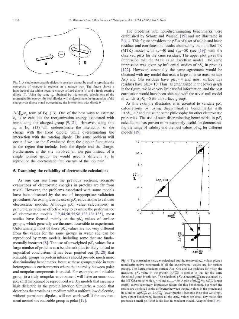
Fig. 3. A single macroscopic dielectric constant cannot be used to reproduce theenergetics of charges in proteins in a unique way. The figure shows ahypothetical site with a negative charge, a fixed dipole (a) and a freely rotatingdipole (b). Using the same εp, obtained by microscopic calculations of thereorganization energy, for both dipoles will underestimate the interaction of thecharge with dipole a and overestimate the interactions with dipole b.
Fig. 4. The correlation between calculated and the observed pKa values gives anondiscriminative benchmark if all the experimental values are for surfacegroups. The figure considers surface Asp, Glu and Lys residues for which themeasured pKa value in the protein (pKapp
obs) is similar to that for the samefunctional group in solution. The calculated pKa values (pKapp
calc) are evaluated bythe MTK(S) model with εp=40 and εwater=80. A plot of pKapp
obs vs. pKappcalc (upper
graph) shows seemingly impressive results for this benchmark, but when theresults are displayed as the difference between the pKa values in the protein andin solution (ΔpK app
obs vs. ΔpK appcalc, lower graph) it becomes clear that we simply
have a poor benchmark. Because all the ΔpKa values are small, any model thatproduces a small pKa shift looks like an excellent model. Adapted from [19].
1656 A. Warshel et al. / Biochimica et Biophysica Acta 1764 (2006) 1647–1676
ΔUQμp /εp term of Eq. (13). One of the best ways to estimate
εp is to calculate the reorganization energy associated withintroducing the charged group [9,121]. However, using thisεp in Eq. (13) will underestimate the interaction of thecharge with the fixed dipole, while overestimating theinteraction with the rotating dipole. The same problem willoccur if we use the ε̄ evaluated from the dipolar fluctuationsin the region that includes both the dipole and the charge.Furthermore, if the site involved an ion pair instead of asingle ionized group we would need a different εp toreproduce the electrostatic free energy of the ion pair.
5. Examining the reliability of electrostatic calculations
As one can see from the previous sections, accurateevaluations of electrostatic energies in proteins are far fromtrivial. However, the problems associated with some modelshave been obscured by the use of inappropriate validationprocedures. An example is the use of pKa calculations to validateelectrostatic models. Although pKa value calculations, inprinciple, provide an effective way to examine the performanceof electrostatic models [12,44,50,55,96,122,128,135], moststudies have focused mainly on the pKa values of surfacegroups, which generally are the most accessible to experiment.Unfortunately, most of these pKa values are not very differentfrom the values for the same groups in water and can bereproduced by many models, including some that are funda-mentally incorrect [8]. The use of unweighted pKa values for alarge number of proteins as a benchmark thus is likely to lead tounjustified conclusions. It has been pointed out [8,128] thationizable groups in protein interiors should provide much morediscriminating benchmarks, because these groups reside in veryheterogeneous environments where the interplay between polarand nonpolar components is crucial. For example, an ionizablegroup in a truly nonpolar environment will have an enormouspKa shift that cannot be reproduced well bymodels that assume ahigh dielectric in the protein interior. Similarly, a model thatdescribes the protein as a medium with a uniform low dielectric,without permanent dipoles, will not work well if the environ-ment around the ionizable group is polar [12].
The problems with non-discriminating benchmarks wereestablished by Schutz and Warshel [19] and are illustrated inFig. 4. This figure considers the pKas of a set of acidic and basicresidues and correlates the results obtained by the modified TK(MTK) model with εp=40 and εeff=80 (see [19]) with theobserved pKas for the same residues. The upper plot gives theimpression that the MTK is an excellent model. The sameimpression was given by influential studies of pKa in proteins[122]. However, essentially the same agreement would beobtained with any model that uses a large ε, since most surfaceAsp and Glu residues have pKa≈4 and most surface Lysresidues have pKa≈10. Thus, as emphasized in the lower graphin the figure, we have very little useful information, and the bestcorrelation would have been obtained with the trivial null modelin which ΔpKa=0 for all surface groups.
As this example illustrates, it is essential to validate pKa
calculations by using discriminative benchmarks with∣ΔpKa∣>2 and to use the same philosophy for other electrostaticproperties. The use of such discriminating benchmarks in pKa
calculations has proven to be extremely useful for demonstrat-ing the range of validity and the best values of εp for differentmodels [19].
1657A. Warshel et al. / Biochimica et Biophysica Acta 1764 (2006) 1647–1676
A recent study of the PMF for ion penetration through ionchannels [57] provides another example of validation ofelectrostatic models. The issue here is the validity of seeminglyrigorous statistical mechanical expressions such as Eq. (11), andthe problem again is that such approaches have been examinedonly by using insufficiently discriminating benchmarks such asthe torsional potential of the alanine dipeptide rather than systemswith the necessary electrostatic heterogeneity. Furthermore,calculations on an actual ion channel cannot be considered aproper benchmark because it is not clear a priori what is necessaryin order to obtain converging results in such a system. Thisproblem was addressed by Kato and Warshel [57], who used partof a gramicidin channelwith a variable number ofwatermoleculesto establish a system that provided converging simulations (Fig.2). Comparing the PMF for moving an ion through or around thischannel, which should be identical, provides a way of assessingthe hysteresis in the calculations (see Fig. 2).
6. Calibrating calculations by studies of solvation energiesof small molecules
Modeling a biological process can be helped enormously bycalibrating the calculated free energy change relative to theobserved or estimated free energy of a corresponding process inaqueous solution [12,136]. This is especially important forenzymatic reactions, where the catalytic effect is defined relativeto the solution reaction. It also applies to calculations of ligand-binding processes, where one has to compare the solvationenergy of the ligand in its protein site to the correspondingsolvation energy in solution. Early attempts to estimate solvationenergies [137,138] were based on Born's expression for theenergy of a charge in a spherical cavity with a basically arbitraryradius (Eq. (20)) or Onsager's similar expression for a dipole.The first attempts to move toward quantitative evaluation ofsolvation energies branched in two directions. One approachwasto examine the interaction between the solute and a singlesolvent molecule quantum mechanically [139,140]. The otherapproach, which ultimately turned out to be the more successful,was to parameterize the solute–solvent van der Waals interac-tions in a complete solute–solvent system empirically, andevaluate the interaction between the solute andmany (rather thanone) solvent molecules [37]. Although the empirical approachwas regarded initially as having “too many parameters,” it wasrealized eventually that having an atom–solvent parameter foreach type of solute atom is the key requirement in anyquantitative semiempirical solvation model. Recent continuumsolvation models [141–143] have essentially the same numberof parameters as current all-atom models [40,51,52,144]. For asolute with a given set of charges, the crucial step in either typeof model is parameterization of the solute–solvent repulsionterm or the corresponding atomic radius (see [25]). Leaving theselection of reasonable parameter values aside, the nextrequirement is to obtain converging results with the particularmodeling method that is chosen.
The general progress in modeling solvation energies of smallmolecules and ions is quite encouraging, as is apparent from theresults reported by many research groups using a variety of
simulation approaches. FEP calculations have focused on theproper treatment of ionized groups and the importance of properboundary conditions [26,27,33,40,145]. Recently, the validity ofthe LRA treatment [53] in solvation calculations was explored[146] and the LRA formalism was used in a quantummechanicaltreatment [147]. LD calculations were found to give accurateresults when incorporated into quantum mechanical models[148,149] and similar success was reported in continuum[141,150] and GB studies [104]. All of these models apparentlygive similar accuracy once properly parameterized. The deviationfrom experiments after reasonable parameter fitting in some casescould reflect the neglect of charge transfer to the solvent.
These successful calculations on small molecules in solutioncould be viewed as almost trivial in one sense, considering thatthe environment is uniform and the solvation can be related tothe effective atomic radius in a simple way. Unfortunately, theability to reproduce solvation energies in solution does notguarantee that the same treatment will give reliable results forsolvation energies of ligands in proteins, or for the solvationenergy of the entire macromolecule.
7. Evaluation of pKas of ionizable residues in proteins
Ionizable residues in proteins play major roles in almost allbiological processes, including enzymatic reactions, protonpumps and protein stability. Understanding these roles canrequire evaluating both the interactions of the ionized groupsand the energetics of the ionization process itself. Thus anability to calculate pKas of ionizable groups in proteins can becrucial for structure–function correlations as well as forvalidating different treatments of electrostatic energies [12].
Calculations of pKas by all-atom FEP approaches have beendescribed for only a small number of proteins [50,56]. Recentwork includes studies of the pKa of metal-bound watermolecules [151] and proton transfer in proteins [152] as wellas functionally important groups [153–156]. Several all-atomLRA calculations also have been described [54,55]. PDLD/S-LRA calculations have given encouraging results with εp=4[19,54]. Although the LRA treatment considers the proteinreorganization formally, the limit of εp=2 expected for a“perfect” model with implicit induced dipoles has not beenreached, probably because some of the reorganization and/orwater penetration is not captured in the computer time used in thesimulations. Only a few studies (e.g. [36]) include estimates ofthe error range in the calculations. It appears that the error rangeof the all-atommodels is still somewhat disappointing and that insome cases the results of microscopic calculations are simplydisastrous, which perhaps explains we have so few reportedstudies. However, inclusion of proper long-range treatments andinduced dipoles does lead to some improvements [54,56].
Semimacroscopic-continuum models, such as the currentversion of the PB models, which now include the proteinpermanent dipoles and the self-energy term, frequently givereasonable results. Yet most PB models [122,157,158] do nottreat the protein reorganization explicitly, and the so-calledmulti-conformation continuum electrostatic (MCCE) variant[87] considers only the reorganization of polar side chains rather
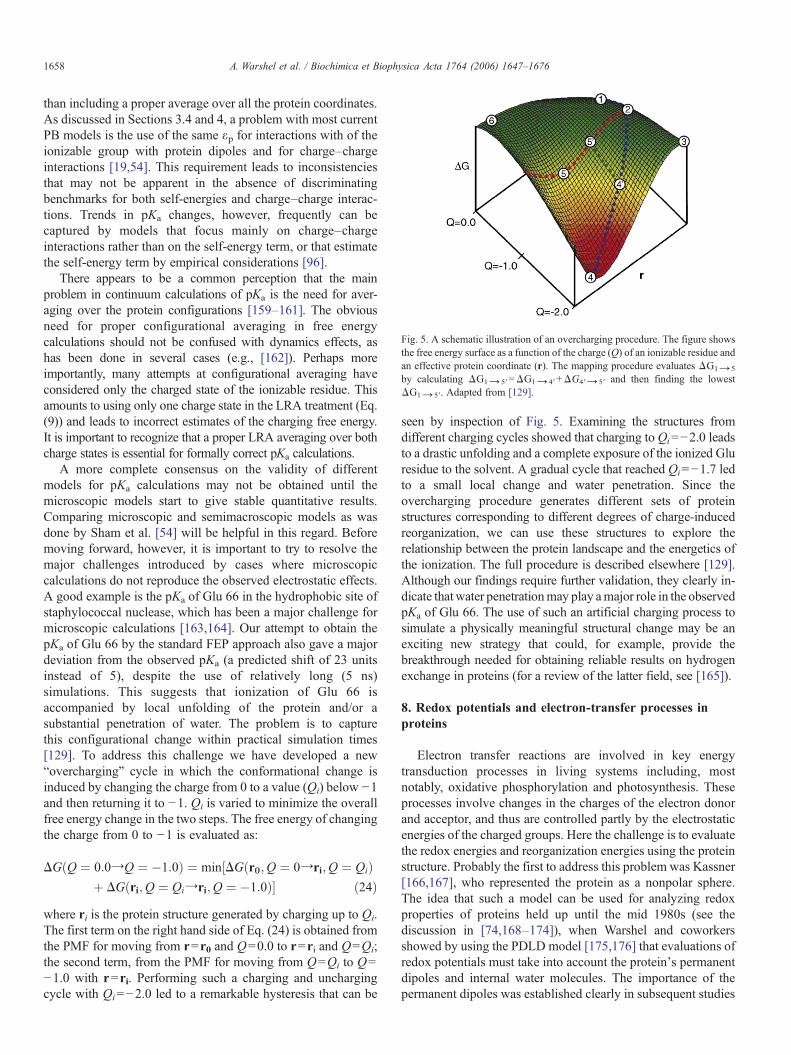
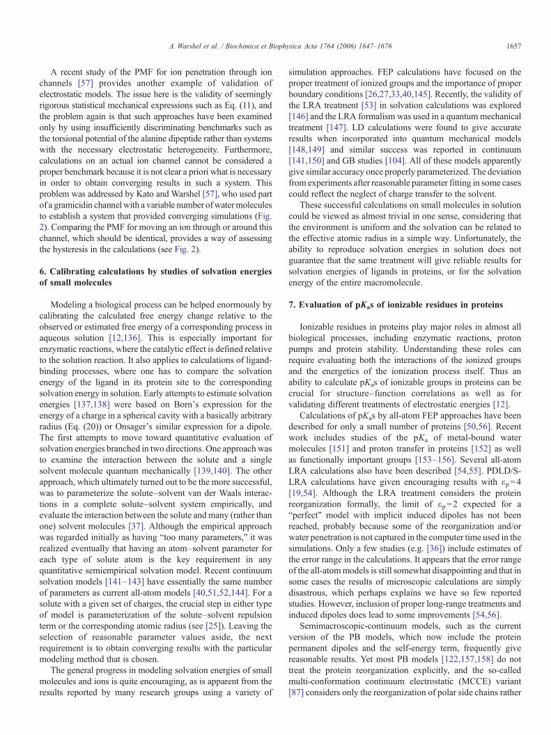
Fig. 5. A schematic illustration of an overcharging procedure. The figure showsthe free energy surface as a function of the charge (Q) of an ionizable residue andan effective protein coordinate (r). The mapping procedure evaluates ΔG1→ 5
by calculating ΔG1→ 5′=ΔG1→ 4′+ΔG4′→ 5′ and then finding the lowestΔG1→ 5′. Adapted from [129].
1658 A. Warshel et al. / Biochimica et Biophysica Acta 1764 (2006) 1647–1676
than including a proper average over all the protein coordinates.As discussed in Sections 3.4 and 4, a problem with most currentPB models is the use of the same εp for interactions with of theionizable group with protein dipoles and for charge–chargeinteractions [19,54]. This requirement leads to inconsistenciesthat may not be apparent in the absence of discriminatingbenchmarks for both self-energies and charge–charge interac-tions. Trends in pKa changes, however, frequently can becaptured by models that focus mainly on charge–chargeinteractions rather than on the self-energy term, or that estimatethe self-energy term by empirical considerations [96].
There appears to be a common perception that the mainproblem in continuum calculations of pKa is the need for aver-aging over the protein configurations [159–161]. The obviousneed for proper configurational averaging in free energycalculations should not be confused with dynamics effects, ashas been done in several cases (e.g., [162]). Perhaps moreimportantly, many attempts at configurational averaging haveconsidered only the charged state of the ionizable residue. Thisamounts to using only one charge state in the LRA treatment (Eq.(9)) and leads to incorrect estimates of the charging free energy.It is important to recognize that a proper LRA averaging over bothcharge states is essential for formally correct pKa calculations.
A more complete consensus on the validity of differentmodels for pKa calculations may not be obtained until themicroscopic models start to give stable quantitative results.Comparing microscopic and semimacroscopic models as wasdone by Sham et al. [54] will be helpful in this regard. Beforemoving forward, however, it is important to try to resolve themajor challenges introduced by cases where microscopiccalculations do not reproduce the observed electrostatic effects.A good example is the pKa of Glu 66 in the hydrophobic site ofstaphylococcal nuclease, which has been a major challenge formicroscopic calculations [163,164]. Our attempt to obtain thepKa of Glu 66 by the standard FEP approach also gave a majordeviation from the observed pKa (a predicted shift of 23 unitsinstead of 5), despite the use of relatively long (5 ns)simulations. This suggests that ionization of Glu 66 isaccompanied by local unfolding of the protein and/or asubstantial penetration of water. The problem is to capturethis configurational change within practical simulation times[129]. To address this challenge we have developed a new“overcharging” cycle in which the conformational change isinduced by changing the charge from 0 to a value (Qi) below −1and then returning it to −1. Qi is varied to minimize the overallfree energy change in the two steps. The free energy of changingthe charge from 0 to −1 is evaluated as:
DGðQ ¼ 0:0YQ ¼ �1:0Þ ¼ min½DGðr0;Q ¼ 0Yri;Q ¼ QiÞþ DGðri;Q ¼ QiYri;Q ¼ �1:0Þ� ð24Þ
where ri is the protein structure generated by charging up to Qi.The first term on the right hand side of Eq. (24) is obtained fromthe PMF for moving from r=r0 and Q=0.0 to r=ri and Q=Qi;the second term, from the PMF for moving from Q=Qi to Q=−1.0 with r=ri. Performing such a charging and unchargingcycle with Qi=−2.0 led to a remarkable hysteresis that can be
seen by inspection of Fig. 5. Examining the structures fromdifferent charging cycles showed that charging toQi=−2.0 leadsto a drastic unfolding and a complete exposure of the ionized Gluresidue to the solvent. A gradual cycle that reached Qi=−1.7 ledto a small local change and water penetration. Since theovercharging procedure generates different sets of proteinstructures corresponding to different degrees of charge-inducedreorganization, we can use these structures to explore therelationship between the protein landscape and the energetics ofthe ionization. The full procedure is described elsewhere [129].Although our findings require further validation, they clearly in-dicate thatwater penetrationmay play amajor role in the observedpKa of Glu 66. The use of such an artificial charging process tosimulate a physically meaningful structural change may be anexciting new strategy that could, for example, provide thebreakthrough needed for obtaining reliable results on hydrogenexchange in proteins (for a review of the latter field, see [165]).
8. Redox potentials and electron-transfer processes inproteins
Electron transfer reactions are involved in key energytransduction processes in living systems including, mostnotably, oxidative phosphorylation and photosynthesis. Theseprocesses involve changes in the charges of the electron donorand acceptor, and thus are controlled partly by the electrostaticenergies of the charged groups. Here the challenge is to evaluatethe redox energies and reorganization energies using the proteinstructure. Probably the first to address this problem was Kassner[166,167], who represented the protein as a nonpolar sphere.The idea that such a model can be used for analyzing redoxproperties of proteins held up until the mid 1980s (see thediscussion in [74,168–174]), when Warshel and coworkersshowed by using the PDLD model [175,176] that evaluations ofredox potentials must take into account the protein's permanentdipoles and internal water molecules. The importance of thepermanent dipoles was established clearly in subsequent studies
1659A. Warshel et al. / Biochimica et Biophysica Acta 1764 (2006) 1647–1676
of iron–sulfur proteins [177,178]. Another interesting factor isthe effect of ionized residues on redox potentials, which oftencan be described well using Coulomb's law with a largeeffective dielectric constant (Eq. (16)) [10,45,99,128].
Microscopic estimates of protein reorganization energieshave been described [45,121,179,180] and have been used instudies of the rate constants of biological electron transport. Thisalso includes studies of nuclear quantum mechanical effectsassociated with fluctuations of the protein's polar groups (forreview see [181]). PB studies of redox proteins also haveprogressed significantly [82,168,171,182,183], although someconfusion still appears to persist on the importance of the proteinpermanent dipoles (see discussion in [74,168]).
To illustrate some of the complexities associated withcalculations of electrostatic free energies in redox proteins, itis instructive to consider computational studies of the initialelectron-transfer step in photosynthetic bacterial reaction centers(RC's). The elucidation of the crystal structure of this systempresented major challenges to both computational and experi-mental approaches. An immediate question was whether theinitial event involved stepwise electron transfer from an excitedbacteriochlorophyll dimer (P*) to a neighboring bacteriochlo-rophyll (BL), followed by electron transfer from BL
− to abacteriopheophytin (HL), or a superexchange process in whichP* transferred an electron directly to HL with the assistance ofelectronic coupling to P+BL
− as a virtual intermediate. In the two-step mechanism, P+BL
− would have to lie close to or below P*BL
in energy, while superexchange would allow P+BL− to be at a
higher energy. Microscopic FEP/US and PDLD calculations thatincluded proper treatments of boundary conditions and long-range interactions gave similar energies for P*BL and P+BL
−,
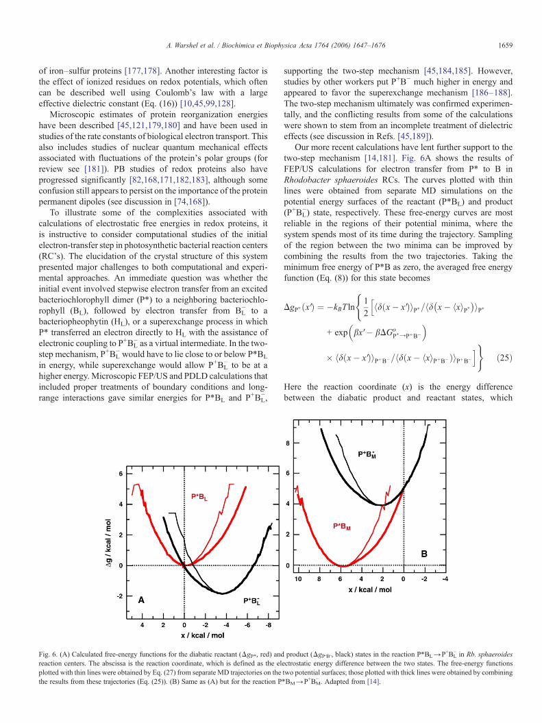
Fig. 6. (A) Calculated free-energy functions for the diabatic reactant (ΔgP*, red) andreaction centers. The abscissa is the reaction coordinate, which is defined as the elplotted with thin lines were obtained by Eq. (27) from separate MD trajectories on thethe results from these trajectories (Eq. (25)). (B) Same as (A) but for the reaction P
supporting the two-step mechanism [45,184,185]. However,studies by other workers put P+B− much higher in energy andappeared to favor the superexchange mechanism [186–188].The two-step mechanism ultimately was confirmed experimen-tally, and the conflicting results from some of the calculationswere shown to stem from an incomplete treatment of dielectriceffects (see discussion in Refs. [45,189]).
Our more recent calculations have lent further support to thetwo-step mechanism [14,181]. Fig. 6A shows the results ofFEP/US calculations for electron transfer from P* to B inRhodobacter sphaeroides RCs. The curves plotted with thinlines were obtained from separate MD simulations on thepotential energy surfaces of the reactant (P*BL) and product(P+BL
−) state, respectively. These free-energy curves are mostreliable in the regions of their potential minima, where thesystem spends most of its time during the trajectory. Samplingof the region between the two minima can be improved bycombining the results from the two trajectories. Taking theminimum free energy of P*B as zero, the averaged free energyfunction (Eq. (8)) for this state becomes
DgP� x Vð Þ ¼ �kBT ln
(12
hd x� x Vð ÞiP�=hd x� hxiP�� �iP�h
m exp bx V� bDGoP�YPþB�
�
� hd x� x Vð ÞiPþB�=hd x� hxiPþB�ð ÞiPþB�
i)ð25Þ
Here the reaction coordinate (x) is the energy differencebetween the diabatic product and reactant states, which
product (ΔgP+B−, black) states in the reaction P*BL→P+BL− in Rb. sphaeroides
ectrostatic energy difference between the two states. The free-energy functionstwo potential surfaces; those plotted with thick lines were obtained by combining*BM→P+BM
− . Adapted from [14].
1660 A. Warshel et al. / Biochimica et Biophysica Acta 1764 (2006) 1647–1676
fluctuates with time during the MD trajectories, andΔGoP*→P+B−
is the overall free-energy change given by the FEP calculations(− 1.9 kcal/mol). The free-energy function for P+B− is obtainedsimilarly. The calculated free energies of the two states areplotted with thick lines in Fig. 6. If ∣ΔGo
P*→P+B−∣ were muchlarger, trajectories on additional mapping potentials would berequired in order to sample the configurational space betweenthe two minima adequately.
One way to check whether the conformational space issampled sufficiently well in a given region of the reactioncoordinate is to see whether the free-energy functions for thereactant and product states satisfy the expression (see Ref. [52])
DgbðxÞ ¼ DgaðxÞ þ x; ð26Þwhich follows from the relationship [52,190],
Dgbðx VÞ ¼ �kBT ln½hdðx� x VÞexpð�x=kBTÞia=hdðx� hxiaÞia�ð27Þ
The averaged free-energy curves plotted with thick lines inFig. 6A conform to Eq. (26) in the region between the twominima, while the individual curves with thin lines do not.
Eqs. (26) and (27) indicate that the entire free-energy curvefor the product state can be obtained from a sufficiently longMD trajectory in the reactant state and vice versa. However,these expressions assume that the trajectory samples the entireconfigurational space adequately, which is unlikely to be thecase for a complex system [180,191]. A single trajectory on thereactant surface generally will not visit all the configurationsthat contribute significantly to the product surface, even if theoverall free-energy change in the reaction is relatively small.Attempting to generate both free-energy surfaces from a singletrajectory, therefore, is not recommended in most cases.
Fig. 6B shows the results of similar FEP calculations forelectron transfer from P* to the neighboring bacteriochlorophyllon the “inactive” branch of electron carriers (BM). The productstate (P+BM
− ) is found to lie about 4 kcal/mol above P*. Thisresult agrees with the PDLD calculations on RCs from therelated species Blastochloris viridis [185], and is in good accordwith the experimental finding that electron transfer occurspredominantly to BL and HL in preference to BM and HM.However, Ceccarelli and Marchi [192] have described MDsimulations that put P+BM
− about 7 kcal/mol below P*. Thediscrepancy with our calculations and experimental findingsseem likely to stem from their use of periodic boundaryconditions, and an Ewald-sum treatment of electrostaticinteractions. They also propagated trajectories only on thereactant state and did not employ FEP, but that is less likely toaccount for the discrepancy. Clearly, fundamental flaws in amodel cannot be remedied simply by increasing the number ofatoms or the length of the MD trajectory, without criticalvalidation studies. As stated in the previous section, the use ofthe spherical SAACS and LRF treatments is more reliable thanthe use of very large systems with periodic boundary conditions.
Despite the successes of consistent microscopic studies ofRCs, it is useful to examine the validity of the results bycomparison with an appropriate macroscopic model. We start by
noting that the free energy change for a reaction such asPB→P+B− can be described macroscopically as
DGelec ¼ DEgas þ DGlsol þ DUQA=e; ð28aÞ
where ΔEgas is the change in the vacuum molecular orbitalenergies of the electron donor and acceptor, ΔGsol
∞ is the changein solvation free energies of the electron carriers at infiniteseparation in a homogeneous medium with dielectric constant ε,andΔUQμ is the change in unscreened electrostatic interactions,including the direct interactions between the donor and acceptor(ΔUQQ).ΔEgas can be estimated either by quantum calculationsor by combining measured Em values with calculated solvationenergies [14,184,185]
DEgas ¼ �FDEm � DGrefsol ð28bÞ
We can use the Born equation (Eq. (20)) to relate the term,ΔGsol
∞ , to the corresponding free energy change in water oranother reference solvent with a high dielectric constant, ΔGsol
∞ :
DGlsol ¼ DGw;l
sol þ ð1� 1=eÞ=ð1� 1=ewÞcDGw;lsol ð1� 1=eÞ:
ð29Þ
This approach is especially convenient if the same referencesolution is used for calculating ΔEgas because it leads to can-cellation of some terms in the final results. Combining Eqs. (28a)and (29) with Eq. (28b) and equating ΔGsol
ref with ΔGsolw,∞, the
free energy change for the electron-transfer reaction becomes,
DGelecc� FDEm þ ðDUQl � DGrefsol Þ=e: ð30Þ
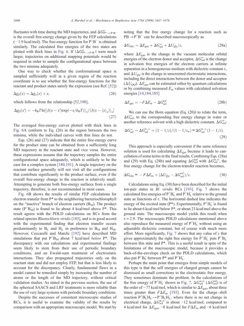
Calculations using Eq. (30) have been described for the initialion-pair states in Bl. viridis RCs [193]. Fig. 7 shows thecalculated free energies of P+BL
− and P+HL− relative to the ground
state as functions of ε. The horizontal dashed line indicates theenergy of the excited state (P*). Experimentally, P+HL
− is foundto lie about 6 kcal/mol below P*, or about 23 kcal/mol above theground state. The macroscopic model yields this result whenε≈2.9. The microscopic PDLD calculations mentioned abovealso reproduce the measured energy of P+HL
− without using anadjustable dielectric constant, but of course with much moreeffort. More significantly, Fig. 7 shows that any value of ε thatgives approximately the right free energy for P+HL
− puts P+BL−
between this state and P*. This is a useful result in spite of thelimitations of the macroscopic model, because it provides aback-of-the-envelope check on the PDLD calculations, whichalso put P+BL
− between P* and P+HL−.
Perhaps the main point that emerges from simple models ofthis type is that the self energies of charged groups cannot bedismissed as small corrections to the electrostatic free energy.They sometimes dominate the problem. In the calculations ofthe free energy of P+HL
− shown in Fig. 7, ΔGsolw,∞ (ΔGsol
ref) is ofthe order of −77 kcal/mol, which is similar toΔEgas, about threetimes greater than FΔEm [193]. Even for the charge–shiftreaction P+BL
−HL→P+BLHL−, where there is no net change in
electrical charge, ΔGsolw,∞ is about −12 kcal/mol, compared to
4 kcal/mol for ΔEgas, −8 kcal/mol for FΔEm and −6 kcal/mol
Fig. 7. Free energies of P+BL− (blue curve) and P+HL
− (green curve) in Bl. viridisRCs as functions of the dielectric constant in a macroscopic model [193]. Allenergies in this figure are expressed relative to the ground state. The horizontaldashed red and green lines are, respectively, the energy of the first excitedsinglet state of P (P*) and an experimental value of the free energy of P+HL
−. Thegas-phase energy for the reaction PHL→P+HL
− was obtained from the measuredredox potentials of P/P+ and HL
−/HL and PDLD calculations of the change insolvation energy for each of the half-cell reactions in situ; that for BL+HL
−→BL−+HL
− was obtained from the redox potentials of bacteriochlorophyll-b andbacteriopheophytin-b and the calculated solvation energies for the molecules insolution. The electrostatic free energy changes for PBLHL→P+BLHL
− andP+BLHL
−→P+BL−HL were calculated using Eq. (30) and were summed to obtain
that for PBLHL→P+BL−HL. See [193] for details.
1661A. Warshel et al. / Biochimica et Biophysica Acta 1764 (2006) 1647–1676
for ΔUQμ [193]. Calculations that neglect the self-energiesclearly should not be given much credence. The occasionalability of such calculations to reproduce an experimental resultcan be misleading in this respect, and, in most cases, probablycan be traced to a cancellation of errors or to the choice of anindiscriminating benchmark; (see [45] for a discussion of howcalculations by Marchi et al. [186], which omitted the solventin and around the protein, gave what is almost certainly muchtoo high an energy for P+BL
−, while fortuitously givingapproximately the correct energy for P+HL
−).Another key question with regard to bacterial RCs is how
slight asymmetries in the protein favor electron transfer from P*mainly to BL and HL while blocking electron transfer to BM andHM. One possibility is that the specificity results fromdifferences in electronic overlap of P* with and BL and BM.However, as shown in Fig. 6, our PDLD/S and FEP calculationsindicated that the specificity probably is explained by theenergetics of the two reactions, because the protein stabilizesP+BL
− significantly more strongly than P+BM− . The calculations
also identified some of the residues that contributed to theasymmetry of the energies [185]. These predictions have heldup well to tests by studies of site-directed mutations thatstabilize P+BM
− or destabilize P+BL− (see, e.g., [194]).
Convergence of microscopic calculations proved to be amajor problem in a recent quantummechanical evaluation of theredox potentials of blue copper proteins [195]. In this case,semimacroscopic models presently give more accurate resultsthan microscopic models.
9. Electrostatic effects in ligand binding to proteins
Reliable evaluations of the free energy of ligand binding canpotentially make important contributions to structure-baseddrug design [196]. Here we have an interplay of electrostatic,hydrophobic and steric effects, but accurate estimates of theelectrostatic contributions are still crucial.
In principle, it is possible to evaluate binding free energiesby performing microscopic FEP calculations of the typeconsidered in Eqs. (7) and (8). The difference between the freeenergies of gradually removing the force field from the ligandin the protein active site and in water should give the freeenergy of binding. This approach, however, encounters majorconvergence problems and, at present, the reported results havebeen disappointing except in cases of very small ligands. Analternative approach is to study the effect of small “mutations”of the ligand, such as replacement of NH2 by OH [51].However, if one is interested in the absolute binding freeenergies for medium-size ligands, it is essential to use simplerapproaches. Perhaps the most useful of these is the LRAapproach of Eq. (9), augmented by estimates of the non-electrostatic effects.
The LRA approach is particularly effective for calculatingelectrostatic contributions to ligand binding energies [53,197].In this approximation one can express the binding energy as
DGbind ¼ 12
Upelec; S
D ESþ Up
elec; S
D ES V� Uw
elec; S
D ES� Uw
elec; S
D ES V
�þ DGnonelec
bind : ð31Þ
HereUelec,ℓ is the electrostatic contribution to the interactionof the ligand with its surroundings; superscripts p and wdesignate protein and water, respectively; and ℓ and ℓ′designate the ligand in its actual charge form and the “nonpolar”ligand created by setting all its atomic charges to zero. In thisexpression the terms ⟨Uelec,ℓ−Uelec,ℓ′⟩, which are required byEq. (9), are replaced by ⟨Uelec,ℓ⟩ since ⟨Uelec,ℓ′⟩=0. Evaluationof the nonelectrostatic contribution ΔGbind
nonelec is still verychallenging, since these contributions might not follow theLRA. A useful option is to evaluate the individual contributionsto the binding free energy from hydrophobic effects, van derWaals interactions, and water penetration [53,197].
Another powerful option is the so-called linear interactionenergy (LIE) approach [198,199]. This approach adopts theLRA approximation for the electrostatic contribution butneglects the ⟨Uelec,ℓ⟩ℓ′ terms. The binding energy is thenexpressed as,
DGbindca Upelec; S
D ES� Uw
elec; S
D ES
h iþ b V Up
vdW; S
D ES� Uw
vdW; S
D ES
h ið32Þ
where α is a constant that is approximately 1/2 in many casesand β′ is an empirical parameter that scales the van der Waals(vdW) component of the protein–ligand interaction. While thegeneral origin of the electrostatic scaling, α, is relatively clear (itshould be around 0.5 [53] with possible small deviations [146]),
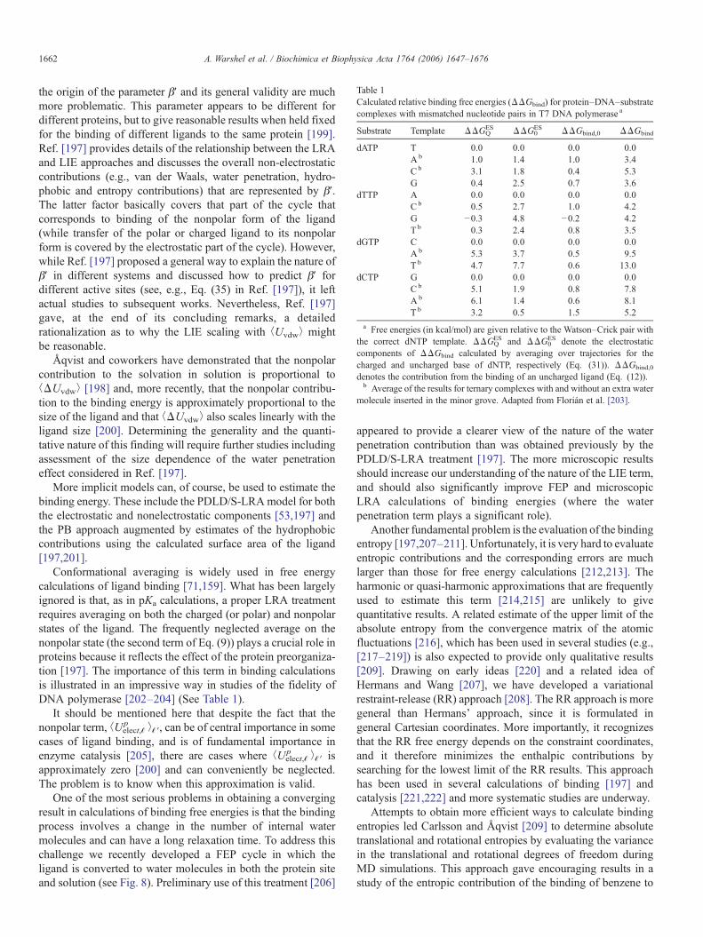
Table 1Calculated relative binding free energies (ΔΔGbind) for protein–DNA–substratecomplexes with mismatched nucleotide pairs in T7 DNA polymerase a
Substrate Template ΔΔGQES ΔΔG0
ES ΔΔGbind,0 ΔΔGbind
dATP T 0.0 0.0 0.0 0.0A b 1.0 1.4 1.0 3.4Cb 3.1 1.8 0.4 5.3G 0.4 2.5 0.7 3.6
dTTP A 0.0 0.0 0.0 0.0Cb 0.5 2.7 1.0 4.2G −0.3 4.8 −0.2 4.2T b 0.3 2.4 0.8 3.5
dGTP C 0.0 0.0 0.0 0.0A b 5.3 3.7 0.5 9.5T b 4.7 7.7 0.6 13.0
dCTP G 0.0 0.0 0.0 0.0Cb 5.1 1.9 0.8 7.8A b 6.1 1.4 0.6 8.1T b 3.2 0.5 1.5 5.2
a Free energies (in kcal/mol) are given relative to the Watson–Crick pair withthe correct dNTP template. ΔΔGQ
ES and ΔΔG0ES denote the electrostatic
components of ΔΔGbind calculated by averaging over trajectories for thecharged and uncharged base of dNTP, respectively (Eq. (31)). ΔΔGbind,0
denotes the contribution from the binding of an uncharged ligand (Eq. (12)).b Average of the results for ternary complexes with and without an extra water
molecule inserted in the minor grove. Adapted from Florián et al. [203].
1662 A. Warshel et al. / Biochimica et Biophysica Acta 1764 (2006) 1647–1676
the origin of the parameter β′ and its general validity are muchmore problematic. This parameter appears to be different fordifferent proteins, but to give reasonable results when held fixedfor the binding of different ligands to the same protein [199].Ref. [197] provides details of the relationship between the LRAand LIE approaches and discusses the overall non-electrostaticcontributions (e.g., van der Waals, water penetration, hydro-phobic and entropy contributions) that are represented by β′.The latter factor basically covers that part of the cycle thatcorresponds to binding of the nonpolar form of the ligand(while transfer of the polar or charged ligand to its nonpolarform is covered by the electrostatic part of the cycle). However,while Ref. [197] proposed a general way to explain the nature ofβ′ in different systems and discussed how to predict β′ fordifferent active sites (see, e.g., Eq. (35) in Ref. [197]), it leftactual studies to subsequent works. Nevertheless, Ref. [197]gave, at the end of its concluding remarks, a detailedrationalization as to why the LIE scaling with ⟨Uvdw⟩ mightbe reasonable.
Åqvist and coworkers have demonstrated that the nonpolarcontribution to the solvation in solution is proportional to⟨ΔUvdw⟩ [198] and, more recently, that the nonpolar contribu-tion to the binding energy is approximately proportional to thesize of the ligand and that ⟨ΔUvdw⟩ also scales linearly with theligand size [200]. Determining the generality and the quanti-tative nature of this finding will require further studies includingassessment of the size dependence of the water penetrationeffect considered in Ref. [197].
More implicit models can, of course, be used to estimate thebinding energy. These include the PDLD/S-LRAmodel for boththe electrostatic and nonelectrostatic components [53,197] andthe PB approach augmented by estimates of the hydrophobiccontributions using the calculated surface area of the ligand[197,201].
Conformational averaging is widely used in free energycalculations of ligand binding [71,159]. What has been largelyignored is that, as in pKa calculations, a proper LRA treatmentrequires averaging on both the charged (or polar) and nonpolarstates of the ligand. The frequently neglected average on thenonpolar state (the second term of Eq. (9)) plays a crucial role inproteins because it reflects the effect of the protein preorganiza-tion [197]. The importance of this term in binding calculationsis illustrated in an impressive way in studies of the fidelity ofDNA polymerase [202–204] (See Table 1).
It should be mentioned here that despite the fact that thenonpolar term, ⟨Uelect,ℓ
p ⟩ℓ′, can be of central importance in somecases of ligand binding, and is of fundamental importance inenzyme catalysis [205], there are cases where ⟨Uelect,ℓ
p ⟩ℓ′ isapproximately zero [200] and can conveniently be neglected.The problem is to know when this approximation is valid.
One of the most serious problems in obtaining a convergingresult in calculations of binding free energies is that the bindingprocess involves a change in the number of internal watermolecules and can have a long relaxation time. To address thischallenge we recently developed a FEP cycle in which theligand is converted to water molecules in both the protein siteand solution (see Fig. 8). Preliminary use of this treatment [206]
appeared to provide a clearer view of the nature of the waterpenetration contribution than was obtained previously by thePDLD/S-LRA treatment [197]. The more microscopic resultsshould increase our understanding of the nature of the LIE term,and should also significantly improve FEP and microscopicLRA calculations of binding energies (where the waterpenetration term plays a significant role).
Another fundamental problem is the evaluation of the bindingentropy [197,207–211]. Unfortunately, it is very hard to evaluateentropic contributions and the corresponding errors are muchlarger than those for free energy calculations [212,213]. Theharmonic or quasi-harmonic approximations that are frequentlyused to estimate this term [214,215] are unlikely to givequantitative results. A related estimate of the upper limit of theabsolute entropy from the convergence matrix of the atomicfluctuations [216], which has been used in several studies (e.g.,[217–219]) is also expected to provide only qualitative results[209]. Drawing on early ideas [220] and a related idea ofHermans and Wang [207], we have developed a variationalrestraint-release (RR) approach [208]. The RR approach is moregeneral than Hermans' approach, since it is formulated ingeneral Cartesian coordinates. More importantly, it recognizesthat the RR free energy depends on the constraint coordinates,and it therefore minimizes the enthalpic contributions bysearching for the lowest limit of the RR results. This approachhas been used in several calculations of binding [197] andcatalysis [221,222] and more systematic studies are underway.
Attempts to obtain more efficient ways to calculate bindingentropies led Carlsson and Åqvist [209] to determine absolutetranslational and rotational entropies by evaluating the variancein the translational and rotational degrees of freedom duringMD simulations. This approach gave encouraging results in astudy of the entropic contribution of the binding of benzene to
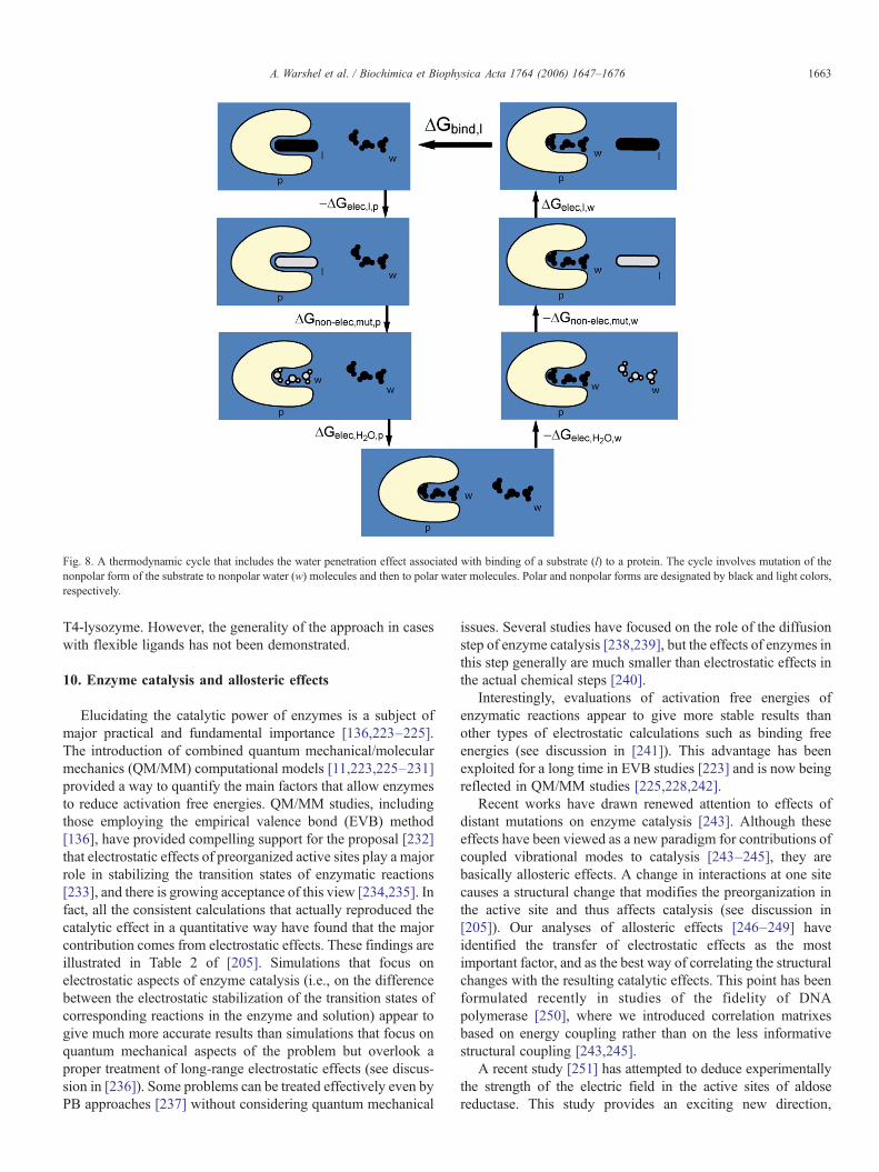
Fig. 8. A thermodynamic cycle that includes the water penetration effect associated with binding of a substrate (l) to a protein. The cycle involves mutation of thenonpolar form of the substrate to nonpolar water (w) molecules and then to polar water molecules. Polar and nonpolar forms are designated by black and light colors,respectively.
1663A. Warshel et al. / Biochimica et Biophysica Acta 1764 (2006) 1647–1676
T4-lysozyme. However, the generality of the approach in caseswith flexible ligands has not been demonstrated.
10. Enzyme catalysis and allosteric effects
Elucidating the catalytic power of enzymes is a subject ofmajor practical and fundamental importance [136,223–225].The introduction of combined quantum mechanical/molecularmechanics (QM/MM) computational models [11,223,225–231]provided a way to quantify the main factors that allow enzymesto reduce activation free energies. QM/MM studies, includingthose employing the empirical valence bond (EVB) method[136], have provided compelling support for the proposal [232]that electrostatic effects of preorganized active sites play a majorrole in stabilizing the transition states of enzymatic reactions[233], and there is growing acceptance of this view [234,235]. Infact, all the consistent calculations that actually reproduced thecatalytic effect in a quantitative way have found that the majorcontribution comes from electrostatic effects. These findings areillustrated in Table 2 of [205]. Simulations that focus onelectrostatic aspects of enzyme catalysis (i.e., on the differencebetween the electrostatic stabilization of the transition states ofcorresponding reactions in the enzyme and solution) appear togive much more accurate results than simulations that focus onquantum mechanical aspects of the problem but overlook aproper treatment of long-range electrostatic effects (see discus-sion in [236]). Some problems can be treated effectively even byPB approaches [237] without considering quantum mechanical
issues. Several studies have focused on the role of the diffusionstep of enzyme catalysis [238,239], but the effects of enzymes inthis step generally are much smaller than electrostatic effects inthe actual chemical steps [240].
Interestingly, evaluations of activation free energies ofenzymatic reactions appear to give more stable results thanother types of electrostatic calculations such as binding freeenergies (see discussion in [241]). This advantage has beenexploited for a long time in EVB studies [223] and is now beingreflected in QM/MM studies [225,228,242].
Recent works have drawn renewed attention to effects ofdistant mutations on enzyme catalysis [243]. Although theseeffects have been viewed as a new paradigm for contributions ofcoupled vibrational modes to catalysis [243–245], they arebasically allosteric effects. A change in interactions at one sitecauses a structural change that modifies the preorganization inthe active site and thus affects catalysis (see discussion in[205]). Our analyses of allosteric effects [246–249] haveidentified the transfer of electrostatic effects as the mostimportant factor, and as the best way of correlating the structuralchanges with the resulting catalytic effects. This point has beenformulated recently in studies of the fidelity of DNApolymerase [250], where we introduced correlation matrixesbased on energy coupling rather than on the less informativestructural coupling [243,245].
A recent study [251] has attempted to deduce experimentallythe strength of the electric field in the active sites of aldosereductase. This study provides an exciting new direction,
1664 A. Warshel et al. / Biochimica et Biophysica Acta 1764 (2006) 1647–1676
although the catalytic effect of aldose reductase is relatively small[252] and, thus, the electric field is smaller than in other enzymes.Furthermore, it is not entirely clear that the preorganization effectcan be directly correlated with the macroscopic field.
Kraut et al. [253] have recently argued that electrostatic effectsdo not provide a major contribution to enzyme catalysis byketosteroid isomerase (KSI). The authors found that the bindingenergies of phenolate ions to the oxyanion hole of the enzyme arenot strongly correlated with the delocalization of the phenolate'scharge (as estimated from NMR shifts). This weak correlationwas assumed to provide an experimental tool for assessing theimportance of electrostatic energies in enzyme catalysis. Thefinding of a very small change in binding energy for significantchanges in delocalization was interpreted as evidence thatelectrostatic contributions do not play a major role in KSI and,presumably, in other enzymes as well. As in many other cases,however, there is a risk of confusing an interpretation ofexperimental facts with a unique energy-based analysis. Carefulstudies have shown that the catalytic effect of KSI is due almostentirely to electrostatic preorganization effects [254]. Theexperimental correlation described by Kraut et al. does notprovide a way to estimate the actual electrostatic contribution or,for that matter, any other contribution. The authors simplypostulated that the electrostatic contributions to both binding andcatalysis must be correlated with the localization of the charge inthe isolated phenolate, and tried to deduce the electrostaticcontribution to catalysis from the localization. The most seriousproblem with this approach is that the catalytic cycle and thebinding cycle are fundamentally very different; the catalytic cycleinvolves transfer of a negative charge from Asp 40 to the enolateoxygen, while binding involves movement of a negative chargefromwater to the protein active site. Calculations of the energeticsof these processes (Warshel, A., Sharma, P. K., Chu, Z. T. andÅqvist, J., unpublished data), we found that the electrostaticcontribution to binding is only about 2 kcal/mol as compared toabout 10 kcal/mol in the catalytic process. The confusion heremight have been due to the fact that the binding energy and thecatalytic energy are of similar magnitudes (about 10 kcal/mol)and, thus, it possible to assume that both of them should reflectsimilar electrostatic contributions. However, most of the bindingenergy involves a nonpolar contribution, while the nonpolarcontribution to catalysis is basically zero. Even if we overlook thedifference between the cycle that leads to kcat and the bindingprocess and compare the binding of the enolate intermediate to thebinding of the phenolates, we can see major differences betweenthe two processes. That is, the electrostatic preorganization effectis, by far, larger in the case of the real substrate than in the case ofthe phenolates. This reflects the fact that the preorganization effectdepends on the orientation of the nonpolar from the ligand (thesecond term in Eq. 9) relative to the oxyanion hole, as well as onthe polarization of the oxyanion hole in the absence of a charge onthe ligand. Apparently, when the phenolate is in the nonpolarstate, it can easily move out of the correct orientation.
Another problem is that a more localized charge on thephenolate will also lead to greater electrostatic stabilization(solvation) in the reference solvent, so that the net effect oncatalysis (i.e., on the ratio of the enzymatic and nonenzymatic
reactions) is smaller than one might anticipate. Thus, theattempt to deduce the electrostatic effect from the pKa of thephenolates is very problematic unless it involves carefulcalculations of the actual electrostatic contributions.
11. Ion Pairs
Ion pairs play major roles in many biological systems. Theseinclude the “salt bridges” that control the Bohr effect inhemoglobin [255], ion-pair type transition states [136], and ionpairs that mediate protein–protein interactions (Section 12),contribute to protein stability (Section 13), or participate inphotobiological processes [256,257]. Reliable analysis of theenergetics of ion pairs in proteins presents a major challengeand has engendered significant conceptual confusions (seediscussions in [10,12]). The most common point of confusion isthe intuitive assumption that nonpolar environments stabilizeion pairs (see the analysis in [256,258]). Examples of thisconfusion are found even in recent studies [259] and in mostdesolvation models of enzyme catalysis [260–262], which arebased on the assumption that enzymes destabilize reactionground states by placing the substrates in nonpolar environ-ments. As has been demonstrated elsewhere [136,241],enzymes do not provide nonpolar active sites for ion-pair typeground states: they provide very polar environments thatstabilize the transition states. In general, ion pairs are not stablein nonpolar sites in proteins. Most ion pairs in proteins arelocated on the surface, and those that are located in proteininteriors are always surrounded by polar environments [136].Forcing an ion pair between amino acids to be in a nonpolarenvironment usually results in proton transfer from the base tothe acid, converting the pair to its nonpolar form.
The energetics of ion pairs in proteins has been studied byboth microscopic FEP calculations [263,264] and semimacro-scopic calculations [19,42,70,265,266]. However, there are veryfew systematic validation studies. An instructive example is thework of Hendsch and Tidor, who used a PB approach with a lowvalue of εp to study the energetics of forming ion pairs in T4lysozyme [267]. The calculated energies for the processesAsp700···His31+→Asp70−···His31+ and Asp70−···His310→Asp70 −···His31+ were −0.69 and −0.77 kcal/mol, respectively.These values can be compared to the observed energies of −4.7and −1.9 kcal/mol. A reanalysis using the PDLD/S-LRA modelgave values of −4.7 and −2.2 kcal/mol, which agree much betterwith experiment [19]. Similarly, Hendsch and Tidor's estimateof 10 kcal/mol destabilization of the Glu11–Arg45 ion pair in T4lysozyme seems to be a significant overestimate relative to theresult of ∼3 kcal/mol obtained by the PDLD/S-LRA approach.It is significant that the Asp70−···His31+ ion pair apparentlydoes not destabilize the protein. Schutz and Warshel [19] foundthe energy of moving this ion pair from infinite separationin water to the protein site (relative to the corresponding energyfor the neutral residues) to be close to zero, whereas Hendschand Tidor's [267] value was 3.46 kcal/mol. The differencebetween the two studies can be traced in part to the estimate ofthe stabilizing effect of the protein permanent dipoles (ΔUQμ).The PDLD/S-LRA calculation gave a ΔUQμ of approximately
1665A. Warshel et al. / Biochimica et Biophysica Acta 1764 (2006) 1647–1676
−5 kcal/mol [19], whereas the PB treatment [267] gave∼−0.26 kcal/mol. The smaller value found in the PBcalculations could reflect the use of an approach that does notallow polar groups to reorient in response to the charges of theion pair. It is the stabilizing effect of the protein polar groups thatplays a crucial role in stabilizing ion pairs in proteins. Clearly,estimates of the energetics of ion pairs depend crucially on thedielectric constant used, and models that do not consider theprotein relaxation explicitly must use a larger εp than the PDLD/S-LRA model.
There is a need for more systematic validation studies thatconsider internal ion pairs with well-defined structure andenergy. The statistical analyses that have been reported so far[268], though instructive, are likely to involve problems similarto those mentioned in Section 5, since they consider mainlysurface groups, a non-discriminative data base, rather than ionpairs in protein interiors.
12. Protein–protein interactions
Protein–protein interactions play a crucial role in manybiological systems, including signal transduction [269], energytransduction [270], assembly processes [22] and electrontransport [271,272]. Here again, electrostatic effects appear tobe a major factor in determining the nature and strength of theinteraction between protein surfaces [273–277].
Calculations of protein–protein interactions are very chal-lenging because of the large surfaces involved and the largestructural changes that can occur upon binding [116,278,279].Considering the protein reorganization upon binding isparticularly difficult and an LRA study that tried to accomplishthis task for the Ras/RAF system [116] did not obtainquantitative results. However, a much simpler approach, inwhich charge–charge interactions were evaluated by Eq. (16),gave encouraging results for the effect of mutations on thebinding energy between the two proteins [116].
Recent PB studies of binding of the proteins by barnase andbarstar also have met with some success, but indicate that theresults are very sensitive to the dielectric model used [276,277].Some progress has also been reported in studies of the effect ofmutations on protein–protein interactions using the LIEapproach, which, however, requires different parameters fromthose used in binding studies [200].
13. Folding and stability
Studies of protein folding by simulation approaches dateback to the introduction of a simplified folding model in 1975[280], which showed that the folding problem is much simplerthan previously thought. The simplified folding model and thesomewhat less physical model developed by Go [281], whichare sometimes classified as off-lattice and on-lattice models,respectively, paved the way to major advances in understandingthe free energy landscape of protein folding [282–285]. Furtherprogress has been achieved by all-atom simulations of peptidesand small proteins [286–290], by MD simulations with united-residue models [291,292], and by using additional structural
information such as the orientation dependence of hydrogenbonds [293]. Some of these simulations involve explicit solventmodels, but most use implicit solvation models ranging fromGB type models [286] to the Langevin dipole model [288].Interesting insights have been provided by the idea of using asimplified folding model as a reference potential for all-atomfree energy simulation [288], but the full potential of thisapproach has not yet been exploited. Another promisingdirection is the use of distributed computing resources [294].
At present, it seems that simplified folding models candescribe the average behavior of all-atom models quite well,although more systematic studies are needed. The use ofimplicit solvent models also looks promising but here againthere is a clear need for detailed validation studies focusing onmeasured electrostatic energies.
Despite the impressive progress in studies of protein folding,we still lack a clear understanding of the contributions ofelectrostatic contributions to thermal stability. Experimentalstudies of mesophilic, thermophilic, and hyperthermophilicproteins have provided an excellent benchmark for studies inthis area [295,296]. In general, the number of ionizable residuesincreases in hyperthermophiles, indicating that charged residuescan be considered as a stabilizing factor. However, somecontinuum studies have suggested that charged and polargroups lead to destabilization [267] [297]. Hendsch and Tidor[267] concluded that internal salt bridges tend to destabilizeproteins, although as discussed in Section 11, this study did notreproduce the relevant observed energies. Other studies [298–301] appear to support the idea that charged residues can help tooptimize protein stability.
The difficulty in reaching clear conclusions on the role ofelectrostatic interactions in protein stability is that we have acompetition between desolvation penalties, stabilization bylocal protein dipoles, and charge–charge interactions. In theoriginal TK model of a hydrophobic protein [5], both isolatedions and ion pairs become unstable in the “protein” interior [10].However, the situation is much more complex in real proteins,where charges are stabilized by polar groups. Here the balancebetween charge–charge interactions and self-energy can dependdrastically on the assumed εp. As stated in previous sections, itseems to us that different values of εp should be used forcharge–charge interactions and self-energies. The above pointcan be clarified by writing the contribution of the ionizableresidues to the folding energy as
DGelecfold ¼ DGelec
f � DGelecuf ¼
� 2:3RTXi
Qðf Þi pKp
i;int ep� �� pH
�
þ166Xi>j
Qðf Þi Qðf Þ
j
rðf Þif eeff ðrðf Þij Þ�2:3RT
Xi
QðufÞi pKw
a;i�pH�
� 166Xi>j
QðufÞi Qðuf Þ
j
80rðufÞij
; ð33Þ
where f and uf designate, respectively, the folded and un-folded form of the protein, and Qi is the charge of group i. This
1666 A. Warshel et al. / Biochimica et Biophysica Acta 1764 (2006) 1647–1676
expression is based on our previous description of theelectrostatic free energies of ionization states in proteins [302]and on the simplified approximation that the charges are fullysolvated by water (ε≈80) in the unfolded state. If we assumethat the same groups are ionized in the folded state, Eq. (33)simplifies to
DGelecfoldc� 2:3RT
Xi
Qi pKpi;int ep
� �� pKwi;w
�
þ 166Xi>j
QiQj1
rðf Þij eðf Þeff ðrjÞ� 1
80rðuf Þij
" #: ð34Þ
The first term in Eq. (34) represents the change of self-energy upon moving a charge from water to its site in the protein(this term depends on εp), and the second term represents theeffect of charge–charge interactions. In general the effectivedielectric constant that enters into the first term and the εij of thesecond term are quite different, and this makes the analysiscomplicated. Schutz and Warshel (unpublished results) usedthis treatment to evaluate the relative folding energies ofhomologous cold-shock proteins from the mesophilic bacteriumBacillus subtilis [303] and the thermophilic bacterium Bacilluscaldolyticus. They were able to reproduce the increased stabilityof the thermophilic protein with εp=8 and εeff≈20. The sameproblem has been addressed by Brooks and coworkers [304],who examined the electrostatic contribution to the enhancedstability of thermophilic proteins with respect to theirmesophilic homologues. These authors found that the calculatedε̄ increases from mesophiles to hypothermophiles, probablyreflecting the different charge distribution. They also exploredthe folding energy using a GB model. It was found that arelatively large εeff (called εp in Ref. [304]) is needed in order toreproduce the observed trend. Since this treatment used thesame εeff for both the self-energy and the charge–charge terms,the conclusion might be still somewhat problematic. At anyrate, it seems to us analysis based on Eq. (34) or the approach ofRef. [304] must be subjected to careful validation studiesfocusing particularly on reproduction of the observed pKas ofthe proteins under consideration.
14. Ion and proton channels
The control of ion permeation by transmembrane channelsunderlies many important biological functions [305]. Under-standing the control of ion selectivity by ion channels is basicallya problem in protein electrostatics that turns out to be a trulychallenging task [306–308]. The primary problem is theevaluation of the free energy profile for transferring a givenion from water to a particular position in the channel. It is alsoessential to evaluate the interaction between ions in the channel ifthe ion current involves a multi-ion process [155]. Early studiesof ion channels focused on the energetics of ions in thegramicidin channel [46,309]. The solution of the structure of theKcsA potassium channel [310] provided a model for realbiological channels and a major challenge for simulationapproaches. Some early studies (e.g., [311,312]) provided se-
vere overestimates of the barriers for ion transport. The firstrealistic results were obtained byÅqvist and Luzhkov [313], whocarried out FEP calculations using the LRF long-range treatmentand SCSSA boundary conditions. Microscopic attempts toobtain the selectivity difference between K+ and Na+ were alsoreported [154]. However, these studies did not evaluate theactivation barriers for the two different ions and thus could notprovide the difference between the corresponding currents.Furthermore, attempts to evaluate the PMF for ion penetrationhave not succeeded in reproducing the actual PMF for movingthe ions from water to the channel (see discussion in [155]).
The semimacroscopic PDLD/S-LRA approach provides apromising alternative that appears to be effective in capturingthe energetics of ion transport through the KcsA channel.Calculations using this approach reproduced the K+/Na+
selectivity and led to estimates of the activation barriers thataccounted for the difference between the Na+ and K+ currents[314]. The LRA treatment was essential in order to capture thechange in the protein (channel) structure when the ion changesfrom K+ to Na+, which is difficult to treat by any other availablemethod. It is important to note that modeling the ion current byLangevin Dynamics or related approaches requires a very fastevaluation of the electrostatic energies at various configurationsof the moving ions. The PDLD/S-LRA approach with adistance-dependent dielectric constant (Eq. (16)) proved to beuseful for this [155].
Cymes et al. [315] have introduced protonating groups one ata time along the channel of the nicotinic-acetylcoline receptorand have evaluated the pKa's of these groups. This approach,coupled with proper structural and computational analysis,should provide a way to validate electrostatic models for ionchannels.
The discovery of aquaporins and their remarkable role inconducting water molecules through cell membranes hasattracted major interest [316–318]. One of the main questionsthat this discovery raises is how the aquaporin channel blocksmovement of protons across the membrane [319–329]. Earlystudies [320,324] suggested that this blockade depends on waterorientational effects that disrupt the Grotthuss mechanism ofproton diffusion [330–332]. Recent work, however, supportsthe view that the blockage is due to an electrostatic barrier[321,323,325,326,329,333]. This conclusion is in line with theearlier general proposal that proton transport in proteins iscontrolled by electrostatic barriers [302,334]. This point isillustrated in Fig. 9, which shows schematically that the energyinvested in orienting the water molecules is much smaller thanthe energy required to move the positive charge of the protonthrough the channel.
Although the importance of an electrostatic barrier to protonmovement now appears to be generally accepted, the origin andmagnitude of this barrier remain controversial. Some workershave attributed the barrier to special structural elements[321,323] and, in particular, to a so-called NPA motif[319,323,329,333], ionized residues [329], and/or helix dipoles[320,325,326]. Burykin and Warshel concluded that the barriercomes mainly from the cost of desolvation as the charge movesfrom bulk water to the center of the channel [327,328]. These
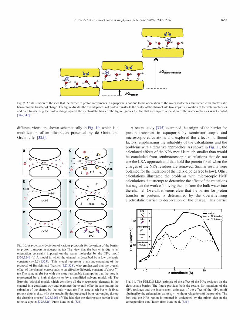
Fig. 9. An illustration of the idea that the barrier to proton movements in aquaporin is not due to the orientation of the water molecules, but rather to an electrostaticbarrier for the transfer of charge. The figure divides the overall process of proton transfer to the center of the channel into two steps: first rotation of the water moleculesand then transferring the proton charge against the electrostatic barrier. The figure ignores the fact that a complete orientation of the water molecules is not needed[346,347].
1667A. Warshel et al. / Biochimica et Biophysica Acta 1764 (2006) 1647–1676
different views are shown schematically in Fig. 10, which is amodification of an illustration presented by de Groot andGrubmuller [325].
Fig. 10. A schematic depiction of various proposals for the origin of the barrierto proton transport in aquaporin. (a) The view that the barrier is due to anorientation constraint imposed on the water molecules by the NPA motif[320,324]. (b) A model in which the channel is described by a low dielectricconstant (ε=2.5) [325]. (This model represents a misunderstanding of theproposal of Burykin and Warshel [327,328], who emphasized that the overalleffect of the channel corresponds to an effective dielectric constant of about 7.)(c) The same as (b) but with the more reasonable assumption that the pore isrepresented by a high dielectric or by a simplified solvent model. (d) TheBurykin–Warshel model, which considers all the electrostatic elements in thechannel in a consistent way and examines the overall effect in substituting thesolvation of the charge by the bulk water. (e) The same as (d) but with fixedprotein dipoles (i.e., with the protein dipoles prevented from rearranging duringthe charging process) [323,326]. (f) The idea that the electrostatic barrier is dueto helix dipoles [325,326]. From Kato et al. [335].
A recent study [335] examined the origin of the barrier forproton transport in aquaporin by semimacroscopic andmicroscopic calculations and explored the effect of differentfactors, emphasizing the reliability of the calculations and theproblems with alternative approaches. As shown in Fig. 11, thecalculated effects of the NPA motif is much smaller than wouldbe concluded from semimacroscopic calculations that do notuse the LRA approach and that hold the protein fixed when thecharges of the NPA residues are removed. Similar results wereobtained for the mutation of the helix dipoles (see below). Othercalculations illustrated the problems with microscopic PMFcalculations that attempt to determine the effect of the mutationsbut neglect the work of moving the ion from the bulk water intothe channel. Overall, it seems clear that the barrier for protontransfer in proteins is determined by the overwhelmingelectrostatic barrier to desolvation of the charge. This barrier
Fig. 11. The PDLD/S-LRA estimate of the effect of the NPA residues on theelectrostatic barrier. The figure provides both the results for mutations of theNPA residues and the inconsistent estimates of the effect of the NPA motifobtained by the calculations using εp=4 without relaxations of the proteins. Thefact that the NPA region is mutated is designated by the minus sign in thecorresponding box. Taken from Kato et al. [335].
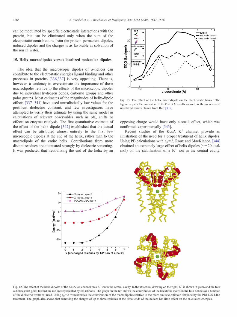
Fig. 13. The effect of the helix macrodipole on the electrostatic barrier. Thefigure depicts the consistent PDLD/S-LRA results as well as the inconsistentunrelaxed results. Taken from Ref. [335].
1668 A. Warshel et al. / Biochimica et Biophysica Acta 1764 (2006) 1647–1676
can be modulated by specific electrostatic interactions with theprotein, but can be eliminated only when the sum of theelectrostatic contributions from the protein permanent dipoles,induced dipoles and the charges is as favorable as solvation ofthe ion in water.
15. Helix macrodipoles versus localized molecular dipoles
The idea that the macroscopic dipoles of α-helices cancontribute to the electrostatic energies ligand binding and otherprocesses in proteins [336,337] is very appealing. There is,however, a tendency to overestimate the importance of thesemacrodipoles relative to the effects of the microscopic dipolesdue to individual hydrogen bonds, carbonyl groups and otherpolar groups. Most estimates of the magnitudes of helix-dipoleeffects [337–341] have used unrealistically low values for thepertinent dielectric constant, and few investigators haveattempted to verify their estimate by using the same model incalculations of relevant observables such as pKa shifts oreffects on enzyme catalysis. The first quantitative estimate ofthe effect of the helix dipole [342] established that the actualeffect can be attributed almost entirely to the first fewmicroscopic dipoles at the end of the helix, rather than to themacrodipole of the entire helix. Contributions from moredistant residues are attenuated strongly by dielectric screening.It was predicted that neutralizing the end of the helix by an
Fig. 12. The effect of the helix dipoles of the KcsA ion channel on a K+ ion in the centα-helices that point toward the ion are represented by red ribbons. The graph on the leof the dielectric treatment used. Using εp=2 overestimates the contribution of the matreatment. The graph also shows that removing the charges of up to three residues a
opposing charge would have only a small effect, which wasconfirmed experimentally [343].
Recent studies of the KcsA K+ channel provide anillustration of the need for a proper treatment of helix dipoles.Using PB calculations with εp=2, Roux and MacKinnon [344]obtained an extremely large effect of helix dipoles (∼−20 kcal/mol) on the stabilization of a K+ ion in the central cavity.
ral cavity. In the structural drawing on the right, K+ is shown in green and the fourft shows the contribution of the backbone atoms in the four helices as a functioncrodipoles relative to the more realistic estimate obtained by the PDLD/S-LRAt the distal ends of the helices has little effect on the calculated energies.
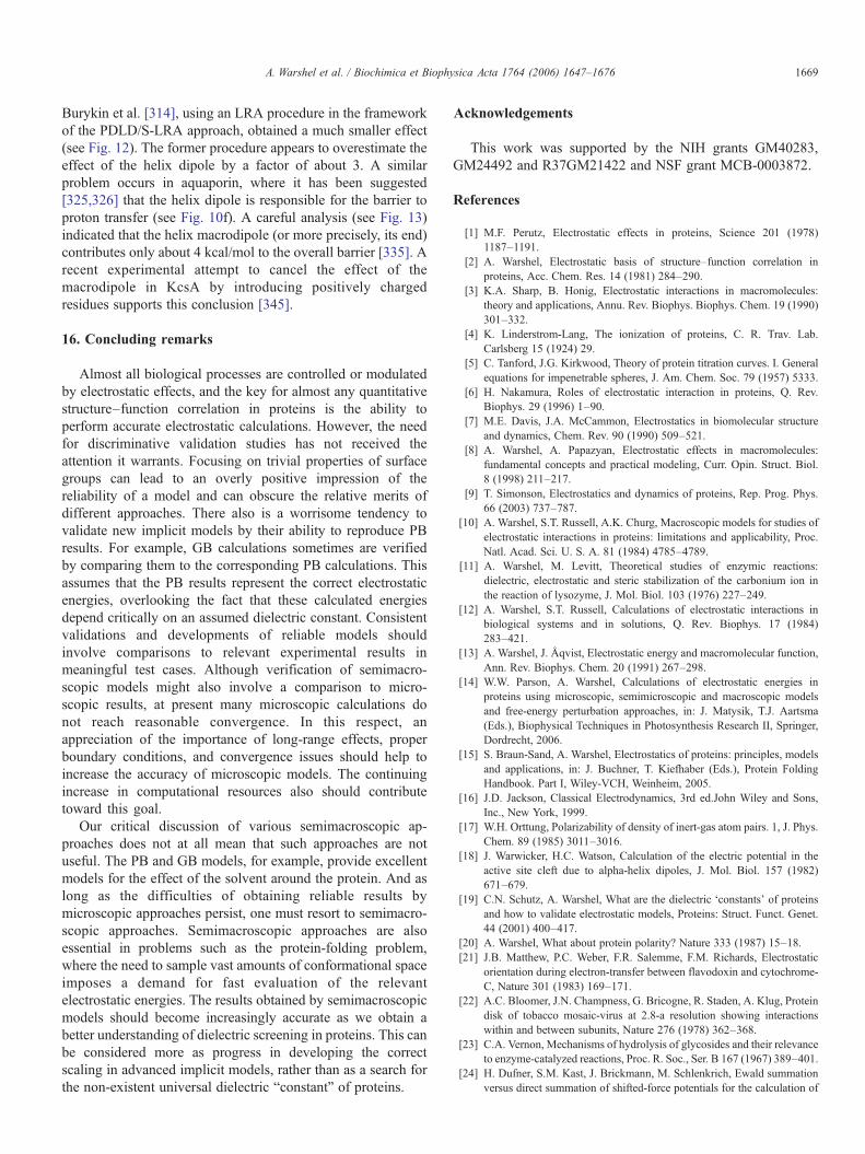
1669A. Warshel et al. / Biochimica et Biophysica Acta 1764 (2006) 1647–1676
Burykin et al. [314], using an LRA procedure in the frameworkof the PDLD/S-LRA approach, obtained a much smaller effect(see Fig. 12). The former procedure appears to overestimate theeffect of the helix dipole by a factor of about 3. A similarproblem occurs in aquaporin, where it has been suggested[325,326] that the helix dipole is responsible for the barrier toproton transfer (see Fig. 10f). A careful analysis (see Fig. 13)indicated that the helix macrodipole (or more precisely, its end)contributes only about 4 kcal/mol to the overall barrier [335]. Arecent experimental attempt to cancel the effect of themacrodipole in KcsA by introducing positively chargedresidues supports this conclusion [345].
16. Concluding remarks
Almost all biological processes are controlled or modulatedby electrostatic effects, and the key for almost any quantitativestructure–function correlation in proteins is the ability toperform accurate electrostatic calculations. However, the needfor discriminative validation studies has not received theattention it warrants. Focusing on trivial properties of surfacegroups can lead to an overly positive impression of thereliability of a model and can obscure the relative merits ofdifferent approaches. There also is a worrisome tendency tovalidate new implicit models by their ability to reproduce PBresults. For example, GB calculations sometimes are verifiedby comparing them to the corresponding PB calculations. Thisassumes that the PB results represent the correct electrostaticenergies, overlooking the fact that these calculated energiesdepend critically on an assumed dielectric constant. Consistentvalidations and developments of reliable models shouldinvolve comparisons to relevant experimental results inmeaningful test cases. Although verification of semimacro-scopic models might also involve a comparison to micro-scopic results, at present many microscopic calculations donot reach reasonable convergence. In this respect, anappreciation of the importance of long-range effects, properboundary conditions, and convergence issues should help toincrease the accuracy of microscopic models. The continuingincrease in computational resources also should contributetoward this goal.
Our critical discussion of various semimacroscopic ap-proaches does not at all mean that such approaches are notuseful. The PB and GB models, for example, provide excellentmodels for the effect of the solvent around the protein. And aslong as the difficulties of obtaining reliable results bymicroscopic approaches persist, one must resort to semimacro-scopic approaches. Semimacroscopic approaches are alsoessential in problems such as the protein-folding problem,where the need to sample vast amounts of conformational spaceimposes a demand for fast evaluation of the relevantelectrostatic energies. The results obtained by semimacroscopicmodels should become increasingly accurate as we obtain abetter understanding of dielectric screening in proteins. This canbe considered more as progress in developing the correctscaling in advanced implicit models, rather than as a search forthe non-existent universal dielectric “constant” of proteins.
Acknowledgements
This work was supported by the NIH grants GM40283,GM24492 and R37GM21422 and NSF grant MCB-0003872.
References
[1] M.F. Perutz, Electrostatic effects in proteins, Science 201 (1978)1187–1191.
[2] A. Warshel, Electrostatic basis of structure–function correlation inproteins, Acc. Chem. Res. 14 (1981) 284–290.
[3] K.A. Sharp, B. Honig, Electrostatic interactions in macromolecules:theory and applications, Annu. Rev. Biophys. Biophys. Chem. 19 (1990)301–332.
[4] K. Linderstrom-Lang, The ionization of proteins, C. R. Trav. Lab.Carlsberg 15 (1924) 29.
[5] C. Tanford, J.G. Kirkwood, Theory of protein titration curves. I. Generalequations for impenetrable spheres, J. Am. Chem. Soc. 79 (1957) 5333.
[6] H. Nakamura, Roles of electrostatic interaction in proteins, Q. Rev.Biophys. 29 (1996) 1–90.
[7] M.E. Davis, J.A. McCammon, Electrostatics in biomolecular structureand dynamics, Chem. Rev. 90 (1990) 509–521.
[8] A. Warshel, A. Papazyan, Electrostatic effects in macromolecules:fundamental concepts and practical modeling, Curr. Opin. Struct. Biol.8 (1998) 211–217.
[9] T. Simonson, Electrostatics and dynamics of proteins, Rep. Prog. Phys.66 (2003) 737–787.
[10] A. Warshel, S.T. Russell, A.K. Churg, Macroscopic models for studies ofelectrostatic interactions in proteins: limitations and applicability, Proc.Natl. Acad. Sci. U. S. A. 81 (1984) 4785–4789.
[11] A. Warshel, M. Levitt, Theoretical studies of enzymic reactions:dielectric, electrostatic and steric stabilization of the carbonium ion inthe reaction of lysozyme, J. Mol. Biol. 103 (1976) 227–249.
[12] A. Warshel, S.T. Russell, Calculations of electrostatic interactions inbiological systems and in solutions, Q. Rev. Biophys. 17 (1984)283–421.
[13] A. Warshel, J. Åqvist, Electrostatic energy and macromolecular function,Ann. Rev. Biophys. Chem. 20 (1991) 267–298.
[14] W.W. Parson, A. Warshel, Calculations of electrostatic energies inproteins using microscopic, semimicroscopic and macroscopic modelsand free-energy perturbation approaches, in: J. Matysik, T.J. Aartsma(Eds.), Biophysical Techniques in Photosynthesis Research II, Springer,Dordrecht, 2006.
[15] S. Braun-Sand, A. Warshel, Electrostatics of proteins: principles, modelsand applications, in: J. Buchner, T. Kiefhaber (Eds.), Protein FoldingHandbook. Part I, Wiley-VCH, Weinheim, 2005.
[16] J.D. Jackson, Classical Electrodynamics, 3rd ed.John Wiley and Sons,Inc., New York, 1999.
[17] W.H. Orttung, Polarizability of density of inert-gas atom pairs. 1, J. Phys.Chem. 89 (1985) 3011–3016.
[18] J. Warwicker, H.C. Watson, Calculation of the electric potential in theactive site cleft due to alpha-helix dipoles, J. Mol. Biol. 157 (1982)671–679.
[19] C.N. Schutz, A. Warshel, What are the dielectric ‘constants’ of proteinsand how to validate electrostatic models, Proteins: Struct. Funct. Genet.44 (2001) 400–417.
[20] A. Warshel, What about protein polarity? Nature 333 (1987) 15–18.[21] J.B. Matthew, P.C. Weber, F.R. Salemme, F.M. Richards, Electrostatic
orientation during electron-transfer between flavodoxin and cytochrome-C, Nature 301 (1983) 169–171.
[22] A.C. Bloomer, J.N. Champness, G. Bricogne, R. Staden, A. Klug, Proteindisk of tobacco mosaic-virus at 2.8-a resolution showing interactionswithin and between subunits, Nature 276 (1978) 362–368.
[23] C.A. Vernon, Mechanisms of hydrolysis of glycosides and their relevanceto enzyme-catalyzed reactions, Proc. R. Soc., Ser. B 167 (1967) 389–401.
[24] H. Dufner, S.M. Kast, J. Brickmann, M. Schlenkrich, Ewald summationversus direct summation of shifted-force potentials for the calculation of
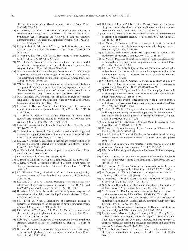
1670 A. Warshel et al. / Biochimica et Biophysica Acta 1764 (2006) 1647–1676
electrostatic interactions in solids—A quantitative study, J. Comp. Chem.18 (1997) 660–675.
[25] A. Warshel, Z.T. Chu, Calculations of solvation free energies inchemistry and biology, in: C.J. Cramer, D.G. Truhlar (Eds.), ACSSymposium Series: Structure and Reactivity in Aqueous Solution.Characterization of Chemical and Biological Systems, ACS, Washing-ton, DC, 1994, pp. 72–93.
[26] F. Figueirido, G.S. Del Buono, R.M. Levy, On the finite size correctionsto the free energy of ionic hydration, J. Phys. Chem., B 101 (1997)5622–5623.
[27] G. Hummer, L.R. Pratt, A.E. Garcia, Free energy of ionic hydration,J. Phys. Chem. 100 (1996) 1206–1215.
[28] Y.Y. Sham, A. Warshel, The surface constrained all atom modelprovides size ¦independent results in calculations of hydration freeenergies, J. Chem. Phys. 109 (1998) 7940–7944.
[29] M.A. Kastenholz, P.H. Hünenberger, Computation of methodology-independent ionic solvation free energies from molecular simulations: I.The electrostatic potential in molecular liquids, J. Chem. Phys. 124(2006) 124106/1–124106–27.
[30] Y.N. Vorobjev, J. Hermans, A critical analysis of methods of calculationof a potential in simulated polar liquids: strong arguments in favor of"Molecule-Based" summation and of vacuum boundary conditions inewald summation, J. Phys. Chem., B 103 (1999) 10234–10242.
[31] M.A. Villarreal, G.G. Montich, On the Ewald artifacts in computersimulations. The case of the octaalanine peptide with charged termini,J. Biomol. Struct. Dyn. 23 (2005) 135.
[32] J. Aqvist, T. Hansson, Analysis of electrostatic potential truncationschemes in simulations of polar solvents, J. Phys. Chem., B 102 (1998)3837–3840.
[33] Y.Y. Sham, A. Warshel, The surface constrained all atom modelprovides size independent results in calculations of hydration freeenergies, J. Chem. Phys. 109 (1998) 7940–7944.
[34] J. Aqvist, Comment on transferability of ion models, J. Phys. Chem. 98(1994) 8253–8255.
[35] S. Kuwajima, A. Warshel, The extended ewald method: a generaltreatment of long-range electrostatic interactions in microscopic simula-tions, J. Chem. Phys. 89 (1988) 3751–3759.
[36] F.S. Lee, A. Warshel, A local reaction field method for fast evaluation oflong-range electrostatic interactions in molecular simulations, J. Chem.Phys. 97 (1992) 3100–3107.
[37] A. Warshel, Calculations of chemical processes in solutions, J. Phys.Chem. 83 (1979) 1640–1650.
[38] S.A. Adelman, Adv. Chem. Phys. 53 (1983) 61.[39] A. Brunger, C.L.B. III, M. Karplus, Chem. Phys. Lett. 105 (1984) 495.[40] G. King, A. Warshel, A surface constrained all-atom solvent model for
effective simulations of polar solutions, J. Chem. Phys. 91 (1989)3647–3661.
[41] J.G. Kirkwood, Theory of solutions of molecules containing widelyserparated charges with special application to zwitterions, J. Chem. Phys.2 (1934) 351.
[42] F.S. Lee, Z.T. Chu, A. Warshel, Microscopic and semimicroscopiccalculations of electrostatic energies in proteins by the POLARIS andENZYMIX programs, J. Comp. Chem. 14 (1993) 161–185.
[43] H. Alper, R.M. Levy, Dielectric and thermodynamic response ofgeneralized reaction field model for liquid state simulations, J. Chem.Phys. 99 (1993) 9847.
[44] S.T. Russell, A. Warshel, Calculations of electrostatic energies inproteins; the energetics of ionized groups in bovine pancreatic trypsininhibitor, J. Mol. Biol. 185 (1985) 389–404.
[45] R.G. Alden, W.W. Parson, Z.T. Chu, A. Warshel, Calculations ofelectrostatic energies in photosynthetic reaction centers, J. Am. Chem.Soc. 117 (1995) 12284–12298.
[46] J. Åqvist, A. Warshel, Energetics of ion permeation through membranechannels. Solvation of Na+ by Gramicidin A, Biophys. J. 56 (1989)171–182.
[47] B. Roux, M. Karplus, Ion transport in the gramicidin channel: free energyof the solvated right-handed dimer in a model membrane, J. Am. Chem.Soc. 115 (1993) 3250–3260.
[48] H.A. Stern, F. Rittner, B.J. Berne, R.A. Friesner, Combined fluctuatingcharge and polarizable dipole models: application to a five-site waterpotential function, J. Chem. Phys. 115 (2001) 2237–2251.
[49] P.Y. Ren, J.W. Ponder, Consistent treatment of inter- and intramolecularpolarization in molecular mechanics calculations, J. Comp. Chem. 23(2002) 1497–1506.
[50] A. Warshel, F. Sussman, G. King, Free energy of charges in solvatedproteins: microscopic calculations using a reversible charging process,Biochemistry 25 (1986) 8368–8372.
[51] P. Kollman, Free energy calculations: applications to chemical andbiochemical phenomena, Chem. Rev. 93 (1993) 2395–2417.
[52] A. Warshel, Dynamics of reactions in polar solvents. semiclassical tra-jectory studies of electron-transfer and proton-transfer reactions, J. Phys.Chem. 86 (1982) 2218–2224.
[53] F.S. Lee, Z.T. Chu, M.B. Bolger, A. Warshel, Calculations of antibody-antigen interactions: microscopic and semi-microscopic evaluation of thefree energies of binding of phosphorylcholine analogs to McPC603, Prot.Eng. 5 (1992) 215–228.
[54] Y.Y. Sham, Z.T. Chu, A. Warshel, Consistent calculations of pKa's ofionizable residues in proteins: semi-microscopic and macroscopicapproaches, J. Phys. Chem., B 101 (1997) 4458–4472.
[55] G.S. Del Buono, F.E. Figueirido, R.M. Levy, Intrinsic pKas of ionizableresidues in proteins: an explicit solvent calculation for lysozyme, Protein:Struct. Funct. Genet. 20 (1994) 85–97.
[56] M. Saito, Molecular dynamics free-energy study of a protein in solutionwith all degrees of freedom and long-range Coulomb interactions, J. Phys.Chem. 99 (1995) 17043–17048.
[57] M. Kato, A. Warshel, Through the channel and around the channel:validating and comparing microscopic approaches for the evaluation offree energy profiles for ion penetration through ion channels, J. Phys.Chem. B 109 (2005) 19516–19522.
[58] A.M. Ferrenberg, R.H. Swendsen, Optimized Monte Carlo data analysis,Phys. Rev. Lett. 63 (1989) 1195–1198.
[59] C. Jarzynski, Nonequilibrium equality for free energy differences, Phys.Rev. Lett. 78 (1997) 2690–2693.
[60] I. Andricioaei, A.R. Dinner, M. Karplus, Self-guided enhanced samplingmethods for thermodynamic averages, J. Chem. Phys. 118 (2003)1074–1084.
[61] B. Roux, The calculation of the potential of mean force using computersimulations, Comput. Phys. Commun. 91 (1995) 275–282.
[62] E.M. Purcell, Electricity and Magnetism, McGraw-Hill Book Company,1965.
[63] Y. Liu, T. Ichiye, The static dielectric-constant of the soft sticky dipolemodel of liquid water—Monte Carlo simulation, Chem. Phys. Lett. 256(1996) 334–340.
[64] R.D. Coalson, A. Duncan, Statistical mechanics of a multipolar gas: alattice field theory approach, J. Phys. Chem. 100 (1996) 2612–2620.
[65] A. Papazyan, A. Warshel, Continuum and dipole-lattice models ofsolvation, J. Phys. Chem. 101 (1997) 11254–11264.
[66] A. Papazyan, A. Warshel, Effect of solvent discreteness on solvation,J. Phys. Chem., B 102 (1998) 5248–5357.
[67] N.K. Rogers, The modeling of electrostatic interactions in the function ofglobular proteins, Prog. Biophys. Mol. Biol. 48 (1986) 37–66.
[68] M. Schaefer, M. Karplus, A comprehensive analytical treatment ofcontinuum electrostatics, J. Phys. Chem. 100 (1996) 1578–1599.
[69] T. HaDuong, S. Phan, M. Marchi, D. Borgis, Electrostatics on particles:phenomenological and orientational density functional theory approach,J. Chem. Phys. 117 (2002) 541–556.
[70] A. Warshel, G. Naray-Szabo, F. Sussman, J.-K. Hwang, How do serineproteases really work? Biochemistry 28 (1989) 3629–3673.
[71] P.A. Kollman, I. Massova, C. Reyes, B. Kuhn, S. Huo, L. Chong, M. Lee,T. Lee, Y. Duan, W. Wang, O. Donini, P. Cieplak, J. Srinivasan, D.A.Case, T.E. Cheatham III, Calculating structures and free energies ofcomplex molecules: combining molecular mechanics and continuummodels, Acc. Chem. Res. 33 (2000) 889–897.
[72] M.K. Gilson, A. Rashin, R. Fine, B. Honig, On the calculation ofelectrostatic interactions in proteins, J. Mol. Bio. 184 (1985)503–516.
1671A. Warshel et al. / Biochimica et Biophysica Acta 1764 (2006) 1647–1676
[73] M. Gilson, K.A. Sharp, B. Honig, Calculatiing the electrostaic potentialof molecules in solutions, J. Comp. Chem. 9 (1987) 327–335.
[74] A. Warshel, A. Papazyan, I. Muegge, Microscopic and semimicroscopicredox calculations: what can and cannot be learned from continuummodels, JBIC 2 (1997) 143–152.
[75] M. Gilson, B. Honig, Calculation of the total electrostatic energy of amacromolecular system. Solvation energies, binding energies andconformational analysis. Proteins: Struct. Funct. Genet. 4 (1988) 7–18.
[76] K.A. Sharp, B. Honig, Calculating total electrostatic energies with thenonlinear Poisson–Boltzmann equation, J. Phys. Chem. 94 (1990)7684–7692.
[77] A. Nicholls, B. Honig, A rapid finite algorithm utilizing successive over-relaxation to solve Poisson–Boltzmann equation, J. Comp. Chem. 12(1991) 435–445.
[78] B. Honig, A. Nicholls, Classical electrostatics in biology and chemistry,Science 268 (1995) 1144–1149.
[79] M. Gunner, A. Nichols, B. Honig, Electrostatic potentials in Rhodop-seudomonas viridis reaction centers: implications for the driving forceand directionality of electron transfer, J. Phys. Chem. 100 (1996)4277–4291.
[80] C.R.D. Lancaster, H. Michel, B. Honig, M.R. Gunner, Calculatedcoupling of electron and proton transfer in the photosynthetic reactioncenter of Rhodopseudomonas viridis, Biophys. J. 70 (1996) 2469–2492.
[81] E. Alexov, M.R. Gunner, Incorporating protein conformational flexibilityinto the calculation of pH-dependent protein properties, Biophys. J. 72(1997) 2075.
[82] B. Rabenstein, G.M. Ullmann, E.W. Knapp, Energetics of electron-transfer and protonation reactions of the quinones in the photosyntheticreaction-center of Rhodopseudomonas viridis, Biochemistry 37 (1998)2488–2495.
[83] E.G. Alexov, M.R. Gunner, Calculated protein and proton motionscoupled to electron transfer: electron transfer from QA- to QB in bacterialphotosynthetic reaction centers, Biochemistry 38 (1999) 8253–8270.
[84] J.E. Nielsen, K.V. Andersen, B. Honig, R.V. Hooft, G. Klebe, G. Vriend,R.C. Wade, Improving macromolecular electrostatics calculations,Protein Eng. 12 (1999) 657–662.
[85] G.M. Ullmann, E. Knapp, Electrostatic models for computing protonationand redox equilibria in proteins, Eur. Biophys. J. Biophys. Lett. 28 (1999)533–551.
[86] M. Gunner, E.G. Alexov, A pragmatic approach to structure basedcalculation of coupled proton and electron transfer in proteins, Biochim.Biophys. Acta 1485 (2000) 63–87.
[87] R.E. Georgescu, E.G. Alexov, M.R. Gunner, Combining conformationalflexibility and continuum electrostatics for calculating pK(a)s in proteins,Biophys. J. 83 (2002) 1731–1748.
[88] W. Rocchia, S. Sridharan, A. Nicholls, E.G. Alexov, A. Chiabrera, B.Honig, Rapid grid-based construction of the molecular surface and theuse of induced surface charge to calcalute reaction field energies:applications to the molecular systems and geometric objects, J. Comp.Chem. 23 (2002) 128–137.
[89] P. Voigt, E. Knapp, Tuning heme redox potentials in the cytochrome csubunit of photosynthetic reaction centers, J. Biol. Chem. 278 (2003)51993–52001.
[90] J. Kim, J. Mao, M. Gunner, Are acidic and basic groups in buried proteinspredicted to be ionized? J. Mol. Biol. 348 (2005) 1283–1298.
[91] W. Rocchia, E.G. Alexov, B. Honig, Extending the applicability of thenonlinear Poisson–Boltzmann equation: multiple dielectric constants andmultivalent ions, J. Phys. Chem., B 105 (2001) 6507–6514.
[92] J.D. Madura, J.M. Briggs, R.C. Wade, M.E. Davis, B.A. Lutis, J.A.McCammon, Electrostatics and diffusion of molecules in solution:simulations with the University of Houston Brownian DynamicsProgram, Comput. Phys. Commun. 91 (1995) 57–95.
[93] N.A. Baker, D. Sept, S. Joseph, M.J. Holst, J.A. McCammon,Electrostatics of manosystems: application to microtubules and theribosome, Proc. Natl. Acad. Sci. U. S. A. 98 (2001) 10037–10041.
[94] E.L. Mehler, G. Eichele, Electrostatic effects in water-accessible regionsof proteins, Biochemistry 23 (1984) 3887–3891.
[95] B.E. Hingerty, R.H. Richie, T.L. Ferrell, J.E. Turner, Dielectric effects in
biopolymers. The theory of ionic saturation revisited, Biopolymers 24(1985) 427–439.
[96] E.L. Mehler, F. Guarnieri, A self-consistent, microscopic modulatedscreened Coulomb potential approximation to calculate pH-dependentelectrostatic effects in proteins, Biophys. J. 75 (1999) 3–22.
[97] J. Guenot, P. Kollman, Molecular dynamics studies of a DNA-bindingprotein. 2. An evaluation of implicit and explicit solvent models for themolecular-dynamics simulation of the Escherichia coli Trp repressor,Protein Sci. 1 (1992) 1185–1205.
[98] J. Guenot, P. Collman, Conformational and energetic effects oftruncating nonbonded interactions in an aqueous protein dynamicssimulation, J. Comput. Chem. 14 (1993) 295–311.
[99] E.T. Johnson, W.W. Parson, Electrostatic Interactions in an IntegralMembrane Protein, Biochemistry 41 (2002) 6483–6494.
[100] R. Constanciel, R. Contreras, Self-consistent field theory of solventeffects representation by continuum models: introduction of dessolvationcontribution, Theor. Chim. Acta 65 (1984) 1–11.
[101] T. Kozaki, K. Morihashi, O. Kikuchi, An MNDO effective charge modelstudy of the solvent effect. The internal rotation about partial doublebonds and the nitrogen inversion in amine, J. Mol. Struct. 168 (1988)265–277.
[102] T. Kozaki, K. Morihashi, O. Kikuchi, MNDO effective charge modelstudy of solvent effect on the potential energy surface of the SN2 reaction,J. Am. Chem. Soc. 111 (1989) 1547–1552.
[103] W.C. Still, A. Tempczyk, R.C. Hawley, T. Hendrickson, Semianalyticaltreatment of solvation for molecular mechanics and dynamics, J. Am.Chem. Soc. 112 (1990) 6127–6129.
[104] G.D. Hawkins, C.J. Cramer, D.G. Truhlar, Parametrized models ofaqueous free-energies of solvation based on pairwise descreening ofsolute atomic charges from a dielectric medium, J. Phys. Chem. 100(1996) 19824–19839.
[105] D. Qiu, P.S. Shenkin, F.P. Hollinger, W.C. Still, The GB/SA continuummodel for solvation. A fast analytical method for the calculation ofapproximate Born radii, J. Phys. Chem. 101 (1997) 3005–3014.
[106] A. Ghosh, C.S. Rapp, R.A. Friesner, Generalized Born Model based on asurface integral formulation, J. Phys. Chem., B 102 (1998) 10983–10990.
[107] D. Bashford, D.A. Case, Generalized born models of macromolecularsolvation effects, Annu. Rev. Phys. Chem. 51 (2000) 129–152.
[108] A. Onufriev, D.A. Case, D. Bashford, Effective Born radii in the gen-eralized Born approximation: the importance of being perfect, J. Comp.Chem. 23 (2002) 1297–1304.
[109] E. Gallicchio, R.M. Levy, AGBNP: an analytic implicit solvent modelsuitable for molecular dynamics simulations and high-resolutionmodeling, J. Comp. Chem. 25 (2004) 479–499.
[110] H. Fan, A.E. Mark, J. Zhu, B. Honig, Comparative study of generalizedBorn models: protein dynamics, Proc. Natl. Acad. Sci. U. S. A. 102(2005) 6760–6764.
[111] J. Zhu, E.G. Alexov, B. Honig, Comparative study of generalized Bornmodels: born radii and peptide folding, J. Phys. Chem., B 109 (2005)3008–3022.
[112] M. Born, Volumen and Hydrationswärme der Ionen, Z. Phys. 1 (1920)45–47.
[113] D. Borgis, N. Levy, M. Marchi, Computing the electrostatic free-energy of complex molecules: the variational Coulomb field ap-proximation, J. Chem. Phys. 119 (2003) 3516–3528.
[114] G. King, F.S. Lee, A. Warshel, Microscopic simulations of macroscopicdielectric constants of solvated proteins, J. Chem. Phys. 95 (1991)4366–4377.
[115] Y.Y. Sham, I. Muegge, A. Warshel, The effect of protein relaxation oncharge–charge interactions and dielectric constants of proteins, Biophys.J. 74 (1998) 1744–1753.
[116] I. Muegge, T. Schweins, R. Langen, A. Warshel, Electrostatic control ofGTP and GDP binding in the oncoprotein p21 ras, Structure 4 (1996)475–489.
[117] T. Simonson, C.L. Brooks, Charge screening and the dielectric constantof proteins: insights from molecular dynamics, J. Am. Chem. Soc. 118(1996) 8452–8458.
[118] P.E. Smith, R.M. Brunne, A.E. Mark, Dielectric properties of trypsin
1672 A. Warshel et al. / Biochimica et Biophysica Acta 1764 (2006) 1647–1676
inhibitor and lysozyme calculated from molecular dynamics simulations,J. Phys. Chem. 97 (1993) 2009–2014.
[119] T. Simonson, D. Perahia, A.T. Brunger, Microscopic theory of thedielectric properties of proteins, Biophys. J. 59 (1991) 670–690.
[120] T. Simonson, D. Perahia, Internal and Interfacial Dielectric Properties ofcytochrome c from molecular dynamics simulations in aqueous solution,Proc. Natl. Acad. Sci. U. S. A. 92 (1995) 1082–1086.
[121] I. Muegge, P.X. Qi, A.J. Wand, Z.T. Chu, A. Warshel, The reorganizationenergy of cytochrome c revisited, J. Phys. Chem., B 101 (1997) 825–836.
[122] J. Antosiewicz, J.A. McCammon, M.K. Gilson, Prediction of pH-dependent properties of proteins, J. Mol. Biol. 238 (1994) 415–436.
[123] B.E. Cohen, T.B. McAnaney, E.S. Park, Y.N. Jan, S.G. Boxer, L.Y. Jan,Probing protein electrostatics with a synthetic fluorescent amino acid,Science 296 (2002) 1700–1703.
[124] S. Borman, Fluorescent probe for proteins: nonnatural amino acid used toinvestigate electrostatic properties within proteins, C&EN 80 (2002) 30.
[125] S. Harvey, P. Hoekstra, Dielectric relaxation spectra of water adsorbed onlysozyme, J. Phys. Chem. 76 (1972) 2987–2994.
[126] M. Gilson, B. Honig, The dielectric constant of a folded protein,Biopolymers 25 (1986) 2097–2119.
[127] J.W. Pitera, M. Falta, W.F.v. Gunsteren, Dielectric properties of proteinsfrom simulation: the effects of solvent, ligands, pH, and temperature,Biophys. J. 80 (2001) 2546–2555.
[128] B. Garcia-Moreno, J.J. Dwyer, A.G. Gittis, E.E. Lattman, D.S. Spencer,W.E. Stites, Experimental measurement of the effective dielectric in thehydrophobic core of a protein, Biophys. Chem. 64 (1997) 211–224.
[129] M. Kato, A. Warshel, Using a charging coordinate in studies of ionizationinduced partial unfolding, J. Phys. Chem., B 110 (2006) 11566–11570.
R. Penfold, J. Warwicker, B. Jonsson, Electrostatic models for calcium bindingproteins, J. Phys. Chem., B 108 (1998) 8599–8610.[131] P.J.W. Debye, L. Pauling, The inter-ionic attraction theory of ionized
solutes. IV. The influence of variation of dielectric constant on thelimiting law for small concentrations, J. Am. Chem. Soc. 47 (1925)2129–2134.
[132] T.J. Webb, The free energy of hydration of ions and the electrostriction ofthe solvent, J. Am. Chem. Soc. 48 (1926) 2589–2603.
[133] T.L. Hill, The electrostatic contribution to hindered rotation in certainions and dipolar ions in solution iii, J. Chem. Phys. 12 (1944)56–61.
[134] V.H. Teixeira, C.A. Cunha, M. Machuqueiro, A.S.F. Oliveira, B.L. Victor,C.M. Soares, A.A. Baptista, On the use of different dielectric constantsfor computing individual and pairwise terms in Poisson–Boltzmannstudies of protein ionization equilibrium, J. Phys. Chem., B 109 (2005)14691–14706.
[135] B. Honig, K. Sharp, R. Sampogna, M.R. Gunner, A.S. Yang, On thecalculation of pKas in proteins, Proteins: Struct. Funct. Genet. 15 (1993)252–265.
[136] A. Warshel, Computer modeling of chemical reactions in enzymes andsolutions, John Wiley and Sons, New York, 1991.
[137] E.M. Kosower, Introduction to Physical Organic Chemistry, Wiley, NewYork, 1968.
[138] N. Mataga, T. Kubota, Molecular Interactions and Electronic Spectra, M.Dekker, New York, 1970.
[139] A. Pullman, B. Pullman, New paths in the molecular orbital approach tosolvation in biological molecules, Q. Rev. Biol. 7 (1975) 506–566.
[140] W.L. Jorgensen, Ab initio molecular orbital study of the geometricproperties and protonation of alkyl chloride, J. Am. Chem. Soc. 100(1978) 1057–1061.
[141] J. Tomasi, B. Mennucci, R. Cammi, M. Cossi, Quantum mechanicalmodels for reactions in solution, in: G. Naray-Szabo, A. Warshel (Eds.),Computational Approaches to Biochemical Reactivity, Kluwer AcademicPublishers, Dordrecht, 1997.
[142] C.J. Cramer, D.G. Truhlar, General parameterized SCF model for freeenergies of solvation in aqueous solution, J. Am. Chem. Soc. 113 (1991)8305–8311.
[143] B. Honig, K. Sharp, A.-S. Yang, Macroscopic models of aqueoussolutions: biological and chemical applications, J. Phys. Chem. 97 (1993)1101–1109.
[144] J. Åqvist, Ion–water interaction potentials derived from free energyperturbation simulations, J. Chem. Phys. 94 (1990) 8021–8024.
[145] F. Figueirido, G.S. Del Buono, R.M. Levy, On finite-size effects incomputer simulations using the Ewald potential, J. Chem. Phys. 103(1995) 6133–6142.
[146] J. Åqvist, T. Hansson, On the validity of electrostatic linear response inpolar solvents, J. Phys. Chem. 100 (1996) 9512–9521.
[147] M. Orozco, F.J. Luque, Generalized linear-response approximation indiscrete methods, Chem. Phys. Lett. 265 (1997) 473–480.
[148] J. Florián, A. Warshel, Langevin dipoles model for ab initiocalculations of chemical processes in solution: parametrization andapplication to hydration free energies of neutral and ionic solutes, andconformational analysis in aqueous solution, J. Phys. Chem., B 101(1997) 5583–5595.
[149] N.O.J. Malcolm, J.J.W. McDouall, Assessment of the Langevin dipolessolvation model for Hartree–Fock wavefunctions, J. Mol. Struct.(Theochem) 336 (1996) 1–9.
[150] B. Marten, K. Kim, C. Cortis, R.A. Friesner, R.B. Murphy, M.N.Ringnalda, D. Sitkoff, B. Honig, New model for calculation of solvationfree energies: correction of self-consistent reaction field continuumdielectric theory for short-range hydrogen bonding effects, J. Phys.Chem. 100 (1996) 11775–11788.
[151] M. Fothergill, M.F. Goodman, J. Petruska, A. Warshel, Structure–energy analysis of the role of metal ions in phosphodiester bondhydrolysis by DNA polymerase I, J. Am. Chem. Soc. 117 (1995)11619–11627.
[152] J. Åqvist, M. Fothergill, Computer simulation of the triosephosphateisomerase catalyzed reaction, J. Biol. Chem. 271 (1996) 10010–10016.
[153] C.N. Schutz, A. Warshel, The low barrier hydrogen bond (LBHB)proposal revisited: the case of the Asp, His pair in serine proteases,Proteins 55 (2004) 711–723.
[154] V. Luzhkov, J. Aqvist, K+/Na+ selectivity of the KcsA potassium channelfrom microscopic free energy perturbation calculations, Biochim.Biophys. Acta 1548 (2001) 194–202.
[155] A. Burykin, C.N. Schutz, J. Villa, A. Warshel, Simulations of ion currentin realistic models of ion channels: the KcsA potassium channel, Proteins:Struct. Funct. Genet. 47 (2002) 265–280.
[156] V.B. Luzhkov, J. Aqvist, A computational study of ion binding andprotonation states in the KcsA potassium channel, Biochim. Biophys.Acta 1481 (2000) 360–370.
[157] P. Beroza, D.R. Fredkin, Calculation of amino acid pK(a)s in aprotein from a continuum electrostatic model: method and sensitivityanalysis, J. Comp. Chem. 17 (1996) 1229–1244.
[158] A. Koumanov, H. Ruterjans, A. Karshikoff, Continuum electrostaticanalysis of irregular ionization and proton allocation in proteins, Proteins:Struct. Funct. Genet. 46 (2002) 85–96.
[159] T.J. You, D. Bashford, Conformation and hydrogen ion titration ofproteins: a continuum electrostatic model with conformational flexibility,Biophys. J. 69 (1995) 1721–1733.
[160] H.W.T. van Vlijmen, M. Schaefer, M. Karplus, Improving the accuracy ofprotein pK(a) calculations: conformational averaging versus the averagestructure, Proteins 33 (1998) 145–158.
[161] H.X. Zhou, M. Vijayakumar, Modeling of protein conformationalfluctuations in pK(a) predictions, J. Mol. Biol. 267 (1997) 1002–1011.
[162] J.J. Wendoloski, J.B. Matthew, Molecular-dynamics effects on proteinelectrostatics, Proteins 5 (1989) 313–321.
[163] J.J. Dwyer, A.G. Gittis, D.A. Karp, E.E. Lattman, D.S. Spencer, W.E.Stites, E.B. Garcia-Moreno, High apparent dielectric constants in theinterior of a protein reflect water penetration [In Process Citation],Biophys. J. 79 (2000) 1610–1620.
[164] C.A. Fitch, D.A. Karp, K.K. Lee, W.E. Stites, E.E. Lattman, B. Garcia-Moreno, Experimental pK(a) values of buried residues: analysis withcontinuum methods and role of water penetration, Biophys. J. 82 (2002)3289–3304.
[165] S.W. Englander, M.M.G. Krishna, Hydrogen exchange, Nature Struct.Biol. 8 (2001) 1.
[166] R.J. Kassner, Effects of non polar environments on the redox potentials ofheme complexes, Proc. Natl. Acad. Sci. 69 (1972) 2263.
1673A. Warshel et al. / Biochimica et Biophysica Acta 1764 (2006) 1647–1676
[167] R.J. Kassner, A theoretical model for the effects of local nonpolar hemeenvironments on the redox potentials in cytochromes, J. Am Chem. Soc.95 (1973) 2674.
[168] H.-X. Zhou, Control of reduction potential by protein matrix: lesson froma spherical protein model, J. Biol. Inorg. Chem. 2 (1997) 109–113.
[169] I. Bertini, G. Gori-Savellini, C. Luchinat, Are unit charges alwaysnegligible? J. Biol. Inorg. Chem. 2 (1997) 114–118.
[170] A.G. Mauk, G.R. Moore, Control of metalloprotein redox potentials:what does site-directed mutagenesis of hemoproteins tell us? J. Biol.Inorg. Chem. 2 (1997) 119–125.
[171] M.R. Gunner, E. Alexov, E. Torres, S. Lipovaca, The importance of theprotein in controlling the electrochemistry of heme metalloproteins:methods of calculation and analysis, J. Biol. Inorg. Chem. 2 (1997)126–134.
[172] G. Naray-Szabo, Electrostatic modulation of electron transfer in theactive site of heme peroxidases, J. Biol. Inorg. Chem. 2 (1997) 135–138.
[173] F.A. Armstrong, Evaluations of reduction potential data in relation tocoupling, kinetics and function, J. Biol. Inorg. Chem. 2 (1997) 139–142.
[174] N.K. Rogers, G.R. Moore, On the energetics of conformational-changesand Ph dependent redox behavior of electron-transfer proteins, FEBSLett. 228 (1988) 69–73.
[175] A.K. Churg, R.M. Weiss, A. Warshel, T. Takano, On the action ofcytochrome c: correlating geometry changes upon oxidation withactivation energies of electron transfer, J. Phys. Chem. 87 (1983)1683–1694.
[176] A.K. Churg, A. Warshel, Control of redox potential of cytochrome c andmicroscopic dielectric effects in proteins, Biochemistry 25 (1986)1675–1681.
[177] P.J. Stephens, D.R. Jollie, A. Warshel, Protein control of redox potentialsof iron–sulfur proteins, Chem. Rev. 96 (1996) 2491–2513.
[178] P.D. Swartz, B.W. Beck, T. Ichiye, Structural origins of redox potentialsin Fe–S proteins—Electrostatic potentials of crystal structures, Biophys.J. 71 (1996) 2958–2969.
[179] R.B. Yelle, T. Ichiye, Solvation free-energy reaction curves for electron-transfer in aqueous solution—Theory and simulation, J. Phys. Chem., B101 (1997) 4127–4135.
[180] W.W. Parson, Z.T. Chu, A. Warshel, Reorganization energy of the initialelectron-transfer step in photosynthetic bacterial reaction centers,Biophys. J. 74 (1998) 182–191.
[181] A. Warshel, W.W. Parson, Dynamics of biochemical and biophysicalreactions: insight from computer simulations, Q. Rev. Biophys. 34 (2001)563–670.
[182] L. Noodleman, T. Lovell, T.Q. Liu, F. Himo, R.A. Torres, Insights intoproperties and energetics of iron–sulfur proteins from simple clusters tonitrogenase, Curr. Opin. Chem. Biol. 6 (2002) 259–273.
[183] V.H. Teixeira, C.M. Soares, A.M. Baptista, Studies of the reductionand protonation behavior of tetraheme cytochromes using atomicdetail, J. Biol. Inorg. Chem. 7 (2002) 200–216.
[184] S. Creighton, J.-K. Hwang, A. Warshel, W.W. Parson, J. Norris,Simulating the dynamics of the primary charge separation process inbacterial photosynthesis, Biochemistry 27 (1988) 774–781.
[185] W.W. Parson, Z.T. Chu, A. Warshel, Electrostatic control of chargeseparation in bacterial photosynthesis, Biochim. Biophys. Acta 1017(1990) 251–272.
[186] M. Marchi, J.N. Gehlen, D. Chandler, M. Newton, Diabatic surfaces andthe pathway for primary electron transfer in a photosynthetic reactioncenter, J. Am. Chem. Soc. 115 (1993) 4178–4190.
[187] H. Treutlein, K. Schulten, J. Deisenhofer, H. Michel, A. Brünger, M.Karplus, Molecular dynamics simulation of the primary processes in thephotosynthetic reaction center of Rhodopseudomonas viridis, in: J.Breton, A. Verméglio (Eds.), The Photosynthetic Reaction Center.Structure and Dynamics, Plenum Press, London, 1988, pp. 139–150.
[188] H. Treutlein, K. Schulten, C. Niedermeier, J. Deisenhofer, H. Michel, D.DeVault, Electrostatic control of electron transfer in the photosyntheticreaction center of Rhodopseudomonas viridis, in: J. Breton, A. Verméglio(Eds.), The Photosynthetic Bacterial Reaction Center, Plenum Press,London, 1988, pp. 369–377.
[189] R.A. Alden, W.W. Parson, Z.T. Chu, A. Warshel, Macroscopic and
microscopic estimates of the energetics of charge separation in bacterialreaction centers, in: M.E. Michel-Beyerle (Ed.), The Reaction Center ofPhotosynthetic Bacteria: Structure and Dynamics, Springer, Berlin, 1995,pp. 105–116.
[190] M. Tachiya, Relation between the electron transfer rate and the freeenergy change of reaction, J. Phys. Chem. 93 (1989) 7050–7052.
[191] H.X. Zhou, A. Szabo, Microscopic formulation of Marcus theory ofelectron-transfer, J. Chem. Phys. 103 (1995) 3481–3494.
[192] M. Ceccarelli, M. Marchi, Simulation and modeling of theRhodobacter sphaeroides bacterial reaction center II: Primary chargeseparation, J. Phys. Chem., B 107 (2003) 5630–5641.
[193] R.G. Alden, W.W. Parson, Z.T. Chu, A. Warshel, Macroscopic andmicroscopic estimates of the energetics of charge separation inbacterial reaction centers, in: M.E. Beyerle (Ed.), Reaction Centers ofPhotosynthetic Bacteria Structure and Dynamics, Springer Verlag,Berlin, 1996.
[194] J.I. Chuang, S.G. Boxer, D. Holten, C. Kirmaier, High yield of M-sideelectron transfer in mutants of Rhodobacter capsulatus reaction centerslacking the L-side bacteriopheophytin, Biochemistry 45 (2006)3845–3851.
[195] M.H.M. Olsson, G. Hong, A. Warshel, Frozen density functional freeenergy simulations of redox proteins: computational studies of thereduction potential of plastocyanin and rusticyanin, J. Am. Chem. Soc.125 (2003) 5025–5039.
[196] I. Muegge, M. Rarey, Small Molecule Docking and Scoring, in (K.B.Lipkowitzm, D.B. Boyd, eds.). Reviews in computational chemistry,Wiley-VCH, John Wiley and Sons, New York, 2001, pp.1–60
[197] Y.Y. Sham, Z.T. Chu, H. Tao, A. Warshel, Examining methods forcalculations of binding free energies: LRA, LIE, PDLD-LRA, and PDLD/S-LRA calculations of ligands binding to an HIV protease, Proteins:Struct. Funct. Genet. 39 (2000) 393–407.
[198] J. Åqvist, C. Medina, J.-E. Samuelsson, A new method for predictingbinding affinity in computer-aided drug design, Prot. Eng. 7 (1994)385–391.
[199] B.O. Brandsdal, F. Osterberg, M. Almlof, I. Feierberg, V.B. Luzhkov, J.Aqvist, Free energy calculations and ligand binding, Protein Simulations(2003) 123–158.
[200] M. Almlof, J. Aqvist, A.O. Smalas, B.O. Brandsdal, Probing the effect ofpoint mutations at protein–protein interfaces with free energy calcula-tions, Biophys. J. 90 (2006) 433–442.
[201] J.D. Madura, Y. Nakajima, R.M. Hamilton, A. Wierzbicki, A. Warshel,Calculations of the electrostatic free energy contributions to the bindingfree energy of sulfonamides to carbonic anhydrase, Struc. Chem. 7 (1996)131–137.
[202] J. Florian, M.F. Goodman, A. Warshel, Theoretical Investigation of thebinding free energies and key substrate-recognition components of thereplication fidelity of human DNA polymerase β, J. Phys. Chem., B 106(2002) 5739–5753.
[203] J. Florián, A. Warshel, M.F. Goodman, Molecular dynamics free-energysimulations of the binding contribution to the fidelity of T7 DNApolymerase, J. Phys. Chem., B 106 (2002) 5754–5760.
[204] J. Florián, M.F. Goodman, A. Warshel, Computer simulation studies ofthe fidelity of DNA polymerases, Biopolymers 68 (2003) 286–299.
[205] A. Warshel, P.K. Sharma, M. Kato, Y. Xiang, H. Liu, M.H.M. Olsson,Electrostatic Basis for enzyme catalysis, Chem. Rev. 106 (2006)3210–3235.
[206] M. Kato, S. Braun-Sand, A. Warshel, Challenges and Progresses inCalculations of Binding Free Energies; What Does it Take to QuantifyElectrostatic Contributions to Protein Ligand Interactions, in: Computa-tion Biology: Structure-based Drug Design, Royal Society of Chemistry,England, 2006, in press.
[207] J. Hermans, L. Wang, Inclusion of loss of translational and rotationalfreedom in theoretical estimates of free energies of binding. Applicationto a complex of benzene and mutant T4 lysozyme, J. Am. Chem. Soc. 119(1997) 2707–2714.
[208] M. Štrajbl, Y.Y. Sham, J. Villà, Z.T. Chu, A. Warshel, Calculations ofactivation entropies of chemical reactions in solution, J. Phys. Chem., B104 (2000) 4578–4584.
1674 A. Warshel et al. / Biochimica et Biophysica Acta 1764 (2006) 1647–1676
[209] J. Carlsson, J. Aqvist, Absolute and relative entropies from computersimulation with applications to ligand binding, J. Phys. Chem., B 109(2005) 6448–6456.
[210] M.K. Gilson, J.A. Given, B.L. Bush, J.A. McCammon, The statistical-thermodynamic basis for computation of binding affinities: a criticalreview, Biophys. J. 72 (1997) 1047–1069.
[211] H.B. Luo, K. Sharp, On the calculation of absolute macromolecularbinding free energies, Proc. Natl. Acad. Sci. U. S. A. 99 (2002)10399–10404.
[212] S.H. Fleischman, C.L. Brooks, Thermodynamics of aqueous solvation—Solution properties of alcohols and alkanes, J. Chem. Phys. 87 (1987)3029–3037.
[213] R.M. Levy, E. Gallicchio, Computer simulations with explicit solvent:Recent progress in the thermodynamic decomposition of free energiesand in modeling electrostatic effects, Annu. Rev. Phys. Chem. 49 (1998)531–567.
[214] R.M. Levy, M. Karplus, J. Kushick, D. Perahia, Evaluation of theconfigurational entropy for proteins—Application to molecular-dynamics simulations of an alpha-helix, Macromolecules 17 (1984)1370–1374.
[215] M. Karplus, J.N. Kushick, Method for estimating the configurationalentropy of macromolecules, Macromolecules 14 (1981) 325–332.
[216] J. Schlitter, Estimation of absolute and relative entropies of macro-molecules using the covariance-matrix, Chem. Phys. Lett. 215 (1993)617–621.
[217] D. Bakowies, W.F. van Gunsteren, Simulations of apo and holo-fatty acidbinding protein: structure and dynamics of protein, ligand and internalwater, J. Mol. Biol. 315 (2002) 713–736.
[218] H. Schafer, X. Daura, A.E. Mark, W.F. van Gunsteren, Entropycalculations on a reversibly folding peptide: changes in solute freeenergy cannot explain folding behavior, Proteins: Struct. Funct. Genet. 43(2001) 45–56.
[219] H. Schafer, A.E. Mark, W.F. van Gunsteren, Absolute entropies frommolecular dynamics simulation trajectories, J. Chem. Phys. 113 (2000)7809–7817.
[220] A. Warshel, Office of Naval Research, Award Number N00014-91-J-1318, , 1991.
[221] J. Villa, M. Strajbl, T.M. Glennon, Y.Y. Sham, Z.T. Chu, A.Warshel, Howimportant are entropic contributions to enzyme catalysis? Proc. Natl.Acad. Sci. U. S. A. 97 (2000) 11899–11904.
[222] P.K. Sharma, Y. Xiang, M. Kato, A. Warshel, What are the roles ofsubstrate-assisted catalysis and proximity effects in peptide bondformation by the Ribosome? Biochemistry 44 (2005) 11307–11314.
[223] A. Warshel, Computer simulations of enzyme catalysis: methods,progress, and insights, Annu. Rev. Biophys. Biomol. Struct. 32 (2003)425–443.
[224] A. Fersht, Structure and Mechanism in Protein Science. A Guide toEnzyme Catalysis and Protein Folding, W.H. Freeman and Company,New York, 1999.
[225] M. Field, Stimulating enzyme reactions: challenges and perspectives,J. Comp. Chem. 23 (2002) 48–58.
[226] P.A. Bash, M.J. Field, R.C. Davenport, G.A. Petsko, D. Ringe, M.Karplus, Computer simulation and analysis of the reaction pathway oftriosephosphate isomerase, Biochemistry 30 (1991) 5826–5832.
[227] D.S. Hartsough, K.M. Merz Jr., Dynamic force field models: moleculardynamics simulations of human carbonic anhydrase II using a quantummechanical/molecular mechanical coupled potential, J. Phys. Chem. 99(1995) 11266–11275.
[228] C. Alhambra, J. Gao, J.C. Corchado, J. Villà, D.G. Truhlar, Quantummechanical dynamical effects in an enzyme-catalyzed proton transferreaction, J. Am. Chem. Soc. 121 (1999) 2253–2258.
[229] S.A. Marti, J. Moliner, V. Silla, E. Tunon, I. Bertran, J., Transitionstructure selectivity in enzyme catalysis: a QM/MM study of chorismatemutase, Theor. Chem. Acc. 3 (2001) 207–212.
[230] A.J. Mulholland, G.H. Grant, W.G. Richards, Computer modelling ofenzyme catalysed reaction mechanisms, Prot. Eng. 6 (1993) 133–147.
[231] U.C. Singh, P.A. Kollman, A combined Ab Initio quantum mechanicaland molecular mechanical method for carrying out simulations on
complex molecular systems: applications to the CH3Cl+Cl− exchange
reaction and gas phase protonation of polyethers, J. Comput. Chem. 7(1986) 718–730.
[232] A. Warshel, Energetics of enzyme catalysis, Proc. Natl. Acad. Sci.U. S. A. 75 (1978) 5250–5254.
[233] A. Warshel, Electrostatic origin of the catalytic power of enzymes and therole of preorganized active sites, J. Biol. Chem. 273 (1998)27035–27038.
[234] M. Roca, S. Marti, J. Andres, V. Moliner, M. Tunon, J. Bertran, A.H.Williams, Theoretical modeling of enzyme catalytic power: analysis of“cratic” and electrostatic factors in catechol O-methyltransferase, J. Am.Chem. Soc. 125 (2003) 7726–7737.
[235] W. Cannon, S. Benkovic, Solvation, reorganization energy, andbiological catalysis, J. Biol. Chem. 273 (1998) 26257–26260.
[236] G. Náray-Szabó, M. Fuxreiter, A. Warshel, Electrostatic basis of enzymecatalysis, in: G. Náray-Szabó, A. Warshel (Eds.), ComputationalApproaches to Biochemical Reactivity, Kluwer Academic Publishers,Dordrecht, 1997, pp. 237–293.
[237] J. Van Beek, R. Callender, M.R. Gunner, The contribution of electrostaticand van der Waals interactions to the stereospecificity of the reactioncatalyzed by lactate-dehydrogenase, Biophys. J. 72 (1997) 619–626.
[238] C.L. Fisher, D.E. Cabelli, R.A. Hallewell, P. Beroza, E.D. Getzoff, J.A.Tainer, Computational, pulse-radiolytic, and structural investigations oflysine-136 and its role in the electrostatic triad of human Cu, Znsuperoxide dismutase, Proteins: Struct. Funct. Genet. 29 (1997)103–112.
[239] J. Antosiewicz, J.A. McCammon, S.T. Wlodek, M.K. Gilson, Simulationof charge-mutant acetylcholinesterases, Biochemistry 34 (1995)4211–4219.
[240] M. Fuxreiter, A. Warshel, Origin of the catalytic power of acetylcholin-esterase: computer simulation studies, J. Am. Chem. Soc. 120 (1998)183–194.
[241] A. Warshel, J. Villà, M. Štrajbl, J. Florián, Remarkable rate enhancementof orotidine 5′-monophosphate decarboxylase is due to transition statestabilization rather than ground state destabilization, Biochemistry 39(2000) 14728–14738.
[242] Q. Cui, M. Elstner, E. Kaxiras, T. Frauenheim, M. Karplus, A QM/MMimplementation of the self-consistent charge density functional tightbinding (SCC-DFTB) method, J. Phys. Chem., B 105 (2001) 569–585.
[243] S. Hammes-Schiffer, Quantum-classical simulation methods for hydro-gen transfer in enzymes: a case study of dihydrofolate reductase, Curr.Opin. Struct. Biol. 14 (2004) 192–201.
[244] K.F. Wong, T. Selzer, S.J. Benkovic, S. Hammes-Schiffer, Impact ofdistal mutations on the network of coupled motions correlated to hydridetransfer in dihydrofolate reductase, Proc Natl. Acad. Sci. U. S. A. 102(2005) 6807–6812.
[245] I.F. Thorpe, C.L. Brooks, Conformational substates modulate hydridetransfer in dihydrofolate reductase, J. Am. Chem. Soc. 127 (2005)12997–13006.
[246] A. Warshel, R.M. Weiss, Energetics of heme-protein interactions inhemoglobin, J. Am. Chem. Soc. 103 (1981) 446.
[247] T.M. Glennon, J. Villa, A. Warshel, How does GAP catalyze the GTPasereaction of Ras? A computer simulation study, Biochemistry 39 (2000)9641–9651.
[248] M. Strajbl, A. Shurki, A. Warshel, Converting conformational changes toelectrostatic energy in molecular motors: the energetics of ATP synthase,Proc. Natl. Acad. Sci. U. S. A. 100 (2003) 14834–14839.
[249] A. Shurki, A. Warshel, Why does the Ras switch “break” by oncogenicmutations? Proteins: Struct. Funct. Bioinf. 55 (2004) 1–10.
[250] Y. Xiang, P. Oelschlaeger, J. Florian, M.F. Goodman, A. Warshel,Simulating the effect of DNA polymerase mutations on transition stateenergetics and fidelity: evaluating amino acid group contribution andallosteric coupling for ionized residues in human Pol β, Biochemistry 45(2006) 7036–7048.
[251] I.T. Suydam, C.D. Snow, V.S. Pande, S.G. Boxer, Electric fields at theactive site of an enzyme: direct comparison of experiment with theory,Science 313 (2006) 200–204.
[252] P. Várnai, A. Warshel, Computer simulation studies of the catalytic
1675A. Warshel et al. / Biochimica et Biophysica Acta 1764 (2006) 1647–1676
mechanism of human aldose reductase, J. Am. Chem. Soc. 122 (2000)3849–3860.
[253] D.A. Kraut, P.A. Sigala, B. Pybus, C.W. Liu, D. Ringe, G.A. Petsko, D.Herschlag, Testing electrostatic complementarity in enzyme catalysis:hydrogen bonding in the ketosteroid isomerase oxyanion hole, PLoSBiol. 4 (2006) 0501–0519.
[254] I. Feierberg, J. Åqvist, The catalytic power of ketosteroid isomeraseinvestigated by computer simulation, Biochemistry 41 (2002)15728–15735.
[255] M.F. Perutz, J.V. Kilmartin, K. Nishikura, J.H. Fogg, P.J.G. Butler, H.S.Rollema, Identification of residues contributing to the Bohr effect ofhuman-hemoglobin, J. Mol. Biol. 138 (1980) 649–670.
[256] A. Warshel, N. Barboy, Energy storage and reaction pathways in the firststep of the vision process, J. Am. Chem. Soc. 104 (1982) 1469.
[257] A. Warshel, Charge stabilization mechanism in the visual and purplemembrane pigments, Proc. Natl. Acad. Sci. U. S. A. 75 (1978) 2558.
[258] A. Warshel, Calculations of enzymic reactions: calculations of pKa,proton transfer reactions, and general acid catalysis reactions in enzymes,Biochemistry 20 (1981) 3167–3177.
[259] F. Jordan, H. Li, A. Brown, Remarkable stabilization of zwitterionicintermediates may account for a billion-fold rate acceleration by thiamindiphosphate-dependent decarboxylases, Biochemistry 38 (1999)6369–6373.
[260] M.J.S. Dewar, D.M. Storch, Alternative view of enzyme reactions, Proc.Natl. Acad. Sci. U. S. A. 82 (1985) 2225–2229.
[261] J. Crosby, R. Stone, G.E. Lienhard, Mechanisms of thiamine-catalyzedreactions. decarboxylation of 2-(1-Carboxy-1-hydroxyethyl)-3,4-dimethylthiazolium chloride, J. Am. Chem. Soc. 92 (1970) 2891–2900.
[262] J.K. Lee, K.N. Houk, A proficient enzyme revisited: the predictedmechanism for orotidine monophosphate decarboxylase, Science 276(1997) 942–945.
[263] J.-K. Hwang, A. Warshel, Why ion pair reversal by protein engineering isunlikely to succeed, Nature 334 (1988) 270.
[264] R.L. Cutler, A.M. Davies, S. Creighton, A. Warshel, G.R. Moore, M.Smith, A.G. Mauk, Role of arginine-38 in regulation of thecytochrome c oxidation–reduction equilibrium, Biochemistry 28(1989) 3188–3197.
[265] X. Barril, C. Aleman, M. Orozco, F.J. Luque, Salt bridge interactions:stability of the ionic and neutral complexes in the gas phase, in solution,and in proteins, Proteins 32 (1998) 67–79.
[266] A. Warshel and G. Naray-Szabo. Kluwer Academic Publishers 1997.[267] Z.S. Hendsch, B. Tidor, Do salt bridges stabilize proteins—A continuum
electrostatic analysis, Protein Sci. 3 (1994) 211–226.[268] S. Kumar, R. Nussinov, Relationship between ion pair geometries and
electrostatic strengths in proteins, Biophys. J. 83 (2002) 1595–1612.[269] N. Nassar, G. Horn, C. Herrmann, A. Scherer, F. McCormick, A.
Wittinghofer, The 2.2 a crystal structure of the ras-binding domain of theserine/threonine kinase c-Raf1 in complex with Rap1A and a GTPanalogue, Nature 375 (1995) 554–560.
[270] R.I. Menz, J.E. Walker, A.G.W. Leslie, Structure of bovine mitochondrialF1-ATPase with nucleotide bound to all three catalytic sites: implicationsfor the mechanism of rotary catalysis, Cell 106 (2001) 331–341.
[271] D. Flock, V. Helms, Protein–protein docking of electron transfercomplexes: cytochrome c oxidase and cytochrome c, Proteins: Struct.Funct. Genet. 47 (2002) 75–85.
[272] B.L. Victor, J.B. Vicente, R. Rodrigues, S. Oliveira, C. Rodrigues-Pousada, C. Frazao, C.M. Gomes, M. Teixeira, C.M. Soares, Dockingand electron transfer studies between rubredoxin and rubredoxin: oxygenoxidoreductase, J. Biol. Inorg. Chem. 8 (2003) 475–488.
[273] Z. Hu, B. Ma, H. Wolfson, R. Nussinov, Conservation of polar residues ashot spots at protein interfaces, Proteins 39 (2000) 331–342.
[274] R. Guerois, J.E. Nielsen, L. Serrano, Predicting changes in the stability ofproteins and their protein complexes: a study of more than 1000mutations, J. Mol. Biol. 320 (2002) 369–387.
[275] T. Kortemme, D. Baker, A simple physical model for binding energy hotspots in protein–protein complexes, Proc. Natl. Acad. Sci. U. S. A. 99(2002) 14116–14121.
[276] T. Wang, S. Tomic, R.R. Gabdoulline, R.C. Wade, How optimal are the
binding energetics of barnase and barstar, Biophys. J. 87 (2004)1618–1630.
[277] F. Dong, H.-Y. Zhou, Electrostatic contributions to exposed chargesversus semi-buried salt bridges, Biophys. J. 83 (2002) 1341–1347.
[278] C. Wang, O. Schueler-Furman, D. Baker, Improved side-chain modelingfor protein–protein docking, Protein Sci. 14 (2005) 1328–1339.
[279] L.P. Ehrlich, M. Nilges, R.C. Wade, The impact of protein flexibility onprotein–protein docking, Proteins 58 (2005) 126–133.
[280] M. Levitt, A. Warshel, Computer simulations of protein folding, Nature253 (1975) 694.
[281] N. Go, Theoretical-studies of protein folding, Annu. Rev. Biophys. Biol.12 (1983) 183–210.
[282] J.N. Onuchic, P.G. Wolynes, Z. Luthey-Schulten, N.D. Socci, Toward anoutline of the topography of a realistic protein-folding funnel, Proc. Natl.Acad. Sci. U. S. A. 92 (1995) 3626–3630.
[283] A. Godzik, J. Skolnick, A. Kolinski, Simulations of the folding pathwayof triose phosphate isomerase-type-alpha/beta barrel proteins, Proc. Natl.Acad. Sci. U. S. A. 89 (1992) 2629–2633.
[284] J. Karanicolas, C.L. Brooks, The structural basis for biphasic kinetics inthe folding of the WW domain from a formin-binding protein: lessons forprotein design? Proc. Natl. Acad. Sci. U. S. A. 100 (2003) 3954–3959.
[285] K.A. Dill, Dominant forces in protein folding, Biochemistry 29 (1990)7133–7155.
[286] C. Simmerling, B. Strockbine, A.E. Roitberg, All-atom structureprediction and folding simulations of a stable protein, J. Am. Chem.Soc. 124 (2002) 11258–11259.
[287] C.D. Snow, N. Nguyen, V.S. Pande, M. Gruebele, Absolute comparisonof simulated and experimental protein-folding dynamics, Nature 420(2002) 102–106.
[288] Z.Z. Fan, J.K. Hwang, A. Warshel, Using simplified protein represen-tation as a reference potential for all-atom calculations of folding freeenergies, Theor. Chem. Acc. 103 (1999) 77–80.
[289] L.J. Smith, R.M. Jones, W.F. van Gunsteren, Characterization of thedenaturation of human alpha-lactalbumin in urea by molecular dynamicssimulations, Proteins: Struct. Funct. Bioinf. 58 (2005) 439–449.
[290] B. Zagrovic, W.F. van Gunsteren, Comparing atomistic simulation datawith the NMR experiment: how much can NOEs actually tell us?Proteins: Struct. Funct. Bioinf. 63 (2006) 210–218.
[291] A. Liwo, M. Khalili, H.A. Scheraga, Ab initio simulations of protein-folding pathways by molecular dynamics with the united-residue modelof polypeptide chains, Proc. Natl. Acad. Sci. U. S. A. 102 (2005)2265–2266.
[292] M. Khalili, A. Liwo, H.A. Scheraga, Kinetic studies of folding of the B-domain of staphylococcal protein A with molecular dynamics and aunited-residue (UNRES) model of polypeptide chains, J. Mol. Biol. 355(2006) 536–547.
[293] O. Schueler-Furman, C. Wang, P. Bradley, K. Misura, D. Baker, Progressin modeling of protein structures and interactions, Science 310 (2005)638–642.
[294] M. Shirts, V.S. Pande, Computing - Screen savers of the world unite!Science 290 (2000) 1903–1904.
[295] J. Hollien, S. Marqusee, Structural distribution of stability in athermophilic enzyme, Proc. Nat. Acad. Sci. U. S. A. 96 (1999)13674–13678.
[296] K.C. Usher, A.F.A. De la Cruz, F.W. Dahlquist, R.V. Swanson, M.I.Simon, S.J. Remington, Crystal structures of CheY from Thermotogamaritima do not support conventional explanations for the structural basisof enhanced thermostability, Protein Sci. 7 (1998) 403–412.
[297] B. Honig, A.S. Yang, Free-energy balance in protein-folding, Adv.Protein Chem. 46 (1995) 27–58.
[298] L. Xiao, B. Honig, Electrostatic contributions to the stability ofhyperthermophilic proteins, J. Mol. Biol. 289 (1999) 1435–1444.
[299] B. Ibarra-Molero, J.M. Sanchez-Ruiz, Genetic algorithm to designstabilizing surface-charge distributions in proteins, J. Phys. Chem., B 106(2002) 6609–6613.
[300] A. Giletto, C.N. Pace, Buried, charged, non-ion-paired aspartic acid 76contributes favorably to the conformational stability of ribonuclease T-1,Biochemistry 38 (1999) 13379–13384.
1676 A. Warshel et al. / Biochimica et Biophysica Acta 1764 (2006) 1647–1676
[301] G.R. Grimsley, K.L. Shaw, L.R. Fee, R.W. Alston, B.M.P. Huyghues-Despointes, R.L. Thurlkill, J.M. Scholtz, C.N. Pace, Increasing proteinstability by altering long-range coulombic interactions, Protein Sci.8 (1999) 1843–1849.
[302] A. Warshel, Conversion of light energy to electrostatic energy in theproton pump of Halobacterium halobium, Photochem. Photobiol. 30(1979) 285–290.
[303] G. Willimsky, H. Bang, G. Fischer, M.A. Marahiel, Characterization ofCspb, a bacillus-subtilis inducible cold shock gene affecting cell viabilityat low-temperatures, J. Bacteriol. 174 (1992) 6326–6335.
[304] B.N. Dominy, H. Minoux, C.L. Brooks, An electrostatic basis for thestability of thermophilic proteins, Proteins: Struct. Funct. Genet. 57(2004) 128–141.
[305] B. Hille, Ion Channels of Excitable Membranes, 3rd ed.Sinauer Assoc.,Sunderland, MA, 2001.
[306] G. Eisenman, R. Horn, Ionic selectivity revisited: the role of kinetic andequilibrium processes in ion permeation through channels, J. Membr.BIol. 50 (1983) 1025–1034.
[307] G. Eisenman, O. Alvarez, Structure and function of channels andchannelogs as studied by computational chemistry, J. Membr. Biol. 119(1991) 109–132.
[308] V.B. Luzhkov, J. Åqvist, Ions and blockers in potassium channels:insights from free energy simulations, Biochim. Biophys. Acta 1747(2005) 109–120.
[309] P.C. Jordan, Microscopic approaches to ion transport through transmem-brane channels. The model system gramicidin, J. Phys. Chem. 91 (1987)6582–6591.
[310] J.H. Morals-Cabral, Y. Zhou, R. MacKinnon, Energetic optimizationof ion conduction rate by the K+ selectivity filter, Nature 414 (2001)37–42.
[311] I.H. Shrivastava, M.S.P. Sansom, Simulations of ion permeation througha potassium channel: molecular dynamics of KcsA in a phospholipidbilayer, Biophys. J. 78 (2000) 557–570.
[312] T.W. Allen, S. Kuyucak, S.H. Chung, Molecular dynamics study of theKcsA potassium channel, Biophys. J. 77 (1999) 2502–2516.
[313] J. Åqvist, V. Luzhkov, Ion permeation mechanism of the potassiumchannel, Nature 404 (2000) 881–884.
[314] A. Burykin, M. Kato, A. Warshel, Exploring the origin of the ionselectivity of the KcsA potassium channel, Proteins 52 (2003) 412–426.
[315] G.D. Cymes, Y. Ni, C. Grosman, Probing ion-channel pores one proton ata time, Nature 438 (2005) 975–980.
[316] P. Agre, D. Kozono, Aquaporin water channels: molecular mechanismsfor human diseases, FEBS Lett. 555 (2003) 72–78.
[317] K. Murata, K. Mitsuoka, T. Hirai, T. Walz, P. Agre, J.B. Heymann, A.Engel, Y. Fujiyoshi, Structural determinants of water permeation throughaquaporin-1, Nature 407 (2000) 599–605.
[318] H. Sui, B.-G. Han, J.K. Lee, P. Walian, B.K. Jap, Structural basis ofwater-specific transport through the AQP1 water channel, Nature 414(2001) 872–878.
[319] A. Yarnell, Blockade in the cell's waterway, Chem. Eng. News 82 (2004)42–44.
[320] B. de Groot, H. Grubmuller, Water permeation across biologicalmembranes: mechanism and dynamics of aquaporin-1 and GlpF, Science294 (2001) 2353–2357.
[321] B.L. de Groot, T. Frigato, V. Helms, H. Grubmuller, The mechanism ofproton exclusion in the aquaporin-1 water channel, J. Mol. Biol. 333(2003) 279–293.
[322] T.E. Decoursey, Voltage-gated proton channels and other proton transferpathways, Physiol. Rev. 83 (2003) 475–579.
[323] M.O. Jensen, E. Tajkhorshid, K. Schulten, Electrostatic tuning ofpermeation and selectivity in aquaporin water channels, Biophys. J. 85(2003) 2884–2899.
[324] E. Tajkhorshid, P. Nollert, M. Jensen, L. Miercke, R.M. Stroud, K.
Schulten, Control of the selectivity of the aquaporin water channel byglobal orientational tuning, Science 296 (2002) 525–530.
[325] B.L. de Groot, H. Grubmuller, The dynamics and energetics of waterpermeation and proton exclusion in aquaporins, Curr. Opin. Struct. Biol.15 (2005) 176–183.
[326] N. Chakrabarti, B. Roux, R. Pomes, Structural determinants of protonblockage in aquaporins, J. Mol. Biol. 343 (2004) 493–510.
[327] A. Burykin, A. Warshel, On the origin of the electrostatic barrier forproton transport in aquaporin, FEBS Lett. 570 (2004) 41–46.
[328] A. Burykin, A. Warshel, What really prevents proton transport throughaquaporin? Charge self-energy versus proton wire proposals, Biophys. J.85 (2003) 3696–3706.
[329] G.V. Miloshevsky, P.C. Jordan, Water and ion permeation in bAQP1 andGlpF channels: a kinetic Monte Carlo study, Biophys. J. 87 (2004)3690–3702.
[330] N. Agmon, The Grotthuss mechanism, Chem. Phys. Lett. 244 (1995)456–462.
[331] M. Eigen, Angew. Chem., Int. Ed. Engl. 3 (1964).[332] G. Zundel, J. Fritcsh, The Chemical Physics of Solvation, vol. 2, Elsevier,
Amsterdam, 1986.[333] B. Ilan, E. Tajkhorshid, K. Schulten, G.A. Voth, The mechanism of
proton exclusion in aquaporin channels, Proteins: Struct. Funct. Genet. 55(2004) 223–228.
[334] Y. Sham, I. Muegge, A. Warshel, Simulating proton translocations inproteins: probing proton transfer pathways in the Rhodobacter Sphaer-oides Reaction Center, Proteins 36 (1999) 484–500.
[335] M. Kato, A. Pisliakov, A. Warshel, The barrier for proton transport inaquaporins as a test case for electrostatic models: on the role of proteinrelaxation in mutational calculations, Protein: Struct. Genet. Bioinf. 64(2006) 829–844.
[336] A. Wada, The a-helix as an electric macro-dipole, Adv. Biophys. 9 (1976)1–63.
[337] W.G.J. Hol, P.T.V. Duijnen, H.J.C. Berendson, Alpha helix dipole and theproperties of proteins, Nature 273 (1978) 443–446.
[338] B. Roux, S. Berneche, W. Im, Ion channels, permeation and electrostatics:insight into the function of KcsA, Biochemistry 39 (44) (2000)13295–13306.
[339] V.D. Daggett, P.A. Kollman, I.D. Kuntz, Free-energy perturbationcalculations of charge interactions with the helix dipole, Chem. Scr.29A (1989) 205–215.
[340] M. Gilson, B. Honig, The energetics of charge–charge interactions inproteins, Proteins 3 (1988) 32–52.
[341] P.T. Vanduijnen, B.T. Thole, W.G.J. Hol, Role of the active-site helix inPapain, an Abinitio molecular-orbital study, Biophys. Chem. 9 (1979)273–280.
[342] J. Aqvist, H. Luecke, F.A. Quiocho, A. Warshel, Dipoles localized athelix termini of proteins stabilize charges, Proc. Natl. Acad. Sci. 88(1991) 2026–2030.
[343] P.J. Lodi, J.R. Knowles, Direct evidence for the exploitation of an alpha-helix in the catalytic mechanism of triosephosphate isomerase,Biochemistry 32 (1993) 4338–4343.
[344] B. Roux, R. MacKinnon, The cavity and pore helices the KcsA K+channel: electrostatic stabilization of monovalent cations, Science 285(1999) 100–102.
[345] F.C. Chatelain, N. Alagem, Q. Xu, R. Pancaroglu, E. Reuveny, D.L.Minor, The pore helix dipole has a minor role in inward rectifier channelfunction, Neuron 47 (2005) 833–843.
[346] S. Braun-Sand, M. Strajbl, A. Warshel, Studies of proton translocations inbiological systems: simulating proton transport in carbonic anhydrase byEVB based models, Biophys. J. 87 (2004) 2221–2239.
[347] S. Braun-Sand, A. Burykin, Z.T. Chu, A. Warshel, Realistic simulationsof proton transport along the gramicidin channel: demonstrating theimportance of solvation effects, J. Phys. Chem., B 109 (2005) 583–592.