Embed Size (px)
Citation preview

Review
Current status of parasitic ciliates Chilodonella spp.(Phyllopharyngea: Chilodonellidae) in freshwater fishaquaculture
G Bastos Gomes1, D R Jerry1, T L Miller1,2 and K S Hutson1
1 Marine Biology and Aquaculture Sciences, College of Science and Engineering and Centre for Sustainable Tropical
Fisheries and Aquaculture, James Cook University, Townsville, QLD, Australia
2 Fish Health Laboratory, Department of Fisheries Western Australia, South Perth, WA, Australia
Abstract
Freshwater fish farming contributes to more thantwo-thirds of global aquaculture production. Para-sitic ciliates are one of the largest causes of pro-duction loss in freshwater farmed fishes, withspecies from the genus Chilodonella being particu-larly problematic. While Chilodonella spp. include‘free-living’ fauna, some species are involved inmortality events of fish, particularly in high-den-sity aquaculture. Indeed, chilodonellosis causesmajor productivity losses in over 16 species offarmed freshwater fishes in more than 14 coun-tries. Traditionally, Chilodonella species are identi-fied based on morphological features; however,the genus comprises yet uncharacterized crypticspecies, which indicates the necessity for moleculardiagnostic methods. This review synthesizes cur-rent knowledge on the biology, ecology and geo-graphic distribution of harmful Chilodonella spp.and examines pathological signs, diagnostic meth-ods and treatments. Recent advances in moleculardiagnostics and the ability to culture Chilodonellaspp. in vitro will enable the development of pre-ventative management practices and sustainedfreshwater fish aquaculture production.
Keywords: aquatic animal health, Chilodonellidae,ciliate parasites, fish disease, fish farming,free-living ciliates.
Introduction
Most of the world’s aquaculture production takesplace in freshwater. Freshwater fish culture repre-sents two-thirds (44.2 million tonnes) of globalaquaculture production (FAO 2014). Parasitic dis-ease can seriously compromise the sustainability ofthis industry as they cause mortality, slow fishgrowth, lower food conversion rates and decreasedmarketability (see Nowak 2007; Buchmann 2013and Shinn et al. 2015 for reviews). Ciliates areconsidered some of the most harmful parasites ofcultured fish (Lom & Dykov�a 1992) and canfacilitate secondary infections (e.g. bacterial infec-tions) in farmed fishes (Lom & Dykov�a 1992;Hossain et al. 2013; Padua et al. 2013). Ciliatesof the genus Chilodonella (Phyllopharyngea:Chilodonellidae) are primarily free-living,although at least two species, Chilodonella hexas-ticha (Kiernik 1909) and C. piscicola (Zacharias1894; syn. C. cyprini Moroff 1902), can infectanimal hosts (Lom & Dykov�a 1992) and causesevere epizootic outbreaks in wild and farmedfreshwater fishes. Persistent fish infections are aconstant threat to aquaculture production withdirect and indirect economic impacts to producers
Correspondence G Bastos Gomes, James Cook University,
James Cook University Drive, Townsville, QLD 4811, Australia
(e-mail: [email protected])
1� 2016
John Wiley & Sons Ltd
Journal of Fish Diseases 2016 doi:10.1111/jfd.12523
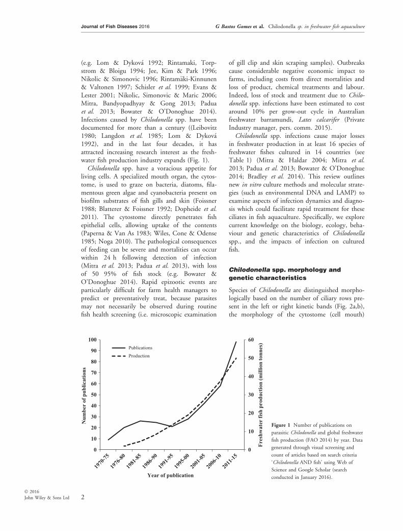
(e.g. Lom & Dykov�a 1992; Rintamaki, Torp-strom & Bloigu 1994; Jee, Kim & Park 1996;Nikolic & Simonovic 1996; Rintam€aki-Kinnunen& Valtonen 1997; Schisler et al. 1999; Evans &Lester 2001; Nikolic, Simonovic & Maric 2006;Mitra, Bandyopadhyay & Gong 2013; Paduaet al. 2013; Bowater & O’Donoghue 2014).Infections caused by Chilodonella spp. have beendocumented for more than a century ((Leibovitz1980; Langdon et al. 1985; Lom & Dykov�a1992), and in the last four decades, it hasattracted increasing research interest as the fresh-water fish production industry expands (Fig. 1).Chilodonella spp. have a voracious appetite for
living cells. A specialized mouth organ, the cytos-tome, is used to graze on bacteria, diatoms, fila-mentous green algae and cyanobacteria present onbiofilm substrates of fish gills and skin (Foissner1988; Blatterer & Foissner 1992; Dopheide et al.2011). The cytostome directly penetrates fishepithelial cells, allowing uptake of the contents(Paperna & Van As 1983; Wiles, Cone & Odense1985; Noga 2010). The pathological consequencesof feeding can be severe and mortalities can occurwithin 24 h following detection of infection(Mitra et al. 2013; Padua et al. 2013), with lossof 50�95% of fish stock (e.g. Bowater &O’Donoghue 2014). Rapid epizootic events areparticularly difficult for farm health managers topredict or preventatively treat, because parasitesmay not necessarily be observed during routinefish health screening (i.e. microscopic examination
of gill clip and skin scraping samples). Outbreakscause considerable negative economic impact tofarms, including costs from direct mortalities andloss of product, chemical treatments and labour.Indeed, loss of stock and treatment due to Chilo-donella spp. infections have been estimated to costaround 10% per grow-out cycle in Australianfreshwater barramundi, Lates calcarifer (PrivateIndustry manager, pers. comm. 2015).Chilodonella spp. infections cause major losses
in freshwater production in at least 16 species offreshwater fishes cultured in 14 countries (seeTable 1) (Mitra & Haldar 2004; Mitra et al.2013; Padua et al. 2013; Bowater & O’Donoghue2014; Bradley et al. 2014). This review outlinesnew in vitro culture methods and molecular strate-gies (such as environmental DNA and LAMP) toexamine aspects of infection dynamics and diagno-sis which could facilitate rapid treatment for theseciliates in fish aquaculture. Specifically, we explorecurrent knowledge on the biology, ecology, beha-viour and genetic characteristics of Chilodonellaspp., and the impacts of infection on culturedfish.
Chilodonella spp. morphology and
genetic characteristics
Species of Chilodonella are distinguished morpho-logically based on the number of ciliary rows pre-sent in the left or right kinetic bands (Fig. 2a,b),the morphology of the cytostome (cell mouth)
0
10
20
30
40
50
60
0
10
20
30
40
50
60
70
80
90
100
Fres
hwat
er fi
sh p
rodu
ctio
n (m
illio
n to
nnes
)
Num
ber
of p
ublic
atio
ns
Year of publication
Publications
Production
Figure 1 Number of publications on
parasitic Chilodonella and global freshwater
fish production (FAO 2014) by year. Data
generated through visual screening and
count of articles based on search criteria
‘Chilodonella AND fish’ using Web of
Science and Google Scholar (search
conducted in January 2016).
2
Journal of Fish Diseases 2016 G Bastos Gomes et al. Chilodonella sp. in freshwater fish aquaculture
� 2016
John Wiley & Sons Ltd
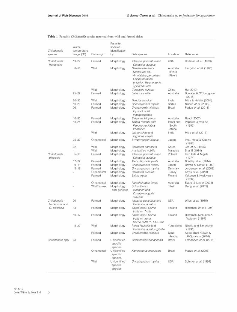
Table 1 Parasitic Chilodonella species reported from wild and farmed fishes
Chilodonella
species
Water
temperature
range (°C) Fish origin
Parasite
species
identification
by Fish species Location Reference
Chilodonella
hexasticha
19–22 Farmed Morphology Ictalurus punctatus and
Carassius auratus
USA Hoffman et al. (1979)
8–13 Wild Morphology Nernatalosa erebi,
Neosilurus sp.,
Amniataba percoides,
Leiopotherapon
unicolor, Melanotaenia
splendidi tatei
Australia
(Finke
River)
Langdon et al. (1985)
– Wild Morphology Carassius auratus China Hu (2012)
25–27 Farmed Morphology Lates calcarifer Australia Bowater & O’Donoghue
(2014)
20–30 Wild Morphology Nandus nandus India Mitra & Haldar (2004)
10–20 Farmed Morphology Oncorhynchus mykiss Serbia Nikolic et al. (2006)
26 Farmed Morphology Oreochromis niloticus,
Gymnotus aff.
inaequilabiatus
Brazil Padua et al. (2013)
10–30 Farmed Morphology Bidyanus bidyanus Australia Read (2007)
13–24 Farmed Morphology Tilapia rendalli and
Pseudocrenilabris
Philander
Israel and
South
Africa
Paperna & Van As
(1983)
– Wild Morphology Labeo rohita and
Cyprinus carpio
India Mitra et al. (2013)
25–30 Ornamental Morphology Symphysodon discus Japan Imai, Hatai & Ogawa
(1985)
22 Wild Morphology Carassius carassius Korea Jee et al. (1996)
– Wild Morphology Aristichthys nobilis Malaysia Shariff (1984)
Chilodonella
piscicola
5–10 Farmed Morphology Ictalurus punctatus and
Carassius auratus
Poland Kazubski & Migala
(1974)
17–27 Farmed Morphology Maccullochella peelii Australia Bradley et al. (2014)
9–11 Farmed Morphology Oncorhynchus masou Japan Urawa & Yamao (1992)
5–18 Farmed Morphology Oncorhynchus mykiss Denmark Jorgensen et al. (2009)
– Ornamental Morphology Carassius auratus Turkey Kayıs et al. (2013)
– Farmed Morphology Salmo trutta Finland Valtonen & Koskivaara
(1994)
– Ornamental Morphology Paracheirodon innesi Australia Evans & Lester (2001)
– Wild/Farmed Morphology
and genetics
Schizothorax
o’connori and
Oxygymnocypris
stewartii
Tibet Deng et al. (2015)
Chilodonella
hexasticha and
C. piscicola
20 Farmed Morphology Ictalurus punctatus and
Carassius auratus
USA Wiles et al. (1985)
13 Farmed Morphology Salmo salar, Salmo
trutta m. Trutta
Finland Rintamaki et al. (1994)
10–17 Farmed Morphology Salmo salar, Salmo
trutta m. trutta,
Salmo trutta m. Lacustris
Finland Rintam€aki-Kinnunen &
Valtonen (1997)
5–22 Wild Morphology Perca fluviatilis and
Carassius auratus gibelio
Yugoslavia Nikolic and Simonovic
(1996)
– Farmed Morphology Oreochromis niloticus Saudi
Arabia
Abdel-Baki, Gewik &
Al-Quraishy (2014)
Chilodonella spp. 23 Farmed Unidentified
specific
species
Odontesthes bonariensis Brazil Fernandes et al. (2011)
– Ornamental Unidentified
specific
species
Xiphophorus maculatus Brazil Piazza et al. (2006)
– Wild Unidentified
specific
species
Oncorhynchus mykiss USA Schisler et al. (1999)
3
Journal of Fish Diseases 2016 G Bastos Gomes et al. Chilodonella sp. in freshwater fish aquaculture
� 2016
John Wiley & Sons Ltd
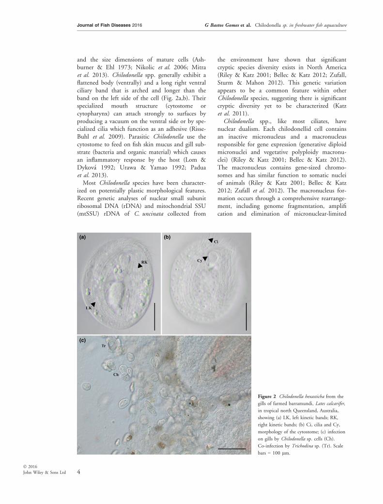
and the size dimensions of mature cells (Ash-burner & Ehl 1973; Nikolic et al. 2006; Mitraet al. 2013). Chilodonella spp. generally exhibit aflattened body (ventrally) and a long right ventralciliary band that is arched and longer than theband on the left side of the cell (Fig. 2a,b). Theirspecialized mouth structure (cytostome orcytopharynx) can attach strongly to surfaces byproducing a vacuum on the ventral side or by spe-cialized cilia which function as an adhesive (Risse-Buhl et al. 2009). Parasitic Chilodonella use thecytostome to feed on fish skin mucus and gill sub-strate (bacteria and organic material) which causesan inflammatory response by the host (Lom &Dykov�a 1992; Urawa & Yamao 1992; Paduaet al. 2013).Most Chilodonella species have been character-
ized on potentially plastic morphological features.Recent genetic analyses of nuclear small subunitribosomal DNA (rDNA) and mitochondrial SSU(mtSSU) rDNA of C. uncinata collected from
the environment have shown that significantcryptic species diversity exists in North America(Riley & Katz 2001; Bellec & Katz 2012; Zufall,Sturm & Mahon 2012). This genetic variationappears to be a common feature within otherChilodonella species, suggesting there is significantcryptic diversity yet to be characterized (Katzet al. 2011).Chilodonella spp., like most ciliates, have
nuclear dualism. Each chilodonellid cell containsan inactive micronucleus and a macronucleusresponsible for gene expression (generative diploidmicronuclei and vegetative polyploidy macronu-clei) (Riley & Katz 2001; Bellec & Katz 2012).The macronucleus contains gene-sized chromo-somes and has similar function to somatic nucleiof animals (Riley & Katz 2001; Bellec & Katz2012; Zufall et al. 2012). The macronucleus for-mation occurs through a comprehensive rearrange-ment, including genome fragmentation, amplification and elimination of micronuclear-limited
(a)
(c)
(b)
Figure 2 Chilodonella hexasticha from the
gills of farmed barramundi, Lates calcarifer,
in tropical north Queensland, Australia,
showing (a) LK, left kinetic bands; RK,
right kinetic bands; (b) Ci, cilia and Cy,
morphology of the cytostome; (c) infection
on gills by Chilodonella sp. cells (Ch).
Co-infection by Trichodina sp. (Tr). Scale
bars = 100 lm.
4
Journal of Fish Diseases 2016 G Bastos Gomes et al. Chilodonella sp. in freshwater fish aquaculture
� 2016
John Wiley & Sons Ltd

sequences (Prescott 1994; Juranek & Lipps 2007;Bellec & Katz 2012). This pervasive rearrange-ment in chilodonellid cells yields a macronucleuswith gene-sized chromosomes which dividethrough amitosis (cell division that happens with-out common features of mitosis) (Riley & Katz2001; Katz & Kovner 2010; Bellec & Katz 2012).Research examining patterns of molecular evolu-tion among ciliates and epigenetic mechanismshas used C. uncinata species as a model organismbecause of this unique nuclear dualism (Bellec &Katz 2012).Ciliates have many unique features related to
their genome structure, including variation in genecopy number (CNV) between individuals within apopulation and gene expression levels (Spring,Pham & Zufall 2013). This modification can beexplained by innate gene duplication in eukary-otes, presence of gene clusters and a comprehen-sive existence of extrachromosomal circular DNA,which may be involved in genome plasticity(Walsh 1987; Cohen & Segal 2009; Bellec &Katz 2012). The study of changes in CNV andexpression level can explain genomic structuralvariations observed when comparing individualswithin a population. CNV can be related to adap-tive evolution of species and also can be associatedwith some human diseases (Spring et al. 2013).Therefore, Chilodonella spp. represents a perfectmodel to study CNV due to the presence of indi-vidual genes on unlinked chromosomes (Riley &Katz 2001; Bellec & Katz 2012).
Life cycle
Chilodonella spp. primarily reproduce by trans-verse binary fission (Lynn 2008; Bellec, Maurer-Alcala & Katz 2014). During binary fission, thezygotic nucleus divides, generating a new nucleus.One nucleus will turn into a micronucleus andthe other will develop into a macronucleus (Riley& Katz 2001; Bellec & Katz 2012). The parentcell then divides by binary fission (asexual andmitotic process) producing two ‘daughter’ cells(offspring) which have the same size (Lynn 2008).Asexual reproduction occurs continually if there issufficient food in the environment (Lynn 2008).Chilodonella spp. can also reproduce by a sexual
process known as conjugation (Lynn 2008; Bellecet al. 2014). Conjugation starts when mature Chi-lodonella spp. cells find other complimentary mat-ing individuals. A mating reaction occurs between
the cells and during this process many nuclearevents occur such as meiosis, exchange of gameticnuclei and fertilization (Lynn 2008). Two excon-jugant cells are formed. If these cells cannot con-jugate, they undergo a period of senescence withdeath temporarily delayed by autogamy or self-fer-tilization (Hausmann & Bradbury 1996; Sugiuraet al. 2005; Lynn 2008). Chilodonella uncinataparticularly starts its sexual cycle by meiosis of themicronucleus (MIC), and then conjugation withexchange of haploid MIC between cells takesplace, followed by nuclear fusion forming a zygo-tic nucleus (Bellec et al. 2014). Chilodonella cellswith similar morphology form a pair and are con-nected to each other by their cytostome. This sex-ual process can be stimulated in Chilodonella cellsin vitro through limited starvation (Lynn 2008).Understanding the asexual division and sexual
cycle of Chilodonella is important for morphologi-cal characterization of species. Generally, speciesare identified using measurements of the macronu-cleus (MAC) and MIC (Fan et al. 2014; Qu et al.2014). However, considering Chilodonella cellsundergo complex processes in their different lifecycle stages, mischaracterization of species couldoccur using MAC and MIC measurements.
Chilodonella spp. in freshwater fish
aquaculture: ecology, known hosts and
geographic distribution
Four Chilodonella species have been isolated fromthe gills and skin of bony fish, including Chilodo-nella uncinata, C. cucullulus, C. hexasticha andC. piscicola (see Migala & Kazubski 1972; Rinta-maki et al. 1994). The most serious mortalities infish aquaculture appear to have been associatedwith infections caused by C. hexasticha and C. pis-cicola (see Hoffman et al. 1979; Mitra & Haldar2004; Mitra et al. 2013; Padua et al. 2013), whilemost reports of C. uncinata and C. cucullulus(Muller, 1976) are from collection made directlyfrom the environment (i.e. not associated with afish host). Chilodonella spp. primarily feed on bac-teria and algae in the environment and on organicmaterial and bacteria when infecting fish (Noga2010; Padua et al. 2013). High number of Chilo-donella cells (Fig. 2c) feeding on fish can causesevere pathological signs, including hyperplasia ofgill epithelium and necrosis in the gill and skin offishes (Ashburner & Ehl 1973; Mitra & Haldar2004; Karvonen et al. 2010; Padua et al. 2013).
5
Journal of Fish Diseases 2016 G Bastos Gomes et al. Chilodonella sp. in freshwater fish aquaculture
� 2016
John Wiley & Sons Ltd
Parasitic Chilodonella spp. exhibit a wide tem-perature tolerance (Table 1). Rapid changes inenvironmental parameters, such as temperatureand oxygen, or increases in organic material inponds, can contribute to the proliferation of Chi-lodonella spp. on fish (Bowater & O’Donoghue2014; Bradley et al. 2014). Additionally, extremetemperatures promote host stress, which can com-promise the host fish’s immune system and affectthe capacity to combat infection (Hossain et al.2008, 2013; Macnab & Barber 2012). Chilodo-nella hexasticha is associated with warmer climatesor seasons between 26 and 31 °C, while Chilodo-nella piscicola exhibits a wide thermal tolerancebetween 4 and 20 °C (Table 1). The ideal envi-ronmental conditions associated with chilodonel-losis (infections caused by Chilodonella spp.) areunidentified (Kepner, Wharton & Coats 1999;Hossain et al. 2008, 2013) and it is not clearwhether specific temperatures impact the repro-duction and proliferation of Chilodonella spp.Clinical signs and detection of infections causedby Chilodonella spp. have been generally foundduring drastic changes in the weather (transitionfrom summer to autumn; dry to wet season; Brad-ley et al. 2014; Hossain et al. 2008, 2013). Thisindicates that stress may predispose fish to infec-tions as it can compromise their immune system(Oidtmann et al. 2011, 2013).Many ciliates are considered microaerophilic
(optimal growth at relatively low levels of oxygen)(Lynn 2008; Fenchel 2014). Ciliates also have achemosensory behaviour which orientates the cellsin the direction of their preferable O2 levels(Fenchel & Bernard 1996; Fenchel 2014). Chilo-donella spp. epizootics rapidly develop in the pres-ence of low levels of dissolved oxygen (Langdonet al. 1985; Garcia et al. 2009; Fenchel 2014).Indeed, Dopheide et al. (2011) proposed thatanaerobic bacteria present in anoxic conditionsmay be some of the most preferable food itemsfor Chilodonella spp. High stocking densities inponds may predispose fish to stress, as low levelsof DO (dissolved oxygen) are common whenponds are overpopulated (Mitra & Haldar 2004;Padua et al. 2013; Bowater & O’Donoghue2014). Although there is some indication of whatconditions might be associated with chilodonel-losis (low oxygen levels, constant temperature fluc-tuation and high levels of organic material inwater/soil) (Fenchel & Bernard 1996; Bowater &O’Donoghue 2014; Fenchel 2014), it is clear that
research is necessary to elucidate the combinationof environmental parameters that facilitate rapidmultiplication of Chilodonella spp..
Clinical presentations and pathology
associated with Chilodonella spp.
infections
Clinical signs associated with Chilodonella spp.infections are not unique, which makes initialdiagnosis of chilodonellosis challenging (Noga2010). Fish infected with Chilodonella spp. canexhibit gasping behaviour, anorexia, skin depig-mentation, ulceration, scale loss, excessive mucusexcretion and gill lesions (Padua et al. 2013). Theclinical signs most commonly observed are a mot-tled/grey appearance on the skin (caused by exces-sive mucus production), lethargy, gill viscousmucus production, swimming slowly near the sur-face and edges, slim appearance (caused by appe-tite loss) and sometimes gill lesions and scale loss(Read 2007; Padua et al. 2013; Bradley et al.2014). Examination of moribund fish is necessaryto avoid misdiagnosis. In general, intense parasiteinfections are accompanied by acute gill lesionssufficient to kill affected fish (Langdon et al.1985; Padua et al. 2013; Bowater & O’Donoghue2014). Fish infected with few cells usually do notexhibit clinical signs of illness (Read 2007). How-ever, Chilodonella cells can rapidly multiply (pop-ulation numbers can double in only a few hours;pers. obs.; Read 2007; Padua et al. 2013).Infestations of Chilodonella spp. cause gill epithe-
lial hypertrophy, hyperplasia and are generallyobserved with complete or partial lamellar fusion fol-lowed by lymphocytic infiltration. Necrosis andsome mild oedema can also be observed (Paperna &Van As 1983; Padua et al. 2013; Bowater &O’Donoghue 2014; Bradley et al. 2014). Fish skinusually demonstrates a non-specific lymphocytic der-matitis (Padua et al. 2013; Bowater & O’Donoghue2014; Bradley et al. 2014).
Diagnostic methods for detecting
Chilodonella spp. in wild and
aquaculture fishes
The lack of host specificity, cosmopolitan distribu-tion and pervasiveness of species in Chilodonellacompromises accurate diagnosis of species thatcause severe disease (Urawa & Yamao 1992;Urawa 1996; Mitra & Haldar 2004; Nikolic et al.
6
Journal of Fish Diseases 2016 G Bastos Gomes et al. Chilodonella sp. in freshwater fish aquaculture
� 2016
John Wiley & Sons Ltd
2006; Jorgensen, Larsen & Buchmann 2009; Gaoet al. 2012). Generally, Chilodonella species areconsidered free-living ciliate protozoans. However,it is unknown whether species considered ‘free-liv-ing’ can also occupy a parasitic lifestyle given opti-mal conditions and opportunity (Noga 2010).Moreover, it is poorly understood how many Chi-lodonella species elicit harmful pathology to theirfish hosts in favourable conditions (Lom &Dykov�a 1992; Noga 2010).Morphological identification of Chilodonella
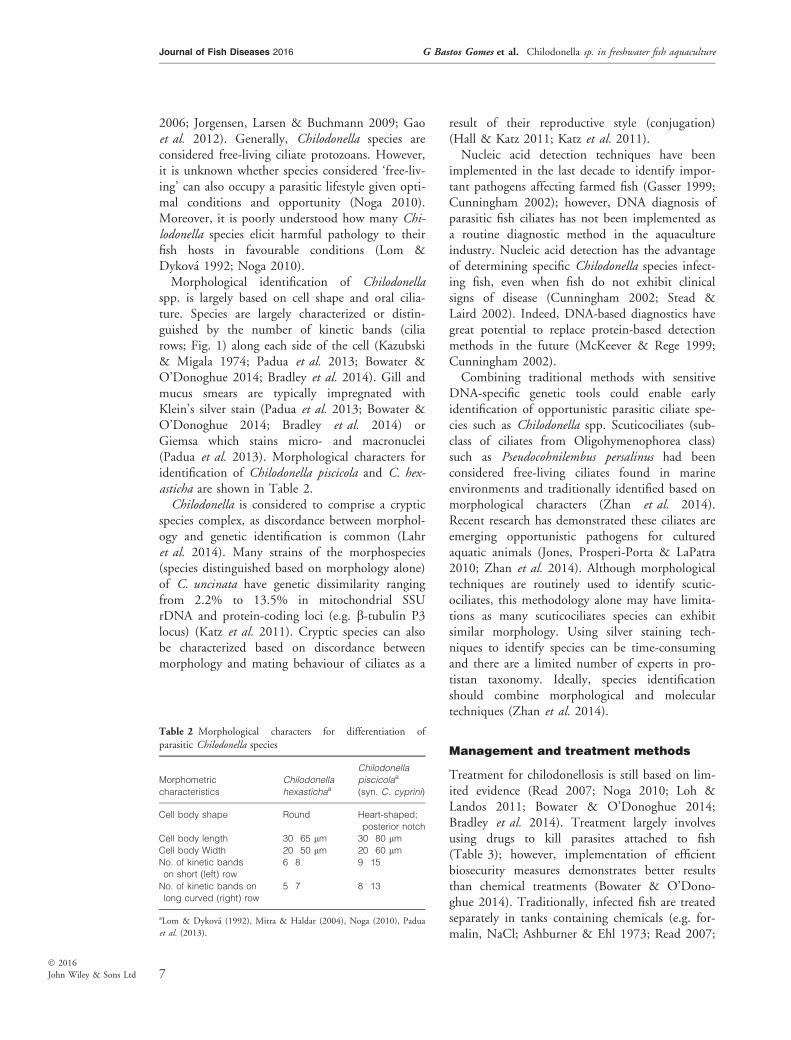
spp. is largely based on cell shape and oral cilia-ture. Species are largely characterized or distin-guished by the number of kinetic bands (ciliarows; Fig. 1) along each side of the cell (Kazubski& Migala 1974; Padua et al. 2013; Bowater &O’Donoghue 2014; Bradley et al. 2014). Gill andmucus smears are typically impregnated withKlein’s silver stain (Padua et al. 2013; Bowater &O’Donoghue 2014; Bradley et al. 2014) orGiemsa which stains micro- and macronuclei(Padua et al. 2013). Morphological characters foridentification of Chilodonella piscicola and C. hex-asticha are shown in Table 2.Chilodonella is considered to comprise a cryptic
species complex, as discordance between morphol-ogy and genetic identification is common (Lahret al. 2014). Many strains of the morphospecies(species distinguished based on morphology alone)of C. uncinata have genetic dissimilarity rangingfrom 2.2% to 13.5% in mitochondrial SSUrDNA and protein-coding loci (e.g. b-tubulin P3locus) (Katz et al. 2011). Cryptic species can alsobe characterized based on discordance betweenmorphology and mating behaviour of ciliates as a
result of their reproductive style (conjugation)(Hall & Katz 2011; Katz et al. 2011).Nucleic acid detection techniques have been
implemented in the last decade to identify impor-tant pathogens affecting farmed fish (Gasser 1999;Cunningham 2002); however, DNA diagnosis ofparasitic fish ciliates has not been implemented asa routine diagnostic method in the aquacultureindustry. Nucleic acid detection has the advantageof determining specific Chilodonella species infect-ing fish, even when fish do not exhibit clinicalsigns of disease (Cunningham 2002; Stead &Laird 2002). Indeed, DNA-based diagnostics havegreat potential to replace protein-based detectionmethods in the future (McKeever & Rege 1999;Cunningham 2002).Combining traditional methods with sensitive
DNA-specific genetic tools could enable earlyidentification of opportunistic parasitic ciliate spe-cies such as Chilodonella spp. Scuticociliates (sub-class of ciliates from Oligohymenophorea class)such as Pseudocohnilembus persalinus had beenconsidered free-living ciliates found in marineenvironments and traditionally identified based onmorphological characters (Zhan et al. 2014).Recent research has demonstrated these ciliates areemerging opportunistic pathogens for culturedaquatic animals (Jones, Prosperi-Porta & LaPatra2010; Zhan et al. 2014). Although morphologicaltechniques are routinely used to identify scutic-ociliates, this methodology alone may have limita-tions as many scuticociliates species can exhibitsimilar morphology. Using silver staining tech-niques to identify species can be time-consumingand there are a limited number of experts in pro-tistan taxonomy. Ideally, species identificationshould combine morphological and moleculartechniques (Zhan et al. 2014).
Management and treatment methods
Treatment for chilodonellosis is still based on lim-ited evidence (Read 2007; Noga 2010; Loh &Landos 2011; Bowater & O’Donoghue 2014;Bradley et al. 2014). Treatment largely involvesusing drugs to kill parasites attached to fish(Table 3); however, implementation of efficientbiosecurity measures demonstrates better resultsthan chemical treatments (Bowater & O’Dono-ghue 2014). Traditionally, infected fish are treatedseparately in tanks containing chemicals (e.g. for-malin, NaCl; Ashburner & Ehl 1973; Read 2007;
Table 2 Morphological characters for differentiation of
parasitic Chilodonella species
Morphometric
characteristics
Chilodonella
hexastichaa
Chilodonella
piscicolaa
(syn. C. cyprini)
Cell body shape Round Heart-shaped;
posterior notch
Cell body length 30�65 lm 30�80 lmCell body Width 20�50 lm 20�60 lmNo. of kinetic bands
on short (left) row
6�8 9�15
No. of kinetic bands on
long curved (right) row
5�7 8�13
aLom & Dykov�a (1992), Mitra & Haldar (2004), Noga (2010), Padua
et al. (2013).
7
Journal of Fish Diseases 2016 G Bastos Gomes et al. Chilodonella sp. in freshwater fish aquaculture
� 2016
John Wiley & Sons Ltd

Bradley et al. 2014). Even though these treat-ments can be used temporarily to reduce infec-tion, they are not highly efficacious (Noga 2010;Bowater & O’Donoghue 2014; Bradley et al.2014). Treating sick fish using only osmotic salt-water baths and replacing them into ponds orcages is not effective (Read 2007; Bradley et al.2014). Once fish are returned to untreated pondwater, which contains Chilodonella cells, re-infec-tion is unavoidable. Moreover, fish are likely to beimmunocompromised by being sick and stressedfrom manual handling.Most common chemicals used to treat fish
infected with Chilodonella spp. include formalin(formaldehyde solution) and potassium perman-ganate. Formalin has bactericidal properties, butits efficacy has never been studied specificallyagainst chilodonellosis. Limited information existsabout formalin bioaccumulation in fish and thepotential implications for human health areunknown (Boyd & Tucker 1998; Boyd & Mas-saut 1999; Wooster et al. 2005).Potassium permanganate (KMnO4) is another
chemical also used to combat Chilodonella spp.infections. KMnO4 can cause serious corrosion to
fish when in contact with gills and skin and it ishighly explosive when in direct contact withorganic substances (Schlenk et al. 2000). Thischemical oxidizes organic and inorganic substancesand kills bacteria. Permanganate oxidizes existingorganic material and other reduced substancestransforming it into relatively non-toxic man-ganese dioxide. Considering most chilodonellosisevents occur when organic material accumulates inponds, KMnO4 use in aquaculture farms must bewell monitored (Boyd & Massaut 1999; Schlenket al. 2000). The chemical can be toxic to phyto-plankton and will reduce the production of dis-solved oxygen by photosynthesis. Therefore,mortality of aquatic organisms is possible, includ-ing beneficial bacteria and ciliates but also para-sitic ciliates (Tucker & Boyd 1977; Tucker 1989;Boyd & Massaut 1999; Schlenk et al. 2000).The drug 35% PEROX-AID� has been the
only form of hydrogen peroxide (H2O2) approvedby the US Food and Drug Administration (FDA)to manage some diseases (e.g. saprolegniasis,bacterial gill disease and columnaris) of freshwa-ter-reared fish (Yanong 2008). There have beenlimited clinical trials examining the efficacy of
Table 3 Common chemicals and dosages used by aquaculture industrya to treat Chilodonella spp. and other ciliate infections in
ornamental and farmed fish
Chemical
Dosage rate for
ornamental tanks
Duration/frequency
of treatment
Dosage rate for
ponds/cages
Duration/frequency
of treatment References
Hydrogen peroxide
(H2O2) animals
200 ppm 30 min 250–500 ppm 1 day
24-h constant aeration
Yanong (2008)
Hydrogen peroxide
(H2O2) 10%
(extra oxygen supply
for soil/water)
– – General dose
250–350 g acre�1Constant aeration and
monitoring
Mostafa &
Kumar (2012)
Formalin (CH2O) 150 ppt 1 h
Not recommended
for young animals
20–30 ppm 4–5 days
24-h constant aeration
Read (2007),
Noga (2010),
Loh & Landos
(2011)
Potassium
permanganate (KMnO4)
20 ppm 60 min 2–3 ppm 2–3 days
24-h constant aeration
Schlenk et al.
(2000), Read
(2007), Noga
(2010), Loh &
Landos (2011)
Copper sulphateb
(CuSO4)
0.15–0.20 ppm Gradually over
2–3 days
0.3 ppm 1 day
24-h constant aeration
Read (2007),
Noga (2010),
Loh & Landos
(2011)
Saltc (NaCl) 10 ppt 60 min
Repeat next day
Non-applicable Non-applicable Read (2007),
Noga (2010),
Loh & Landos
(2011)
aMost are unapproved and non-tested in food fish species.bPond alkalinity must be tested before using copper sulphate.cSalt in ponds is not practical.
8
Journal of Fish Diseases 2016 G Bastos Gomes et al. Chilodonella sp. in freshwater fish aquaculture
� 2016
John Wiley & Sons Ltd
hydrogen peroxide against chilodonellosis. Mosttests using H2O2 against ciliates focus on thedamage to host species (e.g. fish). Nevertheless,hydrogen peroxide has been successfully used totreat Chilodonella spp. infections in Murray cod,Maccullochella peelii (see Bradley et al. 2014).Adding hydrogen peroxide to ponds leads to anincrease in oxygen levels and could mitigate Chilo-donella spp. growth and proliferation aschilodonellids grow well under low levels of oxy-gen (Langdon et al. 1985; Garcia et al. 2009;Fenchel 2014).Copper sulphate (CuSO4) is known in aquacul-
ture for its antiparasitic and algaecide properties,although some countries have banned its use (Wat-son & Yanong 1989; Yanong 2008). Some Murraycod farms in Australia still use this chemical tocombat chilodonellosis (Bradly et al. 2014). Treat-ment concentration needs to be calculated carefullyto prevent toxicity to fish (Watson & Yanong1989). When used for treating ponds containinglarge quantities of algae, copper sulphate can causea drop in oxygen levels which could facilitate Chi-lodonella spp. replication. It has showed that pro-longed use of CuSO4 is not effective againstchilodonellosis and may cause serious damage tofish gills and skin. Copper can also be toxic to thezooplankton (e.g. rotifers) and invertebrates (e.g.snails). Toxicity is even more problematic in lowalkalinity waters. When pond water alkalinity isunknown, or if relying on zooplankton as a foodsource for young fish, this drug may not be the bestoption (Watson & Yanong 1989). Ideally, Chilodo-nella spp. infections should be prevented throughreducing stocking density in ponds, frequent waterquality monitoring, water exchange, weekly fishhealth examination and adequate feed rates (Read2007; Noga 2010; Bowater & O’Donoghue 2014).
Future insights into Chilodonella spp.
biology and epidemiology
Chilodonella spp. can be artificially culturedin vitro in controlled laboratory conditions, whichenables experimental research examining parasitelife cycles and new treatment options. Artificialcultures of some protozoan parasites in vivo and/or in vitro are exceptionally challenging (Crosbieet al. 2012; Pinheiro & Bols 2013) and most par-asitic ciliates need the presence of their host togrow under artificial conditions (e.g. Cryptocaryonirritans). However, eliminating the necessity for
maintaining infected animals greatly increases thefeasibility of culturing parasitic ciliates in vitroand contributes to improved animal welfare(Crosbie et al. 2010, 2012). Chilodonella uncinataand other species in the genus can be culturedin vitro (Bellec & Katz 2012; Bellec et al. 2014).Chilodonella spp. grow well in the dark (roomtemperature, RT) using filtered and sterilisedpond water containing a grain of rice for pro-longed periods (Bellec et al. 2014). Chilodonellauncinata can also be maintained in a cereal wheatgrass media inoculated with Klebsiella sp. withoptimal growth between 25 and 30 °C (Lynn2008). The ability to culture Chilodonella spp. canenable targeted research on biology and epidemi-ology which will advance scientific knowledge onenvironmental and host triggers that facilitateharmful outbreaks.
The future of Chilodonella spp.
detection in aquaculture
The ability to pre-empt outbreaks of chilodonel-losis would be a considerable advantage toindustry. New technologies using sensitive DNA-specific genetic tools can enable early detection ofciliate parasites species affecting freshwater farmedfishes. Environmental DNA (eDNA) is a potentialnew monitoring tool of small amounts of geneticmaterial present in water or soil (Ficetola et al.2008). Novel eDNA techniques and technologieshave the power to identify and quantify species ofciliate protozoans (using qPCR) present in a pondor tank aquaculture system (Bass et al. 2015).Commonly, fish do not present signs of diseaseunless stressed, even when parasites are present inthe environment (Noga 2010). Monitoring waterfrom ponds using eDNA would enable quantifica-tion of protozoan parasites, even when fish arenot presenting clinical signs of infection. Quanti-fying the number of ciliate protozoans in waterand correlating parasite species abundance withlevels of oxygen and temperature could revealimportant ecological patterns associated with Chi-lodonella spp. outbreaks. Determining the ecologi-cal parameters associated with epizootic eventstogether with high numbers of ciliates in the watermay facilitate early preventative parasite manage-ment (e.g. new feed strategies, treatment of thewater before stocking ponds).Another potential new diagnostic tool for aqua-
culture is LAMP (loop-mediated isothermal
9
Journal of Fish Diseases 2016 G Bastos Gomes et al. Chilodonella sp. in freshwater fish aquaculture
� 2016
John Wiley & Sons Ltd
amplification). LAMP is a novel nucleic aciddetection method that can amplify the target spe-cies DNA in isothermal conditions (Notomi et al.2000). This means simple equipment such as aheat block or a water bath can be used to diag-nose presence/absence of pathogen DNA. Thistechnique is ideal for rapid and practical field or‘on-farm’ detection for potential pathogens in asystem. Combining this detection technology witheDNA to monitor infections in aquaculture mayfacilitate the early detection and prophylactictreatment of parasites before epizootic outbreaksoccur. LAMP also has the potential to assist aqua-culture managers to monitor protozoan parasitefauna in the environment as a part of ongoinghealth surveillance programme.The future of a sustainable fish aquaculture
industry relies on the ability to mitigate theimpact of disease on fish production. Research onthe complex interaction between Chilodonellaspp., the environment and fish host is necessary tobetter understand infection dynamics. Molecularcharacterization for the identification of Chilodo-nella species present in fish farms, new diagnostictechniques, treatments and monitoring tools mustbe explored to promote cost-effective managementof parasitic Chilodonella spp. on freshwater fishaquaculture farms.
Acknowledgements
We thank the Australian Society for Parasitologyfor a travel grant which enabled the GBG to workwith Professor Laura Katz, Smith College (Univer-sity of Massachusetts), USA, and develop expertisein the culture of Chilodonella spp. GBG receivedscholarship support from James Cook University.This research was partially funded through theQueensland Government’s Smart Futures Pro-gram, James Cook University and our industrypartners.
References
Abdel-Baki A.-A.S., Gewik M.M. & Al-Quraishy S. (2014)
First records of Ambiphrya and Vorticella spp. (Protozoa,
Ciliophora) in cultured Nile tilapia (Oreochromis niloticus) inthe central region of Saudi Arabia. Saudi Journal ofBiological Sciences 21, 520–523.
Ashburner L.D. & Ehl A.S. (1973) Chilodonellacyprini (Moroff), a parasite of freshwater fish and its
treatment. Australian Society for Limnology Bulletin 5, 3–4.
Bass D., Stentiford G.D., Littlewood D.T.J. & Hartikainen H.
(2015) Diverse applications of environmental DNA methods
in parasitology. Trends in Parasitology 31, 499–513.
Bellec L. & Katz L.A. (2012) Analyses of chromosome copy
number and expression level of four genes in the ciliate
Chilodonella uncinata reveal a complex pattern that suggests
epigenetic regulation. Gene 504, 303–308.
Bellec L., Maurer-Alcala X.X. & Katz L.A. (2014)
Characterization of the life cycle and heteromeric nature of
the macronucleus of the ciliate Chilodonella uncinata using
fluorescence microscopy. Journal of Eukaryotic Microbiology61, 313–316.
Blatterer H. & Foissner W. (1992) Morphology and
infraciliature of some cyrtophorid ciliates (protozoa,
ciliophora) from fresh-water and soil. Archiv FurProtistenkunde 142, 101–118.
Bowater R.O. & O’Donoghue P.J. (2014) An epizootic of
chilodonelliasis in farmed barramundi Lates calcarifer(Bloch), a case report. Journal of Fish Diseases 36, 1–6.
Boyd C.E. & Massaut L. (1999) Risks associated with the use
of chemicals in pond aquaculture. Aquacultural Engineering20, 113–132.
Boyd C.E. & Tucker C.S. (1998) Pond Aquaculture WaterQuality Management. Springer, Boston, MA.
Bradley T., Mccowan C., Cohen S., Ingram B., Green C. &
Mansell P. (2014) Improved fish health management for
integrated inland aquaculture through better management
practices. In Fisheries Research and Development Corporation,Department of Environment and Primary Industries, pp.51.Melbourne.
Buchmann K. (2013) Impact and control of protozoan
parasites in maricultured fishes. Parasitology 142, 168–177.
Cohen S. & Segal D. (2009) Extrachromosomal circular DNA
in eukaryotes: possible involvement in the plasticity of
tandem repeats. Cytogenetic and Genome Research 124, 327–328.
Crosbie P.B.B., Bridle A.R., Leef M.J. & Nowak B.F. (2010)
Effects of different batches of Neoparamoeba perurans andfish stocking densities on the severity of amoebic gill disease
in experimental infection of Atlantic salmon, Salmo salar L.Aquaculture Research 41, e505–e516.
Crosbie P.B.B., Bridle A.R., Cadoret K. & Nowak B.F. (2012)
In vitro cultured Neoparamoeba perurans causes amoebic gill
disease in Atlantic salmon and fulfils Koch’s postulates.
International Journal for Parasitology 42, 511–515.
Cunningham C.O. (2002) Molecular Diagnosis of SalmonidDiseases. Springer, Dordrecht.
Deng Q., Guo Q., Zhai Y., Wang Z. & Gu Z. (2015) First
record of Chilodonella piscicola (Ciliophora: Chilodonellidae)
from two endangered fishes, Schizothorax o’connori andOxygymnocypris stewartii in Tibet. Parasitology Research 114,
3097–3103.
Dopheide A., Lear G., Stott R. & Lewis G. (2011) Preferential
feeding by the ciliates Chilodonella and Tetrahymena spp.
and effects of these protozoa on bacterial biofilm structure
and composition. Applied and Environmental Microbiology77, 4564–4572.
10
Journal of Fish Diseases 2016 G Bastos Gomes et al. Chilodonella sp. in freshwater fish aquaculture
� 2016
John Wiley & Sons Ltd
Evans B.B. & Lester R.J.G. (2001) Parasites of ornamental fish
imported into Australia. Bulletin of the European Associationof Fish Pathologists 21, 51–55.
Fan X., Ma R., Al-Farraj S.A. & Gu F. (2014) Morphological
and molecular characterization of Parafurgasonia zhangi spec.nov. and Chilodonella acuta Kahl, 1931 (Protozoa,
Ciliophora), from a soil habitat of Saudi Arabia.
International Journal of Systematic and EvolutionaryMicrobiology 64, 2385–2394.
FAO (2014) The State of World Fisheries and Aquaculture, pp.23. FAO, Rome.
Fenchel T. & Bernard C. (1996) Behavioural responses in
oxygen gradients of ciliates from microbial mats. EuropeanJournal of Protistology, 32, 55–63.
Fenchel T. (2014) Protozoa and oxygen. Acta Protozoologica53, 3–12.
Fernandes J.M., Portelinha M.K., Rocha C.B., Pouey
J.L.O.F. & Piedras S.R.N. (2011) Occurrence and control
of Chilodonella spp. in pejerrey Odontesthes bonariensis.Arquivo Brasileiro de Medicina Veterin�aria e Zootecnia 63,
788–790.
Ficetola G.F., Miaud C., Pompanon F. & Taberlet P. (2008)
Species detection using environmental DNA from water
samples. Biology Letters 4, 423–425.
Foissner W. (1988) Taxonomy and ecology of some saprobic
ciliates (protozoa, ciliophora).2. family chilodonellidae.
Hydrobiologia 162, 21–45.
Gao S., Huang J., Li J. & Song W. (2012) Molecular
phylogeny of the cyrtophorid ciliates (Protozoa, Ciliophora,
Phyllopharyngea). PLoS One 7, e33198.
Garcia F., Fujimoto R.Y., Martins M.L. & Moraes F.R.
(2009) Protozoan parasites of Xiphophorus spp. (Poeciliidae)and their relation with water characteristics. ArquivoBrasileiro de Medicina Veterin�aria e Zootecnia 61, 156–162.
Gasser R.B. (1999) PCR-based technology in veterinary
parasitology. Veterinary Parasitology 84, 229–258.
Hall M.S. & Katz L.A. (2011) On the nature of species:
insights from Paramecium and other ciliates. Genetica 139,
677–684.
Hausmann K. & Bradbury P.C. (1996) Ciliates: Cells asOrganism. Gustav Fischer Verlag, Stuttgart.
Hoffman G.L., Kazubski S.L., Mitchell A.J. & Smith C.E.
(1979) Chilodonella hexasticha (Kiernili, 1909) (Protozoa,
Ciliata) from North American warm water fish. Journal ofFish Disease 2, 153–157.
Hossain M.D., Hossain M.K., Rahaman M.H., Aktr A. &
Khanom D.A. (2008) Prevalence of ectoparasites of carp
fingerlings at Santaher, Bogra. University Journal of ZoologyRajshahi University 27, 17–19.
Hossain M.D., Islam K., Hossain M.K. & Rahman M.H.
(2013) Environmental impact assessment of fish diseases
on fish production. Journal of Science Foundation 9, 125–131.
Hu Y. (2012) Ciliate ectoparasites (Ciliophora: Trichodinidae/
Chilodonellidae) on gills of Carassius auratus from the
Yangtze River, China, with the description of Trichodina
luzhoues sp. n. Parasitology Research 111, 433–439.
Imai S., Hatai K. & Ogawa M. (1985) Chilodonella hexasticha(Kiernili, 1909) found from the gills of a discus,
Symphysodon discus Heckel, 1940. Japanese Journal ofVeterinary Science 47, 305–308.
Jee B.-Y., Kim K. & Park S. (1996) Chilodonella hexasticha(Protozoa, Ciliata) from Korean freshwater fish. Journal ofFish Pathology 9, 113–118.
Jones S.R., Prosperi-Porta G. & LaPatra S.E. (2010) First
Isolation of Pseudocohnilembus persalinus (Ciliophora:Scuticociliatida) from freshwater-reared Rainbow Trout,
Oncorhynchus mykiss. Journal of Parasitology 96, 1014–1016.
Jorgensen T.R., Larsen T.B. & Buchmann K. (2009) Parasite
infections in recirculated rainbow trout (Oncorhynchusmykiss) farms. Aquaculture 289, 91–94.
Juranek S.A. & Lipps H.J. (2007) New insights into the
macronuclear development in ciliates. International Review ofCytology 262, 21.
Karvonen A., Rintamaki P., Jokela J. & Valtonen E.T. (2010)
Increasing water temperature and disease risks in aquatic
systems: climate change increases the risk of some, but not
all, diseases. International Journal for Parasitology 40, 1483–1488.
Katz L.A. & Kovner A.M. (2010) Alternative processing of
scrambled genes generates protein diversity in the ciliate
Chilodonella uncinata. Journal of Experimental ZoologyPart B-Molecular and Developmental Evolution 314B,
480–488.
Katz L.A., DeBerardinis J., Hall M.S., Kovner A.M.,
Dunthorn M. & Muse S.V. (2011) Heterogeneous rates of
molecular evolution among cryptic species of the ciliate
morphospecies Chilodonella uncinata. Journal of MolecularEvolution 73, 266–272.
Kayıs S., Balta F., Serezli R. & Er A. (2013) Parasites on
different ornamental fish species in Turkey. Journal ofFisheries Sciences.com 7, 114–120.
Kazubski S. & Migala K. (1974) Studies on the distinctness of
Chilodonella cyprini and Chilodonella hexastichachlamydodontidae gymnostomatida ciliate parasites of fishes.
Acta Protozoologica 13, 9–40.
Kepner R.L. Jr, Wharton R.A. Jr & Coats D.W. (1999)
Ciliated protozoa of two antarctic lakes: analysis by
quantitative protargol staining and examination of artificial
substrates. Polar Biology 21, 285–294.
Lahr D.J.G., Laughinghouse H.D., Oliverio A.M., Gao F. &
Katz L.A. (2014) How discordant morphological and
molecular evolution among microorganisms can revise our
notions of biodiversity on Earth. BioEssays 36, 950–959.
Langdon J.S., Gudkovs N., Humphrey J.D. & Saxon E.C.
(1985) Deaths in Australian freshwater fishes associated with
Chilodonella hexasticha infection. Australian VeterinaryJournal 62, 409–441.
Leibovitz L. (1980) Chilodonelliasis. Journal of the AmericanVeterinary Medicine Association 177, 222–223.
Loh R. & Landos M. (2011) Fish Vetting Essentials. Richmond
Publishing, Perth, WA.
Lom J. & Dykov�a I. (1992) Protozoan Parasites of Fishes, pp.315. Elsevier Science Publishers, Amsterdam XI.
11
Journal of Fish Diseases 2016 G Bastos Gomes et al. Chilodonella sp. in freshwater fish aquaculture
� 2016
John Wiley & Sons Ltd
Lynn D.H. (2008) The Ciliated Protozoa: Characterization,Classification, and Guide to the Literature. 3rd edn. Springer,
New York, NY.
Macnab V. & Barber I. (2012) Some (worms) like it hot: fish
parasites grow faster in warmer water, and alter host thermal
preferences. Global Change Biology 18, 1540–1548.
McKeever D.J. & Rege J.E.O. (1999) Vaccines and diagnostic
tools for animal health: the influence of biotechnology.
Livestock Production Science 59, 257–264.
Migala K. & Kazubski S.L. (1972) Occurrence of nonspecific
ciliates on carps (Cyprinus carpi L.) in winter ponds. ActaProtozoologica 9, 229–337.
Mitra A.K. & Haldar D.P. (2004) First record of Chilodonellahexasticha (Kiernik, 1909) Kahl, 1931 (Ciliophora :
Chilodonellidae) infesting a freshwater fish Nandus nandus(Hamilton) from Gangetic West Bengal, India. AnimalBiology 54, 111–118.
Mitra A.K., Bandyopadhyay P.K. &Gong Y. (2013) Studies on
Trichodinid and Chilodonellid Ciliophorans (Protozoa:
Ciliophora) in the Indian freshwater and estuarine fishes with
description of Trichodinella sunderbanensis sp. nov and Trichodinanandusi sp nov. Parasitology Research 112, 1077–1085.
Mostafa S.M. & Kumar B.T. (2012) Aqua chemicals in
shrimp farm: a study from south-west coast of Bangladesh.
The Egyptian Journal of Aquatic Research 38, 275–285.
Nikolic V.P. & Simonovic P.D.(1996) Occurrence of parasitic
ciliates (Protozoa) on perch (Perca fluviatilis) in Lake
Vlasinsko. Annales Zoolologici Fennici 33, 707–710.
Nikolic V., Simonovic P.D. & Maric S.P. (2006) Occurrence
of Chilodonella hexasticha (Ciliophora, Protista) on farmed
rainbow trout (Oncorhynchus mykiss) throughout the season.Acta Veterinaria-Beograd 56, 55–61.
Noga E.J. (2010) Fish Disease: Diagnosis and Treatment, 2ndedn. Wiley-Blackwell, IOWA, IA.
Notomi T., Okayama H., Masubuchi H., Yonekawa T.,
Watanabe K., Amino N. & Hase T. (2000) Loop-mediated
isothermal amplification of DNA. Nucleic Acids Research 28,
e63.
Nowak B.F. (2007) Parasitic diseases in marine cage culture –an example of experimental evolution of parasites?
International Journal for Parasitology 37, 581–588.
Oidtmann B.C., Crane C.N., Thrush M.A., Hill B.J. & Peeler
E.J. (2011) Ranking freshwater fish farms for the risk of
pathogen introduction and spread. Preventive VeterinaryMedicine 102, 329–340.
Oidtmann B., Peeler E., Lyngstad T., Brun E., Bang Jensen B.
& St€ark K.D.C. (2013) Risk-based methods for fish and
terrestrial animal disease surveillance. Preventive VeterinaryMedicine 112, 13–26.
Padua S.B., Martins M.L., Carrijo-Mauad J.R., Ishikawa M.M.,
Jeronimo G.T., Dias-Neto J. & Filarski F. (2013) First record
of Chilodonella hexasticha (Ciliophora: Chilodonellidae) inBrazilian cultured fish: a morphological and pathological
assessment. Veterinary Parasitology 191, 154–160.
Paperna I. & Van As J.G. (1983) The pathology of
Chilodonella hexasticha (Kiernik). Infections in cichlid fishes.
Journal of Fish Biology 23, 441–450.
Piazza R.S., Martins M.L., Guiraldelli L. & Yamashita M.M.
(2006) Parasitic diseases of freshwater ornamental fishes
commercialized in Florian�opolis, Santa Catarina, Brazil.
Boletim do Instituto Pesca 32, 51–57.
Pinheiro M.O. & Bols N. (2013) Use of cell cultures to study
the interactions of ciliates with fish. Springer Science Reviews1, 95–113.
Prescott D.M. (1994) The DNA of ciliated protozoa.
Microbiological Reviews 58, 233–267.
Qu Z., Pan H., Hu X., Li J., Al-Farraj S.A., Al-Rasheid K.A.S.
& Yi Z. (2014) Morphology and molecular phylogeny of
three Cyrtophorid Ciliates (Protozoa, Ciliophora) from
China, including two new species, Chilodonella parauncinatasp. n. and Chlamydonella irregularis sp. n. Journal ofEukaryotic Microbiology 1–13.
Read P. (2007) Diagnosis, treatment and prevention of thediseases of the Australian freshwater fish silver perch (Bidyanusbidyanus). NSW Department of Primary Industries, Grafton,
Grafton, NSW.
Riley J.L. & Katz L.A. (2001) Widespread distribution of
extensive chromosomal fragmentation in ciliates. MolecularBiology and Evolution 18, 1372–1377.
Rintamaki P., Torpstrom H. & Bloigu A. (1994) Chilodonellaspp. at 4 fish farms in northern Finland. Journal ofEukaryotic Microbiology 41, 602–607.
Rintam€aki-Kinnunen P. & Valtonen E.T. (1997)
Epizootiology of protozoans in farmed salmonids at
northern latitudes. International Journal for Parasitology 27,89–99.
Risse-Buhl U., Scherwass A., Schluessel A., Arndt H., Kroewer
S. & Kuesel K. (2009) Detachment and motility of surface-
associated ciliates at increased flow velocities. AquaticMicrobial Ecology 55, 209–218.
Schisler G.J., Walker P.G., Chitum L.A. & Bergersen E.P.
(1999) Gill ectoparasites of juvenile rainbow trout and
brown trout in the upper Colorado river. Journal of AquaticAnimal Health 11, 170–174.
Schlenk D., Colley W.C., El-Alfy A., Kirby R. & Griffin B.R.
(2000) Effects of the oxidant potassium permanganate on the
expression of gill metallothionein mRNA and its relationship
to sublethal whole animal endpoints in channel catfish.
Toxicological Sciences 54, 177–182.
Shariff M. (1984) Occurrence of Chilodonella hexasticha(Kiernik 1909) (Protozoa, Ciliata) on big head carp
Aristichthys nobilis (Richardson) in Malaysia. TropicalBiomedicine 1, 69–75.
Shinn A.P., Pratoomyot J., Bron J.E., Paladini G., Brooker
E.E. & Brooker A.J. (2015) Economic costs of protistan and
metazoan parasites to global mariculture. Parasitology 142,196–270.
Spring K.J., Pham S. & Zufall R.A. (2013) Chromosome copy
number variation and control in the ciliate Chilodonellauncinata. PLoS One 8, e56413.
Stead S.M. & Laird L.M. (2002) Handbook of SalmonFarming. Springer-Praxis, Aberdeen.
Sugiura M., Kawahara S., Iio H. & Harumoto T. (2005)
Developmentally and environmentally regulated expression
12
Journal of Fish Diseases 2016 G Bastos Gomes et al. Chilodonella sp. in freshwater fish aquaculture
� 2016
John Wiley & Sons Ltd
of gamone 1: the trigger molecule for sexual reproduction in
Blepharisma japonicum. Journal of Cell Science 118,2735–2741.
Tucker C.S. (1989) Method for estimating potassium
permanganate disease treatment rates for channel catfish. TheProgressive Fish-Culturist 51, 24–26.
Tucker C.S. & Boyd C.E. (1977) Relationship between
potassium permanganate treatment and water
quality. Transactions of the American Fisheries Society 106,481–488.
Urawa S. (1996) The pathobiology of ectoparasitic protozoans onhatchery-reared Pacific salmon. Scientific Reports of theHokkaido Salmon Hatchery, 1–99.
Urawa S. & Yamao S. (1992) Scanning electron microscopy
and pathogenicity of Chilodonella piscicola (Ciliophora) on
juvenile salmonids. Journal of Aquatic Animal Health 4,
188–197.
Valtonen E.T. & Koskivaara M. (1994) Relationships between
the parasites of some wild and cultured fishes in two lakes
and a fish farm in central Finland. International Journal ofParasitology 24, 109–118.
Walsh J.B. (1987) Persistence of tandem arrays: implications
for satellite and simples sequence DNAs. Genetics 115, 555–567.
Watson C. & Yanong R.P.E. (1989) Use of Copper inFreshwater Aquaculture and Farm Ponds. Institute of Food
and Agricultural Sciences, University of Florida, Gainesville,
FL.
Wiles M., Cone D.K. & Odense P.H. (1985) Studies on
Chilodonella cyprini and C. hexasticha (Protozoa, Ciliata) by
scanning electron microscopy. Canadian Journal of Zoology63, 2483–2487.
Wooster G.A., Martinez C.M., Bowser P.R. & O’Hara D.S.
(2005) Human health risks associated with formalin
treatments used in aquaculture: initial study. North AmericanJournal of Aquaculture 67, 111–113.
Yanong R.P.E. (2008) Use of Hydrogen Peroxide in FinfishAquaculture. Institute of Food and Agricultural Sciences,
University of Florida, Gainesville, FL.
Zhan Z., Stoeck T., Dunthorn M. & Xu K. (2014)
Identification of the pathogenic ciliate Pseudocohnilembuspersalinus (Oligohymenophorea: Scuticociliatia) by
fluorescence in situ hybridization. European Journal ofProtistology 50, 16–24.
Zufall R.A., Sturm M. & Mahon B.C. (2012) Evolution of
germline-limited sequences in two populations of the ciliate
Chilodonella uncinata. Journal of Molecular Evolution 74,
140–146.
Received: 8 April 2016Revision received: 2 June 2016Accepted: 2 June 2016
13
Journal of Fish Diseases 2016 G Bastos Gomes et al. Chilodonella sp. in freshwater fish aquaculture
� 2016
John Wiley & Sons Ltd