Embed Size (px)
Citation preview

3
Penkova R., et al. Variation in polyunsaturated fatty acids in woman...
Review
Regina Komsa-Penkova,Galia Georgieva-Alexandrova,
1Krasimira I. Bakyrdzieva ,2
Pencho T. Tonchev
Department of Chemistry and Biochemistry, Medical University – Pleven1Department of Cardiology,Pulmonology and Endocrinology, Medical University – Pleven2Clinic of Surgery, Univeristy Hospital “Dr. Georgi Stranski” Pleven
Corresponding Author:Regina Komsa-PenkovaDepartment of Chemistry and Biochemistry, Medical University – Pleven1, Sv. Kl. Ohridski str.Pleven, 5800Bulgaria e-mail:
Received: March 12, 2012Revision received: March 17, 2012Accepted: March 19, 2012
Summary
Inappropriate nutrition, along with autoimmune problems, obesity and diabetes is among the risk factors contributing to reproductive health problems. The majority of reproductive failures (22.8% of conceptive mating come to live birth) result from known genetic, anatomic, endocrine, immune, thrombophilic, microbiologic, social factors, and others that are not identified yet. There are some nutritional factors like polyunsaturated fatty acids, which are critical to foetal and infant central nervous system growth and development. Embedded in the cell membrane, arachidonic acid is involved in cell signalling pathways and cell division, and serves as an inflammatory precursor for eicosanoids. The membrane lipids of brain gray matter and the retina contain very high concentrations of docosahexaenoic acid, exceeding 5% of the fatty acids resulting in the presence of docosahexaenoic acid phospholipid species. The balance between dietary n-3 and n-6 affects effects gene activity through epigenetic modulation, changes in immune reactivity, influence reproduction and birth rates, and interfere with normal course of pregnancy. The aim of this review is to summarise and analyse the contribution of the balance between dietary omega-3 (ω-3, n-3) and omega-6 (ω-6, n-6) fatty acids in the diet and its relevance to reproductive health.Key words: omega-3 fatty acids, omega-6 fatty acids, reproductive problem
VARIATION IN POLYUNSATURATED FATTY ACIDS IN WOMAN AND ITS RELEVANCE TO REPRODUCTIVE HEALTH
Introduction
Improper nutrition, along with autoimmune problems, obesity and diabetes is discussed as one of the risk factors contributing to reproductive health problems. Human reproduction is rather an inefficient process with 22.8% of conceptive mating resulting in live birth. According to statistics, EU crude birth rate (10.2/1000) is half of the world average (21.1/1000). The majority of reproductive failures result from known genetic, anatomic, endocrine, immune, thrombophilic, microbiologic, and social factors. However, in approximately 50% of the cases none of the above can be identified. The aim of this review is to summarise and analyse the contribution of the balance between dietary omega-3 (-3, n-3) and omega-6 (-6, n-6) fatty acids in the diet and its relevance to reproductive health.

4
J Biomed Clin Res Volume 5 Number 1, 2012
Certain dietary changes over the past centuries, especially in western countries, have been suggested as a risk factors which increase the incidence of a variety of chronic diseases such as obesity [1], diabetes [2], cardiovascular disease [3, 4], problems in reproductive health, and allergies [5, 6, 7]. Dietary fat should be considered as not only the substrate for energy metabolism, membrane formation and signalling molecules, nevertheless polyunsaturated fatty acids (PUFA) structure, contents and tissue distribution аre closely associated with many heal th outcomes, immunological and i n f l a m m a t o r y r e a c t i o n s , i n c l u d i n g cardiovascular disease morbidity and mortality, reproductive health, and mental and psychiatric health disorders [8, 9].
The balance between dietary n-3 and n-6 fatty acids affects gene activity through epigenetic modulation by food-provided metabolites contribute to the expression of gene products [10, 11, 12], which could lead to changes in immune reactivity, influence reproduction and birth rates, and interfere with normal course of pregnancy.
Dietary fat
Fat consumption is an important part of the diet serving not only as the substrate for energy metabolism, but also as the source of a variety of structural and regulatory components of the body. Dietary fats are mainly triacylglycerol (97%) containing saturated, monounsaturated (MUFA) and polyunsaturated fatty acids (PUFA). Saturated fatty acids (FAs) and MUFAs are provided mainly by animal food and MUFAs and PUFA provided mainly by plant food.
The origin of the FA is important for membrane fluidity, cell signalling, hormonal signal transduction, and trophic, immune and inflammatory reactions. FAs as components of phospholipids and sphingolipids play a structural role in membranes, participate in eicosanoid production in the development of inflammation th rough p ro in f lammatory molecu les : prostaglandins (PG), thromboxanes (TX), leukotrienes (LT) and reduction and resolution of inflammation by epoxyeicosatrienoic acid (EET), anti-inflammatory and prothrombolytic, prostacyclines (PGI2) lipoxines (LX) and resolvins (RL) [13, 14].
However, a comparison between the Western diet today and the ancestral one shows that much
has changed in terms of fat contents. The types of fats in the diet have shifted to increase in saturated and n-6 fatty acid consumption. Moreover, an industrial product of trans FA in the diet induces metabolic changes and the alterations in human health [1-12].
The WHO recommends a PUFA ratio in the diet between 5:1 and 10:1 for n-6:n-3 [15]. In the countries with a westernised lifestyle, the ratio has changed dramatically in favour of n-6 over the last century, reaching a value of 15:1 to 17:1 [16], while anthropological data suggest a ratio below 3:1 (at best 1:1).
Food sources of n-3 and n-6 fatty acids are fish, shellfish, flaxseed (linseed), canola oil (rapeseed), chia seeds, pumpkin seeds, sunflower seeds, hemp oil, soya oil, leafy vegetables and walnuts. Eicosapentaenoic acid (EPA) and docosahexaenoic acid (DHA) may be obtained directly from oily fish such as salmon, herring, mackerel, anchovies, sardines and krill. Plant sources of n-3 contain neither EPA nor DHA and in vegetarian diets the body must convert α-linolenic acid (ALA) to EPA and DHA. The adequate intake of LA and ALA for young women is 12 g/d and 1.1 g/d respectively. Approximately 1.5-3.5 g of n-3 long chain PUFA (LC-PUFA) can be provided by one oily fishmeal. In the absence of oily fish consumption, n-3 LC-PUFA intakes are typically very low and usually do not reach 100 mg day-1.
Essential fatty acidAlthough humans can endogenously synthesize saturated fatty acids and some monounsaturated fatty acids (n-9) by fatty acid synthetase, 9-, 6- and 5-desaturases and/or elongases from carbohydrates and proteins, they lack the enzymes to insert cis double bonds at the n-3 or n-6 positions at the n terminus (-CH3 end) of fatty acids.
Since the fatty acids of the n-3 and n-6 families cannot be synthesized, 17 fats from each of these families are essential starting from linoleic acid (LA) for n-6 series and -linolenic acid (ALA) for n-3 series long chain fatty acids (LCFA). Nutritionally important n-3 and n-6 fatty acids are presented in the Table 1; the structure of the essential fatty acids is given in Figure 1.

5
Table 1. The main polyunsaturated fatty acids of n-3 and n-6 classes
Figure 1. Structure of the essential Fatty acids LA (18: 2n-6) and ALA (18: 3n-3)
In the body, essential fatty acids serve multiple functions. The balance between dietary n-3 and n-6 fatty acids strongly affects their function.LC-PUFAs are modified to produce pro- and anti-inflammatory molecules: · the e icosanoids (p ros tag landins ,
prostacyclins, thromboxanes, leucotrienis, etc., involved in inflammation, immune and many other functions. Some of them like PGE2 and PGF2 are closely associated with the initiation of labor, whereas thromboxane A2 has been associated with preeclampsia);
· the lipoxins from n-6 EFAs and resolvins synthesized from n-3 FA, in the presence of aspirin, down regulate and resolve inflammation;
· the isofurans, neurofurans, isoprostanes, hepoxilins, epoxyeicosatrienoic acids (EETs) and neuroprotectin [18];
EFA form lipid rafts affect cellular signalling;LC-PUFAs act on PPAR and GP 120 receptors and produce a strong effect on metabolism;LC-PUFAs act on DNA thus activate or inhibit transcription factors such as NFB, which is linked to proinflammatory cytokine production;
The endocannabinoids affect mood, behaviour and inflammation.
LC-PUFA BiosynthesisLC-PUFA, such as AA, EPA (20:5n-3) or DHA, can be synthesised endogenously when LA and ALA are available [19]. However, according to Burdge [20] the conversions of ALA to DHA in adults have been found to be very low. When stable isotope-labelled ALA was given to a sample of healthy young male volunteers in the UK who did not take fish oil or do not regularly eat fish, there was very little conversion right through to DHA (EPA 7.9%, DPA 8.1% and DHA 0-0.04%) [14].
Compared with men, women seem to have a slightly higher capacity for LC-PUFA synthesis. Results from a study in non-pregnant females found approximately 10% conversion through to DHA (EPA 21%, DPA 6% and DHA 9%) [21]. The higher conversion rate of ALA to EPA and DHA in women is assumed to be due to oestrogen effects [22].
Thus, an adequate intake of LC-PUFA seems to be necessary, especially during the periods with increased requirements such as pregnancy.
ALA could exert its potentially protective metabolic effects directly or through conversion
Penkova R., et al. Variation in polyunsaturated fatty acids in woman...
Class of
FA
Name Abbrevia-
tion Number of carbon atoms,
position and number of
double bonds
Number
of carbon
atoms
Number
of double
bonds
n-3 PUFA α-Linolenic acid
ALA
18:3n-3
18
3
Eicosapentaenoic
acid EPA 20:5n-3 20 5
Docosahexaenoic
acid
DHA 22:6n-3 22 6
n-6 PUFA Linoleic acid LA 18:2n-6 18 2
Dihomo-gamma-
linolenic acid
DHGLA 20:3n 6 20 3
Arachidonic acid AA 20:4n-6 20 4
J Biomed Clin Res Volume 5 Number 1, 2012
6
to EPA (20:5n-3) and DHA (22:6n-3), which have been shown to reduce plasma triglycerides [23] and blood pressure [24, 25]. A high intake of vegetable oils rich in ALA elevates plasma lipid fractions and neutrophil phospholipids concentrations of EPA to an amount comparable to that of a diet supplemented with fish oil when the background intakes of linoleic acid, EPA, and DHA are low [20].
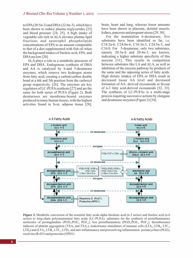
ALA plays a role as a metabolic precursor of EPA and DHA. Endogenous synthesis of DHA and AA is catalyzed by 6-and 5-desaturase enzymes, which remove two hydrogen atoms from fatty acid, creating a carbon/carbon double bond at a 6th and 5th position from the carboxyl group respectively. [26]. The enzymes are key regulators of LC-PUFA synthesis [27] and are the same for both series of PUFA (Figure 2). Both desaturases are membrane-bound enzymes produced in many human tissues, with the highest activities found in liver, adipose tissue [28],
brain, heart and lung, whereas lesser amounts have been shown in placenta, skeletal muscle, kidney, pancreas and pregnant uterus [29, 30].
For the mammalian 6-desaturase, five substrates have been identified so far, i.e. C18:2n-6, C24:6n-6, C18:3n-3, C24:5n-3, and C16:0. For 5-desaturase, only two substrates, namely 20:3n-6 and 20:4n-3, are known, indicating a higher substrate specificity of this enzyme [31]. This results in competition between substrates like LA and ALA, as well as inhibition of the enzyme pathway by products of the same and the opposing series of fatty acids. High dietary intakes of EPA or DHA result in decreased tissue AA level and decreased formation of AA- derived eicosanoids in favour of n-3 fatty acid-derived eicosanoids [32, 33]. The synthesis of LC-PUFAs is a multi-stage process requiring successive actions by elongase and desaturase enzymes (Figure 2) [34].
Figure 2.
rostaglandins (PGD ,PGE , PGF ), less proinflammatory (PGD ,PGE , PGF ), thromboxanes 2 2 2a 3 3 3a
inducers of platelet aggregation (TXA and TXA ), leukotrienes stimulators of immune cells (LTA , LTB , LTC , 2 3 4 4 4
LTD ) and (LTA , LTB , LTC , LTD , and anti-inflammatory and proresolving inflammation prostacyclines (PGI2), 4 5 5 5 5
resolvins (RvD1) and protectins (NPD1)
Metabolic conversion of the essential fatty acids alpha-linolenic acid (n-3 series) and linoleic acid (n-6 series) to long-chain polyunsaturated fatty acids (LC-PUFA), substrates for the synthesis of proinflammatory molecules of p

7
ALA is converted to stearidonic acid by 6 desaturation, and then further elongated to eicosatetraenoic acid (ETA, 20:4n-3). ETA is converted by 5-desaturase to EPA (C20:5n-3), an important n-3 metabolite that serves as a precursor of biologically potent eicosanoids. The major product of the n-3 family DHA (C22:6n-3) is produced by two chain elongations of EPA, followed by a 6-desaturation and a partial -oxidation as it was described by Sprecher [35].
The low activity of 6-desaturase and the translocation to peroxisomes for oxidation may explain the low conversion rate of n-3 docosapentaenoic acid (DPA, 22:5n-3) to DHA in humans [36]. Activity of 6- and 5-desaturases has been demonstrated in human fetal tissue from as early as 17 to 18 weeks of gestation [37], and stable isotope studies have confirmed that preterm and term infants are able to convert ALA to DHA. In the n-6 series LA is converted via intermediates to dihomo-gamma-linolenic acid (20:3n 6) and arachidonic acid (20:4n 6).
The 9-desaturase creates a double bond at the 9th position from the carboxyl end in the synthesis of oleic acid, which can be easily synthesized in the human body from stearic acid. Oleic acid (18:1n-9) could be also the substrate of 6- and 5-desaturases and elongases. Oleic acid is converted via intermediates to “mead acid” (20:3n-9); palmitic acid (16:0) appears to be converted to palmitoleic acid (16:1n-7) and vaccenic acid (18:1n-7). Marked increase in mead acid have previously been reported in the case of severe linoleic acid deficiency [38]. Mead acid is used as a marker of EFA deficiency.
The genes encoding 5-desaturase and 6-desaturase are FADS1 and FADS2, respectively. They form a gene cluster jointly with the gene for fatty acid desaturase 3 (FADS3) on the human chromosome 11q12-q13.1. The presence of a variant T to deletion (T-del) in the promoter of the 6-desaturase gene (FADS2) leads to reduced EPA concentrations in plasma and adipose tissue, suggesting that this variant decreases enzyme activity, and therefore conversion from ALA is insufficient.
Deficiency of PUFAs
Biochemical changes of n-3 fatty acid deficiency include a decrease in plasma and tissue EPA and DHA concentrations [39]. Studies in rodents and nonhuman primates have demonstrated that prolonged feeding with diets containing very low amounts of ALA result in reductions of visual
acuity and cognitive parameters [40]. The effects could be prevented by
supplementation with ALA. The reduction in visual function is accompanied by decreased brain and retina DHA with an increase in DPA (22:5n-6). The compensatory increase in 22 carbon chain n-6 fatty acids results in maintenance of the total amount of n-6 and n-3 polyunsaturated fatty acids in neural tissue.
The offsprings of monkeys fed an n-3 PUFA deficient diet during pregnancy show visual impairments [41]. Supplementation of the infant monkeys with LNA resulted in an increase in the concentration of DHA in neural tissues and an improvement in visual function [42].
This suggests that a deficit in the availability of ALA for conversion, to DHA in particular, was the main mechanism underlying the deficiency symptoms. Changes in learning behaviours in animals fed with ALA deficient diets have also been reported [43]. These studies have involved feeding oils such as sunflower oil, which contains less than 0.1 % of ALA and is high in LA, as the only fat source for prolonged periods.
There is no accepted concentration of plasma or tissue DHA concentrations below which the functions of n-3 fatty acids in visual or neural function are impaired. Similarly, there are no accepted normal ranges for EPA concerning synthesis of EPA-derived eicosanoids or regulation of arachidonic acid metabolism and its eicosanoid metabolites. Clinical signs of essential fatty acid deficiency are generally only found in patients with chronic fat malabsorption on parenteral nutrition and without an enteral or parenteral source of polyunsaturated fat. Early signs of essential fatty acid deficiency include rough and scaly skin, which if left untreated, develops dermatitis [44]. In studies of patients with dermatitis and receiving parenteral nutrition, the ratio of eicosatrienoic acid: arachidonic acid (20:3n-9:20:4n-6) in plasma was elevated. However, in the investigation of patients on parenteral nutrition only it was shown that around 50 % of children receiving long-term total parenteral nutrition lacking ALA had visual dysfunction. These data suggest a decreased availability of DHA for incorporation into neural membranes [45].
Recent studies using stable isotopically labeled fatty acids to investigate the effect of gestational age and intrauterine growth on essential fatty acid desaturation and elongation have shown that the conversion of LA to arachidonic acid occurs as early as 26 weeks of
Penkova R., et al. Variation in polyunsaturated fatty acids in woman...

J Biomed Clin Res Volume 5 Number 1, 2012
8
gestation, and is in fact more active at earlier gestational ages [46]. In addition to its role as a precursor to dihomo-γ-linolenic acid and arachidonic acid, linoleic acid has a specific role in acylceramides, which are important in maintaining the epidermal water barrier.
Two PUFAs, AA and DHA, are critical to foetal and infant central nervous system growth and development. Embedded in the cell membrane phospholipid, AA is involved in cell signalling pathways and cell division, and serves as an inflammatory precursor for eicosanoids. The membrane lipids of brain gray matter and the retina contain very high concentrations of DHA, particularly in the amino phospholipids p h o s p h a t i d y l e t h a n o l a m i n e a n d phosphatidylserine. In these tissues, the concentration of DHA can exceed 50 percent of the fatty acids resulting in the presence of DHA phospholipid species. During n-3 fatty acid deficiency, DHA is definitely retained. Thus, most animal studies investigating the importance of n-3 fatty acids have used rats deprived of n-3 fatty acids for two or more generations.
Benefits of n-3 fatty acids in pregnancy
Most woman, particularly pregnant women do not get enough n-3 fatty acids. To obtain adequate n-3 fatty acids, a variety of sources should be used: vegetable oils, at least two fish servings a week, nuts, and even supplements (fish oil or algae-based DHA).
The synthesis of DHA (C22:6n-3) by the foetus is limited. That is why its transfer across the placenta is of major importance. It has been shown that maternal supplementation with fish oil has been used successfully to increase foetal DHA availability and neonatal DHA status. However, increasing the DHA status of pregnant women with fish oil lowers AA (20:4n-6) concentrations in their infants [47]. Because AA is the second most abundant LC- PUFA in neural tissue, this may not be advantageous. Moreover, high maternal intake of DHA and EPA (C20:5n-3) decreases AA in the placenta as well in foetal liver and brain [48]. It has been found that dietary ALA may be an effective alternative to fish oil for use in increasing maternal and neonatal DHA status [49]. ALA supplementation has been shown to result in the accretion of ALA-derived DHA in the brains of baboon foetuses [50].
Selective transport across the human placenta for individual fatty acids has been suggested as a
mechanism to explain greater concentrations of some PUFAs like DHA and AA in the foetal, rather than maternal, circulation. The placenta plays the key role in pregnancy, forms an anatomical barrier between maternal and foetal circulation, and allows an exchange of gases, nutrients and metabolic products of degradation between mother and foetus. The transporting epithelium, the syncytiotrophoblast surrounding individual villous tree structures within the placenta, contains proteins capable of binding and transporting fatty acids within the polarized syncytiotrophoblast. Fatty acid-binding proteins are cytosolic 14–15 kDa proteins, which play a role in FA uptake and transport. They may modulate FA concentration and in this way influence the functions of enzymes, membranes, ion channels and receptors, gene expression cell growth and differentiation, cellular signalling, gene transcription and cytoprotection. [51]. Placenta contains several types of fatty acid binding proteins (FABP) with a distinct spatio-temporal distribution, fatty acid transporing proteins FATP and FAT/CD36.
The investigated effect of EPA on mRNA expression of fatty acid transport proteins (FATPs) demonstrated that EPA, and not DHA, has been positively correlated with mRNA expression of all membrane proteins. Thus, higher maternal EPA concentrations may increase FATP expression (FATP-4 in particular) which, in turn, has been shown to increase cord blood DHA levels. In pregnancy, the real importance of EPA may be related to its role in mediating DHA and AA concentrations across the placenta rather than its production of the relatively less potent eicosanoids.
Higher EPA concentrations also lead to increased expression of fatty acid-binding proteins FABPs including brain FABP (B-FABP), which is strongly expressed in developing brain cells and has a strong affinity for DHA. EPA levels have been shown to have a marked effect on PPAR expression, an effect not proved to be induced by ALA or by DHA in vitro [52].
In an investigation of pregnant women in Granada by E. Larque et al., [53] healthy pregnant women (n = 136) received, in a double blind randomized trial, 500 mg DHA+150 mg EPA; 400µg N5- methyl-tetrahydrofolic acid (N5- methyl-THFA); 500 mg DHA +400µg N5 methyl-THFA, or placebo during the second half of gestation. Larque et al. analysed the fatty acid composition of maternal and cord blood

9
phospholipids and of placenta; quantifying placental mRNA expression of fatty acid-transport protein 1 (FATP-1), FATP-4, FATP-6, fatty acid translocase, fatty acid– binding protein (FABP) plasma membrane, heart FAPB (H-FABP), adipose (A)-FABP, and brain (B)-FABP (Figure 3) [54].
Figure 3. Fatty acids transporters within the polarized syncytiotrophoblast. The fetal facing basal membrane is on the left from the arrows and the maternal facing - on the right side. FATP4 and pFABPpm are implicated in DHA uptake and FABP1, 3, and 4 may be under the control of HIF. FABP4may be associated with lipid droplets within this tissue (Modified from [54]).
The mRNA expression of FATP-1 and FATP-4 in placenta correlated with DHA in both maternal plasma and placental phospholipids, although only FATP-4 expression significantly corre la ted wi th DHA in cord blood phospholipids. Besides that, the mRNA expression of several membrane lipid carriers correlated with EPA and DHA in placental triacylglycerols and with EPA in placental free fatty acids indicating that these lipid carriers are involved in placental transfer of long-chain polyunsaturated fatty acids.
Despite normal plasma AA and DHA concentrations in women with gestational diabetes [55], low erythrocyte phospholipid long-chain polyunsaturated fatty acids (LC- PUFA) were found in their infants [56]. Fatty acid composition of placenta of women with gestational diabetes had a higher concentration of AA and DHA as compared to placenta of control subjects, suggesting an enhanced uptake of these
two fatty acids. However, AA and DHA concentrations in the foetus were lower than normal.
The importance of PUFA was reported by Danish investigators. They found that women living on the Faroe Islands delivered babies that were 194 g heavier and had gestation lengths 4 days longer than babies born in Denmark [57]. The Faroese diet contained significantly more n-3 fatty acids and less n-6 fatty acids than a Danish diet. Red blood cell fatty acid content (expressed as the ratio of n-3 to n-6) was significantly higher in the Faroese pregnant women than in Danish pregnant women [58].
However, in another trial of Norwegian pregnant women (gestational weeks 17-19) who were randomized to supplementation of 10 ml from cod liver oil or corn oil daily until 3 months after delivery (cod liver oil contain 1183 mg DHA, total 2632 mg n-3 FA) the primary outcomes of gestational length and birth weight did not differ between the two groups [59]. FA content in the blood of the mothers and babies at the end of pregnancy, published later showed higher level of maternal plasma n-3 fatty acids and the ratio of n-3:n-6 FA in the group receiving cod liver oil. Fetal DHA umbilical cord plasma phospholipids were 23% higher in babies born in the group supplemented with cod liver oil. It was found that infants with the highest quartile concentration of DHA in the cord blood had a gestational period 9.3 days longer than those in the lowest quartile [47].Thus, healthy pregnant women taking about 1 g of DHA a day from a marine oil supplement were able to deliver substantially more DHA to the foetus than those who were not given marine oils.
The authors evaluated this same Norwegian group 4 years later in order to cohort the infants. They concluded that the children had higher mental processing scores when born to mothers supplemented with cod liver oil (rich in EPA and DHAn-3 PUFAs) during pregnancy and lactation, as compared with children of mothers who were supplemented with corn oil (rich in ALA n-6 PUFAs).
Thus, it appeared that rather high amounts of n-3 fatty acids need to be consumed to affect gestation and foetal weight.
During the last trimester, the foetus accumulates around 50 to 70 mg per day of DHA. Both maternal DHA intake and maternal circulating DHA concentrations are important for foetal blood concentrations of DHA [60]. DHA concentrations in maternal circulation are
Penkova R., et al. Variation in polyunsaturated fatty acids in woman...
J Biomed Clin Res Volume 5 Number 1, 2012
10
influenced by dietary supply [61]. The accumulation of DHA continues after birth, and DHA is accumulated in the CNS until about 18 months of age.
Because only about 4% to 11% of DHA is retroconverted to EPA, pregnant women who just take DHA supplements, without any dietary EPA, may be unable to produce the right balance of eicosanoids and may limit the transport and uptake of DHA into foetal cells.
Despite experimental and clinical evidence consistent with preferential transfer across the placenta, information from both human and animal studies has shown that the maternal dietary intake of n-6 and n-3 fatty acids influences maternal and foetal AA (20:4n-6) and DHA (22:6n-3) content [62]. A better understanding of the mechanisms involved in the transfer to the neonate and the accumulation is important to improve foetal DHA (22:6n-3) status, as well as the balance between dietary n-3 and n-6 , not only in complicated pregnancies and disorders associated with poor DHA (22:6n-3) status [63] but also in uncomplicated pregnancies to avoid the depletion of n-3) from retinal and neural membranes resulting in reduced visual function, behavioural abnormalities, alterations in the metabolism of several neurotransmitters, decreased membrane receptors and ion channel activities .
References
1. Schmidt MI, Duncan BB. Diabesity: an inflammatory metabolic condition. Clin hem Lab Med. 2005;41:1120-30.
2. Wellen KE, Hotamisligil GS. Inflammation, stress, and diabetes. J Clin Invest. 2005;115:1111-9.
3. Kris-Etherton PM, Harris WS, Appel LJ. Fish consumption, fish oil, omega-3 fatty acids, and c a r d i o v a s c u l a r d i s e a s e . C i r c u l a t i o n . 2002;106(21):2747-57.
4. Wang C, Harris WS, Chung M, Lichtenstein AH, Balk EM, Kupelnick B, Jordan HS. Lau J. n-3 Fatty acids from fish or fish-oil supplements, but not alpha-linolenic acid, benefit cardiovascular disease outcomes in primary- and secondary-prevention studies: a systematic review. Am J Clin Nutr. 2006;84(1):5-17.
5. Stillwell W, Shaikh SR, Zerouga M, Siddiqui R, Wassall SR. Docosahexaenoic acid affects cell signaling by altering lipid rafts. Reprod Nutr Develop. 2005;45(5):559-79.
6. Calder PC. n-3 Fatty acids, inflammation, and immunity – relevance to postsurgical and critically
ill patients. Lipids. 2004;39(12):1147-61.7. Keli SO, Feskens EJ; Kromhout D. Fish
consumption and risk of stroke: The Zutphen Study. Stroke. 1994;25(2):328-32.
8. Leaf A. Prevention of sudden cardiac death by n-3 polyunsaturated fatty acids. Fundam Clin Pharm. 2006;20:525-38.
9. Muskiet FA, Kemperman RF. Folate and longchain polyunsaturated fatty acids in psychiatric disease. J Nutr Biochem. 2006;17:717-27.
10. Benatti P, MC, Peluso G, Nicolai R, Calvani M. Polyunsaturated Fatty Acids: Biochemical, Nutritional and Epigenetic Properties. J Am Coll Nutr. 2004;23(4):281-302.
11. Stillwell W, Shaikh SR, Zerouga M, Siddiqui R, Wassall SR. Docosahexaenoic acid affects cell signaling by altering lipid rafts. Reprod Nutr Develop. 2005;45(5):559-79.
12. Puskas LG, Kitajka K. Nutrigenomic Approaches to Study the Effects of N-3 PUFA Diet in the Central Nervous System. Nutr Health. 2006;18(3):227-32.
13. Shearer GC, Harris WS, Pedersen TL, Newman JW. Detection of omega-3 oxylipins in human plasma and response to treatment with omega-3 acid ethyl esters. J Lipid Res. 2010;51:2074-80.
14. Arita M, Yoshida M, Hong S, Tjonahen E, Glickman JN, Petasis NA et al. Resolvin E1 an endogenous lipid mediator derived from omega-3 eicosapentaenoic acid, protects against 2,4,6-trinitrobenzene sulfonic acid-induced colitis. Proc Natl Acad Sci USA. 2005;102(21):7671-6.
15. World Health Organization, Food and Agriculture Organization of the United Nations. Fats and oils in human nutrition. Report of a joint expert consultation. Rome: FAO Food Nutr Pap. 57; 1994. 147 p.
16. Simopoulos AP. The importance of the ratio of omega-6/omega-3 essential fatty acids. Biomed Pharmacother. 2002;56, 365-79.
17. Institute of Medicine. Food and Nutrition Board. Dietary Reference Intakes for Energy, Carbohydrate, Fiber, Fat, Fatty Acids, Cholesterol, Protein, and Amino Acids. Washington DC: National Academies Press; 2002.
18. Bazan NG, Neuroprotectin D1 (NPD1): a DHA-derived mediator that protects brain and retina against cell injury-induced oxidative stress. Brain Pathol. 2005;15(2):159-66.
19. Voss A, Reinhart M, Sankarappa S, Sprecher H. T h e m e t a b o l i s m o f 7 , 1 0 , 1 3 , 1 6 , 1 9 -docosapentaenoic acid to 4,7,10,13,16,19-docosahexaenoic acid in rat liver is independent of a 4-desaturase. J Biol Chem. 1991;266:19995-20000.
20. Burdge GC, Calder PC. Conversion of alpha-linolenic acid to longer-chain polyunsaturated fatty acids in human adults. Reprod Nutr Dev. 2005;45: 581-97.

11
21. Burdge GC, Wootton SA. Conversion of alpha-l i n o l e n i c a c i d t o e i c o s a p e n t a e n o i c , docosapentaenoic and docosahexaenoic acids in young women. Br J Nutr. 2002;88:411-20.
22. Burdge GC. Metabolism of alpha-linolenic acid in humans. Prostaglandins Leukot Essent Fatty Acids. 2006;75(3):161-8.
23. Harris WS. N-3 fatty acids and serum lipoproteins: human studies. Am J Clin Nutr. 1997; 65(5):1645S-54S.
24. Mori TA, Bao DQ, Burke V, Puddey IB, Beilin LJ. Docosahexaenoic acid but not eicosapentaenoic acid lowers ambulatory blood pressure and heart rate in humans. Hypertension. 1993;34(2):253-60.
25. Morris MC, Sacks F, Rosner B. Does fish oil lower blood pressure? A meta-analysis of controlled trials. Circulation. 1993;88(2):523-33.
26. Innis SM. Perinatal biochemistry and physiology of long-chain polyunsaturated fatty acids. J Pediatr. 2003;143:S1-S8.
27. Nakamura MT, Nara TY. Structure, function, and dietary regulation of delta6, delta5, and delta9 desaturases. Annu Rev Nutr. 2004;24:345-76.
28. Sjogren P, Sierra-Johnson J, Gertow K, Rosell M, Vessby B, de Faire U et al. Fatty acid desaturases in human adipose tissue: relationships between gene expression, desaturation indexes and insulin resistance. Diabetologia. 2008;51(2):328-35.
29. Cho HP, Nakamura M, Clarke SD. Cloning, expression, and fatty acid regulation of the human delta-5 desaturase. J Biol Chem. 1999;274:37335-9.
30. Cho HP, Nakamura MT, Clarke SD. Cloning, expression, and nutritional regulation of the mammalian delta-6 desaturase. J Biol Chem. 1999;274:471-7.
31. Park WJ, Kothapalli KS, Reardon HT, Kim LY, Brenna JT. Novel fatty acid desaturase 3 (FADS3) transcripts generated by alternative splicing. Gene. 2009;446:28-34.
32. Broughton KS, Wade JW. Total fat and (n-3:n-6) fat ratios influence eicosanoid production in mice. J Nutr. 2002;132:88-94.
33. Ferretti A, Nelson GJ, Schmidt PS, Bartolini GL, Kelly DS, Flanagan VP. Dietary docosahexaenoic acid reduces the thromboxane/prostacyclin synthesis ratio in humans. J Nutr Biochem. 1998;32:79-82.
34. Koletzko B, Larque E, Demmelmair H. Placental transfer of long-chain polyunsaturated fatty acids (LC-PUFA). J Perinat Med. 2007; 35(Suppl 1):S5-11.
35. Sprecher H. An update on the pathways of polyunsaturated fatty acid metabolism. Curr Opin Clin Nutr Metab Care. 1999;2(2):135-8.
36. Burdge GC. Alpha-linolenic acid metabolism in men and women: nutritional and biological impl ica t ions . Cl in Nut r Metab Care . 2004;7(2):137-44.
37. De Vrieses SR, Houwelingen ACv, Hornstra G,
Dhont M, Christophe AB. The composition of saturated fatty acids in plasma phospholipids changes in a way to counteract changes in the mean melting point during pregnancy. Lipids. 2001;36(1):15-20.
38. Lundberg WO. The significance of cis, cis, cis-5,8,11-eicosa-trienoic acid etty acid deficiency. Nutr Rev. 1980; 38:233-5.
39. Jeffrey BG, Weisingerb HS, Neuringer M, Mitcheli DC. The role of docosahexaenoic acid in retinal function. Lipids. 2001;36(9):859-71.
40. Willatts P, Forsyth JS, DiModugno MK, Varma S, C o l v i n M . I n f l u e n c e o f l o n g - c h a i n polyunsaturated fatty acids on infant cognitive function. Lipids. 1998;33(10):973-80.
41. Neuringer M, Connor WE, Lin DS, Barstad L, Luck S. Biochemical and functional effects of prenatal and postnatal omega 3 fatty acid deficiency on retina and brain in rhesus monkeys. Proc Natl Acad Sci USA. 1986;83(11): 4021-5.
42. Connor WE, Neuringer M. The effects of n-3 fatty acid deficiency and repletion upon the fatty acid composition and function of the brain and retina. Prog Clin Biol Res. 1988;282:275-94.
43. Innis SM. Perinatal biochemistry and physiology of long-chain polyunsaturated fatty acids. J Pediatr. 2003;143(Suppl 4):S1-8.
44. Jeppesen PB, Hoy CE, Mortensen PB. Essential fatty acid deficiency in patients receiving home parenteral nutri t ion. Am J Clin Nutr. 1998;68(1):126-33.
45. Vinton NE, Heckenlively JR, Laidlaw SA, Martin DA, Foxman SR, Ament ME et al. Visual function in patients undergoing parenteral nutrition. Am J Clin Nutr. 1990;52(5):895-902.
46. Uauy R, Mena P, Wegher B, Nieto S, Salem N. Long chain polyunsaturated fatty acid formation in neonates: Effect of gestational age and intrauterine growth. Pediatr Res. 2000;47(1):127-35.
47. Helland IB, Saugstad OD, Smith L, Saarem K, Solvoll K, Ganes T et al. Similar effects on infants of n-3 and n-6 fatty acids supplementation to pregnant and lactating women. Pediatrics. 2001;108(5):e82. Available at: http://pediatrics. aappublications.org/cgi/content/full/108/5/e82
48. Innis SM, de La Presa Owens S. Dietary fatty acid composition in pregnancy alters neurite membrane fatty acids and dopamine in newborn rat brain. J Nutr. 2001;131(1):118-22.
49. Al MD, van Houwelingen AC, Badart-Smook A, Hornstra G. Some aspects of neonatal essential fatty acid status are altered by linoleicacid supplementation of women during pregnancy. J Nutr. 1995;125: 2822-30.
50. Greiner RC, Winter J, Nathanielsz PW, Brenna JT. Brain docosahexaenoate accretion in fetal baboons: bioequivalence of dietary alpha-linolenic and docosahexaenoic acids. Pediatr Res. 1997;42(6):826-34.
51. Veerkamp JH, Zimmerman AW. Fatty acid-
Penkova R., et al. Variation in polyunsaturated fatty acids in woman...

12
J Biomed Clin Res Volume 5 Number 1, 2012
binding proteins of nervous tissue. J Mol Neurosci. 2001;16(2-3):133-42.
52. Andersen LF, Solvoll K, Drevon CA. Very-long-chain n-3 fatty acids as biomarkers for intake of fish and n-3 fatty acid concentrates. Am J Clin Nutr. 1996;64(3):305-11.
53. Larque E, Krauss-Etschmann S, Campoy C, Hartl D, Linde J, Klingler M et al. Docosahexaenoic acid supply in pregnancy affects placental expression of fatty acid transport proteins. Am J Clin Nutr. 2006;84(4):853-61.
54. Cunningham P, McDermott L. Long Chain PUFA Transport in Human Term Placenta. J Nutr. 2009;139(4):636-9.
55. Wijendran V, Bendel RB, Couch SC, Philipson EH, Thomsen K, Zhang X et al. Maternal plasma phospholipids polyunsaturated fatty acids in pregnancy with and without gestational diabetes mellitus: relations with maternal factors. Am J Clin Nutr. 1999;70(1):53-61.
56. Wijendran V, Bendel RB, Couch SC, Philipson EH, Cheruku S, Lammi-Keefe CJ. Fetal erythrocyte phospholipid polyunsaturated fatty acids are altered in pregnancy complicated with ges ta t ional d iabetes mel l i tus . Lip ids . 2000;35(8):927-31.
57. Olsen SF, Hansen HS, Sorensen TI, Jensen B, Secher NJ, Sommer S et al. Intake of marine fat, rich in (n-3)-polyunsaturated fatty acids, may increase birthweight by prolonging gestation. Lancet. 1986;2(8503):367-9.
58. Olsen SF, Hansen HS, Sommer S, Jensen B, Sørensen TI, Secher NJ et al. Gestational age in relation to marine n-3 fatty acids in maternal erythrocytes: a study of women in the Faroe Islands and Denmark. Am J Obstet Gynecol. 1991;164(5 Pt 1):1203-9.
59. Olsen SF, Sorensen JD, Secher NJ, Hedegaard M, Henriksen TB, Hansen HS et al. Randomised cont ro l led t r ia l o f e ffec t o f f i sh-o i l supplementation on pregnancy duration. Lancet. 1992;339(8800):1003-7.
60. Willatts P. Forsyth JS, DiModugno MK, Varma S, Colvin M.et a l . Effect of long-chain polyunsaturated fatty acids in infant formula on problem solving at 10 months of age. Lancet. 1998;352(9129):688-91.
61. De Vriese SR, Matthys C, De Henauw S, De Backer G, Dhont M, Christophe AB. Maternal and umbilical fatty acid status in relation to maternal diet. Prostaglandins Leukot Essent Fatty Acids. 2002;67(6):389-96.
62. Heird WC, Lapillonne A. The role of essential fatty acids in development. Annu Rev Nutr. 2005;25;549-71.
63. Simmer K, Schulzke SM, Patole S. Long-chain polyunsaturated fatty acid supplementation in preterm infants. Cochrane Database Syst Rev. 2008(1):CD000375.