Embed Size (px)
Citation preview
2239
IntroductionSkeletal muscle’s malleability, which enables remodeling of
the muscle’s structural makeup according to alterations indemand, is a particularly striking phenomenon in the animalkingdom. This plasticity is reflected by the pronouncedadjustments seen in muscular force, endurance and contractilevelocity of mammalian skeletal muscle as a result of analteration in demand (Booth and Baldwin, 1996). This guise iswidely recognized in sports, where distinct adaptation ofmuscle tissue after training in athletes leads to strikingphenotypic modifications that maximize the specificperformance of this contractile tissue.
One notable facet of skeletal muscle plasticity is thespecificity of the adaptive response to a given stimulus (Fluckand Hoppeler, 2003), where the degree of loading and thenumber of muscular contractions appear to be the dominantstimuli for the muscular adaptations. For instance, highlyrepetitive, low-load exercise training will cause differentiationof muscle fibers towards a fatigue-resistance phenotype (Pette,2002). This cellular specialization allows the recruited musclefibers to sustain a high number of slow contractions.Conversely, exercise regimes involving a high degree ofloading provoke an increase in force via fiber hypertrophy. Bycontrast, maintenance of both skeletal muscle mass and
Biological systems have acquired effective adaptivestrategies to cope with physiological challenges and tomaximize biochemical processes under imposedconstraints. Striated muscle tissue demonstrates aremarkable malleability and can adjust its metabolic andcontractile makeup in response to alterations in functionaldemands. Activity-dependent muscle plasticity thereforerepresents a unique model to investigate the regulatorymachinery underlying phenotypic adaptations in a fullydifferentiated tissue.
Adjustments in form and function of mammalianmuscle have so far been characterized at a descriptivelevel, and several major themes have evolved. These implythat mechanical, metabolic and neuronal perturbations inrecruited muscle groups relay to the specific processesbeing activated by the complex physiological stimulus ofexercise. The important relationship between thephenotypic stimuli and consequent muscular modificationsis reflected by coordinated differences at the transcriptlevel that match structural and functional adjustments inthe new training steady state. Permanent alterations ofgene expression thus represent a major strategy for theintegration of phenotypic stimuli into remodeling ofmuscle makeup.
A unifying theory on the molecular mechanism thatconnects the single exercise stimulus to the multi-facetedadjustments made after the repeated impact of themuscular stress remains elusive. Recently, master switcheshave been recognized that sense and transduce theindividual physical and chemical perturbations inducedby physiological challenges via signaling cascades todownstream gene expression events. Molecularobservations on signaling systems also extend the long-known evidence for desensitization of the muscle responseto endurance exercise after the repeated impact of thestimulus that occurs with training. Integrative approachesinvolving the manipulation of single factors and thesystematic monitoring of downstream effects at multiplelevels would appear to be the ultimate method forpinpointing the mechanism of muscle remodeling. Theidentification of the basic relationships underlying themalleability of muscle tissue is likely to be of relevance forour understanding of compensatory processes in othertissues, species and organisms.
Key words: exercise, endurance, hypoxia, gene, transcriptome,morphometry, microarray, PCR.
Summary
The Journal of Experimental Biology 209, 2239-2248Published by The Company of Biologists 2006doi:10.1242/jeb.02149
Review
Functional, structural and molecular plasticity of mammalian skeletal muscle inresponse to exercise stimuli
Martin FlückUnit for Functional Anatomy, Department of Anatomy, University of Berne, Baltzerstrasse 2, Switzerland
e-mail: [email protected]
Accepted 6 February 2006
THE JOURNAL OF EXPERIMENTAL BIOLOGY

2240
oxidative capacity are dependent on the impact of contractilestimuli, as shown by the pronounced deconditioning of musclefunction with inactivity. Thus the profile of muscleperturbation exerts essential control over the musclephenotype. This review sets out our recent findings that buildthe case for the important involvement of gene expression inameliorations of muscle function with repetitive exercisestimuli.
Mechanisms underlying myocellular adaptations toendurance training
The cellular and functional mechanisms underlying theparticular adaptations of the composite muscle tissue toendurance exercise are now well understood. The cellularprocesses behind muscle plasticity involve qualitative andquantitative alterations in muscle fiber cells and associatedstructures. Alterations to endurance training over a period ofweeks to months involve differentiation of the muscle fiberstowards a phenotype with a high mitochondrial volume density(Fluck and Hoppeler, 2003). These myocellular improvementsare assisted by an increase in capillary density and may involvea shift of the contractile character of the fibers towards a slowtype via an exchange of sarcomere components (Fluck andHoppeler, 2003). Collectively, these linked adjustmentscontribute towards maximization of substrate delivery,respiratory capacity and contractile parameters during thefrequent slow contractions that occur with endurance-typeexercise.
The regulatory mechanisms underlying the specificadjustments of muscular organelles to exercise are beginningto be unravelled. The data support the notion that geneexpression underlies muscular adjustments in response tophysical activity (Fig.·1). The model suggests that individualhomeostatic perturbations provoked by exercise are integratedinto alterations in expression levels of diffusible gene copies(i.e. mRNAs), leading to translation of the encoded proteins by
the ribosomal machinery. Enhanced levels of gene transcriptswould therefore support the synthesis of protein componentsand provoke structural remodeling and functional adjustmentsin the long term. Thus changes in mRNA act as a blueprint foradjustment of protein composition (for reviews, see Fluck etal., 2005a; Fluck and Hoppeler, 2003; Booth and Baldwin,1996). In this manner, exercise is known to specifically affectthe rate of synthesis (transcription) and degradation of genetranscripts (Yan et al., 1996; Fluck and Hoppeler, 2003). Geneexpression is therefore an important layer of processing forintegration of exercise stimuli into the adjustments of musclemakeup necessary to match muscle function to alterations indemand.
To test this basic concept we set out to investigate the post-transcriptional processes underlying the tuning of musclemetabolism upon endurance training. The focus of analysiswas on key factors of carbohydrate and lipid metabolization,since these molecule classes constitute the main substrates ofskeletal muscle (Holloszy and Coyle, 1984; van Loon et al.,2001). Both of these organic compounds are imported from thecapillary bed via facilitative processes into the myocellularcompartment. There they reside as myocellular stores untilthey are subjected to controlled metabolization to generatetheir energy equivalents (see Fig.·2). During the catabolicreaction, carbohydrates in the form of glucose are primarilydegraded via anaerobic glycolysis to pyruvate, and eventualcomplete oxidative combustion in the mitochondria via theKrebs cycle. Similarly, triglyceride-derived free fatty acids areimported into mitochondria where they are combusted via beta-oxidation and the Krebs cycle. This latter process producescarbon dioxide and supplies reduction equivalents that lead toATP production via coupling to oxidative phosphorylation.The ATP generated during mitochondrial respiration is thenused to drive energy-dependent processes such as contractions(Fig.·2). From a calorific perspective, the aerobic processeswithin mitochondria are more efficient in generating ATP than
M. Flück
Homeostatic perturbation
Structural–functional adaptations
Instruction
Integration
DNADNA
nucleus
mRNA
gene copying
translation &assembly
local hypoxia, mechanical stress
transcript
gene
protein
signal
Paradigm
gene expression
genome
Fig.·1. Concept of the integration of physiological stimuliin phenotypic responses. Homeostatic perturbations suchas those induced by exercise in muscle are integrated viasignaling pathways into alterations in gene transcription.The diffusible gene copies produced then provide themessage for the instruction of muscle tissue remodelingvia translation and assembly of the encoded proteins.Based upon this relationship it is hypothesized that thesystematic exploration of differences in transcript levelsrelative to phenotypic adjustments arising from the impactof exercise will reveal the strategy underlying muscleplasticity.
THE JOURNAL OF EXPERIMENTAL BIOLOGY

2241Regulatory mechanisms of muscle remodeling
the anaerobic processes. This relates to the principalimplication of oxidative processes in energy allocation withsustained, submaximal types of exercise (Jeukendrup, 2002).
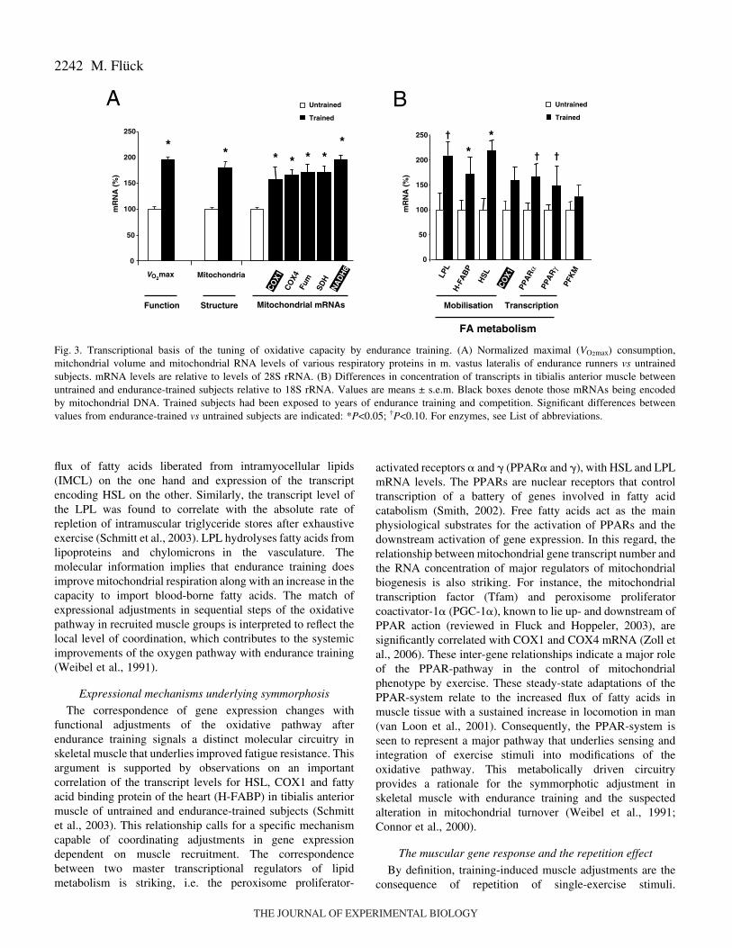
The strategy employed to unravel the regulation ofmetabolic processes involved parallel assessment ofexpression, structural and functional parameters in a majorrecruited muscle group in two ‘steady states’: endurance-trained and untrained subjects, in order to reveal the biologicalrelationships that drive the muscle’s response to repeatedendurance exercise. Oxidative metabolism measurementsincluded the determination of mRNA levels for factorsnecessary for relevant steps of mobilization and oxidativemetabolization of fatty acid in mitochondria as well asmitochondrial volume densities (Fig.·2). Alterationscharacterized in the heavily recruited vastus lateralis muscledemonstrated that mitochondrial respiratory factors areconcomitantly enhanced in endurance-trained athletes(Puntschart et al., 1995). These adjustments in transcriptexpression were in proportion to the augmented mitochondrialvolume density and the increase in systemic maximal oxygenuptake seen in the athletic population compared to untrainedcontrols (Fig.·3A). Note that both mitochondrial- and nuclear-encoded transcripts were increased in a corresponding fashion.Thus mRNA levels of major mitochondrial respiratorysubunits are significantly correlated with mitochondrialvolume density (Fig.·4), suggesting that local adaptations ofmitochondrial transcript number in a major locomotory muscle
group are co-regulated, and matched to maximal respiration ofthe system (i.e. VO2max) during exercise.
Symmorphosis at the molecular level
Molecular examination of the processes underlying theamelioration of metabolic processes with repeated enduranceexercise corroborated observations on the specific co-regulation of oxidative processes in skeletal muscle. Detailedinvestigation of tibialis anterior muscle in competitiveduathletes and untrained male subjects revealed a concomitantenhancement of gene transcript levels for factors involved inmobilization of fatty acids. Furthermore, the study on thismuscle group, which is mostly involved in body balance andfoot control, confirmed the augmented gene message for therespiratory chain component, cytochrome c oxidase subunit I(COX1; Fig.·3B) (Schmitt et al., 2003). Further exploratorycorrelation analysis uncovered significant relationshipsbetween the two main lipases of skeletal muscle, hormone-sensitive lipase (HSL) and alkaline lipoprotein lipase (LPL),with functional elements of lipid metabolism (Fig.·4B). Forinstance, HSL mRNA was significantly correlated with thevolume density of intramyocellular triglycerides andmitochondria, determined by electron microscopy. HSL isknown to reside at the periphery of triglyceride droplets andliberates the entrapped triglycerides for mitochondrialoxidation (for reviews, see Schmitt et al., 2003; Donsmark etal., 2004). The findings now imply a relationship between the
Fig.·2. Metabolic processes in muscle fibers. Themain biochemical processes involved in energygeneration in striated muscle involve the combustionof fatty acids and carbohydrates. Carbohydrates(orange) are imported via facilitative processes fromthe capillary supply lines to the myofibre, where theymay be stored as intramuscular triglycerides orglycogen, respectively, for later combustion. Fattyacid metabolization (green box) is an obligatoryaerobic process that takes place in mitochondria viabeta-oxidation and the Krebs cycle. In contrast, the‘metabolic conversion’ of carbohydrates viaglycolysis in the cytoplasm (orange box) is oxygen-independent and is not necessarily coupled tomitochondrial respiration. This may lead to theproduction of the anaerobic end-product lactate. Thedecomposition of organic backbones in mitochondriaproduces reduction equivalents (and CO2), the formerof which drive the oxygen-dependent generation ofATP via coupling to respiratory chain. Boxed factorsare the crucial proteins involved at successivelyaligned transport, storage and conversion steps ofmetabolic pathways in striated muscle and whosemRNA expression was investigated. Endothelial LPL is involved in transporting fatty acids (FA) from the vasculature through the interstitiuminto the myocellular compartment (Glatz and Storch, 2001; Jeukendrup, 2002). There H-FABP is believed to play a main role in theintramyocellular transport of free FA. HSL liberates free FA from IMCL for mitochondrial oxidation. CPT I is a key enzyme for the uptake ofFA into the mitochondrial matrix. The Krebs cycle enzymes Fum and SDH and the constituents of the electron transport chain, NADH6, COX1and COX4, are then responsible for oxygen-dependent ATP production during mitochondrial respiration. PFKM represents a main control stepfor entry of carbohydrates into the glycolytic pathway. For further explanation, see List of abbreviations.
mitochondrion
beta-oxidation
G-6-P
Fatty acids
IMCL
CO2
ATP
ADP
ADP
ATPKrebs
anaerobic glycolysis
oxidative metabolism
PiADP
PiADPADP
muscle fibre
HSL
COX1
H-FABP
COX5B
PFK
CYC
NADH6
O2capillaryLipid
LPLcarbohydrate
cycleSDH
Fum
glycogen
THE JOURNAL OF EXPERIMENTAL BIOLOGY
2242
flux of fatty acids liberated from intramyocellular lipids(IMCL) on the one hand and expression of the transcriptencoding HSL on the other. Similarly, the transcript level ofthe LPL was found to correlate with the absolute rate ofrepletion of intramuscular triglyceride stores after exhaustiveexercise (Schmitt et al., 2003). LPL hydrolyses fatty acids fromlipoproteins and chylomicrons in the vasculature. Themolecular information implies that endurance training doesimprove mitochondrial respiration along with an increase in thecapacity to import blood-borne fatty acids. The match ofexpressional adjustments in sequential steps of the oxidativepathway in recruited muscle groups is interpreted to reflect thelocal level of coordination, which contributes to the systemicimprovements of the oxygen pathway with endurance training(Weibel et al., 1991).
Expressional mechanisms underlying symmorphosis
The correspondence of gene expression changes withfunctional adjustments of the oxidative pathway afterendurance training signals a distinct molecular circuitry inskeletal muscle that underlies improved fatigue resistance. Thisargument is supported by observations on an importantcorrelation of the transcript levels for HSL, COX1 and fattyacid binding protein of the heart (H-FABP) in tibialis anteriormuscle of untrained and endurance-trained subjects (Schmittet al., 2003). This relationship calls for a specific mechanismcapable of coordinating adjustments in gene expressiondependent on muscle recruitment. The correspondencebetween two master transcriptional regulators of lipidmetabolism is striking, i.e. the peroxisome proliferator-
activated receptors � and � (PPAR� and �), with HSL and LPLmRNA levels. The PPARs are nuclear receptors that controltranscription of a battery of genes involved in fatty acidcatabolism (Smith, 2002). Free fatty acids act as the mainphysiological substrates for the activation of PPARs and thedownstream activation of gene expression. In this regard, therelationship between mitochondrial gene transcript number andthe RNA concentration of major regulators of mitochondrialbiogenesis is also striking. For instance, the mitochondrialtranscription factor (Tfam) and peroxisome proliferatorcoactivator-1� (PGC-1�), known to lie up- and downstream ofPPAR action (reviewed in Fluck and Hoppeler, 2003), aresignificantly correlated with COX1 and COX4 mRNA (Zoll etal., 2006). These inter-gene relationships indicate a major roleof the PPAR-pathway in the control of mitochondrialphenotype by exercise. These steady-state adaptations of thePPAR-system relate to the increased flux of fatty acids inmuscle tissue with a sustained increase in locomotion in man(van Loon et al., 2001). Consequently, the PPAR-system isseen to represent a major pathway that underlies sensing andintegration of exercise stimuli into modifications of theoxidative pathway. This metabolically driven circuitryprovides a rationale for the symmorphotic adjustment inskeletal muscle with endurance training and the suspectedalteration in mitochondrial turnover (Weibel et al., 1991;Connor et al., 2000).
The muscular gene response and the repetition effect
By definition, training-induced muscle adjustments are theconsequence of repetition of single-exercise stimuli.
M. Flück
VO2max Mitochondria
Mitochondrial mRNAs
0
50
100
150
200
250
Untrained
Trained
* * * * **
**
*
A B
PFK
M
FA metabolism
0
50
100
150
200
250
CO
X1
PPA
Rγ
PPA
Rα
Transcription
HSL
H-F
AB
P
LPL
Mobilisation
†
† †
mR
NA
(%
)
mR
NA
(%
)
StructureFunctionSD
H
CO
X1
Fum
CO
X4
NA
DH
6
Untrained
Trained
Fig.·3. Transcriptional basis of the tuning of oxidative capacity by endurance training. (A) Normalized maximal (VO2max) consumption,mitchondrial volume and mitochondrial RNA levels of various respiratory proteins in m. vastus lateralis of endurance runners vs untrainedsubjects. mRNA levels are relative to levels of 28S rRNA. (B) Differences in concentration of transcripts in tibialis anterior muscle betweenuntrained and endurance-trained subjects relative to 18S rRNA. Values are means ± s.e.m. Black boxes denote those mRNAs being encodedby mitochondrial DNA. Trained subjects had been exposed to years of endurance training and competition. Significant differences betweenvalues from endurance-trained vs untrained subjects are indicated: *P<0.05; †P<0.10. For enzymes, see List of abbreviations.
THE JOURNAL OF EXPERIMENTAL BIOLOGY

2243Regulatory mechanisms of muscle remodeling
Adaptations of muscle tissue to increased contractile activityare proposed to be confined to the recovery phase from eachfatiguing bout of exercise (Fluck, 2004; Pilegaard et al., 2000).This would allow an overshoot of cellular adaptations thatsupport the accumulation of incremental remodeling responsesafter each session, and with repetition of the single-exercisestimuli this would support the enhanced enduranceperformance (Fig.·5). The extent to which gene expressionprocesses underlie the continued build-up of muscle structureand performance with each bout of exercise remains to beexplored.
To that end we hypothesized that a systematic explorationof changes in mRNA levels would reveal the molecular
strategies underlying muscle plasticity. We employedmicroarray technology to test whether RNA adaptations in therecovery phase contribute to exercise-induced build-up ofmuscle tissue. This novel technology allows the parallelassessment of adjustments in the levels of hundreds tothousands of transcripts. Nylon filters holding 222 cDNAprobes for muscle-relevant factors were custom-designed forthe detection of reverse-transcribed RNAs after differentrecovery times from exercise (Fluck et al., 2005a).
Exploration of the muscular adjustments revealed a generaltrend for a transient upregulation 8·h after one bout ofergometer exercise (Fig.·6) (Schmutz et al., 2006). This mainresponse to 30·min of bicycling concerned gene families
[mitochondrial RNA]�Vv(mt,f)�VO2max
0
1
2
3
4
3 5 7 9 11Mitochondrial volume density (%)
R 2 =0.56COX1 0
10
20
30
40
R 2 =0.68COX4
Untrained Trained
A BVv(li,f)� [HSL mRNA]�Vv(mt,f)
FA
ATP
IMCL
mitochondria
1/1000mm
r=0.64
r=0.66
mR
NA
/28S
RN
A
HSL
respiration
Fig.·4. Symmorphosis at the RNA level. Transcript–structure correlations bring about significant functional relationships. These concern thecoordination of gene expression between the nuclear and mitochondrial genomes and the match of lipase expression to rate and capacity of fattyacid metabolism. (A) Correlation of mitochondrial-encoded COX1 and nuclear-encoded COX4 mRNA levels with mitochondrial volume densityper fiber, i.e. Vv(mt,f). (B) Micrograph showing an intramyocellular droplet of lipid (IMCL) being enveloped by a mitochondrion. The pathwayinvolved in the oxidative combustion of IMCL-derived fatty acids (FA) in mitochondria is indicated by a green arrow. The suspected localizationof HSL and the correlation coefficients (r) between HSL mRNA with volume density of intramuscular lipids, Vv(li,f), and volume density ofmitochondria, Vv(mt,f), are given in black and white font, respectively.
RecoveryFatigue
RNA
Exercise
�RNARNA
�
Performance
Time
RNA
�
Fig.·5. Microadaptations of transcript expressionrelate to the training effect. Model of the increasein (mitochondrial) RNA and enduranceperformance with repetition of exercise. Each boutof exercise leads to an overshoot of transcriptlevels in the recovery phase from fatiguingexercise (�RNA), which leads via translation to amicroadaptation of the encoded protein and relatedstructure. This relays to the gradual accumulation(�) of mitochondrial volume density and theimproved oxidative capacity with repetition ofendurance exercise. A match of transcriptional,structural and functional parameters is observed inrecruited muscle groups between untrained andendurance-trained steady states. Black stippled andsolid lines indicate the evolution of RNA levels andperformance, respectively, during the training.
THE JOURNAL OF EXPERIMENTAL BIOLOGY

2244
implicated in the oxidative pathway. Multiple factors involvedin the extracellular and myocellular mobilization of fatty acidsas well as mitochondrial beta oxidation and the electrontransport chain were affected. The acute adjustment of themuscle transcriptome related to the oxidative pathway to 6weeks of training was found to recapitulate the knownelevation with years of endurance training (Fig.·3) (Fluck andHoppeler, 2003). The results support the concept thatmicroadaptations in expression after each exercise boutinstruct the structural–functional adjustments of oxidativemuscle metabolism to each exercise bout which accumulatewith repetition of exercise stimuli (Fig.·5).
Conversely, when typical muscle adjustments had beenestablished after 6 weeks of endurance training this transcriptresponse was specifically modified. In particular, the acuteinduction of most transcripts to a matched single exercise boutwas blunted (not shown) (Schmutz et al., 2006), which can beexplained by the increased steady-state mRNA levels andrelates to the reduced adaptive potential in trained individuals(Saltin et al., 1977). These observations reveal that a multi-faceted and coordinated expression program underlies the
specific muscular adjustments with the repeated impact ofexercise with training and relates to the sensitivity of response.
Hypoxia as a stimulus of the exercise response
In the context of the stimuli that instruct muscle plasticity,local hypoxia has been postulated to constitute a main signalfor muscular adjustments to endurance exercise (Hoppeler andVogt, 2001), given that there is a dramatic drop in muscleoxygen tension with the onset of exercise (Richardson et al.,1995; Richardson et al., 2001). Similarly, ambient hypoxiareduces muscular oxygen levels and is known to amplify theexercise-induced local muscle hypoxia (Richardson et al.,1995; Hoppeler et al., 2003). This relates to the long-heldtheory of promotion of respiratory adjustments in chronichypoxia and the hypoxia-induced shift away from the oxidativemetabolisation of fatty acids towards increased utilization ofcarbohydrates via the glycolytic pathway (Reynafarje, 1962;Hoppeler et al., 2003). We therefore speculated that theaddition of a defined normobaric hypoxic stress to the stimulusof a 30·min ergometer-bout would shift the acute musculartranscriptome response towards reduced level adjustments of
M. Flück
A Normoxiam
RN
A/2
8S r
RN
A
1.0
0
pre +1 +8 +24
Desmin
Titin
30 min @threshold
ECH1HADHB
ACADM
ACADL
ACADVL
0
1.0
2.0
3.0
4.0
5.0beta oxidation
Time post exercise (h) Time post exercise (h)
1.0
0
pre +1 +8 +24
Desmin
Titin
ACADL
ECH1ACADVLACADM
ECH10
1.0
2.0
3.0
4.0
B Hypoxia
CPT1
H-FABP
LPL
COX5B
CYC
COX1
0
1.0
2.0
3.0
4.0
5.0
mR
NA
/28S
rR
NA
FA transport and respiration
5.0
beta oxidation
0
1.0
2.0
3.0
4.0
5.0
CPT1
H-FABP
LPL
COX5B
CYC
COX1
30 min @threshold
FA transport & respiration
Fig.·6. Time-course of the muscular exercise response in (A) normoxia and (B) hypoxia. Untrained male subjects exercised for 30·min at theaerobic threshold on a bicycle ergometer while breathing normoxic (21% O2) or hypoxic air (13% O2, N=6/group). Biopsies were harvested duringthe time-course of recovery from this single bout of exercise. Total RNA was isolated and subjected to expression profiling using custom-mademicroarrays (Fluck et al., 2005a; Schmutz et al., 2006). Transcript signals were related to the internal 28S rRNA reference and analyzed forstatistical significance using a Friedman ANOVA. The significant changes of selected transcripts related to the oxidative pathway, such as fattyacid transport, beta oxidation and mitochondrial respiration, are shown in A and B. Significantly altered mRNA levels throughout the time-courseof recovery are underlined. Unaffected transcript levels of the main cytoskeletal factors titin and desmin demonstrate the specificity of effect.
THE JOURNAL OF EXPERIMENTAL BIOLOGY
2245Regulatory mechanisms of muscle remodeling
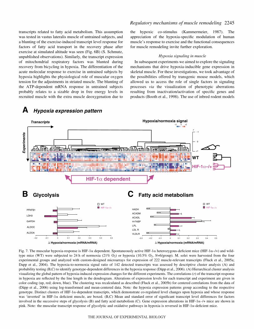
transcripts related to fatty acid metabolism. This assumptionwas tested in vastus lateralis muscle of untrained subjects, anda blunting of the exercise-induced transcript level response forfactors of fatty acid transport in the recovery phase afterexercise at simulated altitude was seen (Fig.·6B) (S. Schmutz,unpublished observations). Similarly, the transcript expressionof mitochondrial respiratory factors was blunted duringrecovery from bicycling in hypoxia. The differentiation of theacute molecular response to exercise in untrained subjects byhypoxia highlights the physiological role of muscular oxygentension for the adjustments in striated muscle. The blunting ofthe ATP-dependent mRNA response in untrained subjectsprobably relates to a sizable drop in free energy levels inrecruited muscle with the extra muscle deoxygenation due to
the hypoxic co-stimulus (Kammermeier, 1987). Theappreciation of the hypoxia-specific modulation of humanmuscle’s response to exercise and the functional consequencesfor muscle remodeling invite further exploration.
Hypoxia signaling in muscle
In subsequent experiments we aimed to explore the signalingmechanisms that drive hypoxia-inducible gene expression inskeletal muscle. For these investigations, we took advantage ofthe possibilities offered by transgenic mouse models, whichallowed us to access the role of single factors in signalingprocesses via the visualization of phenotypic aberrationsresulting from inactivation/activation of specific genes andproducts (Booth et al., 1998). The use of inbred rodent models
+1
0
–1r
Transcripts
Exp
erim
ents
WT
HIF-1α–/+
A Hypoxia expression pattern
HIF-1α dependent
Hypoxia/normoxia signal–2 0 +2
B Glycolysis
–0.2 –0.1 0 0.1 0.2 0.3 0.4 0.5
ALDOA
ALDOC
GAPDH
LDH3
PFKFB1
� Hypoxia/normoxia (mRNA/mRNA) � Hypoxia/normoxia (mRNA/mRNA)
WT
HIF-1α–/+ α–/+
C Fatty acid metabolism
–0.3 –0.2 –0.1 0.0 0.1 0.2 0.3 0.4 0.5
VLDLR
LDL R
LPL
H-FABP
ACADL
ACADM
HADH
WT
HIF-1
Fig.·7. The muscular hypoxia-response is HIF-1� dependent. Spontaneously active HIF-1� heterozygous-deficient mice (HIF-1�–/+) and wild-type mice (WT) were subjected to 24·h of normoxia (21% O2) or hypoxia (10.5% O2, N=6/group). M. solei were harvested from the fourexperimental groups and analyzed with custom-designed microarrays for expression of 222 muscle-relevant transcripts (Fluck et al., 2005a;Dapp et al., 2004). The hypoxia-to-normoxia signal ratio of 142 detected transcripts was assessed by descriptive cluster analysis (A) andprobability testing (B,C) to identify genotype-dependent differences in the hypoxia response (Däpp et al., 2006). (A) Hierarchical cluster analysisvisualizing the global pattern of hypoxia-induced expression changes for the different experiments. The correlations (r) of the transcript responsein hypoxia are reflected by the line length in the dendrogram. Alterations of expression levels for each transcript and experiment are given incolor coding (up, red; down, blue). The clustering was recalculated as described (Fluck et al., 2005b) for centered correlations from the data of(Däpp et al., 2006) using log-transformed and mean-centered data. Note: the hypoxia expression patterns group according to the respectivegenotype. Distinct clusters of HIF-1�-dependent transcripts, which demonstrate co-regulated level changes upon hypoxia and whose responsewas ‘inverted’ in HIF-1� deficient muscle, are boxed. (B,C) Mean and standard error of significant transcript level differences for factorsinvolved in the successive steps of glycolysis (B) and fatty acid metabolism (C). Gene expression alterations in HIF-1�–/+ mice are shown inpink. Note: the muscular transcript response of glycolytic and oxidative pathways in hypoxia is reversed in HIF-1�-deficient mice.
THE JOURNAL OF EXPERIMENTAL BIOLOGY

2246
also has obvious advantages as it permits control overexperimental variables, reducing noise and variability andallowing maximization of the physiological input that drivesmuscle plasticity (for a review, see Wittwer et al., 2004). Weapplied this tool towards elucidation of the role of the alphasubunit of the hypoxia-inducible factor 1 (HIF-1�) in responseto a reduction in ambient oxygen concentration. This factoracts as a regulatory switch for hypoxia sensing in variouscellular systems (Semenza, 2000). In normoxia, HIF-1� israpidly tagged for degradation. Conversely, it is stabilized inan organ-specific manner in hypoxia, permitting its associationwith the HIF-1� subunit to form the DNA-binding HIF-1complex (Pisani and Dechesne, 2005; Stroka et al., 2001; Yuet al., 1998). This heterodimer initiates the transcription ofvarious hypoxia-responsive genes of metabolic processes thatwould be advantageous under the constraint of reducedoxygen, such as capillary growth and glycolysis (for a review,see Hoppeler et al., 2003).
The specific experimental set-up to elucidate the role of HIF-1� in the muscular hypoxia response employed HIF-1�heterozygous-deficient mice exposed to hypoxic vs normoxicair (Fig.·7). Mice with one HIF-1� allele ablated (HIF-1�–/+)demonstrated a 30% lower level of HIF-1� mRNA in the anti-
gravitational soleus muscle under study than control mice(Däpp et al., 2006). Such a partial HIF-1� deficiency has beenshown before to interfere negatively with multiple systemicresponses to hypoxia (Yu et al., 1999). To test this assumption,differences in hypoxia-induced adjustments in transcript levelsin soleus muscle under spontaneous cage activity werecompared between wild-type and HIF-1� heterozygous-deficient mice. Subsequently, genotype-dependent differencesof the effect of a 24·h exposure to hypoxia were analyzed formajor patterns using cluster analysis. This multi-correlationalgorithm identified that the expressional response of muscleto hypoxia was distinct between the two genotypes (Fig.·7A).Detailed inspection of the indicated differences usingprobability testing demonstrated major shifts in hypoxia-induced adjustments in expression related to carbohydratemetabolism with a reduction of the HIF-1� mRNA level. Incontrast, a general level of reduction of transcripts related tofatty acid metabolism was noted in hypoxia and reversed in theHIF-1� heterozygous-deficient mice (Fig.·7C). Conversely,hypoxia-induced mRNA levels of glycolytic factors wereblunted in the mice with reduced HIF-1� levels (Fig.·7B). Aslocal hypoxia is a suspected consequence of ambient oxygenconcentration, the latter finding underscores the suspected role
M. Flück
AAAAAAAA
DNA
Transcriptome
Stimulus
Sensor
Fatty acidmetabolism
Transcription
Translation
Capillarity
nucleus
Sig
nal
inte
gra
tio
nG
ene
exp
ress
ion
Mitochondrialbiogenesis
metabolicmechanical
homeostaticperturbations
PGC-1αPPARα/γ
FFAO2
HIF-1α
ATP
AMP
AMPK
Tension
assembly
gene copying
neuronal
Ca2+
JNK
mitochondrion
transduction
hormonal
cooperation
coordination
Specificity
TFAM
genome
interactome
proteome
physiome
Control level
Fig.·8. Scheme visualizing the integration of the complex stimulus of exercise in recruited skeletal muscle. Different homeostatic perturbations,such as those related to metabolic flux, loading, hormonal and neuronal alterations, are converted by specific sensory molecules into the activationof signaling cascades. These ultimately control muscle fate via the regulation of gene expression. Distinct master switches evolve that relate tothe main themes of the gene expressional response in striated muscle. These phenomena involve the cooperation of gene expressional regulationof metabolic pathways, the coordination between nuclear and mitochondrial genomes and the specificity of the muscular adaptation with respectto the ‘composition’ of the respective exercise stimulus. Consequently, gene expression represents an important layer of control for the processingof physiological information towards a biological outcome.
THE JOURNAL OF EXPERIMENTAL BIOLOGY

2247Regulatory mechanisms of muscle remodeling
of hypoxia as a major regulator of the muscle phenotype.Meanwhile the results also highlight the importance of HIF-1�in the opposing regulation of carbohydrate- and fat-metabolizing processes in muscle.
Signal integration
Within a historical perspective our results extend thebiochemical and cellular exploration of the paradigm of muscleplasticity to show that transcript level adjustments underlie thetuning of the biological processes by exercise. The current datasupport the concept that the structural/functional adjustmentsseen on training reflect the accumulation of transientadjustments in gene expression after repetition of exercisestimuli during training (Fig.·5). The complex stimulus ofexercise provokes a series of homeostatic perturbations inrecruited muscle (Fig.·8). These are sensed by distinctsignaling processes and transduced to downstream activationof gene transcription or a stabilization of transcripts. Knownperturbations include alterations of metabolic, mechanical,hormonal and neuronal factors. With regard to the involvementof metabolic alterations, a drop in oxygen tension, as pointedout in here, an increased flux in free fatty acids and a drop inthe AMP/ATP ratio have evolved as the main components ofthe phenotypic active stimuli in muscle (Fig.·8) (Fluck andHoppeler, 2003). Interaction of these signaling events must beconsidered in order to explain the complex physiologicaloutcome of endurance exercise when combined with co-stimulisuch as hypoxia.
Conclusions
Taking all the above findings together, we infer that acomplex gene response reflects the specificity of the muscularadaptation to different types of exercise. Co-regulation appearsto exist across the oxidative pathway, chromosomes andgenomes. The apparent correlation within gene families andstructure–function relationships reveals that a distinctmolecular circuitry underlies symmorphosis of the pathway ofoxygen, suggesting the involvement of master switches in thecoordination of the local training response. The members ofthese pathways that integrate the homeostatic perturbations inexercised muscle tissue into specific remodeling of muscleorganelles begin to be identified. The well-describedphenomenology of skeletal muscle plasticity and the uniquefeatures of this tissue behavior such as specificity, reversibility,desensitization and accessibility, put muscle into a uniqueposition for future studies on the biological principlesunderlying cell plasticity in vivo.
List of abbreviationsACADL long-chain acyl-CoA dehydrogenaseACADVL very-long-chain acyl-CoA dehydrogenaseACADM medium-chain specific acyl-CoA dehydrogenaseALDOA aldolase AALDOC aldolase CAMPK 5�AMP-activated protein kinase
CPT1 carnitine palmitoly transferase 1CYC cytochrome cCOX1 cytochrome c oxidase subunit 1COX4 cytochrome c oxidase subunit 4COX5B cytochrome c oxidase subunit 5BECH1 enoyl-CoA hydrataseFFA free fatty acidsFum fumaraseGAPDH glyceraldehyde-3-phosphate dehydrogenaseG-6-P glucose-6-phosphateHADH 3-hydroxyacyl-CoA dehydrogenase type IIHADHB 3-hydroxyacyl-CoA dehydrogenase BH-FABP fatty acid binding protein of the heartHIF-1�,� hypoxia-inducible factor 1�,�HSL hormone-sensitive lipaseIMCL intramyocellular lipidsPFK phosphofructokinaseJNK c-jun N-terminal kinaseLDH3 lactate dehydrogenase 3LDL R low-density lipoprotein receptorLPL alkaline lipoprotein lipaseNADH nicotinamide adenine dinucleotide
dehydrogenasePFKM phosphofructo-kinase muscle-typePFKFB1 6-phosphofructo-2-kinase/fructose-2,6-
biphosphatase 1PPAR� peroxisome proliferator-activated receptor �PPAR� peroxisome proliferator-activated receptor �SDH succinate dehydrogenaseTfam mitochondrial transcription factorPGC-1� peroxisome proliferator coactivator-1�VLDLR Very low-density lipoprotein receptorWT wild type
The financial support of the Swiss National ScienceFoundation, the encouragement of Hans Hoppeler and theexperimental support of Prof. Max Gassmann, Dr ChristophDäpp and Dr Silvia Schmutz are greatly acknowledged.
ReferencesBooth, F. W. and Baldwin, K. M. (1996). Muscle plasticity: energy demand
and supply processes. In Handbook of Physiology (ed. L. B. Rowell and J.T. Shepherd), pp. 1074-1123. New York: Oxford University Press.
Booth, F. W., Tseng, B. S., Fluck, M. and Carson, J. A. (1998). Molecularand cellular adaptation of muscle in response to physical training. ActaPhysiol. Scand. 162, 343-350.
Connor, M. K., Bezborodova, O., Escobar, C. P. and Hood, D. A. (2000).Effect of contractile activity on protein turnover in skeletal musclemitochondrial subfractions. J. Appl. Physiol. 88, 1601-1606.
Dapp, C., Schmutz, S., Hoppeler, H. and Fluck, M. (2004). Transcriptionalreprogramming and ultrastructure during atrophy and recovery of mousesoleus muscle. Physiol. Genomics 20, 97-107.
Däpp, C., Gassmann, M., Hoppeler, H. and Flück, M. (2006). Hypoxia-induced gene activity in disused oxidative muscle. Adv. Exp. Biol. Med. Inpress.
Donsmark, M., Langfort, J., Holm, C., Ploug, T. and Galbo, H. (2004).Regulation and role of hormone-sensitive lipase in rat skeletal muscle. Proc.Nutr. Soc. 63, 309-314.
Fluck, M. (2004). Exercise-modulated mitochondrial phenotype; sensors andgene regulation. J. Muscle Res. Cell Motil. 25, 235-237.
THE JOURNAL OF EXPERIMENTAL BIOLOGY

2248
Fluck, M. and Hoppeler, H. (2003). Molecular basis of skeletal muscleplasticity – from gene to form and function. Rev. Physiol. Biochem.Pharmacol. 146, 159-216.
Fluck, M., Dapp, C., Schmutz, S., Wit, E. and Hoppeler, H. (2005a).Transcriptional profiling of tissue plasticity: role of shifts in gene expressionand technical limitations. J. Appl. Physiol. 99, 397-413.
Fluck, M., Schmutz, S., Wittwer, M., Hoppeler, H. and Desplanches, D.(2005b). Transcriptional reprogramming during reloading of atrophied ratsoleus muscle. Am. J. Physiol. 289, R4-R14.
Glatz, J. F. and Storch, J. (2001). Unravelling the significance of cellularfatty acid-binding proteins. Curr. Opin. Lipidol. 12, 267-274.
Holloszy, J. O. and Coyle, E. F. (1984). Adaptations of skeletal muscle toendurance exercise and their metabolic consequences. J. Appl. Physiol. 56,831-838.
Hoppeler, H. and Vogt, M. (2001). Muscle tissue adaptations to hypoxia. J.Exp. Biol. 204, 3133-3139.
Hoppeler, H., Vogt, M., Weibel, E. R. and Fluck, M. (2003). Response ofskeletal muscle mitochondria to hypoxia. Exp. Physiol. 88, 109-119.
Jeukendrup, A. E. (2002). Regulation of fat metabolism in skeletal muscle.Ann. N. Y. Acad. Sci. 967, 217-235.
Kammermeier, H. (1987). High energy phosphate of the myocardium:concentration versus free energy change. Basic Res. Cardiol. 82, S31-S36.
Pette, D. (2002). The adaptive potential of skeletal muscle fibers. Can. J. Appl.Physiol. 27, 423-448.
Pilegaard, H., Ordway, G. A., Saltin, B. and Neufer, P. D. (2000).Transcriptional regulation of gene expression in human skeletal muscleduring recovery from exercise. Am. J. Physiol. 279, E806-E814.
Pisani, D. F. and Dechesne, C. A. (2005). Skeletal muscle HIF-1alphaexpression is dependent on muscle fiber type. J. Gen. Physiol. 126, 173-178.
Puntschart, A., Claassen, H., Jostarndt, K., Hoppeler, H. and Billeter, R.(1995). mRNAs of enzymes involved in energy metabolism and mtDNA areincreased in endurance-trained athletes. Am. J. Physiol. 269, C619-C625.
Reynafarje, B. (1962). Myoglobin content and enzymatic activity of muscleand altitude adaptation. J. Appl. Physiol. 17, 301-305.
Richardson, R. S., Noyszewski, E. A., Kendrick, K. F., Leigh, J. S. andWagner, P. D. (1995). Myoglobin O2 desaturation during exercise.Evidence of limited O2 transport. J. Clin. Invest. 96, 1916-1926.
Richardson, R. S., Newcomer, S. C. and Noyszewski, E. A. (2001). Skeletalmuscle intracellular PO(2) assessed by myoglobin desaturation: response tograded exercise. J. Appl. Physiol. 91, 2679-2685.
Saltin, B., Henriksson, J., Nygaard, E., Andersen, P. and Jansson, E.
(1977). Fiber types and metabolic potentials of skeletal muscles in sedentaryman and endurance runners. Ann. N. Y. Acad. Sci. 301, 3-29.
Schmitt, B., Fluck, M., Decombaz, J., Kreis, R., Boesch, C., Wittwer, M.,Graber, F., Vogt, M., Howald, H. and Hoppeler, H. (2003).Transcriptional adaptations of lipid metabolism in tibialis anterior muscleof endurance-trained athletes. Physiol. Genomics 15, 148-157.
Schmutz, S., Däpp, C., Wittwer, M., Vogt, M., Hoppeler, H. and Flück,M. (2006). Endurance training modulates the muscular transcriptomeresponse to acute exercise. Pflügers Arch. 451, 678-687.
Semenza, G. L. (2000). HIF-1: mediator of physiological andpathophysiological responses to hypoxia. J. Appl. Physiol. 88, 1474-1480.
Smith, S. A. (2002). Peroxisome proliferator-activated receptors and theregulation of mammalian lipid metabolism. Biochem. Soc. Trans. 30, 1086-1090.
Stroka, D. M., Burkhardt, T., Desbaillets, I., Wenger, R. H., Neil, D. A.,Bauer, C., Gassmann, M. and Candinas, D. (2001). HIF-1 is expressedin normoxic tissue and displays an organ-specific regulation under systemichypoxia. FASEB J. 15, 2445-2453.
van Loon, L. J., Greenhaff, P. L., Constantin-Teodosiu, D., Saris, W. H.and Wagenmakers, A. J. (2001). The effects of increasing exerciseintensity on muscle fuel utilisation in humans. J. Physiol. 536, 295-304.
Weibel, E. R., Taylor, C. R. and Hoppeler, H. (1991). The concept ofsymmorphosis: a testable hypothesis of structure-function relationship.Proc. Natl. Acad. Sci. USA 88, 10357-10361.
Wittwer, M., Billeter, R., Hoppeler, H. and Fluck, M. (2004). Regulatorygene expression in skeletal muscle of highly endurance-trained humans.Acta Physiol. Scand. 180, 217-227.
Yan, Z., Salmons, S., Dang, Y. I., Hamilton, M. T. and Booth, F. W. (1996).Increased contractile activity decreases RNA-protein interaction in the 3�-UTR of cytochrome c mRNA. Am. J. Physiol. 271, C1157-C1166.
Yu, A. Y., Frid, M. G., Shimoda, L. A., Wiener, C. M., Stenmark, K. andSemenza, G. L. (1998). Temporal, spatial, and oxygen-regulated expressionof hypoxia-inducible factor-1 in the lung. Am. J. Physiol. 275, L818-L826.
Yu, A. Y., Shimoda, L. A., Iyer, N. V., Huso, D. L., Sun, X., McWilliams,R., Beaty, T., Sham, J. S., Wiener, C. M., Sylvester, J. T. et al. (1999).Impaired physiological responses to chronic hypoxia in mice partiallydeficient for hypoxia-inducible factor 1alpha. J. Clin. Invest. 103, 691-696.
Zoll, J., Steiner, R., Meyer, K., Vogt, M., Hoppeler, H. and Flück, M.(2006). Gene expression in skeletal muscle of coronary artery diseasepatients after concentric and eccentric endurance training. Eur. J. Appl.Physiol. 96, 413-422.
M. Flück
THE JOURNAL OF EXPERIMENTAL BIOLOGY


![[45 ] THE QUANTITATIVE NUTRITIONAL …jeb.biologists.org/content/jexbio/33/1/45.full.pdf · Quantitative nutritional requirements of Drosophila melanogaster 47 spores, and the fluctuations](https://img.pdfslide.us/doc/110x75/5ac1f6ec7f8b9a4e7c8db233/45-the-quantitative-nutritional-jeb-nutritional-requirements-of-drosophila.jpg)

