Embed Size (px)
Citation preview
Review
From RNA world to SARS-CoV-2: the edited story of RNA viral
evolution
Zachary W. Kockler 1 and Dmitry A. Gordenin 2*
1 Genome Integrity and Structural Biology Laboratory, National Institute of Environmental Health Sciences,
US National Institutes of Health, Research Triangle Park, North Carolina, United States of America;
[email protected] 2 Genome Integrity and Structural Biology Laboratory, National Institute of Environmental Health Sciences,
US National Institutes of Health, Research Triangle Park, North Carolina, United States of America;
* Correspondence: [email protected]
Abstract: The current SARS- CoV-2 pandemic underscores the importance of understanding the
evolution of RNA genomes. While RNA is subject to the formation of similar lesions as DNA, the
evolutionary and physiological impacts RNA lesions have on viral genomes are yet to be character-
ized. Lesions that may drive the evolution of RNA genomes can induce breaks that are repaired by
recombination or can cause base substitution mutagenesis, also known as base editing. Over the
past decade or so, base editing mutagenesis of DNA genomes has been subject to many studies,
revealing that exposure of ssDNA is subject to hypermutation that is involved in the etiology of
cancer. However, base editing of RNA genomes has not been studied to the same extent. Recently
hypermutation of single-stranded RNA viral genomes have also been documented though its role
in evolution and population dynamics. Here, we will summarize the current knowledge of key
mechanisms and causes of RNA genome instability covering areas from the RNA world theory to
the SARS- CoV-2 pandemic of today. We will also highlight the key questions that remain as it per-
tains to RNA genome instability, mutations accumulation, and experimental strategies for address-
ing these questions.
Keywords: RNA world theory; messenger RNA; Viral RNA; Genome stability; Viral evolution; Hy-
permutation, APOBEC, ADAR, RNA editing.
1. Introduction
One of the favorite quotes that Miro Radman uses in his presentations is the title of
Theodosius Dobzhansky’s essay “Nothing in Biology Makes Sense Except in the Light of
Evolution” [1]. In his Synthetic Theory of Evolution, Dobzhansky defined two major fac-
tors – genetic variation (i.e., mutation and other types of genome instability) and natural
selection [2] – that interplay in generating new species. Remarkably high instability levels
of RNA genomes accelerate speciation to the levels that often allow documenting evolu-
tion in real time. Besides the unique opportunity for researchers, this, at times, represents
a considerable threat to the species hosting RNA viral genomes, including ongoing pan-
demic of SARS-CoV-2. The latter resulted in recent boom of attention to mechanisms of
genome instability in RNA viruses.
In today’s biological systems, the genetic material making up the genome is primar-
ily DNA. In contrast, a plethora of viruses that infect cellular hosts throughout all king-
doms rely upon RNA as their primary genetic material. Viral RNA, as well as DNA, ge-
nomes use the host organism/biological system to template and synthesize proteins to
perform all functions necessary for creating new virus particles and for transmitting their
genetic information to progeny. Whichever the genetic material of the virus genome, there
is the requirement that the genome remains stable to allow for the transmission of viable
Preprints (www.preprints.org) | NOT PEER-REVIEWED | Posted: 19 April 2021 doi:10.20944/preprints202104.0484.v1
© 2021 by the author(s). Distributed under a Creative Commons CC BY license.

genetic material to progeny, and to prevent the extinction of species [3-6]. However, non-
catastrophic levels of genome instabilities are instrumental for accumulating beneficial
variants to prepare a species to meet the challenges of ever-changing environments and
allow for downstream evolution [7-10]. Therefore, a balance between a stable genome and
instances of genome instability must be met.
To date, there has been numerous studies into the stability/instabilities of DNA ge-
nomes, but the same level of research has not been performed for RNA. This disparity is
important to note because RNA genomes are predicted to be a vital key to biological evo-
lution, as prior to the last universal common ancestor (LUCA) the RNA world theory pre-
dicts the existence of protocells that used RNA as a genetic material. Further, RNA was
also proposed to be used as an enzyme to mediate all metabolic functions (as proteins had
not yet evolved) [11-18]. With the reliance upon RNA for the genetic material, as well as
for cellular function, there must have been efficient replication of RNA within the cell, but
the modes of replication of these protocells are not known. There have been a few pro-
posed mechanisms for self-driven RNA propagation [11-13,19-22], but one such proposed
mechanism gained support from a recent communication [22] of the in vitro evolution of
a holopolymerase ribozyme that can search and identify a promotor, and perform proces-
sive synthesis. This suggests that these protocells may have evolved a ribozyme for effi-
cient RNA synthesis. However, for such a ribozyme to evolve there is the prerequisite for
an RNA synthesis mechanism that would not have utilized a ribozyme. To get a better
picture for how these protocells replicated, along with their likely sources of genome in-
stability, remnants of the mechanisms of protocell replication may still remain within the
genomes of modern RNA viruses.
The modes of replication of modern viruses are known (discussed below) and have
been found to have the highest mutation rates per nucleotide among all biological species
[23]. Viral RNA genomes are not as stable as DNA genomes, and this could be due to
multiple factors including; special features of RNA genomes, RNA virus replication ma-
chinery, high selection pressure, and the susceptibility of viral RNA to environmental
and/or endogenous lesions [24,25]. Thus, these instabilities of RNA virus genomes in turn
should speed up their evolution. These evolutionary insights are especially important in
the light of the current (at time of publishing) COVID-19 pandemic caused by the RNA
Coronavirus SARS- CoV-2. Just how SARS-CoV-2 evolved to become transmissible be-
tween humans is not yet known, but likely a coronavirus infecting an animal host jumped
to infect humans [26,27]. Such a jump would require the introduction of variants through
non-catastrophic events of genome instability to gain an evolutionary advantage. How
these key variants were introduced remains unclear, but in this review, we discuss possi-
ble and likely sources of genome instability that introduced these variants as well as high-
light key remaining questions from the RNA world to SARS-CoV-2 pandemic.
2. Replication-transcription cycles in RNA viruses
2.1. RNA can be a carrier of genetic information through generations
The first protocells of the RNA world theory had to replicate their genome to pass
the genetic material on to progeny, but the mechanism of how they replicated remains
unclear. A recent communication from the Unrau Lab [22] describes in vitro evolution
experiments that results in a holopolymerase ribozyme that can search and identify a pro-
motor followed by processive elongation synthesis. This would suggest that replication
of protocell’s genome could proceed via a ribozyme, but this would require the presence
of two RNA molecules per protocell. This happening in the very first protocells is unlikely,
as a chance of having two copies of RNA -one for the genome and one for the actual rep-
licase ribozyme- arising independently in the same early protocell is low [13,19]. Thus,
prior to ribozyme evolution, there must have been a mechanism for replicating RNA in-
dependent of ribozymes. However, the development of a model for non-enzymatic repli-
cation of RNA genomes is at best in its infancy. Specifically, the problem is that, to date, a
long-tract non-enzymatic RNA replication mechanism in nature has yet to be found (re-
viewed in [11]), which is further compounded by the difficulty to separate long stretches
Preprints (www.preprints.org) | NOT PEER-REVIEWED | Posted: 19 April 2021 doi:10.20944/preprints202104.0484.v1

of replicated RNA strands, should long tract RNA be synthesized. This inability lends to
the likelihood of a hypothesis that, instead of a long tract synthesis, a shorter type of syn-
thesis is utilized [13,21]. This idea has its own problems based on the ends of the replicated
genome. A non-enzymatic replication would be required to begin at one end and continue
through the other end, which would require an improbable standard oligo for all replica-
tion events to act as a primer at the very beginning of the genome. While the other problem
arises at the terminal end where the last base is added at a low efficiency in what is called
the “last base addition problem” [28] due to the imidazolium-bridged dinucleotide inter-
mediates typically requiring two bases to extend, of which is not available at the very end
of the template. These two problems can be resolved if the RNA genome template was
instead a circle, as there would be no beginning and have no end, so the synthesis could
be primed anywhere on the circle and there would be no “last base addition problem”.
Nevertheless, replication of a ssRNA circle to form a dsRNA circle would require efficient
long tract RNA synthesis that, should it be successful, would cause the circle to become
highly strained due to the high bending energy of dsRNA. Therefore, the early protocells
likely would not have a circular RNA genome.
Even with the stated problems above, Szostak and colleagues proposed a new model
for replicating “primordial RNA genomes” through what they call a virtual circular ge-
nome [11]. This virtual circular genome contains multiple oligos that cover the entire cir-
cular RNA genome. Replication of the virtual circular genome will come after the anneal-
ing of the oligos and allow for templated addition of activated monomers, dimers, or tri-
mers to allow for extension of the oligo. These oligos can then switch templates to allow
for a continued elongation of the oligos to slowly replicate the entirety of the genome,
allowing newly synthesized genetic material to pass on to progeny. Further, this mode of
replication would also offer the ability for more than one copy of the RNA genome to be
present in the same protocell, opening the door to the evolution of ribozymes, and con-
sistent with the results observed in [22].
The replicating protocells could have set the track for evolution that ultimately (after
many evolutionary steps) arrived at where we are today. Though, we do not know the
intermediate steps between the protocells to today, it is still possible that remnants of these
protocells remain as pathogens. Specifically, RNA viruses and viroids are two types of
pathogens that use RNA for their genomes. Viroids are the smallest infectious pathogens
known and contain non-protein coding RNAs sized just 200-400 nucleotides [29-31]. How-
ever, most of RNA viruses do encode for some of their own proteins, but otherwise rely
on cellular transcription and translation systems for the necessary proteins. The proteins
that the virus does encode are specific for each virus type including capsid proteins, coat
proteins, and RNA replication machinery resulting in different viral structures and modes
of viral replication.
2.2. Single-strand and double-strand RNA virus genomes.
Generally, there are three classes of RNA viruses, and the key feature for separating
the classes is based on the state in which the viral RNA genome (i.e., RNA packaged into
the virion) is present [32,33]. The first class of RNA viruses maintains their genomes as
double stranded (ds) RNA while the other two classes are single-stranded (ss). The single-
stranded RNA viruses are further separated by the polarity of the genomic strand. The
ssRNA genome can be formed by the positive or (+) strand, which also functions as a
mRNA for viral proteins, or by a negative or (-) strand [32,33]. With these viruses being
maintained differently requires different modes of replication (reviewed in [34]). Specifi-
cally, in positive-strand ssRNA viruses, RNA dependent RNA polymerase (RdRp) uses
the positive genomic strand as a template to create a new negative strand copy (the anti-
genome) that is subsequently used as a template to create large numbers of positive-strand
viral RNA genomes. Alternatively, in negative-strand RNA viruses RdRp uses the ge-
nomic negative-strand as a template to create positive-strand antigenome, that also serve
as mRNAs. RdRp subsequently uses the positive-strand anti-genome as a template to cre-
ate large numbers of negative-strand viral RNA genomes. dsRNA viruses replicate their
Preprints (www.preprints.org) | NOT PEER-REVIEWED | Posted: 19 April 2021 doi:10.20944/preprints202104.0484.v1

genome differently by generating positive-strand mRNAs (templated by the dsRNA ge-
nome) that are also used by RdRp as a template to create dsRNA genomes packaged into
new virus particles. It should be noted that each virus type (even dsRNA viruses) relies
upon multiple copies of ssRNA as intermediates of replication. This is important because
the bases in ssRNA viruses are not protected by hydrogen bonds as they are in dsRNA
viruses, therefore these forms may be more prone to lesions. The polarity of the lesions in
the resulting genomes depends on the virus class (Figure 1). In positive-strand ssRNA and
in dsRNA viruses the predominant ssRNA species is the positive-strand, which is also an
mRNA translated into viral proteins (Figure 1 A and C). In negative-strand ssRNA viruses
(Figure 1B), the negative RNA strand is more abundant. This bias in strand abundance
can affect mutation accumulation bias, which may then be detected as mutation spectra
strand bias (see below). Further, these lesions can become breaks in the RNA genomes
where breakage of dsRNA genome will still maintain an unbroken strand to hold the ge-
nome together, but ssRNA can result in irreversible separation of genome sections. Addi-
tionally, should these lesions be encountered by RdRp the replication will stall resulting
in incomplete copy that can be subsequently utilized by RNA recombination that often
results in genome rearrangements [35].
Preprints (www.preprints.org) | NOT PEER-REVIEWED | Posted: 19 April 2021 doi:10.20944/preprints202104.0484.v1

Figure 1. RNA virus genome type and replication mode define mutation strand bias in the progeny.
Presented are examples of mutagenesis with the ssRNA-specific cytidine deaminase APOBEC and the dsRNA-specific adenosine
deaminase ADAR starting from a single viral genome infecting a cell. Positive (+) strands are shown in blue. Negative (-) strands are
shown in orange. Color codes, same as a strand color, are assigned to original non-mutated nucleotides that will be altered (mutated)
in the next steps. APOBEC mutagenesis and resulting mutant nucleotides are shown in green. ADAR mutagenesis in dsRNA and
resulting mutant nucleotides are shown in purple. ADAR mutagenesis that could occur in ds parts of folded ssRNA molecules is
presumed to be less frequent than ADAR deamination in fully dsRNA and thus not illustrated. Nucleotides that stayed not mutated
in the progeny are shown in black through all steps. Predominant classes of mutations in progeny presented as changes in ssRNA
genomes (panels A and C) or in coding (+) strands RNA of dsRNA genomes (panel B) are shown in boxes. (A) Viruses with positive
(+) ssRNA genome. The infecting (+) strand RNA molecule is used as a template by RNA dependent RNA polymerase (RdRp) to
synthesize a dsRNA with both (+) and (-) strands. A single dsRNA molecule is subsequently used to generate multiple copies of (+)
strand RNA transcripts and/or genomes. A single APOBEC-induced C to U change in the infecting genomic (+) strand ssRNA would
amplify in all viral progeny (C to U mutations). An ADAR-induced A to I (inosine) change in the (+) strand dsRNA would not
reproduce in genomes of viral progeny. In contrast, an ADAR-induced A to I change in the (-) strand dsRNA would be copied into
multiple (+) strand RNA transcripts and thus be amplified in the viral progeny as U to C mutations in genomic (+) strand ssRNA. (B)
Viruses with double-stranded (ds) RNA genomes. Multiple (+) ssRNA transcripts and/or genome precursors are generated by RdRp.
Each (+) ssRNAs precursor is then used to generate a dsRNA genome. Only ADAR-induced A to I mutations in (-) strand are ampli-
fied into multiple dsRNA genomes via copies of (+) strands. Since there are multiple (+) strand intermediates, there is a chance of
detectable level of C to U APOBEC-induced deamination in a fraction of (+) strands. (C) Viruses with negative (-) ssRNA genomes.
Several (+) ssRNA transcripts and/or precursors of (-) ssRNA genomes are generated by RdRp that are then used to generate multiple
(-) ssRNA genomes. (-) ssRNA genomes of infecting particles as well (+) ssRNA precursors can serve as a substrate for APOBEC
mutagenesis. The change (C to U or G to A) recovered by sequencing progeny genomes would be defined by the strand which is
deaminated by APOBEC. Multiple C to U mutant molecules will arise from a single deamination in the infecting (-) ssRNA genome.
Smaller number of G to A changes would result from each deamination event in a (+) strand precursor, but since there may be
multiple precursor copies (shown in the multiple columns), a number of these changes may be comparable with C to U changes.
Preprints (www.preprints.org) | NOT PEER-REVIEWED | Posted: 19 April 2021 doi:10.20944/preprints202104.0484.v1

3. Viral RNA genome rearrangements
Replication of RNA genomes in viruses is rarely perfect, often there is the introduc-
tion of errors [36] (discussed in section 4) as well as the incomplete replication of the ge-
nome. When a genome is incompletely replicated it is either left unrepaired and degraded,
or it is repaired by recombination with another RNA molecule, (reviewed in [35]). Viral
recombination occurs at high rates of 2-20% recombination events per 100 nucleotides [37-
42], and the rate of recombination is dependent on the fidelity of the RNA dependent RNA
polymerase [43-46]. Specifically, RNA dependent RNA polymerases with high fidelity are
associated with a low recombination rate, while RNA dependent RNA polymerases with
low fidelity are associated with high levels of recombination [43-46]. This is due to the
major RNA viral recombination mechanism being initiated by faulty, or incomplete, viral
replication.
Replicative RNA recombination begins when the viral RdRp stalls during replication
of the viral genome followed by the dissociation of the newly synthesized RNA molecule
from the template that subsequently binds to another template where it is used as a primer
to begin synthesis (Figure 2A, reviewed in [35]). The synthesis continues through the end
of the template to complete the RNA molecule [39,40,47-50]. After RdRp stalling and dis-
sociation, should the RNA find its homologous sequence in another identical RNA ge-
nome (Figure 2Ai), the resulting genome will then be identical to the previous template
[39,40,47-51]. However, if the molecule that is utilized as the template is a completely dif-
ferent RNA molecule, which is possible due to the high number of mRNAs in the cell
along with the possibility of infection by other RNA viruses, it will result in a chimeric
RNA molecule containing parts of both RNA sequences (Figure 2Aii). This creates a novel
viral genome. If the recombination event includes new beneficial genes, there may be the
increased fitness of the virus. Such an increase in fitness can be a major driver of evolution
as well as may create new viral disease.
Non-replicative RNA recombination is a much rarer form of RNA recombination,
occurring independent of RdRp, where two molecules are joined at their ends to create a
chimeric molecule (Figure 2B) [52-62]. These events have been documented by modern
sequencing approaches in viruses incapable of replication, but the mechanism for their
formation is not yet known. Even so, a possible avenue for research may come from the
many cell types that are able to recombine mRNA molecules through RNA splicing or
RNA self-splicing to excise introns using small nuclear ribonucleoproteins (snRNPs) or
through ribozymes [63-65]. Potentially, non-replicative recombination could use these
mechanisms to join two different RNA molecules, instead of its more traditional function
of excising introns. Regardless of the mechanism of recombination, what results is the
formation of chimeric molecules that can become a novel viral RNA genome that may
combine beneficial traits that helps with the virus’s overall fitness.
Preprints (www.preprints.org) | NOT PEER-REVIEWED | Posted: 19 April 2021 doi:10.20944/preprints202104.0484.v1

Figure 2. Viral RNA recombination.
(A) Replicative recombination begins after incomplete RNA replication resulting in the dissociation from the template and rebinding
with another RNA molecule to complete replication. (i) RNA template rebinding at a homologous location in an identical RNA
template results in error-free recombination. (ii) RNA template rebinding in an ectopic RNA molecule creates a chimeric molecule.
(B) Non-Replicative RNA recombination occurs through a yet unknown mechanism, which can involve breakage and joining of two
different RNA molecules to create a chimeric RNA molecule.
Preprints (www.preprints.org) | NOT PEER-REVIEWED | Posted: 19 April 2021 doi:10.20944/preprints202104.0484.v1

4. RNA replication errors
4.1. The RdRp’s sequence variation effect on replication fidelity
RNA viral evolution has resulted in a diverse population of virus types that inevita-
bly contain different combinations of genes within each virus. But there is a crucial factor
for RNA virus replication present in all classes of viruses called RNA-dependent RNA
polymerase (RdRp) [66,67]. RdRp is most closely related to eukaryotic reverse transcrip-
tases [66], and functions in replicating viral genomes using another viral RNA genome as
a template. Though RdRp is conserved throughout RNA viruses, the overall RdRp se-
quence is highly variable, with some sequence conservation as low as 10% [68,69]. How-
ever, within this variable sequence of the RdRp, there are seven domains that contain key
conserved residues. These domains are oriented in the order from amino to carboxy ter-
minus G, F, A, B, C, D, and E [68,69] (with some rare exceptions to this order [69]). To-
gether these domains enable the synthesis of new RNA molecules by binding RNA, se-
lecting and stabilizing ribonucleotides, and catalyzing the addition of the ribonucleotides,
reviewed in [67]. Within the RdRp, the combination of conserved regions as well as vari-
able regions has been exploited in metagenomics approaches to provide a clue to their
evolutionary relationship [66,70]. Specifically, the finding that evolutionarily distant host
organisms are infected with related RNA viruses indicate that these viruses did not evolve
linearly, but instead are a result of horizontal transfer of sequences [66,70]. Further, these
same approaches have informed multiple models of viral evolution from the RNA world
to modern day viruses [70].
The variation of the RdRp domains not only allows for the use in evolutionary stud-
ies. Variation of the RdRp domains leads to different levels of RNA replication fidelity,
which is already orders of magnitude less accurate as compared to DNA replication [71-
73]. Within the RdRp domains that are directly involved in ribonucleotide selection or
catalysis there are key conserved aspartate and lysine residues in the center of the domain
that when disrupted greatly alters the RdRp activity [68,69]. However, the remaining se-
quence within the domains are not conserved to the same extent, but each specific se-
quence variation can modify RdRp function by any of the following RdRp functions, in-
cluding RNA binding activity, selection and stabilization of ribonucleotides, and catalyz-
ing the addition of the ribonucleotides. Together, these sequence variations observed be-
tween viruses results in the variable RdRp replication fidelity, reviewed in [67]. Beyond
sequence variability, the fidelity of RdRp synthesis is also affected by environmental fac-
tors such as pH where a change in the pH from pH 6.5 to pH 8.0 can decrease the fidelity
as much as nine times [74]. Further, the presence of nucleoside analogs [75] as well as the
presence of divalent metals [47,51] can decrease the fidelity of the RdRp, which have been
utilized as antiviral treatments [76-79].
4.2. RdRp’s replication fidelity impacts viral evolution
It was argued that the low fidelity of the RdRp drives the evolution of RNA viruses
[80,81]. This idea was supported in multiples studies where viruses (IAV, PV, FMDV,
Chikungunya virus (CHIKV), and Human enterovirus 71 (EV71)) were exposed to nucle-
otide analogs (this increases the mutation rate often used as an antiviral strategy [76-79])
and after a few passages, a subpopulation emerged that became resistant through the ac-
quisition of a mutant RdRp that has a higher replication fidelity [82-86]. Together, this
suggests that a high mutation rate can mediate the formation of advantageous mutations
that can drive evolution, but it also suggests that a higher replication fidelity can result in
a more stable virus propagation. The latter notion is supported by viral strains that con-
tained high fidelity RdRp variants continued stable propagation, however they ultimately
became attenuated [87,88].
With such a high mutation rate observed in viruses, there is a selection for smaller
genomes because a larger genome would have more opportunities for the acquisition of a
deleterious mutation resulting in “error catastrophe” [89-91]. Consequently, there is a bal-
ance of mutagenesis to be high enough to allow for adaptation, but low enough to be able
Preprints (www.preprints.org) | NOT PEER-REVIEWED | Posted: 19 April 2021 doi:10.20944/preprints202104.0484.v1
to maintain a complex genome and prevent error catastrophe. This is believed to be a
selection factor causing a tendency to limit the size of the genome -- for most RNA viruses
to be around 15kb in length [90,91]. Nevertheless, the Nidovirales family of viruses have
RNA genomes upwards of 30kb (maximum of 41kb) [92,93] which is twice as large as a
majority of viruses. A reason for the large viral genome size in Nidovirales remained un-
clear until the Gorbelyna group identified a sequence encoding a 3’ to 5’ exoribonuclease
inside the SARS-CoV nsp14 subunit (also called ExoN). Further, the authors proposed that
ExoN allowed for the increase in genome size by proofreading RdRp errors and thereby
reducing a chance of error catastrophe [94,95]. Subsequently, the 3’ to 5’ exoribonuclease
function of ExoN was found in vitro, and ExoN was also found to be essential for the
viability of the alphacoronavirus HCoV-229E [96]. Similar experiments were conducted
in ExoN-knockout mutants of two betacoronaviruses, MHV and SARS-CoV viruses
[97,98], and revealed that the viruses were still viable, but to a much lower extent as com-
pared to wild type. Also, the ExoN-knockout mutants were deemed to have a “mutator
phenotype” as they had a 15- to 21-fold increase, respectively, in mutations, as compared
to wild type ExoN strains, approximately reaching the mutation frequency of other “non-
nidovirales” viruses [97,98]. Together, this indicated that ExoN may act by proofreading
RdRp errors, which was later supported by the findings of that ExoN can excise mis-
matched nucleotides from a double-stranded RNA substrate [99,100]. In the same work,
it was found that ExoN proofreading activity is enhanced 35-fold by the inclusion of
nsp10, which suggested the formation of a heterodimer that forms to proofread mis-
matches [100]. Together this leads to a repair of mismatches incorporated by RdRp result-
ing in a higher fidelity of RNA synthesis.
Since then, more insights into ExoN have come from structural studies within SARS-
CoV to reveal that ExoN (nsp14) physically interacts with more than just nsp10 to form a
multimeric enzyme complex involved in replication of RNA. ExoN was described to have
an in vitro association with the nsp7/nsp8/nsp12 tripartite complex [101]. nsp8 was pro-
posed to act as a primase as it was shown to be able to synthesize 6-nucleotide long prod-
ucts in vitro [102] to prime synthesis by nsp12, the RdRp of SARS-CoV that lacks a syn-
thesis priming loop, [69,103-106]. However, when nsp8 was studied in conjunction with
the tripartate complex, the primase activity was not identified [101,107], but, instead,
found a 3′-terminal adenylyl transferase activity that may add a 3′-poly(A) tail to tran-
scripts. Therefore, nsp8 is important for RNA synthesis, but more work is required to un-
derstand its role in the context of the tripartite complex. The last subunit of the tripartite
complex, nsp7, works in complex with nsp8 and has been proposed to function as a pro-
cessivity factor [101,108] as well as a primase [102,109]. The exact function of Nsp7 needs
to be investigated further. Altogether, though the exact function of the tripartite complex
remains unclear, it is known that it aids ExoN in its function in the removal of mismatches
and results in fewer mistakes in the newly synthesized viral RNA genome. However, if
selection for a higher evolution rate occurs this would open the door to other modes of
mutations in ExoN containing viruses. Possibly, these mutations could be introduced
through the error-prone bypass of lesions and/or RNA editing.
5. Lesion-induced mutagenesis in viral RNA genomes
5.1. Environmental and endogenous RNA lesions and modifications
Viral genomic RNAs as well as cellular RNAs are the subject to environmental and
endogenous lesions. These lesions can result in RNA breakage or can block RNA replica-
tion leading to repair through recombination, like one-ended breaks in DNA. Broken RNA
genomes would be either lost or participate in recombination like events which can in turn
produce rearranged genomes (see section 3 and Figure 2). Similar to DNA, RNA base le-
sions and modifications can be caused by a variety of endogenous and exogenous agents
(Table 1 and references in footnotes) however, unlike for DNA, most base lesions in RNA
cannot be repaired. The only known exception is for some alkylation products of cytosine
Preprints (www.preprints.org) | NOT PEER-REVIEWED | Posted: 19 April 2021 doi:10.20944/preprints202104.0484.v1

and adenine bases which can be reversed to normal bases by a special class of oxidative
demethylases – AlkB in bacteria or ALKBH family in humans [110-112].
Preprints (www.preprints.org) | NOT PEER-REVIEWED | Posted: 19 April 2021 doi:10.20944/preprints202104.0484.v1
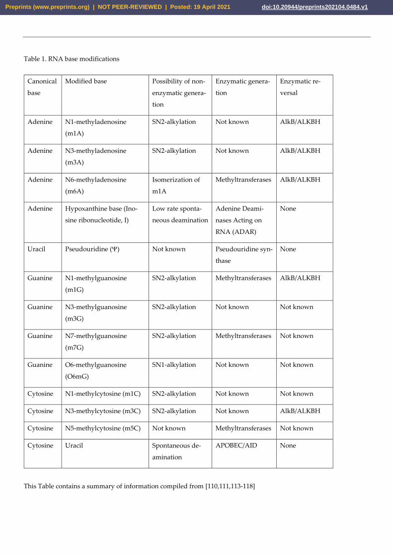
Table 1. RNA base modifications
Canonical
base
Modified base Possibility of non-
enzymatic genera-
tion
Enzymatic genera-
tion
Enzymatic re-
versal
Adenine N1-methyladenosine
(m1A)
SN2-alkylation Not known AlkB/ALKBH
Adenine N3-methyladenosine
(m3A)
SN2-alkylation Not known AlkB/ALKBH
Adenine N6-methyladenosine
(m6A)
Isomerization of
m1A
Methyltransferases AlkB/ALKBH
Adenine Hypoxanthine base (Ino-
sine ribonucleotide, I)
Low rate sponta-
neous deamination
Adenine Deami-
nases Acting on
RNA (ADAR)
None
Uracil Pseudouridine () Not known Pseudouridine syn-
thase
None
Guanine N1-methylguanosine
(m1G)
SN2-alkylation Methyltransferases AlkB/ALKBH
Guanine N3-methylguanosine
(m3G)
SN2-alkylation Not known Not known
Guanine N7-methylguanosine
(m7G)
SN2-alkylation Methyltransferases Not known
Guanine O6-methylguanosine
(O6mG)
SN1-alkylation Not known Not known
Cytosine N1-methylcytosine (m1C) SN2-alkylation Not known Not known
Cytosine N3-methylcytosine (m3C) SN2-alkylation Not known AlkB/ALKBH
Cytosine N5-methylcytosine (m5C) Not known Methyltransferases Not known
Cytosine Uracil Spontaneous de-
amination
APOBEC/AID None
This Table contains a summary of information compiled from [110,111,113-118]
Preprints (www.preprints.org) | NOT PEER-REVIEWED | Posted: 19 April 2021 doi:10.20944/preprints202104.0484.v1
Several enzymatic modifications of RNA bases – pseudouridine (), N6-methyladenine
(m6A), N5-methylcytosine (m5C) and Hypoxanthine (Inosine ribonucleotide (I)) were re-
ported to have physiological functions in RNA viruses ([114] and references therein).
These as well as several other base modifications are also present in cellular mRNAs and
have also multiple functional consequences [117,119,120] and altogether are referred as
the RNA-editome, or as epitranscriptome. For most of RNA base modifications, their full
mutagenic potential is yet to be determined. It is even not clear, if they are present in the
full-size replicating viral genome or only in non-replicating viral mRNAs. Only two kinds
of enzymatic RNA edits – cytidine to uridine (C to U) by APOBEC cytidine deaminases
and adenosine to inosine (A to I) by ADAR adenosine deaminases are known to be carried
into copies of viral genomes resulting in C to U and A to G mutations, respectively. As
discussed in the next section, these two groups of deaminases may have the greatest im-
pact on mutation accumulation in several human RNA viruses.
5.2. Base substitution mutagenesis in RNA viruses
Base substitutions are an important source for viral evolution and population dy-
namics. They are also a common avenue for viruses to escape the host’s adaptive immune
system. Thus, it is important to identify mechanisms underlying the generation of base
substitutions in viral populations. Usual approaches to understanding the mutagenic
mechanisms underlying genome mutations come from a combination of knowledge accu-
mulated in model studies as well as from agnostic documenting features of mutational
spectrum that deviate from the spectrum expected, if mutagenesis would be completely
random. Such “non-random” features of mutational spectra are also called mutational
motifs (by analogy with musical motifs, which combine notes according to the rules of
harmony) or mutational signatures (multiple set of features defining uniqueness of an
object). This synthetic approach turned to be productive in revealing the mutagenic mech-
anisms in human cancers [121-123].
Besides significant mechanistic knowledge, the progress in deciphering cancer mu-
tagenic mechanisms was made possible by accumulating many mutation catalogs – com-
plete lists of de novo mutations in genomes of individual human tumors. Since tumor
tissue is a mixture of a small number of clones, it is possible to identify mutations that had
occurred after a tumor clone, or a small set of clones, have separated from the normal
tissue. First, DNA sequence reads from tumor and from normal tissue are mapped against
a reference genome. A vast majority of the differences with a reference would be common
for normal tissue and for tumor DNA because they come from the common germline of
the same individual. The differences that are present only in the tumor but are not de-
tected in the DNA from normal tissue, are compiled to create the tumor’s mutation catalog.
A similar strategy that was utilized with human cancers can now be applied to sev-
eral RNA viruses, especially because of the accumulation of extensive sequencing data,
most notably for SARS-CoV-2, which was a subject of a gigantic genome resequencing
effort across the world [124] (see also URL https://www.gisaid.org/ ). However, since viral
genomes are small, each genome would contain a small number, or even no, mutations so
a mutation catalogue representative of a population (or of a species) could only be built
from sequences of multiple genomes. Building such a catalog must be done under the
assumption that each genome has been acted upon by similar mutational mechanisms,
which may not be the case for all genomes. Beyond this assumption, other very important
factors must be considered to ensure the proper development of a virus population mu-
tational catalog. Firstly, an important point is that the mutations in the catalog must rep-
resent independent changes rather than a small number of events that are amplified
through the development of a population, or through species evolution. A simplified ex-
ample is shown in (Figure 3) where a viral population starting from a single genome ac-
cumulates mutations over nine rounds of copying. Many mutations in these genomes are
identical, not because they occurred multiple times, but instead because they stem from a
single genome that was then propagated. Therefore, if each of these mutations were
treated as independent events, the mutations that occurred in an early generation would
Preprints (www.preprints.org) | NOT PEER-REVIEWED | Posted: 19 April 2021 doi:10.20944/preprints202104.0484.v1
be counted more times than the actual number of independent mutation events that gen-
erated these mutations. This problem would not occur in a set of non-identical (non-du-
plicated) mutations, as each observed change/mutation would come from an independent
mutation event, and would represent the spectrum of mutational processes (e.g. set of
non-duplicated mutations in a genome at generation 9 on Figure 3). Consequently, to en-
sure an accurate mutational catalog, each mutation in a catalog must represent an inde-
pendent change.
Preprints (www.preprints.org) | NOT PEER-REVIEWED | Posted: 19 April 2021 doi:10.20944/preprints202104.0484.v1
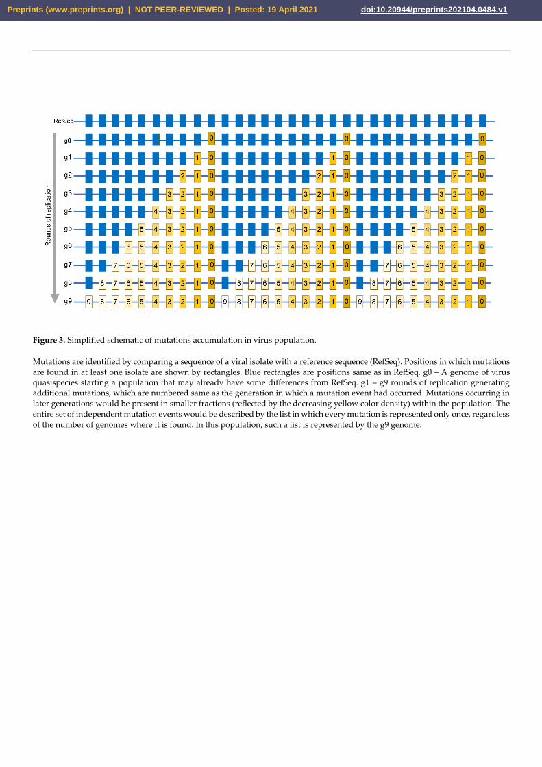
Figure 3. Simplified schematic of mutations accumulation in virus population.
Mutations are identified by comparing a sequence of a viral isolate with a reference sequence (RefSeq). Positions in which mutations
are found in at least one isolate are shown by rectangles. Blue rectangles are positions same as in RefSeq. g0 – A genome of virus
quasispecies starting a population that may already have some differences from RefSeq. g1 – g9 rounds of replication generating
additional mutations, which are numbered same as the generation in which a mutation event had occurred. Mutations occurring in
later generations would be present in smaller fractions (reflected by the decreasing yellow color density) within the population. The
entire set of independent mutation events would be described by the list in which every mutation is represented only once, regardless
of the number of genomes where it is found. In this population, such a list is represented by the g9 genome.
Preprints (www.preprints.org) | NOT PEER-REVIEWED | Posted: 19 April 2021 doi:10.20944/preprints202104.0484.v1
Another important factor for the development of a mutational catalog is the
knowledge of the original genome sequence from which all other genomes stem. This is
required to identify the mutant alleles in every sequenced viral genome as well as to iden-
tify the directionality of the mutation events. The latter is especially important for defining
mutational events across long term population dynamics or during species evolution. In
the cases of identifying mutational signatures in mammalian RNA viruses, a single refer-
ence sequence for the entire dataset is not available, so the reference roles are assigned to
the sequences in the nodes of phylogenetic trees built for genomes of isolates from a pop-
ulation [125], for distant isolates of a single virus species [126-128] or for several related
quasispecies [129]. Each node is taken as a surrogate of the reference sequence for the
genomes in the same clade of a phylogenetic tree.
With the surrogate reference sequence established, it can be utilized to develop a
mutational catalog. This approach allowed for the detection of C to U changes as a prom-
inent or even the major component of mutagenesis in a wide range of mammalian RNA
viruses [130]. Typically, the presence of base substitution type(s) that exceed expectation(s)
for random mutagenesis can be usually explained by several reasons such as base-specific
RdRp errors, mutagenic lesions, enzymatic lesion direct reversal as well as RNA editing.
However, as of now, a single feature of base preference cannot be directly applied to mu-
tation spectra interpretation as the base preferences for RdRp lesions are yet to be deter-
mined. While the base specificities for several RNA damaging agents, damage reversal
enzymes and RNA editing enzymes, are well established they cannot serve as a single
diagnostic indication of a mutagenesis source, because these specificities are often similar
between agents (Table 1). Another factor to account for in the analysis of mutagenesis
results is strand bias of a particular change. In viruses this bias will depend on a preference
of a base modifying factor to single-stranded (ss) or to double-stranded (ds) RNA. It will
be also affected by the kind of RNA forming genome of a virus: positive-strand ssRNA,
negative-stranded ssRNA, or dsRNA (Figure 1A-C). For example, in positive-strand
ssRNA viruses, changes stemming from ssRNA-specific agent modifications of the posi-
tive-strand will come into genomes of viral progeny. As for changes in viral progeny
caused by a dsRNA specific agent, they will be mostly coming from modifications of the
negative-strand (Figure 1A). The base changes shown on all panels of Figure 1 are the
same as C to U changes expected from ssRNA-specific cytidine deaminases APOBEC and
from guanine (G) to inosine (I) dsRNA-specific adenine deaminases ADAR (Figure 4). In-
terestingly, the spectra and strand preference of the two most prevailing kinds of changes
in hypermutated isolates of human vaccine-derived rubella virus corresponded to the pre-
vailing C to U, and U to C, changes in genomic strand of this positive-strand ssRNA virus
shown on Figure 1A [128]. These hypermutated viruses (up to 300 base substitutions in a
9 kb genome) were extracted from granulomas of different children with primary immu-
nodeficiency. Each independent virus isolate stemmed from the attenuated rubella vac-
cine virus; whose known original sequence was used as a reference to build a mutation
catalog from six isolates that contained 993 mutations. C to U, or A to G, changes were the
major mutations in the catalog. Such a pattern of mutations in the rubella vaccine virus
mutation catalog matches the signature of APOBEC cytidine deaminases and supports
the idea that APOBEC enzymes are the major mutator. Recently, It has been established
that APOBEC3A (A3A) enzyme has a preference to the unpaired parts (loops) of folded
RNA structures in mRNAs of human tumors [131]. Importantly, C to U changes in the
positive-strand in hypermutated rubella genomes were the only type of base substitution
that showed a statistically significant density increase in predicted RNA-loops over stems
- sequences with a potential for self-pairing [132]. This lends support for APOBEC muta-
tion being the source for C to U changes, however both strand bias and unpaired loop
preference could be the feature of any agent causing chemical deamination of cytidines in
RNA.
Preprints (www.preprints.org) | NOT PEER-REVIEWED | Posted: 19 April 2021 doi:10.20944/preprints202104.0484.v1

Figure 4. Enzymatic deamination of RNA nucleosides.
(A) APOBEC cytidine deaminase. Deamination of cytidine in ssRNA generates uridine resulting in C→U mutation in the RNA virus
genome. (B) ADAR adenosine deaminase. Deamination of adenosine in dsRNA or in folded and paired ssRNA (forming dsRNA)
generates inosine, which after rounds of copying with RdRp is fixed as A→G mutation.
Preprints (www.preprints.org) | NOT PEER-REVIEWED | Posted: 19 April 2021 doi:10.20944/preprints202104.0484.v1
Deamination of cytosines not only occurs in RNA, cytidine deamination in DNA is
one of the most frequent spontaneous changes and has a preference to ssDNA [115].
Chemicals, such as nitrites, can actively induce these cytosine deamination and mutation
events in ssDNA [133], but APOBEC cytidine deaminases have also been a source for cyt-
idine deamination. It is known that human APOBEC cytidine deaminases have complete
preference to ssDNA and ssRNA over ds polynucleotides. All APOBECs show clear pref-
erence to immediate nucleotide context surrounding deaminated cytidines in DNA. APO-
BEC3G has a preference to cCn context (mutated nucleotide capitalized; n – any nucleo-
tide), while APOBEC1 and all other members of APOBEC3 gene cluster prefer tCn deam-
ination motif [134-137]. The preferred DNA deamination motif for APOBEC3A and APO-
BEC3B was even narrowed to tCa [138]. APOBEC1 was initially discovered as RNA editor
and significant evidence had accumulated by now that members of APOBEC3 gene clus-
ter can also edit mRNA [139], however unlike for DNA, detailed editing signatures in
RNA are yet to be established. We therefore used APOBEC signature motifs established
for DNA to evaluate the mutation spectra in a catalog compiled of hypermutated rubella
genomes (Figure 5 shows example for uCa to uUa motif). This method was initially de-
veloped for evaluating APOBEC mutagenesis in human cancers [140], however it allows
statistical estimate of over-representation with any oligonucleotide motif in mutation da-
tasets [138,141,142]. A fraction of mutations in an oligonucleotide motif among mutations
of a given nucleotide is compared with the presence of the same oligonucleotide in the
genomic context surrounding mutated bases (see also Figure 5 and legend). We found a
high level of enrichment with APOBEC motif uCn and even greater enrichment with more
narrow uCa motif which is also the most preferred DNA editing motif for APOBEC3A
and APOBEC3B [128]. Unlike for APOBEC editing, there is only a multi-motif ADAR ed-
iting web-based Inosine-Predict score tool, which takes into account immediate nucleotide
context for every guanosine position as well as the potential to form a secondary structure.
This tool was developed for ADAR editing in mRNAs [143]. There was slight, albeit sta-
tistically significant increase in Inosine-Predict score for adenine positions involved in A
to G mutations (U to C in complementary strand) mutations as compared to non-mutated
positions of As (or Us) [128].
Preprints (www.preprints.org) | NOT PEER-REVIEWED | Posted: 19 April 2021 doi:10.20944/preprints202104.0484.v1
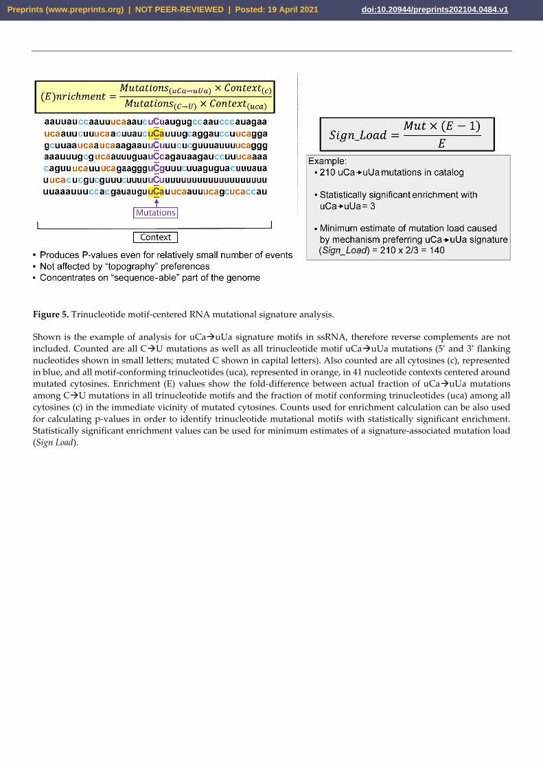
Figure 5. Trinucleotide motif-centered RNA mutational signature analysis.
Shown is the example of analysis for uCa→uUa signature motifs in ssRNA, therefore reverse complements are not
included. Counted are all C→U mutations as well as all trinucleotide motif uCa→uUa mutations (5’ and 3’ flanking
nucleotides shown in small letters; mutated C shown in capital letters). Also counted are all cytosines (c), represented
in blue, and all motif-conforming trinucleotides (uca), represented in orange, in 41 nucleotide contexts centered around
mutated cytosines. Enrichment (E) values show the fold-difference between actual fraction of uCa→uUa mutations
among C→U mutations in all trinucleotide motifs and the fraction of motif conforming trinucleotides (uca) among all
cytosines (c) in the immediate vicinity of mutated cytosines. Counts used for enrichment calculation can be also used
for calculating p-values in order to identify trinucleotide mutational motifs with statistically significant enrichment.
Statistically significant enrichment values can be used for minimum estimates of a signature-associated mutation load
(Sign Load).
Preprints (www.preprints.org) | NOT PEER-REVIEWED | Posted: 19 April 2021 doi:10.20944/preprints202104.0484.v1
Altogether, APOBEC-like and ADAR-like changes represented 86% of 993 mutations
in the catalog from six hypermutated genomes of vaccine derived rubella virus. We then
applied similar, but yet extended analytical and statistical evaluation approaches to eval-
uate mutation load and spectrum accumulated from over 30,000 SARS-CoV-2 genome se-
quences accumulated during first several months of pandemic [132]. In this analysis, we
compared spectrum and signatures with hypermutated isolates of rubella virus. The
unique feature of this dataset is that the starting reference sequence is well defined [144],
so each difference from the reference is a direct trace of mutation event. However, as in
the example on Figure 3, differences from the reference sequence that are identical be-
tween sequenced isolates can represent a record of a single mutation event, which was
then amplified in the population. In fact, some mutations were found in several thousands
of isolates. We therefore used a set of non-duplicated mutations to represent the summary
of mutational processes operating in pandemic SARS-CoV-2 population. This set would
be a minimum estimate of the independent mutation events list, because repeated occur-
rence of the same mutation cannot be excluded, especially for mutations increasing speed
of spreading within a host. To reduce the effects of functional selection we also analyzed
subsets of non-functional (synonymous or non-coding) and functional (non-synonymous).
These mutation sets were analyzed in parallel with the catalog of mutations from hyper-
mutated rubella isolates. Analytical tools initially developed for a single APOBEC motif
derived from prior experimental studies were extended to statistical evaluation of all 192
possible tri-nucleotide base substitution motifs.
We calculated one-sided Fisher’s exact test P-value for each motif and then corrected
P-values for multiple hypotheses testing and applied FDR<0.05 threshold. We found that
mutational processes with the same signatures that were revealed in hypermutated ru-
bella isolates also may operate in the SARS-CoV-2 pandemic population. The main simi-
larities between SARS-CoV-2 and rubella were: (i) the presence of APOBEC-like signature
uCn to uUn in positive strand; (ii) frequent presence of ADAR like A to G and U to C
(shown as in positive strand, will correspond to A to G in the negative-strand); (iii) pref-
erence of loops vs stems for C to U mutations. Also specific to SARS-CoV-2 were the sta-
tistically significant enrichment with the two additional trinucleotide-centered signatures.
Firstly, there was enrichment with mutations in cGn to cAn (reverse complement for nCg
to nUg in the negative-strand) which could reflect increased frequency of cytosine deam-
ination in CpG motifs and C to U changes in the RNA negative-strand of dsRNA interme-
diate producing multiple copies of the positive-strand with the complementary G to A
change (see example of negative strand mutagenesis in Figure 1A). Since enrichment cal-
culations accounted for the presence of a motif in the genomic background, the depletion
of CpG motifs in the viral genome [125,145-148] cannot be due to a single explanation of
such an enrichment. Increased frequency of C to U changes in DNA had been connected
with frequent cytidine methylation in CpG [149] but can also occur in the absence of meth-
ylation in DNA [150,151]. We proposed that increased frequency of cytosine deamination
in RNA CpGs could contribute into increased enrichment with this mutational motif [132].
Secondly, there was increased presence of G to U changes in the positive-strand, which
could reflect C to A changes in negative-strand. These changes could be caused by in-
creased formation of ROS-induced 8-oxoG within cells or during library preparation
[152,153].
Recently Adebali and colleagues performed analysis of large number of SARS-CoV-
2 sequenced genomes organized in a phylogenetic tree [125]. Unlike in [132], this study
used the nodes of the tree as a reference sequence for each sequence within each node.
Despite the two studies having used different approaches for creating representative da-
tasets of independent mutation events in a large collection of SARS-CoV-2 genomes, the
main categories of mutation preferences APOBEC-like, ADAR-like, CpG-like and appar-
ent ROS induced mutations were similar. Overall similar conclusions about prevailing
mutagenic sources and signatures were in works addressing intra-host variations of
SARS-CoV-2 [154,155]. In summary, resequencing of RNA virus genomes suggested ma-
jor mutational processes generating diversity that can lead to development of new virus
Preprints (www.preprints.org) | NOT PEER-REVIEWED | Posted: 19 April 2021 doi:10.20944/preprints202104.0484.v1

forms. These studies also defined several questions and technical developments that
should be addressed in near future.
6. Concluding remarks and future questions.
Emergence of new RNA viral quasispecies pathogenic for humans, especially the
SARS-CoV-2 pandemic, triggered massive research efforts to all aspects of RNA virus
mechanistic studies. Mechanisms underlying instability of RNA virus genomes are im-
portant for better prediction of their evolution, new pathogen emergence, and the devel-
opment of antiviral drugs. Besides that, understanding biological and molecular mechan-
ics that allows this group to flourish rather than be washed away with catastrophic error
rates represents a fundamental question related to general mechanisms of evolution. Be-
low are questions and technological applications that we anticipate being addressed soon.
6.1. Single-molecule sequencing applied to RNA virome in the environment and in single organ-
isms
The sequence of a natural individual viral isolate is usually generated from a refer-
ence-based or de novo alignment of multiple small Illumina reads, thus it does not reflect
the variations of individual viral RNAs but instead is an average of the total population
[156]. However, the recent combination of deep Illumina sequencing, and advanced bio-
informatics, allows intra-host variations to be addressed in genomes of a single viral spe-
cies during an acute infection period [154,155,157,158], so some interhost variation can be
revealed. Further, a combination of bioinformatics with metagenomics focusing on a small
conserved element of RNA viruses, such as RdRp, made it possible to identify and inter-
rogate the content of multispecies virome [66,159]. Even so, the combination of short reads
with metagenomics does have its limitations as it relies on the building arbitrary contigs
from short reads, so the entire genome cannot be assessed [160]. Nevertheless, deep se-
quencing with short reads allowed the identification of multiple new viral species [161],
was successfully used to characterize the viromes in different organisms [162,163]; and
characterize the viromes in different environmental samples [164]. To overcome these
short-read sequencing issues, there are two technologies carrying promise for studies of
viral genome instability by generating long reads from individual polynucleotides, Ox-
ford Nanopore Technology and Pacific Biosciences [165]. Each of the two platforms is
plagued by a rather high sequencing error rate, but even with the current level of accuracy
Oxford Nanopore was used for characterizing viromes [166-168]. Any further increase in
sequencing accuracy may cause a revolution with viral genome instability research. While
the field awaits this increase of accuracy of the core technologies, there is a way to reduce
false positive mutation calls by adding unique molecule identifier (UMI) barcodes added
by either limited number of PCR cycles or by ligation to increase the accuracy in both
platforms [169].
6.2. Impact of RdRp misincorporation and proofreading onto viral mutation rates
Low accuracy of RdRp, as compared with replicative DNA polymerases, led to the
proposal that the major source of viral genome mutations is connected with replication
errors [3,170]. Since many viruses have an exonuclease (ExoN or its homologs; see special
section above) appearing to proofread RdRp misincorporation, it is important to collect
more information about the impact of RdRp proofreading into prevention of hypermuta-
tion in RNA viruses. This would be approached by the modification of either, RdRp accu-
racy or ExoN capability by mutations and/or by endogenous or environmental factors. It
is quite possible that the combination of such functional defects can lead to ultra-mutation
phenotypes that would function similar to the synergistic hypermutation observed in cel-
lular organisms when mismatch repair and DNA polymerase proofreading defects are
combined [171-173]. Further, many antiviral drugs are chain-terminating NTP analogs de-
signed to preferentially affect chain extension by RdRp [76]. However, this chain termina-
tion can be counteracted by ExoN [95,174], so search for inhibitors of this enzyme is im-
portant for practical applications
Preprints (www.preprints.org) | NOT PEER-REVIEWED | Posted: 19 April 2021 doi:10.20944/preprints202104.0484.v1
6.3. Are there RNA repair mechanisms besides AlkB direct reversal?
It is long known that RNA is more vulnerable for breakage as compared to DNA
[175] and is at least just as susceptible as DNA to base lesions (Table 1 and references
therein). However, unlike DNA, there is only one well established mechanism to repair
RNA base lesions – direct reversal of alkylation. Currently there are no direct indications
for the existence of other RNA repair mechanisms. Speculations can still be made based
on structural similarity of RNA and DNA resulting in RNA being a substrate or a ligand
for common DNA repair enzyme, e.g., RPA [176], but more research will be needed to
reveal RNA repair mechanisms of they do indeed exist.
6.4. Impact of environmental RNA lesions onto viral genome instability
DNA base lesions are often an impediment to replicative DNA polymerases and re-
quire specialized trans-lesion synthesis (TLS) DNA polymerases to successfully accom-
plish genome duplication. TLS polymerases are often error-prone and results in mutagen-
esis, while the lack of TLS can lead to genome rearrangements or to replication failure
[177,178]. The same cannot be said about RNA genomes as, per current knowledge, there
have been no model studies addressing the impact of environmental RNA damage on
structural or sequence integrity of RNA genomes. Further mechanistic studies to fill this
gap of knowledge are important for understanding the dynamic world of RNA viruses.
6.5. Impact of endogenous RNA lesions onto viral genome instability
Recent studies summarized in section 5 indicated that adenosine deaminases ADAR
and cytidine deaminases APOBEC are the prevailing sources of bases substitutions in sev-
eral human RNA viruses including SARS-CoV-2. Both types of enzymes are the part of
innate immunity, which raises a question about individual levels of RNA virus hypermu-
tation. Interestingly, hypermutation of the vaccine-derived rubella virus was reported in
subjects with primary immunodeficiency in adaptive immune system, which could be the
reason of excessive activation of innate immunity and, consequently, excessive activation
of APOBECs [128]. Another important question is about the level of endogenous hyper-
mutation of RNA viruses in species that may serve reservoirs for the occurrence of new
quasispecies. It is interesting that bats, which are a known coronavirus reservoir have
multiple (4-7) APOBEC3 homologs while most of other mammalian orders have only one
or two versions of APOBEC3 [179,180]. Therefore, studies into these organisms may reveal
insights into the formation of novel viruses.
6.6. Experimental models to define signatures of environmental and endogenous mutagenesis in
RNA
Defining diagnostic mutational signatures can develop into a multiprong scalable
approach to understanding sources and mechanism of mutagenesis in RNA viruses. Mu-
tational signatures turned to be a productive approach for another set of unstable ge-
nomes – human cancer. This could be a pure agnostic analysis of large datasets of genome
instability catalogs [121,123,181], which can be also combined with prior mechanistic
knowledge about different types of mutagenesis [122]. Another approach is to collect
knowledge about mutational signatures in defined systems - mammalian [182,183] or mi-
crobial [138,141], and then utilize the information to build a specific statistical hypothesis
for interrogating datasets of natural variants. High rates of mutation and relative ease of
RNA virus genome sequencing can certainly make these approaches productive and scal-
able.
Author Contributions: Both listed authors (Z.W.K and D.A.G.), contributed to the visualization,
writing, and editing of the manuscript.
Preprints (www.preprints.org) | NOT PEER-REVIEWED | Posted: 19 April 2021 doi:10.20944/preprints202104.0484.v1

Funding: This research was supported by the US National Institute of Health Intramural Research
Program Project Z1AES103266 to D.A.G.
Data Availability Statement: No new data were created or analyzed in this study. Data sharing is
not applicable to this article.
Acknowledgments: We thank Drs. Hamed Bostan and Marcos Morgan for critical reading of the
manuscript
Conflicts of Interests: Both listed Authors have declared no competing interests.
Preprints (www.preprints.org) | NOT PEER-REVIEWED | Posted: 19 April 2021 doi:10.20944/preprints202104.0484.v1

References
1 Dobzhansky, T. Nothing in Biology Makes Sense except in the Light of Evolution. The American Biology Teacher, (1973), 35,
125-12910.2307/4444260.
2 Dobzhansky, T. Genetics and the origin of species / Theodosius Dobzhansky. Third edition, revised. edn, (1951), (Columbia
University Press).
3 Domingo, E. Molecular Basis of Genetic Variation of Viruses,Virus as Populations,Esteban Domingo,Academic PressBoston,
(2016), 35-7110.1016/b978-0-12-800837-9.00002-2.
4 Domingo, E. & Perales, C. Viral quasispecies. PLoS Genet, (2019), 15, e100827110.1371/journal.pgen.1008271.
5 Domingo, E., Sabo, D., Taniguchi, T. & Weissmann, C. Nucleotide sequence heterogeneity of an RNA phage population. Cell,
(1978), 13, 735-74410.1016/0092-8674(78)90223-4.
6 Domingo, E., Sheldon, J. & Perales, C. Viral quasispecies evolution. Microbiol Mol Biol Rev, (2012), 76, 159-
21610.1128/mmbr.05023-11.
7 Eigen, M. & Schuster, P. The hypercycle. A principle of natural self-organization. Part A: Emergence of the hypercycle. Natur-
wissenschaften, (1977), 64, 541-56510.1007/BF00450633.
8 Fornes, J., Tomas Lazaro, J., Alarcon, T., Elena, S. F. & Sardanyes, J. Viral replication modes in single-peak fitness landscapes: A
dynamical systems analysis. J Theor Biol, (2019), 460, 170-18310.1016/j.jtbi.2018.10.007.
9 Schuster, P. Quasispecies on Fitness Landscapes. Current topics in microbiology and immunology, (2016), 392, 61-
12010.1007/82_2015_469.
10 Swetina, J. & Schuster, P. Self-replication with errors. A model for polynucleotide replication. Biophys Chem, (1982), 16, 329-
34510.1016/0301-4622(82)87037-3.
11 Zhou, L., Ding, D. & Szostak, J. W. The virtual circular genome model for primordial RNA replication. RNA, (2021), 27, 1-
1110.1261/rna.077693.120.
12 Joyce, G. F. & Szostak, J. W. Protocells and RNA Self-Replication. Cold Spring Harb Perspect Biol, (2018), 1010.1101/cshper-
spect.a034801.
13 Szostak, J. W., Bartel, D. P. & Luisi, P. L. Synthesizing life. Nature, (2001), 409, 387-390Doi 10.1038/35053176.
14 Jheeta, S. The Routes of Emergence of Life from LUCA during the RNA and Viral World: A Conspectus. Life (Basel), (2015), 5,
1445-145310.3390/life5021445.
15 Robertson, M. P. & Joyce, G. F. The origins of the RNA world. Cold Spring Harb Perspect Biol, (2012), 410.1101/cshper-
spect.a003608.
16 Dworkin, J. P., Lazcano, A. & Miller, S. L. The roads to and from the RNA world. J Theor Biol, (2003), 222, 127-13410.1016/s0022-
5193(03)00020-1.
17 Gilbert, W. Origin of Life - the Rna World. Nature, (1986), 319, 618-618DOI 10.1038/319618a0.
18 Sankaran, N. The RNA World at Thirty: A Look Back with its Author. Journal of molecular evolution, (2016), 83, 169-
17510.1007/s00239-016-9767-3.
19 Orgel, L. E. The origin of life--a review of facts and speculations. Trends Biochem Sci, (1998), 23, 491-49510.1016/s0968-
0004(98)01300-0.
20 Orgel, L. E. Prebiotic chemistry and the origin of the RNA world. Critical reviews in biochemistry and molecular biology, (2004),
39, 99-12310.1080/10409230490460765.
21 Szostak, J. W. An optimal degree of physical and chemical heterogeneity for the origin of life? Philos Trans R Soc Lond B Biol
Sci, (2011), 366, 2894-290110.1098/rstb.2011.0140.
Preprints (www.preprints.org) | NOT PEER-REVIEWED | Posted: 19 April 2021 doi:10.20944/preprints202104.0484.v1
22 Cojocaru, R. & Unrau, P. J. Processive RNA polymerization and promoter recognition in an RNA World. Science, (2021), 371,
1225-123210.1126/science.abd9191.
23 Moya, A., Holmes, E. C. & Gonzalez-Candelas, F. The population genetics and evolutionary epidemiology of RNA viruses. Nat
Rev Microbiol, (2004), 2, 279-28810.1038/nrmicro863.
24 Gago, S., Elena, S. F., Flores, R. & Sanjuan, R. Extremely high mutation rate of a hammerhead viroid. Science, (2009), 323,
130810.1126/science.1169202.
25 Duffy, S., Shackelton, L. A. & Holmes, E. C. Rates of evolutionary change in viruses: patterns and determinants. Nature reviews.
Genetics, (2008), 9, 267-27610.1038/nrg2323.
26 Abdel-Moneim, A. S. & Abdelwhab, E. M. Evidence for SARS-CoV-2 Infection of Animal Hosts. Pathogens, (2020),
910.3390/pathogens9070529.
27 Andersen, K. G., Rambaut, A., Lipkin, W. I., Holmes, E. C. & Garry, R. F. The proximal origin of SARS-CoV-2. Nat Med, (2020),
26, 450-45210.1038/s41591-020-0820-9.
28 Wu, T. & Orgel, L. E. Nonenzymatic template-directed synthesis on oligodeoxycytidylate sequences in hairpin oligonucleotides.
J Am Chem Soc, (1992), 114, 317-32210.1021/ja00027a040.
29 Flores, R., Gago-Zachert, S., Serra, P., Sanjuan, R. & Elena, S. F. Viroids: survivors from the RNA world? Annu Rev Microbiol,
(2014), 68, 395-41410.1146/annurev-micro-091313-103416.
30 Flores, R. & Owens, R. A. Viroids (Pospiviroidae and Avsunviroidae),Reference Module in Life Sciences,Elsevier (2020)
10.1016/b978-0-12-809633-8.21257-0.
31 Flores, R., Serra, P., Minoia, S., Di Serio, F. & Navarro, B. Viroids: from genotype to phenotype just relying on RNA sequence
and structural motifs. Front Microbiol, (2012), 3, 21710.3389/fmicb.2012.00217.
32 Koonin, E. V., Senkevich, T. G. & Dolja, V. V. The ancient Virus World and evolution of cells. Biology direct, (2006), 1,
2910.1186/1745-6150-1-29.
33 Baltimore, D. Expression of animal virus genomes. Bacteriol Rev, (1971), 35, 235-241.
34 Ortin, J. & Parra, F. Structure and function of RNA replication. Annual Review of Microbiology, (2006), 60, 305-32610.1146/an-
nurev.micro.60.080805.142248.
35 Agol, V. I. & Gmyl, A. P. Emergency Services of Viral RNAs: Repair and Remodeling. Microbiol Mol Biol Rev, (2018),
8210.1128/MMBR.00067-17.
36 Domingo, E. Molecular basis of genetic variation of viruses: error-prone replication. Virus as Populations: Composition, Com-
plexity, Quasispecies, Dynamics, and Biological Implications, 2nd Edition, (2020), 35-7110.1016/B978-0-12-816331-3.00002-7.
37 Lai, M. M. C. Genetic-Recombination in Rna Viruses. Current topics in microbiology and immunology, (1992), 176, 21-32.
38 Levy, D. N., Aldrovandi, G. M., Kutsch, O. & Shaw, G. M. Dynamics of HIV-1 recombination in its natural target cells. Proceed-
ings of the National Academy of Sciences of the United States of America, (2004), 101, 4204-420910.1073/pnas.0306764101.
39 Nagy, P. D. & Simon, A. E. New insights into the mechanisms of RNA recombination. Virology, (1997), 235, 1-9DOI
10.1006/viro.1997.8681.
40 Sztuba-Solinska, J., Urbanowicz, A., Figlerowicz, M. & Bujarski, J. J. RNA-RNA Recombination in Plant Virus Replication and
Evolution. Annu Rev Phytopathol, (2011), 49, 415-44310.1146/annurev-phyto-072910-095351.
41 Urbanowicz, A. et al. Homologous crossovers among molecules of brome mosaic bromovirus RNA1 or RNA2 segments in vivo.
J Virol, (2005), 79, 5732-574210.1128/Jvi.79.9.5732-5742.2005.
42 Liao, C. L. & Lai, M. M. C. Rna Recombination in a Coronavirus - Recombination between Viral Genomic Rna and Transfected
Rna Fragments. J Virol, (1992), 66, 6117-6124Doi 10.1128/Jvi.66.10.6117-6124.1992.
Preprints (www.preprints.org) | NOT PEER-REVIEWED | Posted: 19 April 2021 doi:10.20944/preprints202104.0484.v1
43 Kempf, B. J., Peersen, O. B. & Barton, D. J. Poliovirus Polymerase Leu420 Facilitates RNA Recombination and Ribavirin Re-
sistance. J Virol, (2016), 90, 8410-842110.1128/Jvi.00078-16.
44 Peersen, O. B. Picornaviral polymerase structure, function, and fidelity modulation. Virus Research, (2017), 234, 4-2010.1016/j.vi-
rusres.2017.01.026.
45 Woodman, A., Arnold, J. J., Cameron, C. E. & Evans, D. J. Biochemical and genetic analysis of the role of the viral polymerase
in enterovirus recombination. Nucleic acids research, (2016), 44, 6883-689510.1093/nar/gkw567.
46 Xiao, Y. H. et al. RNA Recombination Enhances Adaptability and Is Required for Virus Spread and Virulence. Cell Host Mi-
crobe, (2016), 19, 493-50310.1016/j.chom.2016.03.009.
47 Arnold, J. J., Ghosh, S. K. & Cameron, C. E. Poliovirus RNA-dependent RNA polymerase (3D(pol)). Divalent cation modulation
of primer, template, and nucleotide selection. J Biol Chem, (1999), 274, 37060-3706910.1074/jbc.274.52.37060.
48 Kirkegaard, K. & Baltimore, D. The mechanism of RNA recombination in poliovirus. Cell, (1986), 47, 433-44310.1016/0092-
8674(86)90600-8.
49 Romanova, L. I. et al. The primary structure of crossover regions of intertypic poliovirus recombinants: a model of recombina-
tion between RNA genomes. Virology, (1986), 155, 202-21310.1016/0042-6822(86)90180-7.
50 Simon-Loriere, E. & Holmes, E. C. Why do RNA viruses recombine? Nat Rev Microbiol, (2011), 9, 617-62610.1038/nrmicro2614.
51 Arnold, J. J., Gohara, D. W. & Cameron, C. E. Poliovirus RNA-dependent RNA polymerase (3Dpol): pre-steady-state kinetic
analysis of ribonucleotide incorporation in the presence of Mn2+. Biochemistry, (2004), 43, 5138-514810.1021/bi035213q.
52 Adams, S. D., Tzeng, W. P., Chen, M. H. & Frey, T. K. Analysis of intermolecular RNA-RNA recombination by rubella virus.
Virology, (2003), 309, 258-27110.1016/s0042-6822(03)00064-3.
53 Austermann-Busch, S. & Becher, P. RNA structural elements determine frequency and sites of nonhomologous recombination
in an animal plus-strand RNA virus. J Virol, (2012), 86, 7393-740210.1128/JVI.00864-12.
54 Gallei, A., Pankraz, A., Thiel, H. J. & Becher, P. RNA recombination in vivo in the absence of viral replication. J Virol, (2004), 78,
6271-628110.1128/JVI.78.12.6271-6281.2004.
55 Gmyl, A. P. & Agol, V. I. Diverse Mechanisms of RNA Recombination. Mol Biol, (2005), 39, 529-54210.1007/s11008-005-0069-x.
56 Gmyl, A. P. et al. Nonreplicative RNA recombination in poliovirus. J Virol, (1999), 73, 8958-896510.1128/JVI.73.11.8958-
8965.1999.
57 Gmyl, A. P., Korshenko, S. A., Belousov, E. V., Khitrina, E. V. & Agol, V. I. Nonreplicative homologous RNA recombination:
promiscuous joining of RNA pieces? RNA, (2003), 9, 1221-123110.1261/rna.5111803.
58 Holmblat, B. et al. Nonhomologous recombination between defective poliovirus and coxsackievirus genomes suggests a new
model of genetic plasticity for picornaviruses. mBio, (2014), 5, e01119-0111410.1128/mBio.01119-14.
59 Kleine Buning, M. et al. Nonreplicative RNA Recombination of an Animal Plus-Strand RNA Virus in the Absence of Efficient
Translation of Viral Proteins. Genome Biol Evol, (2017), 9, 817-82910.1093/gbe/evx046.
60 Raju, R., Subramaniam, S. V. & Hajjou, M. Genesis of Sindbis virus by in vivo recombination of nonreplicative RNA precursors.
J Virol, (1995), 69, 7391-740110.1128/JVI.69.12.7391-7401.1995.
61 Scheel, T. K. et al. Productive homologous and non-homologous recombination of hepatitis C virus in cell culture. PLoS patho-
gens, (2013), 9, e100322810.1371/journal.ppat.1003228.
62 Schibler, M., Piuz, I., Hao, W. & Tapparel, C. Chimeric rhinoviruses obtained via genetic engineering or artificially induced
recombination are viable only if the polyprotein coding sequence derives from the same species. J Virol, (2015), 89, 4470-
448010.1128/JVI.03668-14.
Preprints (www.preprints.org) | NOT PEER-REVIEWED | Posted: 19 April 2021 doi:10.20944/preprints202104.0484.v1
63 Wilkinson, M. E., Charenton, C. & Nagai, K. RNA Splicing by the Spliceosome. Annu Rev Biochem, (2020), 89, 359-
38810.1146/annurev-biochem-091719-064225.
64 Zhang, L., Vielle, A., Espinosa, S. & Zhao, R. RNAs in the spliceosome: Insight from cryoEM structures. Wiley Interdiscip Rev
RNA, (2019), 10, e152310.1002/wrna.1523.
65 Cech, T. R. Self-splicing RNA: implications for evolution. Int Rev Cytol, (1985), 93, 3-2210.1016/s0074-7696(08)61370-4.
66 Dolja, V. V. & Koonin, E. V. Metagenomics reshapes the concepts of RNA virus evolution by revealing extensive horizontal
virus transfer. Virus Res, (2018), 244, 36-5210.1016/j.virusres.2017.10.020.
67 te Velthuis, A. J. W. Common and unique features of viral RNA-dependent polymerases. Cell Mol Life Sci, (2014), 71, 4403-
442010.1007/s00018-014-1695-z.
68 Bruenn, J. A. Relationships among the Positive Strand and Double-Strand Rna Viruses as Viewed through Their Rna-Dependent
Rna-Polymerases. Nucleic acids research, (1991), 19, 217-226DOI 10.1093/nar/19.2.217.
69 Gorbalenya, A. E. et al. The palm subdomain-based active site is internally permuted in viral RNA-dependent RNA polymer-
ases of an ancient lineage. J Mol Biol, (2002), 324, 47-6210.1016/S0022-2836(02)01033-1.
70 Krupovic, M., Dolja, V. V. & Koonin, E. V. Origin of viruses: primordial replicators recruiting capsids from hosts. Nat Rev
Microbiol, (2019), 17, 449-45810.1038/s41579-019-0205-6.
71 Domingo, E. & Holland, J. J. RNA virus mutations and fitness for survival. Annual Review of Microbiology, (1997), 51, 151-
178DOI 10.1146/annurev.micro.51.1.151.
72 Drake, J. W. Rates of Spontaneous Mutation among Rna Viruses. Proceedings of the National Academy of Sciences of the United
States of America, (1993), 90, 4171-4175DOI 10.1073/pnas.90.9.4171.
73 Perrino, F. W., Preston, B. D., Sandell, L. L. & Loeb, L. A. Extension of mismatched 3' termini of DNA is a major determinant of
the infidelity of human immunodeficiency virus type 1 reverse transcriptase. Proceedings of the National Academy of Sciences
of the United States of America, (1989), 86, 8343-834710.1073/pnas.86.21.8343.
74 Eckert, K. A. & Kunkel, T. A. Fidelity of DNA synthesis catalyzed by human DNA polymerase alpha and HIV-1 reverse tran-
scriptase: effect of reaction pH. Nucleic Acids Res, (1993), 21, 5212-522010.1093/nar/21.22.5212.
75 Crotty, S. et al. The broad-spectrum antiviral ribonucleoside ribavirin is an RNA virus mutagen. Nat Med, (2000), 6, 1375-
137910.1038/82191.
76 Seley-Radtke, K. L. & Yates, M. K. The evolution of nucleoside analogue antivirals: A review for chemists and non-chemists.
Part 1: Early structural modifications to the nucleoside scaffold. Antiviral Res, (2018), 154, 66-8610.1016/j.antiviral.2018.04.004.
77 De Clercq, E. Antivirals and antiviral strategies. Nat Rev Microbiol, (2004), 2, 704-72010.1038/nrmicro975.
78 De Clercq, E. & Li, G. Approved Antiviral Drugs over the Past 50 Years. Clin Microbiol Rev, (2016), 29, 695-
74710.1128/CMR.00102-15.
79 Jordheim, L. P., Durantel, D., Zoulim, F. & Dumontet, C. Advances in the development of nucleoside and nucleotide analogues
for cancer and viral diseases. Nat Rev Drug Discov, (2013), 12, 447-46410.1038/nrd4010.
80 Holland, J. et al. Rapid evolution of RNA genomes. Science, (1982), 215, 1577-158510.1126/science.7041255.
81 Novella, I. S. et al. Exponential increases of RNA virus fitness during large population transmissions. Proceedings of the Na-
tional Academy of Sciences of the United States of America, (1995), 92, 5841-584410.1073/pnas.92.13.5841.
82 Pfeiffer, J. K. & Kirkegaard, K. A single mutation in poliovirus RNA-dependent RNA polymerase confers resistance to muta-
genic nucleotide analogs via increased fidelity. Proceedings of the National Academy of Sciences of the United States of Amer-
ica, (2003), 100, 7289-729410.1073/pnas.1232294100.
83 Sadeghipour, S., Bek, E. J. & McMinn, P. C. Ribavirin-resistant mutants of human enterovirus 71 express a high replication
fidelity phenotype during growth in cell culture. J Virol, (2013), 87, 1759-176910.1128/JVI.02139-12.
Preprints (www.preprints.org) | NOT PEER-REVIEWED | Posted: 19 April 2021 doi:10.20944/preprints202104.0484.v1
84 Coffey, L. L., Beeharry, Y., Borderia, A. V., Blanc, H. & Vignuzzi, M. Arbovirus high fidelity variant loses fitness in mosquitoes
and mice. Proceedings of the National Academy of Sciences of the United States of America, (2011), 108, 16038-
1604310.1073/pnas.1111650108.
85 Sierra, M. et al. Foot-and-mouth disease virus mutant with decreased sensitivity to ribavirin: implications for error catastrophe.
J Virol, (2007), 81, 2012-202410.1128/JVI.01606-06.
86 Binh, N. T., Wakai, C., Kawaguchi, A. & Nagata, K. Involvement of the N-terminal portion of influenza virus RNA polymerase
subunit PB1 in nucleotide recognition. Biochem Biophys Res Commun, (2014), 443, 975-97910.1016/j.bbrc.2013.12.071.
87 Liu, X. et al. Vaccine-derived mutation in motif D of poliovirus RNA-dependent RNA polymerase lowers nucleotide incorpo-
ration fidelity. J Biol Chem, (2013), 288, 32753-3276510.1074/jbc.M113.484428.
88 Vignuzzi, M., Wendt, E. & Andino, R. Engineering attenuated virus vaccines by controlling replication fidelity. Nat Med, (2008),
14, 154-16110.1038/nm1726.
89 Steinhauer, D. A., Domingo, E. & Holland, J. J. Lack of evidence for proofreading mechanisms associated with an RNA virus
polymerase. Gene, (1992), 122, 281-28810.1016/0378-1119(92)90216-c.
90 Drake, J. W. & Holland, J. J. Mutation rates among RNA viruses. Proceedings of the National Academy of Sciences of the United
States of America, (1999), 96, 13910-13913DOI 10.1073/pnas.96.24.13910.
91 Eigen, M. Error catastrophe and antiviral strategy. Proceedings of the National Academy of Sciences of the United States of
America, (2002), 99, 13374-1337610.1073/pnas.212514799.
92 Bukhari, K. et al. Description and initial characterization of metatranscriptomic nidovirus-like genomes from the proposed new
family Abyssoviridae, and from a sister group to the Coronavirinae, the proposed genus Alphaletovirus. Virology, (2018), 524,
160-17110.1016/j.virol.2018.08.010.
93 Saberi, A., Gulyaeva, A. A., Brubacher, J. L., Newmark, P. A. & Gorbalenya, A. E. A planarian nidovirus expands the limits of
RNA genome size. PLoS pathogens, (2018), 14, e100731410.1371/journal.ppat.1007314.
94 Snijder, E. J. et al. Unique and conserved features of genome and proteome of SARS-coronavirus, an early split-off from the
coronavirus group 2 lineage. J Mol Biol, (2003), 331, 991-100410.1016/s0022-2836(03)00865-9.
95 Ogando, N. S. et al. The Curious Case of the Nidovirus Exoribonuclease: Its Role in RNA Synthesis and Replication Fidelity.
Front Microbiol, (2019), 10, 181310.3389/fmicb.2019.01813.
96 Minskaia, E. et al. Discovery of an RNA virus 3 '-> 5 ' exoribonuclease that is critically involved in coronavirus RNA synthesis.
Proceedings of the National Academy of Sciences of the United States of America, (2006), 103, 5108-
511310.1073/pnas.0508200103.
97 Eckerle, L. D. et al. Infidelity of SARS-CoV Nsp14-Exonuclease Mutant Virus Replication Is Revealed by Complete Genome
Sequencing. PLoS pathogens, (2010), 6ARTN e1000896
10.1371/journal.ppat.1000896.
98 Eckerle, L. D., Lu, X., Sperry, S. M., Choi, L. & Denison, M. R. High fidelity of murine hepatitis virus replication is decreased in
nsp14 exoribonuclease mutants. J Virol, (2007), 81, 12135-1214410.1128/Jvi.01296-07.
99 Bouvet, M. et al. In vitro reconstitution of SARS-coronavirus mRNA cap methylation. PLoS pathogens, (2010), 6,
e100086310.1371/journal.ppat.1000863.
100 Bouvet, M. et al. RNA 3'-end mismatch excision by the severe acute respiratory syndrome coronavirus nonstructural protein
nsp10/nsp14 exoribonuclease complex. Proceedings of the National Academy of Sciences of the United States of America, (2012),
109, 9372-937710.1073/pnas.1201130109.
101 Subissi, L. et al. SARS-CoV ORF1b-encoded nonstructural proteins 12-16: replicative enzymes as antiviral targets. Antiviral Res,
(2014), 101, 122-13010.1016/j.antiviral.2013.11.006.
Preprints (www.preprints.org) | NOT PEER-REVIEWED | Posted: 19 April 2021 doi:10.20944/preprints202104.0484.v1
102 Imbert, I. et al. A second, non-canonical RNA-dependent RNA polymerase in SARS coronavirus. The EMBO journal, (2006), 25,
4933-494210.1038/sj.emboj.7601368.
103 Gorbalenya, A. E., Koonin, E. V., Donchenko, A. P. & Blinov, V. M. Coronavirus genome: prediction of putative functional
domains in the non-structural polyprotein by comparative amino acid sequence analysis. Nucleic Acids Res, (1989), 17, 4847-
486110.1093/nar/17.12.4847.
104 Sevajol, M., Subissi, L., Decroly, E., Canard, B. & Imbert, I. Insights into RNA synthesis, capping, and proofreading mechanisms
of SARS-coronavirus. Virus Research, (2014), 194, 90-9910.1016/j.virusres.2014.10.008.
105 Kirchdoerfer, R. N. & Ward, A. B. Structure of the SARS-CoV nsp12 polymerase bound to nsp7 and nsp8 co-factors. Nat Com-
mun, (2019), 10, 234210.1038/s41467-019-10280-3.
106 Posthuma, C. C., Te Velthuis, A. J. W. & Snijder, E. J. Nidovirus RNA polymerases: Complex enzymes handling exceptional
RNA genomes. Virus Res, (2017), 234, 58-7310.1016/j.virusres.2017.01.023.
107 Tvarogova, J. et al. Identification and Characterization of a Human Coronavirus 229E Nonstructural Protein 8-Associated RNA
3 '-Terminal Adenylyltransferase Activity. J Virol, (2019), 93ARTN e00291-19
10.1128/JVI.00291-19.
108 Zhai, Y. et al. Insights into SARS-CoV transcription and replication from the structure of the nsp7-nsp8 hexadecamer. Nat Struct
Mol Biol, (2005), 12, 980-98610.1038/nsmb999.
109 Velthuis, A. J. W. T., van den Worm, S. H. E. & Snijder, E. J. The SARS-coronavirus nsp7+nsp8 complex is a unique multimeric
RNA polymerase capable of both de novo initiation and primer extension. Nucleic acids research, (2012), 40, 1737-
174710.1093/nar/gkr893.
110 Drablos, F. et al. Alkylation damage in DNA and RNA--repair mechanisms and medical significance. DNA Repair (Amst),
(2004), 3, 1389-140710.1016/j.dnarep.2004.05.004.
111 Feyzi, E. et al. RNA base damage and repair. Current pharmaceutical biotechnology, (2007), 8, 326-
33110.2174/138920107783018363.
112 Thapar, R. et al. RNA Modifications: Reversal Mechanisms and Cancer. Biochemistry, (2019), 58, 312-32910.1021/acs.bio-
chem.8b00949.
113 Pogolotti, A. L., Jr., Ono, A., Subramaniam, R. & Santi, D. V. On the mechanism of DNA-adenine methylase. J Biol Chem, (1988),
263, 7461-7464https://doi.org/10.1016/S0021-9258(18)68520-5.
114 Potuznik, J. F. & Cahova, H. It's the Little Things (in Viral RNA). mBio, (2020), 1110.1128/mBio.02131-20.
115 Lindahl, T. Instability and decay of the primary structure of DNA. Nature, (1993), 362, 709-71510.1038/362709a0.
116 Ge, J. & Yu, Y. T. RNA pseudouridylation: new insights into an old modification. Trends Biochem Sci, (2013), 38, 210-
21810.1016/j.tibs.2013.01.002.
117 Schaefer, M. R. The Regulation of RNA Modification Systems: The Next Frontier in Epitranscriptomics?, (2021), 12, 345.
118 Beranek, D. T. Distribution of methyl and ethyl adducts following alkylation with monofunctional alkylating agents. Mutation
research, (1990), 231, 11-30.
119 Frye, M., Jaffrey, S. R., Pan, T., Rechavi, G. & Suzuki, T. RNA modifications: what have we learned and where are we headed?
Nature reviews. Genetics, (2016), 17, 365-37210.1038/nrg.2016.47.
120 Ontiveros, R. J., Stoute, J. & Liu, K. F. The chemical diversity of RNA modifications. The Biochemical journal, (2019), 476, 1227-
124510.1042/BCJ20180445.
121 Alexandrov, L. B. et al. Signatures of mutational processes in human cancer. Nature, (2013), 500, 415-42110.1038/nature12477.
122 Roberts, S. A. & Gordenin, D. A. Hypermutation in human cancer genomes: footprints and mechanisms. Nature reviews. Can-
cer, (2014), 14, 786-80010.1038/nrc3816.
Preprints (www.preprints.org) | NOT PEER-REVIEWED | Posted: 19 April 2021 doi:10.20944/preprints202104.0484.v1

123 Alexandrov, L. B. et al. The repertoire of mutational signatures in human cancer. Nature, (2020), 578, 94-10110.1038/s41586-020-
1943-3.
124 Shu, Y. & McCauley, J. GISAID: Global initiative on sharing all influenza data - from vision to reality. Euro surveillance : bulletin
Europeen sur les maladies transmissibles = European communicable disease bulletin, (2017), 2210.2807/1560-
7917.ES.2017.22.13.30494.
125 Azgari, C., Kilinc, Z., Turhan, B., Circi, D. & Adebali, O. The Mutation Profile of SARS-CoV-2 Is Primarily Shaped by the Host
Antiviral Defense. (2021), 13, 394.
126 Khrustalev, V. V. & Barkovsky, E. V. Unusual nucleotide content of Rubella virus genome as a consequence of biased RNA-
editing: comparison with Alphaviruses. International journal of bioinformatics research and applications, (2011), 7, 82-
10010.1504/IJBRA.2011.039171.
127 Khrustalev, V. V., Khrustaleva, T. A., Sharma, N. & Giri, R. Mutational Pressure in Zika Virus: Local ADAR-Editing Areas
Associated with Pauses in Translation and Replication. Frontiers in cellular and infection microbiology, (2017), 7,
4410.3389/fcimb.2017.00044.
128 Perelygina, L. et al. Infectious vaccine-derived rubella viruses emerge, persist, and evolve in cutaneous granulomas of children
with primary immunodeficiencies. PLoS pathogens, (2019), 15, e100808010.1371/journal.ppat.1008080.
129 Simmonds, P. Rampant C-->U Hypermutation in the Genomes of SARS-CoV-2 and Other Coronaviruses: Causes and Conse-
quences for Their Short- and Long-Term Evolutionary Trajectories. mSphere, (2020), 510.1128/mSphere.00408-20.
130 Simmonds, P. & Ansari, M. A. Mutation bias implicates RNA editing in a wide range of mammalian RNA viruses. (2021),
2021.2002.2009.43039510.1101/2021.02.09.430395 %J bioRxiv.
131 Jalili, P. et al. Quantification of ongoing APOBEC3A activity in tumor cells by monitoring RNA editing at hotspots. Nat Com-
mun, (2020), 11, 297110.1038/s41467-020-16802-8.
132 Klimczak, L. J., Randall, T. A., Saini, N., Li, J. L. & Gordenin, D. A. Similarity between mutation spectra in hypermutated ge-
nomes of rubella virus and in SARS-CoV-2 genomes accumulated during the COVID-19 pandemic. PLoS One, (2020), 15,
e023768910.1371/journal.pone.0237689.
133 Chan, K. et al. Base damage within single-strand DNA underlies in vivo hypermutability induced by a ubiquitous environmen-
tal agent. PLoS Genet, (2012), 8, e100314910.1371/journal.pgen.1003149.
134 Conticello, S. G. Creative deaminases, self-inflicted damage, and genome evolution. Annals of the New York Academy of Sci-
ences, (2012), 1267, 79-8510.1111/j.1749-6632.2012.06614.x.
135 Green, A. M. & Weitzman, M. D. The spectrum of APOBEC3 activity: From anti-viral agents to anti-cancer opportunities. DNA
Repair (Amst), (2019), 83, 10270010.1016/j.dnarep.2019.102700.
136 Harris, R. S. & Dudley, J. P. APOBECs and virus restriction. Virology, (2015), 479-480, 131-14510.1016/j.virol.2015.03.012.
137 Refsland, E. W. & Harris, R. S. The APOBEC3 family of retroelement restriction factors. Current topics in microbiology and
immunology, (2013), 371, 1-2710.1007/978-3-642-37765-5_1.
138 Chan, K. et al. An APOBEC3A hypermutation signature is distinguishable from the signature of background mutagenesis by
APOBEC3B in human cancers. Nat Genet, (2015), 47, 1067-107210.1038/ng.3378.
139 Lerner, T., Papavasiliou, F. N. & Pecori, R. RNA Editors, Cofactors, and mRNA Targets: An Overview of the C-to-U RNA
Editing Machinery and Its Implication in Human Disease. Genes, (2018), 1010.3390/genes10010013.
140 Roberts, S. A. et al. An APOBEC cytidine deaminase mutagenesis pattern is widespread in human cancers. Nat Genet, (2013),
45, 970-97610.1038/ng.2702.
141 Saini, N. et al. Mutation signatures specific to DNA alkylating agents in yeast and cancers. Nucleic Acids Res, (2020), 48, 3692-
370710.1093/nar/gkaa150.
Preprints (www.preprints.org) | NOT PEER-REVIEWED | Posted: 19 April 2021 doi:10.20944/preprints202104.0484.v1

142 Saini, N. et al. UV-exposure, endogenous DNA damage, and DNA replication errors shape the spectra of genome changes in
human skin. PLoS Genet, (2021), 17, e100930210.1371/journal.pgen.1009302.
143 Eggington, J. M., Greene, T. & Bass, B. L. Predicting sites of ADAR editing in double-stranded RNA. Nat Commun, (2011), 2,
31910.1038/ncomms1324.
144 Wu, F. et al. A new coronavirus associated with human respiratory disease in China. Nature, (2020), 579, 265-26910.1038/s41586-
020-2008-3.
145 Grigoriev, A. Mutational patterns correlate with genome organization in SARS and other coronaviruses. Trends in genetics :
TIG, (2004), 20, 131-13510.1016/j.tig.2004.01.009.
146 Tulloch, F., Atkinson, N. J., Evans, D. J., Ryan, M. D. & Simmonds, P. RNA virus attenuation by codon pair deoptimisation is
an artefact of increases in CpG/UpA dinucleotide frequencies. eLife, (2014), 3, e0453110.7554/eLife.04531.
147 Woo, P. C., Wong, B. H., Huang, Y., Lau, S. K. & Yuen, K. Y. Cytosine deamination and selection of CpG suppressed clones are
the two major independent biological forces that shape codon usage bias in coronaviruses. Virology, (2007), 369, 431-
44210.1016/j.virol.2007.08.010.
148 Xia, X. Extreme genomic CpG deficiency in SARS-CoV-2 and evasion of host antiviral defense. Molecular biology and evolution,
(2020)10.1093/molbev/msaa094.
149 Pfeifer, G. P. Mutagenesis at methylated CpG sequences. Current topics in microbiology and immunology, (2006), 301, 259-281.
150 Alsoe, L. et al. Uracil Accumulation and Mutagenesis Dominated by Cytosine Deamination in CpG Dinucleotides in Mice Lack-
ing UNG and SMUG1. Scientific reports, (2017), 7, 719910.1038/s41598-017-07314-5.
151 Behringer, M. G. & Hall, D. W. Genome-Wide Estimates of Mutation Rates and Spectrum in Schizosaccharomyces pombe Indi-
cate CpG Sites are Highly Mutagenic Despite the Absence of DNA Methylation. G3 (Bethesda), (2015), 6, 149-
16010.1534/g3.115.022129.
152 Boo, S. H. & Kim, Y. K. The emerging role of RNA modifications in the regulation of mRNA stability. Experimental & molecular
medicine, (2020), 52, 400-40810.1038/s12276-020-0407-z.
153 Costello, M. et al. Discovery and characterization of artifactual mutations in deep coverage targeted capture sequencing data
due to oxidative DNA damage during sample preparation. Nucleic Acids Res, (2013), 41, e6710.1093/nar/gks1443.
154 Di Giorgio, S., Martignano, F., Torcia, M. G., Mattiuz, G. & Conticello, S. G. Evidence for host-dependent RNA editing in the
transcriptome of SARS-CoV-2. Sci Adv, (2020), 6, eabb581310.1126/sciadv.abb5813.
155 Graudenzi, A., Maspero, D., Angaroni, F., Piazza, R. & Ramazzotti, D. Mutational signatures and heterogeneous host response
revealed via large-scale characterization of SARS-CoV-2 genomic diversity. iScience, (2021), 24, 10211610.1016/j.isci.2021.102116.
156 Chiara, M. et al. Next generation sequencing of SARS-CoV-2 genomes: challenges, applications and opportunities. Brief Bioin-
form, (2020) 10.1093/bib/bbaa297.
157 Wang, Y. et al. Intra-host variation and evolutionary dynamics of SARS-CoV-2 populations in COVID-19 patients. Genome
Med, (2021), 13, 3010.1186/s13073-021-00847-5.
158 Lythgoe, K. A. et al. SARS-CoV-2 within-host diversity and transmission. Science, (2021), eabg082110.1126/science.abg0821.
159 Wolf, Y. I. et al. Origins and Evolution of the Global RNA Virome. mBio, (2018), 9, e02329-0231810.1128/mBio.02329-18.
160 Simmonds, P. Methods for virus classification and the challenge of incorporating metagenomic sequence data. J Gen Virol,
(2015), 96, 1193-120610.1099/jgv.0.000016.
161 Wolf, Y. I. et al. Doubling of the known set of RNA viruses by metagenomic analysis of an aquatic virome. Nat Microbiol, (2020),
5, 1262-127010.1038/s41564-020-0755-4.
162 Sadeghi, M., Tomaru, Y. & Ahola, T. RNA Viruses in Aquatic Unicellular Eukaryotes. Viruses, (2021), 13, 36210.3390/v13030362.
Preprints (www.preprints.org) | NOT PEER-REVIEWED | Posted: 19 April 2021 doi:10.20944/preprints202104.0484.v1

163 Simmons, H. E. et al. Deep sequencing reveals persistence of intra- and inter-host genetic diversity in natural and greenhouse
populations of zucchini yellow mosaic virus. J Gen Virol, (2012), 93, 1831-184010.1099/vir.0.042622-0.
164 Hjelmso, M. H. et al. Evaluation of Methods for the Concentration and Extraction of Viruses from Sewage in the Context of
Metagenomic Sequencing. PLoS One, (2017), 12, e017019910.1371/journal.pone.0170199.
165 Boldogkoi, Z., Moldovan, N., Balazs, Z., Snyder, M. & Tombacz, D. Long-Read Sequencing - A Powerful Tool in Viral Tran-
scriptome Research. Trends in microbiology, (2019), 27, 578-59210.1016/j.tim.2019.01.010.
166 Lovestad, A. H., Jorgensen, S. B., Handal, N., Ambur, O. H. & Aamot, H. V. Investigation of intra-hospital SARS-CoV-2 trans-
mission using nanopore whole genome sequencing. The Journal of hospital infection, (2021) 10.1016/j.jhin.2021.02.022.
167 Charre, C. et al. Evaluation of NGS-based approaches for SARS-CoV-2 whole genome characterisation. Virus evolution, (2020),
6, veaa07510.1093/ve/veaa075.
168 Li, J. et al. Rapid genomic characterization of SARS-CoV-2 viruses from clinical specimens using nanopore sequencing. Scientific
reports, (2020), 10, 1749210.1038/s41598-020-74656-y.
169 Karst, S. M. et al. High-accuracy long-read amplicon sequences using unique molecular identifiers with Nanopore or PacBio
sequencing. Nat Methods, (2021), 18, 165-16910.1038/s41592-020-01041-y.
170 Fitzsimmons, W. J. et al. A speed-fidelity trade-off determines the mutation rate and virulence of an RNA virus. PLoS biology,
(2018), 16, e200645910.1371/journal.pbio.2006459.
171 Schaaper, R. M. Mechanisms of mutagenesis in the Escherichia coli mutator mutD5: role of DNA mismatch repair. Proceedings
of the National Academy of Sciences of the United States of America, (1988), 85, 8126-813010.1073/pnas.85.21.8126.
172 Schaaper, R. M. Base selection, proofreading, and mismatch repair during DNA replication in Escherichia coli. J Biol Chem,
(1993), 268, 23762-23765.
173 Morrison, A., Johnson, A. L., Johnston, L. H. & Sugino, A. Pathway correcting DNA replication errors in Saccharomyces cere-
visiae. The EMBO journal, (1993), 12, 1467-1473.
174 Robson, F. et al. Coronavirus RNA Proofreading: Molecular Basis and Therapeutic Targeting. Mol Cell, (2020), 79, 710-
72710.1016/j.molcel.2020.07.027.
175 Yan, L. W. L. & Zaher, H. S. How do cells cope with RNA damage and its consequences? Journal of Biological Chemistry, (2019),
294, 15158-1517110.1074/jbc.REV119.006513.
176 Mazina, O. M. et al. Replication protein A binds RNA and promotes R-loop formation. J Biol Chem, (2020), 295, 14203-
1421310.1074/jbc.RA120.013812.
177 Chatterjee, N. & Walker, G. C. Mechanisms of DNA damage, repair, and mutagenesis. Environmental and molecular mutagen-
esis, (2017), 58, 235-26310.1002/em.22087.
178 Vaisman, A. & Woodgate, R. Translesion DNA polymerases in eukaryotes: what makes them tick? Critical reviews in biochem-
istry and molecular biology, (2017), 52, 274-30310.1080/10409238.2017.1291576.
179 Jebb, D. et al. Six reference-quality genomes reveal evolution of bat adaptations. Nature, (2020), 583, 578-58410.1038/s41586-020-
2486-3.
180 Uriu, K., Kosugi, Y., Ito, J. & Sato, K. The Battle between Retroviruses and APOBEC3 Genes: Its Past and Present. (2021), 13, 124.
181 Nik-Zainal, S. et al. The genome as a record of environmental exposure. Mutagenesis, (2015), 30, 763-77010.1093/mutage/gev073.
182 Riva, L. et al. The mutational signature profile of known and suspected human carcinogens in mice. Nat Genet, (2020), 52, 1189-
119710.1038/s41588-020-0692-4.
183 Kucab, J. E. et al. A Compendium of Mutational Signatures of Environmental Agents. Cell, (2019), 177, 821-836
e81610.1016/j.cell.2019.03.001.
Preprints (www.preprints.org) | NOT PEER-REVIEWED | Posted: 19 April 2021 doi:10.20944/preprints202104.0484.v1

Preprints (www.preprints.org) | NOT PEER-REVIEWED | Posted: 19 April 2021 doi:10.20944/preprints202104.0484.v1