Embed Size (px)
Citation preview
REVIEW
Feline glaucoma—a comprehensive review
Gillian J. McLellan*,†,‡,§ and Paul E. Miller†,‡,§Departments of *Ophthalmology and Visual Sciences and †Surgical Sciences, ‡Eye Research Institute, and §Comparative Ophthalmic Research Laboratories,
University of Wisconsin-Madison, Madison, WI 53792, USA
Address communications to:
G. J. McLellan
Tel.: (608) 263 6649
Fax: (608) 263 0543
e-mail: [email protected]
AbstractCats with glaucoma typically present late in the course of disease. It is likely thatglaucoma in cats is under-diagnosed due to its insidious onset and gradual
progression, as well as limitations of some commonly used tonometers in this species.Treatment of glaucoma in feline patients presents a clinical challenge, particularly as
glaucoma is often secondary to other disease processes in cats. In this review, weconsider the clinical features, pathophysiology, and classification of the feline
glaucomas and provide current evidence to direct selection of appropriate treatmentstrategies for feline glaucoma patients.
Key Words: cat, feline, glaucoma, intraocular pressure, pathophysiology, therapy
WHAT IS GLAUCOMA?
Glaucoma is not a single disease entity. Rather, ‘the glauco-mas’ should be considered to represent a large, diverse groupof disorders. In all species, this group of disorders is unifiedin their final common pathway of characteristic optic nerveand retinal pathology resulting in loss of vision. Glaucoma istherefore widely considered a neurodegenerative disease.
The single most important risk factor for the developmentof glaucoma in humans and animals is intraocular pressure(IOP). However, caution should be exercised in diagnosingglaucoma based solely on a single, elevated IOP reading.Elevated IOP in the absence of clinical evidence of glauco-matous optic nerve and retinal damage is termed ocularhypertension, to distinguish it from overt glaucoma. Bothocular hypertension and glaucoma should be distinguishedfrom falsely increased IOP measurements due to impropertonometric technique, inappropriate restraint, patient stress,and corneal factors that may render the tonometer inaccu-rate. Conversely, a diagnosis of glaucoma cannot beexcluded based on a single low or normal IOP reading. Inglaucomatous cats, as in other species including glaucoma-tous dogs and humans,1,2 IOP may fluctuate considerably,both within and between days. The magnitude of circadianfluctuation in IOP observed in glaucomatous cats in onerecent study that utilized a Tono-pen XL,3 was two to eighttimes that reported for normal cats, which demonstrated cir-cadian fluctuations in IOP in the order of 4 mmHg.4 It isclear that a single measurement of IOP obtained during anoffice visit may not accurately reflect the cumulative IOP towhich the eye has been exposed. Furthermore, in advanced
glaucoma, degeneration of the ciliary body may limit aque-ous production, ultimately lowering IOP.5,6 In such cases, adiagnosis of glaucoma relies on the identification of charac-teristic posterior segment changes, supported by othersecondary abnormalities, as described later in this review.
WHAT IS NORMAL IOP IN CATS AND HOW DO
WE MEASURE IT?
As elevations in IOP contribute to the progression of glau-coma, an accurate and reliable means of measuring IOP iscrucial to effective diagnosis and monitoring of this disease.Normal feline IOP varies with the time of day, age, andreproductive status.4,7,8 Normal cats exhibit a pronouncedcircadian rhythm in IOP, with highest values at night (byabout 4–5 mmHg) and a gradual decline in IOP occurringduring the day (with IOP in the mid-to-late afternoon about1–1.5 mmHg lower than morning values). Intraocular pres-sure in cats is also influenced by the age of the subject, beingconsiderably lower in geriatric cats than in young cats;higher in adolescent than in adult cats, and lower in youngkittens within the first few weeks of life than in adolescentcats. In one large study involving 538 cats 7 years of age andolder, normal mean IOP as measured by Tono-pen XL was12.3 ± 4.0 mmHg.7 In those cats in which IOP wasmeasured on multiple occasions over time, IOP decreasedprogressively and it was not unusual for aged cats to havevery low IOPs (£7 mmHg) in the absence of any signs ofanterior uveitis. This reduction in IOP may reflect reductionin active secretion of aqueous humor associated with declin-ing systemic health.7 Marked asymmetry in IOPs, with a
� 2011 American College of Veterinary Ophthalmologists
Veterinary Ophthalmology (2011) 14, Supplement 1, 15–29 DOI:10.1111/j.1463-5224.2011.00912.x
difference of 12 mmHg or more between eyes, or an IOP of25 mmHg or more in older cats (measured with a Tono-penXL), should prompt a thorough evaluation for ocular abnor-malities including glaucoma.7 While reproductive status hasbeen shown to influence IOP in cats, with significantlyhigher IOP observed in estrus females,8 this influence maybe of limited clinical significance in veterinary practice, asrelatively few feline patients are sexually intact. In clinicalsettings, it is also important to be aware that prior applica-tion of mydriatic may lead to substantial increases in IOP inboth normal and glaucomatous cats.9–11
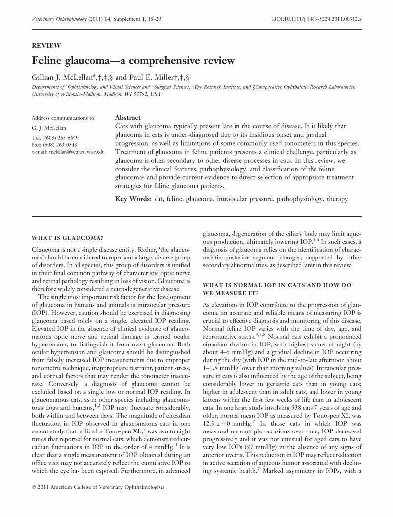
Differences in measurements obtained by differenttonometer types are also clinically significant. It is thereforeimportant that a consistent tonometer type and model isused for clinical monitoring and that the tonometer typeused to measure IOP is specified in the medical record.Veterinary clinicians should also be aware of the commonlimitations of the tonometer that is used. A number of to-nometers have been investigated in cats (Table 1), includingthe Schiøtz indentation tonometer;12 pneumotonometer;13
Mackay-Marg type applanation tonometers (such as theMackay-Marg, Tono-pen, Tonopen XL, and TonoPenVet);13–16 Perkins applanation tonometer, 17 and, mostrecently an induction/impact or rebound tonometer (Tono-Vet�).16,18 Although all these tonometers demonstratedacceptable accuracy within the normal physiological rangein cats, it is noteworthy that most applanation tonometersdramatically underestimated IOP above about 30 mmHgwhen compared to manometry. Despite their widespreadapplication in clinical veterinary practice, the systematicunderestimation of IOP in glaucomatous cats by mostapplanation tonometers may have contributed to an under-estimation of the true prevalence of glaucoma in the felinepopulation. The TonoVet� rebound tonometer is moreaccurate than the Tono-Pen, particularly at IOPs>30 mmHg; does not require application of topical anes-thetic; is well-tolerated by cats, and may be considered most
suitable among current, commercially available tonometers,for diagnosis and monitoring of glaucoma in cats.16,18
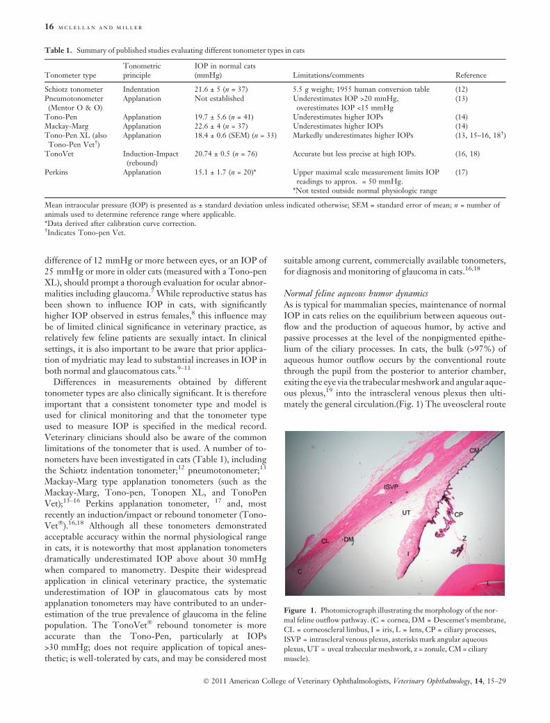
Normal feline aqueous humor dynamicsAs is typical for mammalian species, maintenance of normalIOP in cats relies on the equilibrium between aqueous out-flow and the production of aqueous humor, by active andpassive processes at the level of the nonpigmented epithe-lium of the ciliary processes. In cats, the bulk (>97%) ofaqueous humor outflow occurs by the conventional routethrough the pupil from the posterior to anterior chamber,exiting the eye via the trabecular meshwork and angular aque-ous plexus,19 into the intrascleral venous plexus then ulti-mately the general circulation.(Fig. 1) The uveoscleral route
Table 1. Summary of published studies evaluating different tonometer types in cats
Tonometer typeTonometricprinciple
IOP in normal cats(mmHg) Limitations/comments Reference
Schiotz tonometer Indentation 21.6 ± 5 (n = 37) 5.5 g weight; 1955 human conversion table (12)Pneumotonometer(Mentor O & O)
Applanation Not established Underestimates IOP >20 mmHg,overestimates IOP <15 mmHg
(13)
Tono-Pen Applanation 19.7 ± 5.6 (n = 41) Underestimates higher IOPs (14)Mackay-Marg Applanation 22.6 ± 4 (n = 37) Underestimates higher IOPs (14)Tono-Pen XL (alsoTono-Pen Vet†)
Applanation 18.4 ± 0.6 (SEM) (n = 33) Markedly underestimates higher IOPs (13, 15–16, 18†)
TonoVet Induction-Impact(rebound)
20.74 ± 0.5 (n = 76) Accurate but less precise at high IOPs. (16, 18)
Perkins Applanation 15.1 ± 1.7 (n = 20)* Upper maximal scale measurement limits IOPreadings to approx. = 50 mmHg.
*Not tested outside normal physiologic range
(17)
Mean intraocular pressure (IOP) is presented as ± standard deviation unless indicated otherwise; SEM = standard error of mean; n = number ofanimals used to determine reference range where applicable.*Data derived after calibration curve correction.†Indicates Tono-pen Vet.
Figure 1. Photomicrograph illustrating the morphology of the nor-
mal feline outflow pathway. (C = cornea, DM = Descemet’s membrane,
CL = corneoscleral limbus, I = iris, L = lens, CP = ciliary processes,
ISVP = intrascleral venous plexus, asterisks mark angular aqueous
plexus, UT = uveal trabecular meshwork, z = zonule, CM = ciliary
muscle).
16 m c l e l l a n a n d m i l l e r
� 2011 American College of Veterinary Ophthalmologists, Veterinary Ophthalmology, 14, 15–29
(via the iris and ciliary body stroma to the suprachoroidalcirculation and vortex veins, and through the sclera to theepiscleral tissues) accounts for a very small percentage ofaqueous humor outflow (<3%) in this species.20
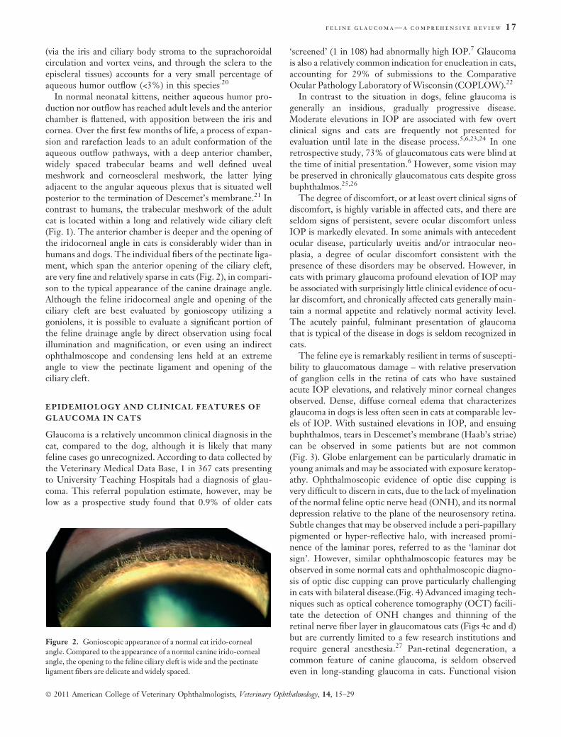
In normal neonatal kittens, neither aqueous humor pro-duction nor outflow has reached adult levels and the anteriorchamber is flattened, with apposition between the iris andcornea. Over the first few months of life, a process of expan-sion and rarefaction leads to an adult conformation of theaqueous outflow pathways, with a deep anterior chamber,widely spaced trabecular beams and well defined uvealmeshwork and corneoscleral meshwork, the latter lyingadjacent to the angular aqueous plexus that is situated wellposterior to the termination of Descemet’s membrane.21 Incontrast to humans, the trabecular meshwork of the adultcat is located within a long and relatively wide ciliary cleft(Fig. 1). The anterior chamber is deeper and the opening ofthe iridocorneal angle in cats is considerably wider than inhumans and dogs. The individual fibers of the pectinate liga-ment, which span the anterior opening of the ciliary cleft,are very fine and relatively sparse in cats (Fig. 2), in compari-son to the typical appearance of the canine drainage angle.Although the feline iridocorneal angle and opening of theciliary cleft are best evaluated by gonioscopy utilizing agoniolens, it is possible to evaluate a significant portion ofthe feline drainage angle by direct observation using focalillumination and magnification, or even using an indirectophthalmoscope and condensing lens held at an extremeangle to view the pectinate ligament and opening of theciliary cleft.
EPIDEMIOLOGY AND CLINICAL FEATURES OF
GLAUCOMA IN CATS
Glaucoma is a relatively uncommon clinical diagnosis in thecat, compared to the dog, although it is likely that manyfeline cases go unrecognized. According to data collected bythe Veterinary Medical Data Base, 1 in 367 cats presentingto University Teaching Hospitals had a diagnosis of glau-coma. This referral population estimate, however, may below as a prospective study found that 0.9% of older cats
‘screened’ (1 in 108) had abnormally high IOP.7 Glaucomais also a relatively common indication for enucleation in cats,accounting for 29% of submissions to the ComparativeOcular Pathology Laboratory of Wisconsin (COPLOW).22
In contrast to the situation in dogs, feline glaucoma isgenerally an insidious, gradually progressive disease.Moderate elevations in IOP are associated with few overtclinical signs and cats are frequently not presented forevaluation until late in the disease process.5,6,23,24 In oneretrospective study, 73% of glaucomatous cats were blind atthe time of initial presentation.6 However, some vision maybe preserved in chronically glaucomatous cats despite grossbuphthalmos.25,26
The degree of discomfort, or at least overt clinical signs ofdiscomfort, is highly variable in affected cats, and there areseldom signs of persistent, severe ocular discomfort unlessIOP is markedly elevated. In some animals with antecedentocular disease, particularly uveitis and/or intraocular neo-plasia, a degree of ocular discomfort consistent with thepresence of these disorders may be observed. However, incats with primary glaucoma profound elevation of IOP maybe associated with surprisingly little clinical evidence of ocu-lar discomfort, and chronically affected cats generally main-tain a normal appetite and relatively normal activity level.The acutely painful, fulminant presentation of glaucomathat is typical of the disease in dogs is seldom recognized incats.
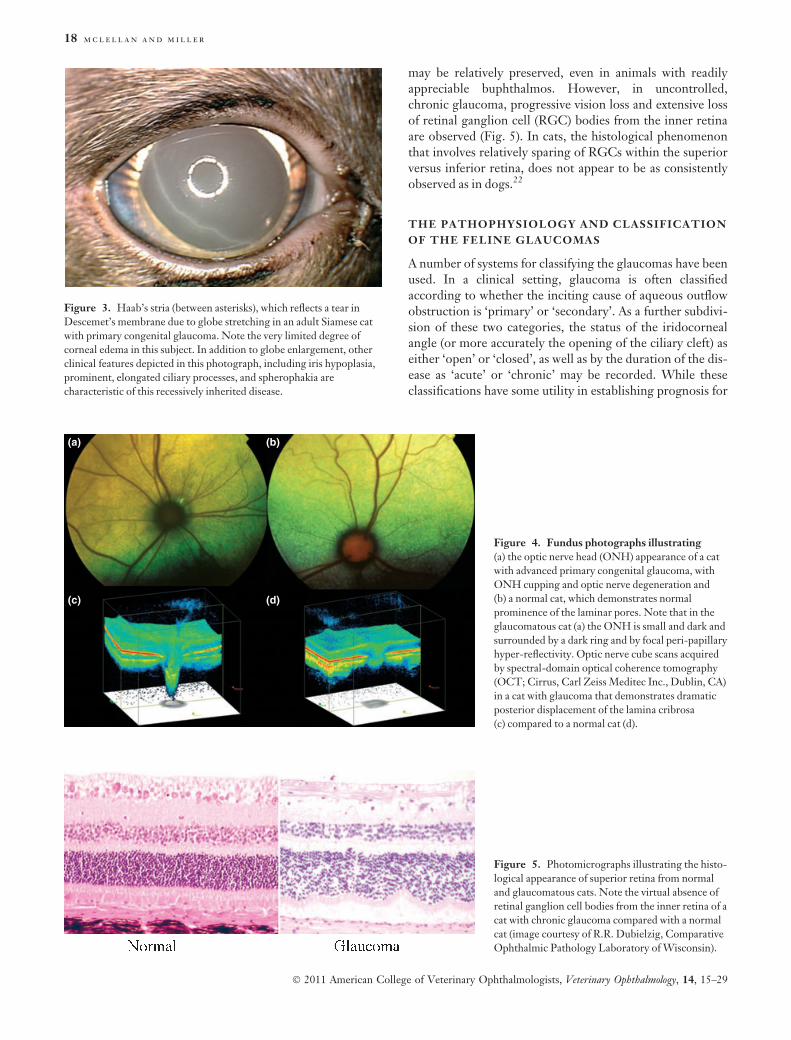
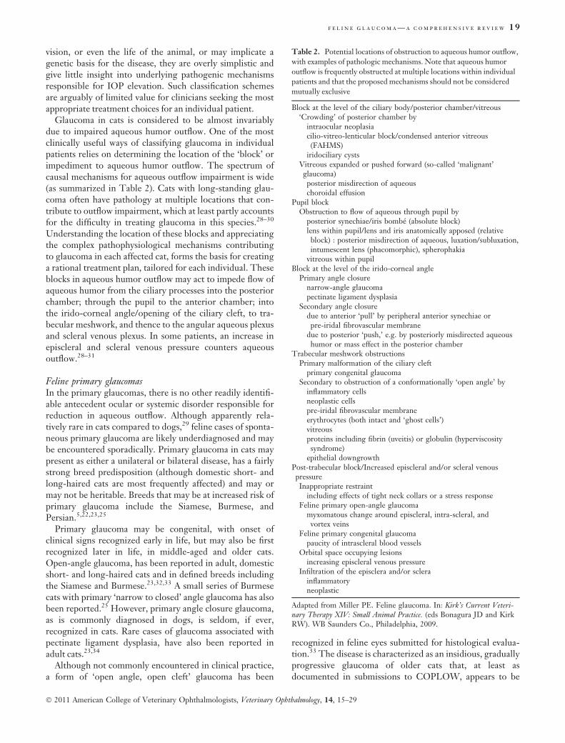
The feline eye is remarkably resilient in terms of suscepti-bility to glaucomatous damage – with relative preservationof ganglion cells in the retina of cats who have sustainedacute IOP elevations, and relatively minor corneal changesobserved. Dense, diffuse corneal edema that characterizesglaucoma in dogs is less often seen in cats at comparable lev-els of IOP. With sustained elevations in IOP, and ensuingbuphthalmos, tears in Descemet’s membrane (Haab’s striae)can be observed in some patients but are not common(Fig. 3). Globe enlargement can be particularly dramatic inyoung animals and may be associated with exposure keratop-athy. Ophthalmoscopic evidence of optic disc cupping isvery difficult to discern in cats, due to the lack of myelinationof the normal feline optic nerve head (ONH), and its normaldepression relative to the plane of the neurosensory retina.Subtle changes that may be observed include a peri-papillarypigmented or hyper-reflective halo, with increased promi-nence of the laminar pores, referred to as the ‘laminar dotsign’. However, similar ophthalmoscopic features may beobserved in some normal cats and ophthalmoscopic diagno-sis of optic disc cupping can prove particularly challengingin cats with bilateral disease.(Fig. 4) Advanced imaging tech-niques such as optical coherence tomography (OCT) facili-tate the detection of ONH changes and thinning of theretinal nerve fiber layer in glaucomatous cats (Figs 4c and d)but are currently limited to a few research institutions andrequire general anesthesia.27 Pan-retinal degeneration, acommon feature of canine glaucoma, is seldom observedeven in long-standing glaucoma in cats. Functional vision
Figure 2. Gonioscopic appearance of a normal cat irido-corneal
angle. Compared to the appearance of a normal canine irido-corneal
angle, the opening to the feline ciliary cleft is wide and the pectinate
ligament fibers are delicate and widely spaced.
f e l i n e g l a u c o m a — a c o m p r e h e n s i v e r e v i e w 17
� 2011 American College of Veterinary Ophthalmologists, Veterinary Ophthalmology, 14, 15–29
may be relatively preserved, even in animals with readilyappreciable buphthalmos. However, in uncontrolled,chronic glaucoma, progressive vision loss and extensive lossof retinal ganglion cell (RGC) bodies from the inner retinaare observed (Fig. 5). In cats, the histological phenomenonthat involves relatively sparing of RGCs within the superiorversus inferior retina, does not appear to be as consistentlyobserved as in dogs.22
THE PATHOPHYSIOLOGY AND CLASSIFICATION
OF THE FELINE GLAUCOMAS
A number of systems for classifying the glaucomas have beenused. In a clinical setting, glaucoma is often classifiedaccording to whether the inciting cause of aqueous outflowobstruction is ‘primary’ or ‘secondary’. As a further subdivi-sion of these two categories, the status of the iridocornealangle (or more accurately the opening of the ciliary cleft) aseither ‘open’ or ‘closed’, as well as by the duration of the dis-ease as ‘acute’ or ‘chronic’ may be recorded. While theseclassifications have some utility in establishing prognosis for
Figure 3. Haab’s stria (between asterisks), which reflects a tear in
Descemet’s membrane due to globe stretching in an adult Siamese cat
with primary congenital glaucoma. Note the very limited degree of
corneal edema in this subject. In addition to globe enlargement, other
clinical features depicted in this photograph, including iris hypoplasia,
prominent, elongated ciliary processes, and spherophakia are
characteristic of this recessively inherited disease.
(a) (b)
(c) (d)
Figure 4. Fundus photographs illustrating
(a) the optic nerve head (ONH) appearance of a cat
with advanced primary congenital glaucoma, with
ONH cupping and optic nerve degeneration and
(b) a normal cat, which demonstrates normal
prominence of the laminar pores. Note that in the
glaucomatous cat (a) the ONH is small and dark and
surrounded by a dark ring and by focal peri-papillary
hyper-reflectivity. Optic nerve cube scans acquired
by spectral-domain optical coherence tomography
(OCT; Cirrus, Carl Zeiss Meditec Inc., Dublin, CA)
in a cat with glaucoma that demonstrates dramatic
posterior displacement of the lamina cribrosa
(c) compared to a normal cat (d).
Figure 5. Photomicrographs illustrating the histo-
logical appearance of superior retina from normal
and glaucomatous cats. Note the virtual absence of
retinal ganglion cell bodies from the inner retina of a
cat with chronic glaucoma compared with a normal
cat (image courtesy of R.R. Dubielzig, Comparative
Ophthalmic Pathology Laboratory of Wisconsin).
18 m c l e l l a n a n d m i l l e r
� 2011 American College of Veterinary Ophthalmologists, Veterinary Ophthalmology, 14, 15–29
vision, or even the life of the animal, or may implicate agenetic basis for the disease, they are overly simplistic andgive little insight into underlying pathogenic mechanismsresponsible for IOP elevation. Such classification schemesare arguably of limited value for clinicians seeking the mostappropriate treatment choices for an individual patient.
Glaucoma in cats is considered to be almost invariablydue to impaired aqueous humor outflow. One of the mostclinically useful ways of classifying glaucoma in individualpatients relies on determining the location of the ‘block’ orimpediment to aqueous humor outflow. The spectrum ofcausal mechanisms for aqueous outflow impairment is wide(as summarized in Table 2). Cats with long-standing glau-coma often have pathology at multiple locations that con-tribute to outflow impairment, which at least partly accountsfor the difficulty in treating glaucoma in this species.28–30
Understanding the location of these blocks and appreciatingthe complex pathophysiological mechanisms contributingto glaucoma in each affected cat, forms the basis for creatinga rational treatment plan, tailored for each individual. Theseblocks in aqueous humor outflow may act to impede flow ofaqueous humor from the ciliary processes into the posteriorchamber; through the pupil to the anterior chamber; intothe irido-corneal angle/opening of the ciliary cleft, to tra-becular meshwork, and thence to the angular aqueous plexusand scleral venous plexus. In some patients, an increase inepiscleral and scleral venous pressure counters aqueousoutflow.28–31
Feline primary glaucomasIn the primary glaucomas, there is no other readily identifi-able antecedent ocular or systemic disorder responsible forreduction in aqueous outflow. Although apparently rela-tively rare in cats compared to dogs,29 feline cases of sponta-neous primary glaucoma are likely underdiagnosed and maybe encountered sporadically. Primary glaucoma in cats maypresent as either a unilateral or bilateral disease, has a fairlystrong breed predisposition (although domestic short- andlong-haired cats are most frequently affected) and may ormay not be heritable. Breeds that may be at increased risk ofprimary glaucoma include the Siamese, Burmese, andPersian.5,22,23,25
Primary glaucoma may be congenital, with onset ofclinical signs recognized early in life, but may also be firstrecognized later in life, in middle-aged and older cats.Open-angle glaucoma, has been reported in adult, domesticshort- and long-haired cats and in defined breeds includingthe Siamese and Burmese.23,32,33 A small series of Burmesecats with primary ‘narrow to closed’ angle glaucoma has alsobeen reported.25 However, primary angle closure glaucoma,as is commonly diagnosed in dogs, is seldom, if ever,recognized in cats. Rare cases of glaucoma associated withpectinate ligament dysplasia, have also been reported inadult cats.23,34
Although not commonly encountered in clinical practice,a form of ‘open angle, open cleft’ glaucoma has been
recognized in feline eyes submitted for histological evalua-tion.33 The disease is characterized as an insidious, graduallyprogressive glaucoma of older cats that, at least asdocumented in submissions to COPLOW, appears to be
Table 2. Potential locations of obstruction to aqueous humor outflow,
with examples of pathologic mechanisms. Note that aqueous humor
outflow is frequently obstructed at multiple locations within individual
patients and that the proposed mechanisms should not be considered
mutually exclusive
Block at the level of the ciliary body/posterior chamber/vitreous‘Crowding’ of posterior chamber by
intraocular neoplasiacilio-vitreo-lenticular block/condensed anterior vitreous(FAHMS)
iridociliary cystsVitreous expanded or pushed forward (so-called ‘malignant’glaucoma)posterior misdirection of aqueouschoroidal effusion
Pupil blockObstruction to flow of aqueous through pupil by
posterior synechiae/iris bombe (absolute block)lens within pupil/lens and iris anatomically apposed (relativeblock) : posterior misdirection of aqueous, luxation/subluxation,intumescent lens (phacomorphic), spherophakia
vitreous within pupilBlock at the level of the irido-corneal angle
Primary angle closurenarrow-angle glaucomapectinate ligament dysplasia
Secondary angle closuredue to anterior ‘pull’ by peripheral anterior synechiae orpre-iridal fibrovascular membrane
due to posterior ‘push,’ e.g. by posteriorly misdirected aqueoushumor or mass effect in the posterior chamber
Trabecular meshwork obstructionsPrimary malformation of the ciliary cleft
primary congenital glaucomaSecondary to obstruction of a conformationally ‘open angle’ by
inflammatory cellsneoplastic cellspre-iridal fibrovascular membraneerythrocytes (both intact and ‘ghost cells’)vitreousproteins including fibrin (uveitis) or globulin (hyperviscositysyndrome)
epithelial downgrowthPost-trabecular block/Increased episcleral and/or scleral venouspressureInappropriate restraint
including effects of tight neck collars or a stress responseFeline primary open-angle glaucoma
myxomatous change around episcleral, intra-scleral, andvortex veins
Feline primary congenital glaucomapaucity of intrascleral blood vessels
Orbital space occupying lesionsincreasing episcleral venous pressure
Infiltration of the episclera and/or sclerainflammatoryneoplastic
Adapted from Miller PE. Feline glaucoma. In: Kirk’s Current Veteri-nary Therapy XIV: Small Animal Practice. (eds Bonagura JD and KirkRW). WB Saunders Co., Philadelphia, 2009.
f e l i n e g l a u c o m a — a c o m p r e h e n s i v e r e v i e w 19
� 2011 American College of Veterinary Ophthalmologists, Veterinary Ophthalmology, 14, 15–29
unilateral in presentation (although asymmetric presenta-tion cannot be excluded due to insufficient follow-up inmost cases). Burmese cats appear to be over-represented. Inaffected cats there is histopathological evidence of RGC lossand subtle myxomatous change within the vascular channelsassociated with aqueous outflow, including the scleralvenous plexus and the vortex veins (Fig. 6). No pathology isrecognizable within the iridocorneal angle or trabecularmeshwork, thus it is proposed that resistance to aqueousoutflow occurs at the level of the scleral venous plexus andvortex veins.33 However, obstruction to outflow at the levelof the trabecular meshwork cannot be excluded based onhistomorphological appearance alone. At this time clinicalcorrelates to accompany these pathological descriptions ofprimary open-angle glaucoma remain sparse, and the clinicalcourse of this particular disease in affected animals remainsunclear.
Sporadic cases of feline congenital or early onset glau-coma associated with various ocular malformations, includ-ing microphakia, ectopia lentis, iridoschisis, pectinateligament dysplasia, multiple iridociliary cysts, and persistentpupillary membranes have been reported in the veterinary
literature.23,35 A research colony founded by breedingSiamese cats has been established in the United States.These cats have relatively symmetric, slowly progressiveglaucoma characterized by elongated ciliary processes, globeenlargement, and spherophakia (Fig. 3). 26 In affectedkittens, postnatal development of the structures of the ciliarycleft is arrested, and affected cats maintain the immatureconformation of their aqueous outflow pathways.36 Themutation responsible for this form of primary congenitalglaucoma has now been identified (M.H. Kuehn, N.M.Ellinwood, K.H. Deckman, M.A. Menotti-Raymond, E.A.Snella and G.J. McLellan, manuscript in process). It wouldappear from sporadic reports in the veterinary literature ofglaucoma occurring secondary to uveitis,37 and of primarymicrophakia with lens luxation,23,38,39 in Siamese cats, thatthis disease may actually have existed in the Siamese catbreed in the USA and Europe for more than 50 years.
Feline secondary glaucomasThe secondary glaucomas, constituting 95–98% of felineglaucoma cases, are associated with antecedent ocular or sys-temic disease processes, such as uveitis, neoplasia, trauma,and intraocular hemorrhage that alter aqueous humordynamics by a range of mechanisms.5–7,23,24,40 Dependingon the underlying pathogenesis, they may be unilateral inpresentation or, less often, bilateral, and are most often seenin adult cats.
Intraocular inflammation, particularly chronic lympho-plasmacytic uveitis, is the most frequently reported cause ofglaucoma in cats.41 Lymphoplasmacytic uveitis withglaucoma accounted for 10% of all feline submissions toCOPLOW.40 Uveitis may lead to elevation of IOP througha number of different pathogenic mechanisms.5,6,23,32,42
While obliteration of the aqueous outflow pathway byinflammatory infiltrates is often proposed as a mechanismfor IOP elevation (Fig. 7), in reality the extent of the inflam-matory infiltrate observed histologically in enucleatedglobes may be minimal. Chronic lymphoplasmacytic uveitismay also lead to lens luxation; condensation, degeneration,and prolapse of vitreous; formation of pre-iridal fibrovascu-lar membranes and synechiae.32,40,43 Angle recession mayalso contribute to aqueous outflow obstruction.40 Althoughidentified histologically in glaucomatous feline eyes,40 therole played by angle recession in the development of feline
Figure 6. Photomicrograph illustrating myxomatous changes sur-
rounding the intrascleral veins (inset) in a cat with primary open angle
glaucoma. The ciliary cleft is open and the trabecular meshwork appears
normal. (Image courtesy of Dr. R.R. Dubielzig, Comparative Ophthal-
mic Pathology Laboratory of Wisconsin).
(a) (b)
Figure 7. Clinical photograph (a) illustrating
features of chronic lymphoplasmacytic uveitis,
including rubeosis iridis and keratic precipates, in a
cat with secondary glaucoma. (b) Photomicrograph
of inflammatory infiltrate which has obliterated the
structures of the ciliary cleft and iridocorneal angle
in a cat with lymphoplasmacytic uveitis and
secondary glaucoma. (Image courtesy of
Dr. R.R. Dubielzig, Comparative Ophthalmic
Pathology Laboratory of Wisconsin).
20 m c l e l l a n a n d m i l l e r
� 2011 American College of Veterinary Ophthalmologists, Veterinary Ophthalmology, 14, 15–29
glaucoma remains unclear and it is possible that thishistomorphological phenomenon may be secondary tobuphthalmia, and associated changes in anterior segmentanatomic relationships. The proposed pathogenesis forangle recession in humans is that blunt force trauma deformsthe globe, pushing the cornea anteriorly and that the sub-sequent rebound effect on the lens exerts traction on theciliary body causing cyclodialysis. Angle recession is mostoften seen histologically in feline eyes with concomitantlymphoplasmacytic uveitis, and a history of trauma is gener-ally lacking, perhaps due to the protracted course of clinicaldisease prior to enucleation in many glaucomatous cats.40
Intraocular neoplasia is another major cause of glaucomain cats.6,23,32,40 In order of frequency, anterior uvealmelanoma (Fig. 8), lymphoma (Fig. 9), post-traumaticocular sarcoma,44,45 and iridociliary epithelial tumors,46
may all be associated with secondary glaucoma. Intraocularneoplasia may be associated with diffuse infiltration andobliteration of the trabecular meshwork and ciliary cleft, orthe development of neovascular glaucoma with angleclosure.40 Diffuse iris melanoma is a relatively commoncause of feline glaucoma. About half of all feline submissionsto COPLOW were diagnosed with diffuse iris melanoma,and just over 10% of all feline submissions were diagnosedwith glaucoma secondary to melanoma. 40
Intraocular hemorrhage, particularly related to systemichypertension in older cats, may also result in secondaryglaucoma. Development of glaucoma secondary to intraocu-lar hemorrhage is of particular concern in animals with‘eight-ball hyphema’ caused by episodes of rebleeding. Tothe authors’ knowledge, glaucoma secondary to retinaldetachment in cats has not been specifically reported in theveterinary literature, and may be less of a concern than indogs.
Lens-associated glaucoma in cats may occur secondary tophacoclastic uveitis or septic implantation syndrome result-ing from lens trauma,32,42,47 or, in the long term, lens trauma
may be associated with the development of post-traumaticocular sarcoma that may present as glaucoma.48 In contrastto dogs, lens luxation in cats is seldom primary and mostoften occurs secondary to glaucoma or uveitis. Even if lensluxation may be considered a ‘primary’ event, as may occurin elderly cats with senile zonular degeneration, or inanimals with hypermature cataract, lens luxation in catsseldom directly results in glaucoma.49 Intumescent cataractand phacomorphic glaucoma are also rarely encountered incats.
Aqueous humor misdirection syndrome is an unusualform of insidious glaucoma, characterized by a uniformlyshallow anterior chamber that has been recognized in oldercats.6,50,51 In addition to a uniformly shallow anterior cham-ber, affected eyes often exhibit dilated pupils. (Fig. 10) Thismydriasis frequently results in anisocoria as the glaucoma isoften unilateral, or at least asymmetric, at the time of initialpresentation. The degree of IOP elevation in affected eyesranges from mild to severe, with modest elevations beingmost typical and in the early stages IOP may actually be nor-mal. In severely affected eyes in one series, a pronouncedmyopia (as much as )16.5 D) was identified, and was attrib-uted to the anteriorly positioned lens.51 Ocular ultrasonog-raphy and histopathology in affected cats reveal thickeningof the anterior vitreous face, anterior displacement of the irisand lens, and clear spaces in the vitreous cavity.(Fig. 11) Thedisease has been termed feline aqueous humor misdirectionsyndrome (FAHMS), as it has been postulated that aqueoushumor is misdirected posteriorly through breaks in the ante-rior vitreous face that act as one-way valves, resulting init becoming progressively trapped within ‘pools’ in the
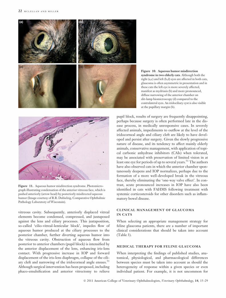
Figure 8. Clinical photograph of an elderly cat with glaucoma
secondary to diffuse anterior uveal melanoma in the right eye. In
addition to the diffuse iris hyperpigmentation, note subtle anisocoria
due to mydriasis in the right eye.
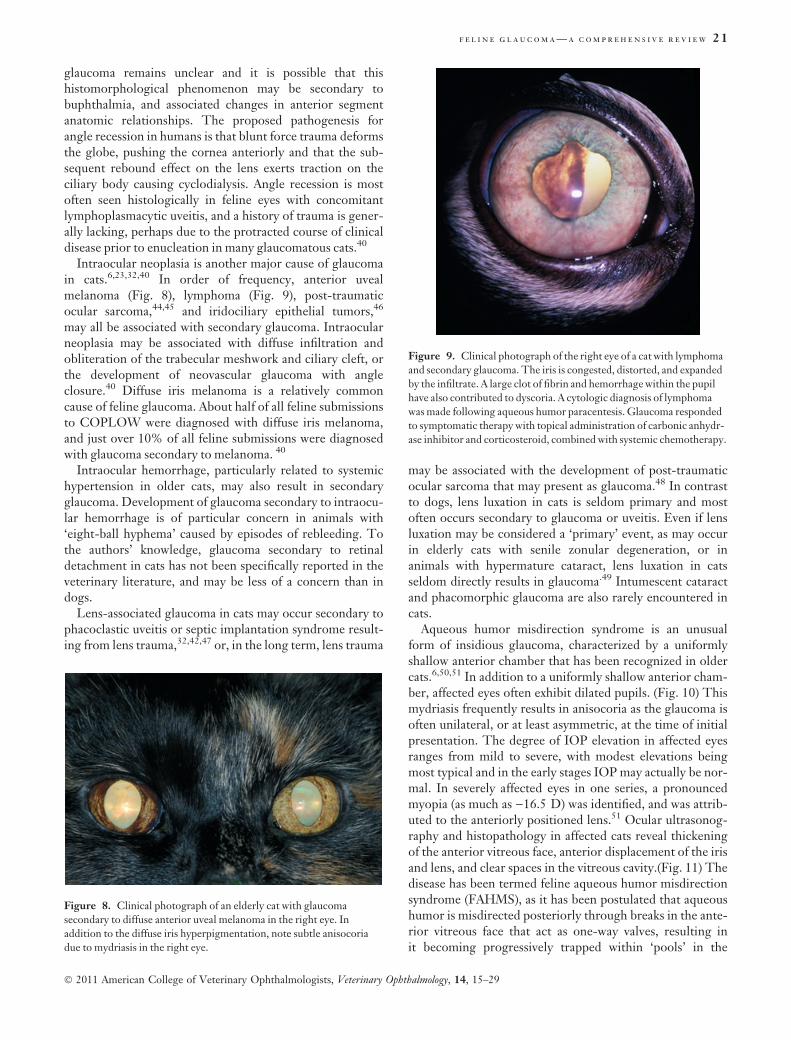
Figure 9. Clinical photograph of the right eye of a cat with lymphoma
and secondary glaucoma. The iris is congested, distorted, and expanded
by the infiltrate. A large clot of fibrin and hemorrhage within the pupil
have also contributed to dyscoria. A cytologic diagnosis of lymphoma
was made following aqueous humor paracentesis. Glaucoma responded
to symptomatic therapy with topical administration of carbonic anhydr-
ase inhibitor and corticosteroid, combined with systemic chemotherapy.
f e l i n e g l a u c o m a — a c o m p r e h e n s i v e r e v i e w 21
� 2011 American College of Veterinary Ophthalmologists, Veterinary Ophthalmology, 14, 15–29
vitreous cavity. Subsequently, anteriorly displaced vitrealelements become condensed, compressed, and juxtaposedagainst the lens and ciliary processes. This juxtaposition,so-called ‘cilio-vitreal-lenticular block’, impedes flow ofaqueous humor produced at the ciliary processes to theposterior chamber, further diverting aqueous humor intothe vitreous cavity. Obstruction of aqueous flow fromposterior to anterior chambers (pupil block) is intensified bythe anterior displacement of the lens, enhancing iris-lenscontact. With progressive increase in IOP and forwarddisplacement of the iris-lens diaphragm, collapse of the cili-ary cleft and narrowing of the iridocorneal angle ensues.51
Although surgical intervention has been proposed, includingphaco-emulsification and anterior vitrectomy to relieve
pupil block, results of surgery are frequently disappointing,perhaps because surgery is often performed late in the dis-ease process, in medically unresponsive cases. In severelyaffected animals, impediments to outflow at the level of theiridocorneal angle and ciliary cleft are likely to have devel-oped and persist after surgery. Given the slowly progressivenature of disease, and its tendency to affect mainly elderlyanimals, conservative management, with application of topi-cal carbonic anhydrase inhibitors (CAIs) when tolerated,may be associated with preservation of limited vision in atleast one eye for periods of up to several years.51 The authorshave also observed cats in which the anterior chamber spon-taneously deepens and IOP normalizes, perhaps due to theformation of a more well-developed break in the vitreousface, thereby eliminating the ‘one-way valve effect’. In con-trast, acute pronounced increases in IOP have also beenidentified in cats with FAHMS following treatment withsystemic corticosteroids for other disorders such as inflam-matory bowel disease.
CLINICAL MANAGEMENT OF GLAUCOMA
IN CATS
When selecting an appropriate management strategy forfeline glaucoma patients, there are a number of importantclinical considerations that should be taken into account(Table 3).
MEDICAL THERAPY FOR FELINE GLAUCOMA
When interpreting the findings of published studies, ana-tomical, physiological, and pharmacological differencesbetween species must be taken into account as should theheterogeneity of response within a given species or evenindividual patient. For example, it is not uncommon for
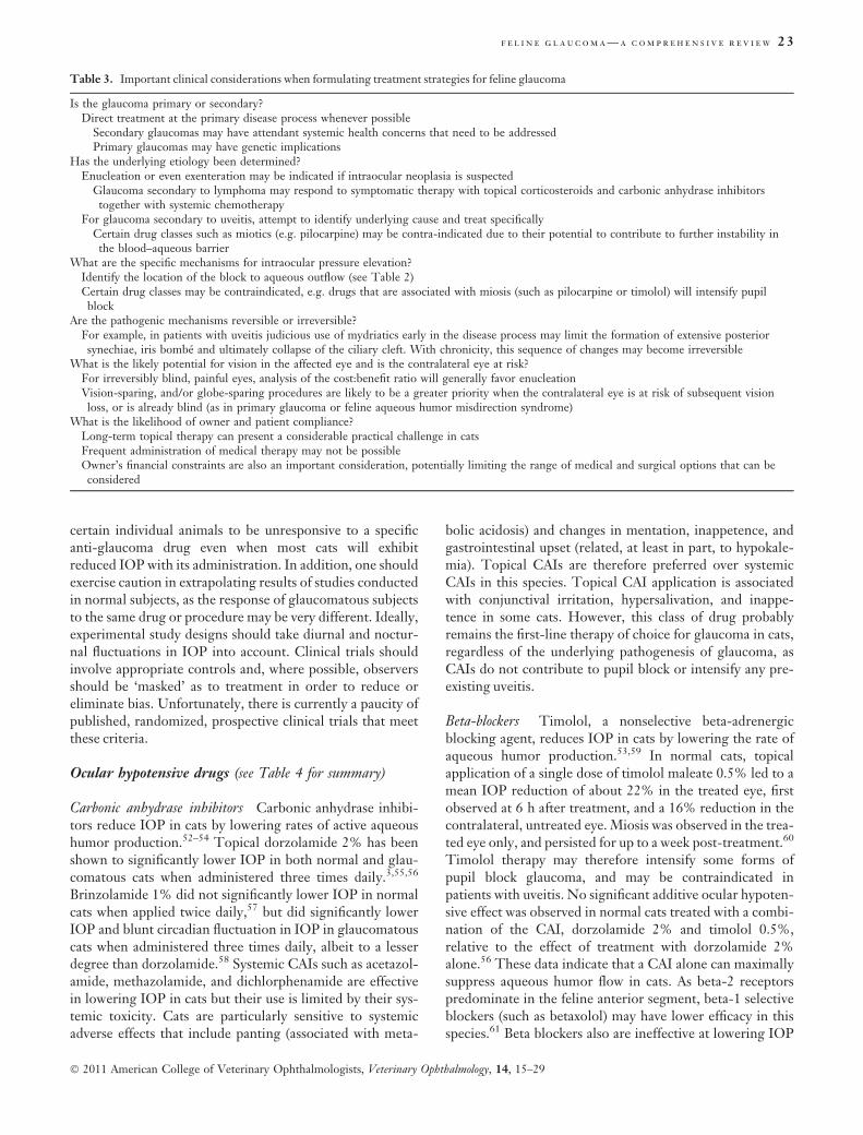
(a) (b)
(c) (d)Figure 10. Aqueous humor misdirection
syndrome in two elderly cats. Although both the
right (a,c) and left (b,d) eyes are affected in both cats,
glaucoma is often asymmetric in presentation and in
these cats the left eye is more severely affected,
manifest as mydriasis (b) and more pronounced,
diffuse narrowing of the anterior chamber on
slit-lamp biomicroscopy (d) compared to the
contralateral eyes. An iridociliary cyst is also visible
at the pupillary margin (b).
Figure 11. Aqueous humor misdirection syndrome. Photomicro-
graph illustrating condensation of the anterior vitreous face, which is
pushed anteriorly (arrow head) by posteriorly misdirected aqueous
humor (Image courtesy of R.R. Dubielzig, Comparative Ophthalmic
Pathology Laboratory of Wisconsin).
22 m c l e l l a n a n d m i l l e r
� 2011 American College of Veterinary Ophthalmologists, Veterinary Ophthalmology, 14, 15–29
certain individual animals to be unresponsive to a specificanti-glaucoma drug even when most cats will exhibitreduced IOP with its administration. In addition, one shouldexercise caution in extrapolating results of studies conductedin normal subjects, as the response of glaucomatous subjectsto the same drug or procedure may be very different. Ideally,experimental study designs should take diurnal and noctur-nal fluctuations in IOP into account. Clinical trials shouldinvolve appropriate controls and, where possible, observersshould be ‘masked’ as to treatment in order to reduce oreliminate bias. Unfortunately, there is currently a paucity ofpublished, randomized, prospective clinical trials that meetthese criteria.
Ocular hypotensive drugs (see Table 4 for summary)
Carbonic anhydrase inhibitors Carbonic anhydrase inhibi-tors reduce IOP in cats by lowering rates of active aqueoushumor production.52–54 Topical dorzolamide 2% has beenshown to significantly lower IOP in both normal and glau-comatous cats when administered three times daily.3,55,56
Brinzolamide 1% did not significantly lower IOP in normalcats when applied twice daily,57 but did significantly lowerIOP and blunt circadian fluctuation in IOP in glaucomatouscats when administered three times daily, albeit to a lesserdegree than dorzolamide.58 Systemic CAIs such as acetazol-amide, methazolamide, and dichlorphenamide are effectivein lowering IOP in cats but their use is limited by their sys-temic toxicity. Cats are particularly sensitive to systemicadverse effects that include panting (associated with meta-
bolic acidosis) and changes in mentation, inappetence, andgastrointestinal upset (related, at least in part, to hypokale-mia). Topical CAIs are therefore preferred over systemicCAIs in this species. Topical CAI application is associatedwith conjunctival irritation, hypersalivation, and inappe-tence in some cats. However, this class of drug probablyremains the first-line therapy of choice for glaucoma in cats,regardless of the underlying pathogenesis of glaucoma, asCAIs do not contribute to pupil block or intensify any pre-existing uveitis.
Beta-blockers Timolol, a nonselective beta-adrenergicblocking agent, reduces IOP in cats by lowering the rate ofaqueous humor production.53,59 In normal cats, topicalapplication of a single dose of timolol maleate 0.5% led to amean IOP reduction of about 22% in the treated eye, firstobserved at 6 h after treatment, and a 16% reduction in thecontralateral, untreated eye. Miosis was observed in the trea-ted eye only, and persisted for up to a week post-treatment.60
Timolol therapy may therefore intensify some forms ofpupil block glaucoma, and may be contraindicated inpatients with uveitis. No significant additive ocular hypoten-sive effect was observed in normal cats treated with a combi-nation of the CAI, dorzolamide 2% and timolol 0.5%,relative to the effect of treatment with dorzolamide 2%alone.56 These data indicate that a CAI alone can maximallysuppress aqueous humor flow in cats. As beta-2 receptorspredominate in the feline anterior segment, beta-1 selectiveblockers (such as betaxolol) may have lower efficacy in thisspecies.61 Beta blockers also are ineffective at lowering IOP
Table 3. Important clinical considerations when formulating treatment strategies for feline glaucoma
Is the glaucoma primary or secondary?Direct treatment at the primary disease process whenever possible
Secondary glaucomas may have attendant systemic health concerns that need to be addressedPrimary glaucomas may have genetic implications
Has the underlying etiology been determined?Enucleation or even exenteration may be indicated if intraocular neoplasia is suspected
Glaucoma secondary to lymphoma may respond to symptomatic therapy with topical corticosteroids and carbonic anhydrase inhibitorstogether with systemic chemotherapy
For glaucoma secondary to uveitis, attempt to identify underlying cause and treat specificallyCertain drug classes such as miotics (e.g. pilocarpine) may be contra-indicated due to their potential to contribute to further instability inthe blood–aqueous barrier
What are the specific mechanisms for intraocular pressure elevation?Identify the location of the block to aqueous outflow (see Table 2)Certain drug classes may be contraindicated, e.g. drugs that are associated with miosis (such as pilocarpine or timolol) will intensify pupilblock
Are the pathogenic mechanisms reversible or irreversible?For example, in patients with uveitis judicious use of mydriatics early in the disease process may limit the formation of extensive posteriorsynechiae, iris bombe and ultimately collapse of the ciliary cleft. With chronicity, this sequence of changes may become irreversible
What is the likely potential for vision in the affected eye and is the contralateral eye at risk?For irreversibly blind, painful eyes, analysis of the cost:benefit ratio will generally favor enucleationVision-sparing, and/or globe-sparing procedures are likely to be a greater priority when the contralateral eye is at risk of subsequent visionloss, or is already blind (as in primary glaucoma or feline aqueous humor misdirection syndrome)
What is the likelihood of owner and patient compliance?Long-term topical therapy can present a considerable practical challenge in catsFrequent administration of medical therapy may not be possibleOwner’s financial constraints are also an important consideration, potentially limiting the range of medical and surgical options that can beconsidered
f e l i n e g l a u c o m a — a c o m p r e h e n s i v e r e v i e w 23
� 2011 American College of Veterinary Ophthalmologists, Veterinary Ophthalmology, 14, 15–29
during sleep,62 due to lower sympathetic tone,61,63 whichmay reduce their efficacy in felines that nap frequently dur-ing the day, and exhibit peak IOP during the nocturnalphase. Timolol may cause bradycardia and bronchoconstric-tion and is probably contraindicated in cats with felineasthma or cardiac disease.64
Cholinergics These miotic drugs are believed to reduceIOP in cats by increasing aqueous outflow.53 Topicaladministration of a single dose of the direct-acting choliner-gic, pilocarpine 2% reduced IOP by about 15% in the trea-ted eye and caused miosis in both the treated and untreatedeye of normal cats.65 This contralateral effect observed fol-lowing unilateral application, indicates that systemic,adverse cholinergic effects might be anticipated duringlong-term treatment. Due to their miotic effect and abilityto destabilize the blood-ocular barrier,66 these drugs aregenerally contra-indicated in animals with pre-existingintraocular inflammation or a tendency to pupil block.Demecarium bromide 0.125%, a cholinesterase inhibitor, isa more potent and longer lasting miotic, but to our knowl-edge its effects have not been specifically evaluated in cats.
Adrenergic agonists The nonspecific adrenergic agonist,epinephrine when applied twice daily reduced IOP in nor-mal cats by about 27% in treated eyes, related to a reductionin both aqueous production, as determined by fluoropho-tometry, and an increase in outflow facility.67 Although thisstudy did not specifically evaluate or report ocular tolerabil-ity, ocular surface irritation might be expected, based on
reported adverse effects in human patients. Dipivefrin, apro-drug of epinephrine, might be expected to exert similareffects to epinephrine but its use has not been specificallyinvestigated in cats. Dipivefrin requires corneal esterases toconvert the pro-drug to its active form, and probably shouldnot be combined with cholinesterase inhibitors.68 Topicaladrenergic agonists are associated with mild pupillarydilation, and do not exacerbate uveitis.
Alpha-2 agonists such as apraclonidine and brimonidinemay reduce IOP by increasing conventional outflow,decreasing aqueous humor production and reducing episcl-eral venous pressure.69 Apraclonidine reduced IOP in nor-mal cats by an average of 24% within 6 h of treatment butalso resulted in miosis that persisted for up to 24 h. Evidenceof systemic toxicity following topical application of 0.5%apraclonidine, consisting of a mean reduction in heart rateof about 12%, and vomiting that was noted in 8/9 cats trea-ted with the drug, precludes use of the commercially avail-able formulation in cats.70
Prostaglandin analogs Species differences in prostanoidreceptor distribution within different ocular tissues havemajor implications for the effects, and efficacy of topicalprostaglandin analog therapy for glaucoma. For example,the intense miosis observed in cats treated with prostaglan-din analogs such as latanoprost, travaprost, unoprostone,and bimatoprost,71–73 is attributable to the presence ofexquisitely well-coupled FP receptors in the iris sphinctermuscle of cats, compared to humans.74,75 Although FPreceptors are largely responsible for the effects of prosta-
Table 4. Summary of currently available anti-glaucoma drugs of potential value in cats
Drug Class Route Frequency Mechanism Contraindications/adverse effects References
Dorzolamide 2% CAI Topical Q 8 h fl production May be transient salivation. Inappetencein some cats. Sterile conjunctivitis
(3,54–56)
Brinzolamide 1% CAI Topical Q 8 h fl production May be less effective but cause less ocularirritation than dorzolamide. Does notaffect IOP in normal cats
(57–58)
Methazolamide,diclorphenamide
acetazolamide
CAI Oral 0.5–2 mg/kgQ 8–24 h
10–25 mg/kg PO;5–10 mg/kg IV
fl production Cats very susceptible to adverse effects(anorexia, GI disturbances, increasedrespiratory rate due to metabolic acidosis)and should be monitored closely
(52–53, 64, 93)
Timolol 0.5% Beta-blocker Topical Q 12 h fl production Avoid in animals with feline asthma,cardiovascular disease or pupil block. Lesseffective during sleep
(53, 59–60)
Betaxolol 0.5% Beta-blocker Topical Q 12 h fl production More Beta 2-selective, may be safer in catswith respiratory or cardiovascular disease.Questionable efficacy in cats due topredominance of b)1 receptors in felineanterior segment
(61);Not studied
Epinephrine 1–2% Adrenergicagonist
Topical Q 6–12 h › outflowfl production
Not reported but local irritation might beanticipated
(67)
Dipivefrin 0.1% Beta-adrenergicagonist
Topical Q 6–12 h › outflowfl production
Do not use with cholinesterase inhibitors Not describedin cats
Pilocarpine 2% Cholinergic Topical Q 6–12 h › outflow Contraindicated where pre-existing uveitis,aqueous humor misdirection, pupil blockand/or phacomorphic glaucoma. Systemictoxicity may be observed
(53, 65–66)
24 m c l e l l a n a n d m i l l e r
� 2011 American College of Veterinary Ophthalmologists, Veterinary Ophthalmology, 14, 15–29
glandin analogs on the canine and human ciliary body, thesereceptors are lacking in the ciliary body of cats and EPreceptors are predominantly responsible for relaxation offeline ciliary muscle.76,77 Significant IOP-lowering effectshave been observed in cats treated with PGA-2 derivativesbut these have been associated with unacceptable ocular sideeffects in humans and nonhuman primates and are not com-mercially available.78 Unfortunately, with the refinement ofprostaglandin analogs to maximize their specificity for FPreceptors on the human ciliary body, to minimize adverseeffects such as ocular inflammation, commercially availableprostaglandin analogs are less likely to have any ocular hypo-tensive effect in cats.
The FP receptor agonists, including latanoprost, travo-prost, and bimatoprost, have no significant IOP-loweringeffect in normal cats.71–73 A recent study (J.E. McDonald,J.A. Kiland, E. Bentley, P.L. Kaufman and G.J. McLellan,manuscript in process) indicates that latanoprost 0.005%transiently lowers IOP in glaucomatous cats following a sin-gle topical application, but this effect is diminished follow-ing 3 weeks of twice daily administration of the drug. In thisrecent study, twice daily application actually led to a ten-dency toward an increase in cumulative IOP exposure rela-tive to the pretreatment period, over the course of a 3-weektreatment period in both normal and glaucomatous cats.These results do not support the use of latanoprost in thetreatment of feline glaucoma, at least at the application fre-quency tested. Although the proposed mechanism for IOPreduction in humans and nonhuman primates is enhanceduveoscleral outflow as a result of alteration in extracellularmatrix of the ciliary body,79 this effect does not account forthe rapid IOP lowering response observed in dogs, that wasalso observed following a single application in glaucomatouscats. In dogs, reduction in aqueous humor flow rates hasbeen proposed as a mechanism for this rapid IOP reduc-tion;80 however, no significant effect on aqueous humor flowrate was observed in latanoprost-treated normal cats by fluo-rophotometry (J.E. McDonald, J.A. Kiland, E. Bentley, P.L.Kaufman and G.J. McLellan, manuscript in process) or inhuman subjects or nonhuman primates.81
Topical corticosteroids: indications andcontraindicationsTopical corticosteroid therapy, most commonly with for-mulations containing either 0.1% dexamethasone or 1%prednisolone, is generally considered to be indicated for themanagement of chronic lymphoplasmacytic uveitis in cats.However, steroid-induced ocular hypertension is an impor-tant consideration both in the management of glaucomatouscats that have evidence of ocular inflammation, and in themonitoring of cats with chronic uveitis that may receivelong-term topical corticosteroid therapy. As is the case in asubset of the human population, normal cats treated witheither topical dexamethasone or 1% prednisolone exhibit asignificant increase in IOP after about 2–3 weeks of treat-ment two or three times daily. The magnitude of the
increase documented in normal cats has ranged from about5 mmHg, up to about 10 mmHg.82,83 Should steroid-induced ocular hypertension prove to be a concern in felinepatients, then a change in the route of administration, orselection of a different topical anti-inflammatory drug maybe warranted. Following withdrawal of topical corticoste-roid therapy in normal cats, IOP was found to return topretreatment values within 6–7 days.82 Although species-specific data are lacking, studies in human ‘steroid respond-ers’ indicate that systemic corticosteroids may result in IOPincreases that are only about 60% of those observed inresponse to topical corticosteroids.84 The authors have clini-cally observed increased difficulty in managing glaucoma incats when systemic corticosteroids are administered forother disorders, such as atopy or inflammatory bowel dis-ease. Studies in humans indicate that other corticosteroids,such as rimexolone, may have less ocular hypertensiveeffects,85 but to our knowledge these have not beenspecifically evaluated in cats.
SURGICAL MANAGEMENT OF FELINE
GLAUCOMA
Gonio-implantationGonio-implantation surgery in glaucomatous cats also pre-sents challenges. Due to the association between chronicuveitis and the development of secondary glaucoma, pre-existing inflammation is common in glaucomatous felineeyes and may contribute to increased risk of shunt failuredue to obstruction of the anterior chamber tube. The degreeof buphthalmos that is also common in cats, even at the timeof first presentation, combined with relatively ‘tight’ eyelidand orbital conformation in this species, may also compli-cate surgical placement of conventional drainage implantsand limit the formation of an adequate sub-conjunctival fil-tering bleb.
Cyclodestructive proceduresThe authors’ experience, as well as outcomes presented inpublished case reports,5,25,34 suggest that success rates fol-lowing laser cyclophotocoagulation or cyclocryotherapy areconsiderably lower in glaucomatous cats than in dogs andoften repeated treatments are required. Disappointingresponses to cyclophotocoagulation may reflect relativelysparse pigmentation within the ciliary body stroma of somecats, since the ocular hypotensive response to cyclophotoco-agulation may in part result from reduced perfusion of theciliary body stroma and processes, as much as specificdestruction of the ciliary epithelium. In addition, thereappears to be marked inter-individual variation in the loca-tion of the ciliary processes relative to the limbus in glauco-matous cats and bupthalmia may further enhance thisvariation. In one experimental study, cyclophotocoagulationwith Nd:YAG laser led to a mean reduction in IOP of about30% in normal cats.86 However, the chronic nature of IOPelevation in most glaucomatous cats and variable degree of
f e l i n e g l a u c o m a — a c o m p r e h e n s i v e r e v i e w 25
� 2011 American College of Veterinary Ophthalmologists, Veterinary Ophthalmology, 14, 15–29
associated globe enlargement make it difficult to determinethe most appropriate site for trans-scleral laser or cryosurgi-cal application based solely on knowledge of the anatomy ofnormal cats. Pre-operative ultrasound biometry, as an aid todetermining the location of the ciliary body in individualglaucomatous cats, could feasibly increase the success rate ofsurgical, cyclodestructive procedures. Both cyclophotocoag-ulation and cyclocryotherapy incite considerable inflamma-tion and are contraindicated in cats with glaucomasecondary to uveitis or neoplasia.
Intravitreal gentamicin Intravitreal injection of gentamicinfor pharmacologic ablation of the ciliary epithelium is widelyconsidered to be contraindicated in cats,24 due to the poten-tial for malignant transformation of the feline lensepithelium when damaged, which may contribute to thedevelopment of life-threatening feline post-traumatic ocularsarcoma.48 Although this is a valid concern, in reality a causalrelationship between intravitreal gentamicin injections andocular sarcoma in cats has not been definitively established.In a recent survey of the COPLOW archive, only five felinecases of malignant intraocular neoplasia (three sarcomas andtwo uveal melanomas) were identified in cats that had a his-tory of prior intravitreal gentamicin injections. In thosecases, it was not possible to determine whether intravitrealgentamicin injections had actually been administered to catswith glaucoma secondary to an occult intraocular neoplasm;neoplasia arose independently of the injection, or whetherthe injection incited neoplastic transformation. (T. Strongand R.R. Dubielzig, personal communication) Nevertheless,pharmacologic cycloablation cannot be considered a treat-ment of choice for feline glaucoma, given that the procedurecarries a relatively low rate of success, about 66% in one lim-ited study.87 Furthermore, malignant intraocular neoplasiais a common cause of secondary glaucoma in this species,that may be unsuspected prior to, and more difficult todetect following intravitreal gentamicin injection.
Enucleation and evisceration in catsFor eyes that are irreversibly blind, or in which the possibil-ity of intraocular neoplasia is suspected or cannot beexcluded on the basis of clinical findings, enucleation war-rants strong consideration. Enucleation is generally pre-ferred over evisceration with intrascleral prosthesisplacement as the success rate of the latter procedure may belower in cats than in dogs.88,89 In addition, the cosmeticresult achieved by implantation of dark-colored spheres incats is often sub-optimal. This has led some to use coloredspheres and to tattoo a slit pupil onto the cornea in an effortto improve the eye’s final post-operative cosmetic appear-ance (Dr Dennis Hacker, personal communication). In theCOPLOW archive, the ratio of corneoscleral shell failuresto evisceration samples indicates that the failure rate for thisprocedure in cats approaches 15%.40 In a recent series,recurrence of malignant intraocular neoplasia, with life-threatening potential, was a major reason for corneoscleral
shell failure, and was diagnosed in over 70% of cats that hadsubsequent enucleation following intra-scleral prosthesisplacement.90
The risk of late complications associated with placementof intra-orbital prostheses following enucleation may also begreater in cats than in dogs, and may necessitate removal ofthe orbital prosthesis in some cats.91,92 In the authors’ expe-rience, irreversibly blind glaucomatous cats tolerate evenbilateral enucleation extremely well. Given the possibility ofsubsequent bilateral involvement and/or malignant intraoc-ular neoplasia in cats presenting with unilateral glaucoma, itis important to remember that valuable lessons can belearned from the histomorphological features of those eyesthat may be considered by clinicians as our ‘failures’,40 thusall enucleated globes and all evisceration specimens shouldbe submitted for histopathological evaluation.
In summary, glaucoma in cats is typically an insidious andgradually progressive disease that is most often secondary toother ocular and/or systemic disease processes. Treatmentof glaucoma is undoubtedly challenging in cats. Adversesystemic and local side effects are commonly encounteredand, coupled with a relatively poor tolerance for the frequentapplication of topical medication by most cats, frequentlylead to poor patient and owner compliance with prescribedtherapy. In some cases, the signs associated with topical orsystemic therapy for glaucoma may be more severe thanminimal signs of ocular discomfort or illness observed priorto therapy. Careful consideration of the ‘cost-to-benefitratio’ for individual cats may favor the adoption of a moreconservative approach of temporization and/or palliativetherapy rather than aggressive vision-sparing medical orsurgical therapy, particularly in older patients. In cats withirreversibly blind, painful eyes, and animals in which intra-ocular neoplasia is suspected or cannot be excluded on thebasis of clinical findings, enucleation is generally the mostappropriate treatment provided that the animal’s generalhealth status permits surgical intervention.
ACKNOWLEDGMENT
The authors are indebted to Dr Richard R. Dubielzig forproviding photographs illustrating ocular pathology infeline glaucoma.
FUNDING
GJM is supported by NIH grant K08EY018609
REFERENCES
1. Drance SM. Diurnal variation of intraocular pressure in treated
glaucoma. Significance in patients with chronic simple glaucoma.
Archives of Ophthalmology 1963; 70: 302–311.
2. Gelatt KN, Gum GG, Gwin RM et al. Primary open angle glau-
coma: inherited primary open angle glaucoma in the beagle.
American Journal of Pathology 1981; 102(2): 292–295.
26 m c l e l l a n a n d m i l l e r
� 2011 American College of Veterinary Ophthalmologists, Veterinary Ophthalmology, 14, 15–29
3. Sigle KJ, Camano-Garcia G, Carriquiry AL et al. The effect of
dorzolamide 2% on circadian intraocular pressure in cats with
primary congenital glaucoma. Veterinary Ophthalmology 2011;
14 (Suppl): 48–53.
4. Del Sole MJ, Sande PH, Bernades JM et al. Circadian rhythm of
intraocular pressure in cats. Veterinary Ophthalmology 2007; 10(3):
155–161.
5. Ridgway MD, Brightman AH. Feline glaucoma: a retrospective
study of 29 clinical cases. Journal of the American Animal HospitalAssociation 1989; 25: 485–490.
6. Blocker T, van der Woerdt A. The feline glaucomas: 82 cases
(1995–1999). Veterinary Ophthalmology 2001; 4(2): 81–85.
7. Kroll MM, Miller PE, Rodan I. Intraocular pressure measurements
obtained as part of a comprehensive geriatric health examination
from cats seven years of age or older. Journal of the American Veteri-nary Medical Association 2001; 219(10): 1406–1410.
8. Ofri R, Shub N, Galin Z et al. Effect of reproductive status on
intraocular pressure in cats. American Journal of Veterinary Research
2002; 63(2): 159–162.
9. Stadtbaumer K, Kostlin RG, Zahn KJ. Effects of topical 0.5%
tropicamide on intraocular pressure in normal cats. VeterinaryOphthalmology 2002; 5(2): 107–112.
10. Stadtbaumer K, Frommlet F, Nell B. Effects of mydriatics on
intraocular pressure and pupil size in the normal feline eye.
Veterinary Ophthalmology 2006; 9(4): 233–237.
11. Espinheira GF, Bentley E, Lin T-L et al. Effects of unilateral
topical administration of 0.5% tropicamide on anterior segment
morphology and intraocular pressure in normal cats and cats with
primary congenital glaucoma. Veterinary Ophthalmology 2011;
14 (Suppl): 75–83.
12. Miller PE, Pickett JP. Comparison of the human and canine
Schiotz tonometry conversion tables in clinically normal cats.
Journal of the American Veterinary Medical Association 1992; 201(7):
1017–1020.
13. Stoiber J, Fernandez V, Lamar PD et al. Ex vivo evaluation of
Tono-Pen and pneumotonometry in cat eyes. Ophthalmic Research2006; 38(1): 13–18.
14. Miller PE, Pickett JP, Majors LJ et al. Evaluation of two applana-
tion tonometers in cats. American Journal of Veterinary Research
1991; 52(11): 1917–1921.
15. Passaglia CL, Guo X, Chen J et al. Tono-Pen XL calibration
curves for cats, cows and sheep. Veterinary Ophthalmology 2004;
7(4): 261–264.
16. Rusanen E, Florin M, Hassig M et al. Evaluation of a rebound
tonometer (Tonovet) in clinically normal cat eyes. Veterinary
Ophthalmology 2010; 13(1): 31–36.
17. Andrade SF, Cremonezi T, Zachi CA et al. Evaluation of the
Perkins handheld applanation tonometer in the measurement of
intraocular pressure in dogs and cats. Veterinary Ophthalmology
2009; 12(5): 277–284.
18. McLellan GJ, Kemmerling JP, Kiland JA. Evaluation of rebound
and applanation tonometry in normal and chronically glaucoma-
tous cats (abstract). 40th Annual Meeting of the American College of
Veterinary Ophthalmologists, Chicago,IL. 2009.
19. Tripathi RC. Ultrastructure of the exit pathway of the aqueous in
lower mammals (a preliminary report on the ‘‘angular aqueous
plexus’’). Experimental Eye Research 1971; 12: 311–314.
20. Bill A. Formation and drainage of aqueous humour in cats. Exper-imental Eye Research 1966; 5: 185–190.
21. Richardson TM, Marks MS, Ausprunk DH et al. A morphologic
and morphometric analysis of the aqueous outflow system of
the developing cat eye. Experimental Eye Research 1985; 41(1):
31–51.
22. Dubielzig RR, Ketring KL, McLellan GJ et al. The glaucomas.
In: Veterinary Ocular Pathology A Comparative Review. Saunders
Elsevier, Oxford, 2010; 419–448.
23. Walde I, Rapp E. Feline glaucoma. Clinical and morphological
aspects (a retrospective study of 38 cases). European Journal ofCompanion Animal Practice 1993; 4: 87–105.
24. Dietrich U. Feline glaucomas. Clinical Techniques in Small AnimalPractice 2005; 20: 108–116.
25. Hampson EC, Smith RI, Bernays ME. Primary glaucoma in
Burmese cats. Australian Veterinary Journal 2002; 80(11):
672–680.
26. McLellan GJ, Betts D, Sigle K et al. Congenital glaucoma in the
Siamese cat- a new spontaneously occurring animal model for
glaucoma research. (abstract). 35th Annual Meeting of the American
College of Veterinary Ophthalmologists, Washington, DC, 2004.
27. McLellan GJ, Seo K, Finch A et al. SD-OCT imaging of the
retina and optic nerve in normal and glaucomatous cats.(abstract).
41st Annual Conference of the American College of Veterinary
Ophthalmologists. ACVO, San Diego, CA, 2010.
28. Bedford PG. The aetiology of canine glaucoma. Veterinary Record
1980; 107(4): 76–82.
29. Gelatt KN, Brooks DE, Samuelson DA. Comparative glaucoma-
tology. I: the spontaneous glaucomas. Journal of Glaucoma 1998;
7(3): 187–201.
30. Gelatt KN, Brooks DE. The canine glaucomas. In: VeterinaryOphthalmology, 3rd edn. (ed. Gelatt KN) Lippincott Williams &
Wilkins, Philadelphia, 1999; 701–754.
31. Pauli AM, Bentley E, Diehl KA et al. Effects of the application of
neck pressure by a collar or harness on intraocular pressure in
dogs. Journal of the American Animal Hospital Association 2006;
42(3): 207–211.
32. Wilcock BP, Peiffer RL Jr., Davidson MG. The causes of
glaucoma in cats. Veterinary Pathology 1990; 27(1): 35–40.
33. Jacobi S, Dubielzig RR. Feline primary open angle glaucoma.
Veterinary Ophthalmology 2008; 11(3): 162–165.
34. Trost K, Peiffer RL Jr., Nell B. Goniodysgenesis associated with
primary glaucoma in an adult European short-haired cat.
Veterinary Ophthalmology 2007; 10(Suppl. 1): 3–7.
35. Brown A, Munger R, Peiffer RL Jr. Congenital glaucoma and
iridoschisis in a Siamese cat. Veterinary and ComparativeOphthalmology 1994; 4(3): 121–124.
36. McLellan GJ, Kuehn MH, Ellinwood NM et al., et al. A feline
model of primary congenital glaucoma- histopathological
and genetic characterization.(abstract). Association for Research inVision and Ophthalmology Annual Meeting, Fort Lauderdale, FL,
2006.
37. Coop MC, Thomas JR. Bilateral glaucoma in the cat. Journal of
the American Veterinary Medical Association 1958; 133: 369–370.
38. Aguirre GD, Bistner SI. Microphakia with lenticular luxation and
subluxation in cats. Veterinary Medicine: Small Animal Clinician1973; 68: 498–500.
39. Molleda JM, Martin E, Ginel PJ et al. Microphakia associated
with lens luxation in the cat. Journal of the American Animal
Hospital Association 1995; 31: 209–212.
40. Dubielzig RR, Ketring KL, McLellan GJ et al. The glaucomas.
In: Veterinary Ocular Pathology: a comparative review. Saunders
Elsevier, Oxford, 2010; 440–446.
41. Peiffer RL Jr., Wilcock BP. Histopathologic study of uveitis in
cats: 139 cases (1978-1988). Journal of the American Veterinary
Medical Association 1991; 198(1): 135–138.
42. McCalla TL, Moore CP, Collier LL. Phacoclastic uveitis with
secondary glaucoma in a cat. Companion Animal Practice 1988;
2(11): 13–17.
f e l i n e g l a u c o m a — a c o m p r e h e n s i v e r e v i e w 27
� 2011 American College of Veterinary Ophthalmologists, Veterinary Ophthalmology, 14, 15–29
43. Peiffer RL Jr., Wilcock BP, Yin H. The pathogenesis and signifi-
cance of pre-iridal fibrovascular membrane in domestic animals.
Veterinary Pathology 1990; 27(1): 41–45.
44. Dubielzig RR, Everitt J, Shadduck JA et al. Clinical and morpho-
logic features of post-traumatic ocular sarcomas in cats. VeterinaryPathology 1990; 27(1): 62–65.
45. Dubielzig RR, Hawkins KL, Toy KA et al. Morphologic features
of feline ocular sarcomas in 10 cats: light microscopy, ultrastruc-
ture, and immunohistochemistry. Veterinary and ComparativeOphthalmology 1994; 4(1): 7–12.
46. Dubielzig RR, Steinberg H, Garvin H et al. Iridociliary epithelial
tumors in 100 dogs and 17 cats: a morphological study. Veterinary
Ophthalmology 1998; 1: 223–231.
47. Dubielzig RR, Ketring KL, McLellan GJ et al. The uvea. In:
Veterinary Ocular Pathology A Comparative Review. Saunders Else-
vier, Oxford, 2010; 245–322.
48. Zeiss CJ, Johnson EM, Dubielzig RR. Feline intraocular tumors
may arise from transformation of lens epithelium. Veterinary
Pathology 2003; 40(4): 355–362.
49. Olivero DK, Riis RC, Dutton AG et al. Feline lens displacement :
a retrospective analysis of 345 cases. Progress in Veterinary andComparative Ophthalmology 1991; 1(4): 239–244.
50. La Croix N, van der Woerdt A, Silverman RH et al. Feline
malignant glaucoma/aqueous misdirection:16 cases. (abstract).
34th Annual Meeting of the American College of VeterinaryOphthalmologists. Coeur d’Alene, Idaho, 2003.
51. Czederpiltz JM, La Croix NC, van der Woerdt A et al. Putative
aqueous humor misdirection syndrome as a cause of glaucoma in
cats: 32 cases (1997-2003). Journal of the American Veterinary
Medical Association 2005; 227(9): 1434–1441.
52. Macri FJ, Dixon RL, Rall DP. Aqueous humor turnover rates in
the cat. I. Effect of acetazolamide. Investigative Ophthalmology1965; 4(5): 927–934.
53. Chiou GCY, Liu HK, Trzeciakowski J. Studies of action mecha-
nism of antiglaucoma drugs with a newly developed cat model.
Life Sciences 1980; 26: 2445–2451.
54. Crumley WR, Rankin AJ. The effect of topical 2% dorzolamide
solution on aqueous humor flow rate and intraocular
pressure in normal cats. (abstract), 41st Annual Conference of the
American College of Veterinary Ophthalmologists, San Diego, CA,
2010.58.
55. Rainbow ME, Dziezyc J. Effects of twice daily application of 2%
dorzolamide on intraocular pressure in normal cats. Veterinary
Ophthalmology 2003; 6(2): 147–150.
56. Dietrich UM, Chandler MJ, Cooper T et al. Effects of topical
2% dorzolamide hydrochloride alone and in combination with
0.5% timolol maleate on intraocular pressure in normal feline
eyes. Veterinary Ophthalmology 2007; 10(Suppl. 1): 95–100.
57. Gray HE, Willis AM, Morgan RV. Effects of topical adminis-
tration of 1% brinzolamide on normal cat eyes. VeterinaryOphthalmology 2003; 6(4): 285–90.
58. McLellan GJ, Lin T-L, Hildreth S et al., et al. Diurnal intraocu-
lar pressure and response to topically administered 1% brinzola-
mide in a spontaneous feline model of primary congenital
glaucoma. (abstract). Annual Meeting of the Association for Research
in Vision and Ophthalmology, Fort Lauderdale, 2009.
59. Liu HK, Chiou GC, Garg LC. Ocular hypotensive effects of timo-
lol in cat eyes. Archives of Ophthalmology 1980; 98(8): 1467–1469.
60. Wilkie DA, Latimer CA. Effects of topical administration of
timolol maleate on intraocular pressure and pupil size in cats.
American Journal of Veterinary Research 1991; 52(3): 436–440.
61. Colasanti BK, Trotter RR. Effects of selective beta 1- and beta
2-adrenoreceptor agonists and antagonists on intraocular pressure
in the cat. Investigative Ophthalmology & Visual Science 1981; 20(1):
69–76.
62. Brubaker RF. Flow of aqueous humor in humans [The Frieden-
wald Lecture]. Investigative Ophthalmology & Visual Science 1991;
32(13): 3145–3166.
63. Liu JH, Bartels SP, Neufeld AH. Effects of timolol on intraocular
pressure following ocular adrenergic denervation. Current EyeResearch 1984; 3(9): 1113–1117.
64. Regnier A. Clinical pharmacology and therapeutics part 2 antimi-
crobials, anti-inflammatory agents, and antiglaucoma drugs. In:
Veterinary Ophthalmology, 3rd edn. (ed. Gelatt KN) Lippincott
Williams and Wilkins, Philadelphia, 1999; 297–336.
65. Wilkie DA, Latimer CA. Effects of topical administration of
2.0% pilocarpine on intraocular pressure and pupil size in cats.
American Journal of Veterinary Research 1991; 52(3): 441–444.
66. Rankin AJ, Krohne SG, Glickman NW et al. Laser flaremetric
evaluation of experimentally induced blood-aqueous barrier dis-
ruption in cats. American Journal of Veterinary Research 2002;
63(5): 750–756.
67. Wang YL, Toris CB, Zhan G et al. Effects of topical epinephrine
on aqueous humor dynamics in the cat. Experimental Eye Research1999; 68(4): 439–445.
68. Anderson JA, Richman JB, Mindel JS. Effects of echothiophate
on enzymatic hydrolysis of dipivefrin. Archives of Ophthalmology
1984; 102(6): 913–916.
69. Toris CB, Tafoya ME, Camras CB et al. Effects of apraclonidine
on aqueous humor dynamics in human eyes. Ophthalmology 1995;
102(3): 456–461.
70. Miller PE, Rhaesa SL. Effects of topical administration of 0.5%
apraclonidine on intraocular pressure, pupil size, and heart rate in
clinically normal cats. American Journal of Veterinary Research
1996; 57(1): 83–86.
71. Studer ME, Martin CL, Stiles J. Effects of 0.005% latanoprost
solution on intraocular pressure in healthy dogs and cats. Ameri-can Journal of Veterinary Research 2000; 61(10): 1220–1224.
72. Bartoe JT, Davidson HJ, Horton MT et al. The effects of
bimatoprost and unoprostone isopropyl on the intraocular pres-
sure in normal cats. Veterinary Ophthalmology 2005; 8(4): 247–252.
73. Regnier A, Lemagne C, Ponchet A et al. Ocular effects of topical
0.03% bimatoprost solution in normotensive feline eyes.
Veterinary Ophthalmology 2006; 9(1): 39–43.
74. Bhattacherjee P, Paterson CA. Studies on prostanoid receptors in
ocular tissues. Journal of Ocular Pharmacology 1994; 10(1): 167–
175.
75. Sharif NA, Kaddour-Djebbar I, Abdel-Latif AA. Cat iris sphincter
smooth-muscle contraction: comparison of FP-class prostaglandin
analog agonist activities. Journal of Ocular Pharmacology and
Therapeutics 2008; 24(2): 152–163.
76. Chen J, Woodward DF. Prostanoid-induced relaxation of precon-
tracted cat ciliary muscle is mediated by EP2 and DP receptors.
Investigative Ophthalmology and Visual Science 1992; 33(11):
3195–3201.
77. Bhattacherjee P, Williams BS, Paterson CA. Responses of intra-
ocular pressure and the pupil of feline eyes to prostaglandin EP1
and FP receptor agonists. Investigative Ophthalmology and Visual
Science 1999; 40(12): 3047–3053.
78. Toris C, Yablonski M, Wang Y-L et al. Prostaglandin A2
increases uveoscleral outflow and trabecular outflow facility in the
cat. Experimental Eye Research 1995; 61: 649–657.
79. Toris CB, Camras CB, Yablonski ME et al. Effects of exogenous
prostaglandins on aqueous humor dynamics and blood-aqueous
barrier function. Survey of Ophthalmology 1997; 41(Suppl. 2):
S69–S75.
28 m c l e l l a n a n d m i l l e r
� 2011 American College of Veterinary Ophthalmologists, Veterinary Ophthalmology, 14, 15–29
80. Ward D.Effects of latanoprost on aqueous humor flow rate in
normal dogs.(abstract). 36th Annual Conference American College of
Veterinary Ophthalmologists. Nashville, TN, 2005.
81. Toris CB, Gabelt BT, Kaufman PL. Update on the mechanism
of action of topical prostaglandins for intraocular pressure reduc-
tion. Survey of Ophthalmology 2008; 53(Suppl. 1): S107–S120.
82. Zhan GL, Miranda OC, Bito LZ. Steroid glaucoma: corticoste-
roid-induced ocular hypertension in cats. Experimental Eye
Research 1992; 54(2): 211–218.
83. Bhattacherjee P, Paterson CA, Spellman JM et al. Pharmacologi-
cal validation of a feline model of steroid-induced ocular
hypertension. Archives of Ophthalmology 1999; 117(3): 361–364.
84. Godel V, Feiler-Ofry V, Stein R. Systemic steroids and ocular
fluid dynamics. II. Systemic versus topical steroids. Acta Ophthal-
mologica (Copenhagen) 1972; 50(5): 664–676.
85. Leibowitz HM, Bartlett JD, Rich R et al. Intraocular pressure-
raising potential of 1.0% rimexolone in patients responding to
corticosteroids. Archives of Ophthalmology 1996; 114(8): 933–937.
86. Rosenberg LF, Burchfield JC, Krupin T et al. Goldenfeld M,
O’Grady RB. Cat model for intraocular pressure reduction after
transscleral Nd:YAG cyclophotocoagulation. Current Eye Research1995; 14(4): 255–261.
87. Bingaman DP, Lindley DM, Glickman NW et al. Intraocular
gentamicin and glaucoma: a retrospective study of 60 dog and cat
eyes (1985-1993). Veterinary and Comparative Ophthalmology 1994;
4(3): 113–119.
88. Koch SA. Intraocular prosthesis in the dog and cat: the failures.
Journal of the American Veterinary Medical Association 1981; 179(9):
883–885.
89. McLaughlin SA, Ramsey DT, Lindley DM et al. Intraocular sili-
cone prosthesis implantation in eyes of dogs and a cat with intra-
ocular neoplasia: nine cases (1983 - 1994). Journal of the American
Veterinary Medical Association 1995; 207(11): 1441–1443.
90. Naranjo C, Dubielzig RR. Histopathological study of the causes
for failure of intrascleral prosthesis in dogs and cats. (abstract).
39th Annual Meeting of the American College of Veterinary
Ophthalmologists. Boston, MA, 2008.
91. Nasisse MP, van Ee RT, Munger RJ et al. Use of methyl methac-
rylate orbital prostheses in dogs and cats: 78 cases (1980-1986).
Journal of the American Veterinary Medical Association 1988; 192(4):
539–542.
92. Hamor RE, Roberts SM, Severin GA. Use of orbital implants
after enucleation in dogs, horses, and cats: 161 cases (1980-1990).
Journal of the American Veterinary Medical Association 1993; 203(5):
701–706.
93. Bill A. Acetazolamide and intrascleral venous pressure. Archives of
Ophthalmology 1963; 69: 236–240.
f e l i n e g l a u c o m a — a c o m p r e h e n s i v e r e v i e w 29
� 2011 American College of Veterinary Ophthalmologists, Veterinary Ophthalmology, 14, 15–29


![A New Glaucoma “VITAL SIGN” - Review of Ophthalmologytype of intervention (e.g., medication, minimally invasive glaucoma sur-gery [MIGS], selective laser trabeculoplasty [SLT],](https://img.pdfslide.us/doc/110x75/5e3244bb16c5527c8a10a4a6/a-new-glaucoma-aoevital-signa-review-of-ophthalmology-type-of-intervention-eg.jpg)