Embed Size (px)
Citation preview
Page 1 of 15(page number not for citation purposes)
Available online http://ccforum.com/content/12/6/237
AbstractIntracerebral hemorrhage is by far the most destructive form ofstroke. The clinical presentation is characterized by a rapidlydeteriorating neurological exam coupled with signs and symptomsof elevated intracranial pressure. The diagnosis is easily estab-lished by the use of computed tomography or magnetic resonanceimaging. Ventilatory support, blood pressure control, reversal ofany preexisting coagulopathy, intracranial pressure monitoring,osmotherapy, fever control, seizure prophylaxis, treatment ofhyerglycemia, and nutritional supplementation are the cornerstonesof supportive care in the intensive care unit. Dexamethasone andother glucocorticoids should be avoided. Ventricular drainageshould be performed urgently in all stuporous or comatose patientswith intraventricular blood and acute hydrocephalus. Emergentsurgical evacuation or hemicraniectomy should be considered forpatients with large (>3 cm) cerebellar hemorrhages, and in thosewith large lobar hemorrhages, significant mass effect, and adeteriorating neurological exam. Apart from management in aspecialized stroke or neurological intensive care unit, no specificmedical therapies have been shown to consistently improveoutcome after intracerebral hemorrhage.
IntroductionIntracerebral hemorrhage (ICH) is defined as the spon-taneous extravasation of blood into the brain parenchyma.Non-traumatic forms of ICH account for 10% to 30% of allstroke hospital admissions [1], leading to catastrophicdisability, morbidity, and a mortality of 30% to 50% at30 days [1]. Death at 1 year varies by different location: 51%for deep, 57% for lobar, 42% for cerebellar and 65% forbrain stem hemorrhages [2]. In a recent population-basedstudy, the overall incidence of ICH was estimated to be 12 to15 cases per 100,000 population [3]. The cost of ICH alone
is estimated to be USD $125,000 per person per year, with atotal cost of USD $6 billion per year in the United Statesalone [4,5] related to both acute and chronic medical carecosts, as well as the loss of productivity.
Depending on the underlying cause of hemorrhage, ICH may beclassified as primary when it originates from the spontaneousrupture of small arterioles damaged by chronic hypertension orcerebral amyloid angiopathy, representing at least 85% of allcases; or secondary when associated with vascular malforma-tions, bleeding related to an ischemic stroke, tumors, abnormalcoagulation [6,7], trauma [8], or vasculitis. In approximately 40%of the cases, blood may also extend into the ventricles - intra-ventricular hemorrhage (IVH) - potentially leading to neurologicaldeath related to acute obstructive hydrocephalus resulting in asubstantial worsening of the prognosis.
Despite ongoing attempts to find effective interventions basedon the physiopathological understanding of this disease,options are limited, and outcomes remain poor. Evidence-based medical therapies for ICH are limited to guidelines oroptions regarding blood pressure (BP) reduction, intracranialpressure (ICP) monitoring, osmotherapy with adequate fluidresuscitation, fever and glycemic control, seizure prophylaxis,and care in a specialized stroke or neurological intensive careunit (ICU) [9]. Recently published guidelines for the manage-ment of spontaneous ICH in adults [2] provide a helpful set ofevidence-based recommendations for the management of thisform of stroke. This review summarizes current therapeuticoptions for ICH based on the American Heart AssociationGuidelines. A summary of the methods for the classification ofthe level of evidence is shown in Table 1 [10].
ReviewClinical review: Critical care management of spontaneousintracerebral hemorrhageFred Rincon1 and Stephan A Mayer2
1Department of Medicine, Cooper University Hospital, The Robert Wood Johnson Medical School University of Medicine and Dentistry of New Jersey,Camden, NJ 08501, USA2Neurological Intensive Care Unit, Division of Stroke and Critical Care, Department of Neurology and the Department of Neurosurgery, College ofPhysicians and Surgeons (SAM), Columbia University, New York, NY 10032, USA
Corresponding author: Stephan A Mayer, [email protected]
Published: 10 December 2008 Critical Care 2008, 12:237 (doi:10.1186/cc7092)This article is online at http://ccforum.com/content/12/6/237© 2008 BioMed Central Ltd
BP = blood pressure; CBF = cerebral blood flow; CPP = cerebral perfusion pressure; CT = computed tomography; EVD = external ventriculardrain; FFP = fresh frozen plasma; GCS = Glasgow Coma Scale; ICH = intracerebral hemorrhage; ICP = intracranial pressure; ICU = intensive careunit; INR = international normalized ratio; IVH = intraventricular hemorrhage; MAP = mean arterial pressure; MRI = magnetic resonance imaging;NIHSS = National Institute of Health Stroke Scale; rFVIIa = recombinant factor VII; RSI = rapid sequence intubation; t-PA = tissue plasminogenactivator.
Page 2 of 15(page number not for citation purposes)
Critical Care Vol 12 No 6 Rincon and Mayer
Risk factorsHypertension is the most important and prevalent risk factor forICH [11]. In a study of 331 patients, hypertension increasedthe risk of ICH by more than two-fold, especially in patientsyounger than 55 years of age who stopped anti-hypertensivetreatment [12]. Hypertension causes a chronic small-vesselvasculopathy characterized by fragmentation, degeneration,and the eventual rupture of small penetrating vessels within thebrain (lipohyalinosis) [13]. Commonly affected structuresinclude the basal ganglia and thalamus (50%), lobar regions(33%), and brainstem and cerebellum (17%) [6,14].
Low cholesterol levels have been implicated as a risk factorfor primary ICH. There is controversy regarding this asso-ciation as the results from case-control and cohort studieshave suggested that lower cholesterol levels imparted anincreased risk of ICH [15-17]. The results of additionalcohort-studies [18] and recent cardiac trials that haveevaluated the effects of statin therapy have failed to confirmthis association [19]. However, the more recent results of theStroke Prevention by Aggressive Reduction in CholesterolLevels Study (SPARCL) showed that in patients with recentstroke or transient ischemic attack, 80 mg of atorvastatin perday reduced the overall incidence of strokes and ofcardiovascular events for over 5 years, despite a smallincrease in the incidence of hemorrhagic stroke [20].
Heavy alcohol intake has been implicated as a risk factor forICH in recent case-control studies [21-25]. This effect maybe mediated in part by hypertension, but those studies thatcontrolled for it have supported an independent association.In theory, alcohol may affect platelet function, coagulationphysiology, and enhance vascular fragility [23].
Cigarette smoking has not been linked to an increased risk ofICH. However, a retrospective study found that smokers withhypertension have an increased risk of ICH [12], an effect
that is mediated by hypertension and not by tobacco abuseper se. Similarly, ICH may be a complication of incidental orchronic cocaine use [26-28].
Non-modifiable risk factors for ICH include advanced age[29,30], male gender, and African-American or Japaneserace/ethnicity [28,31]. Cerebral amyloid angiopathy is animportant risk factor for ICH in the elderly. It is characterizedby the deposition of β-amyloid protein in small- to medium-sized blood vessels of the brain and leptomeninges, whichmay undergo fibrinoid necrosis. It can occur as a sporadicdisorder, in association with Alzheimer’s disease, or withcertain familial syndromes (apolipoprotein ε2 and ε4 allele) [7].
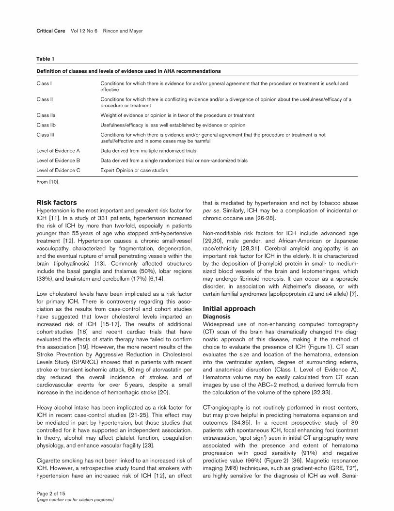
Initial approachDiagnosisWidespread use of non-enhancing computed tomography(CT) scan of the brain has dramatically changed the diag-nostic approach of this disease, making it the method ofchoice to evaluate the presence of ICH (Figure 1). CT scanevaluates the size and location of the hematoma, extensioninto the ventricular system, degree of surrounding edema,and anatomical disruption (Class I, Level of Evidence A).Hematoma volume may be easily calculated from CT scanimages by use of the ABC÷2 method, a derived formula fromthe calculation of the volume of the sphere [32,33].
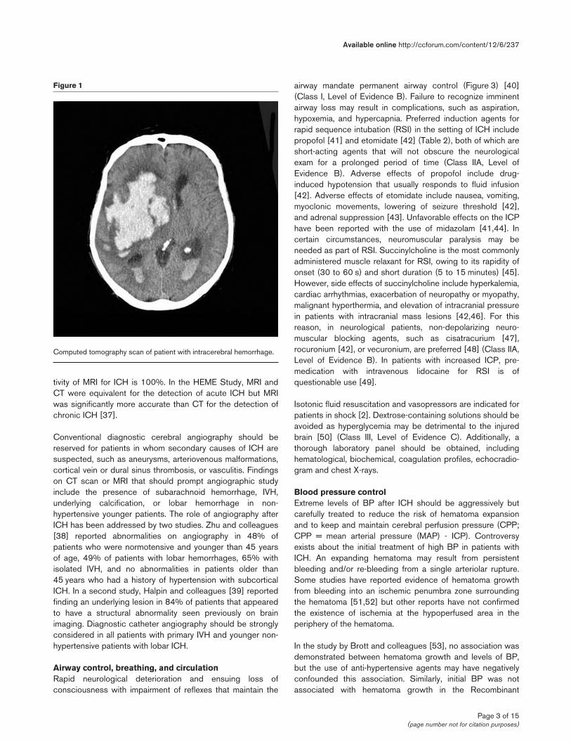
CT-angiography is not routinely performed in most centers,but may prove helpful in predicting hematoma expansion andoutcomes [34,35]. In a recent prospective study of 39patients with spontaneous ICH, focal enhancing foci (contrastextravasation, ‘spot sign’) seen in initial CT-angiography wereassociated with the presence and extent of hematomaprogression with good sensitivity (91%) and negativepredictive value (96%) (Figure 2) [36]. Magnetic resonanceimaging (MRI) techniques, such as gradient-echo (GRE, T2*),are highly sensitive for the diagnosis of ICH as well. Sensi-
Table 1
Definition of classes and levels of evidence used in AHA recommendations
Class I Conditions for which there is evidence for and/or general agreement that the procedure or treatment is useful and effective
Class II Conditions for which there is conflicting evidence and/or a divergence of opinion about the usefulness/efficacy of a procedure or treatment
Class IIa Weight of evidence or opinion is in favor of the procedure or treatment
Class IIb Usefulness/efficacy is less well established by evidence or opinion
Class III Conditions for which there is evidence and/or general agreement that the procedure or treatment is not useful/effective and in some cases may be harmful
Level of Evidence A Data derived from multiple randomized trials
Level of Evidence B Data derived from a single randomized trial or non-randomized trials
Level of Evidence C Expert Opinion or case studies
From [10].
Page 3 of 15(page number not for citation purposes)
tivity of MRI for ICH is 100%. In the HEME Study, MRI andCT were equivalent for the detection of acute ICH but MRIwas significantly more accurate than CT for the detection ofchronic ICH [37].
Conventional diagnostic cerebral angiography should bereserved for patients in whom secondary causes of ICH aresuspected, such as aneurysms, arteriovenous malformations,cortical vein or dural sinus thrombosis, or vasculitis. Findingson CT scan or MRI that should prompt angiographic studyinclude the presence of subarachnoid hemorrhage, IVH,underlying calcification, or lobar hemorrhage in non-hypertensive younger patients. The role of angiography afterICH has been addressed by two studies. Zhu and colleagues[38] reported abnormalities on angiography in 48% ofpatients who were normotensive and younger than 45 yearsof age, 49% of patients with lobar hemorrhages, 65% withisolated IVH, and no abnormalities in patients older than45 years who had a history of hypertension with subcorticalICH. In a second study, Halpin and colleagues [39] reportedfinding an underlying lesion in 84% of patients that appearedto have a structural abnormality seen previously on brainimaging. Diagnostic catheter angiography should be stronglyconsidered in all patients with primary IVH and younger non-hypertensive patients with lobar ICH.
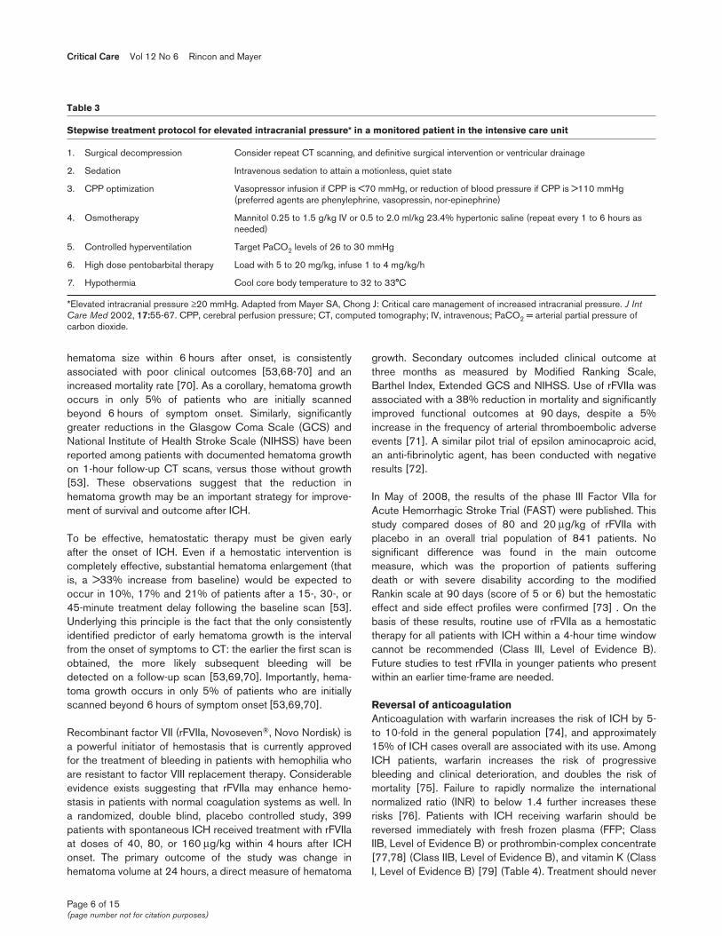
Airway control, breathing, and circulationRapid neurological deterioration and ensuing loss ofconsciousness with impairment of reflexes that maintain the
airway mandate permanent airway control (Figure 3) [40](Class I, Level of Evidence B). Failure to recognize imminentairway loss may result in complications, such as aspiration,hypoxemia, and hypercapnia. Preferred induction agents forrapid sequence intubation (RSI) in the setting of ICH includepropofol [41] and etomidate [42] (Table 2), both of which areshort-acting agents that will not obscure the neurologicalexam for a prolonged period of time (Class IIA, Level ofEvidence B). Adverse effects of propofol include drug-induced hypotension that usually responds to fluid infusion[42]. Adverse effects of etomidate include nausea, vomiting,myoclonic movements, lowering of seizure threshold [42],and adrenal suppression [43]. Unfavorable effects on the ICPhave been reported with the use of midazolam [41,44]. Incertain circumstances, neuromuscular paralysis may beneeded as part of RSI. Succinylcholine is the most commonlyadministered muscle relaxant for RSI, owing to its rapidity ofonset (30 to 60 s) and short duration (5 to 15 minutes) [45].However, side effects of succinylcholine include hyperkalemia,cardiac arrhythmias, exacerbation of neuropathy or myopathy,malignant hyperthermia, and elevation of intracranial pressurein patients with intracranial mass lesions [42,46]. For thisreason, in neurological patients, non-depolarizing neuro-muscular blocking agents, such as cisatracurium [47],rocuronium [42], or vecuronium, are preferred [48] (Class IIA,Level of Evidence B). In patients with increased ICP, pre-medication with intravenous lidocaine for RSI is ofquestionable use [49].
Isotonic fluid resuscitation and vasopressors are indicated forpatients in shock [2]. Dextrose-containing solutions should beavoided as hyperglycemia may be detrimental to the injuredbrain [50] (Class III, Level of Evidence C). Additionally, athorough laboratory panel should be obtained, includinghematological, biochemical, coagulation profiles, echocradio-gram and chest X-rays.
Blood pressure controlExtreme levels of BP after ICH should be aggressively butcarefully treated to reduce the risk of hematoma expansionand to keep and maintain cerebral perfusion pressure (CPP;CPP = mean arterial pressure (MAP) - ICP). Controversyexists about the initial treatment of high BP in patients withICH. An expanding hematoma may result from persistentbleeding and/or re-bleeding from a single arteriolar rupture.Some studies have reported evidence of hematoma growthfrom bleeding into an ischemic penumbra zone surroundingthe hematoma [51,52] but other reports have not confirmedthe existence of ischemia at the hypoperfused area in theperiphery of the hematoma.
In the study by Brott and colleagues [53], no association wasdemonstrated between hematoma growth and levels of BP,but the use of anti-hypertensive agents may have negativelyconfounded this association. Similarly, initial BP was notassociated with hematoma growth in the Recombinant
Available online http://ccforum.com/content/12/6/237
Figure 1
Computed tomography scan of patient with intracerebral hemorrhage.
Activated Factor VII ICH Trial [54]. Moreover, aggressiveblood pressure reduction after ICH may predispose to anabrupt drop in CPP and ischemia, which, in turn, may beaccompanied by elevations of ICP and further neurologicaldamage. In a recent pilot trial of BP reduction after ICH, 14patients with supratentorial ICH were randomized to receiveeither labetalol or nicardipine within 22 hours of onset tolower the MAP by 15%. Cerebral blood flow (CBF) studieswere performed before and after treatment with positronemission tomography and [15O] water. No changes in globalor peri-hematoma CBF were observed [55]. Two additionalstudies have demonstrated that a controlled, pharmaco-logically based reduction in BP has no adverse effects onCBF in humans or animals [56,57]. BP level has beencorrelated with increases in the ICP and volume of thehematoma but it has been very difficult to explain if hyper-tension is the cause of hematoma growth or if this is just aresponse to elevated ICP in the setting of large volume ICHto maintain cerebral perfusion.
In general, the American Heart Association Guidelinesindicate that systolic blood pressures exceeding 180 mmHgor MAP exceeding 130 mmHg should be managed withcontinuous infusion antihypertensive agents such as labetalol,esmolol, or nicardipine [2] (Class IIB, Level of Evidence C).Urapidil, a sympatholytic agent with vasodilator properties isan alternative but it is currently not approved for use in theUS. Use of nitroprusside has drawbacks since this agent mayexacerbate cerebral edema and intracranial pressure [58].
Oral and sub-lingual agents are not preferred, because of theneed for immediate and precise BP control. There are few, ifany, comparative or randomized trials providing definitiveconclusions about the efficacy and safety of comparativeagents.
In comatose patients, it is recommended to monitor ICP andto titrate vasopressors to maintain CPP in the range 70 to90 mmHg. Use of brain tissue oxygen and thermodilution CBFprobes to detect reductions in perfusion related to excessivelowering of BP is gaining in popularity. In general, no matterhow high the BP is, the MAP should not be reduced beyond15% to 30% over the first 24 hours [56]. In the setting ofimpaired blood flow autoregulation, excessive blood pressurereduction may exacerbate ischemia in the area surroundingthe hematoma and worsen perihematomal brain injury [59,60](Table 2; Figure 3). In fact transcranial doppler velocitiesbecome positively correlated with CPP when CPP dropsbelow the left side of the autoregulation curve [61].
Whether more aggressive BP reduction after ICH is safe isthe matter of the ongoing National Institute of NeurologicalDiseases and Stroke supported Antihypertensive Treatmentin Acute Cerebral Hemorrhage (ATACH) pilot study [62].Additionally, the ongoing phase III Intensive Blood PressureReduction in Acute Cerebral Hemorrhage Trial (INTERACT)will test the hypothesis that lowering BP acutely after ICH willreduce the chances of dying or surviving with long-termdisability [63].
Critical Care Vol 12 No 6 Rincon and Mayer
Page 4 of 15(page number not for citation purposes)
Figure 2
Contrast extravasation seen in the hematoma of a patient with acute coagulopathic intracerebral hemorrhage (white arrows).
Though no prospective study has addressed the timing ofconversion from intravenous to oral anti-hypertensive manage-ment, this process can generally be started after 24 to72 hours, as long as the patient’s condition has stabilized [6].
Initial emergency intracranial pressure managementEmergency measures for ICP control are appropriate forstuporous or comatose patients, or those who present acutelywith clinical signs of brain stem herniation (that is, pupillaryabnormalities or motor posturing; Figure 3). The head shouldbe elevated to 30 degrees, 1.0 to 1.5 g/kg of 20% mannitolshould be administered by a rapid infusion, and the patientshould be hyperventilated to a pCO2 of 26 to 30 mmHg (ClassIIA, Level of Evidence B). As a second line therapy, or if thepatient is relatively hypotensive, 0.5 to 2.0 ml/kg of 23.4%saline solution can be administered through a central venousline [64] (Class IIA, Level of Evidence B). These measures aredesigned to lower ICP as quickly and effectively as possible, inorder to ‘buy time’ before a definitive neurosurgical procedure(craniotomy, ventriculostomy (Class IIA, Level of Evidence B), orplacement of an ICP monitor) can be performed (Table 3).Corticosteroids are contraindicated based on the results ofrandomized trials that have failed to demonstrate their efficacy inICH [65,66] (Class III, Level of Evidence B). Neurosurgicalconsultation is warranted for those patients with rapidly declin-ing mental status and hydrocephalus with IVH seen in the initialCT scan. Early placement of a ventricular drain in this case maybe life-saving [67] (Class IIA, Level of Evidence B; Figure 3).
Hemostatic therapyHematoma size is an important determinant of mortality afterICH and early hematoma growth, which is the increase in
Available online http://ccforum.com/content/12/6/237
Page 5 of 15(page number not for citation purposes)
Table 2
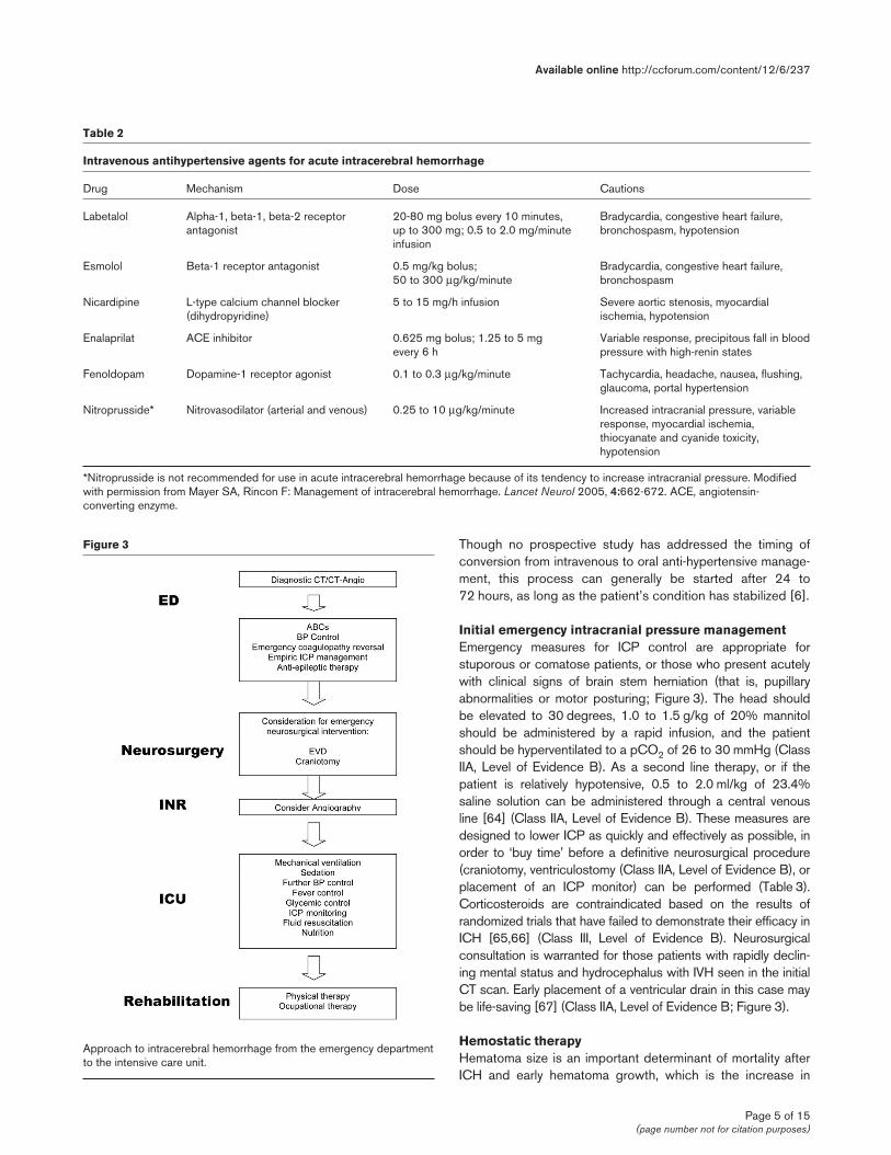
Intravenous antihypertensive agents for acute intracerebral hemorrhage
Drug Mechanism Dose Cautions
Labetalol Alpha-1, beta-1, beta-2 receptor 20-80 mg bolus every 10 minutes, Bradycardia, congestive heart failure, antagonist up to 300 mg; 0.5 to 2.0 mg/minute bronchospasm, hypotension
infusion
Esmolol Beta-1 receptor antagonist 0.5 mg/kg bolus; Bradycardia, congestive heart failure, 50 to 300 μg/kg/minute bronchospasm
Nicardipine L-type calcium channel blocker 5 to 15 mg/h infusion Severe aortic stenosis, myocardial (dihydropyridine) ischemia, hypotension
Enalaprilat ACE inhibitor 0.625 mg bolus; 1.25 to 5 mg Variable response, precipitous fall in blood every 6 h pressure with high-renin states
Fenoldopam Dopamine-1 receptor agonist 0.1 to 0.3 μg/kg/minute Tachycardia, headache, nausea, flushing, glaucoma, portal hypertension
Nitroprusside* Nitrovasodilator (arterial and venous) 0.25 to 10 μg/kg/minute Increased intracranial pressure, variable response, myocardial ischemia, thiocyanate and cyanide toxicity, hypotension
*Nitroprusside is not recommended for use in acute intracerebral hemorrhage because of its tendency to increase intracranial pressure. Modifiedwith permission from Mayer SA, Rincon F: Management of intracerebral hemorrhage. Lancet Neurol 2005, 4:662-672. ACE, angiotensin-converting enzyme.
Figure 3
Approach to intracerebral hemorrhage from the emergency departmentto the intensive care unit.
hematoma size within 6 hours after onset, is consistentlyassociated with poor clinical outcomes [53,68-70] and anincreased mortality rate [70]. As a corollary, hematoma growthoccurs in only 5% of patients who are initially scannedbeyond 6 hours of symptom onset. Similarly, significantlygreater reductions in the Glasgow Coma Scale (GCS) andNational Institute of Health Stroke Scale (NIHSS) have beenreported among patients with documented hematoma growthon 1-hour follow-up CT scans, versus those without growth[53]. These observations suggest that the reduction inhematoma growth may be an important strategy for improve-ment of survival and outcome after ICH.
To be effective, hematostatic therapy must be given earlyafter the onset of ICH. Even if a hemostatic intervention iscompletely effective, substantial hematoma enlargement (thatis, a >33% increase from baseline) would be expected tooccur in 10%, 17% and 21% of patients after a 15-, 30-, or45-minute treatment delay following the baseline scan [53].Underlying this principle is the fact that the only consistentlyidentified predictor of early hematoma growth is the intervalfrom the onset of symptoms to CT: the earlier the first scan isobtained, the more likely subsequent bleeding will bedetected on a follow-up scan [53,69,70]. Importantly, hema-toma growth occurs in only 5% of patients who are initiallyscanned beyond 6 hours of symptom onset [53,69,70].
Recombinant factor VII (rFVIIa, Novoseven®, Novo Nordisk) isa powerful initiator of hemostasis that is currently approvedfor the treatment of bleeding in patients with hemophilia whoare resistant to factor VIII replacement therapy. Considerableevidence exists suggesting that rFVIIa may enhance hemo-stasis in patients with normal coagulation systems as well. Ina randomized, double blind, placebo controlled study, 399patients with spontaneous ICH received treatment with rFVIIaat doses of 40, 80, or 160 μg/kg within 4 hours after ICHonset. The primary outcome of the study was change inhematoma volume at 24 hours, a direct measure of hematoma
growth. Secondary outcomes included clinical outcome atthree months as measured by Modified Ranking Scale,Barthel Index, Extended GCS and NIHSS. Use of rFVIIa wasassociated with a 38% reduction in mortality and significantlyimproved functional outcomes at 90 days, despite a 5%increase in the frequency of arterial thromboembolic adverseevents [71]. A similar pilot trial of epsilon aminocaproic acid,an anti-fibrinolytic agent, has been conducted with negativeresults [72].
In May of 2008, the results of the phase III Factor VIIa forAcute Hemorrhagic Stroke Trial (FAST) were published. Thisstudy compared doses of 80 and 20 μg/kg of rFVIIa withplacebo in an overall trial population of 841 patients. Nosignificant difference was found in the main outcomemeasure, which was the proportion of patients sufferingdeath or with severe disability according to the modifiedRankin scale at 90 days (score of 5 or 6) but the hemostaticeffect and side effect profiles were confirmed [73] . On thebasis of these results, routine use of rFVIIa as a hemostatictherapy for all patients with ICH within a 4-hour time windowcannot be recommended (Class III, Level of Evidence B).Future studies to test rFVIIa in younger patients who presentwithin an earlier time-frame are needed.
Reversal of anticoagulationAnticoagulation with warfarin increases the risk of ICH by 5-to 10-fold in the general population [74], and approximately15% of ICH cases overall are associated with its use. AmongICH patients, warfarin increases the risk of progressivebleeding and clinical deterioration, and doubles the risk ofmortality [75]. Failure to rapidly normalize the internationalnormalized ratio (INR) to below 1.4 further increases theserisks [76]. Patients with ICH receiving warfarin should bereversed immediately with fresh frozen plasma (FFP; ClassIIB, Level of Evidence B) or prothrombin-complex concentrate[77,78] (Class IIB, Level of Evidence B), and vitamin K (ClassI, Level of Evidence B) [79] (Table 4). Treatment should never
Critical Care Vol 12 No 6 Rincon and Mayer
Page 6 of 15(page number not for citation purposes)
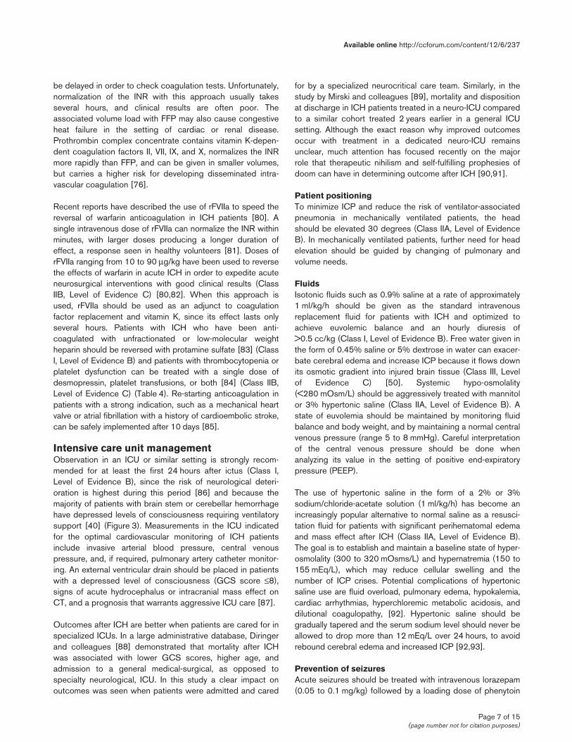
Table 3
Stepwise treatment protocol for elevated intracranial pressure* in a monitored patient in the intensive care unit
1. Surgical decompression Consider repeat CT scanning, and definitive surgical intervention or ventricular drainage
2. Sedation Intravenous sedation to attain a motionless, quiet state
3. CPP optimization Vasopressor infusion if CPP is <70 mmHg, or reduction of blood pressure if CPP is >110 mmHg (preferred agents are phenylephrine, vasopressin, nor-epinephrine)
4. Osmotherapy Mannitol 0.25 to 1.5 g/kg IV or 0.5 to 2.0 ml/kg 23.4% hypertonic saline (repeat every 1 to 6 hours as needed)
5. Controlled hyperventilation Target PaCO2 levels of 26 to 30 mmHg
6. High dose pentobarbital therapy Load with 5 to 20 mg/kg, infuse 1 to 4 mg/kg/h
7. Hypothermia Cool core body temperature to 32 to 33°C
*Elevated intracranial pressure ≥20 mmHg. Adapted from Mayer SA, Chong J: Critical care management of increased intracranial pressure. J IntCare Med 2002, 17:55-67. CPP, cerebral perfusion pressure; CT, computed tomography; IV, intravenous; PaCO2 = arterial partial pressure ofcarbon dioxide.
be delayed in order to check coagulation tests. Unfortunately,normalization of the INR with this approach usually takesseveral hours, and clinical results are often poor. Theassociated volume load with FFP may also cause congestiveheat failure in the setting of cardiac or renal disease.Prothrombin complex concentrate contains vitamin K-depen-dent coagulation factors II, VII, IX, and X, normalizes the INRmore rapidly than FFP, and can be given in smaller volumes,but carries a higher risk for developing disseminated intra-vascular coagulation [76].
Recent reports have described the use of rFVIIa to speed thereversal of warfarin anticoagulation in ICH patients [80]. Asingle intravenous dose of rFVIIa can normalize the INR withinminutes, with larger doses producing a longer duration ofeffect, a response seen in healthy volunteers [81]. Doses ofrFVIIa ranging from 10 to 90 μg/kg have been used to reversethe effects of warfarin in acute ICH in order to expedite acuteneurosurgical interventions with good clinical results (ClassIIB, Level of Evidence C) [80,82]. When this approach isused, rFVIIa should be used as an adjunct to coagulationfactor replacement and vitamin K, since its effect lasts onlyseveral hours. Patients with ICH who have been anti-coagulated with unfractionated or low-molecular weightheparin should be reversed with protamine sulfate [83] (ClassI, Level of Evidence B) and patients with thrombocytopenia orplatelet dysfunction can be treated with a single dose ofdesmopressin, platelet transfusions, or both [84] (Class IIB,Level of Evidence C) (Table 4). Re-starting anticoagulation inpatients with a strong indication, such as a mechanical heartvalve or atrial fibrillation with a history of cardioembolic stroke,can be safely implemented after 10 days [85].
Intensive care unit managementObservation in an ICU or similar setting is strongly recom-mended for at least the first 24 hours after ictus (Class I,Level of Evidence B), since the risk of neurological deteri-oration is highest during this period [86] and because themajority of patients with brain stem or cerebellar hemorrhagehave depressed levels of consciousness requiring ventilatorysupport [40] (Figure 3). Measurements in the ICU indicatedfor the optimal cardiovascular monitoring of ICH patientsinclude invasive arterial blood pressure, central venouspressure, and, if required, pulmonary artery catheter monitor-ing. An external ventricular drain should be placed in patientswith a depressed level of consciousness (GCS score ≤8),signs of acute hydrocephalus or intracranial mass effect onCT, and a prognosis that warrants aggressive ICU care [87].
Outcomes after ICH are better when patients are cared for inspecialized ICUs. In a large administrative database, Diringerand colleagues [88] demonstrated that mortality after ICHwas associated with lower GCS scores, higher age, andadmission to a general medical-surgical, as opposed tospecialty neurological, ICU. In this study a clear impact onoutcomes was seen when patients were admitted and cared
for by a specialized neurocritical care team. Similarly, in thestudy by Mirski and colleagues [89], mortality and dispositionat discharge in ICH patients treated in a neuro-ICU comparedto a similar cohort treated 2 years earlier in a general ICUsetting. Although the exact reason why improved outcomesoccur with treatment in a dedicated neuro-ICU remainsunclear, much attention has focused recently on the majorrole that therapeutic nihilism and self-fulfilling prophesies ofdoom can have in determining outcome after ICH [90,91].
Patient positioningTo minimize ICP and reduce the risk of ventilator-associatedpneumonia in mechanically ventilated patients, the headshould be elevated 30 degrees (Class IIA, Level of EvidenceB). In mechanically ventilated patients, further need for headelevation should be guided by changing of pulmonary andvolume needs.
FluidsIsotonic fluids such as 0.9% saline at a rate of approximately1 ml/kg/h should be given as the standard intravenousreplacement fluid for patients with ICH and optimized toachieve euvolemic balance and an hourly diuresis of>0.5 cc/kg (Class I, Level of Evidence B). Free water given inthe form of 0.45% saline or 5% dextrose in water can exacer-bate cerebral edema and increase ICP because it flows downits osmotic gradient into injured brain tissue (Class III, Levelof Evidence C) [50]. Systemic hypo-osmolality(<280 mOsm/L) should be aggressively treated with mannitolor 3% hypertonic saline (Class IIA, Level of Evidence B). Astate of euvolemia should be maintained by monitoring fluidbalance and body weight, and by maintaining a normal centralvenous pressure (range 5 to 8 mmHg). Careful interpretationof the central venous pressure should be done whenanalyzing its value in the setting of positive end-expiratorypressure (PEEP).
The use of hypertonic saline in the form of a 2% or 3%sodium/chloride-acetate solution (1 ml/kg/h) has become anincreasingly popular alternative to normal saline as a resusci-tation fluid for patients with significant perihematomal edemaand mass effect after ICH (Class IIA, Level of Evidence B).The goal is to establish and maintain a baseline state of hyper-osmolality (300 to 320 mOsms/L) and hypernatremia (150 to155 mEq/L), which may reduce cellular swelling and thenumber of ICP crises. Potential complications of hypertonicsaline use are fluid overload, pulmonary edema, hypokalemia,cardiac arrhythmias, hyperchloremic metabolic acidosis, anddilutional coagulopathy, [92]. Hypertonic saline should begradually tapered and the serum sodium level should never beallowed to drop more than 12 mEq/L over 24 hours, to avoidrebound cerebral edema and increased ICP [92,93].
Prevention of seizuresAcute seizures should be treated with intravenous lorazepam(0.05 to 0.1 mg/kg) followed by a loading dose of phenytoin
Available online http://ccforum.com/content/12/6/237
Page 7 of 15(page number not for citation purposes)
or fosphenytoin (20 mg/kg) (Class I, Level of Evidence B;Figure 3). An alternative to phenytoin infusion is levetiracetam(500 mg q12h, adjusted for renal insufficiency). Side effectsof phenytin infusions include rash, hypotension, arrhythmias,and severe hypocalcemia for the phosphenytoin presentation.Patients with ICH may benefit from prophylactic anti-epilepticdrug therapy, but no randomized trial has addressed theefficacy of this approach. The American Heart AssociationGuidelines have recommended anti-epileptic medication forup to one month, after which therapy should be discontinuedin the absence of seizures [94]. This recommendation issupported by the results of a recent study that showed thatthe risk of early seizures was reduced by prophylactic anti-epileptic drug therapy [95]. The 30-day risk for convulsiveseizures after ICH is approximately 8%, and the risk of overtstatus epilepticus is 1% to 2% [95]. Lobar location and smallhematomas are independent predictors of early seizures [95].The argument for prophylactic anticonvulsant therapy instuporous or comatose ICH patients is bolstered by the factthat continuous electroencephalogram monitoring demon-strates electrographic seizure activity in approximately 25%of these patients, despite prophylactic anti-epileptic drugtherapy [96,97]. The risk of late seizures or epilepsy amongsurvivors of ICH is 5% to 27% [95].
Temperature controlFever (temperature >38.3°C) after ICH is common, particu-larly with IVH [98], and should be treated aggressively(Figure 3; Class I, Level of Evidence C). Sustained fever after
ICH has been shown to be independently associated withpoor outcome after ICH [99]. A large body of experimentalevidence indicates that even small degrees of hyperthermiacan worsen ischemic brain injury by exacerbating excitotoxicneurotransmitter release, proteolysis, free radical and cyto-kine production, blood-brain barrier compromise, and apop-tosis [100,101]. Brain temperature elevations have also beenassociated with hyperemia, exacerbation of cerebral edema,and elevated intracranial pressure [102,103].
As a general standard, acetaminophen and cooling blanketsare recommended for almost all patients with sustained feverin excess of 38.3°C (101.0°F), despite the lack of prospec-tive randomized controlled trials supporting this approach[104,105]. Acetaminophen should be used with caution inpatients with hepatic dysfunction. Newer adhesive surfacecooling systems and endovascular heat exchange cathetershave been shown to be much more effective for maintainingnormothermia [106,107]; however, it remains to be seen ifthese measures can improve clinical outcome.
Management of hyperglycemiaAdmission hyperglycemia is a potent predictor of 30-daymortality in both diabetic and non-diabetic patients with ICH[108]. The detrimental effect of hyperglycemia has been wellstudied in acute vascular syndromes. In ischemic stroke,hyperglycemia occurs in 20% to 40% of patients and isassociated with infarct expansion, worse functional outcome,longer hospital stays, higher medical costs, and an increased
Critical Care Vol 12 No 6 Rincon and Mayer
Page 8 of 15(page number not for citation purposes)
Table 4
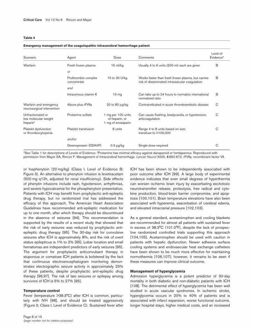
Emergency management of the coagulopathic intracerebral hemorrhage patient
Level of Scenario Agent Dose Comments Evidence*
Warfarin Fresh frozen plasma 15 ml/kg Usually 4 to 6 units (200 ml) each are given B
or
Prothrombin complex 15 to 30 U/kg Works faster than fresh frozen plasma, but carries Bconcentrate risk of disseminated intravascular coagulation
and
Intravenous vitamin K 10 mg Can take up to 24 hours to normalize international Bnormalized ratio
Warfarin and emergency Above plus rFVIIa 20 to 80 μg/kg Contraindicated in acute thromboembolic disease Cneurosurgical intervention
Unfractionated or Protamine sulfate 1 mg per 100 units Can cause flushing, bradycardia, or hypotension, Clow molecular weight of heparin, or anticoagulationheparin† 1 mg of enoxaparin
Platelet dysfunction Platelet transfusion 6 units Range 4 to 8 units based on size; Cor thrombocytopenia transfuse to >100,000
and/or
Desmopressin (DDAVP) 0.3 μg/kg Single dose required C
*See Table 1 for descriptions of Levels of Evidence. †Protamine has minimal efficacy against danaparoid or fondaparinux. Reproduced withpermission from Mayer SA, Rincon F: Management of intracerebral hemorrhage. Lancet Neurol 2005, 4:662-672. rFVIIa, recombinant factor VII.
risk of death [109-111] and it is felt to be secondary to acatecholamine surge and generalized stress response [110].
In the critically ill population, hyperglycemia seems muchmore acutely toxic than in healthy individuals, for whom cellscan protect themselves by down-regulation of glucosetransporters [112]. The acute toxicity of high levels of glucosein critical illness might be explained by an accelerated cellularglucose overload and more pronounced toxic side effects ofglycolysis and oxidative phosphorylation [113]. Neurons andseveral other cell types are insulin independent for glucoseuptake, which is mediated by transporters such as GLUT-1,GLUT-2, and GLUT-3 [114]. These transporters are up-regu-lated by hypoxia and inflammatory mediators such as angio-tensin-II, endothelin-1, vascular endothelial growth factor, andtransforming growth factor-β among others. These trans-porters facilitate glucose entry into the neuron where intra-cellular hyperglycemia can lead to oxidative stress and exag-gerated production of superoxide species [114]. Peroxy-nitrite, superoxide, and other reactive oxygen species lead toinhibition of the glycolytic enzyme glyceraldhyde phosphatedehydrogenase and mitochondrial complexes I and IV, thebasis of mitochondrial dysfunction likely to induce end-organfailure and cellular death [114].
Strict glucose control after ICH is recommended (Class IIA,Level of Evidence C). This approach has been linked toreductions in intracranial pressure, duration of mechanicalventilation, and seizures in an heterogeneous cohort ofcritically ill patients [115].
Elevated intracranial pressure managementLarge volume ICH carries the risk of developing cerebraledema and high ICP (≥15 mmHg or ≥20 mmH2O), and thepresence of IVH further increases the risk of mortality[116,117] (Figure 3). This effect is primarily related to thedevelopment of obstructive hydrocephalus and alterations ofnormal cerebrospinal fluid flow-dynamics. Patients with largevolume ICH, intracranial mass effect, and coma may benefitfrom ICP monitoring, though this intervention has not beenproved to benefit outcomes after ICH [118,119]. Werecommend a stepwise protocol for addressing intracranialhypertension in the ICU setting.
Cerebrospinal fluid drainageInitial cerebrospinal fluid drainage may be a life-savingprocedure particularly in the setting of hydrocephalus andIVH [67] (Class IIA, Level of Evidence B). This techniqueallows for rapid clearance of cerebrospinal fluid, release ofICP, and ICP/CPP monitoring. As a general rule, an ICPmonitor or external ventricular drain (EVD) should be placedin all comatose ICH patients (GCS score of 8 or less) withthe goal of maintaining ICP less than 20 mmH2O(<15 mmHg) and CPP at greater than 70 mmHg, unless theircondition is so dismal that aggressive ICU care is notwarranted. Compared to parenchymal monitors, EVDs carry
the therapeutic advantage of allowing cerebrospinal fluiddrainage, and the disadvantage of a substantial risk ofinfection (approximately 10% during the first 10 days) [120].A small retrospective study failed to show any relationshipbetween changes in ventricular size and level ofconsciousness in ICH patients treated with EVDs [121].
SedationSedation should be used to minimize pain, agitation, anddecrease surges in ICP (Class IIA, Level of Evidence B).Agitation must be avoided, because it can aggravate ICPelevation through straining (increasing thoracic, jugularvenous, and systemic BP), increased cerebral metabolic rateof oxygen, and also may cause uncontrolled hyper-/hypo-ventilation, which both can be detrimental. During an ICPspike, sedation may be all that is necessary to control theICP. The goal of sedation should be a calm, comfortable, andcooperative state in patients with ICP that is well-controlled,and a quiet, motionless state in patients where ICP elevationrequires active management. The preferred regimen is thecombination of a short-acting opioid, such as fentanyl (1 to3 μg/kg/h) or remifentanyl (0.03 to 0.25 μg/kg/minute), toprovide analgesia, and propofol (0.3 to 3 mg/kg/h) becauseof its extremely short half-life, which makes it ideal for periodicinterruption for neurological assessments, which should beperformed on a daily basis unless the patient has demon-strated that the ICP is too unstable (frequent ICP crisis in thesetting of awakening, position changes, fever, and so on) totolerate this. Bolus injections of opioids should be used withcaution in patients with elevated ICP because they cantransiently lower MAP and increase ICP due to autoregulatoryvasodilation of cerebral vessels [122]. Compared to anopiod-based sedation regimen, in one trial propofol wasassociated with lower ICP and fewer ICP interventions inpatients with severe traumatic brain injury [123]. However,propofol has been associated with mithocondrial dysfunctionand multi-organ failure (propofol infusion syndrome). Predis-posing factors include young age, severe critical illness ofcentral nervous system or respiratory origin, exogenous cate-cholamine or glucocorticoid administration, inadequate carbo-hydrate intake and subclinical mitochondrial disease [124].
Cerebral perfusion pressure optimizationTwo prevailing management strategies for the managementof elevated ICP have evolved from the experience in traumaticbrain injury. The ‘Lund concept’ assumes a disruption of theblood brain barrier and recommends manipulations todecrease the hydrostatic BP and increase osmotic pressuresto minimize cerebral blood volume and vasogenic edema byimproving perfusion and oxygenation to the injured areas ofthe brain [125]. This is achieved in theory by maintaining aneuvolemic state with normal hemoglobin, hematocrit, plasmaprotein concentrations, and by antagonizing vasoconstrictionthrough reduction of catecholamine concentration in plasmaand sympathetic outflow. These therapeutic measures attemptto normalize all essential hemodynamic parameters (blood
Available online http://ccforum.com/content/12/6/237
Page 9 of 15(page number not for citation purposes)
pressure, plasma oncotic pressure, plasma and erythrocytevolumes, arterial partial pressure of oxygen (PaO2), andarterial partial pressure of carbon dioxide (PaCO2)). Theintroduction of microdialysis with novel physiological targetsmay optimize the goals of the original Lund protocol. The‘Rosner concept’ emphasizes maintaining a high CPP tominimize reflex vasodilatation or ischemia [126,127] at theexpense of added cardiopulmonary stress (Class IIA, Level ofEvidence B). Computerized bedside graphical displays (ICUPilot®, CMA Micodialysis, Solna, Sweden) can allowclinicians to identify whether ICP and MAP are positivelycorrelated, in which case a low CPP would be preferable, ornegatively correlated, in which case a higher CPP would bedesirable.
Hyperosmolar therapyHyperosmolar therapy [128] should be used after sedationand CPP optimization fail to normalize ICP (Class IIA, Level ofEvidence B). The initial dose of mannitol is 1 to 1.5 g/kg of a20% solution, followed by bolus doses of 0.25 to 1.0 g/kg asneeded to a target osmolality of 300 to 320 mOsm/kg.Additional doses can be given as frequently as once an hour,based on the initial response to therapy with the anticipationof a transient drop in BP. There is little to recommend the useof standing mannitol in patients with normal ICP. In a recenttrial of mannitol for ICH, 128 patients were randomized toreceive low-dose mannitol (100 ml of 20% solution) or shamtherapy every 4 hours for 5 days with a rapid dose taperingschedule over 48 hours. The 1-month mortality rate was 25%in both groups and disability scores at 3 months were notsignificantly different between groups [129]. Hypertonicsaline, such as 23.4% saline solution, can be used as analternative to mannitol, particularly when CPP augmentationis desirable (Class IIA, Level of Evidence B). However, careshould be taken to avoid fluid overload in the setting of heartor kidney failure. Additional side effects of hyperosmolartherapy include kidney failure, rebound ICP, electrolyticimbalance (hypo-/hyper-natremia), and acid/base distur-bances. Despite clinical and animal model support [64], manyissues remain to be clarified, including the exact mechanismof action, best mode and timing of administration, and themost appropriate concentration.
HyperventilationForced hyperventilation is generally used sparingly in the ICUand, for brief periods, in monitored patients, because itseffect on ICP tends to last for only a few hours (Class IIA,Level of Evidence B). Good long term outcomes can occurwhen the combination of osmotherapy and hyperventilation issuccessfully used to reverse transtentorial herniation [130].Overly aggressive hyperventilation to pCO2 levels <25 mmHgmay cause excessive vasoconstriction and exacerbation ofischemia during the acute phase of ICH and should beavoided. Controlled hyperventilation therapy can be optimizedby saturation of jugular vein oxygen and partial brain tissueoxygenation monitoring.
BarbituratesFor cases of severe and intractable intracranial hypertension,barbiturates can control ICP by decreasing cerebralmetabolic activity, which translates into a reduction of theCBF and cerebral blood volume (Class IIB, level of EvidenceB). Pentobarbital can be given in repeated 5 mg/kg bolusesevery 15 to 30 minutes until ICP is controlled (usually 10 to20 mg/kg is required), and then continuously infused at 1 to4 mg/kg/h. An electroencephalogram should be continuouslyrecorded, and the pentobarbital titrated to produce a burst-suppression pattern, with approximately 6 to 8 secondinterbursts, to avoid excessive sedation.
HypothermiaIf pentobarbital fails to control ICP, induced hypothermia to32 to 34°C can effectively lower otherwise refractory ICP(Class IIB, Level of Evidence C) [131]. Hypothermia can beachieved using various surface and endovascular coolingsystems coupled to a rectal, esophageal, pulmonary artery, orbladder thermometer. Complications of hypothermia includenosocomial infection, hypotension, cardiac arrhythmias,coagulopathy, shivering, hyperkalemia, hyperglycemia, andileus. Because these risks increase with the depth andduration of cooling, some advocate for the induction of mildhypothermia (34 to 36°C) if temperature reduction is requiredfor a prolonged period of time to control ICP [104,132,133].
Intraventricular thrombolytic therapyIVH commonly results from extension of ICH into the cerebralventricular system, and is an independent predictor ofmortality after ICH [134]. Commonly, hydrocephalus and IVHare managed with an EVD, but outcomes remain poor [9].Intraventricular administration of the plasminogen activatorurokinase every 12 hours may reduce hematoma size and theexpected mortality rate at 1 month [135]. Several small studieshave reported the successful use of urokinase or tissueplasminogen activator (t-PA) for the treatment of IVH, with thegoal of accelerating the clearance of IVH and improvingclinical outcome [136]. A Cochrane systematic reviewpublished in 2002 summarized the experience of several caseseries providing evidence of safety but no definitive efficacy[137]. No randomized prospective controlled trial hasaddressed the efficacy of intraventricular thrombolysis afterICH. The ongoing Clear IVH Trial (Clot Lysis EvaluatingAccelerated Resoution of Intra Ventricular Hemorrhage), aphase II multicenter study evaluating recombinant t-PAtreatment of patients with IVH, is designed to investigate theoptimum dose and frequency of recombinant t-PAadministered via an intraventricular catheter to safely andeffectively treat IVH and will soon provide some insight intothis issue [138]. When used off-label, a dose of 1 mg of t-PAevery 8 hours (followed by clamping of the EVD for 1 hour) isreasonable until clearance of blood from the third ventricle hasbeen achieved [139]. Doses of 3 mg or more of t-PA for IVHthrombolysis have been associated with an unacceptably highbleeding rate (Daniel Hanley, MD, personal communication).
Critical Care Vol 12 No 6 Rincon and Mayer
Page 10 of 15(page number not for citation purposes)
Deep venous thrombosis prophylaxisPatients with ICH are at high risk for deep vein thrombosisand pulmonary embolism, a potentially fatal complication, dueto limb paresis and prolonged immobilization. Dynamiccompression stockings should be placed on admission [140](Class I, Level of Evidence B). A small prospective trial hasshown that low dose subcutaneous heparin (5000 U BID)starting after the second day significantly reduces thefrequency of venous thromboembolism, with no increase inintracranial bleeding (Class IIB, Level of Evidence B) [141].Treatment with low molecular weight heparin (enoxaparin40 mg daily) is a reasonable alternative if renal function isnormal (Class IIB, Level of Evidence C).
NutritionAs is the case with all critically ill neurological patients,enteral feeding should be started within 48 hours to avoidprotein catabolism and malnutrition. A small-bore nasoduo-denal feeding tube may reduce the risk of aspiration events.
Surgical intervention for intracerebral hemorrhageCraniotomy has been the most studied intervention for thesurgical management of ICH but the results have been dis-couraging. Two earlier smaller trials showed that for patientspresenting with moderate alterations in the state of conscious-ness, surgery reduced the risk of death without improving thefunctional outcome [142] and that ultra-early evacuation mightimprove the 3-month NIHSS [143]. Nevertheless, in a meta-analysis of all prior trials of surgical intervention forsupratentorial ICH, no benefit was demonstrated [144].
The International Surgical Trial in Intracerebral Haemorrhage(STICH) study, a landmark trial of over 1,000 ICH patients,showed that emergent surgical hematoma evacuation viacraniotomy within 72 hours of onset fails to improve outcomecompared to a policy of initial medical management [145].Although the STICH has rightfully dampened the enthusiasmof neurosurgeons for performing surgery, it must be remem-bered that the trial was based on the principle of clinicalequipoise and patients who the local investigator felt wouldmost likely benefit from emergency surgery were not enrolledinto the study. In a post hoc analysis of the STICH, thesubgroup of patients with superficial hematomas and no IVHhad better outcomes in the surgical arm [146]. Thisobservation provided support for the STICH-II, which iscurrently enrolling patients [147]. In contrast to supratentorialICH, there is much better evidence that cerebellar hemor-rhages exceeding 3 cm in diameter benefit from emergentsurgical evacuation as abrupt and dramatic deterioration tocoma can occur within the first 24 hours of onset in thesepatients (Class I, Level of evidence B) [148]. For this reason,it is generally unwise to defer surgery in these patients untilfurther clinical deterioration occurs.
As emergent craniotomy has been unable to improveneurological outcome after ICH, the role of other surgical
techniques such as minimally invasive surgery have gainedimportance over the past decade. The advantages ofminimally invasive surgery over conventional craniotomyinclude reduced operative time, the possibility of performanceunder local anesthesia, and reduced surgical trauma.Endoscopic aspiration of supratentorial ICH was studied in asmall single-center randomized controlled trial (Class IIB,Level of Evidence B) [149]. The study showed that thistechnique provided a reduction of mortality rate at 6 monthsin the surgical group but surgery was more effective insuperficial hematomas and in younger patients (<60 years).
Thrombolytic therapy and surgical removal of hematomas isanother technique that has been studied in a single centerrandomized clinical trial (Class IIB, Level of Evidence B)[143]. Patients in the surgical group had better outcomescores than the medically treated group. Finally, a multi-center randomized control trial examined the utility ofsterotactic urokinase infusion when administered within72 hours to patients with GCS score ≥5 and hematomas≥10 ml [150]; this provided significant reduction in hematomasize and mortality rate at the expense of higher rates of re-bleeding but no significant differences in outcomes measureswas seen.
Hemicraniectomy with duraplasty has been proposed as alife-saving intervention for several neurological catastrophes,such as malignant middle cerebral artery infarction and poorgrade subarachnoid hemorrhage. No randomized controlledtrial has been conducted in patients with ICH. In a recentreport of 12 consecutive patients with hypertensive ICH andtreated with hemicraniectomy, 11 (92%) survived atdischarge and 6 of them (54.5%) had a good functionaloutcome (modified Rankin Score 0 to 3) [151]. Thesepreliminary data support the need for better controlledstudies addressing the role of this surgical technique in ICHpatients (Class IIB, Level of Evidence B).
ConclusionsDespite a long history of devastating outcomes and highmortality, there is optimism that the management of ICH willchange in the future based on new insights into the acutepathophysiology of this disease. A better understanding ofthe dynamic process of hematoma growth, importance ofinflammation triggered by coagulation and products of blooddegradation, and the deleterious effects of fever andhyperglycemia may provide feasible targets for futureinterventions. A recent setback in the form of a negativephase III trial evaluating ultra-early hemostatic therapy withrFVIIa for acute ICH has led researchers to investigate CTangiography as a method for identifying patients with contrastextravasation and an increased risk of active bleeding. Otherpromising approaches that deserve further study includethrombolytic therapy for IVH [138], minimally invasive surgeryfor clot lysis [152], and the development of novel anti-inflammatory agents that target coagulation-induced peri-
Available online http://ccforum.com/content/12/6/237
Page 11 of 15(page number not for citation purposes)
hematomal brain injury, such as thrombin, hemoglobin, matrixmetalloproteinases, and vascular endothelial growth factor[153]. These advances will hopefully lead to new optimism forwhat has historically been one of the most devastatingillnesses in clinical medicine, and establish ICH as a medicallytreatable condition worthy of aggressive ICU support.
Competing interestsSAM has received research grant support, unrestrictededucational grants, consulting fees, and speaking honorariafrom Novo Nordisk A/S; and unrestricted educational grants,consulting fees, speaking honoraria from PDL BioPharma andEKR therapeutics. FR has no potential conflicts of interest toreport.
References1. Sacco RL, Mayer SA: Epidemiology of intracerebral hemor-
rhage. In Intracerebral Hemorrhage. Edited by Feldmann E.Armonk, NY: Futura Publishing Co.; 1994:3-23.
2. Broderick J, Connolly S, Feldmann E, Hanley D, Kase C, KriegerD, Mayberg M, Morgenstern L, Ogilvy CS, Vespa P, Zuccarello M;American Heart Association; American Stroke Association StrokeCouncil; High Blood Pressure Research Council; Quality of Careand Outcomes in Research Interdisciplinary Working Group:Guidelines for the management of spontaneous intracerebralhemorrhage in adults: 2007 update: a guideline from theAmerican Heart Association/American Stroke AssociationStroke Council, High Blood Pressure Research Council, andthe Quality of Care and Outcomes in Research Interdiscipli-nary Working Group. Stroke 2007, 38:2001-2023.
3. Gebel JM, Broderick JP: Intracerebral hemorrhage. Neurol Clin2000, 18:419-438.
4. Holloway RG, Witter DM Jr, Lawton KB, Lipscomb J, Samsa G:Inpatient costs of specific cerebrovascular events at five aca-demic medical centers. Neurology 1996, 46:854-860.
5. Taylor TN, Davis PH, Torner JC, Holmes J, Meyer JW, JacobsonMF: Lifetime cost of stroke in the United States. Stroke 1996,27:1459-1466.
6. Qureshi AI, Tuhrim S, Broderick JP, Batjer HH, Hondo H, HanleyDF: Spontaneous intracerebral hemorrhage. N Engl J Med2001, 344:1450-1460.
7. Skidmore CT, Andrefsky J: Spontaneous intracerebral hemor-rhage: epidemiology, pathophysiology, and medical manage-ment. Neurosurg Clin N Am 2002, 13:281-288, v.
8. Siddique MS, Gregson BA, Fernandes HM, Barnes J, Treadwell L,Wooldridge TD, Mendelow AD: Comparative study of traumaticand spontaneous intracerebral hemorrhage. J Neurosurg2002, 96:86-89.
9. Mayer SA, Rincon F: Treatment of intracerebral haemorrhage.Lancet Neurol 2005, 4:662-672.
10. Measuring and improving quality of care: a report from theAmerican Heart Association/American College of CardiologyFirst Scientific Forum on Assessment of Healthcare Quality inCardiovascular Disease and Stroke. Circulation 2000, 101:1483-1493.
11. Brott T, Thalinger K, Hertzberg V: Hypertension as a risk factorfor spontaneous intracerebral hemorrhage. Stroke 1986, 17:1078-1083.
12. Thrift AG, McNeil JJ, Forbes A, Donnan GA: Three importantsubgroups of hypertensive persons at greater risk of intrac-erebral hemorrhage. Melbourne Risk Factor Study Group.Hypertension 1998, 31:1223-1229.
13. Fisher C: Pathological observations in hypertensive intracere-bral hemorrhage. J Neuropathol Exp Neurol 1971, 30:536-550.
14. Kase CS, Williams JP, Wyatt DA, Mohr JP: Lobar intracerebralhematomas: clinical and CT analysis of 22 cases. Neurology1982, 32:1146-1150.
15. Segal AZ, Chiu RI, Eggleston-Sexton PM, Beiser A, GreenbergSM: Low cholesterol as a risk factor for primary intracerebralhemorrhage: A case-control study. Neuroepidemiology 1999,18:185-193.
16. Iso H, Jacobs DR Jr, Wentworth D, Neaton JD, Cohen JD: Serumcholesterol levels and six-year mortality from stroke in350,977 men screened for the multiple risk factor interventiontrial. N Engl J Med 1989, 320:904-910.
17. Yano K, Reed DM, MacLean CJ: Serum cholesterol and hemor-rhagic stroke in the Honolulu Heart Program. Stroke 1989, 20:1460-1465.
18. Iribarren C, Jacobs DR, Sadler M, Claxton AJ, Sidney S: Low totalserum cholesterol and intracerebral hemorrhagic stroke: isthe association confined to elderly men? The Kaiser Perma-nente Medical Care Program. Stroke 1996, 27:1993-1998.
19. Plehn JF, Davis BR, Sacks FM, Rouleau JL, Pfeffer MA, BernsteinV, Cuddy TE, Moyé LA, Piller LB, Rutherford J, Simpson LM,Braunwald E: Reduction of stroke incidence after myocardialinfarction with pravastatin: the Cholesterol and RecurrentEvents (CARE) study. The Care Investigators. Circulation1999, 99:216-223.
20. Amarenco P, Bogousslavsky J, Callahan A 3rd, Goldstein LB,Hennerici M, Rudolph AE, Sillesen H, Simunovic L, Szarek M,Welch KM, Zivin JA; Stroke Prevention by Aggressive Reductionin Cholesterol Levels (SPARCL) Investigators: High-dose ator-vastatin after stroke or transient ischemic attack. N Engl JMed 2006, 355:549-559.
21. Gill JS, Shipley MJ, Tsementzis SA, Hornby RS, Gill SK, Hitch-cock ER, Beevers DG: Alcohol consumption - a risk factor forhemorrhagic and non-hemorrhagic stroke. Am J Med 1991,90:489-497.
22. Gill JS, Zezulka AV, Shipley MJ, Gill SK, Beevers DG: Stroke andalcohol consumption. N Engl J Med 1986, 315:1041-1046.
23. Gorelick PB: Alcohol and stroke. Stroke 1987, 18:268-271.24. Klatsky AL, Armstrong MA, Friedman GD: Alcohol use and sub-
sequent cerebrovascular disease hospitalizations. Stroke1989, 20:741-746.
25. Thrift AG, Donnan GA, McNeil JJ: Heavy drinking, but not mod-erate or intermediate drinking, increases the risk of intracere-bral hemorrhage. Epidemiology 1999, 10:307-312.
26. Levine SR, Brust JC, Futrell N, Ho KL, Blake D, Millikan CH, BrassLM, Fayad P, Schultz LR, Selwa JF, et al.: Cerebrovascular com-plications of the use of the “crack” form of alkaloidal cocaine.N Engl J Med 1990, 323:699-704.
27. Ariesen MJ, Claus SP, Rinkel GJ, Algra A: Risk factors for intrac-erebral hemorrhage in the general population: a systematicreview. Stroke 2003, 34:2060-2065.
28. Qureshi AI, Mohammad Y, Suri MF, Braimah J, Janardhan V,Guterman LR, Hopkins LN, Frankel MR: Cocaine use and hyper-tension are major risk factors for intracerebral hemorrhage inyoung African Americans. Ethn Dis 2001, 11:311-319.
29. Arboix A, Vall-Llosera A, Garcia-Eroles L, Massons J, Oliveres M,Targa C: Clinical features and functional outcome of intracere-bral hemorrhage in patients aged 85 and older. J Am GeriatrSoc 2002, 50:449-454.
30. Daverat P, Castel JP, Dartigues JF, Orgogozo JM: Death andfunctional outcome after spontaneous intracerebral hemor-rhage. A prospective study of 166 cases using multivariateanalysis. Stroke 1991, 22:1-6.
31. Broderick JP, Brott T, Tomsick T, Miller R, Huster G: Intracerebralhemorrhage more than twice as common as subarachnoidhemorrhage. J Neurosurg 1993, 78:188-191.
32. Kothari RU, Brott T, Broderick JP, Barsan WG, Sauerbeck LR,Zuccarello M, Khoury J: The ABCs of measuring intracerebralhemorrhage volumes. Stroke 1996, 27:1304-1305.
33. Newman GC: Clarification of abc/2 rule for ICH volume. Stroke2007, 38:862.
34. Becker KJ, Baxter AB, Bybee HM, Tirschwell DL, Abouelsaad T,
Critical Care Vol 12 No 6 Rincon and Mayer
Page 12 of 15(page number not for citation purposes)
This article is part of a review series on Stroke,
edited by David Menon.
Other articles in the series can be found online athttp://ccforum.com/series/CC_Stroke
Cohen WA: Extravasation of radiographic contrast is an inde-pendent predictor of death in primary intracerebral hemor-rhage. Stroke 1999, 30:2025-2032.
35. Goldstein JN, Fazen LE, Snider R, Schwab K, Greenberg SM,Smith EE, Lev MH, Rosand J: Contrast extravasation on CTangiography predicts hematoma expansion in intracerebralhemorrhage. Neurology 2007, 68:889-894.
36. Wada R, Aviv RI, Fox AJ, Sahlas DJ, Gladstone DJ, Tomlinson G,Symons SP: CT angiography “spot sign” predicts hematomaexpansion in acute intracerebral hemorrhage. Stroke 2007,38:1257-1262.
37. Kidwell CS, Chalela JA, Saver JL, Starkman S, Hill MD, DemchukAM, Butman JA, Patronas N, Alger JR, Latour LL, Luby ML, BairdAE, Leary MC, Tremwel M, Ovbiagele B, Fredieu A, Suzuki S, Vill-ablanca JP, Davis S, Dunn B, Todd JW, Ezzeddine MA, HaymoreJ, Lynch JK, Davis L, Warach S: Comparison of MRI and CT fordetection of acute intracerebral hemorrhage. JAMA 2004,292:1823-1830.
38. Zhu XL, Chan MS, Poon WS: Spontaneous intracranial hemor-rhage: which patients need diagnostic cerebral angiography?A prospective study of 206 cases and review of the literature.Stroke 1997, 28:1406-1409.
39. Halpin SF, Britton JA, Byrne JV, Clifton A, Hart G, Moore A:Prospective evaluation of cerebral angiography and com-puted tomography in cerebral haematoma. J Neurol NeurosurgPsychiatry 1994, 57:1180-1186.
40. Gujjar AR, Deibert E, Manno EM, Duff S, Diringer MN: Mechani-cal ventilation for ischemic stroke and intracerebral hemor-rhage: indications, timing, and outcome. Neurology 1998, 51:447-451.
41. Diringer MN: Intracerebral hemorrhage: pathophysiology andmanagement. Crit Care Med 1993, 21:152-157.
42. Reynolds SF, Heffner J: Airway management of the critically illpatient: rapid-sequence intubation. Chest 2005, 127:1397-1412.
43. Fellows IW, Bastow MD, Byrne AJ, Allison SP: Adrenocorticalsuppression in multiply injured patients: a complication ofetomidate treatment. Br Med J (Clin Res Ed) 1983, 287:1835-1837.
44. Papazian L, Albanese J, Thirion X, Perrin G, Durbec O, Martin C:Effect of bolus doses of midazolam on intracranial pressureand cerebral perfusion pressure in patients with severe headinjury. Br J Anaesth 1993, 71:267-271.
45. Orebaugh SL: Succinylcholine: adverse effects and alterna-tives in emergency medicine. Am J Emerg Med 1999, 17:715-721.
46. Booij LH: Is succinylcholine appropriate or obsolete in theintensive care unit? Crit Care 2001, 5:245-246.
47. Schramm WM, Jesenko R, Bartunek A, Gilly H: Effects ofcisatracurium on cerebral and cardiovascular hemodynamicsin patients with severe brain injury. Acta Anaesthesiol Scand1997, 41:1319-1323.
48. Schramm WM, Strasser K, Bartunek A, Gilly H, Spiss CK: Effectsof rocuronium and vecuronium on intracranial pressure, meanarterial pressure and heart rate in neurosurgical patients. Br JAnaesth 1996, 77:607-611.
49. Robinson N, Clancy M: In patients with head injury undergoingrapid sequence intubation, does pretreatment with intra-venous lignocaine/lidocaine lead to an improved neurologicaloutcome? A review of the literature. Emerg Med J 2001, 18:453-457.
50. Passero S, Ciacci G, Ulivelli M: The influence of diabetes andhyperglycemia on clinical course after intracerebral hemor-rhage. Neurology 2003, 61:1351-1356.
51. Siddique MS, Fernandes HM, Wooldridge TD, Fenwick JD,Slomka P, Mendelow AD: Reversible ischemia around intrac-erebral hemorrhage: a single-photon emission computerizedtomography study. J Neurosurg 2002, 96:736-741.
52. Rosand J, Eskey C, Chang Y, Gonzalez RG, Greenberg SM,Koroshetz WJ: Dynamic single-section CT demonstratesreduced cerebral blood flow in acute intracerebral hemor-rhage. Cerebrovasc Dis 2002, 14:214-220.
53. Brott T, Broderick J, Kothari R, Barsan W, Tomsick T, SauerbeckL, Spilker J, Duldner J, Khoury J: Early hemorrhage growth inpatients with intracerebral hemorrhage. Stroke 1997, 28:1-5.
54. Broderick JP, Diringer MN, Hill MD, Brun NC, Mayer SA, SteinerT, Skolnick BE, Davis SM: Determinants of intracerebral
hemorrhage growth: an exploratory analysis. Stroke 2007, 38:1072-1075.
55. Powers WJ, Zazulia AR, Videen TO, Adams RE, Yundt KD, Aiya-gari V, Grubb RL Jr, Diringer MN: Autoregulation of cerebralblood flow surrounding acute (6 to 22 hours) intracerebralhemorrhage. Neurology 2001, 57:18-24.
56. Powers WJ, Adams RE, Yundt KD: Acute pharmacologicalhypotension after intracerebral hemorrhage does not changecerebral blood flow. Stroke 1999, 30:242.
57. Qureshi AI, Wilson DA, Hanley DF, Traystman RJ: Pharmacologicreduction of mean arterial pressure does not adversely affectregional cerebral blood flow and intracranial pressure inexperimental intracerebral hemorrhage. Crit Care Med 1999,27:965-971.
58. Rose JC, Mayer SA: Optimizing blood pressure in neurologicalemergencies. Neurocritical Care 2004, 1:287-299.
59. Kuwata N, Kuroda K, Funayama M, Sato N, Kubo N, Ogawa A:Dysautoregulation in patients with hypertensive intracerebralhemorrhage. A SPECT study. Neurosurg Rev 1995, 18:237-245.
60. Mayer SA, Lignelli A, Fink ME, Kessler DB, Thomas CE, SwarupR, Van Heertum RL: Perilesional blood flow and edema forma-tion in acute intracerebral hemorrhage: a SPECT study. Stroke1998, 29:1791-1798.
61. Lang EW, Lagopoulos J, Griffith J, Yip K, Mudaliar Y, MehdornHM, Dorsch NW: Noninvasive cerebrovascular autoregulationassessment in traumatic brain injury: validation and utility. JNeurotrauma 2003, 20:69-75.
62. Antihypertensive Treatment in Acute Cerebral Hemorrhage[http://www.strokecenter.org/trials/TrialDetail.aspx?tid=602]
63. Intensive Blood Pressure Reduction in Acute CerebralHaemorrhage [http://www.clinicaltrials.gov/ct/show/NCT00226096?order=1]
64. Qureshi AI, Wilson DA, Traystman RJ: Treatment of transtentor-ial herniation unresponsive to hyperventilation using hyper-tonic saline in dogs: effect on cerebral blood flow andmetabolism. J Neurosurg Anesthesiol 2002, 14:22-30.
65. Poungvarin N, Bhoopat W, Viriyavejakul A, Rodprasert P,Buranasiri P, Sukondhabhant S, Hensley MJ, Strom BL: Effects ofdexamethasone in primary supratentorial intracerebral hem-orrhage. N Engl J Med 1987, 316:1229-1233.
66. Tellez H, Bauer RB: Dexamethasone as treatment in cere-brovascular disease. 1. A controlled study in intracerebralhemorrhage. Stroke 1973, 4:541-546.
67. Liliang PC, Liang CL, Lu CH, Chang HW, Cheng CH, Lee TC,Chen HJ: Hypertensive caudate hemorrhage prognostic pre-dictor, outcome, and role of external ventricular drainage.Stroke 2001, 32:1195-1200.
68. Fujii Y, Takeuchi S, Sasaki O, Minakawa T, Tanaka R: Multivariateanalysis of predictors of hematoma enlargement in sponta-neous intracerebral hemorrhage. Stroke 1998, 29:1160-1166.
69. Fujii Y, Tanaka R, Takeuchi S, Koike T, Minakawa T, Sasaki O:Hematoma enlargement in spontaneous intracerebral hemor-rhage. J Neurosurg 1994, 80:51-57.
70. Kazui S, Naritomi H, Yamamoto H, Sawada T, Yamaguchi T:Enlargement of spontaneous intracerebral hemorrhage. Inci-dence and time course. Stroke 1996, 27:1783-1787.
71. Mayer SA, Brun NC, Begtrup K, Broderick J, Davis S, DiringerMN, Skolnick BE, Steiner T: Recombinant activated factor VIIfor acute intracerebral hemorrhage. N Engl J Med 2005, 352:777-785.
72. Piriyawat P ML, Yawn DH, Hall CE, Grotta JC: Treatment ofAcute Intracerebral Hemorrhage with epsilon aminocaproicAcid: a pilot study. Neurocritical Care 2004, 1:47-51.
73. Mayer SA, Brun NC, Begtrup K, Broderick J, Davis S, DiringerMN, Skolnick BE, Steiner T: Efficacy and safety of recombinantactivated factor VII for acute intracerebral hemorrhage. N EnglJ Med 2008, 358:2127-2137.
74. Wintzen AR, de Jonge H, Loeliger EA, Bots GT: The risk ofintracerebral hemorrhage during oral anticoagulant therapy: apopulation study. Ann Neurol 1984, 16:553-558.
75. Hart RG, Boop BS, Anderson DC: Oral anticoagulants andintracranial hemorrhage. Facts and hypotheses. Stroke 1995,26:1471-1477.
76. Fredriksson K, Norrving B, Stromblad LG: Emergency reversalof anticoagulation after intracerebral hemorrhage. Stroke1992, 23:972-977.
Available online http://ccforum.com/content/12/6/237
Page 13 of 15(page number not for citation purposes)
77. Makris M, Greaves M, Phillips WS, Kitchen S, Rosendaal FR,Preston EF: Emergency oral anticoagulant reversal: the rela-tive efficacy of infusions of fresh frozen plasma and clottingfactor concentrate on correction of the coagulopathy. ThrombHaemost 1997, 77:477-480.
78. Yasaka M, Sakata T, Minematsu K, Naritomi H: Correction of INRby prothrombin complex concentrate and vitamin K inpatients with warfarin related hemorrhagic complication.Thromb Res 2002, 108:25-30.
79. Heit JA: Perioperative management of the chronically antico-agulated patient. J Thromb Thrombolysis 2001, 12:81-87.
80. Sorensen B, Johansen P, Nielsen GL, Sorensen JC, Ingerslev J:Reversal of the International Normalized Ratio with recombi-nant activated factor VII in central nervous system bleedingduring warfarin thromboprophylaxis: clinical and biochemicalaspects. Blood Coagul Fibrinolysis 2003, 14:469-477.
81. Erhardtsen E, Nony P, Dechavanne M, Ffrench P, Boissel JP,Hedner U: The effect of recombinant factor VIIa (NovoSeven)in healthy volunteers receiving acenocoumarol to an Interna-tional Normalized Ratio above 2.0. Blood Coagul Fibrinolysis1998, 9:741-748.
82. Freeman WD, Brott TG, Barrett KM, Castillo PR, Deen HG Jr,Czervionke LF, Meschia JF: Recombinant factor VIIa for rapidreversal of warfarin anticoagulation in acute intracranial hem-orrhage. Mayo Clin Proc 2004, 79:1495-1500.
83. Wakefield TW, Stanley JC: Intraoperative heparin anticoagula-tion and its reversal. Semin Vasc Surg 1996, 9:296-302.
84. Mannucci PM, Remuzzi G, Pusineri F, Lombardi R, Valsecchi C,Mecca G, Zimmerman TS: Deamino-8-D-arginine vasopressinshortens the bleeding time in uremia. N Engl J Med 1983,308:8-12.
85. Ananthasubramaniam K, Beattie JN, Rosman HS, Jayam V, BorzakS: How safely and for how long can warfarin therapy be with-held in prosthetic heart valve patients hospitalized with amajor hemorrhage? Chest 2001, 119:478-484.
86. Mayer SA, Sacco RL, Shi T, Mohr JP: Neurologic deteriorationin noncomatose patients with supratentorial intracerebralhemorrhage. Neurology 1994, 44:1379-1384.
87. Mayer SA, Chong J: Critical care management of increasedintracranial pressure. J Intensive Care Med 2002, 17:55-67.
88. Diringer MN, Edwards DF: Admission to a neurologic/neuro-surgical intensive care unit is associated with reduced mortal-ity rate after intracerebral hemorrhage. Crit Care Med 2001,29:635-640.
89. Mirski MA, Chang CW, Cowan R: Impact of a neuroscienceintensive care unit on neurosurgical patient outcomes andcost of care: evidence-based support for an intensivist-directed specialty ICU model of care. J Neurosurg Anesthesiol2001, 13:83-92.
90. Hemphill JC, 3rd, Newman J, Zhao S, Johnston SC: Hospitalusage of early do-not-resuscitate orders and outcome afterintracerebral hemorrhage. Stroke 2004, 35:1130-1134.
91. Becker KJ, Baxter AB, Cohen WA, Bybee HM, Tirschwell DL,Newell DW, Winn HR, Longstreth WT Jr: Withdrawal of supportin intracerebral hemorrhage may lead to self-fulfilling prophe-cies. Neurology 2001, 56:766-772.
92. Ziai WC, Toung TJ, Bhardwaj A: Hypertonic saline: first-linetherapy for cerebral edema? J Neurol Sci 2007, 261:157-166.
93. Adrogue HJ, Madias NE: Hypernatremia. N Engl J Med 2000,342:1493-1499.
94. Broderick JP, Adams HP Jr, Barsan W, Feinberg W, Feldmann E,Grotta J, Kase C, Krieger D, Mayberg M, Tilley B, Zabramski JM,Zuccarello M: Guidelines for the management of spontaneousintracerebral hemorrhage: A statement for healthcare profes-sionals from a special writing group of the Stroke Council,American Heart Association. Stroke 1999, 30:905-915.
95. Passero S, Rocchi R, Rossi S, Ulivelli M, Vatti G: Seizures afterspontaneous supratentorial intracerebral hemorrhage. Epilep-sia 2002, 43:1175-1180.
96. Vespa PM, O’Phelan K, Shah M, Mirabelli J, Starkman S, KidwellC, Saver J, Nuwer MR, Frazee JG, McArthur DA, Martin NA: Acuteseizures after intracerebral hemorrhage: A factor in progres-sive midline shift and outcome. Neurology 2003, 60:1441-1446.
97. Claassen J, Jetté N, Chum F, Green R, Schmidt M, Choi H, JirschJ, Frontera JA, Connolly ES, Emerson RG, Mayer SA, Hirsch LJ:Electrographic seizures and periodic discharges after intrac-
erebral hemorrhage. Neurology 2007, 25:1356-1365.98. Commichau C, Scarmeas N, Mayer SA: Risk factors for fever in
the neurologic intensive care unit. Neurology 2003, 60:837-841.
99. Szczudlik A, Turaj W, Slowik A, Strojny J: Hyperthermia is not anindependent predictor of greater mortality in patients withprimary intracerebral hemorrhage. Med Sci Monit 2002, 8:CR702-707.
100. Baena RC, Busto R, Dietrich WD, Globus MY, Ginsberg MD:Hyperthermia delayed by 24 hours aggravates neuronaldamage in rat hippocampus following global ischemia. Neu-rology 1997, 48:768-773.
101. Minamisawa H, Smith ML, Siesjo BK: The effect of mild hyper-thermia and hypothermia on brain damage following 5, 10,and 15 minutes of forebrain ischemia. Ann Neurol 1990, 28:26-33.
102. Clasen RA, Pandolfi S, Laing I, Casey D Jr: Experimental studyof relation of fever to cerebral edema. J Neurosurg 1974, 41:576-581.
103. Rossi S, Zanier ER, Mauri I, Columbo A, Stocchetti N: Brain tem-perature, body core temperature, and intracranial pressure inacute cerebral damage. J Neurol Neurosurg Psychiatry 2001,71:448-454.
104. Schwab S, Georgiadis D, Berrouschot J, Schellinger PD,Graffagnino C, Mayer SA: Feasibility and safety of moderatehypothermia after massive hemispheric infarction. Stroke2001, 32:2033-2035.
105. Mayer S, Commichau C, Scarmeas N, Presciutti M, Bates J,Copeland D: Clinical trial of an air-circulating cooling blanketfor fever control in critically ill neurologic patients. Neurology2001, 56:292-298.
106. Diringer MN: Treatment of fever in the neurologic intensivecare unit with a catheter-based heat exchange system. CritCare Med 2004, 32:559-564.
107. Mayer SA, Kowalski RG, Presciutti M, Ostapkovich ND, McGannE, Fitzsimmons BF, Yavagal DR, Du YE, Naidech AM, Janjua NA,Claassen J, Kreiter KT, Parra A, Commichau C: Clinical trial of anovel surface cooling system for fever control in neurocriticalcare patients. Crit Care Med 2004, 32:2508-2515.
108. Fogelholm R, Murros K, Rissanen A, Avikainen S: Admissionblood glucose and short term survival in primary intracerebralhaemorrhage: a population based study. J Neurol NeurosurgPsychiatry 2005, 76:349-353.
109. Baird TA, Parsons MW, Phanh T, Butcher KS, Desmond PM,Tress BM, Colman PG, Chambers BR, Davis SM: Persistentpoststroke hyperglycemia is independently associated withinfarct expansion and worse clinical outcome. Stroke 2003,34:2208-2214.
110. Capes SE, Hunt D, Malmberg K, Pathak P, Gerstein HC: Stresshyperglycemia and prognosis of stroke in nondiabetic anddiabetic patients: a systematic overview. Stroke 2001, 32:2426-2432.
111. Williams LS, Rotich J, Qi R, Fineberg N, Espay A, Bruno A,Fineberg SE, Tierney WR: Effects of admission hyperglycemiaon mortality and costs in acute ischemic stroke. Neurology2002, 59:67-71.
112. Klip A, Tsakiridis T, Marette A, Ortiz PA: Regulation of expres-sion of glucose transporters by glucose: a review of studies invivo and in cell cultures. Faseb J 1994, 8:43-53.
113. Van den Berghe G: How does blood glucose control withinsulin save lives in intensive care? J Clin Invest 2004, 114:1187-1195.
114. Langouche L, Van den Berghe G: Glucose metabolism andinsulin therapy. Crit Care Clin 2006, 22:119-129, vii.
115. Van den Berghe G, Schoonheydt K, Becx P, Bruyninckx F,Wouters PJ: Insulin therapy protects the central and periph-eral nervous system of intensive care patients. Neurology2005, 64:1348-1353.
116. Nilsson OG, Lindgren A, Brandt L, Saveland H: Prediction ofdeath in patients with primary intracerebral hemorrhage: aprospective study of a defined population. J Neurosurg 2002,97:531-536.
117. Tuhrim S, Horowitz DR, Sacher M, Godbold JH: Volume of ven-tricular blood is an important determinant of outcome insupratentorial intracerebral hemorrhage. Crit Care Med 1999,27:617-621.
118. Bowers SA, Marshall LF: Outcome in 200 consecutive cases of
Critical Care Vol 12 No 6 Rincon and Mayer
Page 14 of 15(page number not for citation purposes)
severe head injury treated in San Diego County: a prospectiveanalysis. Neurosurgery 1980, 6:237-242.
119. Unwin DH, Giller CA, Kopitnik TA: Central nervous system mon-itoring: what helps, what does not. Surg Clin North Am 1991,71:733-747.
120. Lozier AP, Sciacca RR, Romagnoli MF, Connolly ES Jr: Ventricu-lostomy-related infections: a critical review of the literature.Neurosurgery 2002, 51:170-181; discussion 181-172.
121. Adams RE, Diringer MN: Response to external ventriculardrainage in spontaneous intracerebral hemorrhage withhydrocephalus. Neurology 1998, 50:519-523.
122. Albanese J, Durbec O, Viviand X, Potie F, Alliez B, Martin C:Sufentanil increases intracranial pressure in patients withhead trauma. Anesthesiology 1993, 79:493-497.
123. Kelly DF, Goodale DB, Williams J, Herr DL, Chappell ET, RosnerMJ, Jacobson J, Levy ML, Croce MA, Maniker AH, Fulda GJ, LovettJV, Mohan O, Narayan RK: Propofol in the treatment of moder-ate and severe head injury: a randomized, prospectivedouble-blinded pilot trial. J Neurosurg 1999, 90:1042-1052.
124. Kam PC, Cardone D: Propofol infusion syndrome. Anaesthesia2007, 62:690-701.
125. Lundberg N: Continuous recording and control of ventricularfluid pressure in neurosurgical practice. Acta Psychiatr ScandSuppl 1960, 36:1-193.
126. Chambers IR, Banister K, Mendelow AD: Intracranial pressurewithin a developing intracerebral haemorrhage. Br J Neuro-surg 2001, 15:140-141.
127. Fernandes HM, Siddique S, Banister K, Chambers I, WooldridgeT, Gregson B, Mendelow AD: Continuous monitoring of ICPand CPP following ICH and its relationship to clinical, radio-logical and surgical parameters. Acta Neurochir Suppl 2000,76:463-466.
128. Qureshi AI, Suarez JI: Use of hypertonic saline solutions intreatment of cerebral edema and intracranial hypertension.Crit Care Med 2000, 28:3301-3313.
129. Misra UK, Kalita J, Ranjan P, Mandal SK: Mannitol in intracere-bral hemorrhage: a randomized controlled study. J Neurol Sci2005, 234:41-45.
130. Qureshi AI, Geocadin RG, Suarez JI, Ulatowski JA: Long-termoutcome after medical reversal of transtentorial herniation inpatients with supratentorial mass lesions. Crit Care Med2000, 28:1556-1564.
131. Shiozaki T, Sugimoto H, Taneda M, Yoshida H, Iwai A, Yoshioka T,Sugimoto T: Effect of mild hypothermia on uncontrollableintracranial hypertension after severe head injury. J Neurosurg1993, 79:363-368.
132. Schwab S, Spranger M, Schwarz S, Hacke W: Barbiturate comain severe hemispheric stroke: useful or obsolete? Neurology1997, 48:1608-1613.
133. Rincon F, Mayer SA: Therapeutic hypothermia for brain injuryafter cardiac arrest. Semin Neurol 2006, 26:387-395.
134. Hemphill JC, 3rd, Bonovich DC, Besmertis L, Manley GT, John-ston SC: The ICH score: a simple, reliable grading scale forintracerebral hemorrhage. Stroke 2001, 32:891-897.
135. Naff NJ, Carhuapoma JR, Williams MA, Bhardwaj A, Ulatowski JA,Bederson J, Bullock R, Schmutzhard E, Pfausler B, Keyl PM,Tuhrim S, Hanley DF: Treatment of intraventricular hemorrhagewith urokinase : effects on 30-day survival. Stroke 2000, 31:841-847.
136. Coplin WM, Vinas FC, Agris JM, Buciuc R, Michael DB, Diaz FG,Muizelaar JP: A cohort study of the safety and feasibility ofintraventricular urokinase for nonaneurysmal spontaneousintraventricular hemorrhage. Stroke 1998, 29:1573-1579.
137. Lapointe M, Haines S: Fibrinolytic therapy for intraventricularhemorrhage in adults. Cochrane Database Syst Rev2002:CD003692.
138. Evaluating Accelerated Resolution of Intraventricular Hemor-rhage, rt-PA (Cathflo®) Treatment of Brain Hemorrhage[http://www.strokecenter.org/trials/TrialDetail.aspx?tid=607]
139. Nyquist P, Hanley DF: The use of intraventricular thrombolyticsin intraventricular hemorrhage. J Neurol Sci 2007, 261:84-88.
140. Geerts WH, Pineo GF, Heit JA, Bergqvist D, Lassen MR, ColwellCW, Ray JG: Prevention of venous thromboembolism: theSeventh ACCP Conference on Antithrombotic and Throm-bolytic Therapy. Chest 2004, 126(3 Suppl):338S-400S.
141. Boeer A, Voth E, Henze T, Prange HW: Early heparin therapy inpatients with spontaneous intracerebral haemorrhage. J
Neurol Neurosurg Psychiatry 1991, 54:466-467.142. Juvela S, Heiskanen O, Poranen A, Valtonen S, Kuurne T, Kaste
M, Troupp H: The treatment of spontaneous intracerebralhemorrhage. A prospective randomized trial of surgical andconservative treatment. J Neurosurg 1989, 70:755-758.
143. Zuccarello M, Brott T, Derex L, Kothari R, Sauerbeck L, Tew J, VanLoveren H, Yeh HS, Tomsick T, Pancioli A, Khoury J, Broderick J:Early surgical treatment for supratentorial intracerebral hem-orrhage: a randomized feasibility study. Stroke 1999, 30:1833-1839.
144. Fernandes HM, Gregson B, Siddique S, Mendelow AD: Surgeryin intracerebral hemorrhage. The uncertainty continues.Stroke 2000, 31:2511-2516.
145. Mendelow AD, Gregson BA, Fernandes HM, Murray GD, Teas-dale GM, Hope DT, Karimi A, Shaw MD, Barer DH: Early surgeryversus initial conservative treatment in patients with sponta-neous supratentorial intracerebral haematomas in the Inter-national Surgical Trial in Intracerebral Haemorrhage (STICH):a randomised trial. Lancet 2005, 365:387-397.
146. Bhattathiri PS, Gregson B, Prasad KS, Mendelow AD: Intraven-tricular hemorrhage and hydrocephalus after spontaneousintracerebral hemorrhage: results from the STICH trial. ActaNeurochir Suppl 2006, 96:65-68.
147. STICH II Trial [http://www.ncl.ac.uk/stich/]148. Ott KH, Kase CS, Ojemann RG, Mohr JP: Cerebellar hemor-
rhage: diagnosis and treatment. A review of 56 cases. ArchNeurol 1974, 31:160-167.
149. Auer LM, Deinsberger W, Niederkorn K, Gell G, Kleinert R,Schneider G, Holzer P, Bone G, Mokry M, Korner E, et al.: Endo-scopic surgery versus medical treatment for spontaneousintracerebral hematoma: a randomized study. J Neurosurg1989, 70:530-535.
150. Teernstra OP, Evers SM, Lodder J, Leffers P, Franke CL, BlaauwG: Stereotactic treatment of intracerebral hematoma bymeans of a plasminogen activator: a multicenter randomizedcontrolled trial (SICHPA). Stroke 2003, 34:968-974.
151. Murthy JM, Chowdary GV, Murthy TV, Bhasha PS, Naryanan TJ:Decompressive craniectomy with clot evacuation in largehemispheric hypertensive intracerebral hemorrhage. Neuro-critical care 2005, 2:258-262.
152. Minimally Invasive Surgery Olus rtPA for Intracerebral Hemor-rhage Evacuation (MISTIE) [http://www.strokecenter.org/trials/TrialDetail.aspx?tid=690]
153. Rincon F, Mayer SA: Novel therapies for intracerebral hemor-rhage. Curr Opin Crit Care 2004, 10:94-100.
Available online http://ccforum.com/content/12/6/237
Page 15 of 15(page number not for citation purposes)