Embed Size (px)
Citation preview
Histol Histopathol (2001) 16: 981-988 001 : 10.14670/HH-16.981
http://www.ehu.es/histol-histopathol
Histology and Histopathology
Cellular and Molecular Biology
Review
Cellular and molecular basis of fibrous dysplasia P.J. Marie Laboratory of Osteoblast Biology and Pathology, INSEAM Unite 349 affiliated CNAS, Hopital Lariboisiere, Paris, France
Summary . Recent advances have been made in the cellular and molecular mechanisms involved in monostotic and polyostotic fibrous dysplasia, a rare nonmalignant disease causing bone deformations and fractures. The molecular basis of fibrous dysplasia has been clarified when mutations affecting the stimulatory a subunit of G protein (Gs) have been found in dyspla tic bone lesions. The histological analysis of dyspla tic lesions revealed that the mutations in Gsa caused abnormalities in cells of the osteoblastic lineage and therefore in the bone matrix. Further in vitro analyses of bone cells from mutant dysp lastic bone lesions revealed that the abnormal deposition of immature bone matrix in fibrous dysplasia results from decrea ed differentiation and increased proliferation of osteoblastic cells. Finally, the ignaling pathway involved in these osteoblastic abnormalitie has been identified. It i now apparent that the constitutive elevation in cAMP level induced by the Gsa mutations leads to alterations in the expression of several target genes whose promoters contain cAMP-respon ive elements, uch as c-fos, c-jun, Il-6 and I1-11. This in turn affects the transcription and expression of downstream genes and results in the alterations of osteoblast recruitment and function in dysplastic bone lesions. These mechanisms provide a cellular and molecular basis for the alterations in bone cells and bone matrix in fibrous dysplasia.
Key words: Osteoblasts , Bone formation, Fibrous dysplasia, McCune-Albright syndrome, Gsa mutations
Introduction
During the last decade, significant advances have been made in the understanding of cellular and molecular mechanisms involved in the etiology of fibrous dysplasia , a rare nonmalignant bone disease. This has been made possible by the discovery of genetic
Offprint requests to: Pierre J. Marie. Ph.D., Laboratory of Osteoblast Biology and Pathology, INSERM Unite 349 affiliated CNRS, Hopital Lariboisiere. 2 rue Ambroise Pare, 75475 Paris Cedex, France. e-mail: [email protected]
mutations expressed in dysplasic cells, and by the identification at the tissue and cellular levels of the resulting cellular abnormalities. In this review, I will summarize the pathophysiological mechanisms responsible for the bone disorders in fibrous dysplasia, based on results obtained in my and other laboratories.
Pathology and histology of fibrous dysplasia
Fibrous dyspJasia is a benign di ease characterized by focal bone lesions and occuring sporadically. Most patients (70%) present with a single area of dysplasia (monocystic fibrous dysplasia), but some have multiple areas affected (polyostotic fibrous dy plasia). The lesion may be asymptomatic or be present with local pain, deformities, fractures and growth abnormalities during childhood and at puberty (McCune, 1936; Albright et aI. , 1937; Harris et aI., 1962). Several parts of the keleton may be affected, including femur, tibia, humerus, pelvis, skull and rib (Harris et aI. , 1962; Cohen and Howell, 1999). In a small number of cases, fibrous dysplastic lesions may degenerate into malignant tumors, osteosarcoma and chondrosarcoma (Yabut et aI., 1988). A minority (about 3%) of polyostotic patients w i th McCune-Albright syndrome present skin pigmentation called "cafe-au-lait" lesions (McCune, 1936; Albright et aI. , 1937), associated with multiple endocrinopathies. This includes sexual precocity, hyperthyroidism, growth hormone excess and acromegaly, hyperprolactinemia and adrenal hyperplasia resulting from autonomous hypersecretion of hormones. Radiologically, the lesions present in the metaphysis and diaphysis of long bones appear cystic and may expand from the marrow cavity to the surrounding cortex. Histologically, lesions of fibrous dysplasia have variable characteristics depending on their location (Grabias et aI. , 1977; Shenker et aI. , 1994; Marie et aI., 1997; Marie, 1999a; Riminucci et aI., 1999). Craniofacial lesions are often present as dense fibrous bone tissue (Riminucci et aI. , 1999). In long bone, dysplastic lesions are characterized by an accumulation of disconnected trabecular, poorly mineralized bone matrix which is mixed with the normal lamellar bone (Shenker et aI., 1994; Marie et aI., 1997; Marie, 1999a; Riminucci et aI. , 1999) (Fig. 1A). The more severe lesions present with
Mechanisms of fibrous dysplasia
irnmature woven bone which is not replaced by mature lamellar bone (Marie et al., 1997; Riminucci et al., 1999). The lack of connectivity and the immature nature of the bone formed results in decreased mechanical properties of the bone, leading to deformations and fractures.
Osteoblast abnormalitles in flbrous dysplasia
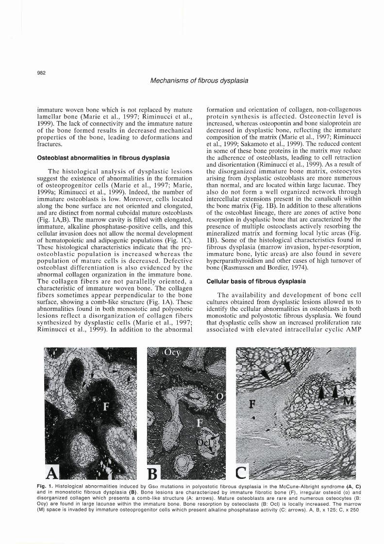
The histological analysis of dysplastic lesions suggest the existente of abnormalities in the formation of osteoprogenitor cells (Marie et al., 1997; Marie, 1999a; Riminucci et al., 1999). Indeed, the number of immature osteoblasts is low. Moreover, cells located along the bone surface are not oriented and elongated, and are distinct from normal cuboidal mature osteoblasts (Fig. lA,B). The marrow cavity is filled with elongated, immature, alkaline phosphatase-positive cells, and this cellular invasion does not allow the normal development of hematopoietic and adipogenic populations (Fig. 1C). These histological characteristics indicate that the pre- osteoblastic population is increased whereas the population of mature cells is decreased. Defective osteoblast differentiation is also evidenced by the abnormal collagen organization in the immature bone. The collagen fibers are not parallelly oriented, a characteristic of immature woven bone. The collagen fibers sometimes appear perpendicular to the bone surface, showing a comb-like structure (Fig. 1A). These abnormalities found in both monostotic and polyostotic lesions reflect a disorganization of collagen fibers synthesized by dysplastic cells (Marie et al., 1997; Riminucci et al., 1999). In addition to the abnormal
formation and orientation of collagen, non-collagenous protein synthesis is affected. Osteonectin leve1 is increased, whereas osteopontin and bone sialoprotein are decreased in dysplastic bone, reflecting the immature composition of the matrix (Marie et al., 1997; Riminucci et al., 1999; Sakamoto et al., 1999). The reduced content in some of these bone proteins in the matrix may reduce the adherence of osteoblasts, leading to cell retraction and disorientation (Riminucci et al., 1999). As a result of the disorganized immature bone matrix, osteocytes arising from dysplastic osteoblasts are more numerous than normal, and are located within large lacunae. They also do not form a well organized network through intercellular extensions present in the canaliculi within the bone matrix (Fig. 1B). In addition to these alterations of the osteoblast lineage, there are zones of active bone resorption in dysplastic bone that are caracterized by the presence of multiple osteoclasts actively resorbing the mineralized matrix and forming local lytic areas (Fig. 1B). Some of the histological characteristics found in fibrous dysplasia (marrow invasion, hyper-resorption, immature bone, lytic areas) are also found in severe hyperparathyroidism and other cases of high turnover of bone (Rasmussen and Bordier, 1974).
Cellular basls of fibrous dysplasia
The availability and development of bone cell cultures obtained from dysplastic lesions allowed us to identify the cellular abnormalities in osteoblasts in both monostotic and polyostotic fibrous dysplasia. We found that dysplastic cells show an increased proliferation rate associated with elevated intracellular cyclic AMP
Flg. 1 stological abnormalities induced by Gsa mutations in polyostotic fibrous dysplasia in the McCune-Albright syndrome (A, C) and in monostotic fibrous dysplasia (8). Bone lesions are characterized by immature fibrotic bone (F), irregular osteoid (o) and disorganized collagen which presents a comb-like structure (A: arrows). Mature osteoblasts are rare and numerous osteocytes (B: Ocy) are found in large lacunae within the immature bone. Bone resorption by osteoclasts (B: Ocl) is locally increased. The marrow (M) space is invaded by immature osteoprogenitor cells wihich present alkaline phosphatase activity (C: arrows). A, B, x 125; C, x 250
Mechanisms of fibrous dysplasia
(cAMP) levels (Marie et al., 1997). Osteoblastic cells isolated from the more severe bone lesions, composed of immature bone matrix, show a less differentiated phenotype and increased proliferative rate compared to cells from the less affected lesions composed of a mixture of mature and immature bone matrix. This indicates that the increased cell proliferation is associated with the less differentiated osteoblast phenotype in the dysplastic lesions. In addition to the increased cell proliferation, dysplastic cells show decreased differentiation, as evidenced by the alteration of osteocalcin mRNA expression and protein synthesis (Marie et al., 1997; Sakamoto et al., 1999; Hopyan et al., 1999). However, dysplastic cells respond normally to 1,25-dihydroxyvitamin D which enhances osteocalcin expression in osteoblasts (Marie et al., 1997). The expression of Runx21Cbfa1, a master transcription factor known to stimulate osteocalcin expression (Karsenty, 2000), was found to be increased in dysplastic bone cells (Sakamoto et al., 1999). One possible explanation for the increased Runx2lCbfal expression with regard to the reduced osteocalcin expression is that dysplastic cells express high levels of Msx2, a transcription factor that is expressed early during osteoblast differentiation and that inhibits osteocalcin expression (Newberry et al., 1997). The concomittant decrease in cell differentiation and increased proliferation of pre-osteoblastic cells in dysplastic lesions lead to a rapid deposition of an immature, poorly organised woven bone characteristic of the bone lesion at the histological leve1 (Marie et al., 1997; Riminucci et al., 1999).
Molecular basis of fibrous dysplasia
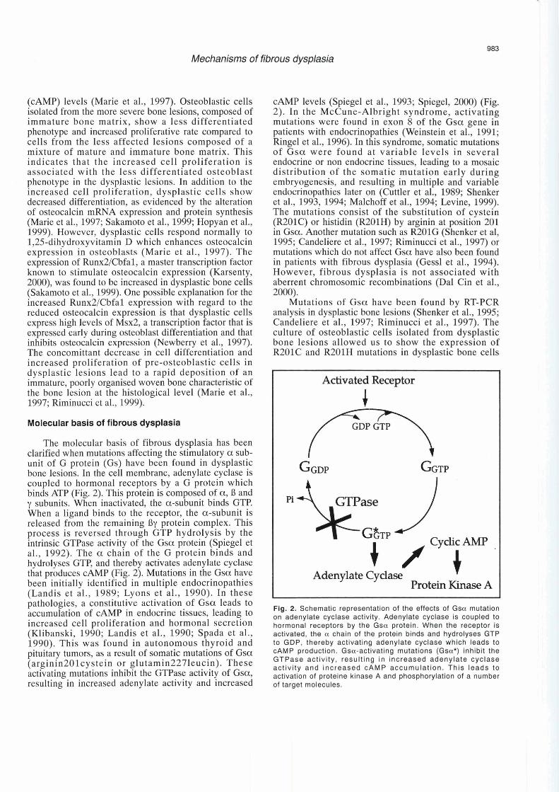
The molecular basis of fibrous dysplasia has been clarified when mutations affecting the stimulatory a sub- unit of G protein (Gs) have been found in dysplastic bone lesions. In the cell membrane, adenylate cyclase is coupled to hormonal receptors by a G protein which binds ATP (Fig. 2). This protein is composed of a , 13 and y subunits. When inactivated, the a-subunit binds GTP. When a ligand binds to the receptor, the a-subunit is released from the remaining By protein complex. This process is reversed through GTP hydrolysis by the intrinsic GTPase activity of the Gsa protein (Spiegel et al., 1992). The a chain of the G protein binds and hydrolyses GTP, and thereby activates adenylate cyclase that produces cAMP (Fig. 2). Mutations in the Gsa have been initially identified in multiple endocrinopathies (Landis et al., 1989; Lyons et al., 1990). In these pathologies, a constitutive activation of Gsa leads to accumulation of cAMP in endocrine tissues, leading to increased cell proliferation and hormonal secretion (Klibanski, 1990; Landis et al., 1990; Spada et al., 1990). This was found in autonomous thyroid and pituitary tumors, as a result of somatic mutations of Gsa (arginin20lcystein or glutamin227leucin). These activating mutations inhibit the GTPase activity of Gsa, resulting in increased adenylate activity and increased
cAMP levels (Spiegel et al., 1993; Spiegel, 2000) (Fig. 2). In the McCune-Albright syndrome, activating mutations were found in exon 8 of the Gsa gene in patients with endocrinopathies (Weinstein et al., 1991; Ringel et al., 1996). In this syndrome, somatic mutations of G s a were found at variable levels in several endocrine or non endocrine tissues, leading to a mosaic distribution of the somatic mutation early during embryogenesis, and resulting in multiple and variable endocrinopathies later on (Cuttler et al., 1989; Shenker et al., 1993, 1994; Malchoff et al., 1994; Levine, 1999). The mutations consist of the substitution of cystein (R201C) or histidin (R201H) by arginin at position 201 in Gsa. Another mutation such as R201G (Shenker et al, 1995; Candeliere et al., 1997; Riminucci et al., 1997) or mutations which do not affect Gsa have also been found in patients with fibrous dysplasia (Gessl et al., 1994). However, fibrous dysplasia is not associated with aberrent chromosomic recombinations (Da1 Cin et al., 2000).
Mutations of Gsa have been found by RT-PCR analysis in dysplastic bone lesions (Shenker et al., 1995; Candeliere et al., 1997; Riminucci et al., 1997). The culture of osteoblastic cells isolated from dysplastic bone lesions allowed us to show the expression of R201C and R201H mutations in dysplastic bone cells
Adivated Receptor
GDP GTP f i GGDP GGTP
m&:Tp J + r ""i""' . Adenylate Cyclase
Protein Kinase A
Fig. 2. Schematic representation of the effects of Gsa mutation on adenylate cyclase activity. Adenylate cyclase is coupled to hormonal receptors by the Gsa protein. When the receptor is activated, the a chain of the protein binds and hydrolyses GTP to GDP, thereby activating adenylate cyclase which leads to cAMP production. Gsa-activating mutations (Gsa*) inhibit the GTPase activity. resulting in increased adenylate cyclase act iv i ty and increased cAMP accumulation. This leads to activation of proteine kinase A and phosphorylation of a number of target rnolecules.
Mechanisms of fibrous dysplasia
(Shenker et al., 1995). These mutations are expressed in both monostotic and polyostotic fibrous dysplasia (Shenker et al., 1995; Alman et al., 1996) and represent the molecular basis of the cellular abnormalities in the two types of fibrous dysplasia. Interestingly, further analyses indicated that not al1 bone cells within the dysplastic lesions express such mutations (Riminucci et al., 1997). This mosaic distribution allows cell survival because the mutations are lethal (Happle, 1986). It is therefore believed that Gsa mutations which appear early during embryogenesis induce the generalized abnormalities found in the McCune-Albright syndrome, whereas the somatic mutations appearing later in life induce more restricted pathologies such as thyroid and pituitary adenomas, and monostotic fibrous dysplastic lesions. Thus, the endocrine and skeletal lesions associated with these mutations reflect a variable phenotypic expression of the mutations resulting from a somatic mosaicism of a same molecular alteration (Ringel et al., 1996; Spiegel, 1996; Cohen and Howell, 1999). In dysplastic lesions, G s a mutations are expressed in osteoprogenitor cells present in the marrow stroma (Marie et al., 1997; Bianco et al., 2000). The frequence of expression of mutations is higher in polyostotic than in monostotic fibrous dysplasia (Stanton et al., 1999). In terms of functional activity, dysplastic cells from monostotic and polyostotic lesions are able to be osteogenic in vitro in the presence of glucocorticoids which stimulate osteoblastic cell differentiation. However, mutated dysplastic cells are not osteogenic in vivo, probably because of the lethality induced by the mutation. Only mixed mutated and non mutated cells reconstituting the natural mosaic are able to form dysplastic lesions in vivo (Bianco et al., 1998). This indicates that Gsa mutations are responsible for the formation of dysplastic bone lesions, and that the presence of mutated and nonmutated cells is required for the development of the lesion (Bianco et al., 1998).
Molecular mechanisms of fibrous dysplasia
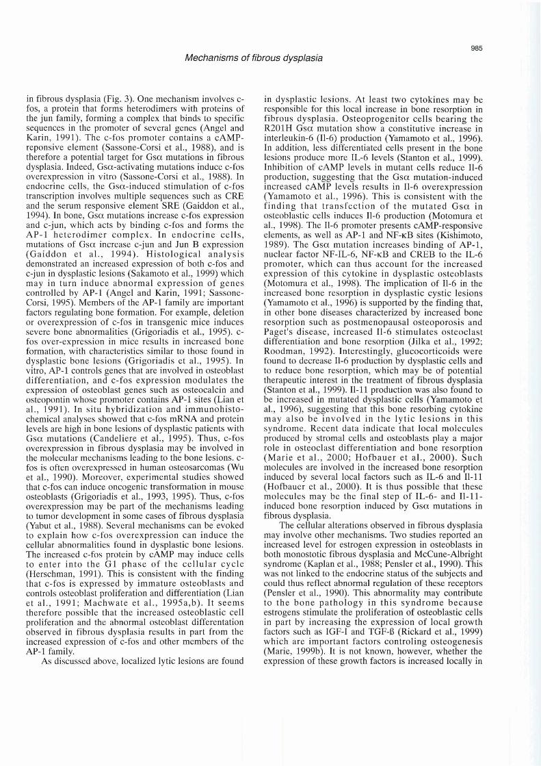
The formation of dysplastic bone lesions resulting from Gsa mutations involves severa1 mechanisms. In theory, activating mutations of Gsa should induce stimulation of adenylate cyclase and increased cAMP accumulation (Fig. 2). Accordingly, we found that Gsa mutations induce a constitutive accumulation of cAMP in osteoblastic cells in the McCune-Albright syndrome (Marie et al., 1997) and this was confirmed by other groups (Candeliere et al., 1997; Bianco et al., 1998). The downstream events leading to the abnormal cellular phenotype in osteoblastic cells are linked to cAMP signaling. The signaling pathways induced by cAMP are known to tightly control gene expression (Borrelli et al., 1992). cAMP accumulation activates protein kinase A which then phosphorylates transcription factors that are members of the cAMP-response-element-binding- protein (CREB) family. This leads to binding of these proteins to DNA domains called cAMP-responsive
element (or CRE) in the promoter of target genes (Montminy et al., 1990). This pathway controls the expression of multiple genes in several endocrine and nonendocrine tissues. For example, in pituitary cells, the increased activity of PKA induced by Gsa-activating mutations stimulate the promotor activity of prolactin and growth hormone (Tian et al., 1994; Gaiddon et al., 1995), leading to endocrinopathies associated with the mutation. In osteoblastic cells, a rise in cAMP induced by Gsa mutations also induce multiple effects (Fig. 3). One of the more rapid effects is cell retraction which is observed in dysplastic cells (Riminucci et al., 1997). This may be explained by the rapid disassembling effect of cAMP on cytoskeletal proteins leading to cell retraction (Lomri and Marie, 1990). The rise in cAMP in mutated dysplastic cells may also be responsible for the decreased expression of bone matrix proteins, such as osteopontin (Noda and Rodan, 1989) whose promoter contains cAMP-responsive elements (Kopp et al., 1989; Kerr et al., 1993). The rise in cAMP is also likely to be involved in the increased bone resorption found in dysplastic lytic lesions. Such local stimulation of bone resorption is found in severe cases of hyperparathyroidism in which high levels of parathyroid hormone induces elevated intracellular cAMP levels in osteoblasts (Lomri and Marie, 1990) and results in increased osteoclastic bone resorption by increasing bone resorbing cytokines (see below).
The elevation in cAMP induced by the Gsa is a an important mechanism leading to bone cell abnormalities
Gsa* + c fos
t - + c Jun -+ AP-1
Alteration of Bone Matrix Pro teins
Lesions
Fig. 3. Molecular rnechanisrns involved in fibrous dysplasia. The rnutations in Gsa (Gsa*) result in increased intracellular cAMP levels that rnodulate the expression of several genes whose promoter contains a cAMP-responsive elernent (CRE). This pathway leads to abnormal production of bone rnatrix proteins and to increased c- fos express ion, leading to abnormal recruitrnent and function of osteoblastic cells, and to abnormal bone rnatrix production. The cAMP overproduction induced by the mutation also leads to increased production of interleukin-6 and -1 1 (11-6, 11-1 1) which in turn stimulate osteoclastic bone resorption. These rnolecular mechanisms induced by the Gsa rnutations result in the forrnation of dysplastic bone lesions.
Mechanisms of fibrous dysplasia
in fibrous dysplasia (Fig. 3). One mechanism involves c- fos, a protein that forms heterodimers with proteins of the jun family, forming a complex that binds to specific sequences in the promoter of several genes (Angel and Karin, 1991). The c-fos promoter contains a cAMP- reponsive element (Sassone-Corsi et al., 1988), and is therefore a potential target for Gsa mutations in fibrous dysplasia. Indeed, Gsa-activating mutations induce c-fos overexpression in vitro (Sassone-Corsi et al., 1988). In endocrine cells, the Gsa-induced stimulation of c-fos transcription involves multiple sequences such as CRE and the serum responsive element SRE (Gaiddon et al., 1994). In bone, Gsa mutations increase c-fos expression and c-jun, which acts by binding c-fos and forms the AP-1 heterodimer complex. In endocrine cells, mutations of Gsa increase c-jun and Jun B expression (Gaiddon et al., 1994). Histological analysis demonstrated an increased expression of both c-fos and c-jun in dysplastic lesions (Sakamoto et al., 1999) which may in turn induce abnormal expression of genes controlled by AP-1 (Angel and Karin, 1991; Sassone- Corsi, 1995). Members of the AP-1 family are important factors regulating bone formation. For example, deletion or overexpression of c-fos in transgenic mice induces severe bone abnormalities (Grigoriadis et al., 1995). c- fos over-expression in mice results in increased bone formation, with characteristics similar to those found in dysplastic bone lesions (Grigoriadis et al., 1995). In vitro, AP-1 controls genes that are involved in osteoblast differentiation, and c-fos expression modulates the expression of osteoblast genes such as osteocalcin and osteopontin whose promoter contains AP-1 sites (Lian et al., 1991). In situ hybridization and immunohisto- chemical analyses showed that c-fos mRNA and protein levels are high in bone lesions of dysplastic patients with Gsa mutations (Candeliere et al., 1995). Thus, c-fos overexpression in fibrous dysplasia may be involved in the molecular mechanisms leading to the bone lesions. c- fos is often overexpressed in human osteosarcomas (Wu et al., 1990). Moreover, experimental studies showed that c-fos can induce oncogenic transformation in mouse osteoblasts (Grigoriadis et al., 1993, 1995). Thus, c-fos overexpression may be part of the mechanisms leading to tumor development in some cases of fibrous dysplasia (Yabut et al., 1988). Several mechanisms can be evoked to explain how c-fos overexpression can induce the cellular abnormalities found in dysplastic bone lesions. The increased c-fos protein by cAMP may induce cells to enter into the G1 phase of the cellular cycle (Herschman, 1991). This is consistent with the finding that c-fos is expressed by immature osteoblasts and controls osteoblast proliferation and differentiation (Lian et al., 1991; Machwate et al., 1995a,b). It seems therefore possible that the increased osteoblastic cell proliferation and the abnormal osteoblast differentation observed in fibrous dysplasia results in part from the increased expression of c-fos and other members of the AP-1 family.
As discussed above, localized lytic lesions are found
in dysplastic lesions. At least two cytokines may be responsible for this local increase in bone resorption in fibrous dysplasia. Osteoprogenitor cells bearing the R201H Gsa mutation show a constitutive increase in interleukin-6 (11-6) production (Yamamoto et al., 1996). In addition, less differentiated cells present in the bone lesions produce more IL-6 levels (Stanton et al., 1999). Inhibition of cAMP levels in mutant cells reduce 11-6 production, suggesting that the Gsa mutation-induced increased cAMP levels results in 11-6 overexpression (Yamamoto et al., 1996). This is consistent with the finding that transfection of the mutated G s a in osteoblastic cells induces 11-6 production (Motomura et al., 1998). The 11-6 promoter presents cAMP-responsive elements, as well as AP-1 and NF-KB sites (Kishimoto, 1989). The Gsa mutation increases binding of AP-1, nuclear factor NF-IL-6, NF-KB and CREB to the IL-6 promoter, which can thus account for the increased expression of this cytokine in dysplastic osteoblasts (Motomura et al., 1998). The implication of 11-6 in the increased bone resorption in dysplastic cystic lesions (Yamamoto et al., 1996) is supported by the finding that, in other bone diseases characterized by increased bone resorption such as postmenopausal osteoporosis and Paget's disease, increased 11-6 stimulates osteoclast differentiation and bone resorption (Jilka et al., 1992; Roodman, 1992). Interestingly, glucocorticoids were found to decrease 11-6 production by dysplastic cells and to reduce bone resorption, which may be of potential therapeutic interest in the treatment of fibrous dysplasia (Stanton et al., 1999). 11-11 production was also found to be increased in mutated dysplastic cells (Yamamoto et al., 1996), suggesting that this bone resorbing cytokine may also be involved in the lytic lesions in this syndrome. Recent data indicate that local molecules produced by stromal cells and osteoblasts play a major role in osteoclast differentiation and bone resorption (Marie et al., 2000; Hofbauer et al., 2000). Such molecules are involved in the increased bone resorption induced by several local factors such as IL-6 and 11-11 (Hofbauer et al., 2000). It is thus possible that these molecules may be the final step of IL-6- and 11-11- induced bone resorption induced by Gsa mutations in fibrous dysplasia.
The cellular alterations observed in fibrous dysplasia may involve other mechanisms. Two studies reported an increased leve1 for estrogen expression in osteoblasts in both monostotic fibrous dysplasia and McCune-Albright syndrome (Kaplan et al., 1988; Pensler et al., 1990). This was not linked to the endocrine status of the subjects and could thus reflect abnormal regulation of these receptors (Pensler et al., 1990). This abnormality may contribute to the bone pathology in this syndrome because estrogens stimulate the proliferation of osteoblastic cells in part by increasing the expression of local growth factors such as IGF-1 and TGF-B (Rickard et al., 1999) which are important factors controling osteogenesis (Marie, 1999b). It is not known, however, whether the expression of these growth factors is increased locally in
Mechanisms of fibrous dysplasia
fibrous dysplasia. Finally, a rise in the expression of the parathyroid hormone related peptide (PTHrP) has been recently reported in osteoblastic cells of patients with the McCune-Albright syndrome. A treatment with 1,25- dihydroxy-vitamin D3 can reduce the production of PTHrP in vitro as well as bone markers in this disease. It is thus possible that a local increase in the expression of PTHrP, which acts on bone cells in a similar way to PTH, could contribute to the bone cell alterations in the McCune Albright syndrome (Fraser et al., 2000).
Conclusion
The discovery that Gsa mutations are expressed in dysplastic bone cells provided a basis for the cellular and molecular mechanisms involved in the osteoblast alterations observed in fibrous dysplasia. I t is now apparent that the abnormal osteoblastic cell proliferation and differentiation obsewed in fibrous dysplasia result f rom increased cAMP accumulation, leading to alteration in the expression of target genes including c- fos, c-jun, 11-6, 11-11, which in turn modulate the transcription and expression of downstream genes and result in the alterations of osteoblast and osteoclast recruitment and function in the dysplastic bone lesions. These pathways provide a cellular and molecular basis for the bone alterations in fibrous dysplasia. Further analysis of the molecular pathways activated by the Gsa mutations in dysplastic bone cells may lead to the identification of target genes for cellular therapies and allow us in the future to develop a therapeutic approach of the skeletal alterations in fibrous dysplasia.
References
Albright F., Butler A.M., Hampton A.O. and Smith P. (1937). Syndrome characterized by osteotis fibrosa disseminata, areas of pigmentat ion and endocrine dysfunction, with precocious puberty in females: report of five cases. N. Engl. J. Med. 216, 727-746.
Alman B.A., Greel D.A. and Wolfe H.J. (1996). Activating mutations of Gs protein in monostotic fibrous lesions of bone. J. Orthop. Res. 14, 31 1-31 5.
Angel P. and Karin M. (1991). The role of Jun, Fos and the AP-1 complex in cell-proliferation and transformation. Biochim. Biophys. Acta 10, 129-157.
Bianco P., Kuznetsov S.A., Riminucci M., Fisher L.W., Spiegel A.M. and Robey P.G. (1998). Reproduction of human fibrous dysplas ia of bone in immunocompromised mice by transplanted mosaics of normal and Gsa-mutated skeletal progenitor cells. J. Clin. Invest. 101, 1737-1744.
Bianco P., Riminucci M., Majolagbe A,, Kuznetsov S.A., Collins M.T., Mankani M.H., Corsi A., Bone H.G., Wientroub S.. Spiegel A.M., Fisher L.W. and Robey P.G. (2000). Mutations of the G N A S I gene, s t romal ce l l dysfunct ion, and osteomalacic changes in non-McCune-Albr ight f ibrous dysplasia of bone. J. Bone Miner. Res. 15, 120-128.
Borrelli E., Montmayeur J.P., Foulkes N.S. and Sassone-Corsi P. (1992). Signal transduction and gene control: the cAMP
pathway. Crit. Rev. Oncog. 3, 321-338. Candeliere G.A., Glorieux F.H., Prud'homme D.J. and St Arnaud
R. (1995). lncreased expression of the c-fos proto-oncogene in bone from patients with fibrous dysplasia. N. Engl. J. Med. 332, 1546-1 551.
Candeliere G.A, Roughley P.J. and Glorieux F.H. (1997). Polymerase chain reaction-based technique for the selective enrichment and analysis of mosaic arg201 mutations in Galpha S from patients with fibrous dysplasia of bone. Bone 21, 201 -206.
Cohen M.M. and Howel l R.E. (1999). Et iology of f ibrous dysplasia and McCune-Albr ight syndrome. Int. J. Oral Maxillofac. Surg. 28, 366-371.
Cuttler L., Jackson J.A., Saeed uz-Zafar M., Levitsky L.L., Mellinger R.C. and Frohman L.A. (1989). Hypersecretion of growth hormone and prolactin in McCune-Albright syndrome. J. Clin. Endocrinol. Metab. 68, 1148-1 154.
Dal Cin P., Sciot R., Brys P., De Wever l., Dorfman H., Fletcher C.D., Jonsson K., Mandahl N,, Mertens F., Mitelman F., Rosai J., Rydholm A,. Samson l., Tallini G., Van den Berghe H., Vanni R. and Willen R. (2000). Recurrent chromosome aberrations in fibrous dysplasia of the bone: a report of the CHAMP study group. Chromosomes and morphology. Cancer Genet. Cytogenet. 122, 30-32.
Fraser W.D., Walsh C.A., Birch M.A., Durham B., Dillon J.P., McCreavy D. and Gal lagher J .A. (2000) . Parathyroid hormone-related protein in the aetiology of fibrous dysplasia of bone in the McCune-Albright syndrome. Clin. Endocrinol. 53, 621 -628.
Gaiddon C., Boutillier A.L., Monnier D., Mercken L. and Loeffler J.P. (1994). Genomic effects of the putative oncogene Gsa. J. Biol. Chem. 269, 22663-22671.
Gaiddon C., Mercken L., Bancroft C. and Loeffler J.P. (1995). Transcr ip t ional e f fects i n GH3 ce l ls of G s a mutants associated with human pitui tary tumors: stimulation of adenosine 3',5'-monophosphate response element-binding protein-mediated transcription and of prolactin and growth hormone promoter activity via protein kinase A. Endocrinol., 136, 4331 -4338.
Gessl A,, Freissmuth M.. Czech T., Matula C., Hainfellner J.A., Buchfelder M. and Vierhapper H. (1994). Growth hormone- prolactin-thyrotropin-secreting pituitary adenoma in atypical McCune-Albright syndrome with functionally normal Gs alpha protein. J. Clin. Endocrinol. Metab. 79, 11 28-1 134.
Grabias S.L. and Campbell C.J. (1977). Fibrous dysplasia. Orthop. Clin. North Am. 8, 771-783.
Grigoriadis A.E., Schellander K., Wang Z.Q. and Wagner E.F. (1993). Osteoblasts are target cells for transformation in c- fos transgenic mice. J. Cell Biol. 122, 685-701.
Grigoriadis A.E.. Wang Z.Q. and Wagner E.F. (1995). Fos and bone cell development: lessons from a nuclear oncogene. Trends Genet. 11, 436-441.
Happle R. (1986). The McCune-Albright syndrome: a lethal gene surviving by mosaicism. Clin. Genet. 29, 321-324.
Harris W.H, Dudley H.R. and Barry R.J. (1962). The natural history of fibrous dysplasia. J. Bone J. Surg. 44A, 207-233.
Herschman H.R. (1991). Primary response genes induced by growth factors and tumor promoters. Annu. Rev. Biochem. 60, 281 -319.
Mechanisms of fibrous dysplasia
formation. Am. J. Pathol. 151, 1587-1600. Riminucci M., Liu B., Corsi A,, Shenker A,, Spiegel A.M., Robey
P.G. and Bianco P. (1999). The histopathology of fibrous dysplasia of bone in patients with activating rnutations of the Gs a lpha gene: s i te-spec i f ic pat terns and recurrent histological hallrnarks. J. Pathol. 187, 249-258.
Ringel M.D., Schwindinger W.F. and Levine M.A. (1996). Clinical implications of genetic defects in G proteins. The molecular basis of McCune-Albright syndrome and Albright hereditary osteodystrophy. Medicine 75, 171 -184.
Roodman G.D. (1992). Interleukin-6: an osteotropic factor? J. Bone Miner. Res. 7, 475-478.
Sakamoto A., Oda Y., lwamoto Y. and Tsuneyoshi M. (1999). A comparative study of fibrous dysplasia and osteofibrous dysplasia with regard to expressions of c-fos and c- jun products and bone matrix proteins: a clinicopathologic review and immunohistochemical study of c-fos, c- jun, type I collagen, osteonectin, osteopontin, and osteocalcin. Hum. Pathol. 30, 1418-1426.
Sassone-Cors i P. (1995) . Signal ing pathways and c- fos transcriptional response - links to inherited diseases. N. Engl. J. Med. 332, 1576-1577.
Sassone-Corsi P., Visvader J., Ferland L., Mellon P.L. and Verma I .M. (1 988) . lnduct ion of proto-oncogene fos t ranscr ip t ion through the adenylate cyclase pathway: characterization of a cAMP-responsive element. Genes Dev. 2, 1529-1538.
Schwindinger W. and Levine M.A. (1993). McCune-Albright syndrome. Trends Endocrinol. Metab. 4, 238-242.
Shenker A., Weinstein L.S., Moran A., Pescovitz O.H., Charest N.J.. Boney C.M., Van Wyk J.J., Merino M.J., Feuillan P.P. and Spiegel A.M. (1993) . Severe endocr ine and nonendocr ine mani festat ions of the McCune-Albr ight syndrome associated with activating mutations of stimulatory G protein Gs. J. Pediatr. 123, 509-51 8.
Shenker A., Weinstein L.S., Sweet D.E. and Spiegel A.M. (1994). An activating Gsa mutation is present in fibrous dysplasia of bone in the McCune-Albright syndrome. J. Clin. Endocrinol. Metab. 79, 750-755.
Shenker A., Chanson P., Weinstein L.S., Chi P., Spiegel A.M., Lomri A. and Marie P.J. (1995). Osteoblastic cells derived from isolated lesions of fibrous dysplasia contain activating somatic mutations of the Gsa gene. Hum. Mol. Genet. 4, 1675-1 676.
Spada A., Arosio M., Bochicchio D., Bazzoni N,, Vallar L.,
Bassetti M. and Faglia G. (1990). Clinical, biochemical, and morpholog ica l corre la tes in pat ients bear ing growth hormone-secret ing p i tu i tary tumors with or without constitutively active adenylyl cyclase. J. Clin. Endocrinol. Metab. 71. 1421-1426.
Spiegel A.M. (1996). Genetic basis of endocrine disease. Mutations in G proteins and G protein-coupled receptors in endocrine disease. J. Clin. Endocrinol. Metab. 81, 2434- 2442.
Spiegel A.M. (2000). G protein defects in signal transduction. Horm. Res. 53 (suppl. 3), 17-22.
Spiegel A.M., Shenker A. and Weinsten L.S. (1992). Receptor- effector coupling by G proteins: implications for normal and abnormal signal transduction. Endcr. Rev. 13, 536-560.
Spiegel A.M, Weinste in L.S. and Shenker A. (1993) . Abnormalit ies in G protein-coupled signal transduction pathways in human disease. J. Clin. Invest. 92, 11 19-1125.
Stanton R.P., Hobson G.M., Montgomery B.E., Moses P.A., Smi th-Ki rwin S.M. and Funanage V.L. (1999). Glucocorticoids decrease interleukin-6 levels and induce mineralization of cultured osteogenic cells from children with fibrous dysplasia. J. Bone Miner. Res. 14, 1104-1 11 4.
Tian J . , Chen J. and Bancroft C. (1994) . Expression of constitutively active Gs alpha-subunits in GH3 pituitary cells stimulates prolactin promoter activity. J. Biol. Chem. 269, 33- 36.
Weinstein L.S., Shenker A.. Gejman P.V, Merino M.J., Friedman E. and Spiegel A.M. (1991). Activating mutations of the stimulatory G protein in the McCune-Albright syndrome. N. Engl. J. Med. 325, 1688-1695.
Wu J.X., Carpenter P.M., Gresens C., Keh R., Niman H., Morris J.W. and Mercola D. (1990). The proto-oncogene c-fos is over-expressed in the majority of human osteosarcomas. Oncogene 5, 989-1 000.
Yabut S.M., Kenan S.. Sissons H.A. and Lewis M.M. (1988). Malignant transformation of fibrous dysplasia. A case report and review of the literature. Clin. Orthop. 281-289.
Yamarnoto T., Ozono K., Kasayama S., Yoh K., Hiroshima K., Takagi M., Matsumoto S., Michigami T., Yamaoka K., Kishimoto T. and Okada S. (1996). lncreased IL-6 production by cells isolated from the fibrous bone dysplasia tissues in patients with McCune-Albright syndrome. J. Clin. Invest. 98, 30-35.
Accepted May 9, 2001


![Lamellar Bone is an Incremental Tissue Reconciling Enamel ... · as the scaling of bone mass to body mass is an axiom of vertebrate hard tissue biology [15]. Because enamel and bone](https://img.pdfslide.us/doc/110x75/5fa2f5bb05de466cf30f1103/lamellar-bone-is-an-incremental-tissue-reconciling-enamel-as-the-scaling-of.jpg)