Embed Size (px)
Citation preview

The Plant Cell, Vol. 4, 879-887, August 1992 O 1992 American Society of Plant Physiologists
REVIEW ARTICLE
The Remarkable Biology of Pollen
Patricia Bedinger Department of Biology, University of North Carolina, Chapel Hill, North Carolina 27599-3280
INTRODUCTION
To reproduce, higher plants utilize a unique multicellular mi- croorganism: the male gametophyte, or pollen grain. The independent lifetime of flowering plant gametophytes is greatly abbreviated compared to that of gametophytes of more primi- tive plants, yet the angiosperm pollen grain must be able to survive at least briefly free from the sporophytic plant and per- form a number of specialized functions before fertilization is accomplished.
Although the pollen grain is a rather simple two- or three- celled organism, cytogenetic and mutagenesis experiments indicate that higher plants make a significant investment in genetic material devoted to gametophyte production (Carlson, 1977; Birchler and Schwartz, 1979; Kindiger et al., 1991). Mo- lecular studies identifying anther- and pollen-specific genes support the notion that the construction of a functional male gametophyte requires a rather large pool of such genes (Kamalay and Goldberg, 1980; Willing and Mascarenhas, 1984; McCormick, 1991). This is not so surprising given the special- ized structures, mechanisms for rapid growth, and cell-cell communication systems during pollen-pistil interactions that have evolved to allow for both the survival of the gametophyte free of the sporophyte and the efficient delivery of sperm to the embryo sac.
Studies of certain aspects of the molecular mechanisms of gametophyte development and function have progressed very rapidly. Examples of these include the identification of cis-acting elements determining tissue-specific gene expression in pollen and tapetal cells (Ursin et al., 1989; Koltunow et al., 1990; McCormick et al., 1991) and of mechanisms of self-incom- patibility in pollen-pistil interactions (McClure et al., 1989, 1990; Thorsness et al., 1991). The intent of this article is to focus on a few less well studied but intriguing aspects of pollen development and function, with an emphasis on pollen devel- opment in maize. For more general reviews of microsporo- genesis, the reader is referred to Giles and Prakash (1987) and Mascarenhas (1989). For recent reviews of gene expression during pollen development, see Mascarenhas (1990) and McCormick (1991).
OVERVIEW OF POLLEN DEVELOPMENT
Pollen development takes place within the anther. Four anther wall layers (the epidermis, the endothecium, the middle layer, and the tapetum) enclose the fluid-filled locule, as shown in Figure 1. This locule contains the sporogenic cells that will undergo meiosis. The layer of anther cells adjacent to the loc- ule is known as the tapetum, which is a tissue that is intimately involved in microsporogenesis (see below). Figure 2 summa- rizes pollen development in maize. At 50 to 60 days after seeding, the microsporocytes are encased in an impermeable P-l,9glucan (callose) wall (Figure 2A), which effectively iso- lates the meiocytes from other cells (Knox and Heslop-Harrison, 1970). Each microsporocyte undergoes two meiotic divisions over a period of approximately 3 days, producing a tetrad of four haploid cells called microspores that are still encased within a callose wall (Figure 28). After dissolution of the cal- lose wall, the free young microspores grow rapidly for about 5 days. During this period, the outer pollen wall, or exine, is synthesized (Figure 2C). The centrally located nucleus migrates toward the cell periphery to a position opposite the pollen pore.
As the young microspores grow, they fill with multiple small vacuoles (Figure 2D) that eventually coalesce into a single large vacuole, compressing the cytoplasm into a small region op- posite the pollen pore (Figure 2E). About 5 days after meiosis, the asymmetric division called microspore mitosis occurs, producing two cells with very different fates (Figure 2F). The bicellar product of microspore mitosis is, by definition, pollen. During the following 7 days, further maturation steps occur. In maize, the generative cell within the young pollen divides again to form two sperm cells (Figure 2G); in many other plant species, this second mitosis takes place only after germina- tion of the pollen tube. The pollen grain then secretes the cellulosic and pectic inner pollen wall, or intine. The pollen grain accumulates starch granules until the grain is entirely engorged (Figure 2H). It appears that some mRNAs synthe- sized at this time may be stored for translation during germination (Mascarenhas et al., 1984; Mascarenhas, 1990). The pollen dehydrates, reducing the water content to 40 to 58% at the time of dehiscence (Barnabas, 1985). Further

880 The Plant Cell
Bepidermisendothecium
middle layertapetum
— microspore
anther anther cross-section anther loculeFigure 1. Structure of Maize Anther.
(A) Diagram of anther. The arrow indicates the plane of the cross-section shown in (B).(B) Diagram of cross-section of the anther. The dotted box indicates a single anther locule.(C) A micrograph of an anther locule, with cell layers identified.
dehydration of the pollen grain after its release from the anthermay cause domains of the vegetative cell plasma membraneto enter an unstable gel/liquid crystal state (Kerhoas et al.,1987). The maize pollen grain hydrates upon interaction withthe silk, and a pollen tube is rapidly germinated through thepore to transport the sperm to the embryo sac.
THE TAPETUM: POLAR SECRETORY CELLS
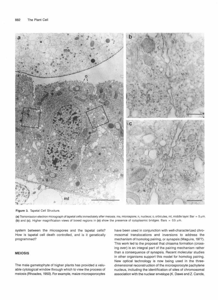
As is the case for all higher organisms, the production of ga-metes in higher plants requires the participation of both thedeveloping haploid gametophytic cells and accessory somatic(in the case of plants, sporophytic) diploid cells. The tapelumof higher plants plays a critical role in pollen development(Pacini et al., 1985). During meiosis, the tapetal cells undergoa dramatic differentiation into binucleate, polar secretory cellslacking a primary cell wall. Figure 3 shows that these cellsare packed with ribosomes, mitochondria, endoplasmic retic-ulum, Golgi, and many vesicles on the locular face of theplasma membrane and are connected by cytoplasmic bridgesearly in development. The tapetum may remain associated withthe anther wall (in which case it is termed secretory or pari-etal, as in maize), or the tapetal cells may actually fuse to forma syncytium that invades the anther locule, engulfing the de-veloping microspores (in which case it is termed amoeboid,as in Tradescantia). Secretory vesicles are transported in apolar fashion to the locular surface of the cells.
Several tapetal cell functions are known. It has long beenproposed that the tapetal cells play a nutritive role for the micro-spores, in direct analogy to nurse cells in mammalian systems.A second tapetal cell function is to release the young haploidmicrospores from the callose wall enclosing the meiotic tetrad
by the secretion of a p-1,3-glucanase, or callase (Steiglitz, 1977).The timing of callase secretion appears to be critical for nor-mal pollen development. In one case of cytoplasmic malesterility in petunia, the timing of callose wall dissolution ispremature, leading to collapse of the developing microspores(Izhar and Frankel, 1971). This result has recently been repeatedwith a more defined system using transgenic plants. Worrallet al. (1992) fused a modified callase gene to an Arabidopsispromoter known to be active in the tapetum during meiosis(Scott et al., 1991; Paul et al., 1992). Partial or complete malesterility was observed in transgenic tobacco plants express-ing the callase gene during meiosis. Meiosis was apparentlynormal in the absence of a callose wall, but pollen wall devel-opment was abnormal. Instead of a highly organized patternof exine deposition, an electron-dense material that may besporopollenin appeared to be randomly distributed on themicrospore surface. How the presence of callose may con-tribute to the formation of a normal pollen wall remains to beclarified. A rather surprising result of this study was that thetapetal cells as well as the microspores exhibited abnormali-ties. This raises the intriguing possibility that the tapetal cellsdo not function independently of the microspores—in otherwords, the two cell types are somehow interdependent duringpollen development.
A third proposed role for the tapetal cells is the productionof precursors for the biosynthesis of the outer pollen wall, orexine. This aspect of tapetal cell function is described in moredetail below. As the microspores become vacuolate, the tape-tal cells undergo cell death. Even at this stage, the tapetal cellscontribute to pollen development with the deposition of cellremnants called tryphine or pollenkitt on the maturing pollensurface. These substances are largely lipoidal in nature andmay function to protect the pollen grain from dehydration orto allow it to attract and adhere to insect pollinators.

Biology of Pollen 881
Other functions of the tapetum remain more mysterious. Genetic studies have identified recessive, sporophytic muta- tions at many different loci that cause male sterility (Beadle, 1932; Albertsen and Phillips, 1981; Kaul, 1988). Most cytologi- cal evidence points to the tapetal cells as the affected sporophytic cell type. This is almost certainly the case with cytoplasmic male sterility type T in maize, where irregularities in tapetal cell morphology are observed prior to pollen abor- tion (Warmke and Lee, 1977). It is possible that the majority of these mutations adversely affect pollen nutrition dueto the malfunctioning of the tapetum. However, some male sterile (ms) mutations appear to have more specific phenotypic effects. Severa1 of the male sterile mutants of maize (ms7, msl) are defective in pollen wall biosynthesis, a finding consistent with the proposed role of the tapetum in exine production. Other ms mutations affect the progression of microspores through microspore mitosis, causing failure of chromosome conden- sation (ms14) o1 precocious chromosome condensation (ms13) (Albertsen and Phillips, 1981). The finding that a deficiency in a purine salvage pathway enzyme leads to abnormal pol- len mitosis and male sterility in Arabidopsis (Regan and Moffatt, 1990) suggests that one way that chromosome behavior in microspores can be influenced is in the control of DNA precur- sor production.
A greater understanding of specific tapetal functions will be accomplished by isolating sporophytic male sterility genes. The generation of new male sterile mutants using transpos-
able element-containing maize lines should allow the isolation of such genes through transposon tagging methods (P Bedinger, A. Broadwater, C. Loukides and S. Stephenson, un- published data). Similarly, T-DNA mutagenesis in Arabidopsis has identified severa1 new male sterility genes that should be amenable to cloning (C. Makaroff and K. Feldmann, personal communication). Tomato YAC libraries, in conjunction with an extensive RFLP map, are currently being used to isolate male sterility genes by chromosome walking (S. McCormick, per- sonal communication).
Tapetal cells provide an excellent target for the control of fertility through genetic engineering. Male sterility has been induced in tobacco through the selective destruction of the tapetum by fusing the promoter of a gene expressed specifi- cally in tapetal cells to a cytotoxic ribonuclease gene (Mariani et al., 1990). Recent progress using a specific inhibitor of this ribonuclease to control its activity demonstrates the tremen- dous potential for the impact of biotechnology on agriculture, in this case, by providing a genetic means of producing hy- brid seed (Mariani et al., 1992).
The importance of the tapetum in microsporogenesis, first suggested by cytological studies and now confirmed by molec- ular studies, is beyond dispute. What remains to be elucidated are the molecular mechanisms of tapetal function. How is the dramatic differentiation of the tapetum during meiosis con- trolled? What are the different specific roles of the tapetum during microsporogenesis? 1s there a two-way communication
+ + meios is
C f r e e young A po l len mo the r B t e t r a d o f haploid c e l l (me iocy te) mic rospores mic rospores
D m u l t i p l e sma l l vacuoles
1
b
microspore m i t o s i s
I lUCl cu3 generat ive c e l l 1 dehydration div is ion, i n t i n e g r o w t h
ma tu re po l len
E s ing ie la rge vacuole
Figure 2. Schematic Diagram of Pollen Development in Maize.
Morphologically distinct stages of the developing pollen within the locule are depicted and are described in the text.
882 The Plant Cell
ml
Figure 3. Tapetal Cell Structure.(a) Transmission electron micrograph of tapetal cells immediately after meiosis. ms, microspore; n, nucleus; o, orbicules, ml, middle layer. Bar = 5 urn.(b) and (c). Higher magnification views of boxed regions in (a) show the presence of cytoplasmic bridges. Bars = 0.5 um.
system between the microspores and the tapetal cells?How is tapetal cell death controlled, and is it geneticallyprogrammed?
MEIOSIS
The male gametophyte of higher plants has provided a valu-able cytological window through which to view the process ofmeiosis (Rhoades, 1950). For example, maize microsporocytes
have been used in conjunction with well-characterized chro-mosomal translocations and inversions to address themechanism of homolog pairing, or synapsis (Maguire, 1977).This work led to the proposal that chiasma formation (cross-ing over) is an integral part of the pairing mechanism ratherthan a consequence of synapsis. Recent molecular studiesin other organisms support this model for homolog pairing.New optical technology is now being used in the three-dimensional reconstruction of the microsporocyte pachytenenucleus, including the identification of sites of chromosomalassociation with the nuclear envelope (K. Dawe and Z. Cande,
Biology of Pollen 883
personal communication). If these studies can be extended to earlier meiotic stages, the relationship between crossing over and synapsis may be further clarified.
Genetic studies in maize have identified many mutants defec- tive in meiosis (Golubovskaya, 1979). Mutants with particularly interesting phenotypes include those affecting chromosome behavior, such as asynaptic (reduced homolog pairing) and sticky (chromosomes stick together); those affecting spindle structure, such as divergent spindle (lack of focused spindle pole bodies in the first meiotic division); and those affecting control of cell division, such as polymitotic (supernumerary cytokinesis following the second division). The use of these mutants in conjunction with the cytological approaches de- scribed above should greatly enhance our understanding of fundamental meiotic processes, including how chromosome homologs pair, what factors might function specifically in meiotic as opposed to mitotic spindles, and how cytokinesis is normally coupled to chromosome replication and separation.
A UNIQUE CELL WALL
One of the most distinctive features of pollen grains is the pol- len wall. The outermost wall, or exine, is ornamented in a species-specific fashion, as illustrated in Figure 4. The exine is composed largely of a material called sporopollenin. The extraordinary chemical resistance of sporopollenin was first reported more than 150 years ago (John, 1814), but its molec- ular structure is not yet understood. Until fairly recently, sporopollenin was thought to be a carotenoid polymer, but more recent inhibitor and physical data indicate that this is not the case (Prahl et al., 1985; Guilford et al., 1988). It is clear that sporopollenin is one of the most resistant biopolymers known; this property is presumably essential for the survival of the pollen grain free from the sporophyte prior to fertilization. The resistance of sporopollenin has been of great value to evolu- tionary and archeological researchers, who are able to use the distinctive pollen exine patterns to “sample” the plant com- munity present at any particular time.
Exine biosynthesis is a joint effort on the part of tapetal cells and microspores. It is thought that the microspore (or even meiocyte) elaborates a surface template system known as the primexine or glycocalyx (Heslop-Harrison, 1971; Rowley, 1973) that determines the patterning of exine deposition. The mech- anism by which primexine elements are distributed in a species-specific pattern in the microspore plasma membrane is currently unknown. The role of the tapetal cells in exine for- mation appears to be in the secretion of sporopollenin precursors that are then polymerized onto the template sys- tem. Orbicule structures composed of sporopollenin form on the locular face of the tapetal cells (Figure 3), probably as a byproduct of exine biosynthesis.
Although there have been a number of elegant morphologi- cal studies on pollen wall development (Skvarla and Larson,
1966; El-Ghazaly and Jensen, 1986), biochemical studies of this unique and highly specialized pollen structure are Only beginning. Exine purification has allowed the production of specific antibodies (Southworth, 1988; Southworth et al., 1988) and the first characterization of potential structural proteins (Chay et al., 1992). The study of mutants defective in the for- mation of the exine (Morton et al., 1989) may provide additional approaches for understanding the synthesis, function, and ba- sis of pattern formation of this unusual extracellular matrix.
MICROSPORE MITOSIS: A DEVELOPMENTAL SWITCH
After meiosis and an initial burst of growth and exine synthesis, the haploid microspores undergo a cytological reorganization in preparation for a key event in pollen development, the asym- metric division known as microspore mitosis. As is the case for many morphological events in plant development, an asym- metric division marks the initiation of a new developmental program. The pollen system provides a rare and exciting op- portunity to study the cellular basis for the establishment and maintenance of polarity within a single cell. The cytological reorganization prior to microspore mitosis involves nuclear migration to a specific location near the cell periphery. In or- chids (which lack the impermeable exine that interferes with immunolocalization studies), a nove1 microtubule system has been detected at the site of nuclear migration and subsequent division (Brown and Lemmon, 1991). In maize, the production of a large vacuole positions the entire cytoplasm in this region. If the role of organelles and cytoskeletal elements in estab- lishing specific cytoplasmic domains in the microspore can be elucidated, the basis for many asymmetric morphogenic events in plant development may also be revealed.
An asymmetric, cone-shaped spindle perpendicular to the plasma membrane is utilized in this division (Brumfield, 1941; Brown and Lemmon, 1992). The nucleus adjacent to the plasma membrane becomes cellularized, forming the gener- ative cell. Whereas the chromatin of the generative cell is highly condensed, the vegetative nucleus is larger, has more nuclear pores, and has decondensed chromatin, indicating that it is the transcriptionally active nucleus (LaFountain and LaFountain, 1973; Wagner et al., 1990). The basis for the striking differ- ences in chromatin structure and activity in the two nuclei may lie in the components of the cytoplasmic domains established prior to division.
Microspore mitosis appears to be a critical point in commit- ment to the gametophytic pathway, analogous to other events in development where an aysmmetric division signals a com- mitment to differentiate, such as the first division in FUCUS (Quatrano, 1990) and C. elegans (Strome, 1989) embryo de- velopment or the asymmetric division prior to guard cell differentiation (Cho and Wick, 1989). Anther and microspore culture experiments suggest that uninucleate microspores can be more easily induced than postmitotic young pollen to enter

884 The Plant Cell
Figure 4. Scanning Electron Micrographs of Pollen.(a) Maize.(b) Amaryllis.(c) Liatris, a composite.(d) Redbud, a legume (with stigma).(e) Tobacco.(f) Impatiens.Bars = 10 urn.
a sporophytic developmental program, producing androgenicstructures (Gaillard et al., 1991). Studies of RNA and proteinpopulations (Bedinger and Edgerton, 1990) and of protein syn-thesis in isolated microspores (Mandaron et al., 1990) indicatethat a major developmental switch in gene expression occursat this time. Studies of pollen-specific gene expression are con-sistent with this hypothesis in that the majority of such genesappear to become actively transcribed only after microsporemitosis (Stinson et al., 1987; McCormick, 1991). The role thatthe division of the microspore nucleus plays in the transcrip-
tional activation of a gametophytic developmental program isan intriguing area for future research.
POLLEN TUBE GERMINATION AND GROWTH
Upon the interaction of the mature pollen with a stigma, thepollen hydrates and the pollen tube emerges from a pore onthe exine surface. In the grasses, tube germination takes place

Biology of Pollen 885
within a few minutes after the pollen lands on a silk. The pol- len tube then elongates at astounding rates. For example, the rate of maize pollen tube growth can approach 1 cmlhr (Miller, 1919; Barnabas and Fridvalszky, 1984), a growth rate rivaled in biology only by neurite growth under certain conditions. Given this extremely rapid growth, it is likely that many of the transcripts and proteins that accumulate in the maturing maize pollen grain are synthesized in preparation for this effort. Very little is currently known about the “tip growth” mode of pollen tubes and root hairs in higher plants. Given that tip growth con- sists of the rapid polar extension of a single cell, there could be fundamental differences between this mode of growth and the standard mode of cellular growth in plants. However, the recent discovery of a hydroxyproline-rich glycoprotein-like gene expressed in maturing maize pollen suggests that pollen tube growth, like that of other plant cells, could involve hydroxypro- line-rich glycoprotein deposition during wall formation (A. Broadwater, K. Lowrey, A. Rubinstein, and P. Bedinger, unpub- lished data).
As the pollen tube elongates, the pollen cytoplasm, vegeta- tive nucleus, and sperm cells are transported within the tip of the pollen tube through the transmitting tissue to the ovule. This process, therefore, represents an extremely rare and strik- ing example of cell migration in plant development (Sanders and Lord, 1989). In the case of maize, these structures may migrate over 30 cm within about 24 hr-a truly remarkable feat.
FUTURE DlRECTlONS
As we look to the future use of genetic engineering in agricul- ture, the control of plant reproduction has a high priority. Therefore, it is essential that we understand the biological mechanisms that determine fertility. In addition to the practi- cal applications of pollen research, the pollen developmental pathway is an appealing system for more basic investigation. Pollen development is a relatively simple system that comprises many fundamental processes. For example, the interactions between the tapetal cells and the developing gametophytes may model the interactions that take place between cells in more complex systems. Previous views that the tapetal cells act simply to provide nutrition to the microspores need to be modified to include more subtle and varied roles in micro- sporogenesis, such as the cooperative construction of an extracellular structure, the exine. The study of tapetal cell dif- ferentiation, sporophytic and gametophytic male sterility genes, and exine biosynthesis should shed light on the mechznisms involved in these interactions with the microspores.
Another key component in develapment and differentiation is the establishment and maintenance of the cellular domains that lead to polarization of cells. Polarity plays an essential role in pollen development at severa1 points, including microspore mitosis and pollen tube growth. Cytological and molecular studies of these processes could elucidate basic mechanisms in polar cellular organization. In addition, the availability of mutants and molecular tools makes microsporogenesis an
excellent system for the study of the mechanism of chromo- some homolog pairing during meiosis and the molecular basis of developmental switches.
ACKNOWLEDGMENTS
I would like to acknowledge Ken Kassenbrock, Rebecca Chasan, and Patricia Pukkila for critical reading of the manuscript, and Zac Cande, Kelly Dawe, Roy Brown, Chris Makaroff, Ken Feldmann, and Sheila McCormick for sharing of results prior to publication. I would also like to thank Patricia Gensel for SEM micrographs, Tony Perdue for TEM micrographs, and Susan Whitfield for assistance in producing the figures. Research in my laboratory is supported by U.S. Department of Agriculture Grant No. 91-37304 and National lnstitutes of Health Grant No. GM38516.
Received May 11, 1992; accepted June 15, 1992.
REFERENCES
Albertsen, M.C., and Phillips, R.L. (1981). Developmental cytology of 13 genetic male sterile loci in maize. Can. J. Genet. Cytol. 23,
Barnabas, B. (1985). Effect of water loss on germination ability of maize (i’ea mays) pollen. Ann. Bot. 48, 861-864.
Barnabas, B., and Fridvalszky, L. (1984). Adhesion and germination of differently treated maize pollen grains on the stigma. Acta Bot. Hung. 30, 329-332.
Beadle, G.W. (1932). Genes in maize for pollen sterility. Genetics 17,
Bedinger, P., and Edgerton, M.D. (1990). Developmental staging of maize microspores reveals a transition in developing microspore proteins. Plant Physiol. 92, 474-479.
Birchler, J.A., and Schwartz, D. (1979). Mutational study of the alco- hol dehydrogenase-1 FCm duplication in maize. Biochem. Genet.
Brown, R.C., and Lemmon, B.E. (1991). Pollen development in or- chids. 3. A nove1 generative pole microtubule system predicts unequal pollen mitosis. J. Cell Sci. 99, 273-281.
Brown, R.C., and Lemmon, B.E. (1992). Pollen development in or- chids. 4. Cytoskeleton and ultrastructure of the unequal pollen mitosis in Phalaenopsis. Protoplasma, in press.
Brumfield, R.T. (1941). Asymmetrical spindles in the first microspore division of certain angiosperms. Am. J. Bot. 28, 713-722.
Carlson, W.R. (1977). The cytogenetics of corn. In Corn and Corn Im- provement, G.F. Sprague, ed (Madison, WI: American Society of Agronomy), pp. 225-304.
Chay, C., Buehler, E.G., Thorn, J.M., Whelan, T., and Bedinger, P. (1992). Purification of maize pollen exines and analysis of as- sociated proteins. Plant Physiol., in press.
Cho, S.-O., and Wick, S.M. (1989). Microtubule orientation during stomatal differentiation in grasses. J. Cell Sci. 92, 581-594.
El-Ghazaly, G., and Jensen, W.A. (1986). Studies of thedevelopment of wheat (Triicum aestivum) pollen. I. Formation of the pollen wall and Ubische bodies during development. Grana 25, 1-29.
195-208.
413-431.
17, 1173-1180.
886 The Plant Cell
Gaillard, A., Vergne, P., and Beckert, M. (1991). Optimization of maize microspore isolation and culture conditions for reliable plant regener- ation. Plant Cell Rep. 10, 55-58.
Giles, K.L., and Prakash, J. (eds) (1987). Pollen: Cytology and Devel- opment. Int. Rev. Cytol. 107, 1-455.
Golubovskaya, I.N. (1979). Genetic control of meiosis. Int. Rev. Cytol.
Guilford, W.L., Schneider, D.M., Labovitz, J., and Opella, S.J. (1988). High resolution solid state 13C NMR spectroscopy of sporopollenins from different plant taxa. Plant Physiol. 86, 134-136.
Heslop-Harrison, J. (1971). The pollen wall: Structure and develop- ment. In Pollen: Development and Physiology, J. Heslop-Harrison, ed (London: Butteworth), pp. 75-98.
Izhar, S., and Frankel, R. (1971). Mechanisms of male sterility in fetu- nia: The relationship between pH, callase activity in the anthers, and the breakdown of the microsporogenesis. Theor. Appl. Genet.
John, J.J. (1814). Uber befruchtenstrasse nebst eine analyse des tul- pen pollens. J. Chemie Physik 12, 244-261.
Kamalay, J.C., and Goldberg, R.B. (1980). Regulation of structural gene expression in tobacco. Cell 19, 935-946.
Kaul, M.L.H. (1988). Male Sterility in Higher Plants. (Berlin: Springer- Verlag).
Kerhoas, C., Gay, G., and Dumas, C. (1987). A rnultidisciplinary ap- proach to the study of the plasma membrane of Zea mays pollen during controlled dehydration. Planta 171, 1-10,
Kindiger, B., Beckett, J.B., and Coe, E.H., Jr. (1991). Differential ef- fects of specific chromosomal deficiencies on the development of the maize pollen grain. Genome 34, 579-594.
Knox, R.B., and Heslop-Harrison, J. (1970). Direct demonstration of the low permeability of the angiosperm meiotic tetrad using afluoro- genic ester. Z. Pflanzenphysiol. 62, 451-459.
Koltunow, A.M., Treuttner, J., Cox, K.H., Wallroth, M., andGoldberg, R.B. (1990). Different temporal and spatial gene expression patterns occur during anther development. Plant Cell 2, 1201-1224.
LaFountain, J.R., and LaFountain, K.L. (1973). Comparison of den- sity of nuclear pores on vegetative and generative nuclei in pollen of Tradescanfia. Exp. Cell Res. 78, 472-476.
Maguire, M.P. (1977). Homologous chromosome pairing. Philos. Trans. R. SOC. Lond. B 277, 245-258.
Mandaron, P., Niogret, M.F., Mache, R., and Moneger, F. (1990). In vitro protein synthesis in isolated microspores of Zea mays at sev- era1 stages of development. Theor. Appl. Genet. 80, 134-138.
Mariani, C., De Beuckeleer, M., Treuttner, J., Leemans, J., and Goldberg, R.B. (1990). lnduction of male sterility in plants by a chi- meric ribonuclease gene. Nature 347, 737-741.
Mariani, C., Goselle, V., De Beuckeleer, M., De Block, M., Goldberg, R.B., De Greef, W., and Leemans, J. (1992). A chimaeric ribo- nuclease-inhibitor gene restores fertility to male sterile plants. Nature 357, 384-387.
Mascarenhas, J.P. (1989). The male gametophyte of flowering plants. Plant Cell 1, 657-664.
Mascarenhas, J.P. (1990). Gene activity during pollen development. Annu. Rev. Plant Physiol. MOI. Biol. 41, 317-338.
Mascarenhas, N.T., Bashe, D., Eisenberg, A., Willing, R.P., Xiao, C.M., and Mascarenhas, J.P. (1984). Messenger RNAs in corn pol- len and protein synthesis during germination and pollen tube growth. Theor. Appl. Genet. 68, 323-326.
58, 247-290.
41, 104-108.
McClure, B.A., Haring, V., Ebert, P.R., Anderson, M.A., Simpson, R.J., Sakiyama, F., and Clarke, A.E. (1989). Style self-incompatibility gene products of Nicotiana alata are ribonucleases. Nature 342, 955-957.
McClure, B.A., Gray, J.E., Anderson, M.A., and Clarke, A.E. (1990). Self-incompatibility in Nicotiana alata involves degradation of pol- len rRNA. Nature 347, 757-760.
McCormick, S. (1991). Molecular analysis of male gametogenesis in plants. Trends Genet. 7, 289-303.
McCormick, S., Yamaguchi, J., and Twell, D. (1991). Deletion analy- sis of pollen-expressed promoters. In Vitro Cell. Dev. Biol. 27,15-20.
Miller, E.C. (1919). Development of the pistillate spikelet and fertiliza- tion in Zea mays. J. Agric. Res. 18, 255-267.
Morton, C.M., Lawson, D.L., and Bedinger, P. (1989). Morphologi- cal study of the maize male sterile mutant ms7. Maydica 34,239-245.
Pacini, E., Franchi, G.G., and Hesse, M. (1985). The tapetum: Itsform, function and possible phylogeny in fmbryophyta. PI. Syst. Evol. 149,
Paul, W., Hodge, R., Smartt, S., Draper, J., and Scott, R. (1992). The isolation and characterization of the tapetum-specific Arabidopsis thaliana AS gene. Plant MOI. Biol., in press.
Prahl, A.K., Springstubbe, H., Grumbach, K., and Weirmann, R. (1985). Studies on sporopollenin biosynthesis: The effect of inhibi- tors of carotenoid biosynthesis on sporopollenin accumulation. Z. Naturforsch. 40, 621-626.
Quatrano, R.S. (1990). Polar axis fixation and cytoplasmic localiza- tion in Fucus. In Geneticsof Pattern Formation and Growth Control, A.P. Mahowald, ed (New York: Wiley-Liss), pp. 31-46.
Regan, S.M., and Moffatt, B.A. (1990). Cytochemical analysis of pol- len development in wild-type Arabidopsis and a male-sterile mutant. Plant Cell 2, 877-889.
155-185.
Rhoades, M.M. (1950). Meiosis in maize. J. Hered. 41, 58-67. Rowley, J.R. (1973). Formation of pollen exine bacules and microchan-
nels on a glycocalyx. Grana 13, 129-138. Sanders, L.C., and Lord, E.M. (1989). Directed movement of latex par-
ticles in the gynoecia of three species of flowering plants. Science
Scott, R., Hodge, R., Paul, W., Soufleri, I., and Draper, J. (1991). The molecular biology of anther differentiation. Plant Sci. 80,167-191.
Skvarla, J.J., and Larson, D.L. (1966). Fine structural studies of Zea mays pollen. I. Cell membranes and exine ontogeny. Am. J. Bot.
Southworth, D. (1988). lsolation of exines from gymnosperm pollen. Am. J. Bot. 75, 15-21.
Southworth, D., Singh, M.B., Hough, T., Smart, I.J., Taylor, P., and Knox, R.B. (1988). Antibodies to pollen exines. Planta 176,482-487.
Steiglitz, H. (1977). Role of P-1,3-glucanase in postmeiotic microspore release. Dev. Biol. 57, 87-97.
Stinson, J.R., Eisenberg, AR., Willing, R.P., Pe, M.E., Hanson, D.D., and Mascarenhas, J.P. (1987). Genes expressed in the male gameto- phyte of flowering plants and their isolation. Plant Physiol. 83,
Strome, S. (1989). Generation of cell diversity during early embryo- genesis in the nematode Caenorhabditis elegans. Int. Rev. Cytol.
Thorsness, M.K., Kandasamy, M.K., Nasrallah, M.E., and Nasrallah, J.B. (1991). A Brassica S-locus gene promoter targets toxic gene
243, 1606-1608.
53, 1112-1125.
442-447.
114, 81-123.

Biology of Pollen 887
expression and cell death to the pistil and pollen of transgenic Nico- fiana. Dev. Biol. 143, 173-184.
Ufsln, V.M., Yamaguchl, J., and McCormlck, S. (1989). Gametophytic and sporophytic expression of anther-specific genes in developing tomato anthers. Plant Cell 1, 727-736.
Wagner, V.T., Crestl, M., Salvatlcl, P., and Tleul, A. (1990). Changes in volume, surface area, and frequency of nuclear pores on the vegetative nucleus of tobacco pollen in fresh, hydrated and activated conditions. Planta 181. 304-309.
Warmke, H.E., and Lee, S.-L.J. (1977). Mitochondrial degeneration in Tem cytoplasmic male-sterile corn anthers. J. Hered. 68,213-222.
Willlng, R.P., and Mascarenhas, J.P. (1984). Analysis of the complexity and diversity of mRNAs from pollen and shoots of Tredescantia. Plant Physiol. 75, 865-868.
Worrall, D., Hlrd, D.L., Hodge, R., Paul, W., Draper, J., and Scott, R. (1992). Premature dissolution of the microsporocyte callose wall causes male sterility in transgenic tobacco. Plant Cell 4, 759-771.

DOI 10.1105/tpc.4.8.879 1992;4;879-887Plant Cell
P BedingerThe remarkable biology of pollen.
This information is current as of June 2, 2018
Permissions https://www.copyright.com/ccc/openurl.do?sid=pd_hw1532298X&issn=1532298X&WT.mc_id=pd_hw1532298X
eTOCs http://www.plantcell.org/cgi/alerts/ctmain
Sign up for eTOCs at:
CiteTrack Alerts http://www.plantcell.org/cgi/alerts/ctmain
Sign up for CiteTrack Alerts at:
Subscription Information http://www.aspb.org/publications/subscriptions.cfm
is available at:Plant Physiology and The Plant CellSubscription Information for
ADVANCING THE SCIENCE OF PLANT BIOLOGY © American Society of Plant Biologists