Embed Size (px)
Citation preview
Introduction Positron emission tomography (PET) is rapidly increasing its role in nuclear medicine imaging, thanks to the development of new tracers and more accurate techniques in images acquisi-tion, which allows the patients to be scanned in a relative short time and in whole body modal-ity. 18F-fluoro-deoxyglucose (18F-FDG) is still the most widespread among PET tracers, since it is useful in oncologic and not oncologic field (infection, inflammation, brain blood perfusion, epilepsy, etc.), but also because of its long half life (about 120 minutes) [1], which permits PET centers not to be obliged to produce the tracer on site. 18F-FDG PET has though proven ineffective in a number of neoplasms, such as differentiated hepatocellular carcinoma (HCC) [2], or well dif-ferentiated lung adenocarcinoma [3], where it
resulted in false negatives. Moreover, 18F-FDG can turn positive in inflammation, thus being a false positive if the scan is performed to evalu-ate tumor localization. It has also been proven that physiological 18F-FDG uptake in the urinary tract makes the tracer not helpful to investigate urinary tract malignancy [4-7], especially in pri-mary evaluation of bladder cancer, while it is considered a good tool in detecting lymph node (LN) involvements and distance recurrence both in bladder and in renal malignancy. Regarding renal cancer, some authors noticed a correla-tion between 18F-FDG-avidity and tumor grade, GLUT-1 receptors and degree of tumor necrosis [6]. For these reasons, new metabolic tracers have been developed. 11C-choline or 18F-choline, for instance, are widely used to investigate not only prostate cancer [5-6] but also HCC or urological and brain tumor [7-9], while 11C-methionine is
Am J Nucl Med Mol Imaging 2012;2(1):33-47 www.ajnmmi.us /ISSN:2160-8407/ajnmmi1106004
Review Article The clinical use of PET with 11C-acetate Ilaria Grassi, Cristina Nanni, Vincenzo Allegri, Joshua James Morigi, Gian Carlo Montini, Paolo Castellucci, Stefano Fanti Unità Operativa di Medicina Nucleare, Policlinico S. Orsola-Malpighi, Bologna, Italy Received June 30, 2011; accepted December 9, 2011; Epub December 15, 2011; Published January 1, 2012 Abstract: The aim of this review is to evaluate clinical applications of 11C-acetate positron emission tomography (PET). Acetate is quickly metabolized into acetyl-CoA in human cells. In this form it can either enter into the tricarboxylic acid cycle, thus producing energy, as happens in the myocardium, or participate in cell membrane lipid synthesis, as hap-pens in tumor cells. 11C-acetate PET was originally employed in cardiology, to study myocardial oxygen metabolism. More recently it has also been used to evaluate myocardial perfusion, as well as in oncology. The first studies of 11C-acetate focused on its use in prostate cancer. Subsequently, 11C-acetate was studied in other urological malignan-cies, as well as renal cell carcinoma and bladder cancer. Well differentiated hepatocellular carcinoma represents an 18F-fluoro-deoxyglucose (18F-FDG) PET pitfall, so many authors have proposed to use 11C-acetate in addition to 18F-FDG in studying this tumor. 11C-acetate PET has also been used in other malignancies, such as brain tumors and lung carcinoma. Some authors reported a few cases in which 11C-acetate PET incidentally found multiple myeloma or rare tumors, such as thymoma, multicentric angiomyolipoma of the kidney and cerebellopontine angle schwannoma. Lastly, 11C-acetate PET was also employed in a differential diagnosis case between glioma and encephalitis. The nu-merous studies on 11C-acetate have demonstrated that it can be used in cardiology and oncology with no contraindi-cations apart from pregnancy and the necessity of a rapid scan. Despite its limited availability, this tracer can surely be considered to be a promising one, because of its versatility and capacity to even detect non 18F-FDG-avid neo-plasm, such as differentiated lung cancer or hepatocellular carcinoma. Keywords: Positron emission tomography (PET), 11C-acetate, cancer, cardiology, prostate cancer, liver cancer, brain tumor
11C-acetate PET
34 Am J Nucl Med Mol Imaging 2012;2(1):33-47
used to investigate brain tumor [8-9]. Among the less diffuse tracers, 11C-acetate seems to have a promising role in PET investigations. As we will discuss more extensively further, 11C-acetate is rapidly picked-up by cells and me-tabolized into acetyl-CoA. It is doubly involved in cell metabolism, in fact in heart cells acetyl-CoA is rapidly converted into carbon dioxide (CO2) and water (H2O), while in cancer cells acetyl-CoA is employed to build membrane fatty acid. For its versatile uptake mechanism, it can be con-sidered useful in many diagnostic fields and particularly in cardiologic and oncologic studies. The aim of this work is to evaluate the current role of 11C-acetate PET in nuclear medicine in-vestigations and to predict future employment of the tracer. General features of the tracer Cell metabolism of acetate Acetate, or acetic acid, is a molecule quickly picked-up by cells and converted into acetyl-CoA by acetyl-CoA synthetase (EC 6.2.1.1 according to Enzyme Commission Number). In this form, it can be involved in two different and opposite methabolic pathways, the first being anabolic and the second being cathabolic. In particular, it can be used to synthesize cholesterol and fatty acids, thus forming cell membrane (anabolic pathway), or it can be oxidized (cathabolic way) in mitochondria by the tricarboxylic acid cycle (TCA) to CO2 and H2O, thus producing energy. Only in a few cases, acetate may be converted into amino acids. The predominant pathway is strictly linked with the type of cell: in myocardial tissue, acetate is mainly metabolized to CO2 via the TCA, as Randle and his colleague put in evidence in 1970 in preclinical studies [10], while tumor cells over-express the enzyme fatty acid syn-thetase, [FAS(EC 2.3.1.85 according to Enzyme Commission Number)] [11], thus converting most of the acetate into fatty acids and incorpo-rating them into intracellular phosphatidylcho-line membrane microdomains, that are impor-tant for tumor growth and metastasis [12]. In 2001 Yoshimoto and his colleagues studied the uptake of acetate, labelled with 14C, by four tu-mor cell lines in vitro [13]. They noticed that acetate uptake was higher than 18F-FDG and that tumor cells incorporated 14C into the lipid-
soluble fraction (phosphatidylcholine and neu-tral lipids). Vavere and his co-workers demonstrated both in vitro and in vivo that inhibition of FAS reduces 11C-acetate uptake [14], thus confirming the hypothesis that 11C-acetate uptake in tumors is related to FAS expression. Some authors, such as Schiepers [15], are not in agreement with this theory, as will be discussed further (see 11C-acetate PET in prostate cancer), since the cathabolic pathway is more rapid than the ana-bolic one and, if the scan starts into 20 min-utes, also in tumor cells the TCA cycle way will be predominant. In cancer cells, above all in prostatic ones, another enzyme has proven to be over-expressed and involved in the increase of fatty acid synthesis: acetyl-CoA carboxylase (6.4.1.2 according to Enzyme Commission Num-ber) [16]. Radiopharmaceutical synthesis of 11C-acetate For nuclear medicine purposes, acetate is la-belled with 11C and the derived compound is called 11C-acetate. 11C-acetate is produced by proton bombardment of natural nitrogen through the 14N(p,a)11C nuclear reaction. A gas mixture of 2% oxygen in nitrogen will produce radioactive CO2 (11CO2), while 5% hydrogen in nitrogen will produce methane (11CH4). Many methods have been developed to product automatically 11C-acetate, starting from Grig-nard reagent, that is to say methyl magnesium chloride or bromide (CH3MgBr or CH3MgCl), and based on reaction of methyl magnesium bro-mide or chloride and 11CO2. In 1995, Kruijer and his co-workers suggested a practical method to product 11C-acetate [17]: this method gives 11C-acetate ready for injection within only 15 min-utes. In 2002, Moerlein suggested another method [18], based on five steps (trapping, heating, extraction, filtration, and assay), which guarantees 223-300 mCi of acetate within 23 min. Finally Roeda suggested an improvement of 11C-acetate synthesis by using less Grignard reagent and commercial cartridges [19]. Dosimetry of 11C-acetate Regarding dosimetry, it was estimated in healthy volunteers by intravenous injection of 14.2 mCi of 11C-Acetate [20]. The organs receiv-ing the highest adsorbed doses were pancreas
11C-acetate PET
35 Am J Nucl Med Mol Imaging 2012;2(1):33-47
(62.9 mrad/mCi), bowel (mrad/mCi), kidneys (34.0 mrad/mCi), and spleen (34.0 mrad/mCi). The tracer has not urinary excretion. According to this biodistribution and considering the short half-life of 11C, extimated to be of about 20.38 minutes [21], images are often obtained soon. For example, a patient with prostate cancer is usually scanned 10-20 min after intravenous injection [22], while, in case of heart studies, the scan starts immediately after injection, even when dobutamine infusion is performed [23-24]. Lastly, in patients with suspected or certain hepatocellular carcinoma the scan usually starts 10 minutes after injection [25]. Most au-thors suggest to keep the patients on fasting. Main applications of 11C-acetate PET 11C-acetate PET has been used to to measure myocardial oxygen consumption, to study pros-tate cancer, HCC, renal cell carcinoma (RCC), bladder carcinoma and brain tumors. Some authors referred about rare conditions inciden-tally found with 11C-acetate PET, as well as thy-moma, cerebellopontine angle schwannoma, angiomyolipoma of the kidney, or encephalitis, and finally multiple myeloma was tried to be studied with this tracer. 11C-acetate PET in cardiologic studies Historically, the first application of 11C-acetate PET was in heart studies. In 1987 Brown and co-workers published a paper regarding the use of 14C and 11C-acetate PET in studying myocardial oxygen utilization (oxidative metabolic rate) in male New Zealand rabbits [26]. The authors noticed that the average steady state extraction fraction of 11C-acetate was significantly higher in ischemic hearts than in normal hearts and that 14C-acetate oxidation is strictly connected with oxygen consumption rate. 11C-acetate clearance was demonstrated to be closely correlated with 14C-acetate one. In 1989 the same author and his colleagues demonstrated in dogs that the myocardial turn-over rate constant (k) can be measured non-invasively with 11C-acetate PET and that myocar-dial oxidative metabolism (MVO2) is independ-ent from myocardial substrate utilization [27]. In 1991 Lear tried to clarify myocardial 11C-acetate kinetics and above all to explain the relationship between 11C-acetate clearance and myocardium oxygen metabolism, suggesting that acetate (and after acetyl-CoA) radiolabeled
carbon atom can be lost as CO2 (for the most part) but it can also be incorporated into amino-acids through transaminases [28]. A study investigated the role of 11C-acetate PET in evaluating myocardial blood flow (MBF) in normal subjects and in subjects with hypertrofic cardiomyopathy [29]. Four different models for calculating MBF with 11C-acetate were com-pared and finally 11C-acetate PET results were compared with 15O-H2O PET. The authors estab-lished that 11C-acetate PET is a good tracer to study MBF and that the best model was the one based on a single tissue compartment with standardized correction for recirculating me-tabolites and for partial volume and spill over. In 2010, Sörensen and his colleagues evalu-ated feasibility of 11C-acetate PET in “myocardial perfusion, oxidative metabolism, cardiac effi-ciency and pump function at rest and during supine bicycle exercise” [30]. They performed 11C-acetate PET to five athletes during rest and supine bicycle stress, and they were able to ob-tain some parameters related to cardiac func-tion (MBF, oxidative metabolic rate, cardiac out-put, cardiac efficiency) in a non-invasive way. Finally, Arakawa and his co-workers evaluated abnormal energy production and response to L-arginine administration in mitochondrial cardio-myopathy by using 11C-acetate PET [31]. Accord-ing to the authors, 11C-acetate PET is a useful non-invasive method to investigate the change in oxidative methabolism typical of mitochon-drial cardiomyopathy, that is to say the shift from aerobiosis into anaerobiosis; this is evi-dent as an increased acetate uptake in myocar-dial cells. To have a synoptic vision of all the clinical applications of 11C-acetate PET in cardio-logic studies (Table 1). 11C-acetate PET in oncology: prostate cancer Prostate cancer is the most spread cancer (excluding skin cancer) among occidental men and the second leading cause of cancer-related death in men [32]. Epidemiology of this tumor is still discussed, risk factors have been identified in age, race, genetic susceptibility, but other ones are still discussed, as like as hormones, vasectomy, smoking, obesity and sedentariness [33]. A recent work by Heijmink and his colleagues examines all the diagnostic investigation tech-

11C-acetate PET
36 Am J Nucl Med Mol Imaging 2012;2(1):33-47
nique in detecting prostate cancer [34]: trans-rectal ultrasound (TRUS), specifically with intra-venous contrast agents, is an excellent tool for population screening and it can be used also to direct biopsy while magnetic resonance imaging (MRI) allows for highly accurate detection and localization of prostate carcinoma, above all in patients with prior negative ultrasound guided biopsies. Bonekamp and his colleague put in evidence that MRI can also be a guide for tar-geted prostate biopsy, which is an alternative to the current standard of transrectal ultrasono-graphy-guided biopsy [35]. Lastly, both spectros-copy and magnetic resonance spectroscopic imaging (MRSI) are considered as a valid imag-ing method [36]. As regards PET, gold standard in prostate can-cer detection is considered to be choline, la-belled with 11C or 18F, as we previously dis-cussed in the introduction. 11C-acetate PET has been extensively used to evaluate prostate can-cer. Schiepers and his co-workers studied a ki-netic model of 11C-acetate in prostate cancer and they concluded that acetate is used as sub-strate for many intracellular processes (inside mitochondria for energy metabolism, in the cyto-sol for lipid synthesis), but if the acquisition scan starts into 20 minutes, the only possible pathway is oxidation in the TCA cycle to CO2 and H2O [15]. Other Authors, as Vāvere [14], are not in agreement with this hypothesis, as discussed in the introduction. On the other hand, also the most widespread tracer used in studying prostatic cancer, 11C or 18F-choline, is also considered as a marker of membrane cell proliferation. One of the first papers regarding this tracer [37] put in evi-dence that prostatic cells, and above all neo-
plastic ones, are choline-avid because they in-corporate it in phosphatidylcholine and so in membrane, whose synthesis is increased in prostatic tumor cells. The use of 11C-acetate PET has been tested in detecting primary tumor (Figure 1), in staging, and in particular in evaluating lymph-node in-volvement and distant metastasis, as well as detecting relapse, even when prostate-specific antigen (PSA) is low (Table 2). As regards detect-ing primary tumor, in 2002 Oyama and his co-workers enrolled 22 patients with histologically proved prostate adenocarcinoma and subjected them to 11C-acetate PET [22]. Eighteen of these patients also underwent 18F-FDG PET. The re-sults of this study were surprising: 11C-acetate PET showed primary prostate cancer lesions in all of patients (sensitivity of 100%), while pri-mary lesions were seen in 15 of 18 patients scanned with 18F-FDG (sensitivity of 83%).
Table 1. Role of 11C-acetate PET in myocardic studies Aim of the study Results References
Investigating MBF in normal subjects and in subjects with hypertrofic cardio-myopathy, in comparison with 15O-water PET
11C-acetate PET is a good tracer to study MBF.
[29]
Obtaining information about MBF, oxidative metabolic rate, cardiac out-put, cardiac efficiency
11C-acetate PET is good for evaluating MBF, oxida-tive metabolic rate, cardiac output and cardiac effi-ciency both in rest and in stress conditions.
[30]
Studying mithocondrial cardiomyopa-thy and its response to L-arginine ad-ministration
11C-acetate PET is a useful non-invasive method to investigate the change in oxidative methabolism typical of mitochondrial cardiomyopathy, that is to say the shift from aerobiosis into anaerobiosis.
[31]
Figure 1. 11C-acetate PET scan (fused image) of a patient with prostate cancer. The figure shows in-creased 11C-acetate uptake in the prostate of this patient.
11C-acetate PET
37 Am J Nucl Med Mol Imaging 2012;2(1):33-47
As regards lymph node metastases, 5 patients in Oyama’s work had lymph node metastases, and 11C-acetate PET showed all of these sites, while 18F-FDG PET saw intrapelvic accumulation in only 2/5 patients. As regards bone metasta-sis, in Oyama’study 7 of the patients had proved bone metastases: high 11C-acetate accumula-tion was observed in 6/7 patients, while 18F-FDG showed bone accumulation in 4/7 cases. Kotzerke found no difference between 11C-acetate and 11C-choline in detecting bone me-tastasis [38]. In a report by Fricke, sensitivity of 11C acetate PET in bone metastasis detection was found to be 83% [39]. As regards relapse, many authors investigated the role of 11C-acetate PET in prostate cancer in detecting relapse [40-41], finding the tracer able to detect local recurrence. If 11C-acetate PET/CT (Computed Tomography) is performed, early evaluation of relapse is also possible. Some authors suggested performing 11C-acetate PET to detect residual or progressive subclinical disease when PSA level is very low (<1 ng/mL) after radical prostatectomy [42]: their studies concluded that 11C-acetate PET/CT is able to detect residual or recurrent disease in about half the patients with PSA levels of <1 ng/mL, but it can’t be considered the only diag-nostic tool this case. Sondblom evaluated 22 patients who had undergone radical prostatec-tomy and had an increasing PSA [43] and he suggested to use 11C-acetate in detecting recur-rence after radical prostatectomy even when PSA is 0.5ng/mL, but he found three false posi-tive. Some authors proposed to use 11C-acetate PET (and contrast-enhanced MRI) to evaluate cancer aggressiveness [44]. For this purpose, 21 pa-tients with untreated localized prostate cancer
were enrolled. Sensitivity, specificity, and accu-racy in detecting primary tumor were found to be 80%, 29%, and 71%, respectively for 11C-acetate PET/CT, and 89%, 29%, and 79%, re-spectively, for contrast-enhanced MRI, but they both failed in giving information about cancer aggressiveness. Other authors put in evidence that, using 11C-acetate, Standardized Uptake Value (SUV) and early-to-late-activity ratio (E/L ratio) for the normal prostate and for benignant prostatic hyperplasia (BPH) overlap significantly with those for prostate cancer and stressed the importance of careful interpretation of images [45]. 11C-acetate PET in oncology: HCC HCC is the third leading cause of cancer mortal-ity worldwide and its incidence in the United States continues to increase [46]. Risk factors have been identified as previous infection by Hepatitis B (HBV) or Hepatitis C (HCV) Virus, alcohol, aflatoxin B1, drugs (steroids), hemo-chromatosis and other conditions which led to cirrhosis (Wilson's disease or primary sclerosing cholangitis). New diagnostic techniques are needed, to estabilish a more capillary screening and treatment of localized-stage tumors. Traditionally, diagnostic tools in evaluating HCC are considered: ultrasound (US) and contrast-enhanced ultrasound (CEUS), which can dis-criminate between HCC and other liver lesions [47], MRI [48] and CT, even if it has lower sensi-tivity than MRI [49]; all the described tech-niques can also guide liver biopsy. One of the first study about using 11C-acetate PET in detecting HCC and other liver masses was published by Ho and his co-workers in 2003 and it evaluated a total of 45 patients (39
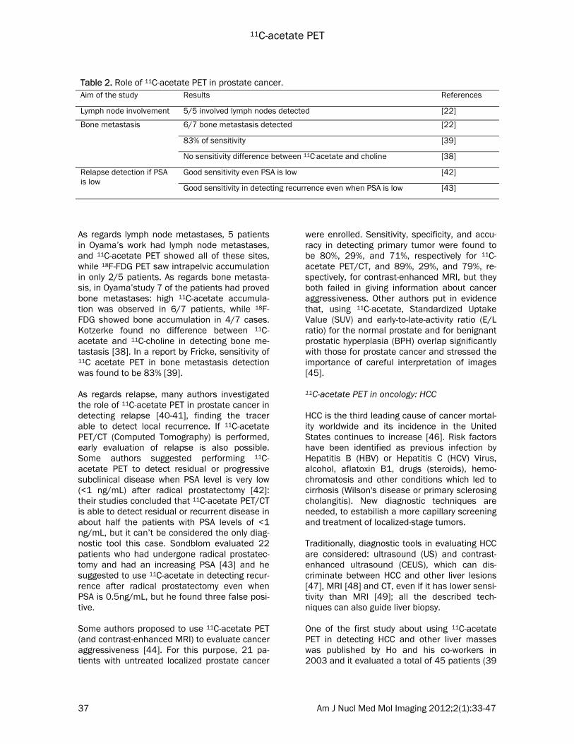
Table 2. Role of 11C-acetate PET in prostate cancer. Aim of the study Results References
Lymph node involvement 5/5 involved lymph nodes detected [22]
Bone metastasis 6/7 bone metastasis detected [22]
83% of sensitivity [39]
No sensitivity difference between 11C-acetate and choline [38]
Relapse detection if PSA is low
Good sensitivity even PSA is low [42]
Good sensitivity in detecting recurrence even when PSA is low [43]
11C-acetate PET
38 Am J Nucl Med Mol Imaging 2012;2(1):33-47
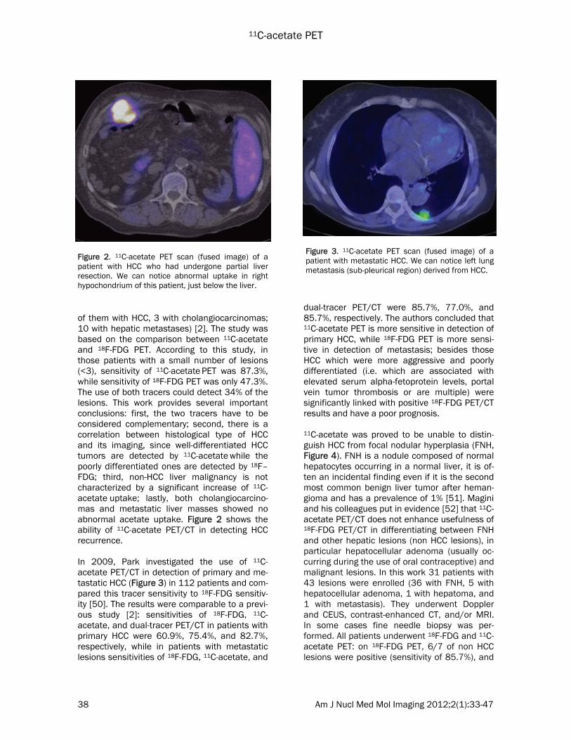
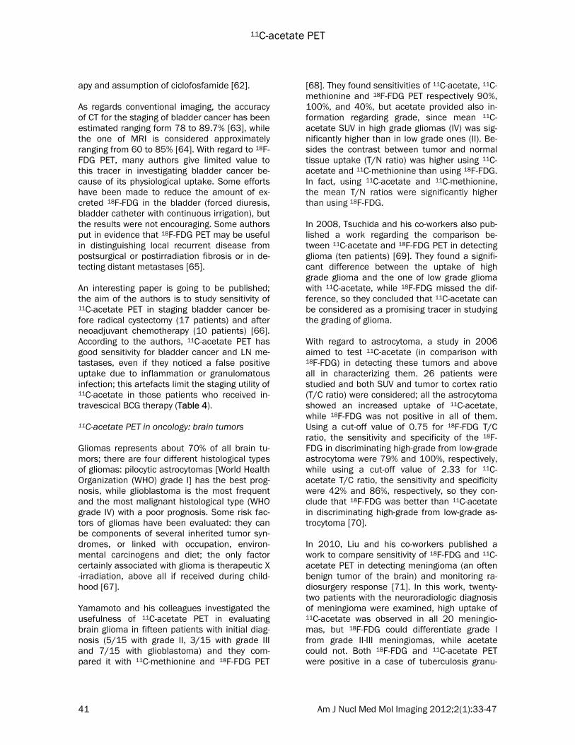
of them with HCC, 3 with cholangiocarcinomas; 10 with hepatic metastases) [2]. The study was based on the comparison between 11C-acetate and 18F-FDG PET. According to this study, in those patients with a small number of lesions (<3), sensitivity of 11C-acetate PET was 87.3%, while sensitivity of 18F-FDG PET was only 47.3%. The use of both tracers could detect 34% of the lesions. This work provides several important conclusions: first, the two tracers have to be considered complementary; second, there is a correlation between histological type of HCC and its imaging, since well-differentiated HCC tumors are detected by 11C-acetate while the poorly differentiated ones are detected by 18F–FDG; third, non-HCC liver malignancy is not characterized by a significant increase of 11C-acetate uptake; lastly, both cholangiocarcino-mas and metastatic liver masses showed no abnormal acetate uptake. Figure 2 shows the ability of 11C-acetate PET/CT in detecting HCC recurrence. In 2009, Park investigated the use of 11C-acetate PET/CT in detection of primary and me-tastatic HCC (Figure 3) in 112 patients and com-pared this tracer sensitivity to 18F-FDG sensitiv-ity [50]. The results were comparable to a previ-ous study [2]: sensitivities of 18F-FDG, 11C-acetate, and dual-tracer PET/CT in patients with primary HCC were 60.9%, 75.4%, and 82.7%, respectively, while in patients with metastatic lesions sensitivities of 18F-FDG, 11C-acetate, and
dual-tracer PET/CT were 85.7%, 77.0%, and 85.7%, respectively. The authors concluded that 11C-acetate PET is more sensitive in detection of primary HCC, while 18F-FDG PET is more sensi-tive in detection of metastasis; besides those HCC which were more aggressive and poorly differentiated (i.e. which are associated with elevated serum alpha-fetoprotein levels, portal vein tumor thrombosis or are multiple) were significantly linked with positive 18F-FDG PET/CT results and have a poor prognosis. 11C-acetate was proved to be unable to distin-guish HCC from focal nodular hyperplasia (FNH, Figure 4). FNH is a nodule composed of normal hepatocytes occurring in a normal liver, it is of-ten an incidental finding even if it is the second most common benign liver tumor after heman-gioma and has a prevalence of 1% [51]. Magini and his colleagues put in evidence [52] that 11C-acetate PET/CT does not enhance usefulness of 18F-FDG PET/CT in differentiating between FNH and other hepatic lesions (non HCC lesions), in particular hepatocellular adenoma (usually oc-curring during the use of oral contraceptive) and malignant lesions. In this work 31 patients with 43 lesions were enrolled (36 with FNH, 5 with hepatocellular adenoma, 1 with hepatoma, and 1 with metastasis). They underwent Doppler and CEUS, contrast-enhanced CT, and/or MRI. In some cases fine needle biopsy was per-formed. All patients underwent 18F-FDG and 11C-acetate PET: on 18F-FDG PET, 6/7 of non HCC lesions were positive (sensitivity of 85.7%), and
Figure 2. 11C-acetate PET scan (fused image) of a patient with HCC who had undergone partial liver resection. We can notice abnormal uptake in right hypochondrium of this patient, just below the liver.
Figure 3. 11C-acetate PET scan (fused image) of a patient with metastatic HCC. We can notice left lung metastasis (sub-pleurical region) derived from HCC.

11C-acetate PET
39 Am J Nucl Med Mol Imaging 2012;2(1):33-47
33/36 FNH finds were true-negative (specificity of 91.7%), while using 11C-acetate PET, only 2/7 of non-HCC disease lesions were positive (sensitivity of 28.6%), and 34/36 FNH finds were true negative (specificity of 94.4%). A study published by Huo suggests to perform a dual time point 11C-acetate PET in order to dis-tinguish FNH from HCC [53]. According to this study, both FNH and HCC are 11C-acetate-avid, but in the first case the uptake decreased, be-ing so lower in late acquisition than in the early one, while in the second case the uptake in-creased, being so lower in the early acquisition than in the late one. In Table 3 main applica-tions of 11C-acetate are summarized.
11C-acetate PET in oncology: RCC RCC, also-called renal adenocarcinoma, arises from the cells of the renal tubule and it is rela-tively rare if compared with other cancers, but an increase in its incidence was observed in the past five decades in the US [54]. If the patients have localized RCC, the prognosis is good, but those with advanced disease do not respond to the majority of traditional treatment options. In the past, RCC was found in patients present-ing with pain in the flank, but now the first diag-nostic step is considered US, while CT and MRI provide information about staging system. Some authors tried to investigate RCC with 18F-FDG PET, for instance, Schöder and his col-league put in evidence that 18F-FDG PET is not useful in diagnosis, staging and recurrence de-tection of renal cell carcinoma, even if it is char-acterized by a high specificity and positive pre-dictive value [55]. Low sensitivity of 18F-FDG is well explained by Kochhar and his colleague [56]: they underline that the tracer is excreted via the urinary tract, but also that, in RCC, expression of GLUT-1 is variable and finally they justified the lack of up-take in some RCC because big tumors are char-acterized by central necrosis. In 1995 Shreve and his co-workers were the first to be interested in using 11C-acetate to de-tect renal tumor [57]. They enrolled 18 patients, who underwent 30-minutes dynamic PET, and noticed that 11C-acetate uptake was quick and that the tracer had not urinary clearance. They also concluded that RCC had 11C-acetate up-
Figure 4. 11C-acetate PET scan (fused image) of a patient with proved FNH. We can notice abnormal uptake of acetate in the liver (VII segment); this find-ing could not be distinguished from a focal HCC.
Table 3. Role of 11C-acetate PET in HCC Aim of the study Results References
Comparison be-tween 11C-acetate and 18F-FDG in detection of liver masses
Sensitivity of 11C-acetate: 87.3%, sensitivity of 18F-FDG 47.3%,34% of lesions are avid of both tracers; the two tracers are complementary (specificity of 100%); 11C-acetate is more useful in well differentiated neoplasm while in cholangiocarcino-mas and in metastatic liver masses no abnormal uptake has been detected
[2]
Detecting primary HCC
11C-acetate is more sensitive (75.4%) in detecting primary HCC than 18F-FDG (60.9%)
[50]
Detecting HCC metastasis
11C-acetate is less sensitive in detecting metastasis (77%) than 18F-FDG (85.7%); the two tracers are complementary
[50]
Distinguishing HCC from FNH
11C-acetate is not useful in distinguish HCC from FNH [52]
Dual point 11C-acetate PET can distinguish HCC from FNH [53]

11C-acetate PET
40 Am J Nucl Med Mol Imaging 2012;2(1):33-47
take image similar to the normal tissue, but the rate of clearance was significantly lower in can-cer cells. Therefore, if the acquisition starts be-yond 10 min of tracer administration, a differen-tiation between neoplastic and normal cells was possible (see Table 4). In 2008, Oyama and his co-workers found a sensitivity of 11C-acetate PET in detecting RCC of 70% (14/20 lesions histologically proven); in particular, papillar carcinoma (1/20) showed a greater uptake than clear-cell carcinoma, while benign cyst turned to be negative. They finally suggest to start acquisition at least 15 minutes after injection, to give the tracer the opportunity to be entrapped in neoplastic cells [58] (Table 4). Figure 5 shows the ability of 11C-acetate PET in detecting bone metastasis deriving from RCC. In 2006 a case report was published about the ability of 11C-acetate PET in detecting a renal oncocytoma, which was incidentally found be-cause prostate cancer was suspected [59]. Re-nal oncocytomas are uncommon and often be-nign tumors of the renal collecting duct, they can be hardly distinguished from RCC using non-invasive methods. More recently, 11C-acetate PET is becoming a tool to evaluate sunitinib response [60]. Sunit-inib is a multitargeted tyrosine kinase inhibitor which turns out to have a promising role in RCC therapy. Madeddu and her colleague published a case report regarding the use of 11C-acetate PET as an early predictor of this therapy re-sponse (Table 4). Other authors tried to evalu-ate therapy response with 18F-FDG PET and they noticed a correlation between decreasing of this
tracer uptake and positive response to therapy, concluding that 18F-FDG uptake is still high in more aggressive tumor [61]. 11C-acetate PET in oncology: bladder carcinoma Bladder cancer is the fourth most common ma-lignancy among western men, following pros-tate, lung, and colon cancers (for 5% to 10% in Europe and the United States). Epidemiological risk factors are considered male sex, smoking, exposure to β-naphthylamine, 4-aminobiphenyl (ABP), benzidine, polychlorinated biphenyls, formaldehyde, asbestos, solvents (benzene, dioxane, and methylene chloride), aluminium, iron, previous urinary tract infections, radiother-
Table 4. Role of 11C-acetate PET in RCC and bladder carcinoma Aim of the study Results References
Detecting primary RCC with 11C-acetate dynamic PET
RCC had 11C-acetate is similar in normal tissue and in RCC, but the rate of clearance is lower in cancer cells, so the acquisition is suggested to be stated beyond 10 min of tracer administration
[57]
Sensitivity of 11C-acetate PET in detecting RCC is 70%; no abnormal uptake is seen in benign cysts
[58]
Evaluating sunitinib re-sponse in RCC
11C-acetate PET is an early predictor of this therapy re-sponse (case report)
[60]
Staging bladder cancer before radical cystectomy and after neoadjuvant che-motherapy
Good sensitivity in detecting bladder cancer and LN metas-tases; false positive uptake can be due to inflammation, infection and previous intravescical BCG therapy
[66
Figure 5. 11C-acetate PET scan (fused image) of a patient with metastatic RCC. The figure shows bone metastasis and in particular we can notice abnormal uptake of the tracer in the left ischium.
11C-acetate PET
41 Am J Nucl Med Mol Imaging 2012;2(1):33-47
apy and assumption of ciclofosfamide [62]. As regards conventional imaging, the accuracy of CT for the staging of bladder cancer has been estimated ranging form 78 to 89.7% [63], while the one of MRI is considered approximately ranging from 60 to 85% [64]. With regard to 18F-FDG PET, many authors give limited value to this tracer in investigating bladder cancer be-cause of its physiological uptake. Some efforts have been made to reduce the amount of ex-creted 18F-FDG in the bladder (forced diuresis, bladder catheter with continuous irrigation), but the results were not encouraging. Some authors put in evidence that 18F-FDG PET may be useful in distinguishing local recurrent disease from postsurgical or postirradiation fibrosis or in de-tecting distant metastases [65]. An interesting paper is going to be published; the aim of the authors is to study sensitivity of 11C-acetate PET in staging bladder cancer be-fore radical cystectomy (17 patients) and after neoadjuvant chemotherapy (10 patients) [66]. According to the authors, 11C-acetate PET has good sensitivity for bladder cancer and LN me-tastases, even if they noticed a false positive uptake due to inflammation or granulomatous infection; this artefacts limit the staging utility of 11C-acetate in those patients who received in-travescical BCG therapy (Table 4). 11C-acetate PET in oncology: brain tumors Gliomas represents about 70% of all brain tu-mors; there are four different histological types of gliomas: pilocytic astrocytomas [World Health Organization (WHO) grade I] has the best prog-nosis, while glioblastoma is the most frequent and the most malignant histological type (WHO grade IV) with a poor prognosis. Some risk fac-tors of gliomas have been evaluated: they can be components of several inherited tumor syn-dromes, or linked with occupation, environ-mental carcinogens and diet; the only factor certainly associated with glioma is therapeutic X-irradiation, above all if received during child-hood [67]. Yamamoto and his colleagues investigated the usefulness of 11C-acetate PET in evaluating brain glioma in fifteen patients with initial diag-nosis (5/15 with grade II, 3/15 with grade III and 7/15 with glioblastoma) and they com-pared it with 11C-methionine and 18F-FDG PET
[68]. They found sensitivities of 11C-acetate, 11C-methionine and 18F-FDG PET respectively 90%, 100%, and 40%, but acetate provided also in-formation regarding grade, since mean 11C-acetate SUV in high grade gliomas (IV) was sig-nificantly higher than in low grade ones (II). Be-sides the contrast between tumor and normal tissue uptake (T/N ratio) was higher using 11C-acetate and 11C-methionine than using 18F-FDG. In fact, using 11C-acetate and 11C-methionine, the mean T/N ratios were significantly higher than using 18F-FDG. In 2008, Tsuchida and his co-workers also pub-lished a work regarding the comparison be-tween 11C-acetate and 18F-FDG PET in detecting glioma (ten patients) [69]. They found a signifi-cant difference between the uptake of high grade glioma and the one of low grade glioma with 11C-acetate, while 18F-FDG missed the dif-ference, so they concluded that 11C-acetate can be considered as a promising tracer in studying the grading of glioma. With regard to astrocytoma, a study in 2006 aimed to test 11C-acetate (in comparison with 18F-FDG) in detecting these tumors and above all in characterizing them. 26 patients were studied and both SUV and tumor to cortex ratio (T/C ratio) were considered; all the astrocytoma showed an increased uptake of 11C-acetate, while 18F-FDG was not positive in all of them. Using a cut-off value of 0.75 for 18F-FDG T/C ratio, the sensitivity and specificity of the 18F-FDG in discriminating high-grade from low-grade astrocytoma were 79% and 100%, respectively, while using a cut-off value of 2.33 for 11C-acetate T/C ratio, the sensitivity and specificity were 42% and 86%, respectively, so they con-clude that 18F-FDG was better than 11C-acetate in discriminating high-grade from low-grade as-trocytoma [70]. In 2010, Liu and his co-workers published a work to compare sensitivity of 18F-FDG and 11C-acetate PET in detecting meningioma (an often benign tumor of the brain) and monitoring ra-diosurgery response [71]. In this work, twenty-two patients with the neuroradiologic diagnosis of meningioma were examined, high uptake of 11C-acetate was observed in all 20 meningio-mas, but 18F-FDG could differentiate grade I from grade II-III meningiomas, while acetate could not. Both 18F-FDG and 11C-acetate PET were positive in a case of tuberculosis granu-

11C-acetate PET
42 Am J Nucl Med Mol Imaging 2012;2(1):33-47
loma; 11C-acetate performed better in monitor-ing five patients who had received gamma-knife surgery. This work also provides an explanation to the uptake mechanism of 11C-acetate in these tumors: probably it is metabolized by as-trocytes and it is quickly incorporated into gluta-mate and glutamine within the first 15 minutes from injection, while after 30 minute it is used for FFA synthesis. To have a synoptical vision of 11C-acetate PET usefulness in investigating brain tumors, see Table 5. Other 11C-acetate PET clinical applications Multiple myeloma is a rare neoplasm (accounting for about 0.8% of all new cancer cases) originating from plasma cells; it is usually investigated with routine laboratory exams, bone marrow examination, conventional radiog-raphy of the bone region suspected to be in-volved, MRI and CT, but also 18F-FDG PET [72]. A recent case report relates about a multiple myeloma incidentally found by 11C-acetate PET, in a man who was affected by HCC [73]. Lung cancer is the leading cause of cancer-related mortality not only in the United States but also around the world; risk factors can be
considered cigarette smoking (active and pas-sive), pollution, professional exposure to silica or asbesto, genetic factors, lack of physical ac-tivity, a diet poor in vitamins [74]. It is usually classified in non small cell lung cancer (NSLC, 85% of all lung cancers in the US) and small cell lung cancer; among NSLC, bronchioloalveolar carcinoma accounts for less than 3% of all lung cancer and it is more frequent in male [75], while lung adenocarcinoma is the most frequent histological type in females (smokers or non-smokers) and in non-smoking males and its incidence trend seems to be increasing even if there are many geographical differences [76]. A paper published by Shibata and co-workers evaluated the usefulness of 11C-acetate PET for lung adenocarcinoma imaging and in particular to evaluate its aggressiveness [3]. They com-pared these results with 18F-FDG PET (Table 6). According to the authors, 11C-acetate PET has a good sensitivity (better than 18F-FDG) in detect-ing bronchioloalveolar carcinoma and well-differentiated adenocarcinoma (stage IA) and there is also a significant correlation between ki67 staining scores and tracer uptake, while 18F-FDG uptake is superior in tumors with patho-logical advanced stages (lymphatic, vascular
Table 5. Role of 11C-acetate PET brain tumors Aim of the study Results References
Evaluating usefulness of 11C-acetate PET in detecting brain glioma, in comparison with 11C-methionine and 18F-FDG
Sensitivity of 11C-acetate is 90%, lower than 11C-methionine (100%) but higher than 18F-FDG one (40%).
[68]
Evaluating usefulness of 11C-acetate PET in grading brain glioma, in comparison with 11C-methionine and 18F-FDG
11C-acetate can provide information about tumor grading, since mean SUV in high grade gliomas is significantly higher than in low grade ones
[68]
11C-acetate showed a significant difference between the uptake of high grade glioma and the one of low grade glioma while 18F-FDG does not.
[69]
Evaluating usefulness of 11C-acetate PET in grading brain astrocytoma, in comparison with 18F-FDG
Sensitivity and specificity of the 18F-FDG in discrimi-nating high-grade from low-grade astrocytoma were 79% and 100%, respectively, while for 11C-acetate sensitivity and specificity were 42% and 86%;18F-FDG is more accurate in grading brain astrocytoma
[70
Evaluating usefulness of 11C-acetate PET in detecting meningioma
Good sensitivity of 11C-acetate [71
Evaluating usefulness of 11C-acetate PET in grading meningioma
18F-FDG could differentiate grade I from grade II-III meningiomas, while 11C acetate could not.
[71
Evaluating usefulness of 11C-acetate PET in monitoring radiosurgery response in men-ingioma
11C-acetate performed better than 18F-FDG in moni-toring radiosurgery response
[71

11C-acetate PET
43 Am J Nucl Med Mol Imaging 2012;2(1):33-47
and/or pleural involvements). A multicentric study involving seven Japanese institutes suggested that 11C-acetate can substi-tute 18F-FDG in the imaging of differentiated adenocarcinoma, since 18F-FDG sensitivity is lower in these cases [77]. The authors tried to give an explanation to this result: well differenti-ated adenocarcinoma have a slow glucose me-tabolism, while membrane lipid synthesis is rapid. In 2006 Ohtsuka and colleagues reported three cases of thymoma (a rare and often benign tu-mor originating from thymus cells and usually detected by CT), investigated with 11C-acetate PET [78]. All of three patients with thymoma had a positive scan with 11C-acetate, while 18F-FDG PET scan missed one case. The uptake mecha-nism of acetate is not clear in thymoma, even if the authors supposed it is similar to the other tumors uptake and different from the myocar-dial one. A case report dealing with a patient who had amnesia and syncope episodes and whose MRI was inconclusive (encephalitis or glioma?) was published in 2009 [79]. 18F-FDG PET was per-formed, and it was still inconclusive. Finally a 11C-acetate PET was performed and it did not demonstrate any abnormal uptake, thus sug-gesting the inflammatory (and not neoplastic) nature of the illness. In the same year Lee and his co-workers published another case report describing the usefulness of 11C-acetate in de-tecting cerebellopontine angle Schwannoma, a very rare tumor, occurring in a ten years old child; the tracer was found to be able to detect the recurrence of the tumor [80]. In 2011 Ho and his colleagues published a case report regarding a multicentric angiomyolipoma
of the kidney [81]; the diagnosis was not possi-ble with CT and 18F-FDG PET, while using 11C-acetate PET/CT identified an exophytic lesion in the left kidney and left para-aortic nodes. In Table 7 incidental findings with 11C-acetate PET are summarized. Conclusion The large number of published works demon-strates a clear interest in development of ace-tate PET. 11C is a short half life isotope and needed to be produced on site, so 11C-acetate is not readily available; nonetheless many centres started to use 11C-acetate. It is probably due to its versatility, since into the cells it is can go through a double way, the catabolic one (via TCA cycle), which made it useful for cardiologic studies, and an anabolic one (via FAS), which made it useful for oncological purpose. Many neoplasm with a relatively low grade of proliferation, as like as well differentiated HCC or lung carcinomas, are not 18F-FDG-avid, so in these cases a 11C-acetate PET has been sug-gested; on the contrary, acetate uptake is less dependent from inflammatory states than 18F FDG, thus permitting an easier differential diag-nosis between neoplasm and inflammation. In our opinion, 11C-acetate PET should be consid-
Table 6. Role of 11C-acetate PET in lung cancer Aim of the study Results References
Evaluating usefulness of 11C-acetate PET in detecting bron-chioloalveolar and well differenti-ated adenocarcinoma
11C-acetate PET has a good sensitivity (better than 18F-FDG one) in detecting bronchioloalveolar carcinoma and well-differentiated adenocarcinoma (stage IA)
[3]
11C-acetate is more sensitive than 18F-FDG for detecting differ-entiated adenocarcinoma.
[77]
Evaluating usefulness of 11C-acetate PET in detecting aggres-siveness of lung adenocarcinoma
18F-FDG uptake is superior in tumors with pathological ad-vanced stages (lymphatic, vascular and/or pleural involve-ments).
[3]
Table 7. Incidental findings with 11C-acetate PET Type of malignancy References
Multiple myeloma [73]
Thymoma [78]
Cerebellopontine angle schwannoma [80]
Angiomyolipoma of the kidney [81]

11C-acetate PET
44 Am J Nucl Med Mol Imaging 2012;2(1):33-47
ered as a promising tracer, alone or combining with other tracers. Address correspondence to: Dr. Ilaria Grassi, Medicina Nucleare, Policlinico Sant’Orsola- Malpighi, via Massarenti 9, 40138 Bologna Tel: +39 0516363196, +39 0516363185; Fax: 051/6363956; E-mail: [email protected] References [1] de Beco V, Le Bars D, Scherrmann JM.
18Fluorine in radiopharmacy. Ann Pharm Fr 2008; 66: 60-65.
[2] Ho CL, Yu SC, Yeung DW. 11C-acetate PET im-aging in hepatocellular carcinoma and other liver masses. J Nucl Med 2003; 44: 213-21.
[3] Shibata H, Nomori H, Uno K, Iyama K, Tomiyoshi K, Nakashima R, Sakaguchi K, Goya T, Takanami I, Koizumi K, Suzuki T, Kaji M, Horio H. 11C-acetate for positron emission tomography imaging of clinical stage IA lung adenocarcinoma: comparison with 18F-fluoro deoxyglucose for imaging and evaluation of tumor aggressiveness. Ann Nucl Med 2009; 23: 609-16.
[4] Hain SF, Maisey MN. Positron emission tomo-graphy for urological tumors. BJU Int 2003; 92: 159-64.
[5] Krause BJ, Souvatzoglou M, Treiber U. Imaging of prostate cancer with PET/CT and radioac-tively labeled choline derivates. Urol Oncol 2011 (in press).
[6] Murphy RC, Kawashima A, Peller PJ. The Utility of 11C-Choline PET/CT for Imaging Prostate Cancer: A Pictorial Guide. AJR Am J Roentgenol 2011; 196: 1390-8.
[7] Bouchelouche K, Oehr P. Positron emission tomography and positron emission tomogra-phy/computerized tomography of urological malignancies: an update review. J Urol 2008; 179: 34-45.
[8] Shinoda J, Asano Y, Yano H. Usability of 11C-methionine PET in diagnosis of glioma. Gan To Kagaku Ryoho 2010; 37: 1027-33.
[9] Okita Y, Kinoshita M, Goto T, Kagawa N, Ki-shima H, Shimosegawa E, Hatazawa J, Hashi-moto N, Yoshimine T. 11C-methionine uptake correlates with tumor cell density rather than with microvessel density in glioma: A stereo-tactic image-histology comparison. Neuroi-mage 2010; 49: 2977-82.
[10] Randle PJ, England PJ, Denton RM. Control of the tricarboxylate cycle and its interactions with glycolysis during acetate utilization in rat heart. Biochem J 1970; 117: 677-95.
[11] Swinnen JV, Heemers H, Deboel L, Foufelle F, Heyns W, Verhoeven G. Stimulation of tumor-associated fatty acid synthase expression by growth factor activation of the sterol regula-tory element-binding protein pathway.
Oncogene 2000; 19: 5173-81. [12] Swinnen JV, Van Veldhoven PP, Timmermans
L, De Schrijver E, Brusselmans K, Vanderhoydonc F, Van de Sande T, Heemers H, Heyns W, Verhoeven G. Fatty acid synthase drives the synthesis of phospholipids partition-ing into detergent-resistant membrane micro-domains. Biochem Biophys Res Commun 2003; 302: 898-903.
[13] Yoshimoto M, Waki A, Yonekura Y, Sadato N, Murata T, Omata N, Takahashi N, Welch MJ, Fujibayashi Y. Characterization of acetate metabolism in tumor cells in relation to cell proliferation: acetate metabolism in tumor cells. Nucl Med Biol 2001; 28: 117-22.
[14] Vāvere AL, Kridel SJ, Wheeler FB, Lewis JS. 11C-acetate as a PET radiopharmaceutical for imaging fatty acid synthase expression in prostate cancer. Nucl Med 2008; 49: 327-34.
[15] Schiepers C, Hoh CK, Nuyts J, Seltzer M, Wu C, Huang SC, Dahlbom M. 11C-acetate kinetics of prostate cancer. J Nucl Med 2008; 49: 206-15.
[16] Brusselmans K, De Schrijver E, Verhoeven G, Swinnen JV. RNA interference-mediated si-lencing of the acetyl-CoA-carboxylase-alpha gene induces growth inhibition and apoptosis of prostate cancer cells. Cancer Res 2005; 65: 6719-25.
[17] Kruijer PS, Ter Linden T, Mooij R, Visser FC, Herscheid JDM. A practical method for the preparation of 11C-acetate. Appl Radiat Isot 1995; 46: 317-21.
[18] Moerlein SM, Gaehle GG, Welch MJ. Robotic preparation of Sodium Acetate 11C Injection for use in clinical PET. Nucl Med Biol 2002; 29: 613-21.
[19] Roeda D, Dolle F, Crouzel C. An improvement of 11C-acetate synthesis-non-radioactive con-taminants by irradiation-induced species ema-nating from the 11C carbon dioxide production target. Appl Radiat Isot 2002; 57: 857-60.
[20] Seltzer MA, Jahan SA, Sparks R, Stout DB, Satyamurthy N, Dahlbom M, Phelps ME, Barrio JR. Radiation dose estimates in humans for 11C-acetate whole-body PET. J Nucl Med 2004; 45: 1233-6.
[21] AK Solomon. Half-Life of C11. Phys Rev 1941; 60: 279.
[22] Oyama N, Akino H, Kanamaru H, Suzuki Y, Muramoto S, Yonekura Y, Sadato N, Yama-moto K, Okada K. 11C-acetate PET Imaging of Prostate Cancer. J Nucl Med 2002; 43: 181-6.
[23] Klein LJ, Visser FC, Nurmohamed SA, Vink A, Peters JH, Knaapen P, Kruijer PS, Herscheid JD, Teule GJ, Visser CA. Feasibility of planar myocardial 11C-acetate imaging. J Nucl Cardiol 2000; 7: 221-7.
[24] Sörensen J, Valind S, Andersson LG. Simulta-neous quantification of myocardial perfusion, oxidative metabolism, cardiac efficiency and pump function at rest and during supine bicy-

11C-acetate PET
45 Am J Nucl Med Mol Imaging 2012;2(1):33-47
cle exercise using 11C-acetate PET-a pilot study. Clin Physiol Funct Imaging 2010; 30: 279-84.
[25] Ho CL, Yu SC, Yeung DW. 11C-acetate PET im-aging in hepatocellular carcinoma and other liver masses. J Nucl Med 2003; 44: 213-21.
[26] Brown M, Marshall DR, Sobel BE, Bergmann SR. Delineation of myocardial oxygen utiliza-tion with 11C-labeled acetate. Circulation 1987; 76: 687-96.
[27] Brown MA, Myears DW, Bergmann SR. Validity of estimates of myocardial oxidative metabo-lism with 11C- acetate and positron emission tomography despite altered patterns of sub-strate utilization. J Nucl Med 1989; 30: 187-93.
[28] Lear JL. Relationship between myocardial clearance rates of 11C-acetate-derived radio-label and oxidative metabolism: physiologic basis and clinical significance. J Nucl Med 1991; 32: 1957-60.
[29] Timmer SA, Lubberink M, Germans T, Götte MJ, ten Berg JM, ten Cate FJ, van Rossum AC, Lammertsma AA, Knaapen P. Potential of 11C-acetate for measuring myocardial blood flow: Studies in normal subjects and patients with hypertrophic cardiomyopathy. J Nucl Cardiol 2010; 17: 264-75.
[30] Sörensen J, Valind S, Andersson LG. Simulta-neous quantification of myocardial perfusion, oxidative metabolism, cardiac efficiency and pump function at rest and during supine bicy-cle exercise using 11C-acetate PET-a pilot study. Clin Physiol Funct Imaging 2010; 30: 279-84.
[31] Arakawa K, Kudo T, Ikawa M, Morikawa N, Kawai Y, Sahashi K, Lee JD, Kuriyama M, Miyamori I, Okazawa H, Yoneda M. Abnormal myocardial energy-production state in mito-chondrial cardiomyopathy and acute response to L-arginine infusion. 11C-acetate kinetics revealed by positron emission tomography. Circ J 2010; 74: 2702-11.
[32] Sarma AV, Schottenfeld D. Prostate cancer incidence, mortality, and survival trends in the United States: 1981-2001. Semin Urol Oncol 2002; 20: 3-9.
[33] Hsing AW, Chokkalingam AP. Prostate cancer epidemiology. Front Bio sci 2006; 11: 1388-1413.
[34] Heijmink SW, Fütterer JJ, Strum SS, Oyen WJ, Frauscher F, Witjes JA, Barentsz JO. State-of-the-art uroradiologic imaging in the diagnosis of prostate cancer. Acta Oncol 2011; 50: 25-38.
[35] Bonekamp D, Jacobs MA, El-Khouli R, Stoianovici D, Macura KJ. Advancements in MR Imaging of the Prostate: From Diagnosis to Interventions. Radiographics 2011; 31: 677-703.
[36] Defeo EM, Wu CL, McDougal WS, Cheng LL. A decade in prostate cancer: from NMR to me-
tabolomics. Nat Rev Urol 2011 (in press). [37] Hara T, Kosaka N, Kishi H. PET imaging of
prostate cancer using 11C-choline. J Nucl Med 1998; 39: 990-5.
[38] Kotzerke J, Volkmer BG, Neumaier B, Gschwend JE, Hautmann RE, Reske SN. Car-bon-11 acetate positron emission tomography can detect local recurrence of prostate can-cer. Eur J Nucl Med Mol Imaging 2002; 29: 1380-84.
[39] Fricke E, Machtens S, Hofmann M, van den Hoff J, Bergh S, Brunkhorst T, Meyer GJ, Karstens JH, Knapp WH, Boerner AR. Positron emission tomography with 11C-acetate and 18F-FDG in prostate cancer patients. Eur J Nucl Med Mol Imaging 2003; 30: 607-11.
[40] Reske SN, Blumstein NM, Glatting G. PET and PET/CT in relapsing prostate carcinoma. Urologe A 2006; 45: 1240, 1242-1244, 1246-1248, 1250.
[41] Martino P, Scattoni V, Galosi AB, Consonni P, Trombetta C, Palazzo S, Maccagnano C, Liguori G, Valentino M, Battaglia M, Barozzi L. Role of imaging and biopsy to assess local recurrence after definitive treatment for pros-tate carcinoma (surgery, radiotherapy, cryotherapy, HIFU). World J Urol 2011 (in press).
[42] Vees H, Buchegger F, Albrecht S, Khan H, Husarik D, Zaidi H, Soloviev D, Hany TF, Miral-bell R. 18F-choline and/or 11C-acetate posi-tron emission tomography: detection of resid-ual or progressive subclinical disease at very low prostate-specific antigen values (<1 ng/mL) after radical prostatectomy. BJU Int 2007; 99: 1415-20.
[43] Sandblom G, Sörensen J, Lundin N, Häggman M, Malmström PU. Positron emission tomogra-phy with 11C-acetate for tumor detection and localization in patients with prostate-specific antigen relapse after radical prostatectomy. Urology 2006; 67: 996-1000.
[44] Jambor I, Borra R, Kemppainen J, Lepomäki V, Parkkola R, Dean K, Alanen K, Arponen E, Nurmi M, Aronen HJ, Minn H. Functional imag-ing of localized prostate cancer aggressive-ness using 11C-acetate PET/CT and 1H-MR spectroscopy. J Nucl Med 2010; 51: 1676-83.
[45] Kato T, Tsukamoto E, Kuge Y, Takei T, Shiga T, Shinohara N. Accumulation of 11C-acetate in normal prostate and benign prostatic hyper-plasia: comparison with prostate cancer. Eur J Nucl Med Mol Imaging 2002; 29: 1492-5.
[46] Altekruse SF, McGlynn KA, Reichman ME. Hepatocellular carcinoma incidence, mortality, and survival trends in the United States from 1975 to 2005. J Clin Oncol 2009; 27: 1485-91.
[47] Martie A, Sporea I, Popescu A, Sirli R, Dănilă M, Serban C, Ardelean M, Bota S, Sendroiu M, Chisevescu D. Contrast enhanced ultrasound for the characterization of hepatocellular car-

11C-acetate PET
46 Am J Nucl Med Mol Imaging 2012;2(1):33-47
cinoma. Med Ultrason 2011; 13: 108-13. [48] Bargellini I. Hepatocellular carcinoma: MR
staging and therapeutic decisions. Abdom Imaging 2011 (in press).
[49] Rode A. Radiological diagnosis of hepatocellu-lar carcinoma in 2010. Cancer Radiother 2011; 15: 7-12.
[50] Park JW, Kim JH, Kim SK, Kang KW, Park KW, Choi JI, Lee WJ, Kim CM, Nam BH. A prospec-tive evaluation of 18F-FDG and 11C-acetate PET/CT for detection of primary and metas-tatic hepatocellular carcinoma. J Nucl Med 2008; 49: 1912-21.
[51] Leclera, Arrivé L. Focal nodular hyperplasia. Clin Res Hepatol Gastroenterol 2011; 35: 159-60.
[52] Magini G, Farsad M, Frigerio M, Serra C, Colecchia A, Jovine E, Vivarelli M, Feletti V, Golfieri R, Patti C, Fanti S, Franchi R, Lodi F, Boschi S, Bernardi M, Trevisani F. 11C-acetate does not enhance usefulness of F-18 FDG PET/CT in differentiating between focal nodu-lar hyperplasia and hepatic adenoma. Clin Nucl Med 2009; 34: 659-65.
[53] Huo L, Wu Z, Zhuang H, Fu Z, Dang Y. Dual time point 11C-acetate PET imaging can poten-tially distinguish focal nodular hyperplasia from primary hepatocellular carcinoma. Clin Nucl Med 2009; 34: 874-7.
[54] Drucker BJ. Renal cell carcinoma: current status and future prospects. Cancer Treat Rev 2005; 31: 536-45.
[55] Schöder H, Larson SM. Positron emission tomography for prostate, bladder, and renal cancer. Semin Nucl Med 2004; 34: 274-92.
[56] Kochhar R, Brown RK, Wong CO, Dun-nick NR, Frey KA, Manoharan P Role of FDG PET/CT in imaging of renal lesions. J Med Imaging Radiat Oncol 2010; 54: 347-57.
[57] Shreve P, Chiao PC, Humes HD, Schwaiger M, Gross MD. 11C-acetate PET imaging in renal disease. J Nucl Med 1995; 36: 1595-1601.
[58] Oyama N, Okazawa H, Kusukawa N, Kaneda T, Miwa Y, Akino H, Fujibayashi Y, Yonekura Y, Welch MJ, Yokoyama O. 11C-acetate PET imag-ing for renal cell carcinoma. Eur J Nucl Med Mol Imaging 2009; 36: 422-7.
[59] Shriki J, Murthy V, Brown J. Renal oncocytoma on 11C-acetate positron emission tomography: Case report and literature review. Mol Imag Biol 2006; 8: 208-11.
[60] Maleddu A, Pantaleo MA, Castellucci P, Astorino M, Nanni C, Nannini M, Busato F, Di Battista M, Farsad M, Lodi F, Boschi S, Fanti S, Biasco G. 11C-acetate PET for early prediction of sunitinib response in metastatic renal cell carcinoma. Tumori 2009; 95: 382-4.
[61] ME, Winge Main AK, Hagen G, Fjeld JG, Fosså SD, Lilleby W. Combined positron emission tomography/computed tomography in sunit-
inib therapy assessment of patients with me-tastatic renal cell carcinoma. Clin Oncol (R Coll Radiol) 2011; 23: 339-43.
[62] Kirkali Z, Chan T, Manoharan M, Algaba F, Busch C, Cheng L, Kiemeney L, Kriegmair M, Montironi R, Murphy WM, Sesterhenn IA, Ta-chibana M, Weider J. Bladder cancer: epidemi-ology, staging and grading, and diagnosis. Urology 2005; 66: 4-34.
[63] Knox MK, Rivers Bowerman MD, Bardgett HP, Cowan NC. Multidetector computed tomogra-phy with triple-bolus contrast medium admini-stration protocol for preoperative anatomical and functional assessment of potential living renal donors. Eur Radiol 2010; 20: 2590-9.
[64] Tekes A, Kamel I, Imam K, Szarf G, Schoenberg M, Nasir K, Thompson R, Bluemke D. Dynamic MRI of bladder cancer: evaluation of staging accuracy. Am J Roentgenol 2005; 184: 121-7.
[65] Bouchelouche K, Oehr P. Positron emission tomography and positron emission tomogra-phy/computerized tomography of urological malignancies: an update review. J Urol 2008; 179: 34-45.
[66] Schöder H, Ong SC, Reuter VE, Cai S, Burnazi E, Dalbagni G, Larson SM, Bochner BH. Initial Results with 11C-acetate Positron Emission Tomography/Computed Tomography (PET/CT) in the Staging of Urinary Bladder Cancer. Mol Imaging Biol 2011 (in press).
[67] Ohgaki H. Epidemiology of brain tumors. Methods Mol Biol 2009; 472: 323-42.
[68] Yamamoto Y, Nishiyama Y, Kimura N, Kameyama R, Kawai N, Hatakeyama T, Kaji M, Ohkawa M. 11C-acetate PET in the evaluation of brain glioma: comparison with 11C-methionine and 18F-FDG-PET. Mol Imaging Biol 2008; 10: 281-7.
[69] Tsuchida T, Takeuchi H, Okazawa H, Tsujikawa T, Fujibayashi Y. Grading of brain glioma with 11C-acetate PET: comparison with 18F-FDG PET. Nucl Med Biol 2008; 35: 171-6.
[70] Liu RS, Chang CP, Chu LS, Chu YK, Hsieh HJ, Chang CW, Yang BH, Yen SH, Huang MC, Liao SQ, Yeh SH. PET imaging of brain astrocytoma with 11C-acetate. Eur J Nucl Med Mol Imaging 2006; 33: 420-7.
[71] Liu RS, Chang CP, Guo WY, Pan DH, Ho DM, Chang CW Kim SK. 11C-acetate versus 18F-FDG PET in detection of meningioma and monitor-ing the effect of gamma-knife Radiosurgery. J Nucl Med 2010; 51: 883-91.
[72] Palumbo A, Anderson K. Multiple myeloma. N Engl J Med 2011; 364: 1046-60.
[73] Lee SM, Kim TS, Lee JW, Kwon HW, Kim YI, Kang SH, Kim SK. Incidental finding of an 11C-acetate PET-positive multiple myeloma. Ann Nucl Med 2010; 24: 41-4.
[74] Molina JR, Jang P. cassivi SD, Schild SE, Adjei. Non-Small Cell Lung Cancer: Epidemiology, Risk Factors, Treatment, and Survivorship.
11C-acetate PET
47 Am J Nucl Med Mol Imaging 2012;2(1):33-47
Mayo Clin Proc 2008; 83: 584-94. [75] Falk RT, Pickle LW, Fontham ET, Greenberg
SD, Jacobs HL, Correa P, Fraumeni JF Jr. Epi-demiology of bronchioloalveolar carcinoma. Cancer Epidemiol Biomarkers Prev 1992; 1: 339-44.
[76] Charloux A, Quoix E, Wolkove N, Small D, Pauli G, Kreisman H. The increasing incidence of lung adenocarcinoma: reality or artefact? A review of the epidemiology of lung adenocarci-noma. Int J Epidemiol 1996; 28: 14-23.
[77] Nomori H, Shibata H, Uno K, Iyama K, Honda Y, Nakashima R, Sakaguchi K, Goya T, Taka-nami I, Koizumi K, Suzuki T, Kaji M, Horio H. 11C-acetate can be used in place of 18F-fluorodeoxyglucose for positron emission to-mography imaging of non-small cell lung can-cer with higher sensitivity for well-differentiated adenocarcinoma. J Thorac On-col 2008; 3: 1427-32.
[78] Ohtsuka T, Nomori H, Watanabe K, Naruke T, Suemasu K, Kosaka N, Uno K. Positive imag-ing of thymoma by 11C-acetate positron emis-sion tomography. Ann Thor Surg 2006; 81: 1132-4.
[79] Wang HC, Zhao J, Zuo CT, Zhang ZW, Xue FP, Liu P, Hua FC, Tan HB, Guan YH. Encephalitis depicted by a combination of 11C-acetate and F-18 FDG PET/CT. Clin Nucl Med 2009; 34: 952-4.
[80] Lee SM, Kim TS, Kim SK. Cerebellopontine Angle Schwannoma on 11C-acetate PET/CT. Clin Nucl Med 2009; 34: 831-3.
[81] Ho CL, Chen S, Ho KMT, Ng WK, Leung YL, Cheng TKC. 11C-acetate PET/CT in Multicentric Angiomyolipoma of the Kidney. Clin Nucl Med 2011; 36: 407-8.

![EEssttuuddoo ccoommbbiinnaaddoo ddee PPEETT 1 ccoomm … Barroca.pdf · perform Positron Emission Tomography (PET) with the radiotracer [11C]PiB that binds to the Aβ plaques. Recently,](https://img.pdfslide.us/doc/110x75/5e81c5a637b0ea2762150a9b/eessttuuddoo-ccoommbbiinnaaddoo-ddee-ppeett-1-ccoomm-perform-positron-emission.jpg)

![Cellular/Molecular FunctionalCharacterizationof5-HT1B … · 2017. 10. 28. · hemodynamics. We used PET-MR imaging with the PET radio-ligand [11C]AZ10419369 administered as a bolus](https://img.pdfslide.us/doc/110x75/60ad0aed6329c946012475a8/cellularmolecular-functionalcharacterizationof5-ht1b-2017-10-28-hemodynamics.jpg)

![Use of [ C]Choline PET-CT as a Noninvasive Method …clincancerres.aacrjournals.org/content/clincanres/17/24/7673.full.pdf · Imaging, Diagnosis, Prognosis Use of [11C]Choline PET-CT](https://img.pdfslide.us/doc/110x75/5b5c867e7f8b9a68368cf226/use-of-ccholine-pet-ct-as-a-noninvasive-method-imaging-diagnosis-prognosis.jpg)

![APPLICATION IN PET RADIOCHEMISTRY. - Politecnico di … · 1.5.2 Nucleophilic fluorination ... Figure 1.13. [18F]FECH and [11C]Choline uptake mechanism . Figure 1.14. Radiolabelled](https://img.pdfslide.us/doc/110x75/5af4e91e7f8b9a190c8da922/application-in-pet-radiochemistry-politecnico-di-nucleophilic-fluorination.jpg)


![spiral.imperial.ac.uk · Web viewSeventeen healthy male volunteers received [11C]CIMBI-36 PET scans before and 3 hours after an oral dose of d-amphetamine (0.5 mg/kg). Dynamic PET](https://img.pdfslide.us/doc/110x75/61495146080bfa6260148833/web-view-seventeen-healthy-male-volunteers-received-11ccimbi-36-pet-scans-before.jpg)


![[11C]docetaxel PET studies in lung cancer patients](https://img.pdfslide.us/doc/110x75/587e016b1a28abd44b8b891f/11cdocetaxel-pet-studies-in-lung-cancer-patients.jpg)

![Multimodal analysis using [11C]PiB-PET/MRI for functional](https://img.pdfslide.us/doc/110x75/61ff326588a357094244a349/multimodal-analysis-using-11cpib-petmri-for-functional-.jpg)