Embed Size (px)
Citation preview
Hindawi Publishing CorporationGastroenterology Research and PracticeVolume 2011, Article ID 917941, 9 pagesdoi:10.1155/2011/917941
Review Article
Radiation Proctitis: Current Strategies in Management
Nhue L. Do, Deborah Nagle, and Vitaliy Y. Poylin
Division of Colon and Rectal Surgery, Beth Israel Deaconess Medical Center, Harvard Medical School Boston, 330 Brookline Avenue,Stoneman 9, Boston, MA 02215, USA
Correspondence should be addressed to Vitaliy Y. Poylin, [email protected]
Received 31 January 2011; Revised 15 June 2011; Accepted 23 August 2011
Academic Editor: Jean S. Wang
Copyright © 2011 Nhue L. Do et al. This is an open access article distributed under the Creative Commons Attribution License,which permits unrestricted use, distribution, and reproduction in any medium, provided the original work is properly cited.
Radiation proctitis is a known complication following radiation therapy for pelvic malignancy. The majority of cases are treatednonsurgically, and an understanding of the available modalities is crucial in the management of these patients. In this paper, wefocus on the current treatments of radiation proctitis.
1. Introduction
Radiation causes both apoptosis and cell death secondaryto damage to DNA, proteins, and lipids and usually affectsrapidly proliferating cells such as those found in cancer [1].Today, radiation to the pelvis is an important constituentin the treatment of pelvic malignancies and is administeredeither as neoadjuvant or adjuvant therapy. Initially, radiationtherapy (RT) was believed to provide no benefit for patients,especially patients with rectal cancer, due to the resistanceof these cancers to radiation. Only later was it discoveredthat higher doses were required to overcome that resistance.However, higher doses of radiation have resulted in collateraldamage to organs in or surrounding the field of radiation.Currently, the most frequent complication of radiation ther-apy, secondary to the utilization of higher doses, is proctitisafter treatment for prostate cancer [2].
Radiation therapy can be administered via external beamradiation or radioactive implants termed brachytherapy. Ex-ternal beam radiation is typically administered by an externalphoton generator with a variety of sources including gammarays, electron beams, and X-rays via a four-beam approachwhich results in significant exposure to surrounding organs[3]. Particular to prostate cancer treatment, certain regions ofthe rectum receive a dose similar to, if not equal to, the dosedelivered to the prostate. Newer modalities of external beamradiation delivery attempt to deliver equivocal or higher dos-es but with decreased toxicity to surrounding areas; these
modalities include three-dimensional conformal radiationtherapy (3D-CRT) [4] and intensity-modulated radiationtherapy (IMRT) [5]. 3D-CRT utilizes CT scanning to focusradiation fields with a constant dose rate [4], while IMRTutilizes computational methods for beam orientation anddose adjustments within the same beam field [5]. Bothmethods allow for the use of higher doses of radiation fortarget tissues with less exposure of normal tissue. Newermethods are currently being developed and tested, whichutilize heavy particles such as protons and neutrons, to im-prove outcomes with reduced toxicity. These methods havethe potential to deliver optimal doses of radiation to thetarget tissue with reduced damage to normal adjacent tissue;long-term outcomes are not clear, and these techniquescurrently come at significantly higher cost [3].
Brachytherapy has two methods of delivery that attemptto achieve the same goal of localized exposure of target tissue.The most common approach necessitates the implantationof radioactive pellets (typically iodine 125 or palladium103) into the target tissue with a gradual release overtime [6]. An alternative method involves the use of hollowcatheters that can be progressively filled with increasingamounts of radioactive pellets over time. Brachytherapy re-duces exposure damage to surrounding tissue based on care-ful positioning of the implanted pellets [6]. The rate ofcolorectal complications with brachytherapy is generallylower compared to external beam radiation. A study byLesperance et al. show a decrease for both acute (6% versus
2 Gastroenterology Research and Practice
43%) and chronic (2% versus 21%) complications withbrachytherapy when compared to external beam radiation[7, 8].
The incidence of radiation proctitis is not clear due to thelack of consensus on its definition and reporting method-ologies. There is a general agreement that the incidence islikely related to the dose of radiation, area of exposure, meth-od of delivery, and the use of cytoprotective agents. The dosesgenerally delivered to the pelvis vary from 45 to 50 Gy foradjuvant or neoadjuvant treatment for prostate or anorectalmalignancies; up to 90 Gy is considered the definitive therapyfor gynecological malignancies [9, 10]. It is generally agreedthat treatments <45 Gy cause very few side effects. Dosesbetween 45 and 70 Gy, which is the dosage range for mosttreatments, cause more complications, but the complicationstend to be of lesser intensity [9, 10]. Doses above 70 Gy causesignificant and long standing injury to the surrounding area[9, 10]. The Radiation Therapy Oncology Group (RTOG)and the European Organization for Research and Treatmentof Cancer (EORTC) individually attempted to classify thedegree of radiation proctitis into a graded system of symp-toms progressing from no symptoms to fatal complications(Table 1) [11, 12]. External beam radiation studies haveseen incidence rates of radiation proctitis range from 2% to39% [7–10] depending upon the severity/grade of proctitis,whereas IMRT studies have seen incidence rates from 1% to9% [11–13] and particle radiation therapy in the range of1% [14]. Although particle radiation therapy seems to havea lower incidence of proctitis, it is limited by cost and onlyavailable at specialized facilities. The incidence of proctitis inpatients treated with brachytherapy alone is 8% to 13% andup to 21% when used in combination with other modalities[15].
Radiation proctitis is generally classified as acute orchronic, usually delineated by the timeframe of symptoms inrelation to the treatment as well as the presenting symptomsand signs.
2. Acute Proctitis
Acute radiation proctitis is defined as an inflammatoryprocess involving only the superficial mucosa [2] that occursalmost immediately after the initiation of therapy or up to3 months after the onset of therapy [16]. Symptoms includ-ing diarrhea, nausea, cramps, tenesmus, urgency, mucusdischarge, and minor bleeding will develop in up to 20%of patients necessitating an interruption in treatment [17].Grossly, radiation proctitis is an inflammatory process of therectal mucosa that appears edematous, beefy red, and mayhave ulceration or sloughing. Microscopically, there is a lossor distortion of the microvillus architecture with hyperemia,edema, and ulceration [2]. Colonoscopic biopsy of theinflammatory rectal mucosa is usually not recommendeddue to the increased risk of bleeding and fistula formation.Acute radiation proctitis does not increase the risk of chronicradiation proctitis [2] and is usually self-limiting with thediscontinuation of the radiation therapy.
Table 1
Grade Symptoms or signs Overall management
0 No symptoms None
1
Occasional urgency andoccasional pain; superficialulceration <1 cm2, occultbleeding, and mild stricture
Outpatientmanagement; no lifestyleadjustments
2
Intermittent urgency andintermittent pain; superficialulceration >1 cm2,occasional bleeding, andmoderate stricture
Outpatientmanagement; somelifestyle adjustments
3
Persistent urgency andpersistent pain; deepulceration, persistentbleeding, severe stricture
Possible short hospitaladmission or minorsurgical intervention;major lifestyleadjustments
4
Refractory urgency anduncontrollable pain; grosshemorrhage, perforation,fistula, complete obstruction
Long-term hospitaladmission or majorsurgical intervention
5Sepsis, multiorgan failure,and death
Fatal complications
3. Chronic Proctitis
The time frame of chronic radiation proctitis can beginearly, even during the acute phase of radiation proctitis, butsymptoms may not become apparent until months to yearslater after the cessation of therapy (median 8–12 monthsafter the completion of therapy) [2]. Symptoms of chronicproctitis may include those of acute radiation proctitis butmay further include severe bleeding, strictures, perforation,fistula, and bowel obstruction. The pathological process isdifferent from the acute phase and ultimately involves thecompromise of blood supply to the rectal wall which resultsin full-thickness ischemia and fibrotic changes [2]. Grossly,the intestines are pale, noncompliant with telangiectasias,and may have strictures, ulcerations, fistulas, or heavybleeding [18]. Microscopically, there is focal distortion anddestruction of small arteries and arterioles with intimalfibrosis [18]. Chronic radiation proctitis has a significantlylarger effect on the quality of life of individuals afflictedby this complication when compared to patients with acuteproctitis [2].
The incidence of chronic radiation proctitis is estimatedat 2%–20% [18]. Tissue biopsies are usually inconclusivewith an inflammatory picture, and a diagnosis is madeonly after the exclusion of coexisting disease [2]. Since analteration of blood supply, especially of small vessels, is asignificant part of chronic radiation proctitis, conditionsthat further affect microvascular circulation such as diabetesand peripheral vascular disease may increase the risk ofdeveloping proctitis [9, 10]. Another mechanism that mayhave some contribution to the development of chronicradiation proctitis is oxidative stress; antioxidant agents arebeing utilized more recently to protect tissue damage inradiation injury [19]. Furthermore, it appears that patients

Gastroenterology Research and Practice 3
with inflammatory bowel disease (IBD) may be at anincreased risk of developing radiation proctitis as well asother complications from external beam irradiation [20].A retrospective study by Willet, which included 28 patientswith IBD, showed an overall incidence rate of complicationsof 46% at 32 months. The incident rate is higher than pre-vious studies of patients without IBD and may be attributedpartly to the inherent propensity of these patients to developbowel complications as well as their immunosuppressed statefrom IBD steroid treatment [20]. There is some suggestionthat limiting the exposure of already immunocompromisedbowel, which is possible with the use of brachytherapy, maydecrease the incidence of complications in patients with IBD[21].
While treatment and followup for HIV has improvedto the point where it can now be considered a chroniccondition, a number of HIV/AIDS associated malignancieshave been rising. Some of these malignancies include cervicalcancer, anal cancer, and lymphomas, all of which may requireradiation therapy that can affect the rectum. There have beena number of reports suggesting higher complication rates inAIDS patients after radiation therapy. The etiology is unclear,but it has been suggested that the systemic glutathionedeficiency seen in HIV patients leads to the depletion of ra-dioprotective thiols and increased oxidative injury. For HIVpositive patients with anal cancer and CD4 counts less than200, Hoffman et al. [22] reports increased toxicity (bleeding,and mucositis) and decreased tolerance, thereby suggestingthe need for lower treatment doses. Other studies suggest asimilar toxicity profile [23] and recommend no change in thetreatment dose. At this time, there are no reported conclusivedata on radiation proctitis in patients with HIV/AIDS andprostate cancer.
4. Diagnosis
Radiation proctitis should be suspected in any patient whohas had pelvic radiation exposure and presents with thesymptoms mentioned above. The majority are diagnosedafter colonoscopy or sigmoidoscopy with features dem-onstrating pallor, friability, and telangiectasias. Other causesusually need to be excluded such as infection or inflamma-tory bowel disease. Fistulizing disease including rectovaginal,rectourethral, and rectovesicular fistulas could be late pre-senting symptoms of radiation proctitis but usually will bepreceded by other symptoms.
5. Preventative Measures
Although modifications of radiation techniques and dosesare continually being studied to decrease the incidence ofradiation proctitis, trials investigating preventive methodshave been disappointing to date. Small double-blind ran-domized control trials have shown no difference in the ratesor severity of chronic radiation proctitis with the use ofrectal (200 µg) misoprostol and oral (3 gm b.i.d.) or rectal(3 gm daily) sucralfate [24–27]. Amifostine administeredintravenously (340 mg/m2 daily) has shown some promise
in small trials in preventing symptoms of acute proctitis aswell as decreasing the severity of chronic proctitis symptoms.The follow-up period, however, was relatively short [28–30].Overall, preventative measures have not made a significantcontribution to decrease the incidence of radiation proctitis.
6. Treatments
6.1. Acute Proctitis. While acute proctitis is self-limiting, upto 20% of patients undergoing external beam radiation willrequire short interruptions in their treatments to improvesymptoms. Supportive medical management is usually theonly treatment required and includes hydration, antidiar-rheals, and possibly steroid or 5-aminosalicylate enemas.Cessation of therapy is usually the definitive treatment andsurgical interventions are rarely needed.
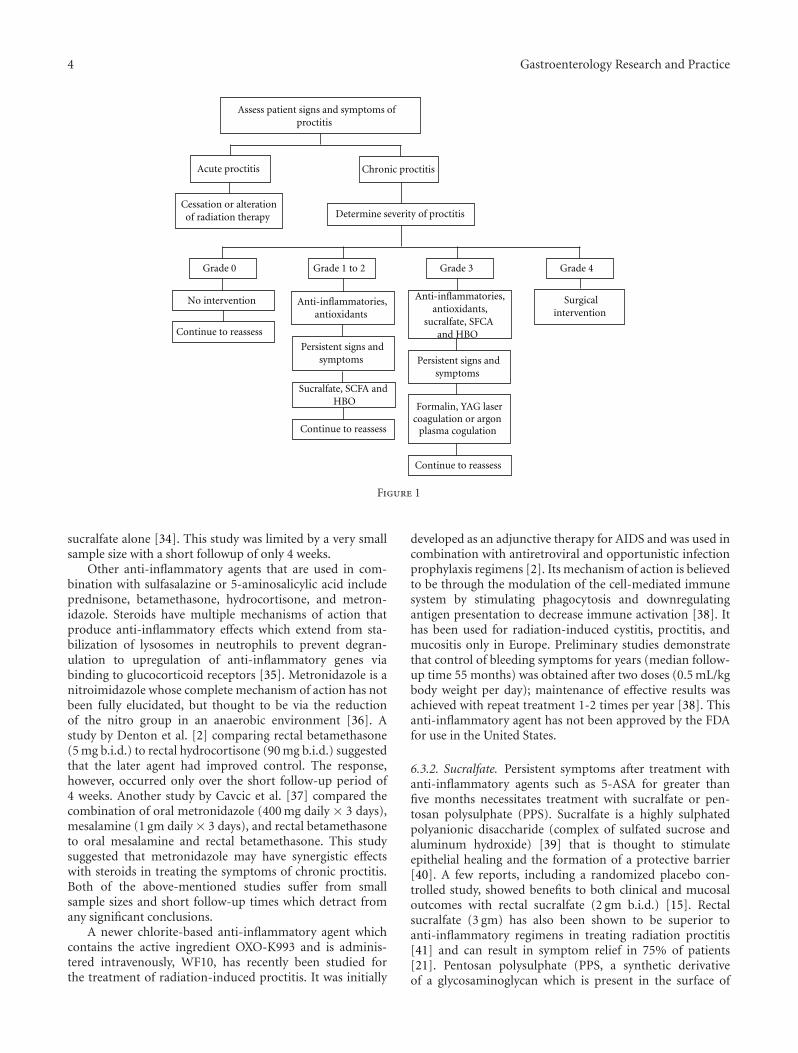
6.2. Chronic Proctitis. The management of chronic proc-titis can be divided into noninvasive treatments (anti-in-flammatory agents, sucralfate, short-chain fatty acids, hyper-baric, antioxidants) and invasive treatments (ablation andsurgery). Although there is considerable variation in themanagement strategies for chronic proctitis, there is gener-ally a strategy of using the least invasive interventions firstwith gradual progression as symptoms and signs worsen(Figure 1).
6.3. Noninvasive Treatments. Noninvasive therapy for chron-ic radiation proctitis begins with the use of oral, rectal, orgaseous agents. These agents consist of nonsteroidal anti-inflammatory drugs, sucralfate, short-chain fatty acids(SCFA), hyperbaric oxygen (HBO), and antioxidants.
6.3.1. Anti-Inflammatory Agents. Anti-inflammatory agentssuch as sulfasalazine or 5-aminosalicylic acid (mesalamine)are usually first line treatments, but they have low efficacyeven in combination with other agents such as steroids andantibiotics. The mechanism of action of both nonsteroidalanti-inflammatory agents listed above is thought to bethrough the inhibition of prostaglandin synthesis or viathe lipoxygenase pathway of arachidonic acid metabolism[31]. Other suggested effects include the inhibition of folate-dependent enzymes and free radical scavenging activity [31].Sulfasalazine is re-excreted in the bile after absorption via thesmall bowel and split into sulfapyridine and 5-aminosalicylicacid by colonic bacteria [32]. Studies have suggested that 5-aminosalicylic acid is the active metabolite of sulfasalazine[33]. A single randomized controlled trial by Kochhar etal. [34] comparing sulfasalazine (500 mg t.i.d) with rectalsteroids (prednisolone 20 mg b.i.d.) to rectal sucralfate (2 gmb.i.d.) had a clinical improvement in 53% (8 of 15) of patientswith oral sulfasalazine plus rectal steroids compared to a94% (16 of 17) improvement with rectal sucralfate alone.Objective data from this study showed 47% (7 of 15) hadimproved endoscopic finding in the sulfasalazine plus rectalsteroids group as compared to 71% (12 of 17) in the rectal
4 Gastroenterology Research and Practice
Assess patient signs and symptoms ofproctitis
Grade 0 Grade 1 to 2 Grade 3 Grade 4
No intervention Anti-inflammatories,antioxidants
Continue to reassess
Formalin, YAG laser coagulation or argon
plasma cogulation
Surgicalintervention
Acute proctitis Chronic proctitis
Cessation or alterationof radiation therapy Determine severity of proctitis
Persistent signs andsymptoms
Persistent signs andsymptoms
Sucralfate, SCFA and HBO
Anti-inflammatories,antioxidants,
sucralfate, SFCA and HBO
Continue to reassess
Continue to reassess
Figure 1
sucralfate alone [34]. This study was limited by a very smallsample size with a short followup of only 4 weeks.
Other anti-inflammatory agents that are used in com-bination with sulfasalazine or 5-aminosalicylic acid includeprednisone, betamethasone, hydrocortisone, and metron-idazole. Steroids have multiple mechanisms of action thatproduce anti-inflammatory effects which extend from sta-bilization of lysosomes in neutrophils to prevent degran-ulation to upregulation of anti-inflammatory genes viabinding to glucocorticoid receptors [35]. Metronidazole is anitroimidazole whose complete mechanism of action has notbeen fully elucidated, but thought to be via the reductionof the nitro group in an anaerobic environment [36]. Astudy by Denton et al. [2] comparing rectal betamethasone(5 mg b.i.d.) to rectal hydrocortisone (90 mg b.i.d.) suggestedthat the later agent had improved control. The response,however, occurred only over the short follow-up period of4 weeks. Another study by Cavcic et al. [37] compared thecombination of oral metronidazole (400 mg daily × 3 days),mesalamine (1 gm daily × 3 days), and rectal betamethasoneto oral mesalamine and rectal betamethasone. This studysuggested that metronidazole may have synergistic effectswith steroids in treating the symptoms of chronic proctitis.Both of the above-mentioned studies suffer from smallsample sizes and short follow-up times which detract fromany significant conclusions.
A newer chlorite-based anti-inflammatory agent whichcontains the active ingredient OXO-K993 and is adminis-tered intravenously, WF10, has recently been studied forthe treatment of radiation-induced proctitis. It was initially
developed as an adjunctive therapy for AIDS and was used incombination with antiretroviral and opportunistic infectionprophylaxis regimens [2]. Its mechanism of action is believedto be through the modulation of the cell-mediated immunesystem by stimulating phagocytosis and downregulatingantigen presentation to decrease immune activation [38]. Ithas been used for radiation-induced cystitis, proctitis, andmucositis only in Europe. Preliminary studies demonstratethat control of bleeding symptoms for years (median follow-up time 55 months) was obtained after two doses (0.5 mL/kgbody weight per day); maintenance of effective results wasachieved with repeat treatment 1-2 times per year [38]. Thisanti-inflammatory agent has not been approved by the FDAfor use in the United States.
6.3.2. Sucralfate. Persistent symptoms after treatment withanti-inflammatory agents such as 5-ASA for greater thanfive months necessitates treatment with sucralfate or pen-tosan polysulphate (PPS). Sucralfate is a highly sulphatedpolyanionic disaccharide (complex of sulfated sucrose andaluminum hydroxide) [39] that is thought to stimulateepithelial healing and the formation of a protective barrier[40]. A few reports, including a randomized placebo con-trolled study, showed benefits to both clinical and mucosaloutcomes with rectal sucralfate (2 gm b.i.d.) [15]. Rectalsucralfate (3 gm) has also been shown to be superior toanti-inflammatory regimens in treating radiation proctitis[41] and can result in symptom relief in 75% of patients[21]. Pentosan polysulphate (PPS, a synthetic derivativeof a glycosaminoglycan which is present in the surface of
Gastroenterology Research and Practice 5
the bladder, vessels, and the gastrointestinal tract lining) isthought to reduce epithelial permeability and prevent ad-herence similar to sucralfate [42].
6.3.3. SCFA. Short chain fatty acids (SCFA) are the mainoxidative fuel of the colonic mucosa and also serve to stim-ulate colonic mucosal proliferation [43]. They are thoughtto be produced by the anaerobic bacteria of the colon fromnonabsorbed carbohydrates. The most significant product ofSCFA is butyric acid [44]. SCFA also exerts a vasodilatoryeffect on the arteriole walls to improve blood flow [45].Radiation-induced injury results in ischemia and loss ofmicrovillus architecture which may result in the impairmentof SCFA absorption, thereby contributing to the changesseen with chronic radiation proctitis [3]. Supplementationwith SFCA enemas may accelerate healing by improving thedeficiency experienced by the colonocytes. Two randomizedstudies looked at butyrate enemas (40 mM butyrate) andfound nonsignificant improvement in symptoms and signscontrary to case reports suggesting some benefit. Both stud-ies, however, were severely underpowered [46–48]. Furtherstudies are necessary to evaluate the possible benefits of SCFAfor chronic proctitis.
6.3.4. HBO. Hyperbaric oxygen therapy (HBO) stems fromthe pathological process of ischemia involving the compro-mise of blood flow to the rectal wall. The benefit of HBO istheoretically achieved through the decrease of tissue hypoxia,possibly through its angiogenic and antibacterial effects [49,50]. Data on the use of HBO in chronic radiation proctitis islimited. A review by Bennett et al. [51] found an increasedchance of improvement with hyperbaric oxygen treatment(RR 1.75, number needed to treat = 5). However, thedegree of benefit, the cumulative effects, or duration of theresponse cannot be quantified because of the methodologyand quality of the data. One randomized-controlled trial byClarke compared patients treated with 90 minutes of 100%oxygen at 2 atmospheres with patients treated for 90 minuteswith 21% oxygen at approximately 1 atmosphere. Resultssuggested some benefit with symptom improvement after30 treatments [52]. Most studies demonstrate that HBO isexpensive and not readily available in most areas except inhighly specialized centers due to the requirement for multipletreatments and specialized equipment.
6.3.5. Antioxidants. Oxidative stress is thought to be amajor mechanism in the development of chronic radiationproctitis; agents with antioxidant properties have been usedin an attempt to limit tissue damage in radiation injury. Ina study by Kennedy et al., which only included 10 patients,the use of Vitamin E and C significantly decreased the rateof diarrhea and urgency [19]. The benefit of Vitamin Ahas been looked at by itself and in combination with othertherapies. In a double-blind study by Ehrenpreis, the useof Vitamin A significantly reduced proctitis symptoms andthe effects extended to patients in the placebo group aftercrossover [53]. Patel reported that the addition of VitaminA to treatment with 8% formalin increased the success
rate of formalin and shortened the time needed to achieveimprovement [54].
6.4. Invasive Treatments
6.4.1. Ablative Procedures. Ablative techniques are reservedfor the treatment of symptoms refractory to medical man-agement and include formalin, endoscopic coagulation, andargonplasma coagulation. These techniques can be associ-ated with complications that include bleeding, perforation,fistulas, and stenosis.
6.4.2. Formalin. In radiation proctitis, vascular telangiectasiaand nonhealing mucosal ulceration, perhaps due to anunderlying obliterative arteritis, may lead to severe recurrenthemorrhage. While there are no prospective studies offormalin treatment, formalin is considered to be a safe andeffective way to treat radiation proctitis causing significantbleeding. Formalin scleroses and seals fragile neovasculaturein radiation damaged tissues to prevent further bleedingthrough chemical cauterization [16, 55, 56]. Two methodsof formalin application have been described since its firstreported use in 1986 which are the rectal instillation of 4%formalin solution or direct topical application of a 10%formalin solution [55]. The topical application directly to themucosa is thought to produce a more targeted local chemicalcauterization; however, its success is entirely dependent onaccurate localization. Topical formalin (10% formaldehydesolution) is generally applied through a rigid proctoscope,flexible endoscope, or by direct application with formalinsoaked gauze [2, 57, 58] Contact with formalin for 2-3minutes (until slight blanching of the mucosa is achieved)is allowed and believed to cause chemical cauterization.It is a procedure that can be performed in the office orprocedure suite with reported success rates of 70%–80% withfew patients requiring repeat application [55]. A few studiesreported similar efficacy and a better complication profilewith lower doses of formalin (down to 4% solution) [56], butonly a few patients were examined. The perianal skin needsto be protected during the procedure to prevent strictureand skin damage. To prevent topical damage, flushing andirrigation through flexible endoscope has been proposed asa way of administration. Formalin application is generallysafe, but bleeding, strictures, and fistulas have been reported.Direct injection of formalin allows for the more preciseadministration of treatment and potentially fewer strictures,but it is not feasible with large mucosal areas effected andcould lead to fistula formation. Minor side effects were notfrequently reported. The duration of the treatment effectscannot be assessed reliably from the data available but appearto last a minimum of 3 months. The absence of quality of lifedata means the impact of this treatment from the patient’sperspective cannot be addressed.
6.4.3. Endoscopic Coagulation. A variety of endoscopic coag-ulation devices are effective for the control of radiation-induced bleeding through coagulation of focal bleedingtelangiectasias. Most of the studies currently available are
6 Gastroenterology Research and Practice
retrospective and show an improvement of symptoms anda decreased recurrence after treatment with YAG laser coag-ulation or argon plasma coagulation [2]. Both methods arebased on the delivery of thermal coagulation and should bereserved for patients suffering from significant hemorrhagicproctitis [59]. Several treatment sessions are often required.
Argon plasma coagulation uses high-frequency energy(monopolar diathermy) transmitted to the tissue throughan ionized gas in a noncontact fashion [59]. Its ability tocontrol bleeding throughout the gastrointestinal tract hasbeen demonstrated, and it has very limited tissue pene-tration, making it attractive for treatment of superficialbleeding. Available studies are retrospective and have a smallnumber of patients [60, 61]. However, all studies haveshown higher hemoglobin and fewer symptoms in patientswith hemorrhagic proctitis who failed medical therapy.Single sessions have been reported to significantly improvesymptoms [62], but on average, two to three treatmentsare needed to achieve this result. Improvements persistedfor a number of months after therapy was finished [2, 63].Most of the complications reported were mild and includedcramps, mucus discharge, and stricture [2]. However, signif-icant complications including large ulcers, perforations, andrectourethral and rectovaginal fistulas have been reported.
YAG lasers have the same theoretical benefit as argonplasma coagulation with a limited depth of penetration andthe possibility for precise application. Evidence for their useis similar to those for argon plasma coagulation with onlya few studies available, including those with fewer than 10patients. [64, 65]. Based on studies currently available, YAGlasers may be useful in the treatment of radiation proctitis,but the data are not strong enough to support their wide use.For both argon plasma coagulation and YAG lasers, cost andavailability may also present significant obstacles.
6.4.4. Surgery. Although surgery is often necessary for adiagnosis of radiation proctitis and as an adjunct to some ofthe treatment options described above, it is considered a lastresort for patients with radiation proctitis. Fewer than 10%[66] of patients ultimately require surgery. When required,it is directed at specific symptoms and complications ofradiation proctitis such as intractable bleeding, perforation,strictures, and fistulas. Rarely, surgery has been used to treatuncontrollable pain. Gas and stool incontinence are commonsymptoms that often accompany these complications andshould be considered when deciding on the need for sur-gical intervention. When surgery is being considered, thepathophysiology of radiation damage must be taken intoaccount. Microvascular damage caused by radiation not onlycauses the symptoms of radiation proctitis, but also cansignificantly impair healing after any surgery. Fecal diversionwith either a colostomy or ileostomy is a common rea-son patients are referred to surgeons. Diverting the stoolstream decreases symptoms of pain, tenesmus, drainage, andinfection but rarely eliminates them completely. An ostomycan also improve symptoms related to incontinence andstricture, but it has a limited effect on bleeding. At leastone study [67] showed significant improvement in bleeding
from a diverting loop colostomy but not complete resolution.Diversion will improve symptoms but without additional in-terventions, the improvements in symptoms are unlikely topersist after an ostomy reversal.
Rectourethral, rectovaginal and rectovesicular fistulaspresent with infections, pain, or incontinence symptoms andare one of the more common complications of proctitis.Surgical treatment options have traditionally included localexcision and reconstruction such as an advancement flap.Due to poorly vascularized tissues and low healing rates,however, these interventions should not be used as theonly treatment modalities. Additional options to improvesuccess of fistula repairs include pedunculated flaps, suchas a gracilis or martius flap, which are used to facilitatehealing by introducing well-vascularized healthy tissue.Diversion of stool or the urinary stream with an ostomyor a suprapubic catheter should be considered in almost allcases where repair is attempted. In some patients, a completediversion will improve symptoms and their quality of life tothe point that they do not require any further intervention[66, 68–70] even though the underlying problem is notrepaired. In cases of complicated fistulous disease, especiallywhen accompanied by significant pain and incontinence,a proctectomy or pelvic exenteration with or without re-construction is recommended. While this is a definitive treat-ment, it is accompanied by significant morbidity, includingexceedingly high rates of anastomotic leaks in cases ofreconstruction and high rates of perineal wound complica-tions when reconstruction is not attempted [69, 70]. Whenconsidering reconstruction for these patients, a temporarydiversion should be part of the initial operation. It is alsoimportant to discuss with patients that although theseprocedures may be a technical success, they often resultin unacceptable long-term morbidity including complicatedscarring, stricture, and incontinence. In cases of abdominalperineal resections, bringing a well-vascularized tissue toclose the wound, such as rectus abdominus, gracilis or glutealV-Y flap, will significantly decrease rates of postoperativewound complications.
In cases of severe and intractable bleeding, surgical op-tions are very limited, because a diversion will rarely controlthe bleeding completely. Diverting loop colostomy was de-scribed in one study as a successful way of controllingbleeding [67], but in many instances, proctectomy may bethe only option available. When strictures are an issue, adiversion of the stool stream will often result in a significantimprovement of symptoms. Other options include resectionwith reconstruction when the stricture is higher in therectum or an advancement flap (mucosa or skin) when therectal stricture is in the anus. In all cases, given the poorquality of the tissues, at least a temporary diversion should beconsidered. When surgical treatment is needed, most studiesdemonstrate poor outcomes with high complication rates(15%–80%), and a mortality of 3%–9% [66, 68–70].
7. Conclusion
Radiation proctitis is a relatively rare complication of radia-tion therapy. Rates of both acute and chronic proctitis have
Gastroenterology Research and Practice 7
been decreasing with improved radiation therapy techniquesthat allow for the targeted delivery of higher doses of ra-diation. It is important to note that radiation proctitis is aresult of radiation doses that are beyond the ability of thenormal tissue to repair or recover from injury. There is recentevidence that the impairment of the rectum’s ability to healmay also mean that other organs exposed to the same highradiation doses may be at increased risk of malignant trans-formation [71]. It has been suggested that patients exposedto higher doses of radiation may need to be more closelyscreened for other malignancies [71], but further studiesneed to be conducted before definitive recommendations canbe made. Although there are no good preventive measuresavailable at this point, most instances of proctitis are self-limited and respond to medical management. A combinationof sucralfate, steroids, and pain control have been successfulin most cases to improve symptoms. In more severe cases,especially with bleeding, chemical (formalin) or thermal(endoscopic coagulation) treatments have been successful.Surgery is rarely required to treat this condition, but whenperformed can lead to significant improvements. Surgery,however, also results in the increased risk of postsurgicalcomplications. More studies are needed to prospectively lookat both the prevention and treatment of radiation proctitis aswell as patients with special consideration such as those withIBD and HIV/AIDS.
References
[1] M. Xiao and M. H. Whitnall, “Pharmacological countermea-sures for the acute radiation syndrome,” Current MolecularPharmacology, vol. 2, no. 1, pp. 122–133, 2009.
[2] A. Denton, A. Forbes, J. Andreyev, and E. J. Maher, “Non sur-gical interventions for late radiation proctitis in patients whohave received radical radiotherapy to the pelvis,” CochraneDatabase of Systematic Reviews, no. 1, Article ID CD003455,2002.
[3] T. Terasawa, T. Dvorak, S. Ip, G. Raman, J. Lau, and T.A. Trikalinos, “Systematic review: charged-particle radiationtherapy for cancer,” Annals of Internal Medicine, vol. 151, no.8, pp. 556–565, 2009.
[4] E. Melian, G. S. Mageras, Z. Fuks et al., “Variation inprostate position quantitation and implications for three-dimensional conformal treatment planning,” InternationalJournal of Radiation Oncology Biology Physics, vol. 38, no. 1,pp. 73–81, 1997.
[5] B. A. Fraass, M. L. Kessler, D. L. McShan et al., “Optimizationand clinical use of multisegment intensity-modulated radia-tion therapy for high-dose conformal therapy,” Seminars inRadiation Oncology, vol. 9, no. 1, pp. 60–77, 1999.
[6] S. A. Shah, R. R. Cima, E. Benoit, E. L. Breen, and R. Bleday,“Rectal complications after prostate brachytherapy,” Diseasesof the Colon and Rectum, vol. 47, no. 9, pp. 1487–1492, 2004.
[7] R. N. Lesperance, R. J. Kjorstadt, J. B. Halligan, and S. R.Steele, “Colorectal complications of external beam radiationversus brachytherapy for prostate cancer,” American Journal ofSurgery, vol. 195, no. 5, pp. 616–620, 2008.
[8] T. E. Schultheiss, W. R. Lee, M. A. Hunt, A. L. Hanlon, R.S. Peter, and G. E. Hanks, “Late GI and GU complicationsin the treatment of prostate cancer,” International Journal of
Radiation Oncology Biology Physics, vol. 37, no. 1, pp. 3–11,1997.
[9] L. R. Coia, “Late effects of radiation therapy on the gastro-intestinal tract,” International Journal of Radiation OncologyBiology Physics, vol. 31, no. 5, pp. 1213–1236, 1995.
[10] C. J. Beard, K. J. Propert, P. P. Rieker et al., “Complicationsafter treatment with external-beam irradiation in early-stage prostate cancer patients: a prospective multiinstitutionaloutcomes study,” Journal of Clinical Oncology, vol. 15, no. 1,pp. 223–229, 1997.
[11] M. V. Pilepich, J. M. Krall, and W. T. Sause, “Correlation ofradiotherapeutic parameters and treatment related morbidityin carcinoma of the prostate—analysis of RTOG study 75-06,”International Journal of Radiation Oncology Biology Physics,vol. 13, no. 3, pp. 351–357, 1987.
[12] M. J. Zelefsky, Z. Fuks, M. Hunt et al., “High-dose intensitymodulated radiation therapy for prostate cancer: early toxicityand biochemical outcome in 772 patients,” InternationalJournal of Radiation Oncology Biology Physics, vol. 53, no. 5,pp. 1111–1116, 2002.
[13] M. J. Zelefsky, E. J. Levin, M. Hunt et al., “Incidence of laterectal and urinary toxicities after three-dimensional confor-mal radiotherapy and intensity-modulated radiotherapy forlocalized prostate cancer,” International Journal of RadiationOncology Biology Physics, vol. 70, no. 4, pp. 1124–1129, 2008.
[14] A. L. Zietman, K. Bae, J. D. Slater et al., “Randomizedtrial comparing conventional-dose with high-dose confor-mal radiation therapy in early-stage adenocarcinoma of theprostate: long-term results from proton radiation oncologygroup/american college of radiology 95-09,” Journal of ClinicalOncology, vol. 28, no. 7, pp. 1106–1111, 2010.
[15] S. I. Zeitlin, J. Sherman, A. Raboy, G. Lederman, and P. Albert,“High dose combination radiotherapy for the treatment oflocalized prostate cancer,” Journal of Urology, vol. 160, no. 1,pp. 91–96, 1998.
[16] A. S. Denton, H. J. N. Andreyev, A. Forbes, and E. J. Maher,“Systematic review for non-surgical interventions for themanagement of late radiation proctitis,” British Journal ofCancer, vol. 87, no. 2, pp. 134–143, 2002.
[17] G. Cotti, V. Seid, S. Araujo, A. H. Souza, D. R. Kiss, andA. Habr-Gama, “Conservative therapies for hemorrhagic ra-diation proctitis: a review,” Revista do Hospital das Clınicas.,vol. 58, no. 5, pp. 284–292, 2003.
[18] P. P. Tagkalidis and J. J. Tjandra, “Chronic radiation proctitis,”ANZ Journal of Surgery, vol. 71, no. 4, pp. 230–237, 2001.
[19] M. Kennedy, K. Bruninga, E. A. Mutlu, J. Losurdo, S.Choudhary, and A. Keshavarzian, “Successful and sustainedtreatment of chronic radiation proctitis with antioxidantvitamins E and C,” American Journal of Gastroenterology, vol.96, no. 4, pp. 1080–1084, 2001.
[20] C. G. Willett, C. J. Ooi, A. L. Zietman et al., “Acute andlate toxicity of patients with inflammatory bowel disease un-dergoing irradiation for abdominal and pelvic neoplasms,” In-ternational Journal of Radiation Oncology Biology Physics, vol.46, no. 4, pp. 995–998, 2000.
[21] J. Phan, D. A. Swanson, L. B. Levy, R. J. Kudchadker, T. L.Bruno, and S. J. Frank, “Late rectal complications after pros-tate brachytherapy for localized prostate cancer,” Cancer, vol.115, no. 9, pp. 1827–1839, 2009.
[22] R. Hoffman, M. L. Welton, B. Klencke, V. Weinberg, and R.Krieg, “The significance of pretreatment CD4 count on theoutcome and treatment tolerance of HIV-positive patients

8 Gastroenterology Research and Practice
with anal cancer,” International Journal of Radiation OncologyBiology Physics, vol. 44, no. 1, pp. 127–131, 1999.
[23] N. Housri, R. Yarchoan, and A. Kaushal, “Radiotherapy forpatients with the human immunodeficiency virus: are specialprecautions necessary?” Cancer, vol. 116, no. 2, pp. 273–283,2010.
[24] T. Kertesz, M. K.A. Herrmann, A. Zapf et al., “Effect ofa prostaglandin—given rectally for prevention of radiation-induced acute proctitis—on late rectal toxicity: results ofa phase III randomized, placebo-controlled, double-blindstudy,” Strahlentherapie und Onkologie, vol. 185, no. 9, pp.596–602, 2009.
[25] A. Kneebone, H. Mameghan, T. Bolin et al., “Effect of oral su-cralfate on late rectal injury associated with radiotherapy forprostate cancer: a double-blind, randomized trial,” Interna-tional Journal of Radiation Oncology Biology Physics, vol. 60,no. 4, pp. 1088–1097, 2004.
[26] P. C. O’Brien, C. I. Franklin, M. G. Poulsen, D. J. Joseph, N.S. Spry, and J. W. Denham, “Acute symptoms, not rectallyadministered sucralfate, predict for late radiation proctitis:longer term follow-up of a phase III trial—Trans-Tasman Ra-diation Oncology Group,” International Journal of RadiationOncology Biology Physics, vol. 54, no. 2, pp. 442–449, 2002.
[27] N. Hovdenak, H. Sørbye, and O. Dahl, “Sucralfate does notameliorate acute radiation proctitis: randomised study andmeta-analysis,” Clinical Oncology, vol. 17, no. 6, pp. 485–491,2005.
[28] T. Liu, Y. Liu, S. He, Z. Zhang, and M. M. Kligerman, “Use ofradiation with or without WR-2721 in advanced rectal cancer,”Cancer, vol. 69, no. 11, pp. 2820–2825, 1992.
[29] H. Athanassiou, D. Antonadou, N. Coliarakis et al., “Protectiveeffect of amifostine during fractionated radiotherapy inpatients with pelvic carcinomas: results of a randomized trial,”International Journal of Radiation Oncology Biology Physics,vol. 56, no. 4, pp. 1154–1160, 2003.
[30] D. M. Keefe, M. M. Schubert, L. S. Elting et al., “Updatedclinical practice guidelines for the prevention and treatmentof mucositis,” Cancer, vol. 109, no. 5, pp. 820–831, 2007.
[31] M. A. Peppercorn, “Sulfasalazine. Pharmacology, clinical usetoxicity, and related new drug development,” Annals of InternalMedicine, vol. 101, no. 3, pp. 377–386, 1984.
[32] C. P. Rains, S. Noble, and D. Faulds, “Sulfasalazine: a review ofits pharmacological properties and therapeutic efficacy in thetreatment of rheumatoid arthritis,” Drugs, vol. 50, no. 1, pp.137–156, 1995.
[33] U. Klotz, K. Maier, C. Fischer, and K. Heinkel, “Therapeuticefficacy of sulfasalazine and its metabolites in patients withulcerative colitis and Crohn’s disease,” New England Journal ofMedicine, vol. 303, no. 26, pp. 1499–1502, 1980.
[34] R. Kochhar, F. Patel, S. C. Scharma et al., “Radiation-induced proctosigmoiditis: prospective, randomized, double-blind controlled trial of oral sulfasalazine plus rectal steroidsversus rectal sucralfate,” Digestive Diseases and Sciences, vol. 36,no. 1, pp. 103–107, 1991.
[35] L. A. Schwiebert, L. A. Beck, C. Stellato, C. A. Bickel, B. S.Bochner, and R. P. Schleimer, “Glucocorticosteroid inhibitionof cytokine production: relevance to antiallergic actions,”Journal of Allergy and Clinical Immunology, vol. 97, no. 1, pp.143–152, 1996.
[36] C. D. Freeman, N. E. Klutman, and K. C. Lamp, “Metronida-zole. A therapeutic review and update,” Drugs, vol. 54, no. 5,pp. 679–708, 1997.
[37] J. Cavcic, J. Turcic, P. Martinac et al., “Metronidazole in thetreatment of chronic radiation proctitis: clinical trial,” Cro-atian Medical Journal, vol. 41, no. 3, pp. 314–318, 2000.
[38] V. Veerasarn, W. Boonnuch, and C. Kakanaporn, “A phaseII study to evaluate WF10 in patients with late hemorrhagicradiation cystitis and proctitis,” Gynecologic Oncology, vol. 100,no. 1, pp. 179–184, 2006.
[39] B. F. McGraw and E. G. Caldwell, “Sucralfate,” Drug Intelli-gence and Clinical Pharmacy, vol. 15, no. 7-8, pp. 578–580,1981.
[40] R. Nagashima, “Mechanisms of action of sucralfate,” Journalof Clinical Gastroenterology, vol. 3, supplement 2, pp. 117–127,1981.
[41] G. Sanguineti, P. Franzone, M. Marcenaro, F. Foppiano, and V.Vitale, “Sucralfate versus mesalazine versus hydrocortisone inthe prevention of acute radiation proctitis during conformalradiotherapy for prostate carcinoma: a randomized study,”Strahlentherapie und Onkologie, vol. 179, no. 7, pp. 464–470,2003.
[42] P. W. Grigsby, M. V. Pilepich, and C. L. Parsons, “Preliminaryresults of a phase I/II study of sodium pentosanpolysulfatein the treatment of chronic radiation-induced proctitis,”American Journal of Clinical Oncology, vol. 13, no. 1, pp. 28–31, 1990.
[43] B. Darcy-Vrillon, C. Cherbuy, M. T. Morel, M. Durand, andP. H. Duee, “Short chain fatty acid and glucose metabolism inisolated pig colonocytes: modulation by NH+
4 ,” Molecular andCellular Biochemistry, vol. 156, no. 2, pp. 145–151, 1996.
[44] P. H. Duee, B. Darcy-Vrillon, F. Blachier, and M. T. Morel,“Fuel selection in intestinal cells,” Proceedings of the NutritionSociety, vol. 54, no. 1, pp. 83–94, 1995.
[45] C. Aalkjaer, “Short chained fatty acids and the colon: how dothey cause vasodilatation?” Journal of Physiology, vol. 538, no.3, p. 674, 2002.
[46] N. A. Talley, F. Chen, D. King, M. Jones, and N. J. Talley,“Short-chain fatty acids in the treatment of radiation proctitis:a randomized, double-blind, placebo-controlled, cross-overpilot trial,” Diseases of the Colon and Rectum, vol. 40, no. 9,pp. 1046–1050, 1997.
[47] A. Pinto, P. Fidalgo, M. Cravo et al., “Short chain fatty acidsare effective in short-term treatment of chronic radiationproctitis: randomized, double-blind, controlled trial,” Diseasesof the Colon and Rectum, vol. 42, no. 6, pp. 788–796, 1999.
[48] R. Al-Sabbagh, F. A. Sinicrope, J. H. Sellin, Y. Shen, and L.Roubein, “Evaluation of short-chain fatty acid enemas: treat-ment of radiation proctitis,” Diseases of the Colon & Rectum,vol. 40, no. 9, pp. 1046–1050, 1997.
[49] J. J. Feldmeier and N. B. Hampson, “A systematic review ofthe literature reporting the application of hyperbaric oxygenprevention and treatment of delayed radiation injuries: anevidence based approach,” Undersea and Hyperbaric Medicine,vol. 29, no. 1, pp. 4–30, 2002.
[50] R. E. Marx, “A new concept in the treatment of osteora-dionecrosis,” Journal of Oral and Maxillofacial Surgery, vol. 41,no. 6, pp. 351–357, 1983.
[51] M. H. Bennett, J. Feldmeier, N. Hampson, R. Smee, and C.Milross, “Hyperbaric oxygen therapy for late radiation tissueinjury,” Cochrane Database of Systematic Reviews, no. 3, ArticleID CD005005, 2005.
[52] R. E. Clarke, L. M. C. Tenorio, J. R. Hussey et al., “Hyperbaricoxygen treatment of chronic refractory radiation proctitis:a randomized and controlled double-blind crossover trialwith long-term follow-up,” International Journal of RadiationOncology Biology Physics, vol. 72, no. 1, pp. 134–143, 2008.
Gastroenterology Research and Practice 9
[53] E. D. Ehrenpreis, A. Jani, J. Levitsky, J. Ahn, and J. Hong,“A prospective, randomized, double-blind, placebo-controlledtrial of retinol palmitate (vitamin A) for symptomatic chronicradiation proctopathy,” Diseases of the Colon and Rectum, vol.48, no. 1, pp. 1–8, 2005.
[54] P. Patel, G. Subhas, A. Gupta, Y. J. Chang, V. K. Mittal, andA. McKendrick, “Oral vitamin A enhances the effectivenessof formalin 8% in treating chronic hemorrhagic radiationproctopathy,” Diseases of the Colon and Rectum, vol. 52, no. 9,pp. 1605–1609, 2009.
[55] E. M. Haas, H. R. Bailey, and I. Farragher, “Application of10 percent formalin for the treatment of radiation-inducedhemorrhagic proctitis,” Diseases of the Colon and Rectum, vol.50, no. 2, pp. 213–217, 2007.
[56] S. Parikh, C. Hughes, E. P. Salvati et al., “Treatment ofhemorrhagic radiation proctitis with 4 percent formalin,”Diseases of the Colon and Rectum, vol. 46, no. 5, pp. 596–600,2003.
[57] S. F. Counter, D. P. Froese, and M. J. Hart, “Prospective eval-uation of formalin therapy for radiation proctitis,” AmericanJournal of Surgery, vol. 177, no. 5, pp. 396–398, 1999.
[58] V. Mathai and F. Seow-Choen, “Endoluminal formalin ther-apy for haemorrhagic radiation proctitis,” British Journal ofSurgery, vol. 82, no. 2, p. 190, 1995.
[59] J. J. Tjandra and S. Sengupta, “Argon plasma coagulation isan effective treatment for refractory hemorrhagic radiationproctitis,” Diseases of the Colon and Rectum, vol. 44, no. 12,pp. 1759–1765, 2001.
[60] A. C. Fantin, J. Binek, W. R. Suter, and C. Meyenberger, “Argonbeam coagulation for treatment of symptomatic radiation-induced proctitis,” Gastrointestinal Endoscopy, vol. 49, no. 4,pp. 515–518, 1999.
[61] R. A. Silva, A. J. Correia, L. M. Dias, H. L. Viana, and R. L.Viana, “Argon plasma coagulation therapy for hemorrhagicradiation proctosigmoiditis,” Gastrointestinal Endoscopy, vol.50, no. 2, pp. 221–224, 1999.
[62] M. P. Swan, G. T. C. Moore, W. Sievert, and D. A. Devonshire,“Efficacy and safety of single-session argon plasma coagu-lation in the management of chronic radiation proctitis,”Gastrointestinal Endoscopy, vol. 72, no. 1, pp. 150–154, 2010.
[63] C. S. Higuera, M. I. M. Arribas, S. J. R. Gomez, A. P. Villoria, J.M. Moreno, and A. B. Gonzalez, “Efficacy and safety of argonplasma coagulation for the treatment of hemorrhagic radia-tion proctitis,” Revista Espanola de Enfermedades Digestivas,vol. 96, no. 11, pp. 758–764, 2004.
[64] T. J. Alexander and R. M. Dwyer, “Endoscopic Nd:YAG lasertreatment of severe radiation injury of the lower gastrointesti-nal tract: long-term follow-up,” Gastrointestinal Endoscopy,vol. 34, no. 5, pp. 407–411, 1988.
[65] M. Ventrucci, M. P. Di Simone, P. Giulietti, and G. De Luca,“Efficacy and safety of Nd:YAG laser for the treatment ofbleeding from radiation proctocolitis,” Digestive and LiverDisease, vol. 33, no. 3, pp. 230–233, 2001.
[66] S. W. Jao, R. W. Beart, and L. L. Gunderson, “Surgicaltreatment of radiation injuries of the colon and rectum,”American Journal of Surgery, vol. 151, no. 2, pp. 272–277, 1986.
[67] J. Ayerdi, K. Moinuddeen, A. Loving, J. Wiseman, and N.Deshmukh, “Diverting loop colostomy for the treatment ofrefractory gastrointestinal bleeding secondary to radiationproctitis,” Military Medicine, vol. 166, no. 12, pp. 1091–1093,2001.
[68] V. E. Pricolo and P. C. Shellito, “Surgery for radiation injury tothe large intestine: variables influencing outcome,” Diseases ofthe Colon and Rectum, vol. 37, no. 7, pp. 675–684, 1994.
[69] P. F. Anseline, I. C. Lavery, and V. W. Fazio, “Radiation injuryof the rectum. Evaluation of surgical treatment,” Annals ofSurgery, vol. 194, no. 6, pp. 716–724, 1981.
[70] M. E. Lucarotti, R. A. Mountford, and D. C. C. Bartolo,“Surgical management of intestinal radiation injury,” Diseasesof the Colon and Rectum, vol. 34, no. 10, pp. 865–869, 1991.
[71] A. M. Nieder, M. P. Porter, and M. S. Soloway, “Radiationtherapy for prostate cancer increases subsequent risk ofbladder and rectal cancer: a population based cohort study,”Journal of Urology, vol. 180, no. 5, pp. 2005–2010, 2008.

Submit your manuscripts athttp://www.hindawi.com
Stem CellsInternational
Hindawi Publishing Corporationhttp://www.hindawi.com Volume 2014
Hindawi Publishing Corporationhttp://www.hindawi.com Volume 2014
MEDIATORSINFLAMMATION
of
Hindawi Publishing Corporationhttp://www.hindawi.com Volume 2014
Behavioural Neurology
EndocrinologyInternational Journal of
Hindawi Publishing Corporationhttp://www.hindawi.com Volume 2014
Hindawi Publishing Corporationhttp://www.hindawi.com Volume 2014
Disease Markers
Hindawi Publishing Corporationhttp://www.hindawi.com Volume 2014
BioMed Research International
OncologyJournal of
Hindawi Publishing Corporationhttp://www.hindawi.com Volume 2014
Hindawi Publishing Corporationhttp://www.hindawi.com Volume 2014
Oxidative Medicine and Cellular Longevity
Hindawi Publishing Corporationhttp://www.hindawi.com Volume 2014
PPAR Research
The Scientific World JournalHindawi Publishing Corporation http://www.hindawi.com Volume 2014
Immunology ResearchHindawi Publishing Corporationhttp://www.hindawi.com Volume 2014
Journal of
ObesityJournal of
Hindawi Publishing Corporationhttp://www.hindawi.com Volume 2014
Hindawi Publishing Corporationhttp://www.hindawi.com Volume 2014
Computational and Mathematical Methods in Medicine
OphthalmologyJournal of
Hindawi Publishing Corporationhttp://www.hindawi.com Volume 2014
Diabetes ResearchJournal of
Hindawi Publishing Corporationhttp://www.hindawi.com Volume 2014
Hindawi Publishing Corporationhttp://www.hindawi.com Volume 2014
Research and TreatmentAIDS
Hindawi Publishing Corporationhttp://www.hindawi.com Volume 2014
Gastroenterology Research and Practice
Hindawi Publishing Corporationhttp://www.hindawi.com Volume 2014
Parkinson’s Disease
Evidence-Based Complementary and Alternative Medicine
Volume 2014Hindawi Publishing Corporationhttp://www.hindawi.com