Embed Size (px)
Citation preview
Review ArticlePredictive Symptoms and Signs of Severe Dengue Disease forPatients with Dengue Fever: A Meta-Analysis
H. Zhang,1,2 Y. P. Zhou,1 H. J. Peng,2 X. H. Zhang,3 F. Y. Zhou,1 Z. H. Liu,1 and X. G. Chen2
1 Department of Infectious Diseases and Hepatology Unit, Nanfang Hospital, Southern Medical University,Guangzhou 510515, China
2 Key Laboratory of Prevention and Control for Emerging Infectious Diseases of Guangdong Higher Institutes,School of Public Health and Tropical Medicine, Southern Medical University, Guangzhou 510515, China
3Department of Infectious Diseases, TheThird Affiliated Hospital of Sun Yat-sen University, Guangzhou 510630, China
Correspondence should be addressed to Y. P. Zhou; [email protected] and X. G. Chen; [email protected]
Received 29 April 2014; Revised 3 June 2014; Accepted 11 June 2014; Published 1 July 2014
Academic Editor: Jianfeng Dai
Copyright © 2014 H. Zhang et al.This is an open access article distributed under theCreative CommonsAttribution License, whichpermits unrestricted use, distribution, and reproduction in any medium, provided the original work is properly cited.
The aim of the meta-analysis was to provide more solid evidence for the reliability of the new classification. A systematic literaturesearch was performed using PubMed, Armed Forces Pest Management Board Literature Retrieval System, and Google Scholar upto August 2012. A pooled odds ratio (OR) was calculated using either a random-effect or a fixed-effect model. A total of 16 paperswere identified. Among the 11 factors studied, five symptoms demonstrated an increased risk for SDD, including bleeding [OR:13.617; 95% confidence interval (CI): 3.281, 56.508], vomiting/nausea (OR: 1.692; 95% CI: 1.256, 2.280), abdominal pain (OR: 2.278;95% CI: 1.631, 3.182), skin rashes (OR: 2.031; 95% CI: 1.269, 3.250), and hepatomegaly (OR: 4.751; 95% CI: 1.769, 12.570). Amongthe four bleeding-related symptoms including hematemesis, melena, gum bleeding, and epistaxis, only hematemesis (OR: 6.174;95% CI: 2.66, 14.334; 𝑃 < 0.001) and melena (OR: 10.351; 95% CI: 3.065, 34.956; 𝑃 < 0.001) were significantly associated withSDD. No significant associations with SDD were found for gender, lethargy, retroorbital pain, diarrhea, or tourniquet test, whereasheadache appeared protective (OR: 0.555; 95% CI: 0.455, 0.676). The meta-analysis suggests that bleeding (hematemesis/melena),vomiting/nausea, abdominal pain, skin rashes, and hepatomegaly may predict the development of SDD in patients with DF, whileheadache may predict otherwise.
1. Introduction
Dengue is an infectious disease caused by dengue virus(DENV). It is endemic inmany tropical and subtropical areas.Patients infectedwithDENVhave a wide spectrumof clinicalmanifestation, ranging from silent infections with no symp-toms to amild flu-like syndrome, dengue fever (DF), or severedengue disease (SDD), including dengue haemorrhagic fever(DHF) and dengue shock syndrome (DSS) [1–3]. Recently,DF has become one of the most challenging public healthproblems in affected regions, as the DF incidence increasesrapidly worldwide [4]. There are approximately 2.5 billionpeople at risk for DF worldwide. Fifty million people wouldacquire DENV annually, and half a million among themwould develop dengue hemorrhagic fever, including 22,000deaths [5].
Several methods have been used for the diagnoses of DF.However, there lacks an accuratemeans to predict the severityof disease at early stages of the infection. Since patients withmild or classical DF can develop SDD later [2], it is importantto look for symptoms/signs to facilitate the early predictionof the progression into SDD. The establishment of predic-tive symptoms/signs is essential for preventing unnecessaryhospitalization, reducing disease burden, and controllingpotential SDD. Based on the dengue guidelines (2009), thewarning symptoms/signs for SDD include abdominal pain ortenderness, persistent vomiting, mucosal bleed, lethargy, andrestlessness.
Published studies about symptoms/signs that are associ-ated with SDD have been inconclusive. For instance, Khanet al. found thatmaleDF patients weremore likely to progressinto DHF (OR: 2.3, 95% CI: 1.1–4.5, 𝑃 value 0.021) [6], while
Hindawi Publishing CorporationBioMed Research InternationalVolume 2014, Article ID 359308, 10 pageshttp://dx.doi.org/10.1155/2014/359308
2 BioMed Research International
there was no association with SDD [7, 8]. The frequenciesof symptoms/signs of vomiting/nausea, abdominal pain,skin rashes, and bleeding were also found to be correlatedwith SDD [6–11]. However, the published studies were notable to conclude that these symptoms/signs are associatedwith SDD. In addition, although some findings such asviral factors, varying host immune conditions, host immunereactions, and laboratory tests can predict SDD [12], theclinicalmanifestationsmight always offer the earliestmarkersin predicting SDD. For example, patients with nonseveredengue could be clustered into two groups: one with warningsigns, such as abdominal pain, mucosal bleeding, and liverenlargement, and the other without those signs [2], as mostof the warning signs were associated with an indication forICU admission and were severe, even for the relationship ofdeath [13].
Because of these inconsistent reports, more accuratemethods to predict SDD are needed. We conducted themeta-analysis to identify which clinical symptoms/signs areassociated with SDD and to help find better methods topredict the development of SDD in patients with DF.
2. Materials and Methods
2.1. Literature Searches. Our study was performed accordingto the recommendations of the PRISMA Statement [14],which is available in supporting information (see TableS1 available online at http://dx.doi.org/10.1155/2014/359308).Computerized searches were conducted on NCBI PubMed,Armed Forces Pest Management Board Literature RetrievalSystem, and Google Scholar. As few studies before 2000met the criteria of WHO guidelines (1997), the search timewindow was set between January 1, 2000, and August 1, 2012,with no language limit. Because severe dengue disease (SDD)is classified asDHF andDSS, we used the following keywordsfor searching: dengue fever, DF, dengue haemorrhagic fever,DHF, dengue shock syndrome, DSS, and clinical diagnosis.We also manually searched the reference lists of the retrievedarticles to identify more qualified studies.
2.2. Inclusion and Exclusion Criteria. Studies were eligible forinclusion if they met the following criteria: (1) retrospective,prospective, or cross-sectional studies providing the details ofsymptoms/signs as well as any information regarding gender,vomiting/nausea, abdominal pain, skin rashes, bleeding,headache, lethargy, retroorbital pain, diarrhea, hepatomegaly,or tourniquet test; (2) the symptoms/signs of DF and SDDwere distinguished; (3) cases with DF in the study wereconfirmed by laboratory tests; cases with SDD were definedby one ormore of the following: plasma leakage thatmay leadto shock (dengue shock) and/or fluid accumulation, with orwithout respiratory distress, and/or severe bleeding, and/orsevere organ impairment. When two or more publicationsreported the same study, we chose the most recent one.Reports providing inadequate information were excluded.
2.3. Quality Assessment. The quality of the selected stud-ies was assessed independently by two authors using the
Newcastle-Ottawa Scale (NOS) [15]. The NOS uses differ-ent tools for case-control and cohort studies and consistsof 3 parameters of quality: selection, comparability, andexposure/outcome assessment.The NOS assigns a maximumof 4 points for selection, 2 for comparability, and 3 forexposure or outcome. We assigned NOS scores of 1–3, 4–6,and 7–9 for low, intermediate, and high-quality studies,respectively. Discrepancies were settled by consensus afterjoint reevaluation of the original studies.
2.4. Data Extraction. For each eligible manuscript, the fol-lowing information was extracted: (1) first author’s nameand year of publication; (2) study design (prospective, retro-spective, or cross-sectional); (3) study populations (children,adults, or both); (4) distinctive numbers of patients withspecific symptoms in DF and SDD groups.
2.5. Statistical Analysis. The prevalence rates of specificsymptoms/signs in DF and SDD groups were compared bycalculating an odds ratio (OR) with a 95% confidenceinterval (CI) using either a fixed-effect model or a random-effect model. Predictive factors of interest included gen-der, vomiting/nausea, abdominal pain, skin rashes, bleed-ing (hematemesis, melena, gum bleeding, and epistaxis),headache, lethargy, retroorbital pain, diarrhea, hepatomegaly,and tourniquet test.
Heterogeneity between studies was assessed using boththeChi-square testwith a𝑃 value≤0.10 and the inconsistencyindex (𝐼2) with a cut-off of 50% [16]. To explore the potentialsources of heterogeneity among studies, subgroup analysesand metaregression were performed on the strata of studydesign, study population, and publication year.
Potential publication bias was comprehensively assessedby Begg’s funnel plot and Egger’s rank correlation test ofasymmetry. Publication bias was determined present whenthe 𝑃 value ≤0.10 by Egger’s or Begg’s test. All statisticalanalyses were performed using STATA version 11.0 (STATACorporation, College Station, TX, USA).
3. Results
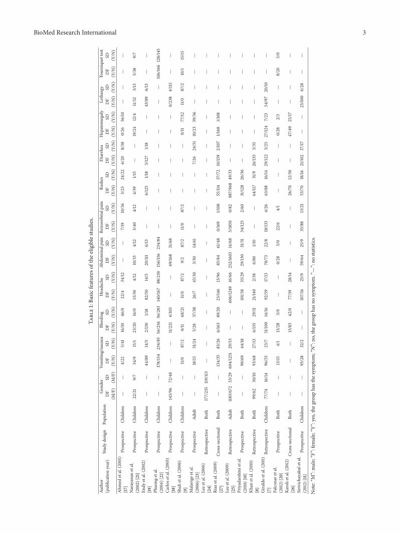
3.1. Study Characteristics and Quality. The search strategyidentified 446 citations. Sixteen articles published between2000 and 2012 were ultimately included in this meta-analysisbased on the inclusion and exclusion criteria (Figure 1). Thefinal collection consists of 10 prospective [9–11, 17–23], fourretrospective [7, 8, 24, 25], and two cross-sectional studies[26, 27]. As listed in Table 1, eight of the 16 studies reported ona population study of children, two on adults and six on both.The factor of gender was included in five studies. The clinicalsymptoms/signs of vomiting/nausea were included in 13studies, abdominal pain in 13, skin rashes in 10, bleeding in 13,headache in 13, lethargy in 6, retroorbital pain in 9, diarrheain 7, hepatomegaly in 8, and tourniquet test in 4 studies. Fourcommon kinds of bleeding symptoms were present in thesestudies, including hematemesis in five, melena in four, gumbleeding in seven, and epistaxis in five. Based on the NOSscores, 12 studies (75%) were of high quality and the otherfour (25%) were acceptable.
BioMed Research International 3
Table1:Ba
sicfeatures
ofthee
ligiblestu
dies.
Author
(pub
licationyear)
Stud
ydesig
nPo
pulation
Gender
Vomiting
/nausea
Bleeding
Headache
Abdo
minalpain
Retro
orbitalp
ain
Rashes
Diarrhea
Hepatom
egaly
Lethargy
Tourniqu
ettest
DF
(M/F)
SD (M/F)
DF
(Y/N
)SD (Y/N
)DF
(Y/N
)SD (Y/N
)DF
(Y/N
)SD (Y/N
)DF
(Y/N
)SD (Y/N
)DF
(Y/N
)SD (Y/N
)DF
(Y/N
)SD (Y/N
)DF
(Y/N
)SD (Y/N
)DF
(Y/N
)SD (Y/N
)DF
(Y/N
)SD (Y/N
)DF
(Y/N
)SD (Y/N
)Ahm
edetal.(2001)
[17]
Prospective
Child
ren
——
4/22
5/41
16/10
46/0
22/4
34/12
——
7/19
10/36
3/23
24/22
6/20
8/38
0/26
36/10
——
——
Narayanan
etal.
(2002)
[21]
Prospective
Child
ren
22/21
9/7
34/9
15/1
23/20
16/0
13/30
4/12
10/33
4/12
3/40
4/12
4/39
1/15
——
19/24
12/4
11/32
3/13
5/38
9/7
Endy
etal.(2002)
[19]
Prospective
Child
ren
——
44/89
14/5
2/130
1/18
82/50
14/5
20/11
16/13
——
6/125
1/18
5/127
1/18
——
43/89
6/13
——
Phuo
ngetal.
(200
4)[22]
Prospective
Child
ren
——
178/134
234/85
56/256
36/283
140/167
88/230
156/156
234/84
——
——
——
——
——
106/166
128/145
Carlo
setal.(2005)
[18]
Prospective
Child
ren
143/96
72/48
——
11/221
6/105
——
69/16
851/68
——
——
——
——
0/238
4/115
——
Shah
etal.(2006)
[9]
Prospective
Child
ren
——
11/0
87/2
0/11
68/21
11/0
87/2
9/2
87/2
11/0
87/2
——
——
0/11
77/12
11/0
87/2
10/1
15/15
Malavigee
tal.
(200
6)[23]
Prospective
Adult
——
18/15
51/24
5/28
37/38
26/7
45/30
3/30
14/61
——
——
7/26
24/51
10/23
39/36
——
——
Leee
tal.(200
6)[24]
Retro
spectiv
eBo
th177/235
119/11
3—
——
——
——
——
——
——
——
——
——
—
Riaz
etal.(2009)
[27]
Cross-sectional
Both
——
134/35
83/26
6/163
89/20
23/14
613/96
85/84
61/48
0/169
1/108
55/114
37/72
10/15
92/107
1/168
1/108
——
——
Leee
tal.(200
9)[25]
Retro
spectiv
eAd
ult
1183/672
53/29
604/1251
29/53
——
606/1249
16/66
252/1603
14/68
5/1850
0/82
887/968
49/33
——
——
——
——
Priyadarshinietal.
(2010)
[10]
Prospective
Both
——
90/69
44/18
——
101/5
833/29
29/13
031/31
34/12
52/60
31/12
826/36
——
——
——
——
Khanetal.(2010)
[8]
Retro
spectiv
eBo
th99/62
30/10
93/68
27/13
6/155
29/11
21/14
02/38
6/80
1/10
——
44/117
31/9
26/13
55/35
——
——
——
Gira
ldoetal.(2011)
[7]
Retro
spectiv
eCh
ildren
77/74
16/14
96/55
23/7
51/10
014/16
92/59
17/13
78/73
22/8
18/13
34/26
63/88
16/14
29/12
25/25
27/12
47/23
54/97
20/10
——
Falcon
aretal.
(2012)
[20]
Prospective
Both
——
13/15
4/1
13/28
5/0
——
0/28
5/0
22/6
4/1
——
——
0/28
2/3
——
8/20
5/0
Karolietal.(2012)
[26]
Cross-sectional
Both
——
——
13/83
42/0
77/19
28/14
——
——
26/70
12/30
——
47/49
25/17
——
——
Siriv
ichayaku
letal.
(2012)
[11]
Prospective
Child
ren
——
95/28
32/2
——
107/16
25/9
59/64
25/9
35/88
13/21
53/70
18/16
21/10
217/17
——
23/10
06/28
——
Note:“M
”:male;“F”:female;“Y
”:yes,theg
roup
hasthe
symptom
;“N”:no
,the
grou
phasn
osymptom
.“—”:no
statistic
s.
4 BioMed Research International
Table 2: Results of meta-analysis for the clinical manifestations between DF and SDD.
Clinical manifestation Number of studies Odds ratio (95% CI) Test for OR Test of heterogeneity Publication bias𝑃 𝐼
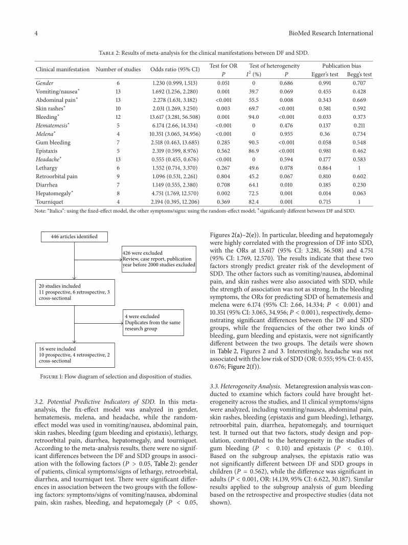
2 (%) 𝑃 Egger’s test Begg’s testGender 6 1.230 (0.999, 1.513) 0.051 0 0.686 0.991 0.707Vomiting/nausea∗ 13 1.692 (1.256, 2.280) 0.001 39.7 0.069 0.455 0.428Abdominal pain∗ 13 2.278 (1.631, 3.182) <0.001 55.5 0.008 0.343 0.669Skin rashes∗ 10 2.031 (1.269, 3.250) 0.003 69.7 <0.001 0.581 0.592Bleeding∗ 12 13.617 (3.281, 56.508) 0.001 94.0 <0.001 0.033 0.373Hematemesis∗ 5 6.174 (2.66, 14.334) <0.001 0 0.476 0.137 0.211Melena∗ 4 10.351 (3.065, 34.956) <0.001 0 0.955 0.36 0.734Gum bleeding 7 2.518 (0.463, 13.685) 0.285 90.5 <0.001 0.058 0.548Epistaxis 5 2.319 (0.599, 8.976) 0.562 86.9 <0.001 0.981 0.462Headache∗ 13 0.555 (0.455, 0.676) <0.001 0 0.594 0.177 0.583Lethargy 6 1.552 (0.714, 3.370) 0.267 49.6 0.078 0.864 1Retroorbital pain 9 1.096 (0.531, 2.261) 0.804 45.2 0.067 0.810 0.602Diarrhea 7 1.149 (0.555, 2.380) 0.708 64.1 0.010 0.185 0.230Hepatomegaly∗ 8 4.751 (1.769, 12.570) 0.002 72.5 0.001 0.014 0.063Tourniquet 4 2.194 (0.395, 12.206) 0.369 82.4 0.001 0.715 1Note: “Italics”: using the fixed-effect model, the other symptoms/signs: using the random-effect model; ∗significantly different between DF and SDD.
446 articles identified
426 were excludedReview, case report, publication year before 2000 studies excluded
20 studies included11 prospective, 6 retrospective, 3 cross-sectional
4 were excludedDuplicates from the same research group
16 were included10 prospective, 4 retrospective, 2 cross-sectional
Figure 1: Flow diagram of selection and disposition of studies.
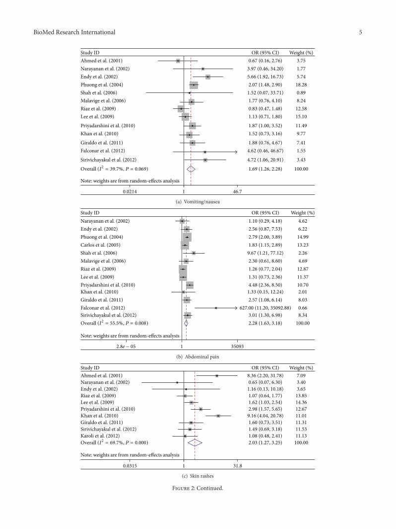
3.2. Potential Predictive Indicators of SDD. In this meta-analysis, the fix-effect model was analyzed in gender,hematemesis, melena, and headache, while the random-effect model was used in vomiting/nausea, abdominal pain,skin rashes, bleeding (gum bleeding and epistaxis), lethargy,retroorbital pain, diarrhea, hepatomegaly, and tourniquet.According to the meta-analysis results, there were no signif-icant differences between the DF and SDD groups in associ-ation with the following factors (𝑃 > 0.05, Table 2): genderof patients, clinical symptoms/signs of lethargy, retroorbital,diarrhea, and tourniquet test. There were significant differ-ences in association between the two groups with the follow-ing factors: symptoms/signs of vomiting/nausea, abdominalpain, skin rashes, bleeding, and hepatomegaly (𝑃 < 0.05,
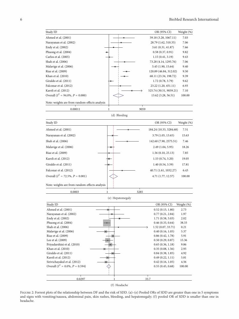
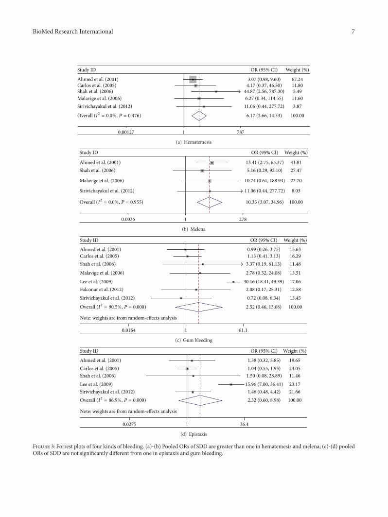
Figures 2(a)–2(e)). In particular, bleeding and hepatomegalywere highly correlated with the progression of DF into SDD,with the ORs at 13.617 (95% CI: 3.281, 56.508) and 4.751(95% CI: 1.769, 12.570). The results indicate that these twofactors strongly predict greater risk of the development ofSDD. The other factors such as vomiting/nausea, abdominalpain, and skin rashes were also associated with SDD, whilethe strength of association was not as strong. In the bleedingsymptoms, the ORs for predicting SDD of hematemesis andmelena were 6.174 (95% CI: 2.66, 14.334; 𝑃 < 0.001) and10.351 (95% CI: 3.065, 34.956; 𝑃 < 0.001), respectively, demo-nstrating significant differences between the DF and SDDgroups, while the frequencies of the other two kinds ofbleeding, gum bleeding and epistaxis, were not significantlydifferent between the two groups. The details were shownin Table 2, Figures 2 and 3. Interestingly, headache was notassociatedwith the low risk of SDD (OR: 0.555; 95%CI: 0.455,0.676; Figure 2(f)).
3.3. Heterogeneity Analysis. Metaregression analysis was con-ducted to examine which factors could have brought het-erogeneity across the studies, and 11 clinical symptoms/signswere analyzed, including vomiting/nausea, abdominal pain,skin rashes, bleeding (epistaxis and gum bleeding), lethargy,retroorbital pain, diarrhea, hepatomegaly, and tourniquettest. It turned out that two factors, study design and pop-ulation, contributed to the heterogeneity in the studies ofgum bleeding (𝑃 < 0.10) and epistaxis (𝑃 < 0.10).Based on the subgroup analyses, the epistaxis ratio wasnot significantly different between DF and SDD groups inchildren (𝑃 = 0.562), while the difference was significant inadults (𝑃 < 0.001, OR: 14.139, 95% CI: 6.622, 30.187). Similarresults applied to the subgroup analysis of gum bleedingbased on the retrospective and prospective studies (data notshown).
BioMed Research International 5
Note: weights are from random-effects analysis
Sirivichayakul et al. (2012)
Narayanan et al. (2002)
Khan et al. (2010)Priyadarshini et al. (2010)
Falconar et al. (2012)
Lee et al. (2009)
Giraldo et al. (2011)
Ahmed et al. (2001)
Shah et al. (2006)Phuong et al. (2004)
Riaz et al. (2009)
Endy et al. (2002)
Malavige et al. (2006)
Study ID
1.69 (1.26, 2.28)
4.72 (1.06, 20.91)
OR (95% CI)
3.97 (0.46, 34.20)
1.52 (0.73, 3.16)1.87 (1.00, 3.52)
4.62 (0.46, 46.67)
1.13 (0.71, 1.80)
1.88 (0.76, 4.67)
0.67 (0.16, 2.76)
1.52 (0.07, 33.71)2.07 (1.48, 2.90)
0.83 (0.47, 1.48)
5.66 (1.92, 16.73)
1.77 (0.76, 4.10)
100.00
3.43
Weight (%)
1.77
9.7711.49
1.55
15.10
7.41
3.75
0.8918.28
12.58
5.74
8.24
0.0214 1 46.7
Overall (I2 = 39.7%, P = 0.069)
(a) Vomiting/nausea
Study ID OR (95% CI) Weight (%)
Note: weights are from random-effects analysis
Priyadarshini et al. (2010)
Malavige et al. (2006)
Sirivichayakul et al. (2012)
Lee et al. (2009)
Endy et al. (2002)
Khan et al. (2010)
Falconar et al. (2012)
Riaz et al. (2009)
Giraldo et al. (2011)
Carlos et al. (2005)Shah et al. (2006)
Narayanan et al. (2002)
Phuong et al. (2004)
2.28 (1.63, 3.18)
4.48 (2.36, 8.50)
2.30 (0.61, 8.60)
3.01 (1.30, 6.98)
1.31 (0.73, 2.36)
2.56 (0.87, 7.53)
1.33 (0.15, 12.24)
627.00 (11.20, 35092.88)
1.26 (0.77, 2.04)
2.57 (1.08, 6.14)
1.83 (1.15, 2.89)9.67 (1.21, 77.12)
1.10 (0.29, 4.18)
2.79 (2.00, 3.89)
100.00
10.70
4.69
8.34
11.37
6.22
2.01
0.66
12.87
8.03
13.232.26
4.62
14.99
1 350932.8e − 05
Overall (I2 = 55.5%, P = 0.008)
(b) Abdominal pain
Study ID OR (95% CI) Weight (%)
Note: weights are from random-effects analysis
Priyadarshini et al. (2010)
Ahmed et al. (2001)
Endy et al. (2002)
Sirivichayakul et al. (2012)
Khan et al. (2010)
Lee et al. (2009)
Karoli et al. (2012)
Riaz et al. (2009)
Narayanan et al. (2002)
Giraldo et al. (2011)
2.03 (1.27, 3.25)
2.98 (1.57, 5.65)
8.36 (2.20, 31.78)
1.16 (0.13, 10.18)
1.49 (0.69, 3.18)
9.16 (4.04, 20.78)
1.62 (1.03, 2.54)
1.08 (0.48, 2.41)
1.07 (0.64, 1.77)
0.65 (0.07, 6.30)
1.60 (0.73, 3.51)
100.00
12.67
7.09
3.65
11.53
11.01
14.36
11.13
13.85
3.40
11.31
10.0315 31.8
Overall (I2 = 69.7%, P = 0.000)
(c) Skin rashes
Figure 2: Continued.
6 BioMed Research International
Study ID OR (95% CI) Weight (%)
Note: weights are from random-effects analysis
Giraldo et al. (2011)Falconar et al. (2012)
Endy et al. (2002)Phuong et al. (2004)
Karoli et al. (2012)
Malavige et al. (2006)
Narayanan et al. (2002)
Shah et al. (2006)
Khan et al. (2010)Riaz et al. (2009)
Ahmed et al. (2001)
Carlos et al. (2005)
13.62 (3.28, 56.51)
1.72 (0.78, 3.79)23.22 (1.20, 451.11)
3.61 (0.31, 41.87)0.58 (0.37, 0.91)
525.74 (30.51, 9059.21)
5.45 (1.90, 15.64)
28.79 (1.62, 510.35)
73.28 (4.14, 1295.76)
68.11 (23.34, 198.72)120.89 (46.84, 312.02)
59.18 (3.28, 1067.11)
1.15 (0.41, 3.19)
100.00
9.626.93
7.669.82
7.10
9.40
7.06
7.06
9.399.50
7.03
9.43
0.00011 1 9059
Overall (I2 = 94.0%, P = 0.000)
(d) Bleeding
Study ID OR (95% CI) Weight (%)
Note: weights are from random-effects analysis
Karoli et al. (2012)
Giraldo et al. (2011)
Shah et al. (2006)
Narayanan et al. (2002)
Falconar et al. (2012)
Malavige et al. (2006)
Ahmed et al. (2001)
Riaz et al. (2009)
4.71 (1.77, 12.57)
1.53 (0.74, 3.20)
1.40 (0.54, 3.59)
142.60 (7.90, 2575.51)
3.79 (1.05, 13.65)
40.71 (1.61, 1032.27)
2.49 (1.04, 5.95)
184.24 (10.33, 3284.68)
1.56 (0.10, 25.13)
100.00
19.05
17.81
7.46
15.63
6.43
18.26
7.51
7.85
10.0003 3285
Overall (I2 = 72.5%, P = 0.001)
(e) Hepatomegaly
Study ID OR (95% CI) Weight (%)
Lee et al. (2009)
Phuong et al. (2004)
Sirivichayakul et al. (2012)Karoli et al. (2012)
Narayanan et al. (2002)
Riaz et al. (2009)Malavige et al. (2006)
Khan et al. (2010)Giraldo et al. (2011)
Shah et al. (2006)
Endy et al. (2002)
Priyadarshini et al. (2010)
Ahmed et al. (2001)
0.55 (0.45, 0.68)
0.50 (0.29, 0.87)
0.46 (0.33, 0.64)
0.42 (0.16, 1.05)0.49 (0.22, 1.11)
0.77 (0.21, 2.84)
0.86 (0.42, 1.78)0.40 (0.16, 1.05)
0.35 (0.08, 1.56)0.84 (0.38, 1.85)
1.52 (0.07, 33.71)
1.71 (0.58, 5.03)
0.65 (0.36, 1.18)
0.52 (0.15, 1.80)
100.00
15.36
38.33
4.565.81
1.97
5.915.37
2.954.92
0.21
2.02
9.86
2.73
10.0297 33.7
Overall (I2 = 0.0%, P = 0.594)
(f) Headache
Figure 2: Forrest plots of the relationship between DF and the risk of SDD. (a)–(e) Pooled ORs of SDD are greater than one in 5 symptomsand signs with vomiting/nausea, abdominal pain, skin rashes, bleeding, and hepatomegaly; (f) pooled OR of SDD is smaller than one inheadache.
BioMed Research International 7
Study ID
Sirivichayakul et al. (2012)
Ahmed et al. (2001)
Shah et al. (2006)Carlos et al. (2005)
Malavige et al. (2006)
6.17 (2.66, 14.33)
OR (95% CI)
11.06 (0.44, 277.72)
3.07 (0.98, 9.60)
44.87 (2.56, 787.30)4.17 (0.37, 46.50)
6.27 (0.34, 114.55)
100.00
Weight (%)
3.87
67.24
5.4911.80
11.60
10.00127 787
Overall (I2 = 0.0%, P = 0.476)
(a) Hematemesis
Study ID OR (95% CI) Weight (%)
Sirivichayakul et al. (2012)
Shah et al. (2006)Ahmed et al. (2001)
Malavige et al. (2006)
10.35 (3.07, 34.96)
11.06 (0.44, 277.72)
5.16 (0.29, 92.10)13.41 (2.75, 65.37)
10.74 (0.61, 188.94)
100.00
8.03
27.4741.81
22.70
10.0036 278
Overall (I2 = 0.0%, P = 0.955)
(b) Melena
Note: weights are from random-effects analysis
Falconar et al. (2012)
Carlos et al. (2005)
Malavige et al. (2006)
Lee et al. (2009)
Sirivichayakul et al. (2012)
Shah et al. (2006)
Ahmed et al. (2001)
2.52 (0.46, 13.68)
2.08 (0.17, 25.31)
1.13 (0.41, 3.13)
2.78 (0.32, 24.08)
30.16 (18.41, 49.39)
0.72 (0.08, 6.34)
3.37 (0.19, 61.13)
0.99 (0.26, 3.75)
OR (95% CI)
100.00
12.58
16.29
13.51
17.06
13.45
11.48
15.63
10.0164 61.1
Study ID Weight (%)
Overall (I2 = 90.5%, P = 0.000)
(c) Gum bleeding
Note: weights are from random-effects analysis
Ahmed et al. (2001)
Lee et al. (2009)Shah et al. (2006)Carlos et al. (2005)
Sirivichayakul et al. (2012)2.32 (0.60, 8.98)
1.38 (0.32, 5.85)
15.96 (7.00, 36.41)1.50 (0.08, 28.89)1.04 (0.55, 1.93)
1.46 (0.48, 4.42)100.00
19.65
23.1711.4624.05
21.66
10.0275 36.4
Study ID OR (95% CI) Weight (%)
Overall (I2 = 86.9%, P = 0.000)
(d) Epistaxis
Figure 3: Forrest plots of four kinds of bleeding. (a)-(b) Pooled ORs of SDD are greater than one in hematemesis and melena; (c)-(d) pooledORs of SDD are not significantly different from one in epistaxis and gum bleeding.
8 BioMed Research International
3.4. Publication Bias. Funnel plots showed no publicationbias in the studies covering vomiting/nausea, abdominalpain, skin rashes, bleeding, or retroorbital pain (Figure S1and Table 2). The P values of Egger’s and Begg’s tests alsosuggested that publication bias had little impact on theresults. There were three signs, bleeding, gum bleeding, andhepatomegaly showing publication bias (Egger’s test: 𝑃 =0.041, 0.058, 0.014).
4. Discussion
The present study is the meta-analysis to comprehensivelyevaluate the correlation of clinical symptoms/signs with thedevelopment of SDD in patients with DF.The results showedthat a total of five symptoms/signs significantly predictdengue patients progressing into SDD: vomiting/nausea,abdominal pain, skin rashes, bleeding, and hepatomegaly.The other five factors were not associated with the dis-ease progression, including tourniquet versus nontourniquet,female versus male patients, lethargy, retroorbital pain, anddiarrhea. We found that patients with bleeding after DENVinfection had approximately a 14-fold increased risk forprogression into SDD (includingDHF andDSS).When com-pared with the frequencies of leucopenia and thrombocy-topenia, haemorrhagicmanifestations, such as gum bleeding,epistaxis, and gastrointestinal bleeding, are less frequent, butnot rare [28]. Our analysis included four kinds of bleeding:hematemesis, melena, gum bleeding, and epistaxis. Previousstudies showed that the frequencies of hematemesis, melena,gum bleeding, and epistaxis were higher in SDD patientsthan in DF patients, but none of them were related tothe risk of development of SDD in patients with thesesymptoms [1, 17, 18, 25]. A recent study also showed that thegastrointestinal bleeding was associated with DSS, althoughit is not a strong association (OR = 1.84) [29]. According toour meta-analysis, the two kinds of gastrointestinal bleedingthat strongly predicted SDD were hematemesis (OR: 6.174;95% CI: 2.66, 14.334; 𝑃 < 0.001) and melena (OR: 10.351;95% CI: 3.065, 34.956; 𝑃 < 0.001), while the other two kindsof bleeding were not significant risk factors. The other fourclinical symptoms and signs proved significant for predictingthe progression into SDD are vomiting/nausea (OR: 1.692;95% CI: 1.256, 2.280), abdominal pain (OR: 2.278; 95% CI:1631, 3.182), skin rashes (OR: 2.031; 95% CI: 1.269, 3.250), andhepatomegaly (OR: 4.751; 95%CI: 1.769, 12.570). Although thevomiting/nausea, abdominal pain, and skin rashes showeda weak association with SDD compared with DF patients,these warnings must be taken seriously as recent studiesdemonstrated that these symptoms were associated with themortality caused by dengue [30, 31]. We found that patientswith hepatomegaly after DENV infection had approximatelya 5-fold increased risk of progression into SDD; however, theCI was with a wide range. The possible reason is that the rateof hepatomegaly was significantly higher in adults than theelderly [32].
The most accepted hypothesis for progression of DFis that subneutralizing levels of DENV-specific antibodiesexacerbate the disease by means of an antibody-dependentenhancement of infection (ADE) [33], which induces
a complicated immunopathogenesis in the host. The extentof vascular permeability is enhanced as a result of ADE [34]and patients with SDD as well as alterations of endothelialcells have been shown to experience thrombocytopenia andcoagulation disorders [35].These significant symptoms/signs,especially the bleeding (hematemesis/melena) and hep-atomegaly, are manifested in patients with SDD as a resultof the aforementioned alterations. In the in vivo model forADE-induced SDD, gastrointestinal bleeding and viral RNAincreased in the liver were observed [36]. Additionally, basedon skin biopsies, IgM, beta 1 C-globulin, dengue antigen,and fibrinogen deposits were found to be present within orabout blood vessel walls of dermal papillae or in the bloodvessels [37], implying that skin rashes that appeared in DHFwere caused by an immunopathologic process. So in patientswith SDD, the host immune system plays a central role intriggering symptoms like bleeding, hepatomegaly, and skinrashes, which could be used to triage patients in need ofintensive care.
The unassociated factors/manifestations were gender,lethargy, retroorbital pain, diarrhea, and positivity of atourniquet test. However, the World Health Organization(WHO) has published guidelines stating that positivity of atourniquet test may be included in the clinical case definitionof dengue haemorrhagic fever [38], and an altered level ofconsciousness such as lethargy should be paid extra attention[2]. Although the results from this meta-analysis showedunexpected absence of associations, relaxing vigilance overthe patients with these symptoms/signs is not recommended,because the results were generated from a random-effectmodel that tends to be overconservative.
Furthermore, headache was a protective factor againstSDD after DENV infection (OR: 0.555; 95% CI: 0.455, 0.676),implying that dengue patients with headache had a lowerprobability to develop into SDD. The protective effect hasbeen proved by a retrospective cohort study [25]. However,in another study, BALB/c mice were infected with differentstrains of DENV which were isolated from DSS or DFpatients, respectively, and in the mice infected with the strainfromDSS patients, DENV-1 isolates appeared to be primarilyneurotropic, whereas in the cases of other strains the virusturned to mainly infect lung and liver [39]. Suggesting thathigh frequency of headache could occur in patients withSDD.
There are limitations in the present study. Firstly, theresults will not apply tomulticenter prospective studies, sincethe present meta-analysis only included retrospective andsingle-center prospective studies. These designs could noteliminate recall and selection biases. Hence, the true associ-ations between these symptoms/signs and the developmentof SDD might have been distorted. Secondly, the definitionsof DF and SDD within these studies may have varied, whichbrought uncertainty into determining cases. Lastly, someof the results were based on a random-effect model thatmight weaken the validity of the analysis. Nonetheless, thisstudy explored a new approach to identify the correlationsof the symptoms/signs after DENV infection with the riskof progression into SDD, which can greatly facilitate theprevention of SDD.
BioMed Research International 9
5. Conclusions
This meta-analysis identified clinical symptoms and signsthat significantly predicted DF patients progressing intosevere dengue. DF patients with vomiting/nausea, abdominalpain, skin rashes, bleeding (hematemesis/melena), and hep-atomegaly were more likely to develop SDD, while patientswith headache had a lower risk of progression into SDD.Other factors such as gender, lethargy, retroorbital pain,diarrhea, and positive tourniquet test are not associated withSDD. Further studies, especially ones with larger sample sizesand prospective, are warranted to confirm the findings.
Abbreviations
DENV: Dengue virusDF: Dengue feverSDD: Severe dengue diseaseDHF: Dengue haemorrhagic feverDSS: Dengue shock syndromeNOS: The Newcastle-Ottawa ScaleADE: Antibody-dependent enhancement of infectionWHO: TheWorld Health Organization.
Conflict of Interests
On behalf of all authors, the corresponding author states thatthere is no conflict of interests.
Authors’ Contribution
All authors were involved in the study design, includingdeveloping the search strategies and project protocol. H.Zhang, H. J. Peng, X. H. Zhang, F. Y. Zhou, Z. H. Liu, andX. G. Chen were responsible for supervising the project andperforming the literature search and data extraction. H. J.Peng, F. Y. Zhou, and Z. H. Liu assessed the quality of studies.H. Zhang performed data analysis and drafted the paper. X.H. Zhang was responsible for the language editorial. Y. P.Zhou and X. G. Chen revised the paper.
Acknowledgments
Thisworkwas supported byGrants from theNationalNaturalScience Foundation of China (30771899) to Y. P. Zhou andNIH (AI083202-02) to X. G. Chen.
References
[1] G. N. Malavige, S. Fernando, D. J. Fernando, and S. L. Senevi-ratne, “Dengue viral infections,” Postgraduate Medical Journal,vol. 80, no. 948, pp. 588–601, 2004.
[2] Research SPF, Diseases TIT, DiseasesWHOD, EpidemicWHO,and P. Alert, Dengue, Guidelines for Diagnosis, Treatment,Prevention and Control, World Health Organization, 2009.
[3] C. Chevillon and A.-B. Failloux, “Questions on viral populationbiology to complete dengue puzzle,”Trends inMicrobiology, vol.11, no. 9, pp. 415–421, 2003.
[4] J. G. Rigau-Perez, G. G. Clark, D. J. Gubler, P. Reiter, E. J.Sanders, and A. V. Vorndam, “Dengue and dengue haemor-rhagic fever,” Lancet, vol. 352, no. 9132, pp. 971–977, 1998.
[5] Impact of Dengue, http://www.who.int/csr/disease/dengue/impact/en/.
[6] E. Khan, J. Siddiqui, S. Shakoor, V. Mehraj, B. Jamil, and R.Hasan, “Dengue outbreak in Karachi, Pakistan, 2006: experi-ence at a tertiary care center,” Transactions of the Royal Societyof Tropical Medicine and Hygiene, vol. 101, no. 11, pp. 1114–1119,2007.
[7] D. Giraldo, C. Sant’Anna, A. R. Perisse et al., “Characteristics ofchildren hospitalized with dengue fever in an outbreak in Riode Janeiro, Brazil,” Transactions of the Royal Society of TropicalMedicine and Hygiene, vol. 105, no. 10, pp. 601–603, 2011.
[8] E. Khan, M. Kisat, N. Khan, A. Nasir, S. Ayub, and R. Hasan,“Demographic and clinical features of dengue fever in Pakistanfrom 2003-2007: A retrospective cross- sectional study,” PLoSONE, vol. 5, no. 9, Article ID e12505, pp. 1–7, 2010.
[9] G. S. Shah, S. Islam, and B. K. Das, “Clinical and laboratoryprofile of dengue infection in children,” Kathmandu UniversityMedical Journal, vol. 4, no. 13, pp. 40–43, 2006.
[10] D. Priyadarshini, R. R. Gadia, A. Tripathy et al., “Clinical find-ings and pro-inflammatory cytokines in dengue patients inWestern India: a facility-based study,” PLoS ONE, vol. 5, no. 1,Article ID e8709, 2010.
[11] C. Sirivichayakul, K. Limkittikul, P. Chanthavanich et al.,“Dengue infection in children in ratchaburi, thailand: a cohortstudy II. clinical manifestations,” PLoS Neglected Tropical Dis-eases, vol. 6, no. 2, Article ID e1520, 2012.
[12] J. A. Pawitan, “Dengue virus infection: predictors for severedengue.,” Acta Medica Indonesiana, vol. 43, no. 2, pp. 129–135,2011.
[13] F. R. Lima, M. G. Croda, D. A. Muniz, I. T. Gomes, K. R. Soares,and M. R. Cardoso, “Evaluation of the traditional and revisedWorld Health Organization classifications of dengue cases inBrazil,” Clinics, vol. 68, pp. 1299–1304, 2013.
[14] D. Moher, A. Liberati, J. Tetzlaff, and D. G. Altman, “Preferredreporting items for systematic reviews and meta-analyses: thePRISMA statement,” PLoS Medicine, vol. 6, no. 7, Article IDe1000097, 2009.
[15] G. Wells, B. Shea, O. Connell D, J. Peterson, and V. Welch,“The Newcastle-Ottawa Scale (NOS) for assessing the qualityof nonrandomised studies in meta-analyses,” Ottawa HospitalResearch Institute, 2012.
[16] J. P. T. Higgins, S. G. Thompson, J. J. Deeks, and D. G. Altman,“Measuring inconsistency in meta-analyses,” British MedicalJournal, vol. 327, no. 7414, pp. 557–560, 2003.
[17] F. U. Ahmed, C. B. Mahmood, J. D. Sharma, S. M. Hoque, R.Zaman, and M. S. Hasan, “Dengue and dengue haemorrhagicfever in children during the 2000 outbreak in Chittagong,Bangladesh,” Dengue Bulletin, vol. 25, pp. 33–39, 2001.
[18] C. C. Carlos, K. Oishi, M. T. D. D. Cinco et al., “Comparisonof clinical features and hematologic abnormalities betweendengue fever and dengue hemorrhagic fever among children inthe Philippines,”TheAmerican Journal of Tropical Medicine andHygiene, vol. 73, no. 2, pp. 435–440, 2005.
[19] T. P. Endy, S. Chunsuttiwat, A. Nisalak et al., “Epidemiologyof inapparent and symptomatic acute dengue virus infection:a prospective study of primary school children in KamphaengPhet, Thailand,” American Journal of Epidemiology, vol. 156, no.1, pp. 40–51, 2002.
10 BioMed Research International
[20] C. M. Falconar, A. K. Falconar, and C. M. Romero-Vivas,“Simple prognostic criteria can definitively identify patientswho develop severe versus non-severe Dengue disease, or haveother febrile illnesses,” Journal of ClinicalMedicine Research, vol.4, no. 1, pp. 33–44, 2012.
[21] M. Narayanan, M. A. Aravind, N. Thilothammal, R. Prema, C.S. R. Sargunam, and N. Ramamurty, “Dengue fever epidemicin Chennai—a study of clinical profile and outcome,” IndianPediatrics, vol. 39, no. 11, pp. 1027–1033, 2002.
[22] C. X. T. Phuong, N. T. Nhan, R. Kneen et al., “Clinical diagnosisand assessment of severity of confirmed dengue infections inVietnamese children: is theworld health organization classifica-tion system helpful?”TheAmerican Journal of TropicalMedicineand Hygiene, vol. 70, no. 2, pp. 172–179, 2004.
[23] G. N. Malavige, V. G. N. S. Velathanthiri, E. S. Wijewickrama etal., “Patterns of disease among adults hospitalized with dengueinfections,” QJM, vol. 99, no. 5, pp. 299–305, 2006.
[24] M. S. Lee, K. P. Hwang, T. C. Chen, P. L. Lu, and T. P. Chen,“Clinical characteristics of dengue and dengue hemorrhagicfever in a medical center of southern Taiwan during the 2002epidemic,” Journal of Microbiology, Immunology and Infection,vol. 39, no. 2, pp. 121–129, 2006.
[25] V. J. Lee, D. C. Lye, Y. Sun, and Y. S. Leo, “Decision treealgorithm in deciding hospitalization for adult patients withdengue haemorrhagic fever in Singapore,” Tropical Medicineand International Health, vol. 14, no. 9, pp. 1154–1159, 2009.
[26] R. Karoli, J. Fatima, Z. Siddiqi, K. I. Kazmi, and A. R. Sultania,“Clinical profile of dengue infection at a teaching hospital inNorth India,” The Journal of Infection in Developing Countries,vol. 6, no. 7, pp. 551–554, 2012.
[27] M. M. Riaz, K. Mumtaz, M. S. Khan et al., “Outbreak of denguefever in Karachi 2006: a clinical perspective,” Journal of thePakistan Medical Association, vol. 59, no. 6, pp. 339–344, 2009.
[28] J. G. Rigau-Perez, G. G. Clark, D. J. Gubler, P. Reiter, E. J.Sanders, and A. V. Vorndam, “Dengue and dengue haemor-rhagic fever,”The Lancet, vol. 352, no. 9132, pp. 971–977, 1998.
[29] N. T. Huy, T. van Giang, D. H. Thuy et al., “Factors associatedwith dengue shock syndrome: a systematic review and meta-analysis,” PLOS Neglected Tropical Diseases, vol. 7, article e2412,2013.
[30] L. A. Mena, J. Fernandez, A. Morales, Y. Soto, J. Feris-Iglesias,and M. O. Brito, “Disease severity and mortality caused bydengue in a Dominican pediatric population,” The AmericanJournal of Tropical Medicine and Hygiene, vol. 90, pp. 169–172,2014.
[31] T. L. Thein, Y. S. Leo, D. A. Fisher et al., “Risk factors forfatality among confirmed adult dengue inpatients in Singapore:a matched case-control study,” PLoS ONE, vol. 8, Article IDe81060, 2013.
[32] E. K. Rowe, Y. S. Leo, J. G. Wong et al., “Challenges indengue fever in the elderly: atypical presentation and risk ofsevere dengue and hospita-acquired infection,” PLoS NeglectedTropical Diseases, vol. 8, no. 4, Article ID e2777, 2014.
[33] S. B. Halstead, “Neutralization and antibody-dependent enha-ncement of dengue viruses,”Advances in Virus Research, vol. 60,pp. 421–467, 2003.
[34] S. B. Halstead, S. Mahalingam, M. A. Marovich, S. Ubol, andD. M. Mosser, “Intrinsic antibody-dependent enhancementof microbial infection in macrophages: disease regulation byimmune complexes,”The Lancet Infectious Diseases, vol. 10, no.10, pp. 712–722, 2010.
[35] C. Krishnamurti, S. Kalayanarooj, M. A. Cutting et al., “Mech-anisms of hemorrhage in dengue without circulatory collapse,”TheAmerican Journal of Tropical Medicine and Hygiene, vol. 65,no. 6, pp. 840–847, 2001.
[36] R. M. Zellweger, T. R. Prestwood, and S. Shresta, “Enhancedinfection of liver sinusoidal endothelial cells in a mouse modelof antibody-induced severe dengue disease,” Cell Host andMicrobe, vol. 7, no. 2, pp. 128–139, 2010.
[37] S. Boonpucknavig, V. Boonpucknavig, N. Bhamarapravati, andS. Nimmannitya, “Immunofluorescence study of skin rash inpatients with dengue hemorrhagic fever,” Archives of Pathologyand Laboratory Medicine, vol. 103, no. 9, pp. 463–466, 1979.
[38] WHO, Dengue Haemorrhagic Fever: Diagnosis, Treatment,Prevention and Control, World Health Organization, Geneva,Switzerland, 2nd edition, 1997.
[39] A. Tuiskunen, M. Wahlstrom, J. Bergstrom, P. Buchy, I. Leparc-Goffart, and A. Lundkvist, “Phenotypic characterization ofpatient dengue virus isolates in BALB/c mice differentiatesdengue fever and dengue hemorrhagic fever fromdengue shocksyndrome,” Virology Journal, vol. 8, article 398, 2011.

Submit your manuscripts athttp://www.hindawi.com
Hindawi Publishing Corporationhttp://www.hindawi.com Volume 2014
Anatomy Research International
PeptidesInternational Journal of
Hindawi Publishing Corporationhttp://www.hindawi.com Volume 2014
Hindawi Publishing Corporation http://www.hindawi.com
International Journal of
Volume 2014
Zoology
Hindawi Publishing Corporationhttp://www.hindawi.com Volume 2014
Molecular Biology International
GenomicsInternational Journal of
Hindawi Publishing Corporationhttp://www.hindawi.com Volume 2014
The Scientific World JournalHindawi Publishing Corporation http://www.hindawi.com Volume 2014
Hindawi Publishing Corporationhttp://www.hindawi.com Volume 2014
BioinformaticsAdvances in
Marine BiologyJournal of
Hindawi Publishing Corporationhttp://www.hindawi.com Volume 2014
Hindawi Publishing Corporationhttp://www.hindawi.com Volume 2014
Signal TransductionJournal of
Hindawi Publishing Corporationhttp://www.hindawi.com Volume 2014
BioMed Research International
Evolutionary BiologyInternational Journal of
Hindawi Publishing Corporationhttp://www.hindawi.com Volume 2014
Hindawi Publishing Corporationhttp://www.hindawi.com Volume 2014
Biochemistry Research International
ArchaeaHindawi Publishing Corporationhttp://www.hindawi.com Volume 2014
Hindawi Publishing Corporationhttp://www.hindawi.com Volume 2014
Genetics Research International
Hindawi Publishing Corporationhttp://www.hindawi.com Volume 2014
Advances in
Virolog y
Hindawi Publishing Corporationhttp://www.hindawi.com
Nucleic AcidsJournal of
Volume 2014
Stem CellsInternational
Hindawi Publishing Corporationhttp://www.hindawi.com Volume 2014
Hindawi Publishing Corporationhttp://www.hindawi.com Volume 2014
Enzyme Research
Hindawi Publishing Corporationhttp://www.hindawi.com Volume 2014
International Journal of
Microbiology