Embed Size (px)
Citation preview

SAGE-Hindawi Access to ResearchInternational Journal of InflammationVolume 2011, Article ID 514623, 9 pagesdoi:10.4061/2011/514623
Review Article
Oxidative Stress and Inflammation in Heart Disease:Do Antioxidants Have a Role in Treatment and/or Prevention?
Fredric J. Pashkow1, 2
1 John A. Burns School of Medicine, University of Hawai‘i, Honolulu, HI, USA2 Manoa Innovation Center, 2800 Woodlawn Drive, Honolulu, HI 96822, USA
Correspondence should be addressed to Fredric J. Pashkow, [email protected]
Received 15 February 2011; Revised 26 April 2011; Accepted 20 June 2011
Academic Editor: Ichiro Manabe
Copyright © 2011 Fredric J. Pashkow. This is an open access article distributed under the Creative Commons Attribution License,which permits unrestricted use, distribution, and reproduction in any medium, provided the original work is properly cited.
Inflammation triggered by oxidative stress is the cause of much, perhaps even most, chronic human disease including humanaging. The oxidative stress originates mainly in mitochondria from reactive oxygen and reactive nitrogen species (ROS/RNS) andcan be identified in most of the key steps in the pathophysiology of atherosclerosis and the consequential clinical manifestations ofcardiovascular disease. In addition to the formation of atherosclerosis, it involves lipid metabolism, plaque rupture, thrombosis,myocardial injury, apoptosis, fibrosis and failure. The recognition of the critical importance of oxidative stress has led to theenthusiastic use of antioxidants in the treatment and prevention of heart disease, but the results of prospective, randomizedclinical trials have been overall disappointing. Can this contradiction be explained and what are its implications for the discovery/development of future antioxidant therapeutics?
1. Introduction
While the importance of inflammation in illnesses where thephenomenon is overt, such as following trauma or infectionhas been recognized since ancient times, its presence andcrucial role in the manifestation of many diseases neverpreviously recognized as inflammatory is relatively recent.In such instances, the source of the inflammation is alsooften imperceptible [1]. This is especially relevant to themany pervasive chronic diseases that are still responsiblefor so much human suffering. We are currently achievinga major understanding of what is involved in the initiationof the inflammatory signaling cascade as well as the com-plex signaling pathways themselves that transcribe and coun-terregulate the molecular messengers (cytokines) that gen-erate the biological combatants such as the inflammatoryenzymes associated with the numerous relevant pathologies.In this paper we will overview some of the details of ouremerging understanding of inflammation and its principalsource, oxidative stress. Further, we will critically review thehistorical advocacy of antioxidants for the treatment andprevention of inflammatory-initiating oxidative stress, andproffer explanations for the failure to date of antioxidants
to achieve therapeutic success. Finally, we will discuss theappropriateness of oxidative stress as a therapeutic target incardiovascular disease [2] and the implications this has in usmoving forward in the discovery and development of newsafe and effective cardiovascular drugs.
2. Inflammation: A Major Causeof Human Disease
While inflammation occurring as a consequence of oxidativestress is not the only biological manifestation of excessROS/RNS [3], inflammation resulting from oxidative stressis the cause of much human disease [4]. Typical examplesare dyslipidemia [5], thrombosis [6, 7], metabolic syndrome[8], type 2 diabetes [9], nonalcoholic steatohepatitis (NASH)[10–12], macular degeneration [13], and neurodegenerativediseases such as Alzheimer’s [14]. Inflammation is also akey factor in all aspects of coronary disease including theinitiation and progression of atherosclerotic plaque, plaquerupture, and thrombosis (atherothrombosis), especially inrecurrent thrombosis where oxidative stress is known to playa significant role [15] (Figure 4), including in those withnormal cholesterol levels and in those being treated with
2 International Journal of Inflammation
Table 1: Human proof-of-concept studies demonstrating effectiveness of various antioxidant regimens on cardiovascular endpoints.
Study n Intervention Endpoint Antiox Rx results Placebo results P value
SPACE [16] 196 Vit E 800 IU/day Composite endpoint1 16% 33% = 0.014
IEISS [17] 125
Vit A 50,000 IU/day,Vit C 1,000 mg/day,Vit E 400 mg/day,β-carotene 25 mg/day
Individual componentscores2 20.6 30.6 “Sig. less”
VCE-MI [18] 61 Vit C&E 600 mg/day SAECG3 No Δ “Sig. Δ” 3 <0.002
PART [19] 101 Probucol 1,000 mg/day Restenosis p PCI 23% 58% = 0.001
ASAP [20] 520d-alpha-tocopherol 91 mg,Vit C 250 mg/day
Carotide IMT 0.011 mm/year-1 0.020 mm/year-1 = 0.008
MVP [21] 317
β-carotene 30,000 i.u.,Vit C 500 mg/day,Vit E 700 IU/day,Probucol 500 mg/day
Restenosis p PCI 28.9% 38.9% “Sig. less”
SPACE: Secondary Prevention with Antioxidants of Cardiovascular disease in Endstage renal disease; IEISS: Indian Experiment of Infarct Survival Study;VCE-MI: Vitamins C&E on Myocardial Infarction; PART: Probucol Angioplasty Restenosis Trial; ASAP: Antioxidant Supplementation in AtherosclerosisPrevention; MVP: Multivitamins and Probucol Study Group.1Composite Endpoint: myocardial infarction (fatal and nonfatal), ischemic stroke, non-AV fistular peripheral vascular disease, and unstable angina.2Individual Component Scores: mean infarct size (creatine kinase and creatine kinase-MB gram equivalents), serum glutamic-oxaloacetic transaminase,cardiac enzyme lactate dehydrogenase increased, and, QRS score in the electrocardiogram.3SAECG: Signal-average electrocardiogram components consist of increase in mean QRS and low-amplitude (<40 microV) signal durations, a decrease in theroot-mean-square voltage of the last 40 ms of the QRS complex.
“statins” and antiplatelet agents. This inflammation, causedby oxidative stress, could be a target for a great next wave ofcardiovascular therapeutics.
3. Role of Oxidative Stress
Oxidative stress has been identified as critical in most ofthe key steps in the pathophysiology of atherosclerosis andacute thrombotic events, including dyslipidemia leadingto atheroma formation, the oxidation of LDL, endothelialdysfunction, plaque rupture, myocardial ischemic injury,and recurrent thrombosis (i.e., the secondary, or subsequentclot that often occurs after initial thrombolysis). The roleof oxidative stress in the connection between the variouscoronary disease risk factors such as elevated blood pressure,diabetes and cigarette smoking, and the clinical sequelae ofdisease associated with vasoconstriction, thrombosis, plaquerupture, and vascular remodeling has been recognized byMoreno and Fuster [22] but its recognition as a specifictherapeutic target represents an elevation in its importancein the oxidative stress hypothesis [23]. Proinflammatorycytokines are also involved in cardiac muscle dysfunction andin the complex syndrome of heart failure [24–27]. Oxidativestress has been implicated as well in diabetic cardiomyopathy[28, 29], congestive cardiomyopathy [30], and hypertensiveheart disease [31].
4. Potential Role of Antioxidants
Recently, progress has been made regarding the source ofthe oxidative stress and an understanding has been achievedregarding the role of the signaling cascade that moderatesthe resulting inflammatory process. However, as far back as
the late 1940’s (and perhaps before), antioxidants such asvitamin E have been suggested as potentially useful in thetreatment of vascular disease [32]. Studies on the inhibitionof experimental cholesterol arteriosclerosis in animals werepublished around 1949-1950 and specific discussions of theuse of vitamin E in the treatment of cardiovascular diseaseappeared the same year [33, 34].
Over the years an oxidative stress hypothesis supportedby epidemiologic and observational evidence that encour-aged belief in and the use of antioxidants [35, 36]. Forexample, studies of fruit and vegetable consumption, thoseparticularly rich in vitamin C and other antioxidants, corre-lated with a reduction in CVD mortality [37]. Further, theplasma level of vitamin E was inversely related to mortalityfrom ischemic heart disease [38]. Numerous observationalstudies, such as the Nurses’ Health Study, reported signifi-cantly reduced risk in those taking vitamin E [39].
5. Human Proof-of-Concept StudiesInitially Encouraging
Up to the year 2000, several “smallish” trials using variouscombinations of antioxidant vitamins and drugs werereported as “positive.” This produced optimism in the com-munity of antioxidant advocates. The conclusions from aselection of these Phase 2-type studies are summarized inthe remainder of this paragraph and Table 1. In hemodialysispatients with prevalent cardiovascular disease, supple-mentation with 800 IU/day vitamin E reduced compositecardiovascular disease endpoints and myocardial infarctionaccording to Boaz and colleagues in Israel [16]. Singh andassociates reported results from a study in India suggestingthat combined treatment with antioxidant vitamins A, E, C,

International Journal of Inflammation 3
and beta-carotene in patients with recent acute myocardialinfarction might be protective against cardiac necrosis andoxidative stress and could be beneficial in preventing com-plications and in reduction of the cardiac event rate in ACSpatients [17]. Chamiec and colleagues reported results sup-porting the hypothesis that in patients with AMI, oxygen-freeradical-induced cellular damage contributes to alterations inelectric function of the heart as seen on the signal-averagedECG (SAECGs) and that vitamins C and E could reduce thesealterations [18]. Yokoi et al. reported that probucol admin-istered beginning 4 weeks before PTCA appears to reducesubsequent restenosis rates [19]. Salonen et al reportedthat combined supplementation with reasonable doses ofboth vitamin E and slow-release vitamin C could retard theprogression of common carotid atherosclerosis in men [20].And finally, Tardif and his colleagues reported from Montr-eal that the antioxidant probucol, with or without a combi-nation of antioxidant vitamins, is effective in reducing therate of restenosis after balloon coronary angioplasty [21].
It is important to note that these studies are all fairlymodest in size with the exception of ASAP [20] whichenrolled more than 500 subjects, and that with the exceptionof SPACE [16], use a surrogate measure for the primaryendpoint. Note that all of the therapies tested, again withthe exception of SPACE, involved subgroups where morethan one antioxidant or combinations of therapy wereused. Finally, while the statistical analyses suggest overallsignificance of the studies’ findings, only those receiving thedrug probucol with or without multivitamins demonstratedsignificant effect in the Mutivitamins and Probucol StudyGroup [21]. The implications of these observations will bediscussed further below.
6. Larger Randomized ClinicalTrials Unsupportive
But problems developed with the performance of largerrandomized clinical trials [41]. An earlier meta-analysis of6 large (>1,000 subjects) randomized trials of vitamin E withpooled data from over 77,000 subjects and 6 trials of β-carotene in over 131,000 subjects showed that the use ofvitamin E was a “wash” (P = 0.94) (Figure 1(a)) and thatβ-carotene use was associated with a worse outcome (P =0.003) (Figure 1(b)) [40].
The Cambridge Heart Antioxidant Study (CHAOS)buoyed hopes for believers in the oxidant stress hypothesiswhen it demonstrated a significant reduction in nonfatal MI(P = 0.0001) but offsetting that finding was an insignificantdifference in cardiovascular deaths (P = 0.78) [42]. However,in a large, long-term trial of male physicians, neither vitaminE nor vitamin C supplementation reduced the risk of majorcardiovascular events [43]. In women at high risk for CVDthere were no overall effects of ascorbic acid, vitamin E or β-carotene on cardiovascular events [44]. Hence, there is a needfor a better understanding and more scientific evidence ofthe relative contribution of major nutraceutical constituentsto the inhibition of the progression of atherosclerosis and itsclinical consequences [45].
Studies looking at progression of atherosclerosis usingvitamins E, C or β-carotene have also been inconclusive [47].However, certain specific patient populations or clinicalcircumstances showed promise: for example, reduction inthe development of transplant atherosclerosis [48]. In acutecoronary syndrome, N-Acetylcysteine (NAC) appeared toproduce a statistically significant improvement in cardiacindex in STEMI patients treated with streptokinase and NTG[49], and in a more recent study, a significant reduction ofin-hospital deaths in patients undergoing primary PCI [50].
7. Why the Failure of So ManyAntioxidant Trials?
The question is, if oxidative stress is so critical in the develop-ment and manifestations of coronary heart disease, why is itthat so many of the larger antioxidant trials have failed [52]?Before specifically attempting to answer this critical question,let us go back to basics for a moment. The utilization ofoxygen as an integral part of the process for generatingmetabolic energy (i.e., mainly via the Electron TransportChain or ETC on the inner membrane of the mitochondria)produces reactive oxygen species (ROS) [53, 54]. Thesereactive oxygen species can damage cells or componentsof cells by oxidizing DNA or proteins or starting chemicalchain reactions such as lipid peroxidation which incidentally,occurs mainly inside the bilayer membrane of cells, nuclei,and mitochondria [55]. ROS can be quite destabilizing tomembrane integrity, but they do have important usefulfunctions, such as the maintenance of balanced intracellularredox signaling. The function of antioxidant systems is not toremove these oxidants entirely, but instead to keep them at alevel below which they will trigger the inflammatory cascade,a series of intracellular and intranuclear signaling that resultsin the release of destructive inflammatory cytokines [56, 57].
8. Inflammation Is Complex
Pathological inflammation, a complex whole-cellular path-way, is a cascade that begins with the production of ex-cess free radicals that frequently arise from mitochondriaresponding to internal or environmental stress and that trig-ger several signaling steps that endup producing the substan-ces that actually cause the classical signs of redness, swelling,and pain in inflammation [1]. NF-kappa-B, one of the keysignaling molecules, is a transcription factor that upregulatesthe production of downstream inflammatory mediators,including tumor necrosis factor-alpha (TNF-alpha) [58], in-ducible NO synthase (iNOS), cyclooxygenase-2 (COX-2),and interleukein-1beta (IL-1beta). Recent studies have sug-gested that CD40-CD40L interactions themselves regulateoxidative stress and affect various signaling pathways in boththe immunological and cardiovascular systems [59]. Normalcellular functions “suckup” these unwanted ROS allowingthe signaling molecules, or cellular pathways such as NF-kappa-B, to operate normally along with the downstreamproducts of these pathways, creating a balanced ROS envi-ronment and normal cellular health.

4 International Journal of Inflammation
Trial
ATBC (n = 29133)
CHAOS (n = 2002)
GISSI (n = 11324)
HOPE (n = 9541)
HPS (n = 20536)
PPP (n = 4495)
Pooled (n = 77031)
Odds ratio (95% CI)
Absolute event rates
P = 0.46
P = 0.74
P = 0.45
P = 0.54
P = 0.34
P = 0.6
P = 0.94
Vitamin Eworse
Vitamin Ebetter
5%
2.6%
5.5%
7.2%
8.6%
1%
6%
0 0.5 1 1.5 2
5.2%
2.4%
5.8%
6.9%
8.2%
1.1%
6%
Vitamin E Controltreatment
Breslow-day test: P = 0.73
(a)
Trial
ATBC (n = 29133)
CARET (n = 18314)
NSCP (n = 1621)
PHS (n = 22071)
WHS (n = 39876)
Pooled (n = 131551)
Odds ratio (95% CI)
Absolute event rates
P = 0.08
P = 0.002
P = 0.34
P = 0.14
P = 0.32
P = 0.7
P = 0.003
5.3%
2.4%
8.6%
0.7%
3.1%
0.1%
3.4%
β caroteneworse
β carotene
better
0 0.5 1 1.5 2
β carotene Controltreatment
Breslow-day test: P = 0.12
4.9%
1.7%
8.2%
1.5%
2.8%
0.1%
3.1%
HPS (n = 20536)
(b)
Figure 1: (a) Meta-analysis of large randomized trials of vit E versus placebo. Meta-analysis of 7 randomized trials involving 77,031 patientscomparing the risk of cardiovascular death among those randomized to placebo or vitamin E (Breslow-Day test, P = 0.73). ATBC: α-Tocopherol, β-Carotene Cancer Prevention trial; CHAOS: Cambridge Heart Antioxidant Study; CI: Confidence Interval; GISSI – GruppoItaliano per lo Studio della Sopravivenza nell’Infarto; HOPE: Heart Ourcomes Prevention Evaluation; HPS: Heart Protection Study; PPP:Primary Prevention Project, modified from [40]. (b) Meta-analysis of Randomized Trials of β-carotene versus Placebo. Meta-analysis of 6randomized trials involving 131,551 patients comparing the risk of cardiovascular death among those randomized to placebo or β-carotene(Breslow-Day test, P = 0.12). ATBC: α-Tocopherol, β-Carotene Cancer Prevention trial; CARET: β-Carotene and Retinol Efficacy Trial; HPS:Heart Protection Study; NSCP: Nambour Skin Cancer Prevention; PHS: Physicians’ Health Study; WHS: Women’s Health Study, modifiedfrom [40].
9. Erroneous Assumptions InfluencedTrial Design
So why have so many antioxidant therapies failed when testedin randomized clinical trials? Studies were conducted basedupon epidemiologic findings of benefit from surveys doc-umenting increased intake of dietary fruits/vegetables [60].
The well-known biases of such studies aside, the assump-tion has been that “known” nutritional compounds that is,vitamin E, vitamin C, and β-carotene, are mainly responsiblefor the benefit. The favorable effects shown by some studiesrelating antioxidant dietary intake and cardiovascular diseasemay have been exerted by other chemicals present in foods.Flavonoids, for example, are ideal candidates, since they

International Journal of Inflammation 5
†
‡
∗
∗
∗
Astaxanthin
Zeaxanthin
Lutein
β-carotene
Lycopene
−30 −20 −10 0 10 20 30
Differences in relative electron density
−60 −30 0 30 60 90 120 150
Increase/decrease LOOH formationversus controls (%)
Figure 2: Antioxidant activity depends on molecular structure and localization (“Alignment”) in cellular/mitochondrial Membrane. Extentof deviation in the X-ray crystallographic pattern correlates with an increase in lipid peroxide formation. Correlation between membranestructure changes and LOOH formation (LOOH, lipid peroxide). Differences in relative electron density as a function of treatment withvarious carotenoids in POPC membranes containing a C/P mole ratio of 0.2. For the peroxidation study, various carotenoids (10 μM) wereincorporated into DLPC membranes and underwent lipid peroxidation at 37◦C for 48 h. Expressed as percent increase or decrease in LOOH(lipid peroxide) formation compared to controls containing no carotenoids. ∗P < 0.001 versus control; ‡P < 0.01 versus control; †P < 0.05versus control; n = 5 ∼ 6. (POPC, 1-palmitoyl 2-oleoyl-3-sn-glycerophosphatidylcholine), adapted from McNulty et al. [46].
are plentiful in foods containing antioxidant vitamins (i.e.,fruits and vegetables) and are potent antioxidants [61]. Otherexamples include cocoa [62, 63] and the active ingredients inwine [64, 65].
The naıve assumption is that all antioxidants are essen-tially the same. Nothing can be further from the truth. Allantioxidants are not the same: they may work in substan-tially different ways (chain-breaking versus singlet oxygenquenching, e.g.), and in different locations (e.g., in thebilayer membrane versus the cytoplasm) and very small dif-ferences in molecular structure can have profound influenceon biological activity. In the carotenoid family, for example,distinct effects occur in lipid peroxidation due to mem-brane structure changes. β-carotene, which misaligns whenlocalized in the bilayer membrane is highly disruptive struc-turally and can be functionally pro-oxidant when compar-ed to structurally similar members of the family that aligncompletely (Figure 2) [46]. These contrasting effects of caro-tenoids on lipid peroxidation may explain the clinical out-comes observed in various randomized trials.
10. Understanding Antioxidant Mode ofAction Is Critically Important
Further, lack of understanding of the mode of action [66] hasled to erroneous clinical designs and patient selection. Littleattempt was made to scale the antioxidant potential of thetherapy to the underlying oxidative stress. Atherosclerosis isa multifactorial disease and LDL is oxidized by all major cells
of the arterial wall during the development of atherosclerosisvia more than one mechanism. The various LDL oxidationpathways produce several lipid peroxidation products suchas isoprostanes from several fatty acids, oxysterols fromunesterified and esterified cholesterol, hydroxy fatty acids,lipid peroxides, and aldehydes. Intervention trials shouldbe accompanied by measurements of one or more of theserelevant biomarkers at intervals during the study and thecorrelation of the biomarkers to the therapeutic interventionneeds to be established. In addition to the markers in usefor lipid peroxidation, there is a need to include markers forendothelial dysfunction, monocyte adhesion, macrophageuptake of lipoproteins, thrombotic, and inflammatory pro-cesses [67].
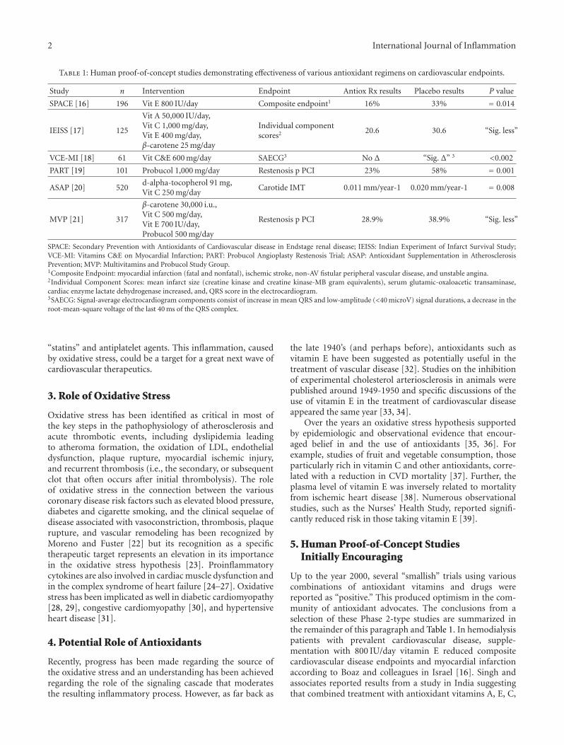
Our recognition of the connection between oxidativestress, inflammatory signaling, and such critical manifes-tations of atherosclerotic cardiovascular disease as athero-thrombosis is growing. The cellular membranes of endothe-lial cells can possess oxidized phospholipids with protrudingsn-2-oxidized fatty acid acyl chains into the extracellularspace. This conformation renders them accessible to interactwith scavenger receptors and other pattern recognitionreceptors on the surface of platelets or probing macrophagesof the circulatory and the immune system (Figure 3) [51].
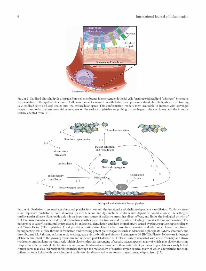
Oxidative stress is an important mediator of both ab-normal platelet function and dysfunctional endothelium-dependent vasodilation in the setting of cardiovascular dis-ease. Superoxide anion is an important source of oxidativestress, has direct effects, and limits the biological activity
6 International Journal of Inflammation
Senescent cell membrane
Senescent cell
Oxidizedlipid
Receptor
Macrophagecell membrane
Macrophage
Figure 3: Oxidized phospholipids protrude from cell membranes in senescent endothelial cells forming oxidized lipid “whiskers.” Schematicrepresentation of the lipid whisker model. Cell membranes of senescent endothelial cells can possess oxidized phospholipids with protrudingsn-2-oxidized fatty acid acyl chains into the extracellular space. This conformation renders them accessible to interact with scavengerreceptors and other pattern recognition receptors on the surface of platelets or probing macrophages of the circulatory and the immunesystem, adapted from [51].
Disrupted endothelium/adherent platelets
TF
Reactive oxygen species
Nitric oxide
Inflammatorymediators
(−)
(−)(−)
Antioxidants
(+)
(+)
(+)
(+)
(+)
(+)
(+)
Platelet activationand recruitment
Platelet disaggregation
Coagulationfactors
Thrombus formation
Reactive oxygen species
Inflammatorymediators O2
−
H2O2
Figure 4: Oxidative stress mediates abnormal platelet function and dysfunctional endothelium-dependent vasodilation. Oxidative stressis an important mediator of both abnormal platelet function and dysfunctional endothelium-dependent vasodilation in the setting ofcardiovascular disease. Superoxide anion is an important source of oxidative stress, has direct effects, and limits the biological activity ofNO. Excessive vascular superoxide production drives further platelet activation and recruitment leading to greater thrombus formation. Theoccurrence of superficial intimal injury caused by endothelial denudation and deep intimal injury caused by plaque rupture expose collagenand Tissue Factor (TF) to platelets. Local platelet activation stimulates further thrombus formation and additional platelet recruitmentby supporting cell-surface thrombin formation and releasing potent platelet agonists such as adenosine diphosphate (ADP), serotonin, andthromboxane A2. A thrombus forms as platelets aggregate via the binding of bivalent fibrinogen to GP IIb/IIIa. Platelet NO release influencesplatelet recruitment to the growing thrombus and impaired platelet-derived NO release is likely associated with acute coronary and strokesyndromes. Antioxidants may indirectly inhibit platelets through scavenging of reactive oxygen species, many of which alter platelet function.Despite the different subcellular locations of water- and lipid-soluble antioxidants, these antioxidant pathways in platelets are closely linked.Antioxidants may also indirectly inhibit platelets through the metabolism of reactive oxygen species, many of which alter platelet function.Inflammation is linked with the evolution of cardiovascular disease and acute coronary syndromes, adapted from [15].

International Journal of Inflammation 7
of NO. Excessive vascular superoxide production drives fur-ther platelet activation and recruitment leading to greaterthrombus formation. The occurrence of superficial intimalinjury caused by endothelial denudation and deep intimalinjury caused by plaque rupture expose collagen and tissuefactor [62] to platelets. Local platelet activation stimulatesfurther thrombus formation and additional platelet recruit-ment by supporting cell-surface thrombin formation andreleasing potent platelet agonists such as adenosine diphos-phate (ADP), serotonin, and thromboxane A2. A thrombusforms as platelets aggregate via the binding of bivalentfibrinogen to GP IIb/IIIa. Platelet NO release influencesplatelet recruitment to the growing thrombus and impairedplatelet-derived NO release is likely associated with acutecoronary and stroke syndromes (Figure 4) [15]. Thus, anti-oxidants may indirectly inhibit platelets through scavengingof reactive oxygen species, many of which alter plateletfunction. Despite the different subcellular locations of water-and lipid-soluble antioxidants, these antioxidant pathways inplatelets are closely linked. Antioxidants may also indirectlyinhibit platelets through the metabolism of reactive oxygenspecies, many of which directly alter platelet function.
Oxidative stress and inflammation are intimately linkedwith both the evolution of cardiovascular disease and acutecoronary syndromes. It should be no surprise that ox-LDLlevels show a significant positive correlation with the severityof acute coronary syndromes and that the more severelesions also contain a significantly higher percentage of ox-LDL-positive macrophages. Such observations suggest thatincreased levels of ox-LDL relate to plaque instability inhuman coronary atherosclerotic lesions [68, 69].
11. Dose-Response Documentation Lacking
Implicit in the randomized trials is that the dose of antiox-idant tested (usually vitamin E), effectively suppressed ox-idative stress but this was never determined [70]. In fact,studies suggest that the dosages of the compounds testedand/or the duration of therapy was not adequate. In onetime-course study, maximum suppression of plasma F2-isoprostane concentrations did not occur until 16 weeksof supplementation. In the dose-ranging study there was alinear trend between the dosage of vitamin E and percentagereduction in plasma F2-isoprostane concentrations whichreached significance at doses of 1600 IU (35 ± 2%, P <0.035) and 3200 IU (49 ± 10%, P < 0.005) [70]. Whethersuch dosages in human subjects would be safe and if thecompound was administered early enough in the lifecycle ofthe disease process are other essential considerations.
12. Safety Now an Overarching Issue
Safety is the overarching issue in drug development today,but little was done historically to determine if the antioxidantbeing tested for an inflammatory-mediated cardiovascularmanifestation is safely tolerated at the levels required toprovide therapeutic relief [71, 72]. Since the high standardsof chemistry, manufacturing, and controls (CMC) requiredfor pharmaceutical drugs are unlikely to be applied to nutra-
ceutical or dietary-supplement-type products, many of thequestions regarding safety and efficacy of antioxidants willmost likely be answered in the future related to the devel-opment of proprietary prodrugs seeking regulatory approv-al. These antioxidant drug candidates, incidentally, will mostlikely have greater druglikeness and bioavailability.
13. Conclusion
Despite the lack of significant randomized clinical trial datasupporting their use, more than $20b is still being spentannually on the antioxidant vitamins A, C, and E with morethan 6 million tons of the latter projected to be consumedannually on a global basis [73]. My belief is that antioxi-dants because of their provenance as “natural products” or“nutritional supplements” and their presumption of safetyand efficacy generated from the results of epidemiologicand observational studies early-on, have not been subjectedto the same stringent developmental requirements that areapplied to new pharmaceutical drug candidates. Biologicallyactive compounds, formulated properly, administered inappropriate amounts for an appropriate duration to the rightpatients will be required to achieve all the requirements thattruly define therapeutic success.
References
[1] W. Huang and C. K. Glass, “Nuclear receptors and inflamma-tion control: molecular mechanisms and pathophysiologicalrelevance,” Arteriosclerosis, Thrombosis, and Vascular Biology,vol. 30, no. 8, pp. 1542–1549, 2010.
[2] T. Munzel, T. Gori, R. M. Bruno, and S. Taddei, “Is oxidativestress a therapeutic target in cardiovascular disease?” EuropeanHeart Journal, vol. 31, no. 22, pp. 2741–2749, 2010.
[3] R. A. Roberts, R. A. Smith, S. Safe, C. Szabo, R. B. Tjalkens,and F. M. Robertson, “Toxicological and pathophysiologicalroles of reactive oxygen and nitrogen species,” Toxicology, vol.276, no. 2, pp. 85–94, 2010.
[4] F. Martinon, “Signaling by ROS drives inflammasome activa-tion,” European Journal of Immunology, vol. 40, no. 3, pp. 616–619, 2010.
[5] E. Hopps et al., “A novel component of the metabolicsyndrome: the oxidative stress,” Nutrition, Metabolism andCardiovascular Diseases, vol. 20, no. 1, pp. 72–77, 2009.
[6] J. Xu, F. Lupu, and C. T. Esmon, “Inflammation, innateimmunity and blood coagulation,” Hamostaseologie, vol. 30,no. 1, pp. 5–9, 2010.
[7] J. A. Leopold and J. Loscalzo, “Oxidative risk for atherothrom-botic cardiovascular disease,” Free Radical Biology andMedicine, vol. 47, no. 12, pp. 1673–1706, 2009.
[8] A. Iyer, D. P. Fairlie, J. B. Prins, B. D. Hammock, and L.Brown, “Inflammatory lipid mediators in adipocyte functionand obesity,” Nature Reviews Endocrinology, vol. 6, no. 2, pp.71–82, 2010.
[9] H. Kaneto, N. Katakami, M. Matsuhisa, and T. A. Matsuoka,“Role of reactive oxygen species in the progression of type 2diabetes and atherosclerosis,” Mediators of Inflammation, vol.2010, Article ID 453892, 2010.
[10] E. Hijona, L. Bujanda, L. Hijona, and J. I. Arenas, “Inflamma-tory mediators of hepatic steatosis,” Mediators of Inflamma-tion, vol. 2010, Article ID 837419, 2010.

8 International Journal of Inflammation
[11] B. L. Copple, H. Jaeschke, and C. D. Klaassen, “Oxidative stressand the pathogenesis of cholestasis,” Seminars in Liver Disease,vol. 30, no. 2, pp. 195–204, 2010.
[12] C. D. Byrne, “Fatty liver: role of inflammation and fattyacid nutrition,” Prostaglandins Leukotrienes and Essential FattyAcids, vol. 82, no. 4-6, pp. 265–271, 2010.
[13] A. J. Augustin and J. Kirchhof, “Inflammation and thepathogenesis of age-related macular degeneration,” ExpertOpinion on Therapeutic Targets, vol. 13, no. 6, pp. 641–651,2009.
[14] G. Candore, M. Bulati, C. Caruso et al., “Inflammation,cytokines, immune response, apolipoprotein E, cholesterol,and oxidative stress in alzheimer disease: therapeutic impli-cations,” Rejuvenation Research, vol. 13, no. 2-3, pp. 301–313,2010.
[15] J. E. Freedman, “Oxidative stress and platelets,” Arteriosclerosis,Thrombosis, and Vascular Biology, vol. 28, no. 3, pp. s11–s16,2008.
[16] M. Boaz, S. Smetana, T. Weinstein et al., “Secondary preven-tion with antioxidants of cardiovascular disease in endstagerenal disease (SPACE): randomised placebo-controlled trial,”The Lancet, vol. 356, no. 9237, pp. 1213–1218, 2000.
[17] R. B. Singh, M. A. Niaz, S. S. Rastogi, and S. Rastogi, “Use-fulness of antioxidant vitamins in suspected acute myocardialinfarction (the Indian Experiment of Infarct Survival-3),”American Journal of Cardiology, vol. 77, no. 4, pp. 232–236,1996.
[18] T. Chamiec, K. Herbaczynska-Cedro, and L. Ceremuzynski,“Effects of antioxidant vitamins C and E on signal-averagedelectrocardiogram in acute myocardial infarction,” AmericanJournal of Cardiology, vol. 77, no. 4, pp. 237–241, 1996.
[19] H. Yokoi, H. Daida, Y. Kuwabara et al., “Effectiveness ofan antioxidant in preventing restenosis after percutaneoustransluminal coronary angioplasty: the probucol angioplastyrestenosis trial,” Journal of the American College of Cardiology,vol. 30, no. 4, pp. 855–862, 1997.
[20] J. T. Salonen, K. Nyyssonen, R. Salonen et al., “AntioxidantSupplementation in Atherosclerosis Prevention (ASAP) study:a randomized trial of the effect of vitamins E and C on 3-year progression of carotid atherosclerosis,” Journal of InternalMedicine, vol. 248, no. 5, pp. 377–386, 2000.
[21] J. C. Tardif, G. Cote, J. Lesperance et al., “Probucol andmultivitamins in the prevention of restenosis after coronaryangioplasty,” The New England Journal of Medicine, vol. 337,no. 6, pp. 365–372, 1997.
[22] P. R. Moreno and V. Fuster, “New aspects in the pathogenesisof diabetic atherothrombosis,” Journal of the American Collegeof Cardiology, vol. 44, no. 12, pp. 2293–2300, 2004.
[23] V. J. Dzau, E. M. Antman, H. R. Black et al., “The cardiovascu-lar disease continuum validated: clinical evidence of improvedpatient outcomes: part I: Pathophysiology and clinical trialevidence (risk factors through stable coronary artery disease),”Circulation, vol. 114, no. 25, pp. 2850–2870, 2006.
[24] D. L. Mann and J. B. Young, “Basic mechanisms in conges-tive heart failure. Recognizing the role of proinflammatorycytokines,” Chest, vol. 105, no. 3, pp. 897–904, 1994.
[25] D. L. Mann, “Inflammatory mediators and the failing heart:past, present, and the foreseeable future,” Circulation Research,vol. 91, no. 11, pp. 988–998, 2002.
[26] G. Torre-Amione, “Immune activation in chronic heartfailure,” American Journal of Cardiology, vol. 95, no. 11, 2005.
[27] A. Blum, “Heart failure—new insights,” The Israel MedicalAssociation Journal, vol. 11, no. 2, pp. 105–111, 2009.
[28] H. Bugger and E. D. Abel, “Mitochondria in the diabeticheart,” Cardiovascular Research, vol. 88, no. 2, pp. 229–240,2010.
[29] M. Khullar, A. A. R. S. Al-Shudiefat, A. Ludke, G. Binepal,and P. K. Singal, “Oxidative stress: a key contributor to dia-betic cardiomyopathy,” Canadian Journal of Physiology andPharmacology, vol. 88, no. 3, pp. 233–240, 2010.
[30] S. Pankuweit, V. Ruppert, and B. Maisch, “Inflammation indilated cardiomyopathy,” Herz, vol. 29, no. 8, pp. 788–793,2004.
[31] A. U. Shahbaz, Y. Sun, S. K. Bhattacharya et al., “Fibrosis inhypertensive heart disease: molecular pathways and cardio-protective strategies,” Journal of Hypertension, vol. 28, no. 1,pp. S25–S32, 2010.
[32] A. Hall Ratcliffe, “Vitamin E in intermittent claudication,” TheLancet, vol. 2, no. 6590, pp. 1128–1130, 1949.
[33] M. E. Eisen and H. Gross, “Vitamin E in arteriosclerotic heartand peripheral vascular disease,” New York State Journal ofMedicine, vol. 49, no. 20, pp. 2422–2424, 1949.
[34] I. S. Ravin and K. H. Katz, “Vitamin E in the treatment ofangina pectoris,” The New England Journal of Medicine, vol.240, no. 9, pp. 331–333, 1949.
[35] E. B. Rimm, M. J. Stampfer, A. Ascherio, E. Giovannucci, G.A. Colditz, and W. C. Willett, “Vitamin E consumption andthe risk of coronary heart disease in men,” The New EnglandJournal of Medicine, vol. 328, no. 20, pp. 1450–1456, 1993.
[36] S. A. Stanner, J. Hughes, C. N. M. Kelly, and J. Buttriss, “Areview of the epidemiological evidence for the ’antioxidanthypothesis’,” Public Health Nutrition, vol. 7, no. 3, pp. 407–422,2004.
[37] A. J. Verlangieri, J. C. Kapeghian, S. El-Dean, and M.Bush, “Fruit and vegetable consumption and cardiovascularmortality,” Medical Hypotheses, vol. 16, no. 1, pp. 7–15, 1985.
[38] K. F. Gey and P. Puska, “Plasma vitamins E and A inverselycorrelated to mortality from ischemic heart disease in cross-cultural epidemiology,” Annals of the New York Academy ofSciences, vol. 570, pp. 268–282, 1989.
[39] D. H. Emmert and J. T. Kirchner, “The role of vitamin E in theprevention of heart disease,” Archives of Family Medicine, vol.8, no. 6, pp. 537–542, 1999.
[40] D. P. Vivekananthan, M. S. Penn, S. K. Sapp, A. Hsu, andE. J. Topol, “Use of antioxidant vitamins for the preventionof cardiovascular disease: meta-analysis of randomised trials,”The Lancet, vol. 361, no. 9374, pp. 2017–2023, 2003.
[41] G. Bjelakovic, D. Nikolova, L. L. Gluud, R. G. Simonetti, andC. Gluud, “Mortality in randomized trials of antioxidant sup-plements for primary and secondary prevention: systematicreview and meta-analysis,” Journal of the American MedicalAssociation, vol. 297, no. 8, pp. 842–857, 2007.
[42] N. G. Stephens, A. Parsons, P. M. Schofield et al., “Randomisedcontrolled trial of vitamin E in patients with coronary disease:Cambridge Heart Antioxidant Study (CHAOS),” The Lancet,vol. 347, no. 9004, pp. 781–786, 1996.
[43] H. D. Sesso, J. E. Buring, W. G. Christen et al., “VitaminsE and C in the prevention of cardiovascular disease in men:the physicians’ health study II randomized controlled trial,”Journal of the American Medical Association, vol. 300, no. 18,pp. 2123–2133, 2008.
[44] N. R. Cook, C. M. Albert, J. M. Gaziano et al., “A randomizedfactorial trial of vitamins C and E and beta carotene in thesecondary prevention of cardiovascular events in women:results from the women’s antioxidant cardiovascular study,”Archives of Internal Medicine, vol. 167, no. 15, pp. 1610–1618,2007.

International Journal of Inflammation 9
[45] L. Badimon, G. Vilahur, and T. Padro, “Nutraceuticals andatherosclerosis: human trials,” Cardiovascular Therapeutics,vol. 28, no. 4, pp. 202–215, 2010.
[46] H. P. McNulty, J. Byun, S. F. Lockwood, R. F. Jacob, andR. P. Mason, “Differential effects of carotenoids on lipidperoxidation due to membrane interactions: X-ray diffractionanalysis,” Biochimica et Biophysica Acta, vol. 1768, no. 1, pp.167–174, 2007.
[47] J. Bleys, E. R. Miller, R. Pastor-Barriuso, L. J. Appel, andE. Guallar, “Vitamin-mineral supplementation and the pro-gression of atherosclerosis: a meta-analysis of randomizedcontrolled trials,” American Journal of Clinical Nutrition, vol.84, no. 4, pp. 880–887, 2006.
[48] J. C. Fang, S. Kinlay, J. Beltrame et al., “Effect of vitamins Cand E on progression of transplant-associated arteriosclerosis:a randomised trial,” The Lancet, vol. 359, no. 9312, pp. 1108–1113, 2002.
[49] M. A. Arstall, J. Yang, I. Stafford, W. H. Betts, and J.D. Horowitz, “N-acetylcysteine in combination with nitro-glycerin and streptokinase for the treatment of evolvingacute myocardial infarction: safety and biochemical effects,”Circulation, vol. 92, no. 10, pp. 2855–2862, 1995.
[50] G. Marenzi, E. Assanelli, I. Marana et al., “N-acetylcysteineand contrast-induced nephropathy in primary angioplasty,”The New England Journal of Medicine, vol. 354, no. 26, pp.2773–2782, 2006.
[51] M. E. Greenberg, X. M. Li, B. G. Gugiu et al., “The lipidwhisker model of the structure of oxidized cell membranes,”Journal of Biological Chemistry, vol. 283, no. 4, pp. 2385–2396,2008.
[52] D. Steinberg and J. L. Witztum, “Is the oxidative modifica-tion hypothesis relevant to human atherosclerosis? Do theantioxidant trials conducted to date refute the hypothesis?”Circulation, vol. 105, no. 17, pp. 2107–2111, 2002.
[53] J. Hirst, M. S. King, and K. R. Pryde, “The production ofreactive oxygen species by complex I,” Biochemical SocietyTransactions, vol. 36, no. 5, pp. 976–980, 2008.
[54] G. Lenaz, “The mitochondrial production of reactive oxygenspecies: mechanisms and implications in human pathology,”IUBMB Life, vol. 52, no. 3-5, pp. 159–164, 2001.
[55] S. Raha and B. H. Robinson, “Mitochondria, oxygen freeradicals, disease and ageing,” Trends in Biochemical Sciences,vol. 25, no. 10, pp. 502–508, 2000.
[56] S. G. Rhee, “Cell signaling H2O2, a necessary evil for cellsignaling,” Science, vol. 312, no. 5782, pp. 1882–1883, 2006.
[57] M. Valko, D. Leibfritz, J. Moncol, M. T. D. Cronin, M.Mazur, and J. Telser, “Free radicals and antioxidants in normalphysiological functions and human disease,” InternationalJournal of Biochemistry and Cell Biology, vol. 39, no. 1, pp. 44–84, 2007.
[58] K. Vlantis and M. Pasparakis, “Role of TNF in pathologiesinduced by nuclear factor κb deficiency,” Current Directions inAutoimmunity, vol. 11, pp. 80–93, 2010.
[59] M. Rizvi, D. Pathak, J. E. Freedman, and S. Chakrabarti,“CD40-CD40 ligand interactions in oxidative stress, inflam-mation and vascular disease,” Trends in Molecular Medicine,vol. 14, no. 12, pp. 530–538, 2008.
[60] S. R. Steinhubl, “Why have antioxidants failed in clinicaltrials?” American Journal of Cardiology, vol. 101, no. 10, pp.S14–S19, 2008.
[61] A. Cherubini, G. B. Vigna, G. Zuliani, C. Ruggiero, U.Senin, and R. Fellin, “Role of antioxidants in atherosclerosis:epidemiological and clinical update,” Current PharmaceuticalDesign, vol. 11, no. 16, pp. 2017–2032, 2005.
[62] E. L. Ding, S. M. Hutfless, X. Ding, and S. Girotra, “Chocolateand prevention of cardiovascular disease: a systematic review,”Nutrition and Metabolism, vol. 3, article no. 2, 2006.
[63] L. Scheid, A. Reusch, P. Stehle, and S. Ellinger, “Antioxidanteffects of cocoa and cocoa products ex vivo and in vivo: isthere evidence from controlled intervention studies?” CurrentOpinion in Clinical Nutrition and Metabolic Care, vol. 13, no.6, pp. 737–742, 2010.
[64] J. B. Ruidavets, P. Ducimetiere, A. Evans et al., “Patterns ofalcohol consumption and ischaemic heart disease in culturallydivergent countries: the Prospective Epidemiological Study ofMyocardial Infarction (PRIME),” BMJ, vol. 341, no. 7783, p.1146, 2010.
[65] R. Rodrigo, A. Miranda, and L. Vergara, “Modulation ofendogenous antioxidant system by wine polyphenols inhuman disease,” Clinica Chimica Acta, vol. 412, no. 5-6, pp.410–424, 2011.
[66] S. L. Archer, J. Stamler, A. Moag-Stahlberg et al., “Associa-tion of dietary supplement use with specific micronutrientintakes among middle-aged american men and women: theINTERMAP study,” Journal of the American Dietetic Associa-tion, vol. 105, no. 7, pp. 1106–1114, 2005.
[67] M. Aviram, “Review of human studies on oxidative damageand antioxidant protection related to cardiovascular diseases,”Free Radical Research, vol. 33, pp. S85–S97, 2000.
[68] M. Anselmi, U. Garbin, P. Agostoni et al., “Plasma levels ofoxidized-low-density lipoproteins are higher in patients withunstable angina and correlated with angiographic coronarycomplex plaques,” Atherosclerosis, vol. 185, no. 1, pp. 114–120,2006.
[69] S. Ehara, M. Ueda, T. Naruko et al., “Elevated levels of oxidizedlow density lipoprotein show a positive relationship with theseverity of acute coronary syndromes,” Circulation, vol. 103,no. 15, pp. 1955–1960, 2001.
[70] L. J. Roberts, J. A. Oates, M. F. Linton et al., “The relationshipbetween dose of vitamin E and suppression of oxidative stressin humans,” Free Radical Biology and Medicine, vol. 43, no. 10,pp. 1388–1393, 2007.
[71] A. Jerome-Morais, A. M. Diamond, and M. E. Wright,“Dietary supplements and human health: for better or forworse?” Molecular Nutrition and Food Research, vol. 55, no. 1,pp. 122–135, 2011.
[72] P. Mason, “One is okay, more is better? Pharmacologicalaspects and safe limits of nutritional supplements,” Proceed-ings of the Nutrition Society, vol. 66, no. 4, pp. 493–507, 2007.
[73] P. M. Parker, The World Market for Unmixed Vitamin Eand Derivatives: A 2011 Global Trade Perspective, Icon GroupInternational, 2011.

Submit your manuscripts athttp://www.hindawi.com
Stem CellsInternational
Hindawi Publishing Corporationhttp://www.hindawi.com Volume 2014
Hindawi Publishing Corporationhttp://www.hindawi.com Volume 2014
MEDIATORSINFLAMMATION
of
Hindawi Publishing Corporationhttp://www.hindawi.com Volume 2014
Behavioural Neurology
EndocrinologyInternational Journal of
Hindawi Publishing Corporationhttp://www.hindawi.com Volume 2014
Hindawi Publishing Corporationhttp://www.hindawi.com Volume 2014
Disease Markers
Hindawi Publishing Corporationhttp://www.hindawi.com Volume 2014
BioMed Research International
OncologyJournal of
Hindawi Publishing Corporationhttp://www.hindawi.com Volume 2014
Hindawi Publishing Corporationhttp://www.hindawi.com Volume 2014
Oxidative Medicine and Cellular Longevity
Hindawi Publishing Corporationhttp://www.hindawi.com Volume 2014
PPAR Research
The Scientific World JournalHindawi Publishing Corporation http://www.hindawi.com Volume 2014
Immunology ResearchHindawi Publishing Corporationhttp://www.hindawi.com Volume 2014
Journal of
ObesityJournal of
Hindawi Publishing Corporationhttp://www.hindawi.com Volume 2014
Hindawi Publishing Corporationhttp://www.hindawi.com Volume 2014
Computational and Mathematical Methods in Medicine
OphthalmologyJournal of
Hindawi Publishing Corporationhttp://www.hindawi.com Volume 2014
Diabetes ResearchJournal of
Hindawi Publishing Corporationhttp://www.hindawi.com Volume 2014
Hindawi Publishing Corporationhttp://www.hindawi.com Volume 2014
Research and TreatmentAIDS
Hindawi Publishing Corporationhttp://www.hindawi.com Volume 2014
Gastroenterology Research and Practice
Hindawi Publishing Corporationhttp://www.hindawi.com Volume 2014
Parkinson’s Disease
Evidence-Based Complementary and Alternative Medicine
Volume 2014Hindawi Publishing Corporationhttp://www.hindawi.com