Embed Size (px)
Citation preview

Review ArticleInfluence of Electric, Magnetic, and Electromagnetic Fields onthe Circadian System: Current Stage of Knowledge
Bogdan Lewczuk,1 Grzegorz Redlarski,2,3 Arkadiusz gak,2 Natalia ZióBkowska,1
Barbara Przybylska-Gornowicz,1 and Marek Krawczuk2
1 Department of Histology and Embryology, Faculty of Veterinary Medicine, University of Warmia and Mazury,Oczapowskiego Street 13, 10-719 Olsztyn, Poland
2Department of Mechatronics and High Voltage Engineering, Gdansk University of Technology, Własna Strzecha Street 18A,80-233 Gdansk, Poland
3Department of Electrical Engineering, Power Engineering, Electronics, and Control Engineering, University of Warmia and Mazury,Oczapowskiego Street 11, 10-736 Olsztyn, Poland
Correspondence should be addressed to Bogdan Lewczuk; [email protected]
Received 5 April 2014; Revised 26 May 2014; Accepted 3 June 2014; Published 22 July 2014
Academic Editor: You-Lin Tain
Copyright © 2014 Bogdan Lewczuk et al. This is an open access article distributed under the Creative Commons AttributionLicense, which permits unrestricted use, distribution, and reproduction in any medium, provided the original work is properlycited.
One of the side effects of each electrical device work is the electromagnetic field generated near its workplace. All organisms,including humans, are exposed daily to the influence of different types of this field, characterized by various physical parameters.Therefore, it is important to accurately determine the effects of an electromagnetic field on the physiological and pathologicalprocesses occurring in cells, tissues, and organs. Numerous epidemiological and experimental data suggest that the extremelylow frequency magnetic field generated by electrical transmission lines and electrically powered devices and the high frequencieselectromagnetic radiation emitted by electronic devices have a potentially negative impact on the circadian system. On the otherhand, several studies have found no influence of these fields on chronobiological parameters. According to the current state ofknowledge, some previously proposed hypotheses, including one concerning the key role of melatonin secretion disruption inpathogenesis of electromagnetic field induced diseases, need to be revised. This paper reviews the data on the effect of electric,magnetic, and electromagnetic fields on melatonin and cortisol rhythms—two major markers of the circadian system as well as onsleep. It also provides the basic information about the nature, classification, parameters, and sources of these fields.
1. Introduction
One of the side effects of each electrical device work is theelectromagnetic field generated near its workplace. All organ-isms, including humans, are exposed daily to the influence ofdifferent types of this field, characterized by distinct physicalparameters.Therefore, it is important to accurately determinethe effects of electromagnetic field on organisms. All elec-trically powered devices and transmission lines generate thelow frequency (usually 50 or 60Hz) field, which has a quasi-stationary character and its two components—the electricand magnetic field—can be analysed separately. This fieldis considered as having a potentially negative impact onorganisms, although the mechanism of its biological action
remains unknown. On the other hand, electronic devices,such as mobile phones, television sets or radio transmitters,emit electromagnetic radiation with high frequencies (from300MHz to 300GHz). High energy radiation of this typecauses a thermal effect that may increase the temperature oftissues and organs and also cause serious damage to cells.Theinternational agency for research on cancer (IARC) in 2002classified the extremely low frequency magnetic field gener-ated by electrical devices as possibly carcinogenic to humans[1]. In 2011, the radio frequencies of electromagnetic fieldswere qualified by IARC and WHO as possibly increasing therisk of malignant brain tumour development [2].
The visible part of electromagnetic radiation, with a rela-tively narrow frequency band from 389 to 789 THz, plays
Hindawi Publishing CorporationBioMed Research InternationalVolume 2014, Article ID 169459, 13 pageshttp://dx.doi.org/10.1155/2014/169459

2 BioMed Research International
a key role in the regulation of the diurnal rhythms by havinginfluence on the activity of the suprachiasmatic nucleus viamelanopsin-positive ganglion cells of the retina [3]. However,several reports have provided evidence that electric andmagnetic fields also influence the circadian system. It hasbeen suggested that a deficiency in melatonin secretionmay be responsible for the oncogenic action of the electro-magnetic field [4].
The aim of the paper was to review the data on the effectsof electric,magnetic, and electromagnetic fields onmelatoninand cortisol rhythms, two major markers of the circadiansystem as well as on sleep. We also included information onthe nature, physical parameters, classification, and sources offields, whichmay be useful for biologists andmedical doctors.
2. Nature of Electric, Magnetic, andElectromagnetic Forces
In physical sciences, the electromagnetic field is the stateof space characterised by electrodynamic nature of forcesacting on electrically charged objects. In that context, theelectromagnetic field can be thought of as consisting of twoindependent components [5]:
(i) electric—represented by a state of space, known as anelectric field, in which Coulomb forces act on station-ary electrically charged objects,
(ii) magnetic—represented by a state of space, knownas a magnetic field, in which Lorenz forces act onnonstationary (moving) electrically charged objects(representing electric currents).
It may be interesting to note that according to the specialtheory of relativity, electric and magnetic fields are twoaspects of the same phenomenon depending on a chosenreference frame of observation—an electrical field in onereference frame may be perceived as a magnetic field in a dif-ferent reference frame.
Within the range of their influence, the electromagneticfields may affect physical objects, including living organisms.The effects of this influence depend on many factors. Amongthese, the most important are [5]
(i) field intensity—in the case of the electric field, itsintensity 𝐸 is expressed in volts per metre (V/m),while in the case of themagnetic field (MF) its intensi-ty𝐻 is expressed in amperes per metre (A/m),
(ii) distance 𝑅 from an object expressed in metres (m),(iii) frequency 𝑓 of radiated energy—in the case of time
dependent fields it is expressed in hertz (Hz), whilefor time independent fields their frequency 𝑓 equals0,
(iv) surface power density𝑃 (specific power) representingthe intensity of radiated energy (power) with the areathroughout this energy being radiated, expressed inwatts per square metre (W/m2).
It is worth mentioning at this point that the intensity ofa magnetic field 𝐻 is expressed in amperes per metre (A/m)
according to the SI standards. However, in the literature andscientific practice, very often, the induction of a magneticfield 𝐵 is used instead, which is expressed in tesla (T). Thesequantities—𝐻 and 𝐵—are interrelated through the mediummagnetic permeability 𝜇.
3. Electromagnetic Fields in the Habitat ofLiving Organisms
Electromagnetic radiation and fields have been accompa-nying living organisms since the dawn of life on Earth.However, their current intensity and omnipresence shouldbe attributed, first of all, to human activity—technologicaladvances in modern engineering related to the developmentand practical use of electrical power transmission systems,electrical equipment, and telecommunications.
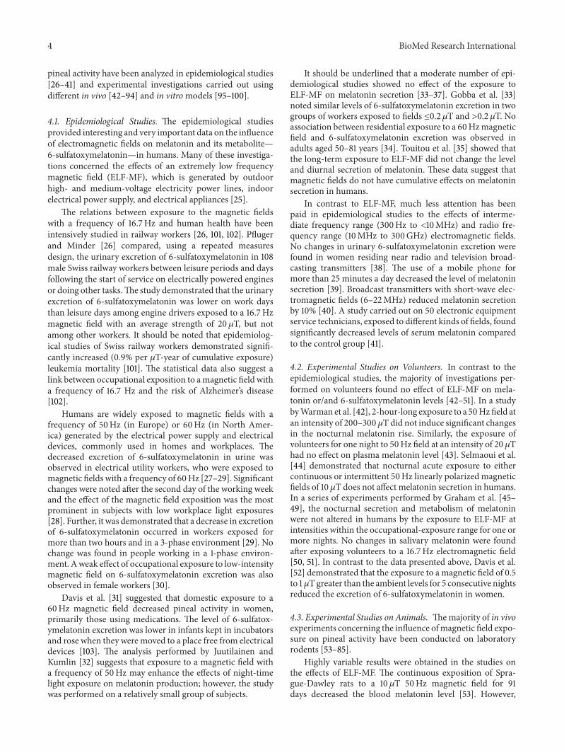
The sources of electromagnetic radiation and fields canbe divided into natural and nonnatural ones. The naturalsources include celestial bodies such as stars and magnetars,Earth and biological processes involving the flow of electricalimpulses in living organisms (Figure 1). The electromagneticradiation that reaches the Earth’s surface from space asmicrowave background radiation is a consequence of thebig bang and the evolution of the universe in the very firstseconds of its existence.This type of radiation is characterisedby its thermal energy distribution as the most perfect blackbody in nature and has a nearly ideal Planck spectrum at atemperature around 2.7 K, while the maximum of its surfacepower density corresponds to the wavelength of 272GHz [6].The solar radiation that reaches the Earth’s surface has rela-tively small surface power density around 3𝜇W/m2 [6] andcomprised of distinctive frequency bands, so-called atmo-sphericwindows, representing those frequency bands that arenot absorbed by the Earth atmosphere. They can be listed as
(i) radio window—represented by electromagneticwavelengths starting from 15MHz up to 300GHz,
(ii) optical window—represented by electromagneticwavelengths starting from 150 THz up to 1000 THz,
(iii) microwave window—represented by electromagneticwavelengths starting from 23.1 THz up to 37.5 THz.
Themagnetic field of Earth is another natural field originatingfrom the planet core that extends to a vast space surroundingEarth, known as the magnetosphere. An important sourceof strong electromagnetic fields is atmospheric discharges,known as lightning. Rapid radiation releases, which accom-pany these natural phenomena, are characterised by highpower densities and high frequencies. In living organisms,electromagnetic fields originate from the transmissionof signals in the nervous system and from structuresautonomously generating electrical impulses (like the heart).
The history of nonnatural sources of electromagneticradiation and fields is relatively short and covers only thelast hundred years. Nonnatural sources of electromagneticradiation or fields are attributed to two groups.Thefirst groupincludes ionising radiation, characterised by a relatively highenergy that may result in the ionisation of matter particles.The presence of this kind of radiation has primarily natural

BioMed Research International 3
Table 1: A list of various sources of electromagnetic fields/radiation influencing living organisms [7].
Level Frequency range Radiation source
Static 0Hz Earth, video screens, magnetic resonance imaging, and otherdiagnostic/scientific equipment, electrolysis, welding
Extremely low frequency fields 0–300Hz Power transmission lines, home wiring, car electric engines, electrictrains and trams, welding devices
Intermediate frequency 300Hz–100 kHzVideo screens, antitheft devices used in cars, homes, shops, cardreaders, metal detectors, magnetic resonance imaging, weldingdevices
Radio frequency 100 kHz–300GHz Radio, television, mobile phones, microwave ovens, radar and radiotransmitters, magnetic resonance imaging
Background (2.7K)
3𝜇W·m−2E
t
40𝜇s
300K
Earth (1.3mW·m−2)
1V·m−1÷ 10kV·m−1
Figure 1: Natural radiation sources present on Earth (based on [6]).
reasons (the statistical annual exposure dose is around2.4mSv). However, nonnatural sources of ionising radiation,such as technical devices, in which various radioactiveisotopes are used, are currently considered to be the mostimportant problems in public health protection. The secondgroup comprises nonionising radiation of energy, which istoo low to ionisematter particles.The common sources of thiskind of radiation are all means used for electrical power pro-duction, transmission, and utilisation (high-voltage powerlines, substations, motors, generators, industrial and domes-tic appliances, home wiring, etc.). Very important sourcesof electromagnetic radiation include telecommunication sys-tems (radio, television, internet, andWi-Fi) as well asmedicaldevices used for diagnosis or therapy.
According to the European Commission, nonionizingradiation can be divided into several levels [7]:
(i) static fields,(ii) extremely low frequency fields (ELF fields),(iii) intermediate frequency fields (IF fields),(iv) radio frequency fields (RF fields).
In order to illustrate the authors’ considerations, typical sour-ces of electromagnetic fields/radiation influencing living
organisms and mentioned above are listed and described inTable 1.
4. Effects of Electric, Magnetic, andElectromagnetic Fields on the DiurnalRhythm of Melatonin Secretion
Melatonin is the main hormone of the circadian timingsystem in all vertebrates including the human [8].The diurnalrhythm of its secretion in the mammalian pineal gland isdriven by the suprachiasmatic nucleus—the central endoge-nous oscillator, directly connected with the retina [8–10].Under physiological condition, the regulatory mechanismsensure that this rhythm is properly entrained to the light-dark cycle and, therefore, the elevated night-time melatoninsecretion can serve for all cells of the body as a clock and acalendar [8, 11, 12]. Melatonin plays a key role in the controlof many physiological processes occurring in daily or season-al rhythms, like sleep, metabolism, and reproduction [13].Moreover, melatonin is also involved in the regulation ofimmune system [14], cardiovascular system [15], and cancerdevelopment [13, 16, 17]. It is also a very potent free radicalscavenger [18].
It is worth to note that the level of melatonin secretiondiffersmarkedly between individuals, in both humans [19, 20]and animals [21, 22]. Based on urinary melatonin measure-ments, the human population could be divided into low andhigh melatonin excretors [19, 20]. The study on the sheepdemonstrated that interindividual variability in a plasmamelatonin level is under strong genetic control and it is relatedto the pineal glandweight andmelatonin secretion, but not tothe hormone catabolism [21]. The individual diurnal profilesof plasma melatonin are highly repeatable on consecutivedays, weeks, and months, in both humans and animals [20,22].The level of nocturnalmelatonin secretion decreaseswithage [23].
Several factors, like light pollution during night or mov-ing across time zones, may lead to the disruption of themelatonin secretion rhythm and circadian disorganization,which undoubtedly has a negative impact on various aspectsof health [13, 14, 16, 24, 25].
The melatonin secretion by the pineal gland is generallyregarded as particularly sensitive to electric, magnetic, andelectromagnetic field influences. The effects of these fields on
4 BioMed Research International
pineal activity have been analyzed in epidemiological studies[26–41] and experimental investigations carried out usingdifferent in vivo [42–94] and in vitromodels [95–100].
4.1. Epidemiological Studies. The epidemiological studiesprovided interesting and very important data on the influenceof electromagnetic fields on melatonin and its metabolite—6-sulfatoxymelatonin—in humans. Many of these investiga-tions concerned the effects of an extremely low frequencymagnetic field (ELF-MF), which is generated by outdoorhigh- and medium-voltage electricity power lines, indoorelectrical power supply, and electrical appliances [25].
The relations between exposure to the magnetic fieldswith a frequency of 16.7Hz and human health have beenintensively studied in railway workers [26, 101, 102]. Pflugerand Minder [26] compared, using a repeated measuresdesign, the urinary excretion of 6-sulfatoxymelatonin in 108male Swiss railway workers between leisure periods and daysfollowing the start of service on electrically powered enginesor doing other tasks.The study demonstrated that the urinaryexcretion of 6-sulfatoxymelatonin was lower on work daysthan leisure days among engine drivers exposed to a 16.7Hzmagnetic field with an average strength of 20𝜇T, but notamong other workers. It should be noted that epidemiolog-ical studies of Swiss railway workers demonstrated signifi-cantly increased (0.9% per 𝜇T-year of cumulative exposure)leukemia mortality [101]. The statistical data also suggest alink between occupational exposition to amagnetic field witha frequency of 16.7 Hz and the risk of Alzheimer’s disease[102].
Humans are widely exposed to magnetic fields with afrequency of 50Hz (in Europe) or 60Hz (in North Amer-ica) generated by the electrical power supply and electricaldevices, commonly used in homes and workplaces. Thedecreased excretion of 6-sulfatoxymelatonin in urine wasobserved in electrical utility workers, who were exposed tomagnetic fields with a frequency of 60Hz [27–29]. Significantchanges were noted after the second day of the working weekand the effect of the magnetic field exposition was the mostprominent in subjects with low workplace light exposures[28]. Further, it was demonstrated that a decrease in excretionof 6-sulfatoxymelatonin occurred in workers exposed formore than two hours and in a 3-phase environment [29]. Nochange was found in people working in a 1-phase environ-ment. Aweak effect of occupational exposure to low-intensitymagnetic field on 6-sulfatoxymelatonin excretion was alsoobserved in female workers [30].
Davis et al. [31] suggested that domestic exposure to a60Hz magnetic field decreased pineal activity in women,primarily those using medications. The level of 6-sulfatox-ymelatonin excretion was lower in infants kept in incubatorsand rose when they weremoved to a place free from electricaldevices [103]. The analysis performed by Juutilainen andKumlin [32] suggests that exposure to a magnetic field witha frequency of 50Hz may enhance the effects of night-timelight exposure on melatonin production; however, the studywas performed on a relatively small group of subjects.
It should be underlined that a moderate number of epi-demiological studies showed no effect of the exposure toELF-MF on melatonin secretion [33–37]. Gobba et al. [33]noted similar levels of 6-sulfatoxymelatonin excretion in twogroups of workers exposed to fields ≤0.2 𝜇T and >0.2𝜇T. Noassociation between residential exposure to a 60Hzmagneticfield and 6-sulfatoxymelatonin excretion was observed inadults aged 50–81 years [34]. Touitou et al. [35] showed thatthe long-term exposure to ELF-MF did not change the leveland diurnal secretion of melatonin. These data suggest thatmagnetic fields do not have cumulative effects on melatoninsecretion in humans.
In contrast to ELF-MF, much less attention has beenpaid in epidemiological studies to the effects of interme-diate frequency range (300Hz to <10MHz) and radio fre-quency range (10MHz to 300GHz) electromagnetic fields.No changes in urinary 6-sulfatoxymelatonin excretion werefound in women residing near radio and television broad-casting transmitters [38]. The use of a mobile phone formore than 25 minutes a day decreased the level of melatoninsecretion [39]. Broadcast transmitters with short-wave elec-tromagnetic fields (6–22MHz) reduced melatonin secretionby 10% [40]. A study carried out on 50 electronic equipmentservice technicians, exposed to different kinds of fields, foundsignificantly decreased levels of serum melatonin comparedto the control group [41].
4.2. Experimental Studies on Volunteers. In contrast to theepidemiological studies, the majority of investigations per-formed on volunteers found no effect of ELF-MF on mela-tonin or/and 6-sulfatoxymelatonin levels [42–51]. In a studybyWarman et al. [42], 2-hour-long exposure to a 50Hzfield atan intensity of 200–300𝜇T did not induce significant changesin the nocturnal melatonin rise. Similarly, the exposure ofvolunteers for one night to 50Hz field at an intensity of 20𝜇Thad no effect on plasma melatonin level [43]. Selmaoui et al.[44] demonstrated that nocturnal acute exposure to eithercontinuous or intermittent 50Hz linearly polarized magneticfields of 10 𝜇T does not affect melatonin secretion in humans.In a series of experiments performed by Graham et al. [45–49], the nocturnal secretion and metabolism of melatoninwere not altered in humans by the exposure to ELF-MF atintensities within the occupational-exposure range for one ormore nights. No changes in salivary melatonin were foundafter exposing volunteers to a 16.7Hz electromagnetic field[50, 51]. In contrast to the data presented above, Davis et al.[52] demonstrated that the exposure to a magnetic field of 0.5to 1 𝜇Tgreater than the ambient levels for 5 consecutive nightsreduced the excretion of 6-sulfatoxymelatonin in women.
4.3. Experimental Studies on Animals. Themajority of in vivoexperiments concerning the influence ofmagnetic field expo-sure on pineal activity have been conducted on laboratoryrodents [53–85].
Highly variable results were obtained in the studies onthe effects of ELF-MF. The continuous exposition of Spra-gue-Dawley rats to a 10 𝜇T 50Hz magnetic field for 91days decreased the blood melatonin level [53]. However,

BioMed Research International 5
another study from the same group failed to demonstrate aconsistent effect of a 100𝜇T 50Hz magnetic field exposureon melatonin levels in rats, as a decline or no changes wereobserved [54]. A decrease in the pineal activity in responseto ELF-MF was also noted in several other experimentsperformed on laboratory rats [55–63] and Djungarian ham-sters [64, 65]. On the other hand, an increased excretion of6-sulfatoxymelatonin was observed in Sprague-Dawley ratsexposed to a magnetic field with a frequency of 50Hz and anintensity of 100 𝜇T for 24 hours [66]. Similarly, Dyche et al.[67] demonstrated that male rats, exposed to the 100𝜇Tmagnetic field for 1 month, have a slightly elevated excretionof 6-sulfatoxymelatonin. Increased melatonin secretion afterexposure to a weak magnetic field was also reported in theDjungarian hamster by Niehaus et al. [68]. In other studiesperformed on rats and hamsters, no changes in melatoninsecretion were observed in response to a magnetic fieldwith a frequency of 50/60Hz [69–77]. The lack of influenceof ELF-MF on pineal activity was also reported for mice[78].
Studies on rodents have provided interesting data con-cerning the effect of radio frequency range of electromagneticfield on pineal activity.The exposure of rats to an electromag-netic field of 900MHz frequency and a specific adsorption of0.9W⋅kg−1 (mobile phone) lasting 2 hours a day and repeatedfor 45 days resulted in a statistically significant decrease inpineal melatonin content [81]. Moreover, a field of 1800MHzfrequency and a power of 200W⋅cm−2 (2 hours per day for32 days; 0.5762W⋅kg−1) disturbed the rhythm of melatoninsecretion in rats [82]. However, in another experiment, theanimals were subjected to a similar field for 30 minutes aday, 5 days a week for 4 weeks and no changes in the level ofmelatonin in rat serum were noted [83]. Similarly, the expo-sure of Djungarian hamsters to an electromagnetic field withfrequencies of 383, 900, and 1800MHz (80mW⋅kg−1) for 60days (24 hours a day) did not result in alternations of themelatonin secretion [84].
Studies on the effects of electric and magnetic fields onnonrodent species have been conducted only occasionally[86–94].The exposure of dairy cattle to a vertical electric fieldof 10 kV/m and a uniform horizontal magnetic field of 30 𝜇Tfor 28 days did not change the nocturnal blood melatoninlevel [86]. Similarly, no changes in melatonin secretion wereobserved in other experiments performed on dairy cows[87, 88] and on lambs [89, 90]. The studies of Americankestrels reveled that a long-term exposure to electromagneticfields (60Hz, 30 𝜇T, 10 kV⋅m−1) caused changes in melatoninsecretion [91]. Themagnetic field increased the level of mela-tonin in the pineal gland and blood serum of trout during thenight [92].
4.4. In Vitro Studies. In vitro studies concerning the effectof electromagnetic fields on melatonin secretion were con-ducted on the pineal glands of Djungarian hamsters [95, 100]and rats [96–99]. The results of experiments with hamsterpineals in the superfusion organ culture demonstrated thatELF-MF with an intensity of 86 𝜇T and a frequency of16.67 or 50Hz caused a decrease in melatonin secretion,
activated by isoproterenol [95]. A reduction in isoproterenol-stimulatedmelatonin secretion and activity of arylalkylamineN-acetyltransferase has also been found in studies of ratpinealocytes after exposure to ELF-MF [96, 97]. On the con-trary, Lewy et al. [98] noted increased activity of melatonin-synthetizing enzymes, while Tripp et al. [99] found nochanges inmelatonin secretion in rat pinealocytes in responseto ELF-MF.
The effect of exposure to an electromagnetic field witha frequency of 1800MHz on melatonin secretion from theDjungarian hamster pineal gland was investigated [100]in the same experimental setup which had been used inexperiments with ELF-MF [95].This study demonstrated thatboth continuous and pulse signals at a specific adsorptionlevel of 800mW⋅kg−1, lasting seven hours, increased the levelof isoproterenol-stimulated melatonin secretion [100].
5. Effects of Electric, Magnetic, andElectromagnetic Fields on the DiurnalRhythm of Cortisol Secretion
Cortisol is an essential steroid hormone produced by theadrenal gland. Like melatonin, it exhibits a constant andreproducible diurnal rhythm under physiological conditions[104–107]. Debono et al. [105] in a study of 33 healthy indi-viduals with 20-minute-interval cortisol profiling over 24hours showed that the cortisol concentration reached thelowest levels at around midnight. It then started to rise at02:00–03:00 and the peak occurred at around 08:30. Next, thecortisol level slowly decreased back to the nadir.Thepeak cor-tisol level in the human bloodwas approximately 399 nmol/L,while the nadir cortisol level was <50 nmol/L. Like manyother physiological processes in the body occurring in dailycycles, the rhythm of cortisol secretion is regulated by thesuprachiasmatic nucleus, located in the hypothalamus.
Cortisol governs hunger and appetite, stress, inflam-matory response, and many other functions [108–110]. Theimportance of cortisol is especially evident when it becomesdeficient in a state known as adrenal insufficiency [111]. Ithas been suggested that cortisol acts as a secondary messen-ger between central and peripheral clocks and may be animportant factor in the synchronization of body circadianrhythms [111]. Alterations in the rhythmic production andlevel of the cortisol lead to significant adverse effects [108,112]. Children with autism frequently show a large variationin day-time patterns of cortisol and significant elevationsin salivary cortisol in response to a nonsocial stressor[113].
Both people and animals live in environments with elec-tromagnetic fields of different origins. They are exposed toelectromagnetic field of natural origin, like the magneticforce of Earth and artificial origins, which results fromhuman activities. Variations in the Earth’s magnetic field areconsequential to all living beings of the planet. In addition,electric and magnetic fields, which exist wherever electricityis generated or transmitted, seem to be very important toexposed organisms.

6 BioMed Research International
5.1. Experimental Studies on Animals. The results of studieson the effects of electromagnetic field on the secretion ofcortisol in animals are very diverse. In Guinea pigs, ELF-MF caused changes in cortisol levels, which depended onthe field frequency and intensity [114]. Exposure of animalsfor 2 h and 4 h per day, over a period of 5 days, to a field of50Hz and 0.207𝜇T showed a significant decrease in cortisollevels [114]. However, in the groups subjected to a field of5Hz and 0.013 𝜇T, no significant changes in cortisol wereobserved after 2 h or 4 h of exposure [114]. In Swiss micecontinuously exposed to a low frequency (50Hz) field for 350days, a decrease in cortisol value was observed on day 190 ofthe experiment [115]. No significant differences were notedon days 90 and 350 of the exposure [115]. An increase in thecortisol level was observed in rats exposed to uniform mag-netic fields of 10−3 T and 10−2 T, 1 hour each day for a periodof ten days [116]. The exposure of female hamsters to mobilephones working at 950MHz for short (10 days, 3 h daily) andlong (60 days, 3 h daily) periods caused a significant increasein cortisol in comparison with the control group [117].
A lack of electromagnetic field effect on cortisol concen-tration was also reported. Burchard et al. [118] showed novariation in cortisol concentration, which could be attributedto the exposure of dairy cows to electric and magnetic fields(vertical electric field 10 kV and horizontal magnetic fieldof 30mT). In ewe lambs, no effect of the exposition to a60Hz magnetic field for 43 weeks on serum cortisol wasalso reported [119]. A lack of electromagnetic field effecton corticosterone concentration, irrespective of the exposurecharacteristics and period, was also found in experiments onrats [120, 121].
5.2. Studies in Humans. The studies concerning the influenceof the Earth’s magnetic force on the human body demon-strated that the serum cortisol values were dependent onthe direction of the head during sleep in relation to theNorth and South Magnetic Poles [122]. The biological effectof exposure to man-made electromagnetic fields on humanswas the subject of several studies [123–127]. Dentistry is oneof the job categories with high exposure to elevated levelsof ELF-MF. Exposure of dentists to the fields emitted bycavitrons caused a decrease in the serum cortisol level incomparison with a control group [123]. Low frequency mag-netic fields are applied in physiotherapy (magnetotherapy andmagnetostimulation). Studies of the long-term application ofthese procedures suggest a regulating influence of magneticfields on cortisol concentration [124]. However, it shouldbe stressed that numerous studies found no effect of themagnetic fields 50/60Hz (1–20𝜇T) and the radio frequencyelectromagnetic fields on a level of cortisol, irrespective of theexperiment time, age, or sex of individuals or sampling time[125–127].
6. Effects of Electric, Magnetic, andElectromagnetic Fields on Sleep
The diurnal rhythms are generated by an internal biologicalclock system that is synchronized to a 24-hour day by
environmental factors, primarily the light-dark cycle. Manyrhythms are overt and easy to recognize, such as the sleep-wake cycle, locomotor activity, and feeding behavior.
The sleep-wake cycle is likely the primary output rhythmof the circadian clock, because the regulation of manybehavior and physiological activities depends on whetherthe organism is asleep or awake. Sleep disorders—frequentlyoccurring clinical symptoms—have been hypothesized to bepartially related to electromagnetic field exposure. In recentyears, there has been an increasing amount of experimentaland epidemiological data on the influence of nonionizingelectromagnetic fields on brain physiology and sleep [40, 128–144].
Sleep is an endogenous, self-sustained cerebral process.It is possible to measure defined and distinguishable phasesof sleep. The low frequency activity (<10Hz) and the sleepspindle frequency activity (approximately 12–15Hz) are twosilent features of nonrapid eye movement (NREM) sleep thatcan be quantified and used as markers of sleep regulatingprocesses [145]. Several experiments have shown that elec-troencephalographic (EEG) spectral power in the alpha (8–12Hz) and spindle (12–14Hz) frequencies is enhanced bothduring and following pulsed-modulated radio frequency fieldexposure [128–133]. Recently, an increase in delta power(<4.5Hz) has also been observed [129]. Mann and Roschke[134] reported a reduction of rapid eye movement (REM)sleep and changes in spectral power of EEG during REMsleep in response to a high frequency electromagnetic fieldemitted by digital mobile radio telephones. Regel et al. [130]performed a study on the influence of radio frequencyelectromagnetic field exposure by varying the signal intensityin three experimental sessions. The analysis of the sleepEEG revealed a dose-dependent increase of power in thespindle frequency range in NREM sleep. This provided thefirst indications of a dose-dependent relation between thefield intensity and its effect on brain physiology. Huber et al.[137] also demonstrated a power increase in the fast spindlefrequency range of EEG during pulse-modulating radio fre-quency field exposure but not in a dose-dependentmanner. Itshould be also stressed that many studies [135, 139–141] failedto show any effects of the radio frequency field exposure onsleep or sleep EEG.
Despite several reports showing an influence of pulsed-modulated radio frequency electromagnetic field on sleepEEG, the mechanism behind these exposure-induced chan-ges is still unclear. Additionally, there is no supporting evi-dence that this effect is related to health consequences suchas alterations in sleep quality [128–130, 136].
To date, there have been few controlled laboratory studieson sleep EEG under low frequency electric and magneticfields. Akerstedt et al. [143] carried out a double-blind, pla-cebo-controlled study on 18 healthy subjects to examinethe effects of a 50Hz magnetic field on sleep. The resultsshowed that sleep efficiency, slow wave sleep, and slowactivity as well as subjective depth of sleep were significantlyreduced under ELF-MF exposure. Although these resultssuggest an interference of the low frequency field, the authorsemphasize that these alterations are still within a normalrange. In a double-blind laboratory study, Graham et al. [144]

BioMed Research International 7
investigated the effect of a 60Hz magnetic field on sleepduring continuous, intermittent, or sham exposures. Theydemonstrated that intermittent exposure resulted in cleardistortion of sleep and altered sleep architecture comparedto sham conditions and continuous exposure. It should beemphasized that field strengths in both cited studies [143, 144]were below those used for medical diagnostic purposes suchas magnetic resonance imaging.
The analysis of epidemiological data concerning the sleepquality and melatonin cycle, collected during ten years inthe area surrounding a short-wave (6–22MHz) broadcastingstation, provided the evidence that electromagnetic fieldexposure only affects poor sleepers and that might be agroup of people who are sensitive to such exposure [40].Thisphenomenon has been described as electromagnetic hyper-sensitivity, EHS. It was also observed in several other reports[146, 147].
Although a biological explanation for an associationbetween exposure to radio frequency electromagnetic fieldand impaired sleep quality has not been identified, it ishypothesized that the suppression of night-time melatoninsecretionmay be involved in this process [148]. Two relativelyrecent studies suggest an association between the decreasedsecretion of melatonin during the night and increasing useof mobile phones emitting a radio frequency field [39, 149].However, four cross-over trials [127, 141, 150, 151] have foundno correlation between the exposure tomobile phone handsetand themelatonin secretion.The hypothesis of an associationbetween melatonin cycle and electromagnetic field exposurerequires further investigation [152].
7. Conclusions
The results of studies on the effects of electric, magnetic, andelectromagnetic fields on melatonin and cortisol secretionas well as on sleep are largely contradictory. The adversedata related to the influence of these physical factors onsecretion of both “circadian” hormones were obtained inall groups of investigations including the epidemiologicalstudies, the studies on volunteers, and the studies on animals.Moreover, in vitro investigations on rodent pineals have alsobrought inconsistent results. The sources of discrepanciesremain unknown; however such factors as an inappropri-ate estimation of exposure level, interferences with otherfactors like light and medication, differences in a phase ofthe circadian rhythm during exposure, and interindividualvariability in the sensitivity to electromagnetic fields seemto be particularly worth of attention. The idea that someindividuals are more sensitive to the electromagnetic fieldthan others, due to genetic background or/and current healthstatus, appears very attractive and should be a subject offurther studies. It is worth to note that inconsistent resultshave been also obtained in the studies dealing with othereffects of electrical, magnetic, and electromagnetic fields onorganism, including their tumor-promoting action [153–157].
Despite divergences in the reported results, ELF-MF andradio frequency electromagnetic field have to be consideredas factors possibly influencing the circadian system function,
because a substantial number of studies demonstrated thechanges inmelatonin and cortisol secretion as well as in sleepafter exposition to these fields. Due to widespread exposureof humans and animals to ELF-MF and radio frequencyelectromagnetic field, the studies on their biological effectsshould be continued. An important and still unsolved issue isrelationships between physical characteristics and biologicaleffects of the fields as well as the mechanisms of field actionon the circadian system.
In light of the existing literature, the hypothesis pointingto the disruption of melatonin secretion, as one of the mainfactors responsible for cancerogenic effects of electrical, mag-netic, or electromagnetic fields [158, 159], is not supportedby the epidemiological and experimental data. Therefore, itshould be currently considered as negatively verified.
Conflict of Interests
The authors declare that there is no conflict of interestsregarding the publication of this paper.
References
[1] International Agency for Research on Cancer, “Non-ionizingradiation, part 1: static and extremely low-frequency (ELF) elec-tric andmagnetic fields,” IARCMonographs on the Evaluation ofCarcinogenic Risks to Humans, vol. 80, pp. 1–395, 2002.
[2] International Agency for Research onCancer, “IARRC classifiesradiofrequency electromagnetic fields as possibly carcinogenicto humans,” Press Release No 2008, 2011.
[3] F. G. Amaral, A. M. Castrucci, J. Cipolla-Neto et al., “Environ-mental control of biological rhythms: effects on development,fertility and metabolism,” Journal of Neuroendocrinology, 2014.
[4] R. G. Stevens and S. Davis, “The melatonin hypothesis: electricpower and breast cancer,” Environmental Health Perspectives,vol. 104, no. 1, pp. 135–140, 1996.
[5] D. Halliday, R. Resnick, and J. Walker, Fundamentals of Physics,Part 3, John Wiley & Sons, New York, NY, USA, 2001.
[6] “Exposure to high frequency electromagnetic fields, biologicaleffects and health consequences (100 kHz-300 GHz),” in Reviewof the Scientific Evidence on Dosimetry, Biological Effects, Epi-demiological Observations, and Health Consequences Concern-ing Exposure to High Frequency Electromagnetic Fields (100 kHzto 300 GHz), P. Vecchia, R. Matthes, G. Ziegelberger James Lin,R. Saunders, and A. Swerdlow, Eds., International Commissionon Non-Ionizing Radiation Protection ICNIRP, 2009.
[7] Possible Effects of Electromagnetic Fields (EMF) on HumanHealth, European Commission, Health &Consumer ProtectionD, Directorate C: Public Health and Risk Assessment, 2007.
[8] P. Pevet, “Melatonin and biological rhythms,” Biological Signalsand Receptors, vol. 9, no. 3-4, pp. 203–212, 2000.
[9] H. Okamura, S. Yamaguchi, and K. Yagita, “Molecular machin-ery of the circadian clock inmammals,”Cell andTissue Research,vol. 309, no. 1, pp. 47–56, 2002.
[10] M. Munch and A. Kawasaki, “Intrinsically photosensitive reti-nal ganglion cells: classification, function and clinical implica-tions,” Current Opinion in Neurology, vol. 26, no. 1, pp. 45–51,2013.
[11] R. J. Reiter, “The melatonin rhythm: both a clock and a calen-dar,” Experientia, vol. 49, no. 8, pp. 654–664, 1993.

8 BioMed Research International
[12] V. Simonneaux and C. Ribelayga, “Generation of the melatoninendocrine message in mammals: a review of the complexregulation of melatonin synthesis by norepinephrine, peptides,and other pineal transmitters,”Pharmacological Reviews, vol. 55,no. 2, pp. 325–395, 2003.
[13] M. Singh and H. R. Jadhav, “Melatonin: functions and ligands,”Drug Discovery Today, 2014.
[14] D. P. Cardinali, L. I. Brusco, R. A. Cutrera, P. Castrillon, and A.I. Esquifino, “Melatonin as a time-meaningful signal in circa-dian organization of immune response,” Biological Signals andReceptors, vol. 8, no. 1-2, pp. 41–48, 1999.
[15] F. Simko, R. J. Reiter,O. Pechanova, andL. Paulis, “Experimentalmodels of melatonin-deficient hypertension,” Frontiers in Bio-science, vol. 18, no. 2, pp. 616–625, 2013.
[16] F. C. Kelleher, A. Rao, and A. Maguire, “Circadian molecularclocks and cancer,” Cancer Letters, vol. 342, no. 1, pp. 9–18, 2014.
[17] B. V. Jardim-Perassi, A. S. Arbab, L. C. Ferreira et al., “Effectof melatonin on tumor growth and angiogenesis in xenograftmodel of breast cancer,” PLoS ONE, vol. 9, no. 4, Article IDe85311, 2014.
[18] J. J. Garcıa, L. Lopez-Pingarron, P. Almeida-Souza et al., “Pro-tective effects of melatonin in reducing oxidative stress andin preserving the fluidity of biological membranes: a review,”Journal of Pineal Research, vol. 56, no. 3, pp. 225–237, 2014.
[19] J. D. Bergiannaki, C. R. Soldatos, T. J. Paparrigopoulos, M.Syrengelas, and C. N. Stefanis, “Low and high melatonin excre-tors among healthy individuals,” Journal of Pineal Research, vol.18, no. 3, pp. 159–164, 1995.
[20] L. Wetterberg, J. D. Bergiannaki, T. Paparrigopoulos et al.,“Normative melatonin excretion: a multinational study,” Psy-choneuroendocrinology, vol. 24, no. 2, pp. 209–226, 1999.
[21] P. Chemineau, A. Daveau, L. Bodin, L. Zarazaga, A. Gomez-Brunet, and B. Malpaux, “Sheep as a mammalian model ofgenetic variability in melatonin,” Reproduction, vol. 59, supple-ment, pp. 181–190, 2002.
[22] A. Rapacz, B. Lewczuk, M. Prusik, and A. Ras, “Diurnal rhythmof plasma melatonin level in mares from spring equinox tosummer solstice,” Bulletin of the Veterinary Institute in Pulawy,vol. 54, no. 4, pp. 693–699, 2010.
[23] R. Hardeland, “Melatonin and the theories of aging: a criticalappraisal of melatonin’s role in antiaging mechanisms,” Journalof Pineal Research, vol. 55, no. 4, pp. 325–356, 2013.
[24] Y. Touitou, O. Coste, G. Dispersyn, and L. Pain, “Disrup-tion of the circadian system by environmental factors: effectsof hypoxia, magnetic fields and general anesthetics agents,”Advanced Drug Delivery Reviews, vol. 62, no. 9-10, pp. 928–945,2010.
[25] Y. Touitou and B. Selmaoui, “The effects of extremely low-frequency magnetic fields on melatonin and cortisol, twomarker rhythms of the circadian system,” Dialogues in ClinicalNeuroscience, vol. 14, no. 4, pp. 381–399, 2012.
[26] D. H. Pfluger and C. E. Minder, “Effects of exposure to 16.7 Hzmagnetic fields on urinary 6-hydroxymelatonin sulfate excre-tion of Swiss railway workers,” Journal of Pineal Research, vol.21, no. 2, pp. 91–100, 1996.
[27] J. B. Burch, J. S. Reif, M. G. Yost, T. J. Keefe, and C. A. Pitrat,“Nocturnal excretion of a urinary melatonin metabolite amongelectric utility workers,” Scandinavian Journal of Work, Envi-ronment and Health, vol. 24, no. 3, pp. 183–189, 1998.
[28] J. B. Burch, J. S. Reif, M. G. Yost, T. J. Keefe, and C. A.Pitrat, “Reduced excretion of amelatoninmetabolite in workers
exposed to 60Hzmagnetic fields,”American Journal of Epidemi-ology, vol. 150, no. 1, pp. 27–36, 1999.
[29] J. B. Burch, J. S. Reif, C.W. Noonan, andM. G. Yost, “Melatoninmetabolite levels in workers exposed to 60-Hz magnetic fields:work in substations and with 3-phase conductors,” Journal ofOccupational and Environmental Medicine, vol. 42, no. 2, pp.136–142, 2000.
[30] J. Juutilainen, R. G. Stevens, L. E. Anderson et al., “Nocturnal 6-hydroxymelatonin sulfate excretion in female workers exposedto magnetic fields,” Journal of Pineal Research, vol. 28, no. 2, pp.97–104, 2000.
[31] S. Davis, W. T. Kaune, D. K. Mirick, C. Chen, and R. G. Stevens,“Residential magnetic fields, light-at-night, and nocturnal uri-nary 6-sulfatoxymelatonin concentration in women,”AmericanJournal of Epidemiology, vol. 154, no. 7, pp. 591–600, 2001.
[32] J. Juutilainen and T. Kumlin, “Occupational magnetic fieldexposure and melatonin: interaction with light-at-night,” Bio-electromagnetics, vol. 27, no. 5, pp. 423–426, 2006.
[33] F. Gobba, G. Bravo, M. Scaringi, and L. Roccatto, “No associa-tion between occupational exposure to ELF magnetic field andurinary 6-sulfatoximelatonin in workers,” Bioelectromagnetics,vol. 27, no. 8, pp. 667–673, 2006.
[34] S. D. Youngstedt, D. F. Kripke, J. A. Elliott, and J. D. Assmus, “Noassociation of 6-sulfatoxymelatonin with in-bed 60-Hz mag-netic field exposure or illumination level among older adults,”Environmental Research, vol. 89, no. 3, pp. 201–209, 2002.
[35] Y. Touitou, J. Lambrozo, F. Camus, and H. Charbuy, “Magneticfields and the melatonin hypothesis: a study of workers chroni-cally exposed to 50-Hzmagnetic fields,”TheAmerican Journal ofPhysiology—Regulatory Integrative and Comparative Physiology,vol. 284, no. 6, pp. R1529–R1535, 2003.
[36] P. Levallois,M.Dumont, Y. Touitou et al., “Effects of electric andmagnetic fields from high-power lines on female urinary excre-tion of 6-sulfatoxymelatonin,” American Journal of Epidemiol-ogy, vol. 154, no. 7, pp. 601–609, 2001.
[37] P. Cocco, M. E. Cocco, L. Paghi et al., “Urinary 6-sulfatoxym-elatonin excretion in humans during domestic exposure to 50hertz electromagnetic fields,” Neuroendocrinology Letters, vol.26, no. 2, pp. 136–142, 2005.
[38] M. L. Clark, J. B. Burch, M. G. Yost et al., “Biomonitoring ofestrogen and melatonin metabolites among women residingnear radio and television broadcasting transmitters,” Journal ofOccupational and Environmental Medicine, vol. 49, no. 10, pp.1149–1156, 2007.
[39] J. B. Burch, J. S. Reif, C.W. Noonan et al., “Melatoninmetaboliteexcretion among cellular telephone users,” International Journalof Radiation Biology, vol. 78, no. 11, pp. 1029–1036, 2002.
[40] E.Altpeter,M. Roosli,M. Battaglia, D. Pfluger, C. E.Minder, andT. Abelin, “Effect of short-wave (6-22 MHz) magnetic fields onsleep quality and melatonin cycle in humans: the Schwarzen-burg shut-down study,” Bioelectromagnetics, vol. 27, no. 2, pp.142–150, 2006.
[41] M. El-Helaly and E. Abu-Hashem, “Oxidative stress, melatoninlevel, and sleep insufficiency among electronic equipmentrepairers,” Indian Journal of Occupational and EnvironmentalMedicine, vol. 14, no. 3, pp. 66–70, 2010.
[42] G. R. Warman, H. Tripp, V. L. Warman, and J. Arendt, “Acuteexposure to circularly polarized 50-Hz magnetic fields of 200-300microT does not affect the pattern ofmelatonin secretion inyoung men,” Journal of Clinical Endocrinology and Metabolism,vol. 88, no. 12, pp. 5668–5673, 2003.

BioMed Research International 9
[43] Y. Kurokawa, H. Nitta, H. Imal, andM. Kabuto, “Acute exposureto 50Hz magnetic fields with harmonics and transient compo-nents: lack of effects on nighttime hormonal secretion in men,”Bioelectromagnetics, vol. 24, no. 1, pp. 12–20, 2003.
[44] B. Selmaoui, J. Lambrozo, and Y. Touitou, “Magnetic fields andpineal function in humans: evaluation of nocturnal acute expo-sure to extremely low frequencymagnetic fields on serummela-tonin and urinary 6-sulfatoxymelatonin circadian rhythms,”Life Sciences, vol. 58, no. 18, pp. 1539–1549, 1996.
[45] C. Graham, M. R. Cook, D. W. Riffle, M. M. Gerkovich, andH. D. Cohen, “Nocturnal melatonin levels in human volunteersexposed to intermittent 60Hz magnetic fields,” Bioelectromag-netics, vol. 17, no. 4, pp. 263–273, 1996.
[46] C. Graham, M. R. Cook, and D. W. Riffle, “Human melatoninduring continuous magnetic field exposure,” Bioelectromagnet-ics, vol. 18, no. 2, pp. 166–171, 1997.
[47] C. Graham, M. R. Cook, A. Sastre, D. W. Riffle, and M. M.Gerkovich, “Multi-night exposure to 60Hz magnetic fields:effects on melatonin and its enzymatic metabolite,” Journal ofPineal Research, vol. 28, no. 1, pp. 1–8, 2000.
[48] C.Graham,M. R. Cook,M.M.Gerkovich, andA. Sastre, “Mela-tonin and 6-OHMS in high-intensitymagnetic fields,” Journal ofPineal Research, vol. 31, no. 1, pp. 85–88, 2001.
[49] C. Graham, A. Sastre, M. R. Cook, and M. M. Gerkovich, “All-night exposure to EMF does not alter urinary melatonin, 6-OHMS or immunemeasures in oldermen andwomen,” Journalof Pineal Research, vol. 31, no. 2, pp. 109–113, 2001.
[50] B. Griefahn, C. Kunemund, M. Blaszkewicz, K. Golka, andG. Degen, “Experiments on effects of an intermittent 16.7-Hzmagnetic field on salivaray melatonin concentrations, rectaltemperature, and heart rate in humans,” International ArchivesofOccupational andEnvironmentalHealth, vol. 75, no. 3, pp. 171–178, 2002.
[51] B. Griefahn, C. Kunemund,M. Blaszkewicz, K. Golka, P.Mehn-ert, and G. Degen, “Experiments on the effects of a continuous16.7Hz magnetic field on melatonin secretion, core body tem-perature, and heart rates in humans,” Bioelectromagnetics, vol.22, no. 8, pp. 581–588, 2001.
[52] S. Davis, D. K. Mirick, C. Chen, and F. Z. Stanczyk,“Effects of 60-Hz magnetic field exposure on nocturnal 6-sul-fatoxymelatonin, estrogens, luteinizing hormone, and follicle-stimulating hormone in healthy reproductive-age women:results of a crossover trial,” Annals of Epidemiology, vol. 16, no.8, pp. 622–631, 2006.
[53] W. Loscher, U. Wahnschaffe, M. Mevissen, A. Lerchl, and A.Stamm, “Effects of weak alternating magnetic fields on noctur-nal melatonin production and mammary carcinogenesis inrats,” Oncology, vol. 51, no. 3, pp. 288–295, 1994.
[54] W. Loscher, M. Mevissen, and A. Lerchl, “Exposure of femalerats to a 100-𝜇T 50Hzmagnetic field does not induce consistentchanges in nocturnal levels of melatonin,” Radiation Research,vol. 150, no. 5, pp. 557–567, 1998.
[55] B. W. Wilson, L. E. Anderson, D. I. Hilton, and R. D. Phillips,“Chronic exposure to 60-Hz electric fields: effects on pinealfunction in the rat,” Bioelectromagnetics, vol. 2, no. 4, pp. 371–380, 1981.
[56] B. W.Wilson, E. K. Chess, and L. E. Anderson, “60-Hz electric-field effects on pineal melatonin rhythms: time course for onsetand recovery,” Bioelectromagnetics, vol. 7, no. 2, pp. 239–242,1986.
[57] R. J. Reiter, L. E. Anderson, R. L. Buschbom, and B. W. Wilson,“Reduction of the nocturnal rise in pineal melatonin levels inrats exposed to 60-Hz electric fields in utero and for 23 daysafter birth,” Life Sciences, vol. 42, no. 22, pp. 2203–2206, 1988.
[58] M. Kato, K. Honma, T. Shigemitsu, and Y. Shiga, “Effects ofexposure to a circularly polarized 50-Hz magnetic field onplasma and pineal melatonin levels in rats,” Bioelectromagnetics,vol. 14, no. 2, pp. 97–106, 1993.
[59] M. Kato, K. Honma, T. Shigemitsu, and Y. Shiga, “Circularlypolarized 50-Hz magnetic field exposure reduces pineal glandand blood melatonin concentrations of Long-Evans rats,” Neu-roscience Letters, vol. 166, no. 1, pp. 59–62, 1994.
[60] M. Kato, K. Honma, T. Shigemitsu, and Y. Shiga, “Recovery ofnocturnal melatonin concentration takes place within one weekfollowing cessation of 50 Hz circularly polarized magnetic fieldexposure for six weeks.,” Bioelectromagnetics, vol. 15, no. 5, pp.489–492, 1994.
[61] B. Selmaoui and Y. Touitou, “Sinusoidal 50-Hz magnetic fieldsdepress rat pineal nat activity and serum melatonin: role ofduration and intensity of exposure,” Life Sciences, vol. 57, no. 14,pp. 1351–1358, 1995.
[62] B. Selmaoui and Y. Touitou, “Age-related differences in serummelatonin and pineal NAT activity and in the response of ratpineal to a 50-Hz magnetic field,” Life Sciences, vol. 64, no. 24,pp. 2291–2297, 1999.
[63] M. Mevissen, A. Lerchl, and W. Loscher, “Study on pinealfunction and DMBA-induced breast cancer formation in ratsduring exposure to a 100-MG, 50-HZmagnetic field,” Journal ofToxicology and Environmental Health A, vol. 48, no. 2, pp. 169–185, 1996.
[64] S. M. Yellon and L. Gottfried, “An acute 60Hz exposure sup-presses the nighttimemelatonin rhythm in the adultDjungarianhamster in short days,” in Annual Review of Research on Biolog-ical Effects of Electric and Magnetic Fields from the Generation,Delivery and Use of Electricity, US Department of Energy: A-22,San Diego, Calif, USA, 1992.
[65] S. M. Yellon, “Acute 60Hz magnetic field exposure effects onthe melatonin rhythm in the pineal gland and circulation of theadult Djungarian hamster,” Journal of Pineal Research, vol. 16,no. 3, pp. 136–144, 1994.
[66] J. Bakos, N. Nagy, G. Thuroczy, and L. D. Szabo, “Urinary6-sulphatoxymelatonin excretion is increased in rats after 24hours of exposure to vertical 50Hz, 100 𝜇Tmagnetic field,”Bioe-lectromagnetics, vol. 18, no. 2, pp. 190–192, 1997.
[67] J. Dyche, A. M. Anch, K. A. J. Fogler, D. W. Barnett, and C.Thomas, “Effects of power frequency electromagnetic fields onmelatonin and sleep in the rat,”EmergingHealthThreats Journal,vol. 5, no. 1, Article ID 10904, 2012.
[68] M. Niehaus, H. Bruggemeyer, H. M. Behre, and A. Ler-chl, “Growth retardation, testicular stimulation, and increasedmelatonin synthesis by weak magnetic fields (50 Hz) in Djun-garian hamsters, Phodopus sungorus,” Biochemical and Biophys-ical Research Communications, vol. 234, no. 3, pp. 707–711, 1997.
[69] M. Kato, K. Honma, T. Shigemitsu, and Y. Shiga, “Horizontalor vertical 50-Hz, 1-𝜇T magnetic fields have no effect on pin-eal gland or plasma melatonin concentration of albino rats,”Neuroscience Letters, vol. 168, no. 1-2, pp. 205–208, 1994.
[70] J. Bakos, N. Nagy, G. Thuroczy, and L. D. Szabo, “Sinusoidal 50Hz, 500 microT magnetic field has no acute effect on urinary6-sulphatoxymelatonin inWistar rats,” Bioelectromagnetics, vol.16, no. 6, pp. 377–380, 1995.

10 BioMed Research International
[71] J. Bakos, N. Nagy, G. Thuroczy, and L. D. Szabo, “One weekof exposure to 50 Hz, vertical magnetic field does not reduceurinary 6-sulphatoxymelatonin excretion of male wistar rats,”Bioelectromagnetics, vol. 23, no. 3, pp. 245–248, 2002.
[72] M. Fedrowitz, J. Westermann, and W. Loscher, “Magnetic fieldexposure increases cell proliferation but does not affect mela-tonin levels in the mammary gland of female sprague Dawleyrats,” Cancer Research, vol. 62, no. 5, pp. 1356–1363, 2002.
[73] T. M. John, G. Y. Liu, and G. M. Brown, “60 Hz magnetic fieldexposure and urinary 6-sulphatoxymelatonin levels in the rat,”Bioelectromagnetics, vol. 19, no. 3, pp. 172–180, 1998.
[74] M. Mevissen, A. Lerchl, M. Szamel, and W. Loscher, “Exposureof DMBA-treated female rats in a 50-Hz, 50 𝜇Tesla magneticfield: effects on mammary tumor growth, melatonin levels, andT lymphocyte activation,” Carcinogenesis, vol. 17, no. 5, pp. 903–910, 1996.
[75] H. Truong, J. C. Smith, and S. M. Yellon, “Photoperiod controlof the melatonin rhythm and reproductive maturation in thejuvenile Djungarian hamster: 60-Hz magnetic field exposureeffects,” Biology of Reproduction, vol. 55, no. 2, pp. 455–460,1996.
[76] S. M. Yellon, “60-Hz magnetic field exposure effects on themelatonin rhythm and photoperiod control of reproduction,”American Journal of Physiology: Endocrinology andMetabolism,vol. 270, no. 5, part 1, pp. E816–E821, 1996.
[77] S. M. Yellon and H. N. Truong, “Melatonin rhythm onset in theadult Siberian hamster: influence of photoperiod but not 60-Hz magnetic field exposure on melatonin content in the pinealgland and in circulation,” Journal of Biological Rhythms, vol. 13,no. 1, pp. 52–59, 1998.
[78] L. de Bruyn, L. de Jager, and J. M. Kuyl, “The influence of long-term exposure of mice to randomly varied power frequencymagnetic fields on their nocturnal melatonin secretion pat-terns,” Environmental Research, vol. 85, no. 2, pp. 115–121, 2001.
[79] L. J. Grota, R. J. Reiter, P. Keng, and S. Michaelson, “Electricfield exposure alters serummelatonin but not pineal melatoninsynthesis in male rats,” Bioelectromagnetics, vol. 15, no. 5, pp.427–437, 1994.
[80] T. Kumlin, P.Heikkinen, J. T. Laitinen, and J. Juutilainen, “Expo-sure to a 50-Hz magnetic field induces a circadian rhythmin 6-hydroxymelatonin sulfate excretion in mice,” Journal ofRadiation Research, vol. 46, no. 3, pp. 313–318, 2005.
[81] K. K. Kesari, S. Kumar, and J. Behari, “900-MHz microwaveradiation promotes oxidation in rat brain,” ElectromagneticBiology and Medicine, vol. 30, no. 4, pp. 219–234, 2011.
[82] F. Qin, J. Zhang, H. Cao et al., “Effects of 1800-MHz radiofre-quency fields on circadian rhythm of plasma melatonin andtestosterone in male rats,” Journal of Toxicology and Environ-mental Health A, vol. 75, no. 18, pp. 1120–1128, 2012.
[83] A. Koyu, F. Ozguner, G. Cesur et al., “No effects of 900MHz and1800MHz electromagnetic field emitted from cellular phoneon nocturnal serum melatonin levels in rats,” Toxicology andIndustrial Health, vol. 21, no. 1-2, pp. 27–31, 2005.
[84] A. Lerchl, H. Kruger, M. Niehaus, J. R. Streckert, A. K. Bitz,and V. Hansen, “Effects of mobile phone electromagnetic fieldsat nonthermal SAR values on melatonin and body weight ofDjungarian hamsters (Phodopus sungorus),” Journal of PinealResearch, vol. 44, no. 3, pp. 267–272, 2008.
[85] R. J. Reiter, D. X. Tan, B. Poeggeler, and R. Kavet, “Inconsistentsuppression of nocturnal pineal melatonin synthesis and serum
melatonin levels in rats exposed to pulsed DC magnetic fields,”Bioelectromagnetics, vol. 19, no. 5, pp. 318–329, 1998.
[86] J. F. Burchard, D. H. Nguyen, and E. Block, “Effects of electricand magnetic fields on nocturnal melatonin concentrations indairy cows,” Journal of Dairy Science, vol. 81, no. 3, pp. 722–727,1998.
[87] J. F. Burchard, D. H. Nguyen, and H. G. Monardes, “Exposureof pregnant dairy heifer to magnetic fields at 60 Hz and 30 𝜇T,”Bioelectromagnetics, vol. 28, no. 6, pp. 471–476, 2007.
[88] M. Rodriguez, D. Petitclerc, J. F. Burchard, D. H. Nguyen, and E.Block, “Blood melatonin and prolactin concentrations in dairycows exposed to 60Hz electric and magnetic fields during 8 hphotoperiods,” Bioelectromagnetics, vol. 25, no. 7, pp. 508–515,2004.
[89] J. M. Lee Jr., F. Stormshak, J. M. Thompson et al., “Melatoninsecretion and puberty in female lambs exposed to environmen-tal electric andmagnetic fields,” Biology of Reproduction, vol. 49,no. 4, pp. 857–864, 1993.
[90] J. M. Lee Jr., F. Stormshak, J. M. Thompson, D. L. Hess, and D.L. Foster, “Melatonin and puberty in female lambs exposed toEMF: a replicate study,” Bioelectromagnetics, vol. 16, no. 2, pp.119–123, 1995.
[91] K. J. Fernie, D. M. Bird, and D. Petitclerc, “Effects of electro-magnetic fields on photophasic circulating melatonin levels inAmerican kestrels,” Environmental Health Perspectives, vol. 107,no. 11, pp. 901–904, 1999.
[92] A. Lerchl, A. Zachmann, M. A. Ali, and R. J. Reiter, “The effectsof pulsing magnetic fields on pineal melatonin synthesis in ateleost fish (brook trout, Salvelinus fontinalis),” NeuroscienceLetters, vol. 256, no. 3, pp. 171–173, 1998.
[93] W. R. Rogers, R. J. Reiter, L. Barlow-Walden, H. D. Smith, andJ. L. Orr, “Regularly scheduled, day-time, slow-onset 60 Hzelectric and magnetic field exposure does not depress serummelatonin concentration in nonhuman primates.,” Bioelectro-magnetics, vol. 3, pp. 111–118, 1995.
[94] W. R. Rogers, R. J. Reiter, H. D. Smith, and L. Barlow-Walden,“Rapid-onset/offset, variably scheduled 60 Hz electric andmagnetic field exposure reduces nocturnal serum melatoninconcentration in nonhuman primates,” Bioelectromagnetics,supplement 3, pp. 119–122, 1995.
[95] H. Brendel, M. Niehaus, and A. Lerchl, “Direct suppressiveeffects of weak magnetic fields (50Hz and 162/3Hz) on mela-tonin synthesis in the pineal gland of Djungarian hamsters(Phodopus sungorus),” Journal of Pineal Research, vol. 29, no. 4,pp. 228–233, 2000.
[96] B. A. Richardson, K. Yaga, R. J. Reiter, and D. J. Morton, “Pulsedstatic magnetic field effects on in-vitro pineal indoleaminemetabolism,” Biochimica et Biophysica Acta: Molecular CellResearch, vol. 1137, no. 1, pp. 59–64, 1992.
[97] L. A. Rosen, I. Barber, and D. B. Lyle, “A 0.5G, 60Hz magneticfield suppresses melatonin production in pinealocytes,” Bioelec-tromagnetics, vol. 19, no. 2, pp. 123–127, 1998.
[98] H. Lewy, O. Massot, and Y. Touitou, “Magnetic field (50Hz) increases N-acetyltransferase, hydroxy-indole-O-methyl-transferase activity and melatonin release through an indirectpathway,” International Journal of Radiation Biology, vol. 79, no.6, pp. 431–435, 2003.
[99] H.M. Tripp, G. R.Warman, and J. Arendt, “Circularly polarisedMF (500 micro T 50 Hz) does not acutely suppress melatoninsecretion from cultured Wistar rat pineal glands,” Bioelectro-magnetics, vol. 24, no. 2, pp. 118–124, 2003.
BioMed Research International 11
[100] I. Sukhotina, J. R. Streckert, A. K. Bitz, V. W. Hansen, and A.Lerchl, “1800MHz electromagnetic field effects on melatoninrelease from isolated pineal glands,” Journal of Pineal Research,vol. 40, no. 1, pp. 86–91, 2006.
[101] C. E. Minder and D. H. Pfluger, “Leukemia, brain tumors, andexposure to extremely low frequency electromagnetic fields inSwiss railway employees,” American Journal of Epidemiology,vol. 153, no. 9, pp. 825–835, 2001.
[102] M. Roosli, M. Lortscher, M. Egger et al., “Mortality fromneurodegenerative disease and exposure to extremely low-frequency magnetic fields: 31 years of observations on Swissrailway employees,” Neuroepidemiology, vol. 28, no. 4, pp. 197–206, 2007.
[103] C. V. Bellieni, M. Tei, F. Iacoponi et al., “Is newborn melatoninproduction influenced by magnetic fields produced by incuba-tors?” Early Human Development, vol. 88, no. 8, pp. 707–710,2012.
[104] J. R. Ingram, J. N. Crockford, and L. R. Matthews, “Ultra-dian, circadian and seasonal rhythms in cortisol secretion andadrenal responsiveness to ACTH and yarding in unrestrainedred deer (Cervus elaphus),” Journal of Endocrinology, vol. 162,no. 2, pp. 289–300, 1999.
[105] M. Debono, C. Ghobadi, A. Rostami-Hodjegan et al., “Modi-fied-release hydrocortisone to provide circadian cortisol pro-files,” Journal of Clinical Endocrinology and Metabolism, vol. 94,no. 5, pp. 1548–1554, 2009.
[106] E. D. Weitzman, D. Fukushima, C. Nogeire, H. Roffwarg, T. F.Gallagher, and L. Hellman, “Twenty-four hour pattern of theepisodic secretion of cortisol in normal subjects,” Journal ofClinical Endocrinology and Metabolism, vol. 33, no. 1, pp. 14–22,1971.
[107] B. Selmaoui and Y. Touitou, “Reproducibility of the circadianrhythms of serum cortisol and melatonin in healthy subjects: astudy of three different 24-h cycles over six weeks,” Life Sciences,vol. 73, no. 26, pp. 3339–3349, 2003.
[108] K. Raspopow, A. Abizaid, K. Matheson, and H. Anisman,“Anticipation of a psychosocial stressor differentially influencesghrelin, cortisol and food intake among emotional and non-emotional eaters,” Appetite, vol. 74, pp. 35–43, 2014.
[109] M. S. Rea, M. G. Figueiro, K. M. Sharkey, andM. A. Carskadon,“Relationship of morning cortisol to circadian phase and risingtime in young adults with delayed sleep times,” InternationalJournal of Endocrinology, vol. 2012, Article ID 749460, 6 pages,2012.
[110] R. H. Straub, “Interaction of the endocrine system with inflam-mation: a function of energy and volume regulation,” ArthritisResearch andTherapy, vol. 16, no. 1, p. 203, 2014.
[111] S. Chan and M. Debono, “Replication of cortisol circadianrhythm: new advances in hydrocortisone replacement therapy,”Therapeutic Advances in Endocrinology and Metabolism, vol. 1,no. 3, pp. 129–138, 2010.
[112] H. Raff and H. Trivedi, “Circadian rhythm of salivary cortisol,plasma cortisol, and plasma ACTH in end-stage renal disease,”Endocrine Connections, vol. 2, no. 1, pp. 23–31, 2012.
[113] B. A. Corbett, S. Mendoza, M. Abdullah, J. A. Wegelin, and S.Levine, “Cortisol circadian rhythms and response to stress inchildren with autism,” Psychoneuroendocrinology, vol. 31, no. 1,pp. 59–68, 2006.
[114] S. Zare, H. Hayatgeibi, S. Alivandi, and A. G. Ebadi, “Effects ofwhole-body magnetic field on changes of glucose and cortisol
hormone in quinea pigs,”The American Journal of Biochemistryand Biotechnology, vol. 1, no. 4, pp. 209–211, 2005.
[115] L. Bonhomme-Faivre, A. Mace, Y. Bezie et al., “Alterations ofbiological parameters in mice chronically exposed to low- fre-quency (50Hz) electromagnetic fields,” Life Sciences, vol. 62, no.14, pp. 1271–1280, 1998.
[116] E. Gorczynska and R. Wegrzynowicz, “Glucose homeostasis inrats exposed to magnetic fields,” Investigative Radiology, vol. 26,no. 12, pp. 1095–1100, 1991.
[117] R. Seyednour and V. Chekaniazar, “Effects of exposure to cel-lular phones 950 mhz electromagnetic fields on progesterone,cortisol and glucose level in female hamsters (Mesocricetusauratus),”Asian Journal of Animal andVeterinary Advances, vol.6, no. 11, pp. 1084–1088, 2011.
[118] J. F. Burchard, D. H. Nguyen, L. Richard, and E. Block, “Bio-logical effects of electric and magnetic fields on productivity ofdairy cows,” Journal of Dairy Science, vol. 79, no. 9, pp. 1549–1554, 1996.
[119] J. M. Thompson, F. Stormshak, J. M. Lee Jr., D. L. Hess, andL. Painter, “Cortisol secretion and growth in ewe lambs chron-ically exposed to electric andmagnetic fields of a 60-Hertz 500-kilovolt AC transmission line,” Journal of Animal Science, vol.73, no. 11, pp. 3274–3280, 1995.
[120] R. Szemerszky, D. Zelena, I. Barna, and G. Bardos, “Stress-related endocrinological and psychopathological effects ofshort- and long-term 50Hz electromagnetic field exposure inrats,” Brain Research Bulletin, vol. 81, no. 1, pp. 92–99, 2010.
[121] J. Martınez-Samano, P. V. Torres-Duran, M. A. Juarez-Oropeza,and L. Verdugo-Dıaz, “Effect of acute extremely low frequencyelectromagnetic field exposure on the antioxidant status andlipid levels in rat brain,” Archives of Medical Research, vol. 43,no. 3, pp. 183–189, 2012.
[122] A. Shrivastava, K. K. Mahajan, V. Karla, and K. S. Negi, “Effectsof electromagnetic forces of Earth on human biological system,”Indian Journal of Preventive and Social Medicine, vol. 40, no. 3,pp. 162–167, 2009.
[123] S.M. J.Mortazavi, S. Vazife-Doost,M. Yaghooti, S.Mehdizadeh,and A. Rajaie-Far, “Occupational exposure of dentists to elec-tromagnetic fields produced by magnetostrictive cavitronsalters the serum cortisol level,” Journal of Natural Science,Biology and Medicine, vol. 3, no. 1, pp. 60–64, 2012.
[124] M. Woldanska-Okonska, J. Czernicki, and M. Karasek, “Theinfluence of the low-frequency magnetic fields of differentparameters on the secretion of cortisol in men,” InternationalJournal of Occupational Medicine and Environmental Health,vol. 26, no. 1, pp. 92–101, 2013.
[125] C. M. Maresh, M. R. Cook, H. D. Cohen, C. Graham, andW. S.Gunn, “Exercise testing in the evaluation of human responses topowerline frequency fields,” Aviation Space and EnvironmentalMedicine, vol. 59, no. 12, pp. 1139–1145, 1988.
[126] B. Selmaoui, J. Lambrozo, and Y. Touitou, “Endocrine functionsin young men exposed for one night to a 50-Hz magneticfield. A circadian study of pituitary, thryroid and adrenocortialhormones,” Life Sciences, vol. 61, no. 5, pp. 473–486, 1997.
[127] K. Radon, D. Parera, D.-M. Rose, D. Jung, and L. Vollrath,“No effects of pulsed radio frequency electromagnetic fields onmelatonin, cortisol, and selectedmarkers of the immune systemin man,” Bioelectromagnetics, vol. 22, no. 4, pp. 280–287, 2001.
[128] S. P. Loughran, A. W. Wood, J. M. Barton, R. J. Croft, B.Thompson, and C. Stough, “The effect of electromagnetic fields

12 BioMed Research International
emitted by mobile phones on human sleep,” NeuroReport, vol.16, no. 17, pp. 1973–1976, 2005.
[129] M. R. Schmid, M. Murbach, C. Lustenberger et al., “Sleep EEGalterations: effects of pulsedmagnetic fields versus pulse-modu-lated radio frequency electromagnetic fields,” Journal of SleepResearch, vol. 21, no. 6, pp. 620–629, 2012.
[130] S. J. Regel, G. Tinguely, J. Schuderer et al., “Pulsed radio-fre-quency electromagnetic fields: dose-dependent effects on sleep,the sleep EEG and cognitive performance,” Journal of SleepResearch, vol. 16, no. 3, pp. 253–258, 2007.
[131] K. Mann and J. Roschke, “Sleep under exposure to high-fre-quency electromagnetic fields,” Sleep Medicine Reviews, vol. 8,no. 2, pp. 95–107, 2004.
[132] A. A. Borbely, R. Huber, T. Graf, B. Fuchs, E. Gallmann, andP. Achermann, “Pulsed high-frequency electromagnetic fieldaffects human sleep and sleep electroencephalogram,” Neuro-science Letters, vol. 275, no. 3, pp. 207–210, 1999.
[133] R. Huber, T. Graf, K. A. Cote et al., “Exposure to pulsed high-frequency electromagnetic field during waking affects humansleep EEG,” NeuroReport, vol. 11, no. 15, pp. 3321–3325, 2000.
[134] K.Mann and J. Roschke, “Effects of pulsed high-frequency elec-tromagnetic fields on human sleep,”Neuropsychobiology, vol. 33,no. 1, pp. 41–47, 1996.
[135] G. Fritzer, R. Goder, L. Friege et al., “Effects of short- and long-term pulsed radiofrequency electromagnetic fields on nightsleep and cognitive functions in healthy subjects,” Bioelectro-magnetics, vol. 28, no. 4, pp. 316–325, 2007.
[136] R. Huber, V. Treyer, A. A. Borbely et al., “Electromagnetic fields,such as those frommobile phones, alter regional cerebral bloodflow and sleep and waking EEG,” Journal of Sleep Research, vol.11, no. 4, pp. 289–295, 2002.
[137] R. Huber, J. Schuderer, T. Graf et al., “Radio frequency elec-tromagnetic field exposure in humans: Estimation of SARdistribution in the brain, effects on sleep and heart rate,”Bioelectromagnetics, vol. 24, no. 4, pp. 262–276, 2003.
[138] R. Huber, V. Treyer, J. Schuderer et al., “Exposure to pulse-modulated radio frequency electromagnetic fields affectsregional cerebral blood flow,” European Journal of Neuroscience,vol. 21, no. 4, pp. 1000–1006, 2005.
[139] P. Wagner, J. Roschke, K. Mann, W. Hiller, and C. Frank,“Human sleep under the influence of pulsed radiofrequencyelectromagnetic fields: a polysomnographic study using stan-dardized conditions,”Bioelectromagnetics, vol. 19, no. 3, pp. 199–202, 1998.
[140] P.Wagner, J. Roschke, K. Mann et al., “Human sleep EEG underthe influence of pulsed radio frequency electromagnetic fields:results from polysomnographies using submaximal high powerflux densities,” Neuropsychobiology, vol. 42, no. 4, pp. 207–212,2000.
[141] K. Mann, J. Roschke, B. Connemann, and H. Beta, “No effectsof pulsed high-frequency electromagnetic fields on heart ratevariability during human sleep,”Neuropsychobiology, vol. 38, no.4, pp. 251–256, 1998.
[142] A. Lowden, T. Akerstedt, M. Ingre et al., “Sleep after mobilephone exposure in subjects with mobile phone-related symp-toms,” Bioelectromagnetics, vol. 32, no. 1, pp. 4–14, 2011.
[143] T. Akerstedt, B. Arnetz, G. Ficca, L. Paulsson, and A. Kallner,“A 50-Hz electromagnetic field impairs sleep,” Journal of SleepResearch, vol. 8, no. 1, pp. 77–81, 1999.
[144] C. Graham, M. R. Cook, H. D. Cohen, D. W. Riffle, S. Hoffman,and M. M. Gerkovich, “Human exposure to 60-Hz magnetic
fields: neurophysiological effects,” International Journal of Psy-chophysiology, vol. 33, no. 2, pp. 169–175, 1999.
[145] H. Landolt, J. V. Retey, K. Tonz et al., “Caffeine attenuateswaking and sleep electroencephalographic markers of sleephomeostasis in humans,”Neuropsychopharmacology, vol. 29, no.10, pp. 1933–1939, 2004.
[146] L. Hillert, N. Berglind, B. B. Arnetz, and T. Bellander, “Preva-lence of self-reported hypersensitivity to electric or magneticfields in a population-based questionnaire survey,” Scandina-vian Journal of Work, Environment and Health, vol. 28, no. 1,pp. 33–41, 2002.
[147] M. Roosli, M. Moser, Y. Baldinini, M. Meier, and C. Braun-Fahrlander, “Symptoms of ill health ascribed to electromagneticfield exposure—a questionnaire survey,” International Journal ofHygiene and Environmental Health, vol. 207, no. 2, pp. 141–150,2004.
[148] R. G. Stevens, “Electric power use and breast cancer: a hypoth-esis,” American Journal of Epidemiology, vol. 125, no. 4, pp. 556–561, 1987.
[149] S. Jarupat, A. Kawabata, H. Tokura, and A. Borkiewicz, “Effectsof the 1900 MHz electromagnetic field emitted from cellularphone on nocturnal melatonin secretion,” Journal of Physiolog-ical Anthropology and Applied Human Science, vol. 22, no. 1, pp.61–63, 2003.
[150] R. de Seze, J. Ayoub, P. Peray, L. Miro, and Y. Touitou, “Evalua-tion in humans of the effects of radiocellular telephones on thecircadian patterns of melatonin secretion, a chronobiologicalrhythm marker,” Journal of Pineal Research, vol. 27, no. 4, pp.237–242, 1999.
[151] A. Bortkiewicz, B. Pilacik, E. Gadzicka, andW. Szymczak, “Theexcretion of 6-hydroxymelatonin sulfate in healthy young menexposed to electromagnetic fields emitted by cellular phone:an experimental study,” Neuroendocrinology Letters, vol. 23,supplement 1, pp. 88–91, 2002.
[152] G. R. Warman, H. M. Tripp, V. L. Warman, and J. Arendt, “Cir-cadian neuroendocrine physiology and electromagnetic fieldstudies: precautions and complexities,” Radiation ProtectionDosimetry, vol. 106, no. 4, pp. 369–373, 2003.
[153] D. D. Hauri, B. Spycher, A. Huss, F. Zimmermann, M. Grotzer,and N. von der Weid, “Exposure to radio-frequency electro-magnetic fields from broadcast transmitters and risk of child-hood cancer: a census-based cohort study,”American Journal ofEpidemiology, vol. 179, no. 7, pp. 843–851, 2014.
[154] M. Kabuto, H. Nitta, S. Yamamoto et al., “Childhood leukemiaand magnetic fields in Japan: a case-control study of childhoodleukemia and residential power-frequency magnetic fields inJapan,” International Journal of Cancer, vol. 119, no. 3, pp. 643–650, 2006.
[155] L. Hardell and C. Sage, “Biological effects from electromagneticfield exposure and public exposure standards,” Biomedicine andPharmacotherapy, vol. 62, no. 2, pp. 104–109, 2008.
[156] B. Selmaoui, A. Bogdan,A.Auzeby, J. Lambrozo, andY. Touitou,“Acute exposure to 50Hz magnetic field does not affect hema-tologic or immunologic functions in healthy young men: aCircadian study,” Bioelectromagnetics, vol. 17, no. 5, pp. 364–372,1996.
[157] Y. Djeridane, Y. Touitou, and R. de Seze, “Influence of electro-magnetic fields emitted by GSM-900 cellular telephones on thecircadian patterns of gonadal, adrenal and pituitary hormonesin men,” Radiation Research, vol. 169, no. 3, pp. 337–343, 2008.

BioMed Research International 13
[158] Y. Touitou, A. Bogdan, J. Lambrozo, and B. Selmaoui, “Is mela-tonin the hormonal missing link between magnetic field effectsand human diseases?” Cancer Causes & Control, vol. 17, no. 4,pp. 547–552, 2006.
[159] D. L. Henshaw and R. J. Reiter, “Do magnetic fields causeincreased risk of childhood leukemia via melatonin disrup-tion?” Bioelectromagnetics, vol. 26, supplement 7, pp. S86–S97,2005.

Submit your manuscripts athttp://www.hindawi.com
Stem CellsInternational
Hindawi Publishing Corporationhttp://www.hindawi.com Volume 2014
Hindawi Publishing Corporationhttp://www.hindawi.com Volume 2014
MEDIATORSINFLAMMATION
of
Hindawi Publishing Corporationhttp://www.hindawi.com Volume 2014
Behavioural Neurology
EndocrinologyInternational Journal of
Hindawi Publishing Corporationhttp://www.hindawi.com Volume 2014
Hindawi Publishing Corporationhttp://www.hindawi.com Volume 2014
Disease Markers
Hindawi Publishing Corporationhttp://www.hindawi.com Volume 2014
BioMed Research International
OncologyJournal of
Hindawi Publishing Corporationhttp://www.hindawi.com Volume 2014
Hindawi Publishing Corporationhttp://www.hindawi.com Volume 2014
Oxidative Medicine and Cellular Longevity
Hindawi Publishing Corporationhttp://www.hindawi.com Volume 2014
PPAR Research
The Scientific World JournalHindawi Publishing Corporation http://www.hindawi.com Volume 2014
Immunology ResearchHindawi Publishing Corporationhttp://www.hindawi.com Volume 2014
Journal of
ObesityJournal of
Hindawi Publishing Corporationhttp://www.hindawi.com Volume 2014
Hindawi Publishing Corporationhttp://www.hindawi.com Volume 2014
Computational and Mathematical Methods in Medicine
OphthalmologyJournal of
Hindawi Publishing Corporationhttp://www.hindawi.com Volume 2014
Diabetes ResearchJournal of
Hindawi Publishing Corporationhttp://www.hindawi.com Volume 2014
Hindawi Publishing Corporationhttp://www.hindawi.com Volume 2014
Research and TreatmentAIDS
Hindawi Publishing Corporationhttp://www.hindawi.com Volume 2014
Gastroenterology Research and Practice
Hindawi Publishing Corporationhttp://www.hindawi.com Volume 2014
Parkinson’s Disease
Evidence-Based Complementary and Alternative Medicine
Volume 2014Hindawi Publishing Corporationhttp://www.hindawi.com