Embed Size (px)
Citation preview
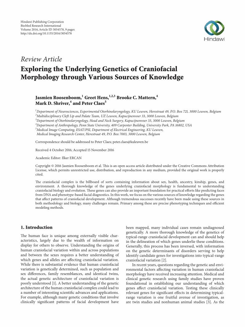
Review ArticleExploring the Underlying Genetics of CraniofacialMorphology through Various Sources of Knowledge
Jasmien Roosenboom,1 Greet Hens,1,2,3 Brooke C. Mattern,4
Mark D. Shriver,4 and Peter Claes5
1Department of Neurosciences, Experimental Otorhinolaryngology, KU Leuven, Herestraat 49, P.O. Box 721, 3000 Leuven, Belgium2Multidisciplinary Cleft Lip and Palate Team, UZ Leuven, Kapucijnenvoer 33, 3000 Leuven, Belgium3Department of Otorhinolaryngology, Head and Neck Surgery, Kapucijnenvoer 33, 3000 Leuven, Belgium4Department of Anthropology, Penn State University, 409 Carpenter Building, University Park, PA 16802, USA5Medical Image Computing, ESAT/PSI, Department of Electrical Engineering, KU Leuven,Medical Imaging Research Center, Herestraat 49, P.O. Box 7003, 3000 Leuven, Belgium
Correspondence should be addressed to Peter Claes; [email protected]
Received 4 October 2016; Accepted 15 November 2016
Academic Editor: Ilker ERCAN
Copyright © 2016 Jasmien Roosenboom et al. This is an open access article distributed under the Creative Commons AttributionLicense, which permits unrestricted use, distribution, and reproduction in any medium, provided the original work is properlycited.
The craniofacial complex is the billboard of sorts containing information about sex, health, ancestry, kinship, genes, andenvironment. A thorough knowledge of the genes underlying craniofacial morphology is fundamental to understandingcraniofacial biology and evolution. These genes can also provide an important foundation for practical efforts like predicting facesfromDNA and phenotype-based facial diagnostics. In this work, we focus on the various sources of knowledge regarding the genesthat affect patterns of craniofacial development. Although tremendous successes recently have been made using these sources inboth methodology and biology, many challenges remain. Primary among these are precise phenotyping techniques and efficientmodeling methods.
1. Introduction
The human face is unique among externally visible char-acteristics, largely due to the wealth of information ondisplay for others to observe. Understanding the origins ofhuman craniofacial variation within and across populationsand between the sexes requires a better understanding ofwhich genes and alleles are affecting craniofacial variation.While there is substantial evidence that human craniofacialvariation is genetically determined, such as population andsex differences, family resemblances, and identical twins,the actual genetic architecture of craniofacial variation ispoorly understood [1]. A better understanding of the geneticarchitecture of the human craniofacial complex could lead toa number of interesting scientific advances and applications.For example, although many genetic conditions that involveclinically significant patterns of facial development have
been mapped, many individual cases remain undiagnosedgenetically. A more thorough knowledge of the genetics oftypical-range craniofacial development can and should helpin the delineation of which genes underlie these conditions.Generally, this process has been inversed, with informationon the genetic determinants of disorders serving to helpidentify candidate genes for investigations into typical-rangecraniofacial variation [2].
In recent years, questions regarding the genetic and envi-ronmental factors affecting variation in human craniofacialmorphology have received increasing attention. Medical andclinical genetic research using family studies have provenfoundational in establishing our understanding of whichgenes affect craniofacial variation. Testing these clinicallyrelevant genes for significant effects in determining typical-range variation is one fruitful avenue of investigation, asare twin studies and nonhuman animal studies [3]. As the
Hindawi Publishing CorporationBioMed Research InternationalVolume 2016, Article ID 3054578, 9 pageshttp://dx.doi.org/10.1155/2016/3054578
2 BioMed Research International
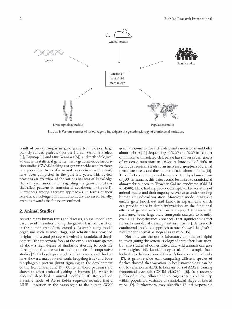
Genetics of craniofacial morphology
Animal studies
GWASFamily studies
Population studies Dysmorphology studies
Figure 1: Various sources of knowledge to investigate the genetic etiology of craniofacial variation.
result of breakthroughs in genotyping technologies, largepublicly funded projects (like the Human Genome Project[4],Hapmap [5], and 1000Genomes [6]), andmethodologicaladvances in statistical genetics, many genome-wide associa-tion studies (GWAS, looking at a genome-wide set of variantsin a population to see if a variant is associated with a trait)have been completed in the past few years. This reviewprovides an overview of the various sources of knowledgethat can yield information regarding the genes and allelesthat affect patterns of craniofacial development (Figure 1).Differences among alternate approaches, in terms of theirrelevance, challenges, and limitations, are discussed. Finally,avenues towards the future are outlined.
2. Animal Studies
As with many human traits and diseases, animal models arevery useful in understanding the genetic basis of variationin the human craniofacial complex. Research using modelorganisms such as mice, dogs, and zebrafish has providedinsights into several processes involved in craniofacial devel-opment. The embryonic faces of the various amniote speciesall show a high degree of similarity, attesting to both thedevelopmental conservation and rationale of comparativestudies [7]. Embryological studies in bothmouse and chickenhave shown a major role of sonic hedgehog (shh) and bonemorphogenic protein (bmp) signaling in the developmentof the frontonasal zone [7]. Genes in these pathways areshown to affect orofacial clefting in humans [8], which isalso well described in animal models [9–11]. Research ona canine model of Pierre Robin Sequence revealed that aLINE-1 insertion in the homologue to the human DLX6
gene is responsible for cleft palate and associated mandibularabnormalities [12]. Sequencing ofDLX5 andDLX6 in a cohortof humans with isolated cleft palate has shown causal effectsof missense mutations in DLX5. A knockout of Nol11 inXenopus Tropicalis leads to an increased apoptosis of cranialneural crest cells and thus to craniofacial abnormalities [13].This effect could be rescued to some extent by a knockdownof p53. In humans, this defect could be linked to craniofacialabnormalities seen in Treacher Collins syndrome (OMIM#154500).These findings provide examples of the versatility ofanimal studies and their ongoing relevance to understandinghuman craniofacial variation. Moreover, model organismsenable gene knock-out and knock-in experiments whichcan provide more in-depth information on the functionaleffects of genetic variants. For example, Attanasio et al.performed some large-scale transgenic analysis to identifyover 4000 long-distance enhancers that significantly affectnormal craniofacial development in mice [14]. A Cre/loxPconditional knock-out approach in mice showed that foxf2 isrequired for normal palatogenesis in mice [15].
Not only can the use of laboratory animals be helpfulin investigating the genetic etiology of craniofacial variation,but also studies of domesticated and wild animals can givenew insights [16]. Lamichhaney et al., for example, havelooked into the evolution of Darwin’s finches and their beaks[17]. A genome-wide scan comparing different species offinches showed that variation in beak morphology can bedue to variation in ALX1. In humans, loss of ALX1 is causingfrontonasal dysplasia (OMIM #136760) [18]. In a recentlypublished study, Pallares and colleagues were able to mapwithin population variance of craniofacial shape of inbredmice [19]. Furthermore, they identified 17 loci responsible
BioMed Research International 3
for variation in skull shape and eight loci responsible forvariation in mandible shape of these mice, with Mn1 as akey gene in skull formation and within population shapevariation.
3. Dysmorphology Studies
At the moment, 8,201 phenotypes are described in theOMIM database (Online Mendelian Inheritance in Man,http://www.omim.org/statistics/entry), of which 4,787 have aknown molecular basis. Approximately 32% of the inheritedhuman disorders are associated with atypical craniofacialcharacteristics [20–22]. These “face signatures” (i.e., the faceshape difference normalized against age and sex matchedcontrols) can provide the additional clues for clinical diag-noses of genetic syndromes [23, 24].
When trying to identify the genetic cause of these syn-dromes, genetic data of affected and unaffected family mem-bers can be compared. Variants that occur in affected familymembers, but not in unaffected family members, can becausal for the syndrome or can be in linkage disequilibrium(the nonrandom association of alleles of different loci) withcausal genetic variants for the condition. Linkage analysis is ameans by which the coinheritance of makers and diagnosticstatus (affected versus unaffected) are formally modeledand tested for statistical significance. Linkage analysis forcraniofacial conditions has been tremendously successful andhas provided much of what we currently know about thegenes affecting human craniofacial morphology.
Although linkage analysis studies have proven to be veryuseful in defining the genes underlying atypical patternsof craniofacial development, researches on how exactly (in3D) the faces of affected and unaffected persons differhave lagged behind mapping studies. A small group ofresearchers have been investigating a subset of conditionsinvolving the development of the face using 3D morpho-metrics (quantitative analysis of form) and have illustratedthe difference between the unaffected and affected faces asface signatures. Face signatures have proven important inunderstanding the effects on the face of some primarilypsychiatric or neurological disorders such as epilepsy [25].Developmentally, the face evolves in concert with the brain,with each influencing the development of the other andsharing genetic signaling pathways [26]: in developmentalneurological disorders the phrase “the face predicts the brain”is commonly used [27]. Since the genetic causes of manysyndromes are partially understood, investigating the facesof these syndromic patients can be very informative. Thegenes and gene regions involved in patterns of atypicalcraniofacial development may also be involved in typical-range craniofacial variation.
De novo generation of a syndrome with a nonspecificcraniofacialmorphology can provide insights into the geneticetiology of craniofacial variation, by finding the location ofthe de novo mutation [20, 28]. The gene region where thismutation is located can either be functionally responsiblefor the craniofacial trait or in linkage disequilibrium withthe variant that is directly affecting the phenotype. Another
opportunity involves investigating whether or not genes andalleles that affect atypical patterns of craniofacial develop-ment also affect typical-range facial variation. An examplefrom the literature is the PAX3 gene which can significantlyaffect typical-range facial variation including the breadth ofthe nasal bridge [2, 29]. Clinically significant mutations inPAX3 can lead to Waardenburg syndrome (OMIM #193500)which features hypertelorism and broad nasal ridges. Thesefacial features were also noticed byClaes et al. in their attemptto replicate SNPs rs7559271 and rs974448 in PAX3, usingspatially dense geometric analysis techniques on normal-range faces [2].
In addition to syndromes, some other patterns of atypicalcraniofacial development also have been widely investigated.In fact, syndromic (associated with other anomalies in thecontext of a known syndrome) or not, about 3% of new-borns have a “major physical anomaly,” meaning a physicalanomaly that has cosmetic or functional significance [30].The best-known example is nonsyndromic cleft lip with orwithout cleft palate (CL/P) with an incidence of 1/700. Thiscongenital condition has a multifactorial etiology, with bothenvironmental and genetic risk factors [31]. It has widely beeninvestigated and studies have revealed several genes affectingcraniofacial morphology [7, 32].
Further, conditions such as craniosynostosis can giveinsight in the genetic etiology of skull development. Cran-iosynostosis occurs through premature closure of the skullsutures and can be recognized by an abnormal skull shape inthe newborn. Genes responsible for this condition are likelyto play a role in typical skull formation [33–35].
Dysmorphology studies have been and will remain valu-able for the identification of individual genes or groupsof genes affecting craniofacial morphology. A completeoverview of these studies is not within the scope of this workand is often specifically described per condition [36–38].The main challenge lies in the limited availability of personsexpressing the same or similar craniofacial configurations.Larger more collaborative efforts for collecting data of thesepatients may be necessary. If 3D images of sufficient numbersof patients with known genetic background can be comparedto unaffected persons, the facial changes diagnostic of thecraniofacial conditions can be distinguished from typical-range effects.
4. Population Studies
Although craniofacial variation both within and amongpopulations is clearly evident, systematic analyses of patternsof this variation using modern morphometric methods havebeen limited. Phenotypic variation, like genetic variation,is the result of four evolutionary forces, namely, geneticdrift, natural selection (both ecological and sexual selection),admixture (interbreeding between two previously isolatedpopulations), and mutation [39]. Evolutionary studies onthe morphology of the human skull exemplify some of theapproaches to understanding the evolution of complex traits[40]. Skeletal analyses suggest that the primary evolutionaryfactor leading to population differentiation and indeed most
4 BioMed Research International
genetic variation in contemporary human populations hasbeen genetic drift: one study showed that 90% of the variationin 3D craniofacial landmark coordinates is shared acrosspopulations while only 10% of the total variation is betweenpopulation variation [41]. Claes and colleagues showed that9.6% of the total facial variation in an African/Europeanmixed population was due to variation in genetic ancestry. Assuch, between population studies, like admixture mapping,will likely provide only a subset of all of the genes affectingcraniofacial variation.
In contrast to the skeletal component, the soft tissuecomponent of the face may have been affected more bynonneutral evolutionary processes [39] such as sexual selec-tion and local adaptation due to its direct exposure tothe environment. For example, one study presented resultssuggesting that variations in the nose and brow area acrossfour Eurasian populations (Han Chinese, Tibetans, Uyghur,and Europeans) were higher than expected under geneticdrift alone [39]. These authors propose that the Europeannose may be an adaptation to colder climates. Furthermore,they speculated that the enlarged brow area in Europeansmight have been influenced by adaptation to specific diets.Sheehan and Nachman showed that also selection for indi-vidual identity signals has shaped patterns of human facialdiversity [42]. Another recent study investigated the patternsof sexual dimorphism in a sample of the faces of persons ofEuropean-derived ancestry and showed large sex differencesin several parts of the face including the chin, brow ridge,and upper cheek region [43]. Comparing these patterns withthose reported in Claes et al., 2012 [43], it seems that thereare differences in the patterns of sexual dimorphism betweena primarily European sample and the approximately halfWestAfrican/half European sample. Differences are seen in thebrow ridges, noses, and the chins.
Once the functional genes and alleles have been identi-fied,molecular evolutionary genetic approaches to investigatethe timing and geographical locations where gene frequencychanges occurred can help us understand the mechanismsbehind changes in the human craniofacial complex acrossevolutionary time.
5. Familial Studies
Familial resemblance in facial features is one of the mainindications that craniofacial shape is genetically regulated.Twin studies are well-known and have been used extensivelyto study the heritability of a wide range of traits andbehaviors [44]. Monozygotic twins share nearly identicalDNA sequences and are the extreme example of familialresemblance providing striking examples of facial similarity.When monozygotic twins are studied in contrast to dizygotictwins, the extent to which genetic factors affect craniofacialmorphology can be investigated. An added advantage oftwins is that they are at the same age, such that comparisonsbetween facial images of twins are not confounded by growthor aging [45–47]. Recently, a large twin study investigatedwhich parts of the face were prone to genetic and envi-ronmental influences [48]. This showed that genetic factors
mostly determine facial size, nasal shape, lips prominence,and interocular distance, while mandibular ramus height andhorizontal facial asymmetry are influenced by environmentalfactors.
Twin andother family studies of craniofacial shape showamoderate to high degree of heritability for a substantial set ofcraniofacial traits [46, 49–51]. Facial height, width, and nasalfeatures, in particular, are more genetically determined thanis facial depth [51]. Furthermore, local facial features withhigh heritability include the orbits, nose, jaw, and teeth.Thesestudies differ in several respects, namely, the study design(twins or parent-offspring), the data acquired (radiographs,3D facial surface scans, medical MRI, or CT), the samplesizes, landmark density, and the type of measurementsextracted and analyzed (e.g., interlandmark distances orprincipal components). Furthermore, from these studies it isclear that some facial regions are more strongly influenced bythe environment than by genes. One example is the variabilitytypically noted in the cheeks due to body weight changeand/or aging [52].
6. Genome-Wide Association Studies (GWAS)
Linkage analysis in families is one way to identify the geneticloci underlying a trait of interest. Genetic association is theother major means for identifying the alleles affecting varia-tion in a trait and is usually employed in a GWAS analyticalframework. In a GWAS, phenotype-genotype associationsare investigated in large population samples [53]. Both qual-itative traits (affected versus unaffected) and continuouslydistributed traits like height can be investigated using GWAS.Although very well established in research to the geneticorigin of disease, GWAS as an attempt to discover geneticvariants responsible for craniofacial morphology is still in itsinfancy.
Liu et al. and Paternoster et al. published the first twoGWAS on typical-range craniofacial genetics in 2012 [29,54]. The GWAS by Liu et al. identified five loci that areassociated with variation in facial morphology in Europeans[54]. They suggested five candidate genes: PRDM16, TP63,C5orf5N, COL17A1, and PAX3. The PAX3 gene was alsoidentified in the GWAS reported by Paternoster et al. andwas the first typical-range facial gene to be confirmed acrossindependent studies [29]. Both GWAS were carried outin Europeans. Recently, three additional GWAS have beenpublished. Adhikari et al. could associate four differentgenomic regions with three nose-related traits and withchin retrusion analyzing 2D frontal photographs of about6000 subjects of Latin-American descent [55]. The strongestassociations in the genomic regions were observed in theEDAR, DCHS2, RUNX2, and GLI3 genes. Furthermore, theywere able to replicate the previously described association ofnasion position and PAX3. Shaffer et al. conducted a GWASanalyzing 20 quantitative facial measurements on 3D imagesof a cohort of about 3100 American individuals of Europeandescent.They found six regions associatedwith variable facialtraits, in which several genes are located which are known tobe associated with craniofacial development: MAFB, PAX9,
BioMed Research International 5
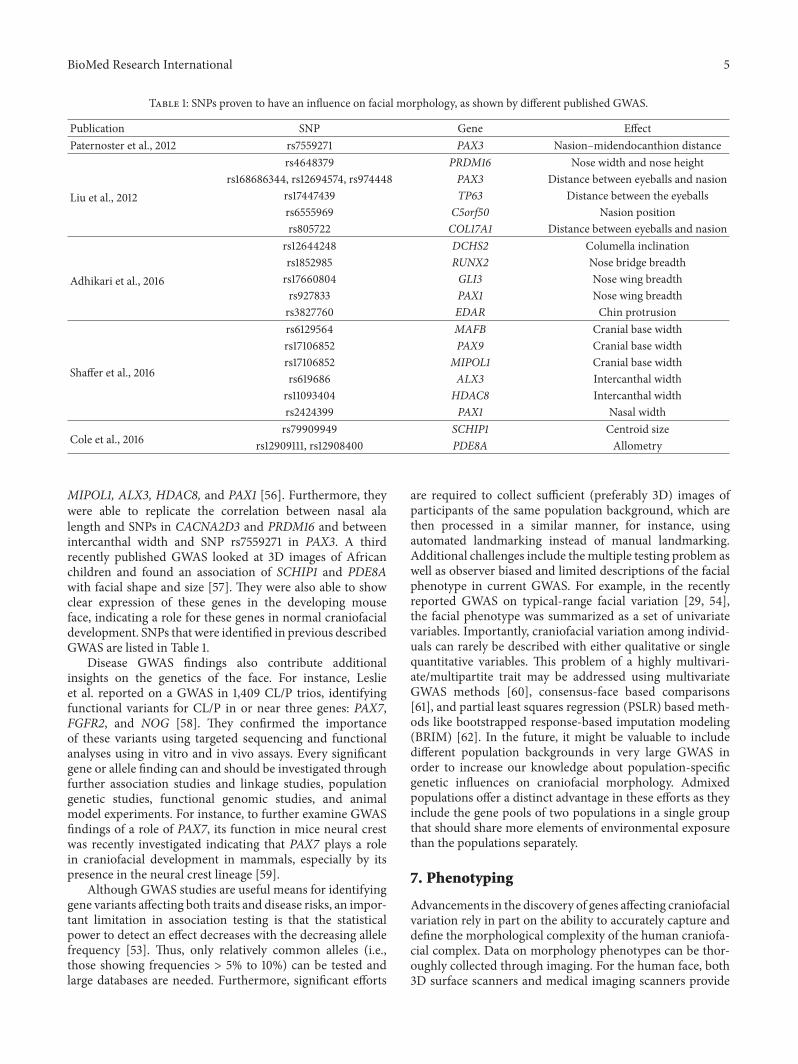
Table 1: SNPs proven to have an influence on facial morphology, as shown by different published GWAS.
Publication SNP Gene EffectPaternoster et al., 2012 rs7559271 PAX3 Nasion–midendocanthion distance
Liu et al., 2012
rs4648379 PRDM16 Nose width and nose heightrs168686344, rs12694574, rs974448 PAX3 Distance between eyeballs and nasion
rs17447439 TP63 Distance between the eyeballsrs6555969 C5orf50 Nasion positionrs805722 COL17A1 Distance between eyeballs and nasion
Adhikari et al., 2016
rs12644248 DCHS2 Columella inclinationrs1852985 RUNX2 Nose bridge breadthrs17660804 GLI3 Nose wing breadthrs927833 PAX1 Nose wing breadthrs3827760 EDAR Chin protrusion
Shaffer et al., 2016
rs6129564 MAFB Cranial base widthrs17106852 PAX9 Cranial base widthrs17106852 MIPOL1 Cranial base widthrs619686 ALX3 Intercanthal widthrs11093404 HDAC8 Intercanthal widthrs2424399 PAX1 Nasal width
Cole et al., 2016rs79909949 SCHIP1 Centroid size
rs12909111, rs12908400 PDE8A Allometry
MIPOL1, ALX3, HDAC8, and PAX1 [56]. Furthermore, theywere able to replicate the correlation between nasal alalength and SNPs in CACNA2D3 and PRDM16 and betweenintercanthal width and SNP rs7559271 in PAX3. A thirdrecently published GWAS looked at 3D images of Africanchildren and found an association of SCHIP1 and PDE8Awith facial shape and size [57]. They were also able to showclear expression of these genes in the developing mouseface, indicating a role for these genes in normal craniofacialdevelopment. SNPs that were identified in previous describedGWAS are listed in Table 1.
Disease GWAS findings also contribute additionalinsights on the genetics of the face. For instance, Leslieet al. reported on a GWAS in 1,409 CL/P trios, identifyingfunctional variants for CL/P in or near three genes: PAX7,FGFR2, and NOG [58]. They confirmed the importanceof these variants using targeted sequencing and functionalanalyses using in vitro and in vivo assays. Every significantgene or allele finding can and should be investigated throughfurther association studies and linkage studies, populationgenetic studies, functional genomic studies, and animalmodel experiments. For instance, to further examine GWASfindings of a role of PAX7, its function in mice neural crestwas recently investigated indicating that PAX7 plays a rolein craniofacial development in mammals, especially by itspresence in the neural crest lineage [59].
Although GWAS studies are useful means for identifyinggene variants affecting both traits and disease risks, an impor-tant limitation in association testing is that the statisticalpower to detect an effect decreases with the decreasing allelefrequency [53]. Thus, only relatively common alleles (i.e.,those showing frequencies > 5% to 10%) can be tested andlarge databases are needed. Furthermore, significant efforts
are required to collect sufficient (preferably 3D) images ofparticipants of the same population background, which arethen processed in a similar manner, for instance, usingautomated landmarking instead of manual landmarking.Additional challenges include themultiple testing problem aswell as observer biased and limited descriptions of the facialphenotype in current GWAS. For example, in the recentlyreported GWAS on typical-range facial variation [29, 54],the facial phenotype was summarized as a set of univariatevariables. Importantly, craniofacial variation among individ-uals can rarely be described with either qualitative or singlequantitative variables. This problem of a highly multivari-ate/multipartite trait may be addressed using multivariateGWAS methods [60], consensus-face based comparisons[61], and partial least squares regression (PSLR) based meth-ods like bootstrapped response-based imputation modeling(BRIM) [62]. In the future, it might be valuable to includedifferent population backgrounds in very large GWAS inorder to increase our knowledge about population-specificgenetic influences on craniofacial morphology. Admixedpopulations offer a distinct advantage in these efforts as theyinclude the gene pools of two populations in a single groupthat should share more elements of environmental exposurethan the populations separately.
7. Phenotyping
Advancements in the discovery of genes affecting craniofacialvariation rely in part on the ability to accurately capture anddefine the morphological complexity of the human craniofa-cial complex. Data on morphology phenotypes can be thor-oughly collected through imaging. For the human face, both3D surface scanners and medical imaging scanners provide
6 BioMed Research International
excellent technological means to capture shape and appear-ance information.Three-dimensional facial surface scanning,such as laser surface and photogrammetric imaging, is well-suited to capturing facial form. This is especially importantin healthy subjects because these scanning techniques arenoninvasive in contrast tomedical computer tomography andX-ray imaging, which use ionizing radiation. However, themajor advantage of the latter systems is that they can be usedto capture the bony structures of the craniofacial complex.Although each phenotyping approach has its advantages,comparison of different kinds of images and measurementsis challenging [2].
Besides the advancement of acquisition techniques tocapture facial morphology, the analytic methods to describecraniofacial shape must also gain in resolution, precision,and power. Shape is unfortunately often still described usingonly a sparse set of specific biological landmarks (thesebeing defined as “a point of correspondence on an objectthat matches between and within populations”), which areindicated manually on each image. This step can introduceoperator error in the placement of landmarks, which can leadto contrasting study outcomes [63]. Furthermore, manualindication is time-consuming and requires skill and training.Finally, because some anatomic regions of the face lackdiscrete features, only a limited number of landmarks can beused, as a consequence salient features of the facial shape areoverlooked [64, 65]. Measurements from sparse landmarkssuch as distances, angles, and ratios, also known as conven-tional morphometric analysis [66], had been the primaryapproach to investigating craniofacial features. However,this conventional morphometric approach oversimplifies the3D craniofacial complex such that facial characteristics ofinterest may be discounted [67]. The previously describedGWAS on typical-range facial variation [29, 54–57] and all ofthe familial studies on facial heritability thus far started fromsparse landmark representation of facial shape. Subsequently,interlandmark distances and/or angles are extracted in com-bination with a set of principal coordinates using principalcomponent analysis on the landmark coordinates. Howeverit is unfounded to assume that each PC represents a distinctand plausible morphological facial trait. Furthermore, allmeasurements, distances, and/or principal coordinates areselected a priori, meaning that many measures will need tobe looked at in order to describe the effects of even oneindependent variable.
Several extensions to spatially dense landmarks have beenproposed, including quasilandmarks and semilandmarks[63].Themain advantage of spatially denser set of landmarksis that they provide more coverage and therefore a fullerdescription of shapes [68]. The challenge however is that thenumber of shape variables almost always exceeds the numberof observations leading to theoretical limitations on the use ofsome statistical methods [69]. Shape regression, for example,is a useful technique to investigate the effect of an indepen-dent variable of interest (in this case genetic, familial, and/orpopulation information) on facial morphology. When work-ing with spatially dense shape representations in contrast toordinary least-square regression more advanced techniquessuch as partial least squares regression should be used to gain
additional facial information. Additional techniques, such asBRIM [62], further aid to increase statistical power to detectgenuine genotype-phenotype correlations.
In the interpretation of the phenotype, one has to becautious and attentive. The term “phenotype” in a clinicalsetting is often used to indicate characteristics that are devi-ating from the “normal” or typical morphology, physiology,and behavior [24]. This gives rise to questions of what istypical. Sometimes, phenotypic features are associated witha genetic disease, without being a deviation of the standard.Endophenotypes, for example, are characteristics (behavioralor anatomical) that are associated with a condition and arepresent in nonaffected family members. They are consideredto be an expression of underlying susceptibility genes for thecondition. For instance, in orofacial clefting, endophenotypicfacial features, such as hypertelorism and midface retrusion,are described in nonaffected first-degree relatives of patients[70, 71]. Objective characterization of these endophenotypesand their underlying genetics can therefore indicate newcandidate genes for this type of multifactorial conditions.
8. Discussion
The interpretation of the genotype is regulated on manydifferent levels, where in addition to genetic variation, epige-netic and distance regulators play a major role as well [14].The epigenetically switching on and off of genes and geneactivity can lead to significant changes on the phenotypelevel. Therefore, it is important to combine the informationretrieved from GWAS with underlying molecular actionson a genetic and an epigenetic level [72]. Only then, acorrect interpretation and verification of the GWAS resultsare possible. Moreover, gene dosage also has an effect oncraniofacial development, which is important in syndromeswith a causal copy number variation and also in typicalcraniofacial development [73]. Another challenge is that anopposite copy number effect (deletion versus duplication forinstance) does not necessarily cause an opposite facial effect,although recently Hammond et al. reported opposite effectson facial morphology in opposite copy number variationof genes [73]. In conclusion, unfolding genotype-phenotypecorrelations on craniofacial morphology is clearly challeng-ing due to its genetic complexity [74].
While progress has beenmade in the uncovering of genesaffecting craniofacial variation using a traditional “predefinedtrait” approach, the adaptation of a phenomic point of view,beyond “phenotyping as usual” [75], has much to offerthese efforts. Large-scale research, multidisciplinary, and up-to-date methods and analysis techniques are essential tosummarize phenotypes as complex as human craniofacialmorphology. Therefore, collaboration not only between butalso within institutes (e.g. between medical doctors, funda-mental scientists, engineers, statisticians, and informaticians)is indispensable [76].
When comparing the findings from different types ofstudies, it is important to keep in mind that there canbe many differences among studies investigating the samehypothesis. Not only study design (e.g., parent-offspring
BioMed Research International 7
studies and animal studies) but also the method of dataacquisition (e.g., radiographs and anthropometric measure-ments), differences in sample size, age of the cohort, and themethod of analysis can influence results obtained. Therefore,it is not always appropriate to simply compare the findingsand conclusions across several studies. An introduction ofguidelines for more standardized research methods can beuseful as well as a central database to register results ofresearch on typical-range and clinically significant cran-iofacial genetics. A good example of such a database isFaceBase (https://www.facebase.org/) [77]. It is also usefulto register negative research results, to avoid unnecessaryreplication of such studies that often involve expensive andtime-consuming data sampling efforts. It would even bemoreoptimal to have a common set of participants on which allcurrent and new methods can be compared and validated.
Through existing and future investigations of facial her-itability, craniofacial evolution, and individual gene effects,a better understanding of the genetic architecture of cran-iofacial morphology is certainly anticipated. With increasedresolution, precision, and power of existing and future anal-ysis techniques we may well get to the point in which ourunderstanding of the genetic determinants of craniofacialvariation can lead to practical facial predictions from DNA.First attempts in this direction were recently demonstratedby Claes et al. and by Fagertun et al. [62, 78]. Although noveland promising, the work is highly preliminary and futurerefinement of the prediction technique is essential.
In conclusion, the genetic architecture of craniofacialmorphology is complex and a challenge to unravel.We expectthat many genes, with alleles causing small average effects,and both gene-gene and gene-environment interactions willplay important roles in determining craniofacial variation.Future studies that investigate different aspects of craniofacialvariation in the context of genetic variation in several humanpopulations and animal models are indispensable. Given therecent development of newmethods for more fully capturingand modeling craniofacial variation, we can expect newgenes to result from both typical and atypical trait-basedresearch. Furthermore, although analytic techniques for thegenome but more importantly for the facial phenome arecertainly advancing, future computational developments inbioinformatics, computational imaging, and developmentalbiology, for example, are still required. Nevertheless, themethodological advances and initial results provide boththe impetus and analytical framework for future multidis-ciplinary studies to investigate the genetic determinants ofhuman facial variation [79].
Competing Interests
The authors declare that there is no conflict of interestsregarding the publication of this paper.
Acknowledgments
Greet Hens is supported by a grant from the Research FundFlanders and by a grant from the research fund Annie
Planckaert-Dewaele. Peter Claes is supported by a CatholicUniversity of Leuven CREA Research fund. Travel costs weresupplied by the Penn State University Center for HumanEvolution and Diversity.
References
[1] L. A. P. Kohn, “The role of genetics in craniofacial morphologyand growth,” Annual Review of Anthropology, vol. 20, no. 1, pp.261–278, 1991.
[2] P. Claes, M. D. Shriver, and G. S. Barsh, “New Entries in thelottery of facial GWAS discovery,” PLOS Genetics, vol. 12, no. 8,Article ID e1006250, 2016.
[3] S. R. F. Twigg and A. O. M. Wilkie, “New insights into cra-niofacial malformations,” Human Molecular Genetics, vol. 24,no. 1, pp. R50–R59, 2015.
[4] E. S. Lander, L. M. Linton, B. Birren et al., “Initial sequencingand analysis of the human genome,” Nature, vol. 409, no. 6822,pp. 860–921, 2001.
[5] International HapMap Consortium, “The International Hap-Map Project,” Nature, vol. 426, no. 6968, pp. 789–796, 2003.
[6] G. D. Schuler, M. S. Boguski, E. A. Stewart et al., “A gene mapof the human genome,” Science, vol. 274, no. 5287, pp. 540–546,1996.
[7] D.Hu,N.M.Young, X. Li, Y. Xu, B.Hallgrımsson, andR. S.Mar-cucio, “A dynamic shh expression pattern, regulated by shh andbmp signaling, coordinates fusion of primordia in the amnioteface,” Development, vol. 142, no. 3, pp. 567–574, 2015.
[8] H. Kurosaka, A. Iulianella, T. Williams, and P. A. Trainor, “Dis-rupting hedgehog and WNT signaling interactions promotescleft lip pathogenesis,”The Journal of Clinical Investigation, vol.124, no. 4, pp. 1660–1671, 2014.
[9] Z. Zhang, Y. Song, X. Zhao, X. Zhang, C. Fermin, and Y. Chen,“Rescue of cleft palate in Msx1-deficient mice by transgenicBmp4 reveals a network of BMP and Shh signaling in theregulation of mammalian palatogenesis,”Development, vol. 129,no. 17, pp. 4135–4146, 2002.
[10] E. Moura, S. M. Cirio, and C. T. Pimpao, “Nonsyndromic cleftlip and palate in boxer dogs: evidence of monogenic autosomalrecessive inheritance,” Cleft Palate-Craniofacial Journal, vol. 49,no. 6, pp. 759–760, 2012.
[11] J. Ben, E. W. Jabs, and S. S. Chong, “Genomic, cDNA and emb-ryonic expression analysis of zebrafish IRF6, the gene mutatedin the human oral clefting disorders Van der Woude andpopliteal pterygium syndromes,” Gene Expression Patterns, vol.5, no. 5, pp. 629–638, 2005.
[12] Z. T.Wolf, E. J. Leslie, B. Arzi et al., “A LINE-1 insertion inDLX6is responsible for cleft palate andmandibular abnormalities in acanine model of Pierre Robin sequence,” PLoS Genetics, vol. 10,no. 4, Article ID e1004257, 2014.
[13] J. N. Griffin, S. B. Sondalle, F. del Viso, S. J. Baserga, and M.K. Khokha, “The ribosome biogenesis factor nol11 is requiredfor optimal rdna transcription and craniofacial development inxenopus,” PLoS Genetics, vol. 11, no. 3, Article ID e1005018, 2015.
[14] C. Attanasio, A. S. Nord, Y. Zhu et al., “Fine tuning of cra-niofacial morphology by distant-acting enhancers,” Science, vol.342, no. 6157, Article ID 1241006, 2013.
[15] J. Xu, H. Liu, Y. Lan, B. J. Aronow, V. V. Kalinichenko, andR. Jiang, “A Shh-Foxf-Fgf18-Shh molecular circuit regulatingpalate development,” PLoS Genetics, vol. 12, no. 1, Article IDe1005769, 2016.
8 BioMed Research International
[16] J. J. Schoenebeck, S. A. Hutchinson, A. Byers et al., “Variation ofBMP3 contributes to dog breed skull diversity,” PLoS Genetics,vol. 8, no. 8, Article ID e1002849, 2012.
[17] S. Lamichhaney, J. Berglund, M. S. Almen et al., “Evolution ofDarwin’s finches and their beaks revealed by genome sequenc-ing,” Nature, vol. 518, no. 7539, pp. 371–375, 2015.
[18] E. Uz, Y. Alanay, D. Aktas et al., “Disruption of ALX1 causesextreme microphthalmia and severe facial clefting: expandingthe spectrum of autosomal-recessive ALX-related frontonasaldysplasia,” American Journal of Human Genetics, vol. 86, no. 5,pp. 789–796, 2010.
[19] L. F. Pallares, P. Carbonetto, S. Gopalakrishnan et al., “Mappingof craniofacial traits in outbred mice identifies major develop-mental genes involved in shape determination,” PLoS Genetics,vol. 11, no. 11, Article ID e1005607, 2015.
[20] F. Hannes, P. Hammond, O. Quarrell, J.-P. Fryns, K. Devriendt,and J. R. Vermeesch, “A microdeletion proximal of the criticaldeletion region is associated with mild Wolf-Hirschhorn syn-drome,” American Journal of Medical Genetics, Part A, vol. 158,no. 5, pp. 996–1004, 2012.
[21] K. L. Jones,M. C. Jones, andM. del Campo, Smith’s RecognizablePatterns of Human Malformation, Elsevier Saunders, Philadel-phia, Pa, USA, 7th edition, 2013.
[22] P. Hammond, F. Hannes, M. Suttie et al., “Fine-grained facialphenotype-genotype analysis in Wolf-Hirschhorn syndrome,”European Journal of Human Genetics, vol. 20, no. 1, pp. 33–40,2012.
[23] P. Hammond, M. Suttie, R. C. Hennekam, J. Allanson, E. M.Shore, and F. S. Kaplan, “The face signature of fibrodysplasiaossificans progressiva,” American Journal of Medical GeneticsPart A, vol. 158, no. 6, pp. 1368–1380, 2012.
[24] G. Baynam, M. Walters, P. Claes et al., “Phenotyping: targetinggenotype’s rich cousin for diagnosis,” Journal of Paediatrics andChild Health, vol. 51, no. 4, pp. 381–386, 2015.
[25] K. Chinthapalli, E. Bartolini, J. Novy et al., “Atypical face shapeand genomic structural variants in epilepsy,” Brain, vol. 135, no.10, pp. 3101–3114, 2012.
[26] K. Aldridge, I. D. George, K. K. Cole et al., “Facial phenotypes insubgroups of prepubertal boys with autism spectrum disordersare correlated with clinical phenotypes,”Molecular Autism, vol.2, no. 1, article 15, 2011.
[27] W. Demyer, W. Zeman, and C. G. Palmer, “The face predictsthe brain: diagnostic significance of median facial anomaliesfor holoprosencephaly (arhinencephaly),” Pediatrics, vol. 34, pp.256–263, 1964.
[28] K. A. Miller, T. Y. Tan, M. F. Welfare et al., “A mouse splice-site mutant and individuals with atypical chromosome 22q11.2deletions demonstrate the crucial role for Crkl in craniofacialand pharyngeal development,” Molecular Syndromology, vol. 5,no. 6, pp. 276–286, 2014.
[29] L. Paternoster, A. I. Zhurov, A. M. Toma et al., “Genome-wide association study of three-dimensional facial morphologyidentifies a variant in PAX3 associated with nasion position,”American Journal of Human Genetics, vol. 90, no. 3, pp. 478–485, 2012.
[30] V. Kumar, A. K. Abbas, N. Fausto, S. L. Robbins, and R. S.Cotran, Eds., Robbins and Cotran Pathologic Basis of Disease,Elsevier Saunders, Philadelphia, Pa, USA, 7th edition, 2005.
[31] M. J. Dixon, M. L. Marazita, T. H. Beaty, and J. C. Murray,“Cleft lip and palate: understanding genetic and environmentalinfluences,” Nature Reviews Genetics, vol. 12, no. 3, pp. 167–178,2011.
[32] S. Boehringer, F. Van Der Lijn, F. Liu et al., “Genetic determina-tion of human facial morphology: links between cleft-lips andnormal variation,” European Journal of Human Genetics, vol. 19,no. 11, pp. 1192–1197, 2011.
[33] S. R. F. Twigg, E.Vorgia, S. J.Mcgowan et al., “Reduced dosage ofERF causes complex craniosynostosis in humans and mice andlinks ERK1/2 signaling to regulation of osteogenesis,” NatureGenetics, vol. 45, no. 3, pp. 308–313, 2013.
[34] V. P. Sharma, A. L. Fenwick, M. S. Brockop et al., “Mutations inTCF12, encoding a basic helix-loop-helix partner of TWIST1,are a frequent cause of coronal craniosynostosis,”Nature Genet-ics, vol. 45, no. 3, pp. 304–307, 2013.
[35] J. A. Hamm and N. H. Robin, “Newborn craniofacial mal-formations: orofacial clefting and craniosynostosis,” Clinics inPerinatology, vol. 42, no. 2, pp. 321–336, 2015.
[36] A. Dixit andM. Suri, “When the face says it all: dysmorphologyin identifying syndromic causes of epilepsy,” Practical Neurol-ogy, vol. 16, no. 2, pp. 111–121, 2016.
[37] T. C. Cox, “Taking it to the max: the genetic and developmentalmechanisms coordinating midfacial morphogenesis and dys-morphology,” Clinical Genetics, vol. 65, no. 3, pp. 163–176, 2004.
[38] M. L. Dentici, A. Di Pede, F. R. Lepri et al., “Kabuki syndrome:clinical andmolecular diagnosis in the first year of life,”Archivesof Disease in Childhood, vol. 100, no. 2, pp. 158–164, 2015.
[39] J. Guo, J. Tan, Y. Yang et al., “Variation and signatures ofselection on the human face,” Journal of Human Evolution, vol.75, pp. 143–152, 2014.
[40] J. H. Relethford, “Population-specific deviations of globalhuman craniometric variation from a neutral model,”AmericanJournal of Physical Anthropology, vol. 142, no. 1, pp. 105–111, 2010.
[41] J. H. Relethford and H. C. Harpending, “Craniometric vari-ation, genetic theory, and modern human origins,” AmericanJournal of Physical Anthropology, vol. 95, no. 3, pp. 249–270,1994.
[42] M. J. Sheehan and M. W. Nachman, “Morphological andpopulation genomic evidence that human faces have evolvedto signal individual identity,” Nature Communications, vol. 5,article 4800, 2015.
[43] P. Claes, M. Walters, M. D. Shriver et al., “Sexual dimorphismin multiple aspects of 3D facial symmetry and asymmetrydefined by spatially dense geometric morphometrics,” Journalof Anatomy, vol. 221, no. 2, pp. 97–114, 2012.
[44] D. Boomsma, A. Busjahn, and L. Peltonen, “Classical twinstudies and beyond,” Nature Reviews Genetics, vol. 3, no. 11, pp.872–882, 2002.
[45] P. Claes, H. Hill, and M. D. Shriver, “Toward DNA-based facialcomposites: preliminary results and validation,”Forensic ScienceInternational: Genetics, vol. 13, pp. 208–216, 2014.
[46] S. M. Weinberg, T. E. Parsons, M. L. Marazita, and B. S. Maher,“Heritability of face shape in twins: a preliminary study using3D stereophotogrammetry and geometric morphometrics,”Dentistry 3000, vol. 1, no. 1, 2013.
[47] F. Amini andA. Borzabadi-Farahani, “Heritability of dental andskeletal cephalometric variables in monozygous and dizygousIranian twins,” Orthodontic Waves, vol. 68, no. 2, pp. 72–79,2009.
[48] J. Djordjevic, A. I. Zhurov, S. Richmond, and T. Cai, “Geneticand environmental contributions to facial morphological vari-ation: a 3D population-based twin study,” PLOSONE, vol. 11, no.9, Article ID e0162250, 2016.
BioMed Research International 9
[49] B. Johannsdottir, F. Thorarinsson, A. Thordarson, and T.E. Magnusson, “Heritability of craniofacial characteristicsbetween parents and offspring estimated from lateral cephalo-grams,” American Journal of Orthodontics & Dentofacial Ortho-pedics, vol. 127, no. 2, pp. 200–207, 2005.
[50] N. Martınez-Abadıas, M. Esparza, T. Sjøvold, R. Gonzalez-Jose,M. Santos, and M. Hernandez, “Heritability of human cranialdimensions: comparing the evolvability of different cranialregions,” Journal of Anatomy, vol. 214, no. 1, pp. 19–35, 2009.
[51] F. B. Naini and J. P. Moss, “Three-dimensional assessment ofthe relative contribution of genetics and environment to variousfacial parameters with the twin method,” American Journal ofOrthodontics and Dentofacial Orthopedics, vol. 126, no. 6, pp.655–665, 2004.
[52] S. Coleman, A. Saboeiro, and R. Sengelmann, “A comparisonof lipoatrophy and aging: volume deficits in the face,” AestheticPlastic Surgery, vol. 33, no. 1, pp. 14–21, 2009.
[53] P. Donnelly, “Progress and challenges in genome-wide associa-tion studies in humans,” Nature, vol. 456, no. 7223, pp. 728–731,2008.
[54] F. Liu, F. van der Lijn, C. Schurmann et al., “A genome-wide association study identifies five loci influencing facialmorphology in Europeans,” PLoS Genetics, vol. 8, no. 9, ArticleID e1002932, 2012.
[55] K. Adhikari, M. Fuentes-Guajardo, M. Quinto-Sanchez et al.,“A genome-wide association scan implicates DCHS2, RUNX2,GLI3, PAX1 and EDAR in human facial variation,” NatureCommunications, vol. 7, Article ID 11616, 2016.
[56] J. R. Shaffer, E. Orlova, M. K. Lee et al., “Genome-wide associa-tion study revealsmultiple loci influencing normal human facialmorphology,” PLOS Genetics, vol. 12, no. 8, Article ID e1006149,2016.
[57] J. B. Cole, M. Manyama, E. Kimwaga et al., “Genomewideassociation study of African children identifies association ofSCHIP1 and PDE8A with facial size and shape,” PLoS Genetics,vol. 12, no. 8, Article ID e1006174, 2016.
[58] E. J. Leslie,M. A. Taub, H. Liu et al., “Identification of functionalvariants for cleft lip with or without cleft palate in or nearPAX7, FGFR2, andNOG by targeted sequencing of GWAS loci,”American Journal of Human Genetics, vol. 96, no. 3, pp. 397–411,2015.
[59] B. Murdoch, C. DelConte, and M. I. Garcıa-Castro, “Pax7lineage contributions to the mammalian neural crest,” PLoSONE, vol. 7, no. 7, Article ID e41089, 2012.
[60] T. E. Galesloot, K. Van Steen, L. A. L. M. Kiemeney, L. L. Janss,and S. H. Vermeulen, “A comparison of multivariate genome-wide association methods,” PLoS ONE, vol. 9, no. 4, Article IDe95923, 2014.
[61] S. Peng, J. Tan, S. Hu et al., “Detecting genetic associationof common human facial morphological variation using highdensity 3D image registration,” PLoS Computational Biology,vol. 9, no. 12, Article ID e1003375, 2013.
[62] P. Claes, D. K. Liberton, K. Daniels et al., “Modeling 3D facialshape from DNA,” PLoS Genetics, vol. 10, no. 3, Article IDe1004224, 2014.
[63] P. Claes, M. Walters, and J. Clement, “Improved facial outcomeassessment using a 3D anthropometric mask,” InternationalJournal of Oral andMaxillofacial Surgery, vol. 41, no. 3, pp. 324–330, 2012.
[64] C. D. L. Thomas, “Three-dimensional quantification of facialshape,” in Computer-Graphic Facial Reconstruction, pp. 55–78,Elsevier Academic Press, London, UK, 2005.
[65] C. D. L. Thomas, P. Claes, A. I. Shaweesh, and J. G. Clement,“Three-dimensional facial shape archetypes for identificationand diagnosis,” in Proceedings of the International SymposiumBiological Shape Analysis, vol. 1, pp. 131–144, Tsukuba, Japan,2009.
[66] P. E. Lestrel, Ed., Biological Shape Analysis: Proceedings of the 1stInternational Symposium, Tsukuba, Japan, 3–6 June 2009,WorldScientific, Singapore, 2011.
[67] R. E. Ward, D. Bixler, and E. R. Raywood, “A study of cephalo-metric features in cleft lip-cleft palate families. I: phenotypicheterogeneity and genetic predisposition in parents of sporadiccases,” Cleft Palate Journal, vol. 26, no. 4, pp. 318–325, 1989.
[68] P. Claes,M.Walters, D. Gillett, D. Vandermeulen, J. G. Clement,and P. Suetens, “The normal-equivalent: a patient-specificassessment of facial harmony,” International Journal of Oral andMaxillofacial Surgery, vol. 42, no. 9, pp. 1150–1158, 2013.
[69] H. Abdi, “Partial least squares regression (pls-regression),” inEncyclopedia for Research Methods for the Social Sciences, pp.792–795, Sage, Thousand Oaks, Calif, USA, 2003.
[70] S. Weinberg, S. Naidoo, K. Bardi et al., “Face shape of unaf-fected parents with cleft affected offspring: combining three-dimensional surface imaging and geometric morphometrics,”Orthodontics and Craniofacial Research, vol. 12, no. 4, pp. 271–281, 2009.
[71] J. Roosenboom, I. Saey, H. Peeters, K. Devriendt, P. Claes,and G. Hens, “Facial characteristics and olfactory dysfunction:two endophenotypes related to nonsyndromic cleft lip and/orpalate,” BioMed Research International, vol. 2015, Article ID863429, 8 pages, 2015.
[72] S. V. Nuzhdin, M. L. Friesen, and L. M. McIntyre, “Genotype-phenotypemapping in a post-GWASworld,”Trends in Genetics,vol. 28, no. 9, pp. 421–426, 2012.
[73] P. Hammond, S. McKee, M. Suttie et al., “Opposite effectson facial morphology due to gene dosage sensitivity,” HumanGenetics, vol. 133, no. 9, pp. 1117–1125, 2014.
[74] B. Hallgrimsson, W. Mio, R. S. Marcucio, and R. Spritz, “Let’sface it–complex traits are just not that simple,” PLoS genetics,vol. 10, no. 11, Article ID e1004724, 2014.
[75] D. Houle, D. R. Govindaraju, and S. Omholt, “Phenomics: thenext challenge,”Nature Reviews Genetics, vol. 11, no. 12, pp. 855–866, 2010.
[76] G. Baynam, M. Walters, P. Claes et al., “The facial evolution:looking backward and moving forward,”Human Mutation, vol.34, no. 1, pp. 14–22, 2013.
[77] H. Hochheiser, B. J. Aronow, K. Artinger et al., “The FaceBaseconsortium: a comprehensive program to facilitate craniofacialresearch,” Developmental Biology, vol. 355, no. 2, pp. 175–182,2011.
[78] J. Fagertun, K. Wolffhechel, T. H. Pers et al., “Predicting facialcharacteristics from complex polygenic variations,” ForensicScience International: Genetics, vol. 19, pp. 263–268, 2015.
[79] P. Claes and M. D. Shriver, “Establishing a multidisciplinarycontext formodeling 3D facial shape fromDNA,”PLoSGenetics,vol. 10, no. 11, Article ID e1004725, 2014.

Submit your manuscripts athttp://www.hindawi.com
Hindawi Publishing Corporationhttp://www.hindawi.com Volume 2014
Anatomy Research International
PeptidesInternational Journal of
Hindawi Publishing Corporationhttp://www.hindawi.com Volume 2014
Hindawi Publishing Corporation http://www.hindawi.com
International Journal of
Volume 2014
Zoology
Hindawi Publishing Corporationhttp://www.hindawi.com Volume 2014
Molecular Biology International
GenomicsInternational Journal of
Hindawi Publishing Corporationhttp://www.hindawi.com Volume 2014
The Scientific World JournalHindawi Publishing Corporation http://www.hindawi.com Volume 2014
Hindawi Publishing Corporationhttp://www.hindawi.com Volume 2014
BioinformaticsAdvances in
Marine BiologyJournal of
Hindawi Publishing Corporationhttp://www.hindawi.com Volume 2014
Hindawi Publishing Corporationhttp://www.hindawi.com Volume 2014
Signal TransductionJournal of
Hindawi Publishing Corporationhttp://www.hindawi.com Volume 2014
BioMed Research International
Evolutionary BiologyInternational Journal of
Hindawi Publishing Corporationhttp://www.hindawi.com Volume 2014
Hindawi Publishing Corporationhttp://www.hindawi.com Volume 2014
Biochemistry Research International
ArchaeaHindawi Publishing Corporationhttp://www.hindawi.com Volume 2014
Hindawi Publishing Corporationhttp://www.hindawi.com Volume 2014
Genetics Research International
Hindawi Publishing Corporationhttp://www.hindawi.com Volume 2014
Advances in
Virolog y
Hindawi Publishing Corporationhttp://www.hindawi.com
Nucleic AcidsJournal of
Volume 2014
Stem CellsInternational
Hindawi Publishing Corporationhttp://www.hindawi.com Volume 2014
Hindawi Publishing Corporationhttp://www.hindawi.com Volume 2014
Enzyme Research
Hindawi Publishing Corporationhttp://www.hindawi.com Volume 2014
International Journal of
Microbiology