Embed Size (px)
Citation preview
Review Article
Modern Principles in the Acute SurgicalManagement of Open Distal Tibial Fractures
ABSTRACT
Over the past two decades, management of open distal tibial fractures
has evolved such that a staged approach, with external fixation and
debridement during the index procedure, followed by definitive fixation
and wound closure at a later date, is often considered the standard of
care. Although definitive treatment of these complex injuries is often
done by a multidisciplinary team of surgeons well versed in
periarticular fracture repair and soft-tissue coverage in the distal
extremity, the on-call orthopaedic surgeon doing the index procedure
must understand the principles and rationale of the staged treatment
algorithm to avoid compromising definitive treatment options and
ensure the best possible patient outcome. The mechanism of injury,
neurovascular status, size and location of soft-tissue injury, fracture
pattern, and concomitant injuries in the polytraumatized patient
should direct the treatment plan and anticipated outcomes. This
review focuses on evaluation and management of these complex
injuries with an emphasis on early aggressive debridement, principles
of initial fracture fixation, and modern options for soft-tissue coverage,
including local and free tissue transfer.
Open tibial fractures account for 11.2% of all open fractures andrepresent a significant source of morbidity and economic cost bur-den.1 Although basic principles of management have remained
constant since Gustilo and Anderson described the importance of earlyantibiotic administration and aggressive surgical débridement, advances inour understanding of fracture biology, fracture fixation, and microsurgicaltechnique have changed the landscape of both routine management andcomplex limb salvage over the past 20 years. A staged approach, with theindex procedure including external fixation and debridement, followed bydefinitive fixation and wound closure at a later date, is often considered thestandard of care. Although definitive treatment is often done by a multi-disciplinary team of surgeons well versed in periarticular fracture repair andsoft-tissue coverage in the distal extremity, the on-call orthopaedic surgeondoing the index procedure must understand the principles and rationale ofthe staged treatment algorithm to avoid compromising definitive treatment
e536 JAAOS® ----- June 1, 2021, Vol 29, No 11 ----- © American Academy of Orthopaedic Surgeons
Babar Shafiq, MD
Jacques Hacquebord, MD
David J. Wright, MD
Ranjan Gupta, MD
From the Department of Orthopaedic Surgery,Johns Hopkins University School of Medicine,Baltimore, MD (Shafiq), the Department ofOrthopaedic Surgery, New York UniversitySchool of Medicine, New York, NY(Hacquebord), and the Department ofOrthopaedic Surgery, University of California,School of Medicine, Los Angeles, CA (Wright andGupta).
None of the following authors or any immediatefamily member has received anything of valuefrom or has stock or stock options held in acommercial company or institution relateddirectly or indirectly to the subject of this article:Shafiq, Hacquebord, Wright, and Gupta.
J Am Acad Orthop Surg 2021;29:e536-e547
DOI: 10.5435/JAAOS-D-20-00502
Copyright 2021 by the American Academy ofOrthopaedic Surgeons.
Copyright © the American Academy of Orthopaedic Surgeons. Unauthorized reproduction of this article is prohibited.
options and ensure the best possible patient outcomes.Although this review focuses specifically on the initialmanagement of open distal tibial fractures, many of theprinciples can also be applied to closed injuries.
Initial ManagementOpen distal tibial fractures have a bimodal distribution,occurring because of high-energy trauma in young pa-tients or lower-energy falls in the elder patients.1 In casesof high-energy trauma, evaluation by a multidisciplinaryteam should proceed according to advanced trauma life-support protocols. Soft-tissue injury and limb deformityassociated with these fractures can be impressive andoften distracts both the patient and physician fromrecognizing other notable injuries, obtaining a detailedhistory, and doing a thorough physical examination.Details about the mechanism of injury, wound con-tamination, and associated injuries help guide initialmanagement, plan definitive fixation, and direct shareddecision-making that will determine the ultimate goalsof treatment in these life-altering injuries. Otherimportant elements of the history include age, pre-morbid functional status, comorbidities, medications,and social history (including smoking status) that mayimpair wound healing or increase the risk of infection.
Physical examination includes inspection to identifythe location, size, and orientation (transverse, longitu-dinal, and stellate) of the open wound(s) in addition tocharacterizing the types of soft tissue involved (skin, fat,fascia, muscle, tendon, nerve, vessels, and bone). Palpa-tion of the entire limb should follow because the inci-dence of associated lower extremity fracture and othermajor system injuries ranges from 27% to 51%.2 Openfractures do not preclude the development of acutecompartment syndrome, and a high index of suspicion isimperative for diagnosis, especially in the obtundedpatient. Acute compartment syndrome will develop in1.4% to 5% of distal tibial fractures and should bemanaged expeditiously.3 Once an open fracture isidentified, antibiotics and tetanus vaccine should beadministered as soon as possible.4 Patzakis demon-strated that delay in antibiotic administration more than3 hours after injury resulted in 1.63 greater odds ofinfection, whereas Lack more recently showed thatadministration more than 66 minutes after injury wasassociated with a 3.78 greater odds of infection.4,5
The neurovascular examination is the most criticalcomponent of the initial examination. Hard signs ofvascular injury include absent or asymmetric pulses,
severe hemorrhage, and expanding hematoma. In theabsence of distal pulses or when signs of hypoperfusionare present, gentle reduction with longitudinal tractionshould be done, followed by repeat vascular examina-tion. If pulses remain absent, doppler examination isindicated to confirm the presence or absence of signals.Although ankle brachial index ,0.90 is suggestive ofvascular injury, doing such an examination in thepresence of an open distal tibial fracture is often notfeasible.6 If repeated examination remains abnormal,CT angiogram or formal on-table angiogram is war-ranted. In patients with active hemorrhage, attempts toclamp or ligate a bleeding vessel in the trauma bay arecontraindicated because this can result in injury toadjacent neurovascular structures (ie, tibial nerve),leading to permanent neurologic deficit and hinderingsubsequent efforts for limb salvage. At the distal tibia,even notable arterial bleeding can often be controlled bydirect pressure, followed by application of a compres-sion dressing. Even if the initial vascular examination isnormal, serial examinations should be done becauseintimal tears or flaps within larger arteries may subse-quently thrombose, resulting in delayed presentationof a dysvascular extremity. One study of high-energytibial plafond fractures identified a 52% incidence ofarterial abnormalities including 7 with complete arterialocclusion, 2 with partial occlusion, and 5 with normalflow but with anatomic disturbances (4 tented and 1entrapped by fracture fragments), with a significantassociation between open fracture and arterial abnor-mality.7 Associated arterial injury is a risk factor for flapfailure at the time of reconstruction and may impact thechoice of surgical approach and soft-tissuereconstruction.
Imaging should consist of dedicated AP, lateral, andmortise views of the ankle and foot and full-length APand lateral views of the tibia and fibula. In fractures withnotable intra-articular comminution, CT is best doneafter external fixation, once basic length, alignment, androtation have been restored. However, early CT imagingbefore external fixation is appropriate when thereis minimal articular comminution or in distal third spiraltibial shaft fractures with question of intra-articularextension, which occurs in 39% to 92.3% of such frac-tures.8,9 CT is also indicated before the index procedureif planning for limited internal fibular fixation or ifimmediate definitive fixation is an option. In such cases,CT helps delineate fracture planes that will guide sur-gical approaches and determine whether acute fibularopen reduction and internal fixation (ORIF) will help orhinder the staged definitive fixation. If free or local
JAAOS® ----- June 1, 2021, Vol 29, No 11 ----- © American Academy of Orthopaedic Surgeons e537
Review
Article
Babar Shafiq, MD, et al
Copyright © the American Academy of Orthopaedic Surgeons. Unauthorized reproduction of this article is prohibited.
tissue transfer is required, a CT angiogram or formalangiogram is indicated to identify potential recipientvessels for microvascular anastomosis and to identifyanatomic variants including anomalous trifurcation ofthe popliteal artery, which has an incidence of 7% to12%.10
Classification of Open FracturesClassification of open fractures has evolved since theGustilo-Anderson classification was first introduced in1976. Although their work established that early anti-biotic delivery and aggressive débridement were funda-mental to open fracture care, the classification servedmainly as a prognostic indicator, with types I, II, andIIIA having relatively low rates of infection andamputation, IIIB fractures having a 52% infection and16% amputation rate, and IIIC having a 42% infectionand 42% amputation rate in their original series.11 Finalclassification is assigned after operative débridementand, therefore, provides little guidance regarding initialsurgical management apart from appropriate antibioticselection. Furthermore, type IIIB fractures encompass awide range of soft-tissue injures, requiring proceduresranging from local soft-tissue rearrangement to freetissue transfer.
Recent classifications have sought to better describedifferent characteristics of the injury and provide guid-ance for treatment. The Ganga Hospital Open InjuryScore (GHOIS) and the Orthopaedic Trauma Associa-tion Open Fracture Classification (OTA-OFC) systemsare two such examples.12,13 These classifications assignindividual scores according to the degree of skin, mus-cle, nerve, bone, and arterial injury and also account forthe degree of contamination (OTA-OFC) or patientcomorbidities (GHOIS). Subsequent studies of thesescoring systems have demonstrated their utility inguiding management. In a retrospective review of 109patients with type IIIA and IIIB open tibial fractures,Rajasekaran12 demonstrated that all fractures withGHOIS score ,14 were successfully salvaged, whereasall of those with score .17 required amputation.Another study found that classification according to theOTA-OFC was predictive of early amputation withodds ratios of 3.4, 4.8, and 6.5 for severity of skininjury, arterial injury, and degree of contamination,respectively.13 Because associated soft-tissue injury,neurovascular injury, and patient comorbidities haveincreasingly been recognized as critical predictors ofoutcomes after open distal tibial fractures, classification
schemes have shifted focus to account for these factorsin an effort to better guide surgical management.
Timing of SurgeryIn the absence of arterial injury, initial surgical manage-ment for open distal tibial fractures should ideally bedone within the first 24 hours after injury. Historically,open fractures have been considered an “orthopaedicemergency” warranting débridement within 6 hours ofinjury. This “6-hour” rule was based largely on clinicalopinion and animal/bacteriology studies conductedbefore the consistent delivery of modern antibiotics.14
Recent studies have demonstrated little or no differencein infection rates between fractures débrided within 6hours or within 24 hours as long as antibiotic man-agement was initiated early.15 Associated life-threatening injuries, hemodynamic stability, and ade-quacy of resuscitation should also be considered beforeproceeding with index débridement and stabilization.
The concept of staged management of distal tibialfractures was first introduced independently in separatestudies by Sirkin andPatterson.16,17 Previous studies hadidentified high rates of wound complications andinfection in up to 40% of patients when ORIF was done3 to 5 days after injury.18,19 By contrast, using a stagedprotocol, Sirkin found an overall infection rate of 5.3%in their series of 56 patients with either open or closeddistal tibial fractures, whereas Patterson noted a 0%rate of deep or superficial infections in their series of 22type III open distal tibial fractures.16,17 These resultshighlighted the importance of appropriate soft-tissuemanagement. More recently, several retrospectivestudies have advocated for early ORIF in select patientswith appropriate soft-tissue envelope.20-22 In their ret-rospective cohort study of 95 patients treated with earlyORIF (within 48 hours of injury), White et al reported a19% infection rate in open fractures and a 2.7%infection rate in closed fractures. Of note, four patientswere excluded from the study because of “local soft-tissue” factors necessitating the placement of tempo-rizing external fixation at the discretion of the treatingsurgeon.22 Meanwhile, Tang et al20 compared early(,36 hours) versus late (10-21 days) ORIF of closedC-type pilon fractures and found no difference in therate of soft-tissue complications between groups with asignificantly shorter hospital stay and time to fractureunion in the “early” group. Overall, recent studies have,at best, shown no difference in infection rates betweenearly definitive and staged fixation groups.
e538 JAAOS® ----- June 1, 2021, Vol 29, No 11 ----- © American Academy of Orthopaedic Surgeons
Management of open distal tibial fractures
Copyright © the American Academy of Orthopaedic Surgeons. Unauthorized reproduction of this article is prohibited.
Principles of DébridementEarly, aggressive débridement is the foundation of sur-gical management for open distal tibial fractures. Theprimary goal during débridement is removal of all for-eign debris and nonviable tissue to establish a clean,healthy wound. This should be done without concernfor the ease of reconstruction. The quality of débride-ment is the most critical surgeon-controlled factor in theprevention of infection and limb preservation becausesubsequent reconstructive efforts will prove futile if aninadequate débridement is done. Débridement should beapproached in a systematic fashion beginning with skin,subcutaneous tissue, fascia, muscle, tendon, and finallybone. The most experienced surgeon available shouldguide the débridement because assessing tissue viabilityis often the most critical and challenging portion of thecase. Every débridement should begin with extension ofthe wound to allow for adequate inspection of deepertissues. Incisions should be extended in a longitudinalfashion with an effort to incorporate transverse or ob-lique traumatic wounds in a curvilinear fashion. In-cisions directly over the subcutaneous border of the tibiashould be avoided because this is the most challengingarea to achieve subsequent soft-tissue coverage. Incisionlength should match the degree of contamination andmechanism of injury, with a larger exposure required formore severe injuries. Débridement of skin and subcu-taneous tissue should be done without the use of tour-niquet because soft-tissue viability can be determinedbased on the presence of punctate bleeding. Muscleviability is often assessed with the use of the 4 Cs:contractility, color, consistency, and capacity forbleeding. Muscle of questionable viability should beexcised because necrotic muscle serves as an idealmedium for bacterial growth. The decision to retain orremove the bone depends on vascularity and whetherthe fragment is diaphyseal, metaphyseal, or articular. Ingeneral, diaphyseal bone without evidence of bleedingand minimal soft-tissue attachment should be removedbecause these fragments can serve as a source of infec-tion and the residual diaphyseal defects can be managedmore easily than metaphyseal or articular defects.Metaphyseal bone has a higher capacity for revascu-larization and should be retained if not grossly con-taminated. All efforts should be made to retain articularfragments. In cases in which tissues of questionableviability are retained, a second look débridement ismandated for reassessment of these tissues before pro-ceeding with reconstructive efforts. In severely con-taminated wounds with significant soft-tissue injury,
several trips to the surgical roommay be required beforean adequate débridement has been achieved.
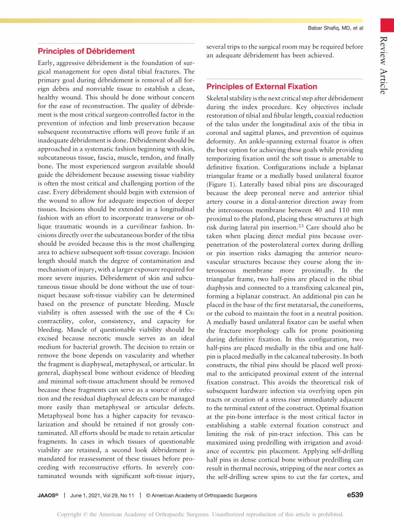
Principles of External FixationSkeletal stability is the next critical step after débridementduring the index procedure. Key objectives includerestoration of tibial and fibular length, coaxial reductionof the talus under the longitudinal axis of the tibia incoronal and sagittal planes, and prevention of equinusdeformity. An ankle-spanning external fixator is oftenthe best option for achieving these goals while providingtemporizing fixation until the soft tissue is amenable todefinitive fixation. Configurations include a biplanartriangular frame or a medially based unilateral fixator(Figure 1). Laterally based tibial pins are discouragedbecause the deep peroneal nerve and anterior tibialartery course in a distal-anterior direction away fromthe interosseous membrane between 40 and 110 mmproximal to the plafond, placing these structures at highrisk during lateral pin insertion.23 Care should also betaken when placing direct medial pins because over-penetration of the posterolateral cortex during drillingor pin insertion risks damaging the anterior neuro-vascular structures because they course along the in-terosseous membrane more proximally. In thetriangular frame, two half-pins are placed in the tibialdiaphysis and connected to a transfixing calcaneal pin,forming a biplanar construct. An additional pin can beplaced in the base of the first metatarsal, the cuneiforms,or the cuboid to maintain the foot in a neutral position.A medially based unilateral fixator can be useful whenthe fracture morphology calls for prone positioningduring definitive fixation. In this configuration, twohalf-pins are placed medially in the tibia and one half-pin is placed medially in the calcaneal tuberosity. In bothconstructs, the tibial pins should be placed well proxi-mal to the anticipated proximal extent of the internalfixation construct. This avoids the theoretical risk ofsubsequent hardware infection via overlying open pintracts or creation of a stress riser immediately adjacentto the terminal extent of the construct. Optimal fixationat the pin-bone interface is the most critical factor inestablishing a stable external fixation construct andlimiting the risk of pin-tract infection. This can bemaximized using predrilling with irrigation and avoid-ance of eccentric pin placement. Applying self-drillinghalf pins in dense cortical bone without predrilling canresult in thermal necrosis, stripping of the near cortex asthe self-drilling screw spins to cut the far cortex, and
JAAOS® ----- June 1, 2021, Vol 29, No 11 ----- © American Academy of Orthopaedic Surgeons e539
Review
Article
Babar Shafiq, MD, et al
Copyright © the American Academy of Orthopaedic Surgeons. Unauthorized reproduction of this article is prohibited.
prominence at the far cortex to achieve full threadpurchase, which may result in adjacent soft-tissueinjury.24,25 Once the provisional frame has beenassembled, indirect reduction can be done by manipu-lation of the calcaneal-transfixing pin. The crossbartrajectory from proximal anterior to distal posterior inthe triangular frame commonly results in an apex-anterior deformity at the fracture if simple longitudinaltraction is applied. This can be minimized with anteriortranslation of the foot during reduction. Finally,although there is debate surrounding acute ORIF of thefibula to provide accurate restoration of length andmore rigid fixation during the index procedure, it isrecommended that ORIF of the fibula be done by thesurgeon who will also be completing the definitive fix-ation because incisions must be carefully planned toavoid a skin bridge less than 5 to 6 cm.26
Initial Wound ManagementLacerationswithout notable underlying soft-tissue injurycan often be closed primarily after a thorough débride-ment and skeletal stabilization. More severe injury tothe skin and underlying tissues may require delayedwound closure, local soft-tissue rearrangement, or freetissue transfer. Accordingly, the traumatic wound iscommonly temporized by application of negative pres-sure wound therapy (NPWT) or antibiotic bead pouch.
An evidence-based consensus statement on the use ofNPWT in the management of open fractures provided a“Grade B” recommendation that “NPWT should beconsidered when primary closure is not possible after orbetween debridements as a bridge to definitive closure.”This recommendation was based on several level II andlevel III studies and one level I study demonstratingdecreased rates of infection in the NPWT group com-pared with wounds treated without NPWT.27 Thestatement also provided a “Grade C” recommendationthat “NPWT may be used to downscale the complexityof closure procedures” based on several level II studiesdemonstrating a decreased number of flap procedureswith corresponding increase in split-thickness skingrafting for coverage of wounds after open fracture.
Polyvinyl alcohol (white) sponges are placed overareas of exposed tendon andbone deepwithin thewoundbecause the pore size in these sponges is small(60-270mm) and the material is hydrophobic comparedwith standard polyurethane ether (black) sponges(400-600 mm, hydrophilic), thereby decreasing theamount of tissue ingrowth and adherence duringremoval. A polyurethane ether sponge can then beplaced over the polyvinyl alcohol sponge and coveredwith commercial adhesive dressings or iodoform-impregnated drapes. The sponges should never beplaced directly over neurovascular structures becausethis can result in nerve injury or severe hemorrhageresulting in death in rare cases. If exposed, nonbraided
Figure 1
Figure demonstrating the common configurations for external fixation of distal tibial fractures include a biplanar triangular frame (A) or amedially-based unilateral fixator (B).
e540 JAAOS® ----- June 1, 2021, Vol 29, No 11 ----- © American Academy of Orthopaedic Surgeons
Management of open distal tibial fractures
Copyright © the American Academy of Orthopaedic Surgeons. Unauthorized reproduction of this article is prohibited.

suture can be used to pull local muscle or other softtissue over these structures before sponge application. Ifthe neurovascular bundle remains exposed, acute flapcoverage should be considered. Alternatively, a non-adherent (ie, petroleum-based gauze) dressing can beplaced over the wound, followed by wet to dry dress-ings. Animal studies have demonstrated that the greatestincrease in granulation tissue occurs with negative125 mmHg of intermittent suction for alternating cyclesof 5 minutes on 2 minutes off.28 However, continuoustherapy can also be used and has been shown to increasegranulation tissue as well.28,29 Local antibiotic deliverythrough antibiotic impregnated polymethlmethacrylate(PMMA) cement has become common practice and canbe used in conjunction with NPWT. In a 2014 sys-tematic review, the addition of antibiotic beads to IVantibiotics in type III open tibial fractures treated withintramedullary nailing resulted in a substantial decreasein infection when compared with IV antibiotics alone(14.4% vs 2.4%).30 However, the effectiveness ofantibiotic beads is decreased when applied in combi-nation with NPWT.31 Modification of the antibioticdelivery to an antibiotic chitosan sponge when used withNPWT demonstrated improved results in an animalstudy and did not seem to decrease the effectiveness ofantibiotic delivery.32
In addition, the antibiotic bead pouch is a techniquepreferred by some surgeons for initial wound manage-ment of open fractures. The technique involves use ofantibiotic-impregnated PMMA cement “beads” placedinto the traumatic wound with a plan to return to thesurgical room for bead removal and staged woundmanagement. In their review of local antibiotic therapyin treatment of open fractures, Zalavras et al33 sug-gested the following antimicrobial dosing (grams ofantibiotic powder per 40 g of PMMA cement): to-bramycin (3.6 g), vancomycin (4 g), cefepime (4 g),cefazolin (6 g), nafcillin (6 g), and imipenem (4 g). Theantibiotic and cement powders are mixed before addingthe catalyzing liquid monomer. “Beads” are then rolledby hand to a size between 5 and 10 mm to maximizesurface area and elution properties. Before final hard-ening, they are strung together on a length of nonab-sorbable suture or 24-gauge wire for ease of laterremoval. A commercially available impermeable dress-ing such as and adhesive iodine dressing or adhesiveplastic dressing can then be placed over the wound tocreate a sealed environment. It is recommended that thepatient return to the surgical roomwithin 48 to 72 hoursfor second stage débridement and replacement of thebead pouch versus primary or flap closure as indi-
cated.34 Bead pouches should be considered in thepresence of notable gross contamination, extensiveareas of exposed bone, or when exposed underlyingstructures such as nerves or vessels prevent the use ofNPWT (Figures 2–5).
Salvage Versus AmputationA complete discussion surrounding the decision for sal-vage versus amputation in these fractures is beyond thescope of this review. However, the surgeon must addresstwo clinical questions. The first is whether salvage istechnically feasible. The second is whether salvage is inthe patient’s best interest. Current indications for lowerextremity amputation in the setting of open fractureinclude sciatic nerve transection and irreparable vas-cular injury. Relative indications include life-threateningpolytrauma in which a prolonged salvage course maythreaten the life of the patient (life-over-limb), a dys-vascular limb with warm ischemia time .6 hours, acrushed foot with nonreconstructable fracture commi-nution, notable pre-existing peripheral vascular disease,and rehabilitation concerns.35 There remains consider-able debate surrounding the absence of plantar sensa-tion as a relative indication for amputation. Althoughseveral studies have included the absence of plantarsensation in predictive scoring algorithms of lowerextremity amputation,36,37 more recent studies havechallenged this hypothesis. For example, in the LEAPstudy, approximately 55% of those with initial absentor abnormal plantar sensation recovered sensation at 2years after injury.38,39
Soft-tissue CoverageA basic understanding of soft-tissue coverage options isimperative in planning for initial and definitive man-agement of open distal tibial fractures. The “recon-structive ladder” provides a conceptual framework forsoft-tissue reconstruction. When deciding on the mostappropriate option for a given wound, the followingprinciples apply: (1) use the simplest option that willachieve optimal function, (2) choose a complex recon-structive method if it will provide the best long-termoutcome, and (3) the choice of reconstructive methodshould be based on the type of donor tissues required(skin, fat, fascia, muscle, tendon, nerve, and bone) andthe required function, durability, shape, and contour ofthose tissues. The so-called “noncritical” portion of thewound includes structures that can heal with primary
JAAOS® ----- June 1, 2021, Vol 29, No 11 ----- © American Academy of Orthopaedic Surgeons e541
Review
Article
Babar Shafiq, MD, et al
Copyright © the American Academy of Orthopaedic Surgeons. Unauthorized reproduction of this article is prohibited.
closure, secondary intention, or skin graft—lower rungson the reconstructive ladder. “Critical” portions of thewound include “white structures” such as nerve, majorvessels, bone (without healthy periosteum), and tendon(without healthy peritenon) that require flap coverage.
Contemporary studies have reaffirmed Godina princi-ples that aggressive débridement and early coverage leadto better outcomes.40,41 Although adjuncts such asNPWT have the potential to extend the acceptablewindow of soft-tissue coverage, it is important to
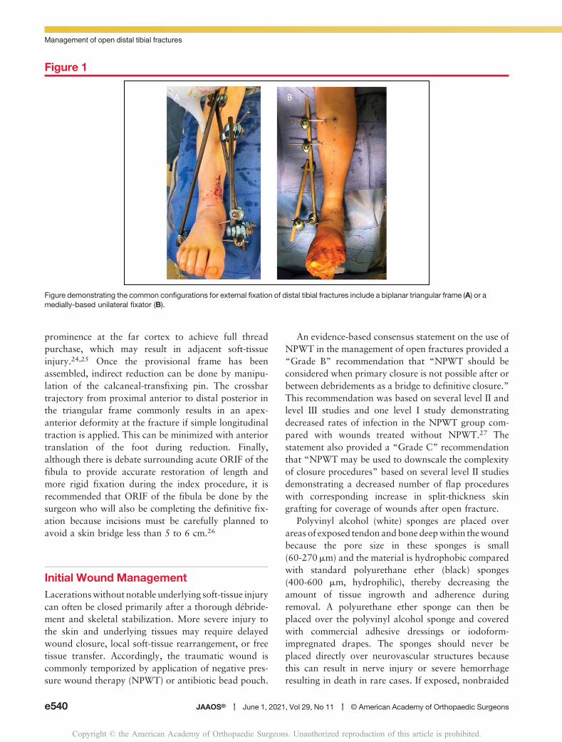
Figure 2
Case 1. A, Figure demonstrating the distal tibial fracture with 4 cm transverse wound involving skin, subcutaneous fat, and fascia alongthe medial aspect of the leg.B andC, Lateral and AP radiographs demonstrating an intra-articular distal tibial fracture with comminutionof both metaphyseal and articular segments (AO/OTA 43.C3 fracture).
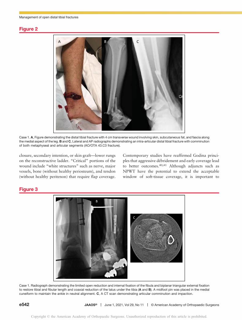
Figure 3
Case 1. Radiograph demonstrating the limited open reduction and internal fixation of the fibula and biplanar triangular external fixationto restore tibial and fibular length and coaxial reduction of the talus under the tibia (A and B). A midfoot pin was placed in the medialcuneiform to maintain the ankle in neutral alignment. C, A CT scan demonstrating articular comminution and impaction.
e542 JAAOS® ----- June 1, 2021, Vol 29, No 11 ----- © American Academy of Orthopaedic Surgeons
Management of open distal tibial fractures
Copyright © the American Academy of Orthopaedic Surgeons. Unauthorized reproduction of this article is prohibited.
remember that early coverage (less than 7 days) shouldbe the goal.41
Local flaps for open distal tibial fractures remainuncommon secondary to the paucity of localmuscle, minimal skin laxity around the foot and ankleand the size and location of the wound. Of the few localoptions, a pedicled flap such as a peroneus brevis flap(based on the terminal perforator of the peroneal artery)
has the potential to provide sufficient coverage aroundthe ankle.42 Other options include the reverse suralartery flap (based on the anastomosis between the ter-minal perforator of the peroneal artery and the super-ficial sural artery) and the reverse soleus flap (based ondistal perforators of the posterior tibial artery). How-ever, because of their relatively small vascular pediclesand the need for almost 180 degrees of rotation to reach
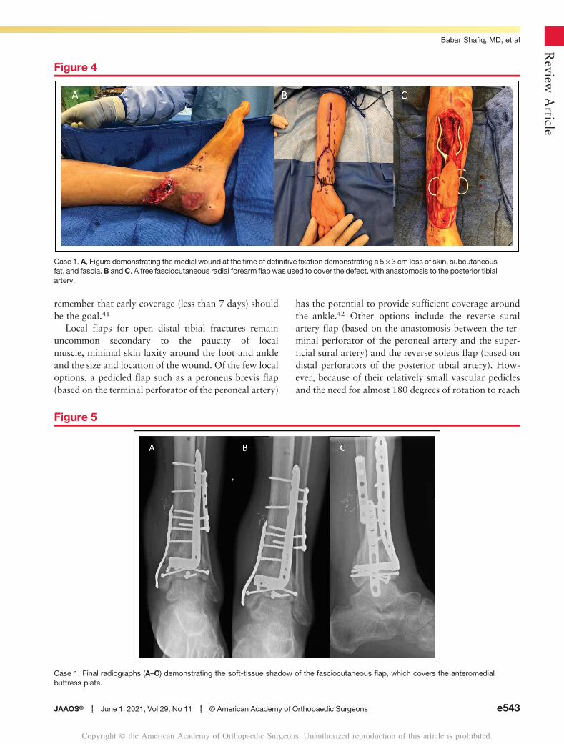
Figure 4
Case 1. A, Figure demonstrating the medial wound at the time of definitive fixation demonstrating a 5 · 3 cm loss of skin, subcutaneousfat, and fascia.B andC, A free fasciocutaneous radial forearm flap was used to cover the defect, with anastomosis to the posterior tibialartery.
Figure 5
Case 1. Final radiographs (A–C) demonstrating the soft-tissue shadow of the fasciocutaneous flap, which covers the anteromedialbuttress plate.
JAAOS® ----- June 1, 2021, Vol 29, No 11 ----- © American Academy of Orthopaedic Surgeons e543
Review
Article
Babar Shafiq, MD, et al
Copyright © the American Academy of Orthopaedic Surgeons. Unauthorized reproduction of this article is prohibited.

the recipient site, these flaps are prone to complicationsand failure.35 Consequently, free tissue transfer is oftenthe best option for critical wounds that result from opendistal tibial fractures. In these instances, the primaryconcern of the orthopaedic surgeon must be to preserveall remaining major vessels of the lower extremity.Current free flap failure rates range between 1% and4% using modern microvascular techniques.43 Apartfrom technical failures, the primary determinant in thesuccess or failure of the flap is the status of the recipientvessel. Recipient vessels within the zone of injury orthose outside the zone of injury that have been damagedindirectly during the débridement or careless placement
of proximal external fixator pins are prone to throm-bosis and resultant flap loss.
Free flaps used in the distal leg are typically eithermuscular or fasciocutaneous. Advantages of fasciocuta-neous flaps include easier elevation in the setting of sec-ondary bone grafting, improved postoperativemonitoring owing to the cutaneous component of theflap, and improved early aesthetic outcome. Muscleflaps, although bulky initially, eventually atrophy andhave good long-term aesthetic outcomes and improvedconformity over a complex three-dimensional surface(eg, wrapping around the ankle and dorsum of foot).They also have the theoretical benefit of improved
Figure 6
Case 2.A andB, Radiographs demonstrating the open distal tibial fracture with extensivemetaphyseal comminution and bone loss andintra-articular extension (AO/OTA type 43.C2).C, Figure demonstrating after débridement and external fixator placement, the elliptical 6· 4 cm anteromedial wound was temporized with placement of antibiotic beads and negative pressure wound therapy.
Figure 7
Case 2. A, Radiographs demonstrating the postinjury day 9 for limited internal fixation of the articular fracture, application of ringedexternal fixator, and placement of an antibiotic cement spacer. B, The anteromedial soft-tissue defect was managed with ananterolateral thigh fasciocutaneous free-flap. C, Construct after free flap.
e544 JAAOS® ----- June 1, 2021, Vol 29, No 11 ----- © American Academy of Orthopaedic Surgeons
Management of open distal tibial fractures
Copyright © the American Academy of Orthopaedic Surgeons. Unauthorized reproduction of this article is prohibited.
antibiotic delivery and bone healing because of higherblood flow. Large retrospective studies have shown min-imal differences in outcomes between the two flaptypes.44-47 For both the soft-tissue surgeon and fracturesurgeon, it is important to note that advantages anddisadvantages exist to each. The common recipient arteryis either the anterior or posterior tibial artery, butselection ultimately depends on the overall quality of thevessel and confirming good in-flow. As mentioned pre-viously, vascular anomalies in the leg have an incidence of7% to 12% in the general cohort.10 For example, the
incidence of peronea magna (dominant peroneal arterywith hypoplastic or aplastic anterior and posterior tibialarteries) is between 0.2% and 8.3% and may limit op-tions for recipient vessels.48,49 The corresponding venacomitantes and the great saphenous vein commonlyprovide venous outflow for the flap. The workhorsefasciocutaneous flap is the anterolateral thigh flap whichis based on the descending branch of the lateral femoralcircumflex artery. The free latissimus dorsi and freegracilis flaps are the most common muscle flaps, espe-cially for larger and more complex wounds (Figures 6–9).
Figure 8
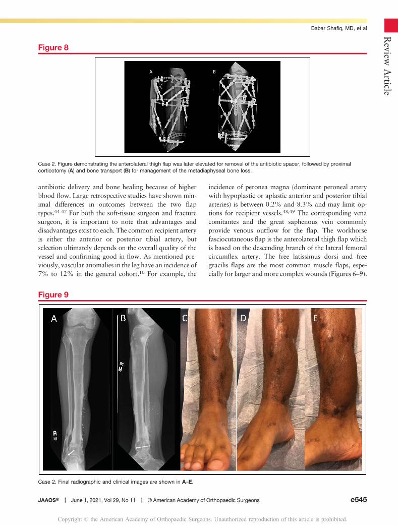
Case 2. Figure demonstrating the anterolateral thigh flap was later elevated for removal of the antibiotic spacer, followed by proximalcorticotomy (A) and bone transport (B) for management of the metadiaphyseal bone loss.
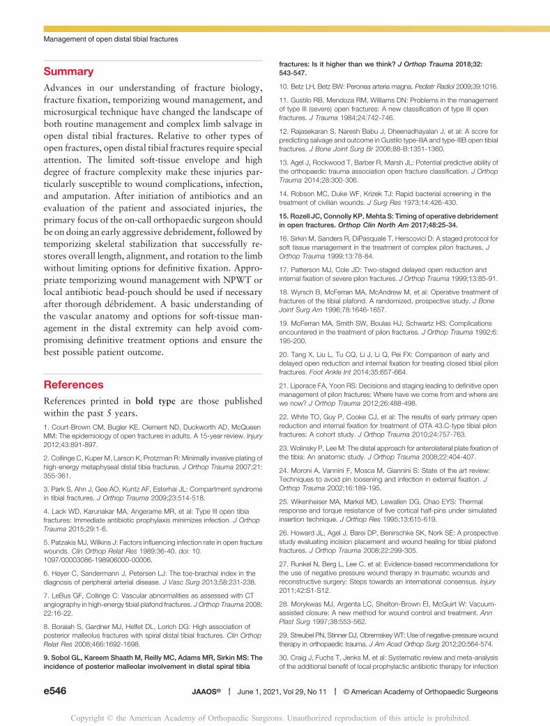
Figure 9
Case 2. Final radiographic and clinical images are shown in A–E.
JAAOS® ----- June 1, 2021, Vol 29, No 11 ----- © American Academy of Orthopaedic Surgeons e545
Review
Article
Babar Shafiq, MD, et al
Copyright © the American Academy of Orthopaedic Surgeons. Unauthorized reproduction of this article is prohibited.
SummaryAdvances in our understanding of fracture biology,fracture fixation, temporizing wound management, andmicrosurgical technique have changed the landscape ofboth routine management and complex limb salvage inopen distal tibial fractures. Relative to other types ofopen fractures, open distal tibial fractures require specialattention. The limited soft-tissue envelope and highdegree of fracture complexity make these injuries par-ticularly susceptible to wound complications, infection,and amputation. After initiation of antibiotics and anevaluation of the patient and associated injuries, theprimary focus of the on-call orthopaedic surgeon shouldbe on doing an early aggressive debridement, followed bytemporizing skeletal stabilization that successfully re-stores overall length, alignment, and rotation to the limbwithout limiting options for definitive fixation. Appro-priate temporizing wound management with NPWT orlocal antibiotic bead-pouch should be used if necessaryafter thorough débridement. A basic understanding ofthe vascular anatomy and options for soft-tissue man-agement in the distal extremity can help avoid com-promising definitive treatment options and ensure thebest possible patient outcome.
ReferencesReferences printed in bold type are those publishedwithin the past 5 years.
1. Court-Brown CM, Bugler KE, Clement ND, Duckworth AD, McQueen
MM: The epidemiology of open fractures in adults. A 15-year review. Injury
2012;43:891-897.
2. Collinge C, Kuper M, Larson K, Protzman R: Minimally invasive plating of
high-energy metaphyseal distal tibia fractures. J Orthop Trauma 2007;21:
355-361.
3. Park S, Ahn J, Gee AO, Kuntz AF, Esterhai JL: Compartment syndrome
in tibial fractures. J Orthop Trauma 2009;23:514-518.
4. Lack WD, Karunakar MA, Angerame MR, et al: Type III open tibia
fractures: Immediate antibiotic prophylaxis minimizes infection. J Orthop
Trauma 2015;29:1-6.
5. Patzakis MJ, Wilkins J: Factors influencing infection rate in open fracturewounds. Clin Orthop Relat Res 1989:36-40. doi: 10.
1097/00003086-198906000-00006.
6. Høyer C, Sandermann J, Petersen LJ: The toe-brachial index in the
diagnosis of peripheral arterial disease. J Vasc Surg 2013;58:231-238.
7. LeBus GF, Collinge C: Vascular abnormalities as assessed with CT
angiography in high-energy tibial plafond fractures. J Orthop Trauma 2008;
22:16-22.
8. Boraiah S, Gardner MJ, Helfet DL, Lorich DG: High association of
posterior malleolus fractures with spiral distal tibial fractures. Clin Orthop
Relat Res 2008;466:1692-1698.
9. Sobol GL, Kareem Shaath M, Reilly MC, Adams MR, Sirkin MS: Theincidence of posterior malleolar involvement in distal spiral tibia
fractures: Is it higher than we think? J Orthop Trauma 2018;32:543-547.
10. Betz LH, Betz BW: Peronea arteria magna. Pediatr Radiol 2009;39:1016.
11. Gustilo RB, Mendoza RM, Williams DN: Problems in the management
of type III (severe) open fractures: A new classification of type III open
fractures. J Trauma 1984;24:742-746.
12. Rajasekaran S, Naresh Babu J, Dheenadhayalan J, et al: A score for
predicting salvage and outcome in Gustilo type-IIIA and type-IIIB open tibial
fractures. J Bone Joint Surg Br 2006;88-B:1351-1360.
13. Agel J, Rockwood T, Barber R, Marsh JL: Potential predictive ability of
the orthopaedic trauma association open fracture classification. J Orthop
Trauma 2014;28:300-306.
14. Robson MC, Duke WF, Krizek TJ: Rapid bacterial screening in thetreatment of civilian wounds. J Surg Res 1973;14:426-430.
15. Rozell JC, Connolly KP, Mehta S: Timing of operative debridementin open fractures. Orthop Clin North Am 2017;48:25-34.
16. Sirkin M, Sanders R, DiPasquale T, Herscovici D: A staged protocol for
soft tissue management in the treatment of complex pilon fractures. J
Orthop Trauma 1999;13:78-84.
17. Patterson MJ, Cole JD: Two-staged delayed open reduction and
internal fixation of severe pilon fractures. J Orthop Trauma 1999;13:85-91.
18. Wyrsch B, McFerran MA, McAndrew M, et al: Operative treatment of
fractures of the tibial plafond. A randomized, prospective study. J Bone
Joint Surg Am 1996;78:1646-1657.
19. McFerran MA, Smith SW, Boulas HJ, Schwartz HS: Complications
encountered in the treatment of pilon fractures. J Orthop Trauma 1992;6:
195-200.
20. Tang X, Liu L, Tu CQ, Li J, Li Q, Pei FX: Comparison of early and
delayed open reduction and internal fixation for treating closed tibial pilon
fractures. Foot Ankle Int 2014;35:657-664.
21. Liporace FA, Yoon RS: Decisions and staging leading to definitive open
management of pilon fractures: Where have we come from and where are
we now? J Orthop Trauma 2012;26:488-498.
22. White TO, Guy P, Cooke CJ, et al: The results of early primary openreduction and internal fixation for treatment of OTA 43.C-type tibial pilon
fractures: A cohort study. J Orthop Trauma 2010;24:757-763.
23. Wolinsky P, Lee M: The distal approach for anterolateral plate fixation of
the tibia: An anatomic study. J Orthop Trauma 2008;22:404-407.
24. Moroni A, Vannini F, Mosca M, Giannini S: State of the art review:
Techniques to avoid pin loosening and infection in external fixation. J
Orthop Trauma 2002;16:189-195.
25. Wikenheiser MA, Markel MD, Lewallen DG, Chao EYS: Thermal
response and torque resistance of five cortical half-pins under simulated
insertion technique. J Orthop Res 1995;13:615-619.
26. Howard JL, Agel J, Barei DP, Benirschke SK, Nork SE: A prospective
study evaluating incision placement and wound healing for tibial plafond
fractures. J Orthop Trauma 2008;22:299-305.
27. Runkel N, Berg L, Lee C, et al: Evidence-based recommendations for
the use of negative pressure wound therapy in traumatic wounds and
reconstructive surgery: Steps towards an international consensus. Injury
2011;42:S1-S12.
28. Morykwas MJ, Argenta LC, Shelton-Brown EI, McGuirt W: Vacuum-
assisted closure: A new method for wound control and treatment. Ann
Plast Surg 1997;38:553-562.
29. Streubel PN, Stinner DJ, ObremskeyWT: Use of negative-pressure wound
therapy in orthopaedic trauma. J Am Acad Orthop Surg 2012;20:564-574.
30. Craig J, Fuchs T, Jenks M, et al: Systematic review and meta-analysis
of the additional benefit of local prophylactic antibiotic therapy for infection
e546 JAAOS® ----- June 1, 2021, Vol 29, No 11 ----- © American Academy of Orthopaedic Surgeons
Management of open distal tibial fractures
Copyright © the American Academy of Orthopaedic Surgeons. Unauthorized reproduction of this article is prohibited.
rates in open tibia fractures treated with intramedullary nailing. Int Orthop
2014;38:1025-1030.
31. Stinner DJ, Hsu JR,Wenke JC: Negative pressure wound therapy reduces
the effectiveness of traditional local antibiotic depot in a large complexmusculoskeletal wound animal model. J Orthop Trauma 2012;26:512-518.
32. Rand BCC, Wenke JC: An effective negative pressure woundtherapy-compatible local antibiotic delivery device. J Orthop Trauma
2017;31:631-635.
33. Zalavras CG, Patzakis MJ, Holtom P: Local antibiotic therapy in the
treatment of open fractures and osteomyelitis. Clin Orthop Relat Res 2004;
427:86-93.
34. Henry SL, Ostermann PAW, Seligson D: The antibiotic bead pouch
technique: The management of severe compound fractures. Clin Orthop
Relat Res 1993:54-62. doi: 10.1097/00003086-199310000-00010.
35. Tornetta P, RicciW,Court-BrownCM,McQueenMM,McKeeM:Rockwood
and Green’s Fractures in Adults. Philadelphia, PA, Wolters Kluwer, 2015.
36. Johansen K, Daines M, Howey T, Helfet D, Hansen ST: Objective
criteria accurately predict amputation following lower extremity trauma. J
Trauma 1990;30:568-573.
37. Russell WL, Sailors DM, Whittle TB, Fisher DF, Burns RP: Limb salvage
versus traumatic amputation: A decision based on a seven-part predictive
index. Ann Surg 1991;213:473-481.
38. Bosse MJ, McCarthy ML, Jones AL, et al; Lower Extremity AssessmentProject (LEAP) Study Group: The insensate foot following severe lower
extremity trauma: An indication for amputation? J Bone Joint Surg Am
2005;87:2601-2608.
39. Loja MN, Sammann A, DuBose J, et al: The mangled extremityscore and amputation: Time for a revision. J Trauma Acute Care Surg
2017;82:518-523.
40. Godina M: Early microsurgical reconstruction of complex trauma of the
extremities. Plast Reconstr Surg 1986;78:285-292.
41. Pincus D, Byrne JP, Nathens AB, et al: Delay in flap coverage past7 days increases complications for open tibia fractures: A cohortstudy of 140 North American Trauma Centers. J Orthop Trauma 2019;33:161-168.
42. Abd-Al-Moktader MA: Distally based peroneus brevis muscle flapfor large leg, ankle, and foot defects: Anatomical finding and clinicalapplication. J Reconstr Microsurg 2018;34:616-623.
43. Pu LLQ: A comprehensive approach to lower extremity free-tissuetransfer. Plast Reconstr Surg Glob Open 2017;5:e1228.
44. Fox CM, Beem HM, Wiper J, Rozen WM, Wagels M, Leong JC: Muscle
versus fasciocutaneous free flaps in heel reconstruction: Systematic review
and meta-analysis. J Reconstr Microsurg 2014;31:59-66.
45. Sabino J, Polfer E, Tintle S, et al: A decade of conflict: Flap coverage
options and outcomes in traumatic war-related extremity reconstruction.
Plast Reconstr Surg 2015;135:895-902.
46. Cho EH, Shammas RL, Carney MJ, et al: Muscle versusfasciocutaneous free flaps in lower extremity traumaticreconstruction: Amulticenter outcomes analysis. Plast Reconstr Surg2018;141:191-199.
47. Lee ZH, Abdou SA, Daar DA, et al: Comparing outcomes forfasciocutaneous versus muscle flaps in foot and ankle free flapreconstruction. J Reconstr Microsurg 2019;35:646-651.
48. Rosson GD, Singh NK: Devascularizing complications of free fibula
harvest: Peronea arteria magna. J Reconstr Microsurg 2005;21:533-538.
49. Kim D, Orron DE, Skillman JJ: Surgical significance of popliteal arterial
variants. A unified angiographic classification. Ann Surg 1989;210:
776-781.
JAAOS® ----- June 1, 2021, Vol 29, No 11 ----- © American Academy of Orthopaedic Surgeons e547
Review
Article
Babar Shafiq, MD, et al
Copyright © the American Academy of Orthopaedic Surgeons. Unauthorized reproduction of this article is prohibited.