Embed Size (px)
Citation preview

Molecular Immunology 40 (2003) 431–443
Review
Antimicrobial peptides in defence of the oral and respiratory tracts
Deirdre A. Devine∗Division of Oral Biology, Leeds Dental Institute, University of Leeds, Leeds LS2 9LU, UK
Received 8 April 2003; accepted 29 April 2003
Abstract
Antimicrobial peptides (AMPs) are components of complex host secretions, acting synergistically with other innate defence molecules tocombat infection and control resident microbial populations throughout the oral cavity and respiratory tract. AMPs are directly antimicrobial,bind lipopolysaccharide (LPS) and lipoteichoic acid, and are immunomodulatory signals. Pathogenic and commensal organisms display avariety of resistance mechanisms, which are related to structure of cell wall components (e.g. LPS) and cytoplasmic membranes, and peptidebreakdown mechanisms. For example, LPS of the AMP-resistant cystic fibrosis pathogenBurkholderia cepaciais under-phosphorylatedand highly substituted with charge-neutralising 4-deoxy-4-aminoarabinose. Additionally, host mimicry by addition of phosphorylcholinecontributes to resistance in oral and respiratory organisms.Porphyromonas gingivalis, Pseudomonas aeruginosaand other pathogensproduce extracellular and membrane-bound proteases that degrade AMPs. Many of these bacterial properties are environmentally regulated.Their modulation in response to host defences and inflammation can result in altered sensitivity to AMPs, and may additionally change otherhost–microbe interactions, e.g. binding to Toll-like receptors. The diversity and breadth of antimicrobial cover and immunomodulatoryfunction provided by AMPs is central to the ability of a host to respond to the diverse and highly adaptable organisms colonising oral andrespiratory mucosa.© 2003 Elsevier Ltd. All rights reserved.
Keywords:Antimicrobial peptides; Defensins; LL-37; Innate defences;Porphyromonas gingivalis; Burkholderia cepacia; Pseudomonas aeruginosa;Periodontal disease; Cystic fibrosis; Proteases; Lipopolysaccharide
1. Introduction
Cationic antimicrobial peptides (AMPs) have emerged ascentral components of mammalian innate defences and areof fundamental relevance to understanding host–microberelationships. The importance of AMPs extends beyondtheir direct antimicrobial activity, as their broad bio-logical activities indicate they are effector molecules,providing communication between innate and adaptiveimmune systems (Yang et al., 2002). An important prop-erty of AMPs is their ability to bind avidly to manypotentially pro-inflammatory molecules released frommicro-organisms, such as lipopolysaccharide (LPS), lipote-ichoic acid (LTA) and DNA. By binding to these molecules,AMPs inhibit responses of host cells and damp-down anundesirable inflammatory response (Scott et al., 1999,2000a,b; Nagaoka et al., 2001). This may be a key function,in which AMPs function alongside or in concert with otherLPS binding molecules, such as lipopolysaccharide bindingprotein (LBP) and bactericidal permeability inducing pro-
∗ Tel.: +44-113-343-6116; fax:+44-113-343-6548.E-mail address:[email protected] (D.A. Devine).
tein (BPI), to regulate immune responses at mucosal sitesassaulted by large numbers of bacteria and, in particular,their released cellular components.
AMPs are synthesised within granules of phagocyticcells or are secreted by epithelia. At each site of production,they form part of a cocktail of antimicrobial substanceswhich in vivo work synergistically to combat infec-tion (Gudmundsson and Agerberth, 1999; Hancock andDiamond, 2000). Their tissue specific expression is alsolikely to be a significant contributor to the tissue tropismdisplayed by pathogenic and resident micro-organisms.AMPs have evolved in response to the positive selectionpressures exerted by colonising micro-organisms (Hughes,1999), and pathogens and commensals alike have developedstrategies for surviving or evading the activities of AMPs(Devine and Hancock, 2002). In humans and other mam-mals, some sites are usually free of micro-organisms (e.g.lung), whilst others (e.g. oral cavity) are heavily colonisedby diverse populations. The oral cavity and respiratory tractare similar in the range of AMPs they express, and increas-ing attention has been paid to the roles of AMPs at thesesites in defence against conditions such as periodontal dis-ease and cystic fibrosis (CF) associated lung disease. AMPs
0161-5890/$ – see front matter © 2003 Elsevier Ltd. All rights reserved.doi:10.1016/S0161-5890(03)00162-7

432 D.A. Devine / Molecular Immunology 40 (2003) 431–443
are additionally important as potential novel anti-infectiveagents for treatment of such diseases.
2. Antimicrobial peptides in oral innate defences
2.1. Production
Tissues of the oral cavity are constantly exposed to in-nate defence components (Table 1) derived from saliva,gingival crevicular fluid and epithelial cells (Dale et al.,2001; Lamont and Jenkinson, 2000; Marsh, 2003). Salivacontains a range of innate defence molecules that areeither directly antimicrobial or interfere with microbialcolonisation or nutrition. Many of these molecules functionsynergistically and some, such as the lysozyme–proteasesystem and histatins (histidine-rich AMPs), are potentiatedby acid, which may be particularly relevant in defenceagainst dental caries. AMPs are important components ofthese oral innate defences and a number are secreted bysalivary glands and epithelial cells and are released fromneutrophils.
Histatins are secreted by salivary glands, following tran-scription from two genes. Subsequent proteolysis of the pri-mary translation products results in at least 12 forms ofsalivary histatin, only some of which are antimicrobial (Xuet al., 1991). Histatins are mainly anti-candidal, althoughthey have been shown to inhibit some Gram-positive bacte-ria and the periodontal pathogenPorphyromonas gingivalis(Murakami et al., 1991).
Like most human epithelia, oral epithelia are pro-tected by production of�-defensins (HBDs). HBD1 isexpressed constitutively in salivary glands, gingiva, buccalmucosa and tongue (Zhao et al., 1996; Krisanaprakornkitet al., 1998; Bonass et al., 1999; Mathews et al., 1999;
Table 1Innate defence molecules in oral fluids and secretions
Salivary components Gingival crevicularfluid components
Epithelial cellsecretions
Histatins �-Defensins(HNP1–4)
�-Defensins(HNP1–4)
�-Defensins (HNP1–4) �-Defensins(HBD1–3)
�-Defensins(HBD1–3)
�-Defensins (HBD1–3) LL-37 HE2�1LL-37 IgG LL-37Secretory IgA IgA IgAMucins IgM CalprotectinLysozyme ComplementProteaseLactoferrinSialoperoxidaseProline-rich proteinsStatherinFibronectinCystatinsTrefoil factor
family proteins
Sahasrebudhe et al., 2000). In saliva, �-defensins are as-sociated with mucin (Sahasrebudhe et al., 2000), whichmay protect them from degradation and could also in-crease their contact with mucin-aggregated bacteria. Suchbinding may also facilitate concerted activities with othermucin-associated molecules synthesised by salivary glands,such as the trefoil factor family (TFF) proteins (Devineet al., 2000). TFFs are wound healing motogenic proteinsinvolved in early restitution of damaged epithelia that alsobind bacteria (dos Santos Silva et al., 2000). Isoforms ofHBD1 have been detected in oral epithelial cell culture su-pernatants (Diamond et al., 2001). Isoforms of HBD1 andHBD2 are produced at other sites and in many mammalianspecies. This may provide added breadth of antimicrobialcover, as minor differences in amino acid sequence can pro-duce significant differences in defensin antimicrobial activ-ity (Raj et al., 2000; Devine and Hancock, 2002). Inducibleexpression of HBD2 and HBD3 have also been observed inoral epithelial cells and salivary glands (Bonass et al., 1999;Mathews et al., 1999; Jia et al., 2000; Krisanaprakornkitet al., 1998; Garcia et al., 2001a; Dunsche et al., 2002), al-though each peptide is induced through different signallingpathways (discussed later). Another putative antimicrobialpeptide with a defensin-like 6-cysteine motif, HE2�1, isexpressed in human gingival epithelia (Jia et al., 2001).
Phagocytic cells are significant sources of AMPs. Neu-trophils are the richest and best-studied sources, butmacrophages, monocytes and dendritic cells also synthesiseHBD1 and HBD2 (Duits et al., 2002) and may be sourcesof these AMPs at a variety of sites. Neutrophil AMPsare found throughout the oral cavity, and increase in con-centration following inflammation.�-Defensins HNP1–4have been detected in saliva, gingival crevicular fluid andin gingival junctional epithelium (McKay et al., 1999;Mizukawa et al., 1999; Dale et al., 2001). The humancathelicidin LL-37 is produced within secondary granulesof neutrophils, but is also secreted by epithelial cells at awide range of sites following induction by microbial prod-ucts or inflammatory mediators. Its secretion (or that of itsprecursor hCAP18) has been detected in tongue and buccalmucosa (Frohm Nilsson et al., 1999) but LL-37 peptidedetected in gingival junctional epithelium was most likelyderived from neutrophil infiltration (Dale et al., 2001).Whilst we have detected LL-37 peptide in salivary glandducts, expression appeared to be low compared with HBD1(Fig. 1).
Other multifunctional molecules with AMP propertiesor activities are also found within oral tissues and fluids.Adrenomedullin is antimicrobial as well as vasodilatory andit is induced by exposure of oral epithelial cells to bacte-ria but not byCandida albicans(Kapas et al., 2001). Somecationic fragments derived from larger proteins exhibit AMPfunctions; for example, fragments of lactoferrin, bacterici-dal permeability inducing peptide, histones, ribosomal pro-tein, haemoglobin and mucin (Devine and Hancock, 2002;Bobek and Situ, 2003).

D.A. Devine / Molecular Immunology 40 (2003) 431–443 433
Fig. 1. Immunostaining of human submandibular gland with antibodiesto �-defensin 1 (A) and LL-37 (B).
2.2. Roles of antimicrobial peptides in oralinnate defences
The micro-organisms encountered by AMPs and otheroral innate defence molecules are numerous and diverse.Close or adjacent sites harbour distinct microbiota; oralmicrobiota differ significantly from upper respiratory tractpopulations despite their proximity and, indeed, there arevariations even within the oral cavity (Hohwy et al., 2001;Rasmussen et al., 2000; Könönen et al., 2002; Marsh,2003). It is estimated that up to 600 species, only 50% ofwhich can be grown in monoculture by conventional meth-ods, are normal inhabitants of the human mouth (Wilsonet al., 1997; Paster et al., 2001). These populations exhibitconsiderable diversity and inter-dependent consortia, ratherthan individual organisms, are associated with diseases(Marsh, 2003). In such cases, it can be difficult to deter-mine which organisms contribute to the aetiology of thedisease and which are bystanders in the process. Nonethe-less, certain organisms are consistently implicated in theaetiology of advanced periodontal diseases, for example,the Gram-negative anaerobeP. gingivalis, which is isolated
in low numbers from healthy subgingival sites but increasesin prevalence during disease. It produces a range of viru-lence determinants, including potent proteases, adhesins,LPS and haemagglutinins (Lamont and Jenkinson, 2000)and is able to survive and grow within oral epithelial cells(Houalet-Jeanne et al., 2001; Rudney et al., 2001).
Proving or examining the roles of AMPs in defence ofthe oral cavity is particularly challenging, because of thecomplexity of the microbiota and the multiplicity of in-nate defence molecules produced. A study of a congenitalcondition associated with severe neutropenia linked pe-riodontal disease with a deficiency of neutrophil AMPs(Putsep et al., 2002). Epithelial AMPs are more numerous(Schutte and McCray Jr., 2002) and greater overlap or re-dundancy of function is likely, as is indicated by the factthat BD1-deficient mice showed no overt signs of ill healthor infection (Morrison et al., 2002; Moser et al., 2002).This may be particularly important in defence of heavilycolonised sites like the mouth, where loss of adequate con-trol of such resident populations could be catastrophic. Atthese sites AMPs not only exhibit a breadth of recognition toaccommodate microbial diversity, but also provide sufficientredundancy to ensure such populations are controlled, avoid-ing disruption of host–microbe homeostatic mechanisms.
Exposure of oral epithelial cells to a range of bacterialproducts, such as LPS, and inflammatory mediators inducesynthesis of�-defensins. Few studies have examined in-duction by organisms that are directly relevant to the oralcavity. However,Krisnaprakornkit et al. (2000)showed thatcell wall extracts ofFusobacterium nucleatuminduced gin-gival epithelial cells to synthesise HBD2, while extracts ofP. gingivalisdid not. This may or may not be related to theability of these two organisms to cause disease. WhilstP.gingivalis is generally recognised as a periodontal pathogenand increases in prevalence with disease,F. nucleatumisisolated with equal frequency from healthy and diseasedsites but has been associated by some groups with peri-odontal disease and additionally demonstrates propertiesassociated with pathogenicity (Haffajee et al., 1999; Hanet al., 2000). To date, a clear cut relationship betweenpathogenicity or commensalism and interactions with AMPshas not emerged, and bacteria employ a range of strategiesfor surviving AMPs (Devine and Hancock, 2002).
Histatins have been somewhat overlooked in many dis-cussions of AMPs, possibly because they appear to bespecific to the mouth. This may indicate that their spectrumof activity, particularly againstCandidaspp., is of primaryimportance to defence of the oral cavity. It may also reflecta need to protect critical cells, for example, secretory cellsin salivary ducts, although AMPs are probably not essentialfor preventing colonisation of other protected sites such asintestinal crypts (Garabedian et al., 1997). Candidaspp. fre-quently colonise oral mucosal surfaces, causing diseasein immunocompromised or antibiotic treated individuals.Reduced salivary flow and lower salivary histatin concen-trations have been linked withCandidacolonisation of oral

434 D.A. Devine / Molecular Immunology 40 (2003) 431–443
mucosa (Jainkittivong et al., 1998). Whether this was a re-flection of the ability ofCandidaspp. to repress secretion ofhistatins was not explored.C. albicansdid not induce oralkeratinocytes to up-regulate adrenomedullin production,in contrast to Gram-positive and Gram-negative bacteria(Kapas et al., 2001). The role in oral ecology of histatinsmay be greater than their principally anti-candidal activitiesindicate since they also: (i) inhibitP. gingivalis and hostproteases (Gusman et al., 2001); (ii) inhibit P. gingivalisadhesion to erythrocytes and streptococci (Murakami et al.,1991, 1992); (iii) suppress induction of cytokines byP.gingivalisouter membrane proteins (Imatani et al., 2000).
3. Antimicrobial peptides in respiratory innate defences
3.1. Production
As in the mouth, the airways are protected by AMPs re-leased from phagocytes and secreted by epithelial cells. Neu-trophil �-defensins HNP1–4 and cathelicidin LL-37, derivedfrom neutrophils and epithelial cells, are detected in airwaysecretions (Bals et al., 1998b; Agerberth et al., 1999). Air-way epithelial cells secrete�-defensin HD5 and�-defensinsHNP1–4 (McCray Jr. and Bentley, 1997; Goldman et al.,1997; Bals et al., 1998a; Singh et al., 1998; Frye et al., 2000;Jia et al., 2001; Harder et al., 2001; Garcia et al., 2001a,b).HBD2 and LL-37 have been shown to reach significant lev-els in bronchial alveolar lavage fluid (Singh et al., 1998;Agerberth et al., 1999). While HBD1 is expressed consti-tutively, the other�-defensins are inducible but not by thesame stimuli. Unlike HBD2, the gene encoding HBD3 doesnot have a NF�B consensus sequence but does have AP1and NF–IL-6 consensus sequences, and theHBD3 gene isup-regulated by IFN-�, not TNF-�, IL-1�, IL-6, PMA ornon-viablePseudomonas aeruginosa(Garcia et al., 2001a).HBD4 is primarily expressed in the lung and is inducedby non-viableP. aeruginosaandStreptococcus pneumoniaebut not by TNF-�, IFN-� or IL-1� (Garcia et al., 2001b).P. aeruginosais a significant respiratory pathogen, and isparticularly associated with high morbidity and mortalityin patients suffering from CF. It has been shown to induce�-defensin synthesis in mouse respiratory epithelia in vivo(Bals et al., 1999a; Morrison et al., 1999). The produc-tion of mucoid extracellular polysaccharide byP. aerugi-nosahas been linked to virulence andHarder et al. (2000)found a mucoidP. aeruginosastrain induced HBD2 pro-duction in respiratory epithelial cells but non-mucoid strainsdid not. This was true for cells derived from CF patients aswell as non-CF individuals. However, CF patients did notup-regulate�-defensins in response to inflammatory medi-ators (Dauletbaev et al., 2002), supporting a contention thatlocal deficiency in innate defences is important in the patho-genesis of CF lung disease (Bals et al., 1998a).
In the respiratory tract, as in the oral cavity, AMPs formpart of a cocktail of antimicrobial molecules (Table 2)
Table 2Innate defence molecules in airway surface fluid
Antimicrobial component Produced by cells
�-Defensins (HNP1–4) Neutrophils�-Defensin (HD5) Epithelial�-Defensins (HBD1–4) Epithelial, macrophages,
monocytes, dendriticLL-37 Neutrophils, epithelialLysozyme Epithelial, neutrophilsPhospholipase A2 Epithelial, neutrophilsIgA EpithelialLactoferrin Epithelial, neutrophilsBactericidal permeability
inducing proteinNeutrophils
Serine proteinase inhibitor Epithelial, macrophagesSurfactant proteins SP-A, SP-D EpithelialAnionic peptides EpithelialProline-rich proteins EpithelialTrefoil factor family proteins Epithelial
many of which increase after infection and inflammation(Diamond et al., 2000; Zhang et al., 2000; Schutte andMcCray Jr., 2002). Synergy has been demonstrated betweenBPI and phospholipase A2, HBD2 and lactoferrin, as wellas between HBD2/HBD4 and lysozyme (Bals et al., 1998a;Garcia et al., 2001b). However, other studies did not confirmsynergy between HBD2, HBD3 or HBD4 and lysozyme(Singh et al., 2000; Garcia et al., 2001a) so this needsfurther clarification. Defensins and many other AMPs areinhibited in vitro by increasing concentrations of NaCl, butsynergy between HNP1 and LL-37 overcame the inhibitoryeffects of NaCl (Nagoaka et al., 2000). AMPs are one ofmany LPS binding molecules produced in the respiratorytract, including BPI, LBP and surfactant-associated proteins(Crouch et al., 2000; Zhang et al., 2000; Augusto et al.,2002). �-Defensins regulate release of serine proteinaseinhibitor (SLPI) from airway epithelial cells (van Weteringet al., 2000). Elafin, which like SLPI is an inhibitor ofneutrophil proteinase activity, is also directly antimicro-bial (Simpson et al., 1999) and is regulated by neutrophildefensins (van Wetering et al., 2000).
3.2. Roles of antimicrobial peptides in respiratoryinnate defences
Inducible and constitutive production of AMPs is knownto occur throughout the respiratory tract and levels of�-defensins and LL-37 increase following infections andinflammation (Ashitani et al., 2001; Dauletbaev et al.,2002; Lee et al., 2002; Schaller-Bals et al., 2002). Theyprotect sites that are heavily colonised, such as the nasalcavity and nasopharynx, and some which are usuallyfree of micro-organisms, e.g. the lung. The diverse resi-dent commensal populations of the upper airways consistof organisms includingStaphylococcusspp., Streptococ-cus spp., Neisseria spp., Moraxella spp., Haemophilusspp. andMycoplasmaspp. Some respiratory pathogens

D.A. Devine / Molecular Immunology 40 (2003) 431–443 435
do not cause clinically overt disease in all hosts, and im-portant pathogens may be carried as part of the normalresident microbiota of individuals for considerable peri-ods of time. Some opportunistic pathogens cause severeinfections in patients with CF. In addition toP. aerugi-nosa, members of theBurkholderia cepaciacomplex areimportant causes of morbidity and mortality in this group(Mohr et al., 2001; Mahenthiralingam et al., 2002). Upto 20% of patients with CF who acquireB. cepaciacom-plex organisms develop the “cepacia syndrome”, a rapidlyfatal necrotising pneumonia, often in conjunction with sep-ticaemia. Virulence and clinical outcome are correlatedto specificB. cepaciagenomovars, many of which havebeen assigned to new species (Mahenthiralingam et al.,2002).
The virulence determinants ofB. cepaciacomplex or-ganisms have received a great deal of attention in recentyears but pathogenicity is not yet fully understood. Adhe-sion and colonisation of respiratory epithelia are impor-tant, and in pulmonary infectionB. cepaciamay exhibit abiofilm mode of growth (Desai et al., 1998). Colonisationmay be aided by the fact that these bacteria are highly re-sistant to a wide range of human and non-human AMPs.In our studies, strains representing six genomovars wereresistant to HNP1, HBD1, HBD2 and LL-37 and othernon-human AMPs (Table 3). However, many were sen-sitive to ovine cathelicidin SMAP-29 and the syntheticD2A-22, both of which have been reported previously tohave activity against selected strains ofB. cepacia(Schwabet al., 1999; Saiman et al., 2001). Whilst virulence is cor-related with particular genomovars of theB. cepaciacom-plex, there was no clear relationship between sensitivity tothese peptides and genomovar. Differences in outer mem-
Table 3Resistance ofB. cepaciacomplex strains representing genomovars I–VIa
to antimicrobial peptides
Peptide Number resistant(number studied)
MCZb ofsensitivestrains
MICc ofsensitivestrains
�-Defensin 1 17 (17) >500 >500�-Defensin 2 17 (17) >500 >500LL-37 17 (17) >500 >500HNP1 17 (17) >500 >500D2A-22 6 (14) 4–250 0.5–250SMAP-29 4 (13) 4–250 0.5 to >500Histatin Dhvar4 14 (14) >500 >500Brevinin 1 16 (16) >500 >500Cecropin B 12 (13) 62 0.5Melittin 16 (17) 1 0.5Polymyxin B 17 (17) >500 >500
a Genomovars II, IV and V have been assigned toBurkholderia multi-vorans, Burkholderia stabilisandBurkholderia vietnamensis, respectively(Mahenthiralingam et al., 2002).
b Minimum concentration of peptide (�g ml−1) producing a zone ofinhibition in double layer agarose assays.
c Minimum inhibitory concentration (�g ml−1) determined in brothmicrodilution assays.
brane structure amongst these strains are currently beingexamined.
An important role for AMPs in the susceptibility of theCF lung to infection has been suggested. It was proposedthat, as a result of the CF defect in ion transport, trachealexudates of CF patients have high concentrations of NaCl,causing inactivity of AMPs towards pathogenic bacteria andthereby contributing to the ability of the latter to infect thesepatients (Goldman et al., 1997). However, not all studieshave confirmed high NaCl concentrations in airway fluidsand it is a paradox that organisms likeB. cepacia, which arenaturally resistant to AMPs, should require salt-inactivationof these innate defences to express their pathogenicity. Also,HBD3 was recently shown to killB. cepaciaregardless ofNaCl concentration (Garcia et al., 2001a). It is likely thatmultiple factors are in operation. It has been suggested thatmore general, uncharacterised, deficiencies in local innatedefence of the CF lung are responsible for the increasedsusceptibility to infection, and AMPs may be a componentof this. B. cepaciacan survive within respiratory epithelialcells, macrophages and in amoeba and this may be relatedto AMP resistance as well as other factors, e.g. a decreasein NO production inB. cepaciainfected macrophages hasbeen reported as well as cytotoxicity to macrophages (Mohret al., 2001).
A number of in vivo experimental models have been usedto determine the functional importance of AMPs in defenceof the respiratory tract, particularly in CF. Over-expressionof the human peptide LL-37 in a CF mouse model re-sulted in increased killing ofP. aeruginosa(Bals et al.,1999c), reduced ability ofP. aeruginosato colonise thelung epithelium and in reduced inflammation and suscep-tibility to septic shock (Bals et al., 1999b). Mice deficientin mBD1 expression did not show any overt signs of illhealth or abnormality (Morrison et al., 2002; Moser et al.,2002). These studies indicated that an individual AMPmay be more important in defence against one organismthan another. For example, mBD1-deficient mice were in-efficient at clearingHaemophilus influenzaefrom lungsand airways (Moser et al., 2002) but eliminatedStaphy-lococcus aureusfrom their lungs as efficiently as wildtype mice (Morrison et al., 2002), while mutant and wildtype mice were equally susceptible to infection, sepsis anddeath following infection withS. pneumoniae(Moser et al.,2002).
In addition to defending against infection, released�-defensins may contribute to epithelial repair in the res-piratory tract through enhancing lung epithelial cell pro-liferation (Aarbiou et al., 2002). To this end, they mayfunction alongside wound healing factors such as the TFFproteins, which are also expressed by respiratory epithelia(dos Santos Silva et al., 2000). AMPs also contribute toprotection against protease-mediated damage through reg-ulation of release of SLPI and elafin. On the other hand,neutrophil AMP release can also have undesirable effects(van Wetering et al., 1999; Devine and Hancock, 2002) and

436 D.A. Devine / Molecular Immunology 40 (2003) 431–443
may contribute to the pathogenesis of certain respiratorydiseases, such as inflammatory lung disease and atheroscle-rosis (through binding of lipoproteins).�-Defensins ad-here to indwelling medical devices and diminish effectivedefences against biofilm formation by inhibiting neu-trophil function, and these AMPs can also enhance theadhesion of respiratory pathogens to respiratory epithelialcells.
Thus, AMPs are components of complex host secretionscontributing to innate defences throughout the oral cav-ity and respiratory tract. They act synergistically with eachother and with other classes of molecule to combat in-fection and control resident microbial populations. Theymay function alongside molecules such as TFF proteinsin wound healing processes, and with other LPS bindingmolecules such as bactericidal permeability inducing proteinand LPS binding protein to regulate responses to bacterialLPS.
Fig. 2. Interactions between antimicrobial peptides and Gram-negative bacterial cells (adapted fromHancock and Chapple, 1999). (a and b) Peptidesbind to the divalent cation-binding sites and associate with the polyanionic outer moieties of LPS, disrupting and expanding the outer membrane andallowing passage of AMPs through the outer membrane. (c) AMPs then bind to the interfacial region of the cytoplasmic membrane. (d) When at sufficientconcentrations, AMPs aggregate within the membrane causing depolarisation and permeabilisation. Some monomers may detach and gain access to thecytoplasm.
4. Mechanisms of antimicrobial peptide resistancedisplayed by oral and respiratory micro-organisms
In many cases antimicrobial peptides are able to killbacteria by depolarising and permeabilising membranes(Fig. 2), but there are some examples in which the lethaltarget is cytoplasmic (reviewed inDevine and Hancock,2002). In Gram-negative bacteria, peptides first associatewith negatively charged moieties of the outer membrane,producing structural cracks. They also bind to the divalentcation-binding sites of polyanionic surface LPS and expandthe outer membrane by displacing divalent cations, whichnormally stabilise outer membrane structure. Disruption ofbarrier function and integrity of the outer membrane then al-lows passage of molecules such as large hydrophobic antibi-otics and the AMPs themselves (termed the self-promoteduptake pathway;Hancock and Chapple, 1999). AMPs thenbind to the interfacial region of the cytoplasmic membrane

D.A. Devine / Molecular Immunology 40 (2003) 431–443 437
and, as they reach sufficient concentrations, aggregatewithin the membrane. Gram-positive bacteria do not havean outer membrane, but AMP binding to outer wall com-ponents such as lipoteichoic acids nonetheless play a rolein the mechanism of action, as changes to these moleculesaffect sensitivity to AMP killing (discussed later).
Resistance of bacteria to killing by peptides is determinedby a number of bacterial properties including charge densityand structure of outer wall components such as LPS, lipidcomposition of the cytoplasmic membrane, the presence ofan electrochemical potential across the cytoplasmic mem-brane, responses of bacterial cells to environmental changesand stresses, and peptide breakdown, transport and effluxmechanisms (Devine and Hancock, 2002; Peschel, 2002).Some of these mechanisms are particularly relevant to oraland respiratory organisms.
4.1. Lipopolysaccharide
Bacterial LPS is an important virulence determinant formany Gram-negative pathogens and it exhibits a numberof important properties, such as immunogenicity, induc-tion of pro-inflammatory cytokines, and protection againstphagocytosis and complement killing. LPS consists ofthree components: lipid A (which anchors the molecule inthe outer membrane) is linked to the 3-deoxy-�-d-manno-oct-2-ulopyranosonic acid (Kdo) of the core oligosaccha-ride, which is in turn linked to the outer component of LPS,the O-polysaccharide. Whilst the O-polysaccharide is highlyvariable, there is more conservation in the core oligosac-charide region and, especially, in lipid A structure (Gronowand Brade, 2001). In lipid A, variations occur in the number
Fig. 3. Structure ofE. coli and P. gingivalis lipid A.
and structure of acyl groups attached to the diglucosaminemoiety and also in the numbers and degree of substitutionof phosphate groups (Fig. 3). The core oligosaccharide canalso vary with respect to phosphorylation and substituentsattached to the conserved Kdo.
The importance of LPS structure in determining resis-tance to AMPs first became clear in studies ofSalmonellaenterica serovar Typhimurium, in which environmentallyregulated two-component signal transduction pathways(phoPQ, which in turn regulates another two-componentpathway,pmrAB) cause LPS modifications that decreasebinding and killing by AMPs and increase pathogenic-ity (Ernst et al., 1999). These modifications includethe partial charge neutralisation of lipid A by additionof 4-deoxy-4-aminoarabinose (Ara4N) to the phosphateresidues attached to the diglucosamine, and addition ofpalmitate to lipid A results in alterations to membrane flu-idity and self-promoted uptake of AMPs. Environmentallyregulated modification of myristate in lipid A to hydrox-ymyristate is thought to be responsible for reduced host cellrecognition by LPS (Ernst et al., 1999).
LPS structure also contributes to AMP resistance in anumber of respiratory pathogens.P. aeruginosapossessessystems for environmental moderation of its LPS to moreresistant phenotypes (Ernst et al., 1999; Macfarlane et al.,2000; Moskowitz et al., 1999). Lipid A from P. aeruginosaisolates from CF patients was highly substituted with Ara4Nand was further altered through the addition of an acylgroup (Ernst et al., 1999; Pier, 2000). The innate resistanceof B. cepaciato AMPs may be in large part explained bythe structure of its LPS. This organism has been shown tolack a self-promoted uptake pathway for AMPs (Hancock,
438 D.A. Devine / Molecular Immunology 40 (2003) 431–443
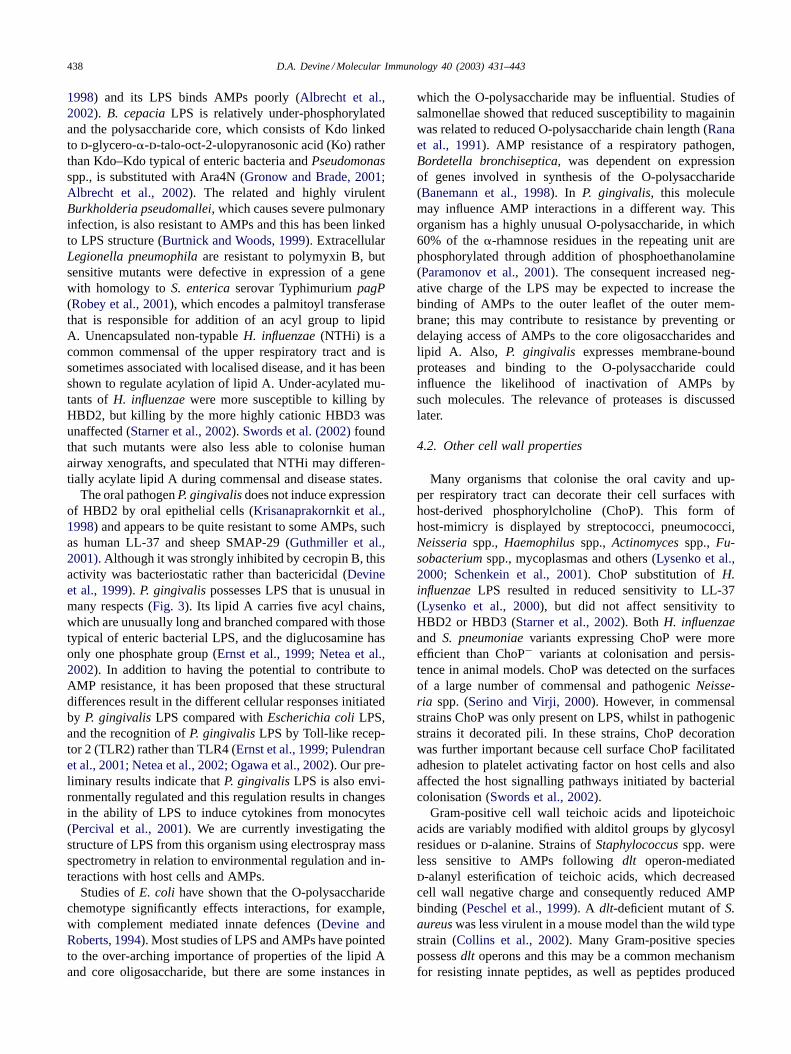
1998) and its LPS binds AMPs poorly (Albrecht et al.,2002). B. cepaciaLPS is relatively under-phosphorylatedand the polysaccharide core, which consists of Kdo linkedto d-glycero-�-d-talo-oct-2-ulopyranosonic acid (Ko) ratherthan Kdo–Kdo typical of enteric bacteria andPseudomonasspp., is substituted with Ara4N (Gronow and Brade, 2001;Albrecht et al., 2002). The related and highly virulentBurkholderia pseudomallei, which causes severe pulmonaryinfection, is also resistant to AMPs and this has been linkedto LPS structure (Burtnick and Woods, 1999). ExtracellularLegionella pneumophilaare resistant to polymyxin B, butsensitive mutants were defective in expression of a genewith homology toS. entericaserovar TyphimuriumpagP(Robey et al., 2001), which encodes a palmitoyl transferasethat is responsible for addition of an acyl group to lipidA. Unencapsulated non-typableH. influenzae(NTHi) is acommon commensal of the upper respiratory tract and issometimes associated with localised disease, and it has beenshown to regulate acylation of lipid A. Under-acylated mu-tants ofH. influenzaewere more susceptible to killing byHBD2, but killing by the more highly cationic HBD3 wasunaffected (Starner et al., 2002). Swords et al. (2002)foundthat such mutants were also less able to colonise humanairway xenografts, and speculated that NTHi may differen-tially acylate lipid A during commensal and disease states.
The oral pathogenP. gingivalisdoes not induce expressionof HBD2 by oral epithelial cells (Krisanaprakornkit et al.,1998) and appears to be quite resistant to some AMPs, suchas human LL-37 and sheep SMAP-29 (Guthmiller et al.,2001). Although it was strongly inhibited by cecropin B, thisactivity was bacteriostatic rather than bactericidal (Devineet al., 1999). P. gingivalispossesses LPS that is unusual inmany respects (Fig. 3). Its lipid A carries five acyl chains,which are unusually long and branched compared with thosetypical of enteric bacterial LPS, and the diglucosamine hasonly one phosphate group (Ernst et al., 1999; Netea et al.,2002). In addition to having the potential to contribute toAMP resistance, it has been proposed that these structuraldifferences result in the different cellular responses initiatedby P. gingivalisLPS compared withEscherichia coliLPS,and the recognition ofP. gingivalisLPS by Toll-like recep-tor 2 (TLR2) rather than TLR4 (Ernst et al., 1999; Pulendranet al., 2001; Netea et al., 2002; Ogawa et al., 2002). Our pre-liminary results indicate thatP. gingivalisLPS is also envi-ronmentally regulated and this regulation results in changesin the ability of LPS to induce cytokines from monocytes(Percival et al., 2001). We are currently investigating thestructure of LPS from this organism using electrospray massspectrometry in relation to environmental regulation and in-teractions with host cells and AMPs.
Studies ofE. coli have shown that the O-polysaccharidechemotype significantly effects interactions, for example,with complement mediated innate defences (Devine andRoberts, 1994). Most studies of LPS and AMPs have pointedto the over-arching importance of properties of the lipid Aand core oligosaccharide, but there are some instances in
which the O-polysaccharide may be influential. Studies ofsalmonellae showed that reduced susceptibility to magaininwas related to reduced O-polysaccharide chain length (Ranaet al., 1991). AMP resistance of a respiratory pathogen,Bordetella bronchiseptica, was dependent on expressionof genes involved in synthesis of the O-polysaccharide(Banemann et al., 1998). In P. gingivalis, this moleculemay influence AMP interactions in a different way. Thisorganism has a highly unusual O-polysaccharide, in which60% of the�-rhamnose residues in the repeating unit arephosphorylated through addition of phosphoethanolamine(Paramonov et al., 2001). The consequent increased neg-ative charge of the LPS may be expected to increase thebinding of AMPs to the outer leaflet of the outer mem-brane; this may contribute to resistance by preventing ordelaying access of AMPs to the core oligosaccharides andlipid A. Also, P. gingivalis expresses membrane-boundproteases and binding to the O-polysaccharide couldinfluence the likelihood of inactivation of AMPs bysuch molecules. The relevance of proteases is discussedlater.
4.2. Other cell wall properties
Many organisms that colonise the oral cavity and up-per respiratory tract can decorate their cell surfaces withhost-derived phosphorylcholine (ChoP). This form ofhost-mimicry is displayed by streptococci, pneumococci,Neisseriaspp., Haemophilusspp., Actinomycesspp., Fu-sobacteriumspp., mycoplasmas and others (Lysenko et al.,2000; Schenkein et al., 2001). ChoP substitution ofH.influenzaeLPS resulted in reduced sensitivity to LL-37(Lysenko et al., 2000), but did not affect sensitivity toHBD2 or HBD3 (Starner et al., 2002). Both H. influenzaeand S. pneumoniaevariants expressing ChoP were moreefficient than ChoP− variants at colonisation and persis-tence in animal models. ChoP was detected on the surfacesof a large number of commensal and pathogenicNeisse-ria spp. (Serino and Virji, 2000). However, in commensalstrains ChoP was only present on LPS, whilst in pathogenicstrains it decorated pili. In these strains, ChoP decorationwas further important because cell surface ChoP facilitatedadhesion to platelet activating factor on host cells and alsoaffected the host signalling pathways initiated by bacterialcolonisation (Swords et al., 2002).
Gram-positive cell wall teichoic acids and lipoteichoicacids are variably modified with alditol groups by glycosylresidues ord-alanine. Strains ofStaphylococcusspp. wereless sensitive to AMPs followingdlt operon-mediatedd-alanyl esterification of teichoic acids, which decreasedcell wall negative charge and consequently reduced AMPbinding (Peschel et al., 1999). A dlt-deficient mutant ofS.aureuswas less virulent in a mouse model than the wild typestrain (Collins et al., 2002). Many Gram-positive speciespossessdlt operons and this may be a common mechanismfor resisting innate peptides, as well as peptides produced

D.A. Devine / Molecular Immunology 40 (2003) 431–443 439
by other Gram-positive bacteria, such as lactococcin, nisinand subtilin (Sahl et al., 1995; Nes et al., 1996).
4.3. Protease production
Many oral and respiratory bacteria, and some importantpathogens, are strongly proteolytic. Extracellular proteasescontribute to nutrient acquisition, tissue destruction andderegulation of inflammatory responses (Potempa et al.,2000). Organisms with proteolytic metabolism also have ef-ficient peptide uptake and transport mechanisms; thus, thereis the potential for such organisms to be protected from an-timicrobial peptides through direct proteolytic degradationor by uptake and transport systems binding and divertingantimicrobial peptides away from target sites.
A relationship has been observed between AMP sensi-tivity and extracellular protease production by some respi-ratory pathogens.Schmidtchen et al. (2002)demonstratedproteolytic degradation of LL-37 by a number of pathogensincluding P. aeruginosaand Streptococcus pyogenes. InP. aeruginosa, this was related to elastase production anddegradation also correlated with sensitivity to killing byLL-37. Elastase was additionally significant in that it in-duced processing of the LL-37 neutrophil precursor pro-protein, hCAP18, to active LL-37. Extracellular proteaseproduction byB. cepaciahas been reported to contribute toantibiotic resistance (Hayashi et al., 2000). We found thateight strains ofB. cepaciarepresenting six genomovars werenot strongly proteolytic and did not degrade�-defensin 1,LL-37, SMAP-29 or D2A-22 (unpublished results).
Oral subgingival anaerobes produce proteases that con-tribute to their nutritional requirements and to their abil-ities to subvert host defences. Strains ofP. gingivalis,Prevotella intermedia, Prevotella nigrescens, Prevotellacorporis and Prevotella pallenssecreted proteases thatcleaved and inactivated a number of AMPs while oralGram-positive organisms, such as streptococci, actino-myces andRothia dentocariosa, did not (Devine et al.,1999). Although P. gingivalis proteases are stronglydown-regulated by environmental temperature, alterationsin growth temperature still resulted in sufficient proteaseproduction to completely inactivate SMAP-29 and D2A-22(Percival et al., 1999; unpublished results). The relationshipbetweenP. gingivalisprotease production and sensitivity toAMPs is complex. The MIC of cecropin B did not correlatewith an ability to degrade AMPs by extracellular proteases(Devine et al., 1999), although for each of the oral anaerobestested the peptide was inhibitory but not bactericidal. It wassuggested that this might have been partly becauseP. gin-givalis proteases inactivated cecropin B slowly (10–15 minfor full inactivation), whereas AMPs generally act rapidly.Treponema pallidumis a protease-producing oral spirocheteassociated with periodontal disease that lacks LPS. It wasresistant to�-defensins, but this was not due to protectionby extracellular proteases (Brissette and Lukehart, 2002).Thus, it appears that in some protease-producing oral bacte-
ria, inner membrane characteristics and LPS structure are ofover-riding importance in determining sensitivity to AMPs.Nonetheless, their proteases may provide indirect protectionfrom AMPs and other host defences. Most Gram-negativebacteria release membrane-bound vesicles from their cellsurfaces and those ofP. gingivalis, P. intermedia, P. ni-grescensandP. aeruginosacontain proteases (Devine et al.,1989; Mayrand and Grenier, 1989; Beveridge, 1999). Thus,proteases may be released and act at sites distant to surfacesof the producer cells, providing protection to accompanyingspecies and deregulating host defences over a broader area.P. aeruginosaelastase and alkaline proteinase,S. pyogenescysteine proteinase andEnterococcus faecalisgelatinase de-grade host cell proteoglycans, releasing dermatan sulphatewhich inhibits HNP1 (Schmidtchen et al., 2002). P. gingi-valis protease degrades CD14, thereby rendering host cellsnon-responsive to LPS (Tada et al., 2002) and preventinginduction AMPs and other host defence molecules.
Most of the earlier studies have considered extracel-lular proteases, but outer membrane associated proteaseswere shown to protectE. coli and S. entericaserovarTyphimurium from the lethal actions of certain AMPs(Stumpe et al., 1998; Guina et al., 2000). P. gingivalisproteases are expressed extracellularly and are membranebound (Curtis et al., 1999, 2001) and these proteases maycontribute to protection. The strong negative charge of thephosphorylated O-polysaccharide may enhance AMP bind-ing without increasing cell death because of protection bymembrane-bound proteases.
5. Environmental regulation of bacterial propertiesrelevant to interactions with antimicrobial peptides
Many of the cellular properties described earlier thatcontribute to AMP resistance are modified in response toalterations in environmental stimuli. Two-component signaltransduction pathways that induce resistance to AMPs andare homologous to those responsible for environmentallyregulated changes toS. entericaserovar Typhimurium LPShave been found in other enteric organisms as well as inP.aeruginosa, Neisseria meningitidis, L. pneumophilaandB.pseudomallei. The modifications associated with AMP re-sistance may affect other host–microbe interactions that arealso highly significant in determining the outcomes of mi-crobial colonisation. Modifications to acyl moieties of lipidA strongly influence interactions between LPS and host re-ceptors, thereby helping determine the signalling pathwaysinitiated. It has been proposed that acylation affects the shapeof lipid A, which in turn determines binding to host cellToll-like receptors (Netea et al., 2002). Under-acylated mu-tants of NTHi stimulated host cells less than wild type bac-teria and elicited a less pro-inflammatory response (Swordset al., 2002). ChoP on teichoic acids ofS. pneumoniaeandon LPS of NTHi facilitates adhesion to platelet activatingfactor on host cells, and the products ofdlt provide other

440 D.A. Devine / Molecular Immunology 40 (2003) 431–443
selective advantages to some bacteria, such as increased acidtolerance, greater intracellular polymer accumulation, andmediation of interbacterial aggregations involved in biofilmestablishment (Clemans et al., 1999; Spatafora et al., 1999;Boyd et al., 2000). Proteases can have many indirect effectsbeyond direct damage of host tissues and AMPs, includingderegulation of inflammatory processes, and inactivationof host cell receptors.P. gingivalis proteases, and othervirulence determinants, are environmentally controlled andappear to be down-regulated by conditions mimicking in-flammation; this attenuation of virulence under certainconditions may contribute to the long-term survival of thisorganism within the hostile environment of the periodontalpocket (Percival et al., 1999; Bonass et al., 1999).
It is not really clear why some epithelial AMPs appearto be expressed constitutively while others require bacterialand inflammatory mediators for induction. HBD1 is muchless potent as an antimicrobial agent than inducible HBD2(van Wetering et al., 1999). It may be that other functionsof HBD1, such as LPS binding, are more important. HBD1may function as a constitutive “sentinel” AMP, binding toLPS and LTA, neutralising them, thereby helping preventan undesirable immune response to low levels of organismsor to resident commensal bacteria. Most bacteria colonisingtissues contain LPS or LTA in their cell walls. It is becom-ing clear that subtle differences in LPS structure, many ofwhich are environmentally regulated, have significant im-pacts on interactions with host cells and synthesis of hostmolecules, including inducible AMPs. It remains to be seenhow these structural properties influence binding to AMPsand the subsequent interactions between AMP–LPS com-plexes and host cells, such as dendritic cells, and how thisimpacts on consequent host responses.
Thus, oral and respiratory AMPs interact with largenumbers of colonising bacteria. The diversity of these pop-ulations can be immense and, in addition to the speciesdiversity evident in resident populations, single species ex-hibit substantial genetic diversity (e.g.Jolley et al., 2000;Hohwy et al., 2001). The complexity of host–microbe inter-actions is further increased by the fact that micro-organismsrapidly adapt to changing environmental conditions, in waysthat may increase survival and pathogenicity. Given thecomplexities of these microbial populations and their ge-netic flexibility, it is essential that host defences are equallyvaried and flexible. AMPs fulfil these requirements and evi-dence is accumulating that demonstrates their importance inprotecting against specific pathogens, modulating residentpopulations and in regulating host responses to bacteria andtheir products, especially LPS.
Acknowledgements
I would like to acknowledge funding from the MRC andthe Ministry of Defence, DSTL (grant number G0000075),the Oral and Dental Research Trust (Proctor and Gamble
Research Award) and the Leeds Hospital Trust SpecialTrustees. Many thanks to my colleagues for helping withinformation and data for this manuscript (Rimondia Per-cival, Michelle Morgan, Zarina Yousuf, Bill Bonass, AlecHigh and Haisal Mohd Hussaini) and to Phil Marsh for hiscritical reading of the manuscript.
References
Aarbiou, J., Ertmann, M., van Wetering, S., van Noort, P., Rook, D., Rabe,K.F., Litvinov, S.V., van Krieken, J.H.J.M., de Boer, W.I., Hiemstra,P.S., 2002. Human neutrophil defensins induce lung epithelial cellproliferation in vitro. J. Leuk. Biol. 72, 167–174.
Agerberth, B., Grunewald, J., Castanos-Velez, E., Olsson, B., Jornvall,H., Wigzell, H., Eklund, A., Gudmundsson, G.H., 1999. Antibacterialcomponents in bronchoalveolar lavage fluid from healthy individualsand sarcoidosis patients. Am. J. Respir. Crit. Care Med. 160, 283–290.
Albrecht, M.T., Wang, W., Shamova, O., Lehrer, R.I., Schiller, N.L., 2002.Binding of protegrin-1 toPseudomonas aeruginosaand Burkholderiacepacia. Respir. Res. 3, 18–28.
Ashitani, J., Mukae, H., Hiratsuka, T., Nakazato, M., Kumamoto,K., Matsukura, S., 2001. Plasma and BAL fluid concentrationsof antimicrobial peptides in patients withMycobacteriumavium–intracellulare infection. Chest 119, 1131–1137.
Augusto, L.A., Li, J., Synguelakis, M., Johansson, J., Chaby, R., 2002.Structural basis for interactions between lung surfactant protein C andbacterial lipopolysaccharide. J. Biol. Chem. 277, 23484–23492.
Bals, R., Wang, X., Wu, Z., Freeman, T., Bafna, V., Zasloff, M., Wilson,J.M., 1998a. Human�-defensin 2 is a salt-sensitive peptide antibioticexpressed in human lung. J. Clin. Invest. 98, 874–880.
Bals, R., Wang, X., Zasloff, M., Wilson, J.M., 1998b. The peptideantibiotic LL-37/hCAP-18 is expressed in epithelia of the human lungwhere it has broad antimicrobial activity at the airway surface. Proc.Natl. Acad. Sci. U.S.A. 95, 9541–9546.
Bals, R., Wang, X., Meegalla, R.L., Wattler, S., Weiner, D.J., Nehls, M.C.,Wilson, J.M., 1999a. Mouse�-defensin 3 is an inducible antimicrobialpeptide expressed in the epithelia of multiple organs. Infect. Immun.67, 3542–3547.
Bals, R., Weiner, D.J., Meegalla, R.L., Wilson, J.M., 1999b. Transfer of acathelicidin peptide antibiotic gene restores bacterial killing in a cysticfibrosis xenograft model. J. Clin. Invest. 103, 1113–1117.
Bals, R., Weiner, D.J., Moscioni, A.D., Meegalla, R.L., Wilson, J.M.,1999c. Augmentation of innate host defence by expression of acathelicidin antimicrobial peptide. Infect. Immun. 67, 6084–6089.
Banemann, A., Deppisch, H., Gross, R., 1998. The lipopolysaccharide ofBordetella bronchiseptaacts as a protective shield against antimicrobialpeptides. Infect. Immun. 66, 5607–5612.
Beveridge, T.J., 1999. Structures of Gram-negative cell walls and theirderived membrane vesicles. J. Bacteriol. 181, 4725–4733.
Bobek, L.A., Situ, H., 2003. MUC7 20-mer: investigation of antimicrobialactivity, secondary structure, and possible mechanism of antifungalaction. Antimicrob. Agents Chemother. 47, 643–652.
Bonass, W.A., High, A.S., Owen, P., Devine, D.A., 1999. Expression of�-defensin genes in human salivary glands. Oral Microbiol. Immunol.14, 371–374.
Boyd, D.A., Cvitkovitch, D.G., Bleiweis, A.S., Kiruikhin, M.Y., Debabov,D.V., Neuhaus, F.C., Hamilton, I.R., 2000. Defects ind-alanyl-lipoteichoic acid synthesis inStreptococcus mutansresults in acidsensitivity. J. Bacteriol. 182, 6055–6065.
Brissette, C.A., Lukehart, S.A., 2002.Treponema denticolais resistant tohuman�-defensins. Infect. Immun. 70, 3982–3984.
Burtnick, M.N., Woods, D.E., 1999. Isolation of polymyxin B-susceptiblemutants ofBurkholderia pseudomalleiand molecular characterizationof genetic loci involved in polymyxin B resistance. Antimicrob. AgentsChemother. 43, 2648–2656.

D.A. Devine / Molecular Immunology 40 (2003) 431–443 441
Clemans, D.L., Kolenbrander, P.E., Debabov, D.V., Zhang, Q., Lunsford,R.D., Sakone, H., Whittaker, C.J., Heaton, M.P., Neuhaus, F.C., 1999.Insertional inactivation of genes responsible for thed-alanylationof lipoteichoic acid inStreptococcus gordoniiDL1 (Challis) affectsintrageneric coaggregations. Infect. Immun. 67, 2464–2474.
Collins, L.V., Kristian, S.A., Weidenmaeir, C., Faigle, M., van Kessel,K.P.M., van Strijp, J.A.G., Gotz, F., Neumeister, B., Peschel, A.,2002. Staphylococcus aureusstrains lackingd-alanine modificationsof teichoic acids are highly susceptible to human neutrophil killingand are virulence attenuated in mice. J. Infect. Dis. 186, 214–219.
Crouch, E., Hartshorn, K., Ofek, I., 2000. Collectins and pulmonary innateimmunity. Immunol. Rev. 173, 52–65.
Curtis, M.A., Kuramitsu, H.K., Lantz, M., Macrina, F.L., Nakayama,K., Potempa, J., Reynolds, E.C., Aduse-Opuku, J., 1999. Moleculargenetics and nomenclature of proteases ofPorphyromonas gingivalis.J. Periodontal Res. 34, 464–472.
Curtis, M.A., Aduse-Opuku, J., Rangarajan, M., 2001. Cysteine proteasesof Porphyromonas gingivalis. Crit. Rev. Oral Biol. Med. 12, 192–216.
Dale, B.A., Kimball, J.R., Krisanaprakornkit, S., Roberts, F., Robinovitch,M., O’Neal, R., Valore, E.V., Ganz, T., Anderson, G.M., Weinberg, A.,2001. Localized antimicrobial peptide expression in human gingiva. J.Periodontal Res. 36, 285–294.
Dauletbaev, N., Gropp, R., Frye, M., Loitsch, S., Wagner, T.O., Bargon,J., 2002. Expression of human�-defensin (HBD-1 and HBD-2) mRNAin nasal epithelia of adult cystic fibrosis patients, healthy individualsand individuals with acute colds. Respiration 69, 46–51.
Desai, M., Buhler, T., Weller, P.H., Brown, M.R., 1998. Increasingresistance of planktonic and biofilm cultures ofBurkholderia cepaciatociprafloxacin and ceftazidime during exponential growth. J. Antimicrob.Chemother. 42, 153–160.
Devine, D.A., Hancock, R.E.W., 2002. Cationic peptides: distribution andmechanisms of resistance. Curr. Pharm. Des. 8, 99–110.
Devine, D.A., Roberts, A.P., 1994. K1, K5 and O antigens ofEscherichiacoli in relation to serum killing via the classical and alternativecomplement pathways. J. Med. Microbiol. 41, 139–144.
Devine, D.A., Gmur, R., Handley, P.S., 1989. Ultrastructure, serogroupingand localization of surface antigens ofBacteroides intermedius. J. Gen.Microbiol. 135, 967–979.
Devine, D.A., Marsh, P.D., Percival, R.S., Rangarajan, M., Curtis, M.A.,1999. Modulation of antibacterial peptide activity by products ofPorphyromonas gingivalisandPrevotellaspp. Microbiology 145, 965–971.
Devine, D.A., High, A.S., Owen, P.J., Poulsom, R., Bonass, W.A., 2000.Trefoil factor expression in human salivary glands. Hum. Pathol. 31,509–515.
Diamond, G., Legarda, D., Ryan, L.K., 2000. The innate immune responseof the respiratory epithelium. Immunol. Rev. 173, 27–38.
Diamond, D.L., Kimball, J.R., Krisanaprakornkit, S., Ganz, T., Dale, B.A.,2001. Detection of�-defensins secreted by human oral epithelial cells.J. Immunol. Methods 256, 65–76.
dos Santos Silva, E., Ulrich, M., Doring, G., Botzenhart, K., Gott, P.,2000. Trefoil factor family domain peptides in the human respiratorytract. J. Pathol. 190, 133–142.
Duits, L.A., Ravensbergen, B., Rademaker, M., Hiemstra, P.S., Nibbering,P.H., 2002. Expression of�-defensin 1 and 2 mRNA by humanmonocytes, macrophages and dendritic cells. Immunology 106, 517–525.
Dunsche, A., Açil, Y., Dommisch, H., Siebert, R., Schröder, J.M., Jepsen,S., 2002. The novel human beta-defensin-3 is widely expressed in oraltissues. Eur. J. Oral Sci. 109, 121–124.
Ernst, R.K., Guina, T., Miller, S.I., 1999. How intracellular bacteriasurvive: surface modifications that promote resistance to host innateimmune responses. J. Infect. Dis. 179, S326–S330.
Frohm Nilsson, M., Sandstedt, B., Sorenson, O., Weber, G., Borregaard,N., Ståhle-Bäckdhal, M., 1999. The human cationic antimicrobialprotein (hCAP18), a peptide antibiotic, is widely expressed in humansquamous epithelia and colocalises with interleukin-6. Infect. Immun.67, 2561–2566.
Frye, M., Bargon, J., Daulbetbaev, N., Weber, A., Wagner, T.O., Gropp,R., 2000. Expression of human alpha-defensin 5 (HD5) mRNA innasal and bronchial epithelial cells. J. Clin. Pathol. 53, 770–773.
Garabedian, E.M., Roberts, L.J.J., McNevin, M.S., Gordon, J.I., 1997.Examining the role of Paneth cells in the small intestine by lineageablation in transgenic mice. J. Biol. Chem. 272, 23729–23740.
Garcia, J.R.C., Jaumann, F., Schulz, S., Krause, A., Rodriguez-Jiminez,J., Forssmann, U., Adermann, K., Klüver, E., Vogelmeier, C., Becker,D., Hedrich, R., Forssmann, W.G., Bals, R., 2001a. Identification of anovel multifunctional�-defensin (human�-defensin 3) with specificantimicrobial activity. Cell Tissue Res. 306, 257–264.
Garcia, J.R.C., Krause, A., Schulz, S., Rodriguez-Jiminez, F.G., Kluver,E., Adermann, K., Forssmann, U., Frimpong-Boateng, A., Bals, R.,Forssmann, W.G., 2001b. Human beta-defensin 4: a novel induciblepeptide with a specific salt-sensitive spectrum of antimicrobial activity.FASEB J. 15, 1819–1821.
Goldman, M.J., Anderson, G.M., Stolzenberg, E.D., Kari, U.P., Zasloff,M., Wilson, J.M., 1997. Human�-defensin-1 is a salt-sensitiveantibiotic in lung that is inactivated in cystic fibrosis. Cell 88, 553–560.
Gronow, S., Brade, H., 2001. Lipopolysaccharide biosynthesis: whichsteps do bacteria need to survive? J. Endotoxin Res. 7, 3–23.
Gudmundsson, G.H., Agerberth, B., 1999. Neutrophil antibacterialpeptides, multifunctional effector molecules in the mammalian immunesystem. J. Immunol. Methods 232, 45–54.
Guina, T., Yi, E.C., Wang, H., Hackett, M., Miller, S.I., 2000. APhoP-regulated outer membrane protease ofSalmonella entericaserovar Typhimurium promotes resistance to alpha-helical antimicrobialpeptides. J. Bacteriol. 182, 4077–4086.
Gusman, H., Travis, J., Helmerhorst, E.J., Potempa, J., Troxler, R.F.,Oppenheim, F.G., 2001. Salivary histatin 5 is an inhibitor of bothhost and bacterial enzymes implicated in periodontal disease. Infect.Immun. 69, 1402–1408.
Guthmiller, J.M., Vargas, K.G., Srikantha, R., Schomberg, L.L.,Weistroffer, P.L., McCray Jr., P.B., Tack, B.F., 2001. Susceptibilitiesof oral bacteria and yeast to mammalian cathelicidins. Antimicrob.Agents Chemother. 45, 3216–3219.
Haffajee, A.D., Socransky, S.S., Ferres, M., Ximenez-Fyvie, L.A., 1999.Plaque microbiology in health and disease. In: Newman, H.N., Wilson,M. (Eds.), Dental Plaque Revisited. Bioline, Cardiff, pp. 255–282.
Han, Y.W., Shi, W., Huang, G.T.J., Haake, S.K., Park, N.H., Kuramitsu,H., Genco, R.J., 2000. Interactions between periodontal bacteria andhuman oral epithelial cells:Fusobacterium nucleatumadheres to andinvades epithelial cells. Infect. Immun. 68, 3140–3146.
Hancock, R.E.W., 1998. Resistance mechanisms inPseudomonasaeruginosaand other nonfermentative Gram-negative bacteria. Clin.Infect. Dis. 27 (Suppl.), S93–S99.
Hancock, R.E.W., Chapple, D.S., 1999. Peptide antibiotics. Antimicrob.Agents Chemother. 43, 1317–1323.
Hancock, R.E.W., Diamond, G., 2000. The role of cationic antimicrobialpeptides in innate host defences. Trends Microbiol. 8, 402–410.
Harder, J., Meyer-Hoffert, U., Teran, L.M., Schwichtenberg, L., Bartels,J., Maune, S., Schröder, J.M., 2000. MucoidPseudomonas aeruginosa,TNF-alpha, and IL-beta, but not IL-6, induce human beta-defensin-2in respiratory epithelia. Am. J. Respir. Cell Mol. Biol. 22, 714–721.
Harder, J., Bartels, J., Christophers, E., Schröder, J.M., 2001. Isolationand characterization of human�-defensin-3, a novel human induciblepeptide antibiotic. J. Biol. Chem. 276, 5707–5713.
Hayashi, A., Abe, M., Kimoto, M., Furukawa, S., Nakazawa, T., 2000.The dsbA–dsbB disulfide bond formation system ofBurkholderiacepacia is involved in the production of protease and alkalinephosphatase, motility, metal resistance, and multi-drug resistance.Microbiol. Immunol. 44, 41–50.
Hohwy, J., Reinholdt, J., Kilian, M., 2001. Population dynamics ofStreptococcus mitisin its natural habitat. Infect. Immun. 69, 6055–6063.
Houalet-Jeanne, S., Pellen-Mussi, P., Tricot-Doleax, S., Apiou, A.,Bonnaure-Mallet, M., 2001. Assessment of internalisation and viability

442 D.A. Devine / Molecular Immunology 40 (2003) 431–443
of Porphyromonas gingivalisin KB epithelial cells by confocalmicroscopy. Infect. Immun. 69, 7146–7151.
Hughes, A.L., 1999. Evolutionary diversification of the mammaliandefensins. Cell. Mol. Life Sci. 56, 94–103.
Imatani, T., Kata, T., Minaguchi, K., Okuda, K., 2000. Histatin 5 inhibitsinflammatory cytokine induction from human gingival fibroblasts byPorphyromonas gingivalis. Oral Microbiol. Immunol. 15, 378–382.
Jainkittivong, A., Johnson, D.A., Yeh, C.K., 1998. The relationshipbetween salivary histatin levels and oral yeast carriage. Oral Microbiol.Immunol. 13, 181–187.
Jia, H.P., Wowk, S.A., Schutte, B.C., Lee, S.K., Vivado, A., Tack, B.F.,Bevins, C.L., McCray Jr., P.B., 2000. A novel murine�-defensinexpressed in tongue, esophagus, and trachea. J. Biol. Chem. 275,33314–33320.
Jia, H.P., Schutte, B.C., Schudy, A., Linzmeier, R., Guthmiller, J.M.,Johnson, G.K., Tack, B.F., Mitros, J.P., Rosenthal, A., Ganz, T., McCrayJr., P.B., 2001. Discovery of new�-defensins using a genomics basedapproach. Gene 263, 211–218.
Jolley, K.A., Kalmusova, J., Feil, E.J., Gupta, S., Musilek, M., Kriz, P.,Maiden, M.C.J., 2000. Carried meningococci in the Czech Republic:a diverse recombining population. J. Clin. Microbiol. 38, 4492–4498.
Kapas, S., Bansal, A., Bhargava, V., Maher, R., Malli, D., Hagi-Pavli, E.,Allaker, R.P., 2001. Adrenomedullin expression in pathogen-challengedoral epithelial cells. Peptides 22, 1485–1489.
Könönen, E., Jousimies-Somer, H., Bryk, A., Kilpi, T., Kilian, M., 2002.Establishment of streptococci in the upper respiratory tract: longitudinalchanges in the mouth and nasopharynx up to 2 years of age. J. Med.Microbiol. 51, 723–730.
Krisanaprakornkit, S., Weinberg, A., Perez, C.N., Dale, B.A., 1998.Expression of the peptide antibiotic human�-defensin-1 in culturedgingival epithelial cells and gingival tissue. Infect. Immun. 66, 4422–4428.
Krisnaprakornkit, S., Kimball, J.R., Weinberg, A., Darveau, R.P.,Bainbridge, B.W., Dale, B.A., 2000. Inducible expression of human�-defensin 2 byFusobacterium nucleatumin oral epithelial cells:multiple signalling pathways and role of commensal bacteria in innateimmunity and the epithelial barrier. Infect. Immun. 68, 2907–2915.
Lamont, R.J., Jenkinson, H.H., 2000. Subgingival colonization byPorphyromonas gingivalis. Oral Microbiol. Immunol. 15, 341–349.
Lee, S.H., Kim, J.E., Lim, H.H., Choi, J.O., 2002. Antimicrobial defensinpeptides of the human nasal mucosa. Ann. Otol. Rhinol. Laryngol.111, 135–141.
Lysenko, E.S., Gould, J., Bals, R., Wilson, J.M., Weiser, J.N.,2000. Bacterial phosphorylcholine decreases susceptibility to theantimicrobial peptide LL-37/hCAP18 expressed in the upper respiratorytract. Infect. Immun. 68, 1664–1671.
Macfarlane, E.L., Kwasnicka, A., Hancock, R.E.W., 2000. Role ofPseudomonas aeruginosaPhoP–PhoQ in resistance to antimicrobialcationic peptides and aminoglycosides. Microbiology 146, 2543–2554.
Mahenthiralingam, E., Baldwin, A., Vandamme, P., 2002.Burkholderiacepacia complex infection in patients with cystic fibrosis. J. Med.Microbiol. 2002, 533–538.
Marsh, P.D., 2003. Are dental diseases examples of ecologicalcatastrophes. Microbiology 149, 279–294.
Mathews, M., Jia, H.P., Guthmiller, J.M., Losh, G., Graham, S., Johnson,G.K., Tack, B.F., McCray Jr., P.B., 1999. Production of�-defensinantimicrobial peptides by the oral mucosa and salivary glands. Infect.Immun. 67, 2740–2745.
Mayrand, D., Grenier, D., 1989. Biological activities of outer membranevesicles. Can. J. Microbiol. 35, 607–613.
McCray Jr., P.B., Bentley, L., 1997. Human airway epithelia express abeta-defensin. Am. J. Respir. Cell Mol. Biol. 16, 343–349.
McKay, M.S., Olson, E., Hesla, M.A., Panyutich, A., Ganz, T., Perkins,S., Rossamando, E.F., 1999. Immunomagnetic recovery of humanneutrophil defensins from the human gingival crevice. Oral Microbiol.Immunol. 14, 190–193.
Mizukawa, N., Sugiyama, K., Ueno, T., Mishima, K., Tagaki, S., Sugahara,T., 1999. Levels of human defensin-1, an antimicrobial peptide, insaliva of patients with oral inflammation. Oral Surg. Oral Med. OralPathol. Oral Radiol. Endod. 87, 539–543.
Mohr, C.D., Tomich, M., Herfst, C.A., 2001. Cellular aspects ofBurkholderia cepaciainfection. Microbes Infect. 3, 425–435.
Morrison, G.M., Davidson, D.J., Dorin, J.R., 1999. A novel mouse�-defensin DefB2, which is upregulated in the airways bylipopolysaccharide. FEBS Lett. 442, 112–116.
Morrison, G.M., Kilanowski, F., Davidson, D., Dorin, J., 2002.Characterization of the mouse beta defensin 1,Defb1, mutant mousemodel. Infect. Immun. 70, 3053–3060.
Moser, C., Weiner, D.J., Lysenko, E., Bals, R., Weiser, J.N., Wilson, J.M.,2002.�-Defensin 1 contributes to pulmonary innate immunity in mice.Infect. Immun. 70, 3068–3072.
Moskowitz, S.M., Ernst, R.K., Miller, S.I., 1999. ThepmrAB locusregulates resistance to the cationic antimicrobial peptides polymyxinB and C18G inPseudomonas aeruginosa. Pediatr. Pulmonol. Suppl.19, 260.
Murakami, Y., Nagata, H., Amano, A., Takagaki, M., Shizukuishi, S.,Tsunemitsu, A., Aimoto, S., 1991. Inhibitory effects of human salivaryhistatins and lysozyme on coaggregation betweenPorphyromonasgingivalis and Streptococcus mitis. Infect. Immun. 59, 3284–3286.
Murakami, Y., Tamagawa, H., Shizukuishi, S., Tsunemitsu, A., Aimoto, S.,1992. Biological role of an arginine residue present in a histidine-richpeptide which inhibits hemagglutination ofPorphyromonas gingivalis.FEMS Microbiol. Lett. 77, 201–204.
Nagoaka, I., Hirota, S., Yomogida, S., Ohwada, A., Hirata, M.,2000. Synergistic actions of antibacterial neutrophil defensins andcathelicidins. Inflamm. Res. 49, 73–79.
Nagaoka, I., Hirota, S., Niyonsaba, F., Hirata, M., Adachi, Y., Tamura,H., Heumann, D., 2001. Cathelicidin family of antibacterial peptidesCAP18 and CAP11 inhibit the expression of TNF-� by blocking thebinding of LPS to CD14+ cells. J. Immunol. 167, 3329–3338.
Nes, I.F., Diep, D.B., Havarstein, L.S., Brurberg, M.B., Eijsink, V., Holo,H., 1996. Biosynthesis of bacteriocins in lactic acid bacteria. Antonievan Leeuwenhoek 70, 113–128.
Netea, M.G., van Deuren, M., Kullberg, B.J., Cavaillon, J.-M., Van derMeer, J.W.M., 2002. Does the shape of lipid A determine the interactionof LPS with Toll-like receptors. Trends Immunol. 23, 135–139.
Ogawa, T., Asai, Y., Hashimoto, M., Takeuchi, O., Kurita, T., Yoshikai,Y., Miyake, K., Akira, S., 2002. Cell activation byPorphyromonasgingivalis lipid A through Toll-like receptor 4- and myeloiddifferentiation factor 88-dependent signalling pathway. Int. Immunol.14, 1325–1332.
Paramonov, N., Bailey, D., Rangarajan, M., Hashim, A., Kelly, G., Curtis,M.A., Houndsell, E.F., 2001. Structural analysis of the polysaccharidefrom the lipopolysaccharide ofPorphyromonas gingivalisstrain W50.Eur. J. Biochem. 268, 4698–4707.
Paster, B.J., Boches, S.K., Galvin, J.L., Ericson, R.E., Lau, C.N., Levanos,V.A., Sahasrabudhe, A., Dewhirst, F.E., 2001. Bacterial diversity inhuman subgingival plaque. J. Bacteriol. 183, 3770–3783.
Percival, R.S., Marsh, P.D., Devine, D.A., Rangarajan, M., Aduse-Opoku,J., Curtis, M.A., 1999. The effect of temperature on growth and proteaseactivity of Porphyromonas gingivalisW50. Infect. Immun. 67, 1917–1921.
Percival, R.S., Marsh, P.D., Devine, D.A., Bonass, W.A., Matthews,J.B., Ingham, E., 2001. Effect of various environmental conditions onPorphyromonas gingivalisgrowth and cytokine production. J. Dent.Res. 80, 1148.
Peschel, A., 2002. How do bacteria resist human antimicrobial peptides?Trends Microbiol. 10, 179–186.
Peschel, A., Otto, M., Jack, R.W., Kalbacher, H., Jung, G., Götz, F.,1999. Inactivation of thedlt operon inStaphylococcus aureusconferssensitivity to defensins, protegrins and other antimicrobial peptides. J.Biol. Chem. 274, 8405–8410.

D.A. Devine / Molecular Immunology 40 (2003) 431–443 443
Pier, G.B., 2000. Role of cystic fibrosis transmembrane conductanceregulator in innate immunity toPseudomonas aeruginosainfections.Proc. Natl. Acad. Sci. U.S.A. 97, 8822–8828.
Potempa, J., Babbula, A., Travis, J., 2000. Role of bacterial proteinases inmatrix destruction and modulation of host responses. Periodontology24, 153–192.
Pulendran, B., Kumar, P., Cutler, C.W., Mohamadzadeh, M., Van Dyke,T., Banchereau, J., 2001. Lipopolysaccharides from distinct pathogensinduce different classes of immune response in vivo. J. Immunol. 167,5067–5076.
Putsep, K., Carlsson, G., Boman, H.G., Andersson, M., 2002. Deficiencyof antibacterial peptides in patients with morbus Kostmann: anobservation study. Lancet 360, 1144–1149.
Raj, P.A., Antonyraj, K.J., Karunakaran, T., 2000. Large-scale synthesisand functional elements for the antimicrobial activity of defensins.Biochem. J. 347, 633–641.
Rana, F.R., Macias, E.A., Sultany, C.M., Modzrakowski, M.C., Blazyk,J., 1991. Interactions between magainin 2 andSalmonella typhimuriumouter membranes: effect of lipopolysaccharide structure. Biochemistry30, 5858–5866.
Rasmussen, T.T., Kirkeby, L.P., Poulsen, K., Reinholdt, J., Kilian, M.,2000. Resident aerobic microflora of the adult human nasal cavity.APMIS 108, 663–675.
Robey, M., O’Connell, W., Cianciotto, N.P., 2001. Identification ofLegionella pneumophila rcp, a pagP-like gene that confers resistanceto cationic antimicrobial peptides and promotes intracellular infection.Infect. Immun. 69, 4276–4286.
Rudney, J.D., Chen, R., Sedgewick, G.J., 2001. IntracellularActinobacillusactinomycetemcomitansand Porphyromonas gingivalisin buccalepithelial cells collected from human subjects. Infect. Immun. 69,2700–2707.
Sahasrebudhe, K.S., Kimball, J.R., Morton, T.H., Weinberg, A., Dale,B.A., 2000. Expression of the antimicrobial peptide, human�-defensin1, in duct cells of minor salivary glands and detection in saliva. J.Dent. Res. 79, 1669–1674.
Sahl, H.G., Jack, R.W., Bierbaum, G., 1995. Biosynthesis and biologicalactivities of lantibiotics with unique post-translational modifications.Eur. J. Biochem. 230, 827–853.
Saiman, L., Tabibi, S., Starner, T.D., San Gabriel, P., Winokur, P.L.,Jia, H.P., McCray Jr., P.B., Tack, B.F., 2001. Cathelicidin peptidesinhibit multiply antibiotic resistant pathogens from patients with cysticfibrosis. Antimicrob. Agents Chemother. 45, 2838–2844.
Schaller-Bals, S., Schulze, A., Bals, R., 2002. Increased levels ofantimicrobial peptides in tracheal aspirates of newborn infants duringinfection. Am. J. Respir. Crit. Care Med. 165, 992–995.
Schenkein, H.A., Berry, C.R., Purkall, D., Burmeister, J.A., Brooks, C.N.,Tew, J.G., 2001. Phosphorylcholine-dependent cross-reactivity betweendental plaque bacteria and oxidised low-density lipoproteins. Infect.Immun. 69, 6612–6617.
Schmidtchen, A., Frick, I.-M., Andersson, E., Tapper, H., Björk, L., 2002.Proteinases of common pathogenic bacteria degrade and inactivate theantibacterial peptide LL-37. Mol. Microbiol. 46, 157–168.
Schutte, B.C., McCray Jr., P.B., 2002.�-Defensins in lung host defence.Ann. Rev. Physiol. 64, 709–748.
Schwab, U., Gilligan, P., Jaynes, J., Henke, D., 1999. In vitroactivities of designed antimicrobial peptides against multidrug-resistantcystic fibrosis pathogens. Antimicrob. Agents Chemother. 43, 1435–1440.
Scott, M.G., Gold, M.R., Hancock, R.E.W., 1999. Interaction of cationicpeptides with lipoteichoic acid and Gram-positive bacteria. Infect.Immun. 67, 6445–6453.
Scott, M.G., Rosenberger, C.M., Gold, M.R., Finlay, B.B., Hancock,R.E.W., 2000a. An�-helical cationic antimicrobial peptide selectivelymodulates macrophage responses to lipopolysaccharide and directlyalters macrophage gene expression. J. Immunol. 165, 3358–3365.
Scott, M.G., Vreugdenhil, A.C.E., Buurman, W.A., Hancock, R.E.W.,Gold, M.R., 2000b. Cutting edge: cationic antimicrobial peptides blockthe binding of lipopolysaccharide (LPS) to LPS binding protein. J.Immunol. 164, 549–553.
Serino, L., Virji, M., 2000. Phosphorylcholine decoration oflipopolysaccharide differentiates commensalNeisseriafrom pathogenicstrains: identification oflicA-type genes in commensalNeisseria. Mol.Microbiol. 35, 1550–1559.
Simpson, A.J., Maxwell, A.I., Govan, J.R.W., Haslett, C., Sallenave, J.-M.,1999. Elafin (elastase-specific inhibitor) has anti-microbial activityagainst Gram-positive and Gram-negative respiratory pathogens. FEBSLett. 452, 309–313.
Singh, P., Jia, H., Wiles, K., Hesselberth, J., Liu, L., Conway, B.,Greenberg, E., Valore, E., Welsh, M., Ganz, T., Tack, B., McCray Jr.,P.B., 1998. Production of beta-defensins by human airway epithelia.Proc. Natl. Acad. Sci. U.S.A. 95, 14961–14966.
Singh, P.K., Tack, B.F., McCray Jr., P.B., Welsh, M.J., 2000. Synergisticand additive killing by antimicrobial factors found in human airwaysurface liquid. Am. J. Physiol. Lung Cell. Mol. Physiol. 279, L799–L805.
Spatafora, G.A., Sheets, M., June, R., Luyimbazi, D., Howard, K.,Hulbert, R., Barnard, D., el Janne, M., Hudson, M.C., 1999. Regulatedexpression of theStreptococcus mutans dltgenes correlates withintracellular polysaccharide accumulation. J. Bacteriol. 181, 2363–2373.
Starner, T.D., Swords, W.E., Apicella, M.A., McCray Jr., P.B., 2002.Susceptibility of nontypeableHaemophilus influenzaeto human�-defensins is influenced by lipooligosaccharide acylation. Infect.Immun. 70, 5287–5289.
Stumpe, S., Schmid, R., Stephens, D.L., Georgiou, G., Bakker, E.P., 1998.Identification of OmpT as the protease that hydrolyzes the antimicrobialpeptide protamine before it enters growing cells ofEscherichia coli.J. Bacteriol. 180, 4002–4006.
Swords, W.E., Chance, D.L., Cohn, L.A., Shao, J., Apicella, M.A., Smith,A.L., 2002. Acylation of the lipooligosaccharide ofHaemophilusinfluenzae and colonization: an htrB mutation diminishes thecolonization of human airway epithelial cells. Infect. Immun. 70, 4661–4668.
Tada, H., Sugawara, S., Nemoto, E., Takahashi, N., Imamura, T.,Potempa, J., Travis, J., Shimauchi, H., Takada, H., 2002. Proteolysisof CD14 on human gingival fibroblasts by arginine-specific cysteineproteinases fromPorphyromonas gingivalisleading to down-regulationof lipopolysaccharide-induced interleukin-8 production. Infect. Immun.70, 3304–3307.
van Wetering, S., Sterk, P., Rabe, K., Hiemstra, P.S., 1999. Defensins:key players or bystanders in infection, injury, and repair in the lung.J. Allergy Clin. Immunol. 104, 1131–1138.
van Wetering, S., van der Linden, A.C., van Sterkenburg, M.A., de Boer,W.I., Kuijpers, A.L., Schalkwijk, J., Hiemstra, P.S., 2000. Regulationof SLPI and elafin release from bronchial epithelial cells by neutrophildefensins. Am. J. Physiol. Lung Cell. Mol. Physiol. 278, L51–L58.
Wilson, M.J., Weightman, A.J., Wade, W.G., 1997. Applications ofmolecular ecology in the characterization of uncultured microorganismsassociated with human disease. Rev. Med. Microbiol. 8, 91–101.
Xu, T., Levitz, S.M., Diamond, R.D., Oppenheim, F.G., 1991. Anticandidalactivity of major human salivary histatins. Infect. Immun. 59, 2549–2554.
Yang, D., Biragyn, A., Kwak, L.W., Oppenheim, J.J., 2002. Mammaliandefensins in immunity: more than just microbicidal. Trends Immunol.23, 291–296.
Zhang, P., Summer, W.R., Bagby, G.J., Nelson, S., 2000. Innate immunityand pulmonary host defence. Immunol. Rev. 173, 39–51.
Zhao, C., Wang, I., Lehrer, R.I., 1996. Widespread expression ofbeta-defensin HBD-1 in human secretory glands and epithelial cells.FEBS Lett. 396, 319–322.