Embed Size (px)
Citation preview
Reversible host cell remodeling underpinsdeformability changes in malaria parasitesexual blood stagesMegan Dearnleya,1, Trang Chub,1, Yao Zhangc,1, Oliver Lookera,1, Changjin Huangc, Nectarios Klonisa, Jeff Yeomand,Shannon Kennya, Mohit Arorab, James M. Osbornee, Rajesh Chandramohanadasb,2, Sulin Zhangc,2,Matthew W. A. Dixona,2, and Leann Tilleya,2,3
aDepartment of Biochemistry and Molecular Biology, Bio21 Molecular Science and Biotechnology Institute, University of Melbourne, Melbourne, VIC 3010,Australia; bPillar of Engineering Product Development, Singapore University of Technology and Design, Singapore 487372; cDepartment of EngineeringScience and Mechanics, The Pennsylvania State University, University Park, PA 16802; dDepartment of Biochemistry, La Trobe University, Melbourne, VIC3086, Australia; and eSchool of Mathematics and Statistics, University of Melbourne, Melbourne, VIC 3010, Australia
Edited by Carolina Barillas-Mury, National Institutes of Health, Bethesda, MD, and approved March 4, 2016 (received for review October 22, 2015)
The sexual blood stage of the human malaria parasite Plasmodiumfalciparum undergoes remarkable biophysical changes as it preparesfor transmission to mosquitoes. During maturation, midstage game-tocytes show low deformability and sequester in the bone marrowand spleen cords, thus avoiding clearance during passage throughsplenic sinuses. Mature gametocytes exhibit increased deformabilityand reappear in the peripheral circulation, allowing uptake by mos-quitoes. Here we define the reversible changes in erythrocyte mem-brane organization that underpin this biomechanical transformation.Atomic force microscopy reveals that the length of the spectrin cross-members and the size of the skeletal meshwork increase in develop-ing gametocytes, then decrease in mature-stage gametocytes. Thesechanges are accompanied by relocation of actin from the erythrocytemembrane to the Maurer’s clefts. Fluorescence recovery after photo-bleaching reveals reversible changes in the level of coupling betweenthe membrane skeleton and the plasma membrane. Treatment ofmidstage gametocytes with cytochalasin D decreases the vertical cou-pling and increases their filterability. A computationally efficientcoarse-grained model of the erythrocyte membrane reveals thatrestructuring and constraining the spectrin meshwork can fully ac-count for the observed changes in deformability.
gametocyte | deformability | spectrin/actin skeleton | AFM |molecular dynamics simulation
The most virulent of the human malaria parasites, Plasmodiumfalciparum causes ∼440,000 deaths annually (1). Pathology is
associated with asexual multiplication within red blood cells (RBCs).The trophozoite (growing) and schizont (dividing) stages (∼24–48 hafter invasion) sequester in deep tissue using adhesive proteinspresented on platform-like structures called “knobs” at the infectedRBC surface. Cytoadhesion enables the parasite to avoid passagethrough the splenic sinuses and thus mechanical clearance from thecirculation. Unfortunately, complications associated with sequestra-tion of infected RBCs in the brain are responsible for much of themalaria-related mortality and morbidity.After a period of asexual cycling, a proportion of blood-stage
parasites commit to sexual development (gametocytogenesis).The intraerythrocytic gametocyte develops through five distinctstages (I–V) over a period of 10–12 d, eventually adopting thecharacteristic crescent (falciform) shape that gives P. falciparumits name. Elongation is driven by assembly of a sheath of mi-crotubules, attached to an inner membrane complex, underneaththe parasite plasma membrane. From stage II to IV, gametocytesdisappear from the circulation (2, 3); however, the mechanism ofsequestration is not well understood. Upon maturation, the mi-crotubule cytoskeleton is disassembled, and stage V gametocytesre-enter the circulation (2, 3). Ingestion of mature gametocytes byan Anophelesmosquito triggers release from the RBCs, followed bysexual recombination in the insect gut, and eventual transmission.
Efforts to control malaria are often thwarted by the presence ofgametocytes in asymptomatic individuals. These infected individualsserve as a reservoir during the low transmission season, ready toretransmit disease when mosquito numbers increase. As a conse-quence, there is intense interest in understanding gametocyte cellbiology with the aim of interfering with this developmental stage.Of particular interest are the molecular and biomechanical
changes that accompany the sequestration and release of game-tocytes. Developing gametocytes (stages II–IV) have significantlyreduced cellular deformability (2, 4, 5). This increased rigidity mayenable gametocytes to become mechanically trapped in the bonemarrow and splenic cords. In contrast, stage V gametocytes exhibitincreased deformability (4–6), which may help them survive in thecirculation, where they can be picked up by mosquitoes.Survival in the circulation requires RBCs to undergo de-
formation without fragmentation, as they transit through the 1.5-to 2-μm interendothelial slits in the spleen. The remarkabledeformability properties of RBCs are thought to derive fromtheir submembranous protein skeleton (7, 8). The skeleton iscomposed of a regular hexagonal array of spectrin heterodimersthat self-associate head-to-head to form tetramers. The tails of
Significance
This study provides, to our knowledge, the first ultrastructural anddynamics analysis of the host red blood cell membrane of Plas-modium falciparum gametocytes, revealing reversible expansion ofthe spectrin–actin skeleton, accompanied by reversible modulationof skeletal coupling to the plasma membrane. We use the mea-sured physical parameters to inform a computationally efficientcoarse-grained model. This model shows that restructuring theskeletal meshwork can fully account for the observed deformabilitychanges.We reveal a critical role for actin remodeling in driving thisreversible biomechanical host cell subversion. This work providesfundamental insights into the molecular changes that underpingametocyte survival in the circulation.
Author contributions: R.C., S.L.Z., M.W.A.D., and L.T. designed research; M.D., T.C., Y.Z.,O.L., C.J.H., N.K., J.Y., S.K., M.A., and M.W.A.D. performed research; T.C., J.M.O., and R.C.contributed new reagents/analytic tools; M.D., T.C., Y.Z., O.L., C.J.H., N.K., J.Y., J.M.O.,R.C., S.L.Z., M.W.A.D., and L.T. analyzed data; and M.D., R.C., S.L.Z., M.W.A.D., and L.T.wrote the paper.
The authors declare no conflict of interest.
This article is a PNAS Direct Submission.
Freely available online through the PNAS open access option.1M.D., T.C., Y.Z., and O.L. contributed equally to this work.2R.C., S.L.Z., M.W.A.D., and L.T. contributed equally to this work.3To whom correspondence should be addressed. Email: [email protected].
This article contains supporting information online at www.pnas.org/lookup/suppl/doi:10.1073/pnas.1520194113/-/DCSupplemental.
4800–4805 | PNAS | April 26, 2016 | vol. 113 | no. 17 www.pnas.org/cgi/doi/10.1073/pnas.1520194113
Dow
nloa
ded
by g
uest
on
Dec
embe
r 25
, 201
9
the spectrin heterodimers are linked into junctional complexescontaining actin oligomers (each with 14–16 protomers), protein4.1R, adducin, and accessory proteins (9). Flexible linkages be-tween the triple-helical segments of spectrin heterodimers, aswell as tetramer dissociation, and breakable linkages into thejunction points, are assumed to accommodate the distortionsimposed by shear forces in the circulation.Vertical interactions connect the skeletal meshwork to the
plasma membrane. A subpopulation of band-3 dimers connectsto spectrin via ankyrin (9). Band-3 dimers also participate in asecond linkage complex that involves glycophorin C. This com-plex is linked to the membrane skeleton via glycophorin C/pro-tein 4.1 interactions as well as band-3/adducin interactions.The molecular determinants of the increased rigidity of mature
asexual parasite-infected RBCs are beginning to be elucidated.Atomic force microscopy (AFM) has revealed reorganization andexpansion of the spectrin network of the host cell membrane (10,11), while cryo-electron tomography suggests that this re-organization occurs as a result of mining of the actin junctions inorder to generate actin filaments that connect parasite-derivedorganelles known as Maurer’s clefts to the knobs (12). A parasite-encoded protein called the Knob-Associated Histidine-RichProtein (KAHRP) is thought to be a major contributor to RBCrigidification (8, 13). KAHRP binds spectrin and self-assemblesinto a structure that distorts the RBC membrane with surfaceprotrusions. Recent modeling suggests that composite strength-ening and strain hardening of the infected RBC membrane resultfrom modified lateral and vertical interactions within the mem-brane skeleton and deposition of rigidifying knob structures (8).In contrast, relatively little is known about host RBC remod-
eling in gametocytes. There are no knobs on gametocytes, andvery limited (if any) surface expression of adhesins (14). In thiswork, we used AFM to investigate the membrane skeletonstructure in gametocyte-infected RBCs and probed the interac-tions between RBC integral membrane proteins and the mem-brane skeleton using fluorescence photobleaching. In stage IIIgametocytes, we observed relocation of actin to Maurer’s clefts,accompanied by expansion of the spectrin skeleton and en-hanced coupling of the membrane skeleton to the plasmamembrane. These changes are reversed in stage V gametocytes.The actin depolymerizing agent cytochalasin D modulates theproperties of stage III gametocytes, consistent with reversibleactin remodeling. Coarse-grained molecular dynamics (CGMD)modeling reveals that enhanced lateral interactions and con-straints on the spectrin motions can fully account for the com-promised biomechanical properties of stage III gametocytes.
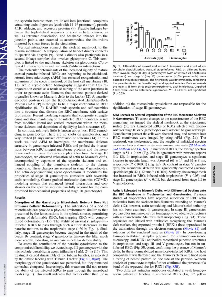
ResultsDisruption of the Gametocyte Microtubule Network Does NotInfluence Cellular Deformability. The interstices of a bed ofmicrobeads can provide a physical environment similar to thatpresented by the fenestrations in the splenic sinuses, permittingpassage of deformable RBCs, but trapping RBCs with compro-mised deformability (15). The ability of asexual P. falciparum-infected RBCs to pass through such a filter decreases as theparasite matures to the trophozoite stage (∼30 h; Fig. 1). Simi-larly, stage III gametocytes become trapped in the mesh of thebeads. By contrast, stage V gametocytes traverse the filter muchmore readily, indicating an increase in cellular deformability.To assess the contribution of the parasite cytoskeleton to the
compromised filterability, we treated stage III gametocytes with themicrotubule destabilizing agent trifluralin (1 μM, 24 h) (16). Thistreatment caused disassembly of the tubulin bundles, as indicatedby the diffuse labeling with Tubulin Tracker (Fig. S1, Right). Themorphology of the gametocytes was perturbed, but they remainedsomewhat elongated. Interestingly, this treatment did not restorethe ability of the infected RBCs to pass through the microbeadmesh (Fig. 1). This result indicates that factors other than (or in
addition to) the microtubule cytoskeleton are responsible for therigidification of stage III gametocytes.
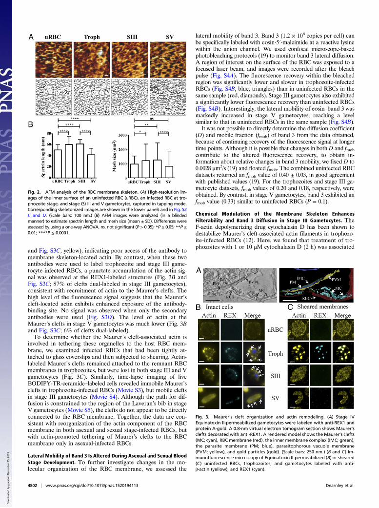
AFM Reveals an Altered Organization of the RBC Membrane Skeletonin Gametocytes. To assess changes to the nanostructure of the RBCmembrane, we imaged the skeletal meshwork at the cytoplasmicsurface (10, 17). Uninfected RBCs or RBCs infected with tropho-zoites or stage III or V gametocytes were adhered to glass coverslips.Nonadherent parts of the cells were sheared away, and remnant hostRBC membranes were imaged by using AFM (Fig. 2A). Themeshwork was skeletonized, and the lengths of individual spectrincross-members and mesh sizes were assessed manually (SI Materialsand Methods and Fig. S2). In uninfected RBCs, the average spectrinlength is 51 ± 9 nm (Fig. 2B), in agreement with previous reports(10, 18). In trophozoites and stage III gametocytes, a significantincrease in spectrin length was observed (61 ± 14 and 62 ± 8 nm,respectively; P < 0.0001) (Fig. 2B). By contrast in stage V gameto-cytes, the membrane skeleton was significantly contracted (averagespectrin length, 42 ± 12 nm; P < 0.0001). Similarly, the average meshsize increased in RBCs infected with trophozoites (P < 0.05) andstage III gametocytes (P < 0.01), but decreased from stage III toV gametocytes.
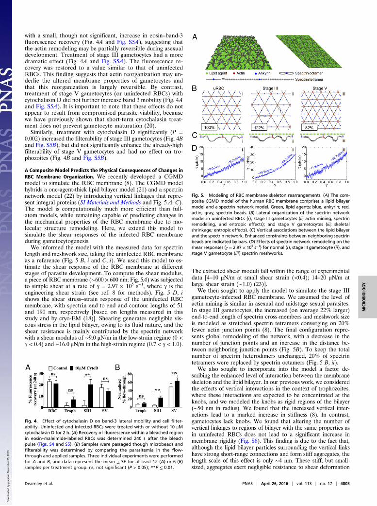
Actin Is Relocated to Maurer’s Clefts, with Differential Docking ontothe RBC Membrane in Trophozoites and Gametocytes. Previousstudies of trophozoites have reported relocation of RBC actinmolecules from the skeleton into filaments extending to Maurer’sclefts (12); however, actin remodeling and Maurer’s cleft tetheringhas not been examined in gametocytes. In stage III gametocytesprepared for immuno-electron tomography, we observed structureswith a characteristic Maurer’s cleft morphology (Fig. 3A). Theseorganelles are labeled with antibodies recognizing the Maurer’scleft marker, ring exported protein-1 (REX1) (best appreciated inthe translations through the electron tomogram (Movie S1) androtations of the rendered features (Movie S2). In pore-formingtoxin-permeabilized samples prepared for immunofluorescencemicroscopy, anti-REX1 antibodies recognized punctate structuresin trophozoites and stage III and V gametocytes, but not in un-infected RBCs (Fig. 3B, cyan), confirming the presence of Maurer’sclefts. In these permeabilized gametocyte-infected RBCs, the hostcompartment was flattened and the Maurer’s clefts were lined up ina “string of beads” pattern on one side of the parasite. Westernanalysis of gametocyte samples further confirmed the presence of aband of the expected size for REX1 (Fig. S3B).Two different antiactin antibodies exhibited a weak homoge-
neous pattern of labeling in uninfected RBCs (Fig. 3B, yellow
Fig. 1. Filterability of asexual and sexual P. falciparum and effect of mi-crotubule destabilization. Asexual stage-infected RBCs at different hoursafter invasion, stage III (day 6) gametocytes (with or without 24-h trifluralintreatment) and stage V (day 10) gametocytes (∼10% parasitemia) werepassaged though microbeads. The filterability was determined by comparingthe parasitemia in the flow-through and applied samples. Data representsthe mean ± SE from three separate experiments, each in triplicate. Unpairedt tests were used to determine significance. **P ≤ 0.01; ns, not significant(P > 0.05).
Dearnley et al. PNAS | April 26, 2016 | vol. 113 | no. 17 | 4801
MICRO
BIOLO
GY
Dow
nloa
ded
by g
uest
on
Dec
embe
r 25
, 201
9
and Fig. S3C, yellow), indicating poor access of the antibody tomembrane skeleton-located actin. By contrast, when these twoantibodies were used to label trophozoite and stage III game-tocyte-infected RBCs, a punctate accumulation of the actin sig-nal was observed at the REX1-labeled structures (Fig. 3B andFig. S3C; 87% of clefts dual-labeled in stage III gametocytes),consistent with recruitment of actin to the Maurer’s clefts. Thehigh level of the fluorescence signal suggests that the Maurer’scleft-located actin exhibits enhanced exposure of the antibody-binding site. No signal was observed when only the secondaryantibodies were used (Fig. S3D). The level of actin at theMaurer’s clefts in stage V gametocytes was much lower (Fig. 3Band Fig. S3C; 6% of clefts dual-labeled).To determine whether the Maurer’s cleft-associated actin is
involved in tethering these organelles to the host RBC mem-brane, we examined infected RBCs that had been tightly at-tached to glass coverslips and then subjected to shearing. Actin-labeled Maurer’s clefts remained attached to the remnant RBCmembranes in trophozoites, but were lost in both stage III and Vgametocytes (Fig. 3C). Similarly, time-lapse imaging of liveBODIPY-TR-ceramide–labeled cells revealed immobile Maurer’sclefts in trophozoite-infected RBCs (Movie S3), but mobile cleftsin stage III gametocytes (Movie S4). Although the path for dif-fusion is constrained to the region of the Laveran’s bib in stageV gametocytes (Movie S5), the clefts do not appear to be directlyconnected to the RBC membrane. Together, the data are con-sistent with reorganization of the actin component of the RBCmembrane in both asexual and sexual stage-infected RBCs, butwith actin-promoted tethering of Maurer’s clefts to the RBCmembrane only in asexual-infected RBCs.
Lateral Mobility of Band 3 Is Altered During Asexual and Sexual BloodStage Development. To further investigate changes in the mo-lecular organization of the RBC membrane, we assessed the
lateral mobility of band 3. Band 3 (1.2 × 10⁶ copies per cell) canbe specifically labeled with eosin-5′-maleimide at a reactive lysinewithin the anion channel. We used confocal microscope-basedphotobleaching protocols (19) to monitor band 3 lateral diffusion.A region of interest on the surface of the RBC was exposed to afocused laser beam, and images were recorded after the bleachpulse (Fig. S4A). The fluorescence recovery within the bleachedregion was significantly lower and slower in trophozoite-infectedRBCs (Fig. S4B, blue, triangles) than in uninfected RBCs in thesame sample (red, diamonds). Stage III gametocytes also exhibiteda significantly lower fluorescence recovery than uninfected RBCs(Fig. S4B). Interestingly, the lateral mobility of eosin–band 3 wasmarkedly increased in stage V gametocytes, reaching a levelsimilar to that in uninfected RBCs in the same sample (Fig. S4B).It was not possible to directly determine the diffusion coefficient
(D) and mobile fraction (fmob) of band 3 from the data obtained,because of continuing recovery of the fluorescence signal at longertime points. Although it is possible that changes in both D and fmobcontribute to the altered fluorescence recovery, to obtain in-formation about relative changes in band 3 mobility, we fixed D to0.0028 μm2/s (19) and floated fmob. The combined uninfected RBCdatasets returned an fmob value of 0.40 ± 0.03, in good agreementwith published values (19). For the trophozoites and stage III ga-metocyte datasets, fmob values of 0.20 and 0.18, respectively, wereobtained. By contrast, in stage V gametocytes, band 3 exhibited anfmob value (0.33) similar to uninfected RBCs (P = 0.1).
Chemical Modulation of the Membrane Skeleton EnhancesFilterability and Band 3 Diffusion in Stage III Gametocytes. TheF-actin depolymerizing drug cytochalasin D has been shown todestabilize Maurer’s cleft-associated actin filaments in trophozo-ite-infected RBCs (12). Here, we found that treatment of tro-phozoites with 1 or 10 μM cytochalasin D (2 h) was associated
Fig. 2. AFM analysis of the RBC membrane skeleton. (A) High-resolution im-ages of the inner surface of an uninfected RBC (uRBC), an infected RBC at tro-phozoite stage, and stage (S) III and V gametocytes, captured in tapping mode.Corresponding skeletonized images are shown in the lower panels and in Fig. S2C and D. (Scale bars: 100 nm.) (B) AFM images were analyzed (in a blindedmanner) to estimate spectrin length and mesh size (mean ± SD). Differences wereassessed by using a one-way ANOVA. ns, not significant (P > 0.05); *P ≤ 0.05; **P ≤0.01; ****P ≤ 0.0001.
Fig. 3. Maurer’s cleft organization and actin remodeling. (A) Stage IVEquinatoxin II-permeabilized gametocytes were labeled with anti-REX1 andprotein A-gold. A 0.8-nm virtual electron tomogram section shows Maurer’sclefts decorated with anti-REX1. A rendered model shows the Maurer’s clefts(MC; cyan), RBC membrane (red), the inner membrane complex (IMC; green),the parasite membrane (PM; blue), parasitophorous vacuole membrane(PVM; yellow), and gold particles (gold). (Scale bars: 250 nm.) (B and C) Im-munofluorescence microscopy of Equinatoxin II-permeabilized (B) or sheared(C) uninfected RBCs, trophozoites, and gametocytes labeled with anti-β-actin (yellow), and REX1 (cyan).
4802 | www.pnas.org/cgi/doi/10.1073/pnas.1520194113 Dearnley et al.
Dow
nloa
ded
by g
uest
on
Dec
embe
r 25
, 201
9
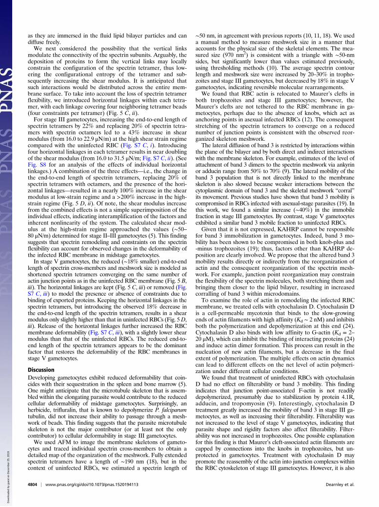
with a small, though not significant, increase in eosin–band-3fluorescence recovery (Fig. 4A and Fig. S5A), suggesting thatthe actin remodeling may be partially reversible during asexualdevelopment. Treatment of stage III gametocytes had a moredramatic effect (Fig. 4A and Fig. S5A). The fluorescence re-covery was restored to a value similar to that of uninfectedRBCs. This finding suggests that actin reorganization may un-derlie the altered membrane properties of gametocytes andthat this reorganization is largely reversible. By contrast,treatment of stage V gametocytes (or uninfected RBCs) withcytochalasin D did not further increase band 3 mobility (Fig. 4Aand Fig. S5A). It is important to note that these effects do notappear to result from compromised parasite viability, becausewe have previously shown that short-term cytochalasin treat-ment does not prevent gametocyte maturation (20).Similarly, treatment with cytochalasin D significantly (P =
0.002) increased the filterability of stage III gametocytes (Fig. 4Band Fig. S5B), but did not significantly enhance the already-highfilterability of stage V gametocytes and had no effect on tro-phozoites (Fig. 4B and Fig. S5B).
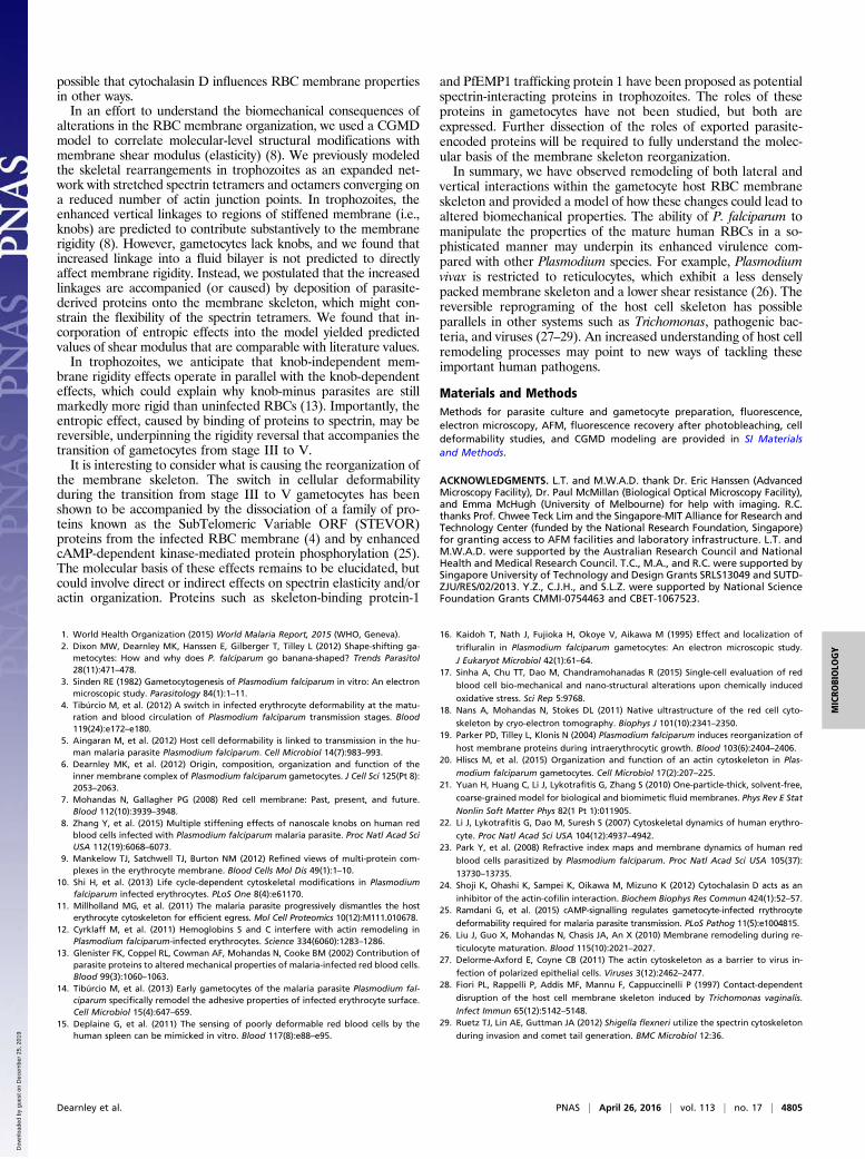
A Composite Model Predicts the Physical Consequences of Changes inRBC Membrane Organization. We recently developed a CGMDmodel to simulate the RBC membrane (8). The CGMD modelhybrids a one-agent-thick lipid bilayer model (21) and a spectrinnetwork model (22) by introducing vertical linkages that repre-sent integral proteins (SI Materials and Methods and Fig. 5 A–C).The model is computationally much more efficient than full-atom models, while remaining capable of predicting changes inthe mechanical properties of the RBC membrane due to mo-lecular structure remodeling. Here, we extend this model tosimulate the shear responses of the infected RBC membraneduring gametocytogenesis.We informed the model with the measured data for spectrin
length and meshwork size, taking the uninfected RBC membraneas a reference (Fig. 5 B, i and C, i). We used this model to es-timate the shear response of the RBC membrane at differentstages of parasite development. To compute the shear modulus,a piece of RBCmembrane (∼600 × 600 nm; Fig. 5A) was subjectedto simple shear at a rate of γ: = 2.97 × 10⁵ s−1, where γ is theengineering shear strain (see ref. 8 for methods). Fig. 5 D, ishows the shear stress–strain response of the uninfected RBCmembrane, with spectrin end-to-end and contour lengths of 51and 190 nm, respectively [based on lengths measured in thisstudy and by cryo-EM (18)]. Shearing generates negligible vis-cous stress in the lipid bilayer, owing to its fluid nature, and theshear resistance is mainly contributed by the spectrin networkwith a shear modulus of ∼9.0 μN/m in the low-strain regime (0 <γ < 0.4) and ∼16.0 μN/m in the high-strain regime (0.7 < γ < 1.0).
The extracted shear moduli fall within the range of experimentaldata [4–10 μN/m at small shear strain (<0.4); 14–20 μN/m atlarge shear strain (∼1.0) (23)].We then sought to apply the model to simulate the stage III
gametocyte-infected RBC membrane. We assumed the level ofactin mining is similar in asexual and midstage sexual parasites.In stage III gametocytes, the increased (on average 22% larger)end-to-end length of spectrin cross-members and meshwork sizeis modeled as stretched spectrin tetramers converging on 20%fewer actin junction points (8). The final configuration repre-sents global remodeling of the network, with a decrease in thenumber of junction points and an increase in the distance be-tween neighboring junction points (Fig. 5B). To keep the totalnumber of spectrin heterodimers unchanged, 20% of spectrintetramers were replaced by spectrin octamers (Fig. 5 B, ii).We also sought to incorporate into the model a factor de-
scribing the enhanced level of interaction between the membraneskeleton and the lipid bilayer. In our previous work, we consideredthe effects of vertical interactions in the context of trophozoites,where these interactions are expected to be concentrated at theknobs, and we modeled the knobs as rigid regions of the bilayer(∼50 nm in radius). We found that the increased vertical inter-actions lead to a marked increase in stiffness (8). In contrast,gametocytes lack knobs. We found that altering the number ofvertical linkages to regions of bilayer with the same properties asin uninfected RBCs does not lead to a significant increase inmembrane rigidity (Fig. S6). This finding is due to the fact that,although the lipid bilayer particles surrounding the vertical linkshave strong short-range connections and form stiff aggregates, thelength scale of this effect is only ∼4 nm. These stiff, but small-sized, aggregates exert negligible resistance to shear deformation
Fig. 4. Effect of cytochalasin D on band-3 lateral mobility and cell filter-ability. Uninfected and infected RBCs were treated with or without 10 μMcytochalasin D for 2 h. (A) Recovery of fluorescence within a bleached regionin eosin–maleimide-labeled RBCs was determined 240 s after the bleachpulse (Figs. S4 and S5). (B) Samples were passaged though microbeads andfilterability was determined by comparing the parasitemia in the flow-through and applied samples. Three individual experiments were performedfor A and B, and data represent the mean ± SE for at least 12 (A) or 6 (B)samples per treatment group. ns, not significant (P > 0.05); **P ≤ 0.01.
Fig. 5. Modeling of RBC membrane skeleton rearrangements. (A) The com-posite CGMD model of the human RBC membrane comprises a lipid bilayermodel and a spectrin network model. Green, lipid agents; blue, ankyrin; red,actin; gray, spectrin beads. (B) Lateral organization of the spectrin networkmodel in uninfected RBCs (i), stage III gametocytes (ii; actin mining, spectrinremodeling, and entropic effects); and stage V gametocytes (iii; skeletalshrinkage; entropic effects). (C) Vertical associations between the lipid bilayerand the spectrin network. Enhanced constraints between neighboring spectrinbeads are indicated by bars. (D) Effects of spectrin network remodeling on theshear responses (γ
:= 2.97 × 10⁵ s−1) for normal (i), stage III gametocyte (ii), and
stage V gametocyte (iii) spectrin meshworks.
Dearnley et al. PNAS | April 26, 2016 | vol. 113 | no. 17 | 4803
MICRO
BIOLO
GY
Dow
nloa
ded
by g
uest
on
Dec
embe
r 25
, 201
9
as they are immersed in the fluid lipid bilayer particles and candiffuse freely.We next considered the possibility that the vertical links
modulate the connectivity of the spectrin subunits. Arguably, thedeposition of proteins to form the vertical links may locallyconstrain the configuration of the spectrin tetramer, thus low-ering the configurational entropy of the tetramer and sub-sequently increasing the shear modulus. It is anticipated thatsuch interactions would be distributed across the entire mem-brane surface. To take into account the loss of spectrin tetramerflexibility, we introduced horizontal linkages within each tetra-mer, with each linkage covering four neighboring tetramer beads(four constraints per tetramer) (Fig. 5 C, ii).For stage III gametocytes, increasing the end-to-end length of
spectrin tetramers by 22% and replacing 20% of spectrin tetra-mers with spectrin octamers led to a 43% increase in shearmodulus (from 16.0 to 22.9 μN/m) at the high shear strain regimecompared with the uninfected RBC (Fig. S7 C, i). Introducingfour horizontal linkages in each tetramer results in near doublingof the shear modulus (from 16.0 to 31.5 μN/m; Fig. S7 C, ii). (SeeFig. S8 for an analysis of the effects of individual horizontallinkages.) A combination of the three effects—i.e., the change inthe end-to-end length of spectrin tetramers, replacing 20% ofspectrin tetramers with octamers, and the presence of the hori-zontal linkages—resulted in a nearly 100% increase in the shearmodulus at low-strain regime and a >200% increase in the high-strain regime (Fig. 5 D, ii). Of note, the shear modulus increasefrom the combined effects is not a simple superimposition of theindividual effects, indicating interamplification of the factors andinherent nonlinearity of the system. The calculated shear mod-ulus at the high-strain regime approached the values (∼50–80 μN/m) determined for stage II–III gametocytes (5). This findingsuggests that spectrin remodeling and constraints on the spectrinflexibility can account for observed changes in the deformability ofthe infected RBC membrane in midstage gametocytes.In stage V gametocytes, the reduced (∼18% smaller) end-to-end
length of spectrin cross-members and meshwork size is modeled asshortened spectrin tetramers converging on the same number ofactin junction points as in the uninfected RBC membrane (Fig. 5 B,iii). The horizontal linkages are kept (Fig. 5 C, iii) or removed (Fig.S7 C, iii) to model the presence or absence of constraints due tobinding of exported proteins. Keeping the horizontal linkages in thespectrin tetramers, but introducing the observed 18% decrease inthe end-to-end length of the spectrin tetramers, results in a shearmodulus only slightly higher than that in uninfected RBCs (Fig. 5D,iii). Release of the horizontal linkages further increased the RBCmembrane deformability (Fig. S7 C, iii), with a slightly lower shearmodulus than that of the uninfected RBCs. The reduced end-to-end length of the spectrin tetramers appears to be the dominantfactor that restores the deformability of the RBC membranes instage V gametocytes.
DiscussionDeveloping gametocytes exhibit reduced deformability that coin-cides with their sequestration in the spleen and bone marrow (5).One might anticipate that the microtubule skeleton that is assem-bled within the elongating parasite would contribute to the reducedcellular deformability of midstage gametocytes. Surprisingly, anherbicide, trifluralin, that is known to depolymerize P. falciparumtubulin, did not increase their ability to passage through a mesh-work of beads. This finding suggests that the parasite microtubuleskeleton is not the major contributor (or at least not the onlycontributor) to cellular deformability in stage III gametocytes.We used AFM to image the membrane skeletons of gameto-
cytes and traced individual spectrin cross-members to obtain adetailed map of the organization of the meshwork. Fully extendedspectrin tetramers have a length of ∼190 nm (18), but in thecontext of uninfected RBCs, we estimated a spectrin length of
∼50 nm, in agreement with previous reports (10, 11, 18). We useda manual method to measure meshwork size in a manner thataccounts for the physical size of the skeletal elements. The mea-sured size (970 nm2) is consistent with a triangle with ∼50-nmsides, but significantly lower than values estimated previously,using thresholding methods (10). The average spectrin contourlength and meshwork size were increased by 20–30% in tropho-zoites and stage III gametocytes, but decreased by 18% in stage Vgametocytes, indicating reversible molecular rearrangements.We found that RBC actin is relocated to Maurer’s clefts in
both trophozoites and stage III gametocytes; however, theMaurer’s clefts are not tethered to the RBC membrane in ga-metocytes, perhaps due to the absence of knobs, which act asanchoring points in asexual infected RBCs (12). The consequentstretching of the spectrin tetramers to converge on a reducednumber of junction points is consistent with the observed reor-ganized skeleton meshwork.The lateral diffusion of band 3 is restricted by interactions within
the plane of the bilayer and by both direct and indirect interactionswith the membrane skeleton. For example, estimates of the level ofattachment of band 3 dimers to the spectrin meshwork via ankyrinor adducin range from 50% to 70% (9). The lateral mobility of theband 3 population that is not directly linked to the membraneskeleton is also slowed because weaker interactions between thecytoplasmic domain of band 3 and the skeletal meshwork “corral”its movement. Previous studies have shown that band 3 mobility iscompromised in RBCs infected with asexual-stage parasites (19). Inthis work, we found a similar increase (∼40%) in the immobilefraction in stage III gametocytes. By contrast, stage V gametocytesexhibited a similar band 3 mobile fraction to uninfected RBCs.Given that it is not expressed, KAHRP cannot be responsible
for band 3 immobilization in gametocytes. Indeed, band 3 mo-bility has been shown to be compromised in both knob-plus and-minus trophozoites (19); thus, factors other than KAHRP de-position are clearly involved. We propose that the altered band 3mobility results directly or indirectly from the reorganization ofactin and the consequent reorganization of the spectrin mesh-work. For example, junction point reorganization may constrainthe flexibility of the spectrin molecules, both stretching them andbringing them closer to the lipid bilayer, resulting in increasedcorralling of band 3 within microdomains.To examine the role of actin in remodeling the infected RBC
membrane, we treated cells with cytochalasin D. Cytochalasin Dis a cell-permeable mycotoxin that binds to the slow-growingends of actin filaments with high affinity (Kd ∼ 2 nM) and inhibitsboth the polymerization and depolymerization at this end (24).Cytochalasin D also binds with low affinity to G-actin (Kd = 2–20 μM), which can inhibit the binding of interacting proteins (24)and induce actin dimer formation. This process can result in thenucleation of new actin filaments, but a decrease in the finalextent of polymerization. The multiple effects on actin dynamicscan lead to different effects on the net level of actin polymeri-zation under different cellular conditions.We found that treatment of uninfected RBCs with cytochalasin
D had no effect on filterability or band 3 mobility. This findingindicates that junction point-associated F-actin is not readilydepolymerized, presumably due to stabilization by protein 4.1R,adducin, and tropomyosin (9). Interestingly, cytochalasin Dtreatment greatly increased the mobility of band 3 in stage III ga-metocytes, as well as increasing their filterability. Filterability wasnot increased to the level of stage V gametocytes, indicating thatparasite shape and rigidity factors also affect filterability. Filter-ability was not increased in trophozoites. One possible explanationfor this finding is that Maurer’s cleft-associated actin filaments arecapped by connections into the knobs in trophozoites, but un-protected in gametocytes. Treatment with cytochalasin D maypromote the reassembly of the actin into junction complexes withinthe RBC cytoskeleton of stage III gametocytes. However, it is also
4804 | www.pnas.org/cgi/doi/10.1073/pnas.1520194113 Dearnley et al.
Dow
nloa
ded
by g
uest
on
Dec
embe
r 25
, 201
9
possible that cytochalasin D influences RBC membrane propertiesin other ways.In an effort to understand the biomechanical consequences of
alterations in the RBC membrane organization, we used a CGMDmodel to correlate molecular-level structural modifications withmembrane shear modulus (elasticity) (8). We previously modeledthe skeletal rearrangements in trophozoites as an expanded net-work with stretched spectrin tetramers and octamers converging ona reduced number of actin junction points. In trophozoites, theenhanced vertical linkages to regions of stiffened membrane (i.e.,knobs) are predicted to contribute substantively to the membranerigidity (8). However, gametocytes lack knobs, and we found thatincreased linkage into a fluid bilayer is not predicted to directlyaffect membrane rigidity. Instead, we postulated that the increasedlinkages are accompanied (or caused) by deposition of parasite-derived proteins onto the membrane skeleton, which might con-strain the flexibility of the spectrin tetramers. We found that in-corporation of entropic effects into the model yielded predictedvalues of shear modulus that are comparable with literature values.In trophozoites, we anticipate that knob-independent mem-
brane rigidity effects operate in parallel with the knob-dependenteffects, which could explain why knob-minus parasites are stillmarkedly more rigid than uninfected RBCs (13). Importantly, theentropic effect, caused by binding of proteins to spectrin, may bereversible, underpinning the rigidity reversal that accompanies thetransition of gametocytes from stage III to V.It is interesting to consider what is causing the reorganization of
the membrane skeleton. The switch in cellular deformabilityduring the transition from stage III to V gametocytes has beenshown to be accompanied by the dissociation of a family of pro-teins known as the SubTelomeric Variable ORF (STEVOR)proteins from the infected RBC membrane (4) and by enhancedcAMP-dependent kinase-mediated protein phosphorylation (25).The molecular basis of these effects remains to be elucidated, butcould involve direct or indirect effects on spectrin elasticity and/oractin organization. Proteins such as skeleton-binding protein-1
and PfEMP1 trafficking protein 1 have been proposed as potentialspectrin-interacting proteins in trophozoites. The roles of theseproteins in gametocytes have not been studied, but both areexpressed. Further dissection of the roles of exported parasite-encoded proteins will be required to fully understand the molec-ular basis of the membrane skeleton reorganization.In summary, we have observed remodeling of both lateral and
vertical interactions within the gametocyte host RBC membraneskeleton and provided a model of how these changes could lead toaltered biomechanical properties. The ability of P. falciparum tomanipulate the properties of the mature human RBCs in a so-phisticated manner may underpin its enhanced virulence com-pared with other Plasmodium species. For example, Plasmodiumvivax is restricted to reticulocytes, which exhibit a less denselypacked membrane skeleton and a lower shear resistance (26). Thereversible reprograming of the host cell skeleton has possibleparallels in other systems such as Trichomonas, pathogenic bac-teria, and viruses (27–29). An increased understanding of host cellremodeling processes may point to new ways of tackling theseimportant human pathogens.
Materials and MethodsMethods for parasite culture and gametocyte preparation, fluorescence,electron microscopy, AFM, fluorescence recovery after photobleaching, celldeformability studies, and CGMD modeling are provided in SI Materialsand Methods.
ACKNOWLEDGMENTS. L.T. and M.W.A.D. thank Dr. Eric Hanssen (AdvancedMicroscopy Facility), Dr. Paul McMillan (Biological Optical Microscopy Facility),and Emma McHugh (University of Melbourne) for help with imaging. R.C.thanks Prof. Chwee Teck Lim and the Singapore-MIT Alliance for Research andTechnology Center (funded by the National Research Foundation, Singapore)for granting access to AFM facilities and laboratory infrastructure. L.T. andM.W.A.D. were supported by the Australian Research Council and NationalHealth and Medical Research Council. T.C., M.A., and R.C. were supported bySingapore University of Technology and Design Grants SRLS13049 and SUTD-ZJU/RES/02/2013. Y.Z., C.J.H., and S.L.Z. were supported by National ScienceFoundation Grants CMMI-0754463 and CBET-1067523.
1. World Health Organization (2015) World Malaria Report, 2015 (WHO, Geneva).2. Dixon MW, Dearnley MK, Hanssen E, Gilberger T, Tilley L (2012) Shape-shifting ga-
metocytes: How and why does P. falciparum go banana-shaped? Trends Parasitol28(11):471–478.
3. Sinden RE (1982) Gametocytogenesis of Plasmodium falciparum in vitro: An electronmicroscopic study. Parasitology 84(1):1–11.
4. Tibúrcio M, et al. (2012) A switch in infected erythrocyte deformability at the matu-ration and blood circulation of Plasmodium falciparum transmission stages. Blood119(24):e172–e180.
5. Aingaran M, et al. (2012) Host cell deformability is linked to transmission in the hu-man malaria parasite Plasmodium falciparum. Cell Microbiol 14(7):983–993.
6. Dearnley MK, et al. (2012) Origin, composition, organization and function of theinner membrane complex of Plasmodium falciparum gametocytes. J Cell Sci 125(Pt 8):2053–2063.
7. Mohandas N, Gallagher PG (2008) Red cell membrane: Past, present, and future.Blood 112(10):3939–3948.
8. Zhang Y, et al. (2015) Multiple stiffening effects of nanoscale knobs on human redblood cells infected with Plasmodium falciparum malaria parasite. Proc Natl Acad SciUSA 112(19):6068–6073.
9. Mankelow TJ, Satchwell TJ, Burton NM (2012) Refined views of multi-protein com-plexes in the erythrocyte membrane. Blood Cells Mol Dis 49(1):1–10.
10. Shi H, et al. (2013) Life cycle-dependent cytoskeletal modifications in Plasmodiumfalciparum infected erythrocytes. PLoS One 8(4):e61170.
11. Millholland MG, et al. (2011) The malaria parasite progressively dismantles the hosterythrocyte cytoskeleton for efficient egress. Mol Cell Proteomics 10(12):M111.010678.
12. Cyrklaff M, et al. (2011) Hemoglobins S and C interfere with actin remodeling inPlasmodium falciparum-infected erythrocytes. Science 334(6060):1283–1286.
13. Glenister FK, Coppel RL, Cowman AF, Mohandas N, Cooke BM (2002) Contribution ofparasite proteins to altered mechanical properties of malaria-infected red blood cells.Blood 99(3):1060–1063.
14. Tibúrcio M, et al. (2013) Early gametocytes of the malaria parasite Plasmodium fal-ciparum specifically remodel the adhesive properties of infected erythrocyte surface.Cell Microbiol 15(4):647–659.
15. Deplaine G, et al. (2011) The sensing of poorly deformable red blood cells by thehuman spleen can be mimicked in vitro. Blood 117(8):e88–e95.
16. Kaidoh T, Nath J, Fujioka H, Okoye V, Aikawa M (1995) Effect and localization of
trifluralin in Plasmodium falciparum gametocytes: An electron microscopic study.
J Eukaryot Microbiol 42(1):61–64.17. Sinha A, Chu TT, Dao M, Chandramohanadas R (2015) Single-cell evaluation of red
blood cell bio-mechanical and nano-structural alterations upon chemically induced
oxidative stress. Sci Rep 5:9768.18. Nans A, Mohandas N, Stokes DL (2011) Native ultrastructure of the red cell cyto-
skeleton by cryo-electron tomography. Biophys J 101(10):2341–2350.19. Parker PD, Tilley L, Klonis N (2004) Plasmodium falciparum induces reorganization of
host membrane proteins during intraerythrocytic growth. Blood 103(6):2404–2406.20. Hliscs M, et al. (2015) Organization and function of an actin cytoskeleton in Plas-
modium falciparum gametocytes. Cell Microbiol 17(2):207–225.21. Yuan H, Huang C, Li J, Lykotrafitis G, Zhang S (2010) One-particle-thick, solvent-free,
coarse-grained model for biological and biomimetic fluid membranes. Phys Rev E Stat
Nonlin Soft Matter Phys 82(1 Pt 1):011905.22. Li J, Lykotrafitis G, Dao M, Suresh S (2007) Cytoskeletal dynamics of human erythro-
cyte. Proc Natl Acad Sci USA 104(12):4937–4942.23. Park Y, et al. (2008) Refractive index maps and membrane dynamics of human red
blood cells parasitized by Plasmodium falciparum. Proc Natl Acad Sci USA 105(37):
13730–13735.24. Shoji K, Ohashi K, Sampei K, Oikawa M, Mizuno K (2012) Cytochalasin D acts as an
inhibitor of the actin-cofilin interaction. Biochem Biophys Res Commun 424(1):52–57.25. Ramdani G, et al. (2015) cAMP-signalling regulates gametocyte-infected rrythrocyte
deformability required for malaria parasite transmission. PLoS Pathog 11(5):e1004815.26. Liu J, Guo X, Mohandas N, Chasis JA, An X (2010) Membrane remodeling during re-
ticulocyte maturation. Blood 115(10):2021–2027.27. Delorme-Axford E, Coyne CB (2011) The actin cytoskeleton as a barrier to virus in-
fection of polarized epithelial cells. Viruses 3(12):2462–2477.28. Fiori PL, Rappelli P, Addis MF, Mannu F, Cappuccinelli P (1997) Contact-dependent
disruption of the host cell membrane skeleton induced by Trichomonas vaginalis.
Infect Immun 65(12):5142–5148.29. Ruetz TJ, Lin AE, Guttman JA (2012) Shigella flexneri utilize the spectrin cytoskeleton
during invasion and comet tail generation. BMC Microbiol 12:36.
Dearnley et al. PNAS | April 26, 2016 | vol. 113 | no. 17 | 4805
MICRO
BIOLO
GY
Dow
nloa
ded
by g
uest
on
Dec
embe
r 25
, 201
9