Embed Size (px)
Citation preview
BRIEF REPORT
Reversible cerebral vasoconstriction syndrome: case report
Oguzhan Oz Æ Seref Demirkaya Æ Semai Bek ÆErdal Eroglu Æ Umit Hıdır Ulas Æ Zeki Odabası
Received: 6 February 2009 / Accepted: 25 March 2009 / Published online: 16 April 2009
� Springer-Verlag 2009
Abstract A 28-year-old woman had thunderclap head-
ache (TCH), after 7 days she had left hemiparesis. She had
a history of oral contraceptive and citalopram medications.
Brain magnetic resonance (MR) angiography demonstrated
multiple stenotic segments. Digital subtraction angiogra-
phy (DSA) showed multiple segments of narrowing in
vessel calibre. Two probable diagnoses performed; primary
angiitis of the central nervous system and reversible cere-
bral vasoconstriction syndrome (RCVS). Because of clin-
ical characteristics and normal cerebrospinal fluid findings
she was set on medication for probable RCVS. Follow-
up MR angiography after 4 weeks and DSA after 7 weeks
demonstrated improvement in vessel calibre. Thus, diag-
nosis RCVS was established. Diagnosis and management
of TCH contain many potential difficulties. Clinicians
should consider the imaging of cerebral arteries, even if
computed tomography scan and lumbar puncture are nor-
mal in TCH. Potential precipitating factors and triggers
should also be known and avoided.
Keywords Reversible cerebral vasoconstriction
syndrome � Thunderclap headache � Digital subtraction
angiography � Selective-serotonin reuptake inhibitors
Introduction
Reversible cerebral vasoconstriction syndrome (RCVS) is
characterized by a sudden onset of thunderclap-like head-
ache, with or without focal neurological deficits and sei-
zures, most commonly in women aged 20–50 years. The
onset of symptoms may be spontaneous, but a clinical
association with pregnancy (postpartum angiopathy), and
the intake of vasoactive drugs has been described. It is a
syndrome that probably includes the previously described
cases of migrainous angiitis and many cases of benign
thunderclap or orgasmic headache that were not further
investigated. The pathophysiology is a reversible segmen-
tal vasoconstriction of cerebral arteries that is frequently
associated with focal cerebral ischemia. RCVS may be also
associated with subarachnoid hemorrhage (SAH) [1–8].
About 60% of the cases of RCVS are secondary, mainly
to postpartum and to the exposure to vasoactive substances.
The main clinical presentation includes multiple recurrent
thunderclap headaches (TCHs) over 1–4 weeks. The major
complications of RCVS are localized cortical subarachnoid
hemorrhages (22%) and parenchymal strokes (7%) that
may lead to permanent sequelae [2].
Primary angiitis of the central nervous system (PACNS)
is an inflammatory disease affecting the small and medium-
sized leptomeningeal and intracranial vessels. Angio-
graphic features of both RCVS and PACNS may be similar
[9].
Case report
A right-handed 28-year-old woman admitted to the emer-
gency department because of a single spontaneous severe
TCH arising from back of neck with nausea. Her
This case was presented as a poster at the European Headache and
Migraine Trust International Congress (EHMTIC), 4–7th September
2008, London, UK entitled ‘‘The Call–Fleming Syndrome: case
report’’.
O. Oz (&) � S. Demirkaya � S. Bek � E. Eroglu �U. H. Ulas � Z. OdabasıNeurology Department, Gulhane Military Medical Academy,
Etlik, Ankara, Turkey
e-mail: [email protected]
123
J Headache Pain (2009) 10:295–298
DOI 10.1007/s10194-009-0117-3
neurological examination was normal. Her brain computed
tomography (CT) was unremarkable. Her severe headache
lasted for 12 h. She had depressive symptoms; citalopram
20 mg and etodolak 400 mg were prescribed. She was
discharged with the diagnosis primary TCH. Her com-
plaints were reduced by the days after this first applying but
she had a moderate headache continuously. After 7 days,
she had left hemiparesis. She was admitted to our neurol-
ogy department and hospitalized.
She had a history of oral contraceptive medication for
5 years because of menstrual cycle disturbances (desoge-
strel 0, 15 mg ? ethinyl estradiol 0, 03 mg).
Clinical examination confirmed left hemiparesis, left
hemihypoesthesia, left central facial paralysis. Babinski
sign was positive on the left side. Blood count and bio-
chemical analysis were normal. Brain CT and cerebrospinal
fluid (CSF) examination were normal. Brain diffusion-
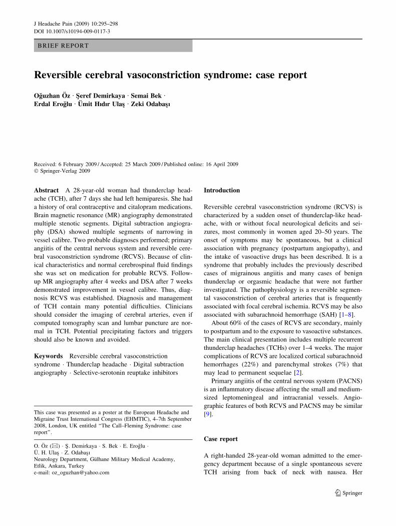
weighted magnetic resonance (MR) imaging showed acute
ischemia within the right centrum semiovale, MR angiog-
raphy demonstrated multiple stenotic segments both in
anterior and posterior circulations particularly in the right
arteria cerebri media (ACM) (Fig. 1a–c). MR venography
was normal. Probabilities of SAH and cerebral venous
thrombosis (CVT) were excluded. The diagnosis of ische-
mic cerebrovascular disease was established. Her oral
contraceptive medication and citalopram discontinued. She
was set on acetylsalicylic acid 300 mg daily.
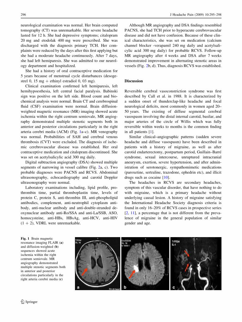
Digital subtraction angiography (DSA) showed multiple
segments of narrowing in vessel calibre (Fig. 2a, c). Two
probable diagnoses were PACNS and RCVS. Abdominal
ultrasonography, echocardiography and carotid Doppler
ultrasonography were normal.
Laboratory examinations including, lipid profile, pro-
thrombin time, partial thromboplastin time, levels of
protein C, protein S, anti-thrombin III, anti-phospholipid
antibodies, complement, anti-neutrophil cytoplasm anti-
body, anti-nuclear antibody and anti-double-stranded de-
oxynuclear antibody anti-Ro/SSA and anti-La/SSB, ASO,
homocysteine, anti-HBs, HBsAg, anti-HCV, anti-HIV
(1 ? 2), VDRL were unremarkable.
Although MR angiography and DSA findings resembled
PACNS, she had TCH prior to hyperacute cerebrovascular
disease and did not have confusion. Because of these clin-
ical characteristics, she was set on medication (calcium
channel blocker -verapamil 240 mg daily and acetylsali-
cylic acid 300 mg daily) for probable RCVS. Follow-up
MR angiography after 4 weeks and DSA after 7 weeks
demonstrated improvement in alternating stenotic areas in
vessels (Fig. 2b, d). Thus, diagnosis RCVS was established.
Discussion
Reversible cerebral vasoconstriction syndrome was first
described by Call et al. in 1988. It is characterized by
a sudden onset of thunderclap-like headache and focal
neurological deficits, most commonly in women aged 20–
50 years. The existing of diffuse segmental cerebral
vasospasm involving the distal internal carotid, basilar, and
major arteries of the circle of Willis which was fully
reversible within weeks to months is the common finding
in all patients [1].
Similar clinical–angiographic patterns (sudden severe
headache and diffuse vasospasm) have been described in
patients with a history of migraine, as well as after
carotid endarterectomy, postpartum period, Guillain–Barre
syndrome, sexual intercourse, unruptured intracranial
aneurysm, exertion, severe hypertension, and after admin-
istration of serotonergic, sympathomimetic medications
(paroxetine, sertraline, trazodone, ephedrin etc), and illicit
drugs such as cocaine [10].
The headaches in RCVS are secondary headaches,
symptom of this vascular disorder, that have nothing to do
with migraine, which is a primary headache without
underlying causal lesion. A history of migraine satisfying
the International Headache Society diagnosis criteria is
found in only 16–20% of RCVS cases in prospective series
[2, 11], a percentage that is not different from the preva-
lence of migraine in the general population of similar
gender and age.
Fig. 1 Brain magnetic
resonance imaging FLAIR (a)
and diffusion-weighted (b)
sequences showed acute
ischemia within the right
centrum semiovale. MR
angiography demonstrated
multiple stenotic segments both
in anterior and posterior
circulations particularly in the
right arteria cerebri media (c)
296 J Headache Pain (2009) 10:295–298
123
Our patient had severe headache arising from back of
neck (thunderclap) with nausea, after a period of 7 days,
she developed left hemiparesis. In RCVS complications
occur with different time courses: cortical subarachnoid
hemorrhages, intracerebral hemorrhages, seizures and
posterior reversible encephalopathy syndrome are early
events occurring during the first week, while ischemic
events including transient ischemic attacks and cerebral
infarcts occur later, during the second week. Ducros et al.
[2] suggest that the underlying disturbance in the control of
cerebral arterial tone first involves small distal arteries
responsible for hemorrhages and then progresses towards
medium and large-sized arteries responsible for ischemic
events. This is exactly what happened in our case that
thunderclap-like headache precedes neurological symp-
toms of a week.
Because of normal initial brain CT scan and normal CSF
examination diagnosis of SAH and because of normal MR
venography diagnosis of CVT were excluded. MR angi-
ography demonstrated multiple stenotic segments both in
anterior and posterior circulations particularly in right
ACM. DSA showed multiple segments of narrowing in
vessel calibre. By these findings two probable diagnoses
performed, PACNS and RCVS. Because of initial clinical
characteristics and normal CSF findings and because of
improving clinical findings she was set on medication for
probable RCVS. Follow-up MR angiography after 4 weeks
and DSA after 7 weeks demonstrated improvement in
vessel calibre.
The medical history of oral contraceptive medication for
5 years because of menstrual cycle disturbances and
citalopram for 5 days were evaluated as important risk
factors. In our case RCVS was spontaneous; unfortunately,
she was treated by citalopram and had an ischemic stroke
after 7 days. Selective-serotonin reuptake inhibitors
(SSRIs) should be avoided in patients evaluated with the
diagnosis of TCH of unknown etiology or ‘‘idiopathic
thunderclap headache’’. Perhaps, citalopram aggravated the
cerebral vasoconstriction.
It is impossible to distinguish RCVS and PACNS on the
basis of angiographic features. Patients with RCVS erro-
neously may be treated as vasculitis, and may be exposed
to long-term treatment by immunosuppressive agents with
serious side effects. In the rare cases of persistent hesitation
between the two diagnoses, it may be a good choice to wait
a few days with a calcium channel blocker (e.g., verapamil,
nimodipine): RCVS is going to stabilize and improve
quickly with regression of the vasoconstriction, and on the
Fig. 2 Digital subtraction
angiography revealed multifocal
segmental vasoconstriction in
anterior circulation (a) which
largely resolved 7 weeks later
(b). Multiple segments of
narrowing in vessel calibre in
basilar artery (c) were also
improved after therapy (d)
J Headache Pain (2009) 10:295–298 297
123
contrary, arterial irregularities in PACNS are not going to
improve so fast. Heavy treatment with immunosuppres-
sants should be reserved to patients having a biopsy proven
vasculitis. Consequently in these patients DSA should be
repeated for definitive diagnosis.
Diagnosis and management of TCH contain many
potential difficulties. Patients applying with TCH, partic-
ularly 20–50 years aged females should first be evaluated
for common conditions, such as SAH and also RCVS must
be considered. Clinicians should consider the imaging of
cerebral arteries, even if CT scan and lumbar puncture are
normal in TCH. Recognition of RCVS may be life-saving,
potential precipitating factors such as postpartum period
and vasoactive substances especially SSRIs and triggers
(sexual intercourse, defecation, sudden emotion, physical
exertion, urination without effort, cough, sneezing, bathing
or showering and sudden head movement) should also be
known and avoided.
Conflict of interest None.
References
1. Call GK, Fleming ML, Sealfon S, Levine H, Kistler JP, Fisher
CM (1988) Reversible cerebral segmental vasoconstriction.
Stroke 19:1159–1170
2. Ducros A, Boukobza M, Porcher R, Sarov M, Valade D, Bousser
MG (2007) The clinical and radiological spectrum of reversible
cerebral vasoconstriction syndrome. A prospective series of 67
patients. Brain 130(Pt 12):3091–3101
3. Singhal AB, Caviness VS, Begleiter AF, Mark EJ, Rordorf G,
Koroshetz WJ (2002) Cerebral vasoconstriction and stroke after
use of serotonergic drugs. Neurology 58:130–133
4. Koopman K, Teune LK, Ter Laan M, Uyttenboogaart M, Vroo-
men PC, De Keyser J, Luijckx GJ (2008) An often unrecognized
cause of thunderclap headache: reversible cerebral vasoconstric-
tion syndrome. J Headache Pain 9(6):389–391
5. Reneman L, Habraken JB, Majoie CB, Booij J, den Heeten GJ
(2000) MDMA (‘Ecstasy’) and its association with cerebrovascular
accidents: preliminary findings. Am J Neuroradiol 21:1001–1007
6. Neudecker S, Stock K, Krasnianski M (2006) Call–Fleming
postpartum angiopathy in the puerperium: a reversible cerebral
vasoconstriction syndrome. Obstet Gynecol 107(2 Pt 2):446–449
(Comment in: Obstet Gynecol. 2006 Feb;107(2 Pt 2):437–438)
7. Moustafa RR, Allen CM, Baron JC (2008) Call–Fleming syn-
drome associated with subarachnoid haemorrhage: three new
cases. J Neurol Neurosurg Psychiatry 79(5):602–605 [Epub 2007
Dec 12]
8. Keyrouz S, Dhar R, Axelrod Y (2008) Call–Fleming syndrome
and orgasmic cephalgia. Headache 48(6):967–971
9. Koopman K, Uyttenboogaart M, Luijckx GJ, De Keyser J,
Vroomen PC (2007) Pitfalls in the diagnosis of reversible cere-
bral vasoconstriction syndrome and primary angiitis of the central
nervous system. Eur J Neurol 14:1085–1087
10. Dodick DW (2003) Reversible segmental cerebral vasoconstric-
tion (Call–Fleming syndrome): the role of calcium antagonists.
Cephalalgia 23(3):163–165 (Comment on: Cephalalgia 2003 Apr;
23(3):218–222)
11. Chen SP, Fuh JL, Lirng JF, Chang FC, Wang SJ (2006) Recurrent
primary thunderclap headache and benign CNS angiopathy:
spectra of the same disorder? Neurology 67(12):2164–2169
298 J Headache Pain (2009) 10:295–298
123