Embed Size (px)
Citation preview

RSC Advances
PAPER
Ope
n A
cces
s A
rtic
le. P
ublis
hed
on 0
3 Ja
nuar
y 20
18. D
ownl
oade
d on
12/
13/2
021
8:46
:05
AM
. T
his
artic
le is
lice
nsed
und
er a
Cre
ativ
e C
omm
ons
Attr
ibut
ion
3.0
Unp
orte
d L
icen
ce.
View Article OnlineView Journal | View Issue
Revealing elastic
aDepartment of Mechanical and Biomedical E
Hong Kong. E-mail: [email protected]
+852-3442-0172; Tel: +852-3442-7174; +852bSchool of Biomedical Sciences, University ocCity University of Hong Kong Shenzhen ResdCentre for Robotics and Automation, City UeCentre for Biosystems, Neuroscience, and
Kong, Hong Kong
† Electronic supplementary informa10.1039/c7ra10750a
Cite this: RSC Adv., 2018, 8, 1030
Received 28th September 2017Accepted 13th December 2017
DOI: 10.1039/c7ra10750a
rsc.li/rsc-advances
1030 | RSC Adv., 2018, 8, 1030–1038
ity of largely deformed cellsflowing along confining microchannels†
Shuhuan Hu, *a Ran Wang,a Chi Man Tsang,b Sai Wah Tsao,b Dong Sunacd
and Raymond H. W. Lam *acde
Deformability is a hallmark of malignant tumor cells. Characterizing cancer cell deformation can reveal how
cancer cell metastasizes through tiny gaps in tissues. However, many previous reports only focus on the
cancer cell behaviors under small deformation regimes, which may not be representative for the behaviors
under large deformations as in the in vivo metastatic processes. Here, we investigate a wide range of cell
elasticity using our recently developed confining microchannel arrays. We develop a relation between the
elastic modulus and cell shape under different deformation levels based on a modified contact theory and
the hyperelastic Tatara theory. We demonstrate good agreements between the model prediction and
experimental results. Strikingly, we discover a clear ‘modulus jump’ of largely deformed cells compared to
that of small deformed cells, offering further biomechanical properties of the cells. Likely, such a modulus
jump can be considered as a label-free marker reflecting the elasticity of intracellular components
including the nucleus during cell translocation in capillaries and tissue constrictions. In essence, we
perform cell classification based on the distinct micromechanical properties of four cell lines, i.e. one
normal cell line (MCF-10A) and three cancer cell lines (MCF-7, MDA-MB-231 and PC3) and achieved
reasonable efficiencies (efficiency >65%). Finally, we study the correlation between large-deformational
elasticity and translocation rates of the floating cells in the microchannels. Together, our results
demonstrate the quantitative analysis of the biomechanical properties of single floating cells, which provide
an additional label-free physical biomarker toward more effective cancer diagnosis.
Introduction
Cancer-related death is oen caused by metastasis, in whichcirculating tumor cells (CTCs) disseminated in the circulationsystem1 metastasize to a second location through blood vessels(hematogenous metastasis2) or lymphatic vasculatures (lym-phogenous metastasis3). These CTCs must deform and squeezethrough small gaps (4–10 mm (ref. 4–6)) over vessels and tissuesgaps. It has been frequently reported that metastatic cancercells are associated with small elasticity.7–10 Further, suchdeformability is highly correlated with the invasiveness ofcancer cells.11,12 For this reason, the quantication of celldeformability or elasticity has been suggested as a promising
ngineering, City University of Hong Kong,
tyu.edu.hk; [email protected]; Fax:
-3442-8577
f Hong Kong, Hong Kong
earch Institute, Shenzhen, China
niversity of Hong Kong, Hong Kong
Nanotechnology, City University of Hong
tion (ESI) available. See DOI:
label-free, toxicity-free and non-destructive cell sorting andclassifying method of oating cells.10,13,14
While novel measurement techniques of cancer cell elas-ticity have been frequently reported in the past two decades,most of them are based on small cell deformation and thelinear elasticity assumption.12,15–17 In fact, the largely deformedcells should reect more representative biomechanical prop-erties for metastasis, as the cancer cells exhibit very largedeformation during invasion and extravasation, in which bothcell nucleus and cytoplasm have to deform altogether. It hasbeen recently pointed out that nuclear deformability ratherthan the cytoplasmic deformability is the rate-limiting factor inthe in vivo metastatic translocation processes.18 Hence, char-acterizing the elasticity of largely deformed cancer cells mayoffer a more-specic label-free marker for cancer diagnosis. Todate, researchers have already provided some techniques todescribe cells with large deformation. Suresh et al. applied thehollow shell hyperelasticity theory and a computational nite-element model to describe mechanical properties of largelydeformed red blood cells during capillary vessel clogging.19,20
Bernick et al. proposed a homogenized material model tocharacterize the time-dependent deformation of neurons forstudying traumatic brain injury (TBI) caused by large physicalcompressions.21
This journal is © The Royal Society of Chemistry 2018

Fig. 1 Key parameters of small and large deformation of a floating cellsqueezing along a confining microchannel.
Paper RSC Advances
Ope
n A
cces
s A
rtic
le. P
ublis
hed
on 0
3 Ja
nuar
y 20
18. D
ownl
oade
d on
12/
13/2
021
8:46
:05
AM
. T
his
artic
le is
lice
nsed
und
er a
Cre
ativ
e C
omm
ons
Attr
ibut
ion
3.0
Unp
orte
d L
icen
ce.
View Article Online
Various measurement techniques for biomechanical cellproperties based on small deformation have been developed inrecent years. However, many of these methods do not supportcells with large deformation or direct deformation of the innercell components such as the nucleus; and an analytical model isstill missing for converting results from the largely deformedcells. For example, while atomic force microscopy (AFM) hasbeen widely used for quantifying and mapping the local stiff-ness of adherent cells,22 its sharp tip with larger indentationscan damage cells.23 Modifying AFM by replacing with a spher-ical tip, scanning force microscopy (SFM) circumvents theproblem of cell damage.21 Micropipette aspiration and opticalstretching are applicable for mechanical measurements ofoating cells.19,20 These techniques can generate larger celldeformation, yet the micropipette aspiration techniquemeasures the cortical stiffness of the cytoplasm whereas theoptical trapping technique only deforms the cellmembrane.19,20,24,25 Recently, a novel microuidic techniqueutilizing a hydraulic shear force to generate hydrodynamicstretching of single oating cells has been reported of itsimplementation of mechanical phenotyping and deformability-based cell sorting.26 Though very effective and with a highthroughput, the hydrodynamic stretching mainly measures thecytoplasmic elasticity. Technically, many other microuidicmethods such as the micro-pillar obstruction inside micro-channels should support generating larger deformations ofcells and their inner components,8 their applications are stilllimited as mechanical phenotyping as the detailed theoreticalanalysis and the quantication of biomechanical properties areyet unavailable.
To implement the phenotyping of large deformation ofoating cells and to address the problems of experiment & theoryset-up, we use our recently developed microuidic elasticity27
microcytometer to quantify mechanical properties of largelydeformed oating cells. Driven by the hydraulic ows, theoating cancer cells are compressed by two conning micro-channel walls until the cell nuclei are also deformed. We use theanalytical model extended from the Hertz–Tatara theory, i.e. thehyperelastic Tatara model, to analyze larger deformation ofoating cells. This mechanical analysis is implemented forelucidating the utility of the large deformation properties asbiomarkers for indicating the structural specialty and abnor-mality of the different cells types (e.g. normal cell line MCF-10A,cancer cell line MCF-7, MDA-MB-231 and PC3). The cell classi-cation method based on the cell micromechanical analysis isintroduced to classify the different types of cells. We furtherimplemented a microuidic model to predict the translocationrate of the oating cells based on the micromechanical analysis.Our micromechanical analysis of the largely deformed oatingcells pointed out the possibility of utilizing these cell physicalproperties as biomarkers for predicting structural distortion ofthe CTCs, which could be useful for biopsy analysis.
Models
We consider a cell moving along a conning channel with inletwidth Win (¼30 mm), outlet width Wout (¼4 mm), channel length
This journal is © The Royal Society of Chemistry 2018
Lchannel (¼300 mm) and tapering angle q (z2.5� as tan q¼ (Win�Wout)/Lchannel), as shown in Fig. 1. Cell deformation is inducedby the hydraulic dragging force Fdrag and the geometricconnement of the sidewalls. The sidewalls are treated witha molecular lubricant (pluoronic F127). The force balance givesFcompress ¼ Fdrag/(2 sin q). We have developed a hyperelasticTatara model to describe the relation between cell stiffness andother related factors. We also estimate the cell stiffness basedon the previous reported Hertz model and Tatara model forcomparison as the followings.
Hertz model
The Hertz contact model provides a general form for sphericalcontact under small deformation. The Young's modulus E isexpressed as:28
E ¼ 3ð1� n2ÞFcompressffiffiffiffiffiffiffiffiffiffiffiffiffiffiffiffiffiffiffiffiffiffiffiffiffiffiffiffiffiffiffiffiffiffiffiffiffiffiffiffiffiffiffiffiDcell
�Dcell �Wdeform
�3q (1)
where Dcell is the cell diameter, Ddeform is the cell deformeddiameter, Wdeform is the cell deformed width and n ¼ 0.5 is thePoisson's ratio of a cell (Fig. 1).
Tatara model
The Tatara model extends descriptions of the Hertz model toa larger deformation regime, in which a non-spherical geometryaer deformation is considered. The Young's modulus E ob-tained by the Tatara model can be expressed as:29
E ¼ 3ð1� n2ÞFcompress
2�Dcell �Wdeform
�a� 2Fcompress
p�Dcell �Wdeform
�f ðaÞ (2)
where a is the contact radius and f(a) is the characteristic lengthof the non-spherical geometry aer deformation as thefollowings:
a ¼ 1
2
ffiffiffiffiffiffiffiffiffiffiffiffiffiffiffiffiffiffiffiffiffiffiffiffiffiffiffiffiffiffiffiffiffiDcell
2 �Wdeform2
qþDdeform �Dcell
� �(3)
RSC Adv., 2018, 8, 1030–1038 | 1031
RSC Advances Paper
Ope
n A
cces
s A
rtic
le. P
ublis
hed
on 0
3 Ja
nuar
y 20
18. D
ownl
oade
d on
12/
13/2
021
8:46
:05
AM
. T
his
artic
le is
lice
nsed
und
er a
Cre
ativ
e C
omm
ons
Attr
ibut
ion
3.0
Unp
orte
d L
icen
ce.
View Article Online
f ðaÞ ¼"
ð1þ nÞDcell2
2�a2 þDcell
2�3=2 þ 1� n2�
a2 þDcell2�1=2#�1
(4)
Hyperelastic Tatara model
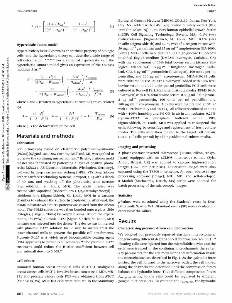
Hyperelasticity is well known as an intrinsic property of biologiccells; and the hyperelastic theory can describe a wide range ofcell deformation.19,20,30–32 For a spherical hyperelastic cell, thehyperelastic Tatara's model gives an expression of the Young'smodulus E as:29
E ¼ 3ð1� n2ÞA2�Dcell �Wdeform
� 1þ 2Ba2
Ddeform2
!Fcompress
a
� 2A
p�Dcell �Wdeform
� 1þ 4Ba2
5Ddeform2
!Fcompress
f ðaÞ (5)
where A and B (related to hyperelastic correction) are calculatedby:
A ¼ ð1� xÞ21� xþ x2
�3; B ¼ 1� x=3
1� xþ x2�3; x ¼ 1� Wdeform
Dcell
(6)
where x is the deformation of the cell.
Materials and methodsFabrication
So lithography based on elastomeric polydimethylsiloxane(PDMS) (Sylgard-184, Dow Corning, Midland, MI) was applied tofabricate the conning microchannels.33 Briey, a silicon moldmaster was fabricated by patterning a layer of positive photo-resist (AZ5214, AZ Electronic Materials, Wiesbaden, Germany),followed by deep reactive ion etching (DRIE; STS Deep SiliconEtcher, Surface Technology Systems, Newport, UK) with a depthof 40 mm, and washing off the photoresist with acetone(Sigma-Aldrich, St. Louis, MO). The mold master wastreated with vaporized (tridecauoro-1,1,2,2-tetrahydrooctyl)-1-trichlorosilane (Sigma-Aldrich, St. Louis, MO) in a vacuumchamber to enhance the surface hydrophobicity. Aerward, thePDMS substrate with micro patterns was casted from the siliconmold. The PDMS substrate was then bonded onto a glass slide(Citoglas, Jiangsu, China) by oxygen plasma. Before the experi-ments, 1% (w/w) pluronic F-127 (Sigma-Aldrich, St. Louis, MO)in water was injected into the device. The device was immersedwith pluronic F-127 solution for 30 min to surface treat theinner channel walls to prevent the possible cell attachments.Pluronic F-127 is a widely used biocompatible coating agent(FDA approved) to prevent cell adhesion.34 The pluronic F-127treatment could reduce the friction coefficient between celland sidewall down to 0.008.35
Cell culture
Immortal human breast epithelial cells MCF-10A, malignantbreast cancer cells MCF-7, invasive breast cancer cells MDA-MB-231 and prostate cancer cells PC3 were obtained from ATCC(Manassas, VA). MCF-10A cells were cultured in the Mammary
1032 | RSC Adv., 2018, 8, 1030–1038
Epithelial Growth Medium (MEGM; CC-3150, Lonza, New YorkCity, NY) added with 0.4% (v/v) bovine pituitary extract (BD,Franklin Lakes, NJ), 0.1% (v/v) human epithelial growth factor(hEGF; Cell Signaling Technology, Beverly, MA), 0.1% (v/v)hydrocortisone (Sigma-Aldrich, St. Louis, MO), 0.1% (v/v)insulin (Sigma-Aldrich) and 0.1% (v/v) of a reagent mixed with30 mg ml�1 gentamicin and 15 mg ml�1 amphotericin (GA-1000,Lonza). MCF-7 cells were cultured in a high-glucose Dulbecco'smodied Eagle's medium (DMEM; Invitrogen, Carlsbad, CA)with the supplement of 10% fetal bovine serum (Atlanta Bio-logical, Atlanta, GA), 0.5 mg ml�1 fungizone (Invitrogen, Carls-bad, CA), 5 mg ml�1 gentamicin (Invitrogen), 100 units per mlpenicillin, and 100 mg ml�1 streptomycin. MDA-MB-231 cellswere cultured in DMEM-F12 (Invitrogen) added with 10% fetalbovine serum and 100 units per ml penicillin. PC-3 cells werecultured in Roswell Park Memorial Institute media (RPMI-1640;Invitrogen) with 10% fetal bovine serum, 0.5 mg ml�1 fungizone,5 mg ml�1 gentamicin, 100 units per ml penicillin, and100 mg ml�1 streptomycin. All cells were maintained at 37 �Cwith 100% humidity and 5%CO2. All cells were cultured at 37 �Cwith�100% humidity and 5% CO2 in air in an incubator. 0.25%trypsin–EDTA in phosphate buffered saline (PBS;Sigma-Aldrich, St. Louis, MO) was applied to re-suspend thecells, following by centrifuge and replacement of fresh culturemedia. The cells were then diluted to the target cell density(�4 � 104 cells per ml) by adding additional culture media.
Imaging and processing
A phase-contrast inverted microscope (TE300, Nikon, Tokyo,Japan) equipped with an sCMOS microscope camera (Zyla,Andor, Belfast, UK) was applied to capture high-resolutionimages (�570 nm per pixel). Fluorescent images were alsocaptured using the TE300 microscope. An open source imageprocessing soware (ImageJ; NIH, MD) and self-developeda Matlab (Mathworks, Natick, MA) script were adopted forbatch processing of the microscopic images.
Statistics
p-Values were calculated using the Student's t-test in Excel(Microso, Seattle, WA). Standard errors (SE) were calculated inexpressing the values.
ResultsCharacterizing pressure driven cell deformation
We adopted our previously reported elasticity microcytometerfor generating different degrees of cell deformation (see ESI†).36
Floating cells were injected into the microuidic device and thecells were trapped in the conning microchannels thereaer.Key parameters for the cell movement and deformation insidethe microchannel are described in Fig. 1. As the hydraulic forcepushed the cell forward to the narrower outlet, the cell movedalong the channels and deformed until the compression forcesbalance the hydraulic force. Thus different compression forcesFcompress acting to the cells could be regulated by differentgauged inlet pressures. To estimate the Fcompress, the hydraulic
This journal is © The Royal Society of Chemistry 2018

Paper RSC Advances
Ope
n A
cces
s A
rtic
le. P
ublis
hed
on 0
3 Ja
nuar
y 20
18. D
ownl
oade
d on
12/
13/2
021
8:46
:05
AM
. T
his
artic
le is
lice
nsed
und
er a
Cre
ativ
e C
omm
ons
Attr
ibut
ion
3.0
Unp
orte
d L
icen
ce.
View Article Online
drag force over the cell body Fdrag was rstly obtained bya laminar ow simulation using COMSOL soware (see ESI†),following by computing the compression force Fcompress byFcompress ¼ Fdrag/(2 sin q). Furthermore as mentioned previouslyin fabrication, the microchannel walls treated with pluronic F-127 has a friction coefficient of 0.008, hence the friction forceon cells was <10% of the drag force. Therefore, the cell defor-mation was mainly caused by Fcompress. In our experiments, thehydraulic dragging Fdrag forces placed on the cells were 1–50 nNand the compression forces were 12–600 nN. We performedexperiments with a human breast epithelial cell line (MCF-10A)to characterize cell deformation under different inlet pressures:100 Pa, 200 Pa, 300 Pa and 400 Pa. The cell position L and thedeformed diameter Ddeform were obtained by an image analysisbased on micrographs as shown Fig. 2a. The deformed widthWdeform was obtained by Wdeform ¼ Win � 2L tan q. The celldiameter Dcell could be obtained by:
Fig. 2 Deformation of cells (MCF-10A) as a function of inlet pressureapplied at the device inlet. (a) Cell deforms in a confining channelunder different inlet pressure levels (100 Pa, 200 Pa, 300 Pa and 400Pa). Scale bar: 30 mm. (b) Statistics of the deformation level against theinlet pressure, n ¼ 31. Error bars represent the standard deviation.
This journal is © The Royal Society of Chemistry 2018
4
3p
�Dcell
2
�3
z 2p
�Ddeform
2
�2�Wdeform
2
�� 2
3p
�Wdeform
2
�3
(7)
The measured cell diameter of MCF-10A is 14.83 � SE 0.45mm, which agrees with the reported values.37 The deformation x
(dened as x ¼ 1 � Wdeform/Dcell) gradually increased with theenhancement of inlet pressure levels (Fig. 2b).
We focused on the transition from small deformation tolarge deformations (i.e. inlet pressure from 100 Pa to 200 Pa). Asshown in Fig. 3, the observations of compression force versusdeformation was plotted (the black dots) to be compared withthe predictions made by the aforementioned three models (thethree lines). A deviation analysis among the three models is alsoavailable in Fig. S2.†
As the classical Hertz model is based on the assumptions oflinear elasticity and small deformation,28 the estimated celldeformation property (the green line) deviated signicantlyfrom observations (black dots) especially when the deformationis larger than 0.3. Such apparent deviation implied that theHertz model is not suitable for describing large cell deforma-tions. Considering that the cell deforms into a non-sphericalobject, Tatara et al. extended the Hertz contact theory to thelarge deformation regime by considering a more detailedgeometrical conguration as a sphere with its upper and lowersections removed as shown in Fig. 1.29 Though this congura-tion led to a closer prediction of the deformation propertycomparing to the Hertz model (Fig. 3, the blue line; Fig. S2a andb†), the deviations between the observations (the black dots)and the model prediction (the blue line) gradually increasedwith the increments of the cell deformations. Thus a moreaccurate model was still needed. On the other hand, ourproposed hyperelastic Tatara model was obtained by furtherintroducing the hyperelasticity modication. (Fig. S2c and d†)
Fig. 3 Cell deformation properties toward large deformations. Thecompression force versus cell deformation under 100 Pa and 200 Pawere plotted and compared with the predictions based on Hertzmodel, Tatara model and hyperelastic Tatara model respectively. Theparameters (Young's modulus and cell diameter) in the predictionswere obtained by the measurement results under 100 Pa.
RSC Adv., 2018, 8, 1030–1038 | 1033

RSC Advances Paper
Ope
n A
cces
s A
rtic
le. P
ublis
hed
on 0
3 Ja
nuar
y 20
18. D
ownl
oade
d on
12/
13/2
021
8:46
:05
AM
. T
his
artic
le is
lice
nsed
und
er a
Cre
ativ
e C
omm
ons
Attr
ibut
ion
3.0
Unp
orte
d L
icen
ce.
View Article Online
As biological cells are intrinsically hyperelastic,16,17,27–29 thismodel should deliver the most accurate description of the celldeformation properties comparing to the previous two models.The prediction made by the hyperelastic Tatara model (Fig. 3,the red line) matched well with the observations (the black dots)of the cell deformations, hence, the hyperelastic Tatara modelgave a highly representative descriptions of cell deformations.
Fig. 5 (a) Cellular and nuclear deformation of a MCF-10A cell at inletpressure of 100 Pa (upper) and 400 Pa (lower). The cell nucleus wasundeformed for the smaller deformation; whereas the nucleusdeformation was created by the direct contact of the channel sidewallswhen the inlet pressure is sufficient high (e.g. 400 Pa). Scale bar:30 mm. (b) Deformation-induced elasticity change of an individual cellrepresented by each polyline. The red sections indicate the ‘modulusjump’.
Deformation-induced ‘modulus jump’
We further increased the inlet pressure to 300 Pa and 400 Pa toinduced nuclear deformations. When a trapped cell reacheda deformation of x ¼ 0.471 � 0.015, we observed a ‘modulusjump’ in the Young's modulus calculated by the hyperelasticTatara model (Fig. 4 and 5). Notably, previous research alsoreported such transition of the overall moduli of a core–shellpolymeric composite.38 Likely, this change in the elasticitymodulus reects a structural heterogeneity between the shelland core parts. We examined that the cell nuclei were deformedby the direct compression from the channel sidewalls undera large enough inlet pressure ($300 Pa) as shown in Fig. 5a. It iswell known that the nuclei have been frequently shown 2–10times stiffer than the cytoplasmic stiffness.39–43 It should not bea surprise to observe a ‘modulus jump’ for a largely deformedcell, as the measured Young's modulus was contributed fromthe nucleus stiffness rather than the cytoplasmic stiffness. Thisreport gave two distinct empirical formulas for description thesmall and large deformation properties: the overall Young'smoduli were given E ¼ V � Ecore + (1 � V) � Eshell under smalldeformation whereas E ¼ L � Ecore + (1 � L) � Eshell under largedeformation, in which V is the core-composite volume ratiowhile L is the core-composite length ratio. Thus it is quitepossible that a rigid core (the nucleus) gave a modulus jump ofthe oating cells and led to the transition of the Young'smodulus, giving the previously frequently reported highernuclear stiffness than cytoplasmic stiffness.41,44–46 We further
Fig. 4 Threemodels for describing cell deformation of MCF-10A cells,i.e., Hertz model, Tatara model and hyperelastic Tatara model. Young'smodulus obtained under different deformations with the normalizedmodulus value scaled as unity for the level under the 100 Pa inletpressure.
1034 | RSC Adv., 2018, 8, 1030–1038
examined that the nucleus diameter is �0.5 of the cell diameter(Fig. S3†); and therefore we oen observed the modulus jump ata deformation �0.5 for most individual cells (Fig. 5b).
Cell classication based on cell physical properties of largedeformation
The physical properties of large cell deformations usually implythe genetic and structural distortions.47 To give an example,four types of cells were used: the breast epithelial cells MCF-10A, breast cancer cells MCF-7, more invasive breast cancercells MDA-MB-231, and prostate cancer cells PC3. A low inletpressure (100 Pa) was applied to induce a small deformation,followed by a high inlet pressure (400 Pa) to induce a largedeformation. Following the same procedures of characterizingMCF-10A cells described in the previous section, the initialmoduli Ei at 100 Pa and the nal moduli Ef at 400 Pa of the fourtypes of cells were calculated by the hyperelastic Tatara model.Under small deformations, the cancerous cells (MCF-7, MDA-MB-231 and PC3) were signicantly soer than the normalbreast cell MCF-10A (Fig. 6a), which indicated the disruption of
This journal is © The Royal Society of Chemistry 2018

Fig. 7 Principle component analysis and cell classification for cellclassification based on cell deformation properties. Cell numbers,MCF-10A: n ¼ 53, MCF-7: n ¼ 73, MDA-MB-231: n ¼ 67, PC3: n ¼ 85;the first principle component is 0.022� Dcell � 2.4 � Ei � 0.048� Ef +1.9 � DE; the second principle component is 0.046 � Dcell + 1.7 � Ei +0.41 � Ef + 2.1 � DE.
Paper RSC Advances
Ope
n A
cces
s A
rtic
le. P
ublis
hed
on 0
3 Ja
nuar
y 20
18. D
ownl
oade
d on
12/
13/2
021
8:46
:05
AM
. T
his
artic
le is
lice
nsed
und
er a
Cre
ativ
e C
omm
ons
Attr
ibut
ion
3.0
Unp
orte
d L
icen
ce.
View Article Online
the cytoskeletal bers in the cytoplasm and conformed toprevious reports.10,48 Under large deformation, however, theMCF-7 and PC3 cells shown no signicances of Young'smodulus comparing to the normal cell type MCF-10A (Fig. 6c).This transition implied a stiffer or larger nuclei of MCF-7 andPC3 cells. In fact, both stiffer and larger nuclei were found incancer cells.49–51 For the modulus jump (Fig. 6b), MDA-MB-231was signicantly lower than MCF-7 (or PC3), which mightimplicated a soer core of MDA-MB-231 than that of MCF-7 (orPC3) since the sizes of the cell body as well as the nuclei of thesethree types of cells were similar (see Fig. S3†). Previous researchfound that the more invasive cancer cells (e.g. MDA-MB-231)undergone epithelial–mesenchymal transition (EMT) usuallycontain soer nuclei.52–54 Hence the nal modulus Ef andmodulus jump DE could provide the insightful information ofthe physical distortion (either stiffening or enlarging) of thenuclei.
Since the physical properties of cell deformation implied thestructural abnormality of cancer cells, these physical propertiescould be used as biomarkers for cell classication. Fordemonstration, the principle component analysis (PCA)55 offour physical parameters (i.e. cell diameter Dcell, initial modulusEi, modulus jump DE and nal modulus Ef) was applied (seeFig. 7). Next, linear discriminant analysis (LDA)56 was applied toclassify the cells. Since MCF-7 and PC3 cells were physicallyundistinguishable in our experiments, the data of the two typeswere combined for the LDA test. These two analyses gavea relatively high efficiency for cell classication: 78.3% for MCF-10A, 65.6% for MCF-7 and PC3, 69.7% for MDA-MB-231.
Critical pressure for a cell translocating through capillarystructures
Our elasticity microcytometer could serve as amicrouidicmodelfor testing the capability of cancer cell translocation with nuclear
Fig. 6 Cell mechanical properties under large deformations. Cell numberBar charts and scattering plots of (a) the initial modulus Ei at 100 Pa, (b) theat 400 Pa. Error bars are standard errors. * indicates p < 0.01.
This journal is © The Royal Society of Chemistry 2018
deformation. For demonstration, we further increased the inletpressures to ush away the trapped cells through the conningmicrochannels with an exit channel width (4 mm) signicantlyshorter than the nucleus diameter. Aer the cells got trapped inthe channels, the inlet pressure was gradually increased and thenumber of the remaining cells was calculated, as shown inFig. 8a. Further, predictions were made based on the deforma-tion properties calculated by the hyperelastic Tataramodel on the
s, MCF-10A: n¼ 53, MCF-7: n¼ 73, MDA-MB-231: n¼ 67, PC3: n¼ 85.modulus jump DE between 100 Pa and 400 Pa, (c) the final modulus Ef
RSC Adv., 2018, 8, 1030–1038 | 1035

Fig. 8 Critical pressures for flushing cells through the confiningmicrochannels with an exit width of 4 mm. (a) Fraction of the remainingcells driven by different inlet pressures (boxes). The predicted values(lines) were computed using the hyperelastic Tatara model. (b) Criticalpressures required for flushing half of the trapped cells away from themicrochannels. Error bars are standard errors. Asterisk indicates p < 0.01.
RSC Advances Paper
Ope
n A
cces
s A
rtic
le. P
ublis
hed
on 0
3 Ja
nuar
y 20
18. D
ownl
oade
d on
12/
13/2
021
8:46
:05
AM
. T
his
artic
le is
lice
nsed
und
er a
Cre
ativ
e C
omm
ons
Attr
ibut
ion
3.0
Unp
orte
d L
icen
ce.
View Article Online
nucleus-deformed cells at 400 Pa. The predictions conformedvery well to the experimental results, which validated the appli-cability of the hyperelastic Tataramodel for prediction cancer celltranslocating in our conning microchannel structure. More-over, we dened and estimated the ‘critical pressure’ for half ofthe trapped cells passing through the microchannels. We foundthat, though the MDA-MB-231 cells and the MCF-7 cells hadsimilar sizes (17.24� SE 0.21 mm versus 17.48 � SE 0.31 mm), thecritical pressure for MDA-MB-231 cells was signicantly lowerthan the MCF-7 cells (0.91 � SE 0.07 kPa versus 1.37 � SE 0.08kPa), which might reect the higher capability of invasion of theMDA-MB-231 cells. Considering that the critical pressure(�1 kPa) discovered in this work was smaller than the normalblood pressure (3.7 kPa) in capillary vessels with a typical diam-eter ranging 4–10 mm in human body.4–6 Very likely, the capillarypressure is already high enough to drive cancer cells, especiallythe invasive ones, through the capillaries. Therefore, the higherliquid pressure could assist cell translocation in the metastaticprocess.
Conclusion
In this work, we have investigated the properties of largelydeformed cancer cells using the elasticity microcytometer. Werstly extended the contact theories for small deformation and
1036 | RSC Adv., 2018, 8, 1030–1038
introduced the hyperelastic Tatara model for quantifying thelarge deformation properties of cells. Interestingly, we observeda “modulus jump” between small deformation and largedeformation, which can be considered as the “rigid-core” effectas it is very likely due to the higher Young's modulus of thenucleus. Next, by applying the hyperelastic Tatara model, thedistinct mechanical properties under large deformation wererevealed for four different types of cells (MCF-10A, MCF-7, MDA-MB-231 and PC3). As changes in the intracellular mechanicalproperties can reect the genetic and structural alterations ofcancer cells, these properties can be considered as the disease-related biomarkers. Cell classication based on these biome-chanical properties was also performed. Finally, we examine therelation between these properties and occurrence of the cellstranslocating through a conning microchannel with a narrowexit channel width (4 mm) comparable to the capillary diameter,providing some insights on the role the elasticity of largelydeformed cancer cells in metastasis. This device can offer a cellcharacterization throughput of �10 cell per min, which ishigher than the continuous ow optical stretcher (1 cell permin)57 and yet lower than the real-time deformability cytometry(6000 cells per min).58 In the future, we may embed microelec-trode arrays along the conning microchannels for real-timecell detection and biomechanical characterization witha signicant higher throughput.59 Importantly, the quantitativemeasurement technique reported in this paper can obtainelasticity and viscosity of cells in both large and small defor-mations; and therefore this technique would induce a morecomprehensive cell characterization for more effective cancerdiagnosis applications.
Conflicts of interest
There are no conicts of interest to declare.
Acknowledgements
We acknowledge nancial supports from the National NaturalScience Foundation of China (project #31500758), GeneralResearch Grant (project #11206014) and Collaborative ResearchFund (project #C1013-15GF) of Hong Kong Research GrantCouncil, Croucher Foundation (Startup grant) and the CityUniversity of Hong Kong (project #7004602).
References
1 P. Mehlen and A. Puisieux, Nat. Rev. Cancer, 2006, 6, 449–458.
2 A. Chambers, G. Naumov, H. Varghese, K. Nadkarni,I. MacDonald and A. Groom, Surg. Oncol. Clin., 2001, 10,243–255.
3 R. K. Jain and B. T. Fenton, J. Natl. Cancer Inst., 2002, 94, 417–421.
4 C.-H. Heldin, K. Rubin, K. Pietras and A. Ostman, Nat. Rev.Cancer, 2004, 4, 806.
5 C. B. Henry and B. R. Duling, Am. J. Physiol.: Heart Circ.Physiol., 1999, 277, H508–H514.
This journal is © The Royal Society of Chemistry 2018

Paper RSC Advances
Ope
n A
cces
s A
rtic
le. P
ublis
hed
on 0
3 Ja
nuar
y 20
18. D
ownl
oade
d on
12/
13/2
021
8:46
:05
AM
. T
his
artic
le is
lice
nsed
und
er a
Cre
ativ
e C
omm
ons
Attr
ibut
ion
3.0
Unp
orte
d L
icen
ce.
View Article Online
6 M. J. Davis, Am. J. Physiol.: Heart Circ. Physiol., 1988, 255,H1114–H1129.
7 J. Guck, S. Schinkinger, B. Lincoln, F. Wottawah, S. Ebert,M. Romeyke, D. Lenz, H. M. Erickson, R. Ananthakrishnanand D. Mitchell, Biophys. J., 2005, 88, 3689–3698.
8 W. Zhang, K. Kai, D. S. Choi, T. Iwamoto, Y. H. Nguyen,H. Wong, M. D. Landis, N. T. Ueno, J. Chang and L. Qin,Proc. Natl. Acad. Sci. U. S. A., 2012, 109, 18707–18712.
9 E. C. Faria, N. Ma, E. Gazi, P. Gardner, M. Brown,N. W. Clarke and R. D. Snook, Analyst, 2008, 133, 1498–1500.
10 T. W. Remmerbach, F. Wottawah, J. Dietrich, B. Lincoln,C. Wittekind and J. Guck, Cancer Res., 2009, 69, 1728–1732.
11 P. Friedl and K. Wolf, Nat. Rev. Cancer, 2003, 3, 362–374.12 S. E. Cross, Y.-S. Jin, J. Rao and J. K. Gimzewski, Nat.
Nanotechnol., 2007, 2, 780–783.13 S. J. Tan, R. L. Lakshmi, P. Chen, W.-T. Lim, L. Yobas and
C. T. Lim, Biosens. Bioelectron., 2010, 26, 1701–1705.14 M. E. Warkiani, B. L. Khoo, L. Wu, A. K. P. Tay,
A. A. S. Bhagat, J. Han and C. T. Lim, Nat. Protoc., 2016,11, 134–148.
15 N. Gavara and R. S. Chadwick, Nat. Nanotechnol., 2012, 7,733–736.
16 R. D. Gonzalez-Cruz, V. C. Fonseca and E. M. Darling, Proc.Natl. Acad. Sci. U. S. A., 2012, 109, E1523–E1529.
17 L. M. Lee and A. P. Liu, Lab Chip, 2015, 15, 264–273.18 C. Beadle, M. C. Assanah, P. Monzo, R. Vallee, S. S. Rosenfeld
and P. Canoll, Mol. Biol. Cell, 2008, 19, 3357–3368.19 C. Lim, M. Dao, S. Suresh, C. Sow and K. Chew, Acta Mater.,
2004, 52, 1837–1845.20 J. Mills, L. Qie, M. Dao, C. Lim and S. Suresh, Mech. Chem.
Biosyst., 2004, 1, 169–180.21 K. B. Bernick, T. P. Prevost, S. Suresh and S. Socrate, Acta
Biomater., 2011, 7, 1210–1219.22 A. Raman, S. Trigueros, A. Cartagena, A. Stevenson,
M. Susilo, E. Nauman and S. A. Contera, Nat. Nanotechnol.,2011, 6, 809–814.
23 A. N. Ketene, E. M. Schmelz, P. C. Roberts and M. Agah,Nanomedicine, 2012, 8, 93–102.
24 D. E. Discher, D. H. Boal and S. K. Boey, Biophys. J., 1998, 75,1584–1597.
25 R. Tran-Son-Tay, D. Needham, A. Yeung and R. Hochmuth,Biophys. J., 1991, 60, 856–866.
26 D. R. Gossett, H. T. Tse, S. A. Lee, Y. Ying, A. G. Lindgren,O. O. Yang, J. Rao, A. T. Clark and D. Di Carlo, Proc. Natl.Acad. Sci. U. S. A., 2012, 109, 7630–7635.
27 S. Hu, G. Liu, W. Chen, X. Li, W. Lu, R. H. Lam and J. Fu,Small, 2016, 12, 2300–2311.
28 D.-H. Kim, B. Li, F. Si, J. M. Phillip, D. Wirtz and S. X. Sun, J.Cell Sci., 2015, 128, 3375–3385.
29 Y. Tatara, J. Eng. Mater. Technol., 1991, 113, 285–291.30 M. F. Beatty, Appl. Mech. Rev., 1987, 40, 1699–1734.31 J. J. O'Hagan and A. Samani, Phys. Med. Biol., 2009, 54, 2557.32 M. Dao, C. T. Lim and S. Suresh, J. Mech. Phys. Solids, 2003,
51, 2259–2280.33 Y. Xia and G. M. Whitesides, Annu. Rev. Mater. Sci., 1998, 28,
153–184.
This journal is © The Royal Society of Chemistry 2018
34 S. F. Khattak, S. R. Bhatia and S. C. Roberts, Tissue Eng.,2005, 11, 974–983.
35 L. Milovanovic and H. Ma, Anal. Methods, 2012, 4, 4303–4309.
36 S. Hu, G. Liu, W. Chen, X. Li, W. Lu, R. H. Lam and J. Fu,Small, 2016, 12, 2300–2311.
37 J. An, J. Lee, S. H. Lee, J. Park and B. Kim, Anal. Bioanal.Chem., 2009, 394, 801–809.
38 T. Kong, L. Wang, H. M. Wyss and H. C. Shum, So Matter,2014, 10, 3271–3276.
39 B. Hampoelz and T. Lecuit, Curr. Opin. Cell Biol., 2011, 23,668–675.
40 F. Guilak, J. R. Tedrow and R. Burgkart, Biochem. Biophys.Res. Commun., 2000, 269, 781–786.
41 H. Liu, J. Wen, Y. Xiao, J. Liu, S. Hopyan, M. Radisic,C. A. Simmons and Y. Sun, ACS Nano, 2014, 8, 3821–3828.
42 G. Ofek, R. M. Natoli and K. A. Athanasiou, J. Biomech., 2009,42, 873–877.
43 N. Caille, O. Thoumine, Y. Tardy and J.-J. Meister, J.Biomech., 2002, 35, 177–187.
44 E. T. Belt, R. Fijneman, E. van den Berg, H. Bril, P. Delis-vanDiemen, M. Tijssen, H. van Essen, E. de Lange-de Klerk,J. Belien and H. Stockmann, Eur. J. Cancer, 2011, 47, 1837–1845.
45 P. Isermann and J. Lammerding, Curr. Biol., 2013, 23,R1113–R1121.
46 N. D. Willis, T. R. Cox, S. F. Rahman-Casans, K. Smits,S. A. Przyborski, P. van den Brandt, M. van Engeland,M. Weijenberg, R. G. Wilson and A. De Bruıne, PLoS One,2008, 3, e2988.
47 V. Swaminathan, K. Mythreye, E. T. O'Brien, A. Berchuck,G. C. Blobe and R. Superne, Cancer Res., 2011, 71, 5075–5080.
48 M. Coughlin, D. Bielenberg, G. Lenormand, M. Marinkovic,C. Waghorne, B. Zetter and J. Fredberg, Clin. Exp. Metastasis,2013, 30, 1–14.
49 D. Zink, A. H. Fischer and J. A. Nickerson, Nat. Rev. Cancer,2004, 4, 677.
50 L. Kong, G. Schafer, H. Bu, Y. Zhang, Y. Zhang andH. Klocker, Carcinogenesis, 2012, 33, 751–759.
51 V. Backman, M. B. Wallace, L. Perelman, J. Arendt, R. Gurjar,M. Muller, Q. Zhang, G. Zonios, E. Kline and T. McGillican,Nature, 2000, 406, 35.
52 C. Gjerdrum, C. Tiron, T. Høiby, I. Stefansson, H. Haugen,T. Sandal, K. Collett, S. Li, E. McCormack andB. T. Gjertsen, Proc. Natl. Acad. Sci. U. S. A., 2010, 107,1124–1129.
53 L. Cicchillitti, G. Corrado, M. Carosi, M. E. Dabrowska,R. Loria, R. Falcioni, G. Cutillo, G. Piaggio and E. Vizza,Oncotarget, 2017, 8, 7935.
54 T. Harada, J. Swi, J. Irianto, J.-W. Shin, K. R. Spinler,A. Athirasala, R. Diegmiller, P. D. P. Dingal, I. L. Ivanovskaand D. E. Discher, J. Cell Biol., 2014, 204, 669–682.
55 H. Abdi and L. J. Williams, Wiley Interdiscip. Rev. Comput.Stat., 2010, 2, 433–459.
56 A. J. Izenman, in Modern multivariate statistical techniques,Springer, 2013, pp. 237–280.
RSC Adv., 2018, 8, 1030–1038 | 1037

RSC Advances Paper
Ope
n A
cces
s A
rtic
le. P
ublis
hed
on 0
3 Ja
nuar
y 20
18. D
ownl
oade
d on
12/
13/2
021
8:46
:05
AM
. T
his
artic
le is
lice
nsed
und
er a
Cre
ativ
e C
omm
ons
Attr
ibut
ion
3.0
Unp
orte
d L
icen
ce.
View Article Online
57 J. Guck, S. Schinkinger, B. Lincoln, F. Wottawah, S. Ebert,M. Romeyke, D. Lenz, H. M. Erickson, R. Ananthakrishnan,D. Mitchell, J. Kas, S. Ulvick and C. Bilby, Biophys. J., 2005,88, 3689–3698.
58 O. Otto, P. Rosendahl, A. Mietke, S. Goler, C. Herold,D. Klaue, S. Girardo, S. Pagliara, A. Ekpenyong andA. Jacobi, Nat. Methods, 2015, 12, 199–202.
1038 | RSC Adv., 2018, 8, 1030–1038
59 S. Hu, C. Yang, D. Hu and R. H. Lam, Microuidicbiosensing of viscoelastic properties of normal andcancerous human breast cells, in 12th IEEE InternationalConference on Nano/Micro Engineered and Molecular Systems(NEMS), IEEE, Los Angeles, 2017, pp. 90–95.
This journal is © The Royal Society of Chemistry 2018