Embed Size (px)
Citation preview
User Manual
NuPAGE® Technical Guide General information and protocols for using the NuPAGE® electrophoresis system
Rev. date: 29 October 2010
Manual part no. IM-1001
MAN0003188

2
Contents
NuPAGE® Precast Gels................................................................................................. 5 General Information .......................................................................................................................................5 Description of the NuPAGE® Electrophoresis System ..............................................................................6 NuPAGE® Gel Specifications ........................................................................................................................8 Gel Selection ....................................................................................................................................................9 Well Volume..................................................................................................................................................10 Gel Staining ...................................................................................................................................................11
Methods ....................................................................................................................... 12 General Guidelines for Samples and Buffers............................................................................................12 Preparing Buffers for Denaturing Electrophoresis ..................................................................................14 Preparing Buffers for Non-Denaturing Electrophoresis .........................................................................16 Electrophoresis of NuPAGE® Gels .............................................................................................................17 Opening Novex® Pre-Cast Gel Cassettes...................................................................................................19 Silver Staining ...............................................................................................................................................20 Coomassie Staining ......................................................................................................................................24 SYPRO® Ruby Staining ................................................................................................................................28 Gel Drying .....................................................................................................................................................31 Western Blotting ...........................................................................................................................................34 Using ZOOM® Gels ......................................................................................................................................40 Calibrating Protein Molecular Weight.......................................................................................................42 Troubleshooting............................................................................................................................................45
Appendix...................................................................................................................... 48 Accessory Products ......................................................................................................................................48 Recipes............................................................................................................................................................50 Gel Migration Chart .....................................................................................................................................54 Gel Conversion Chart...................................................................................................................................55 Technical Support .........................................................................................................................................56 Purchaser Notification .................................................................................................................................58 References ......................................................................................................................................................59
3

4
NuPAGE® Precast Gels
General Information
Purpose of the Guide
The NuPAGE® Technical Guide contains information about the NuPAGE® Electrophoresis System and is intended to supplement the NuPAGE® Bis-Tris Gel Instruction Card (IM-8042) and the NuPAGE® Tris-Acetate Gel Instruction Card (IM-1025). Complete protocols for sample preparation, buffer preparation, electrophoresis, staining, and blotting are provided in this guide.
For additional information, contact Technical Support (see page 56) or download the manuals from our website at www.invitrogen.com.
For description of the NuPAGE® electrophoresis system, see page 6–7.
Storage and Shelf life
Store NuPAGE® Novex®Bis-Tris Gels at 4–25C and NuPAGE® Novex® Tris-Acetate Gels at +4C.
The NuPAGE® Novex® Bis-Tris Gels have a shelf life of 12 months when stored at 4–25C.
The NuPAGE® Novex® Tris-Acetate Gels have a shelf life of 8 months when stored at 4C.
Do not freeze NuPAGE® Gels.
Using expired gels or improperly stored gels may result in poor band resolution.
Packaging The NuPAGE® Pre-Cast Gels are individually packaged in clear pouches with
10 mL of Packaging Buffer.
Handling the Gels The Packaging Buffer contains low levels of residual acrylamide monomer and
0.02% sodium azide. Gloves should be worn at all time when handling gels.
Warning: This product contains a chemical (acrylamide) known to the state of California to cause cancer. To obtain a SDS, see page 56.
Intended Use For research use only. Not intended for human or animal diagnostic or
therapeutic uses.
5
Description of the NuPAGE® Electrophoresis System
Introduction The NuPAGE® Bis-Tris Electrophoresis System is a revolutionary neutral pH, pre-cast, discontinuous SDS-PAGE mini-gel system providing maximum stability of both proteins and gel matrix during electrophoresis, and better band resolution than other gel systems.
The most widely used gel system for separating a broad range of proteins by SDS-PAGE is the Laemmli system (Laemmli, 1970). The highly alkaline operating pH of the Laemmli system may cause band distortion, loss of resolution, or artifact bands. The major causes of poor band resolution with the Laemmli system are:
Hydrolysis of polyacrylamide at the high gel casting pH of 8.7 resulting in a short shelf life of 4–6 weeks
Chemical modifications such as deamination and alkylation of proteins due to the high pH (9.5) of the separating gel
Reoxidation of reduced disulfides from cysteine containing proteins as the redox state of the gel is not constant
Cleavage of Asp-Pro bond of the proteins when heated at 100C in the Laemmli sample buffer, pH 5.2 (Kubo, 1995).
Advantages of the NuPAGE® Electrophoresis System
The neutral operating pH (pH 7.0) of the NuPAGE® Gels and buffers provide following advantages over the Laemmli system:
Longer shelf life of 8–12 months due to improved gel stability (see page 5)
Improved protein stability during electrophoresis at neutral pH resulting in sharper band resolution and accurate results (Moos et al, 1998)
Complete reduction of disulfides under mild heating conditions (70C for 10 minutes) and absence of cleavage of Asp-Pro bonds using the NuPAGE® LDS Sample buffer (pH >7.0 at 70C)
Reduced state of the proteins maintained during electrophoresis and blotting of the proteins when using the NuPAGE® Antioxidant
NuPAGE® Electrophoresis System Components
The NuPAGE® Electrophoresis System consists of:
NuPAGE® Novex® Bis-Tris [Bis (2-hydroxyethyl) imino-tris (hydroxymethyl) methane-HCl] Pre-Cast Gels for separating small to mid-size molecular weight proteins
NuPAGE® Novex® Tris-Acetate Pre-Cast Gels for separating large molecular weight proteins
NuPAGE® LDS (Lithium dodecyl sulfate) Sample Buffer NuPAGE® Reducing Agent NuPAGE® Antioxidant NuPAGE® MES [2-(N-morpholino) ethane sulfonic acid] SDS or MOPS [3-
(N-morpholino) propane sulfonic acid] SDS Running Buffer for NuPAGE® Novex® Bis-Tris Gels
NuPAGE® Tris-Acetate SDS Running Buffer for NuPAGE® Novex® Tris-Acetate Gels
NuPAGE® Transfer Buffer for blotting of NuPAGE® Novex® Pre-Cast Gels
Continued on next page
6
Description of the NuPAGE® Electrophoresis System, Continued
NuPAGE® Bis-Tris Discontinuous Buffer System
The NuPAGE® Bis-Tris discontinuous buffer system involves three ions:
Chloride () from the gel buffer and serves as a leading ion due to its high affinity to the anode relative to other anions in the system. The gel buffer ions are Bis-Tris+ and Cl (pH 6.4).
MES or MOPS () serve as the trailing ion in the running buffer. The running buffer ions are Tris+, MOPS/MES, and dodecylsulfate (pH 7.3–7.7).
Bis-Tris (+) is the common ion present in the gel buffer and running buffer. The combination of the lower pH gel buffer (pH 6.4) and the running buffer (pH 7.3–7.7) results in a significantly lower operating pH of 7 during electrophoresis.
NuPAGE® Tris-Acetate Discontinuous Buffer System
The NuPAGE® Tris-Acetate discontinuous buffer system involves three ions:
Acetate () from the gel buffer and serves as a leading ion due to its high affinity to the anode relative to other anions in the system. The gel buffer ions are Tris+ and Acetate (pH 7.0).
Tricine () from the running buffer serves as the trailing ion. The running buffer ions are Tris+, Tricine, and dodecylsulfate (pH 8.3).
Tris (+) is the common ion present in the gel buffer and running buffer. The Tris-Acetate system also operates at a significantly lower operating pH of 8.1 during electrophoresis.
Separation Range of Proteins
The NuPAGE® Gels have a wider range of separation on a single gel and also separate proteins evenly through the low and high molecular weight ranges compared to existing gels. Due to these advantages, most proteins are well resolved on one of the five NuPAGE® gels (see Applications, page 9).
By combining any of the NuPAGE® Novex® Bis-Tris Gels with the MES SDS or MOPS SDS Running Buffer, you can obtain six separation ranges for resolving proteins over a wide molecular weight range of 1–200 kDa. The NuPAGE® Novex® Tris-Acetate gels resolve proteins in the molecular weight range of 36–400 kDa.
7
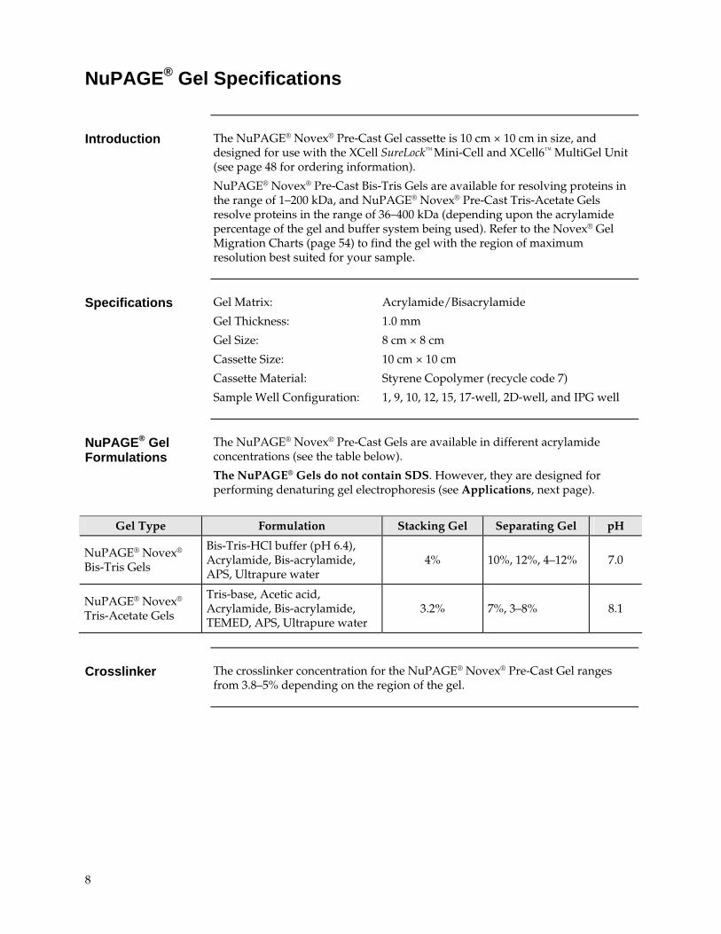
NuPAGE® Gel Specifications
Introduction The NuPAGE® Novex® Pre-Cast Gel cassette is 10 cm × 10 cm in size, and
designed for use with the XCell SureLock™ Mini-Cell and XCell6™ MultiGel Unit (see page 48 for ordering information).
NuPAGE® Novex® Pre-Cast Bis-Tris Gels are available for resolving proteins in the range of 1–200 kDa, and NuPAGE® Novex® Pre-Cast Tris-Acetate Gels resolve proteins in the range of 36–400 kDa (depending upon the acrylamide percentage of the gel and buffer system being used). Refer to the Novex® Gel Migration Charts (page 54) to find the gel with the region of maximum resolution best suited for your sample.
Specifications Gel Matrix: Acrylamide/Bisacrylamide
Gel Thickness: 1.0 mm
Gel Size: 8 cm × 8 cm
Cassette Size: 10 cm × 10 cm
Cassette Material: Styrene Copolymer (recycle code 7)
Sample Well Configuration: 1, 9, 10, 12, 15, 17-well, 2D-well, and IPG well
NuPAGE® Gel Formulations
The NuPAGE® Novex® Pre-Cast Gels are available in different acrylamide concentrations (see the table below).
The NuPAGE® Gels do not contain SDS. However, they are designed for performing denaturing gel electrophoresis (see Applications, next page).
Gel Type Formulation Stacking Gel Separating Gel pH
NuPAGE® Novex® Bis-Tris Gels
Bis-Tris-HCl buffer (pH 6.4), Acrylamide, Bis-acrylamide, APS, Ultrapure water
4% 10%, 12%, 4–12% 7.0
NuPAGE® Novex® Tris-Acetate Gels
Tris-base, Acetic acid, Acrylamide, Bis-acrylamide, TEMED, APS, Ultrapure water
3.2% 7%, 3–8% 8.1
Crosslinker The crosslinker concentration for the NuPAGE® Novex® Pre-Cast Gel ranges
from 3.8–5% depending on the region of the gel.
8
Gel Selection
Choosing a NuPAGE® Gel for Your Application
To obtain the best results, it is important to choose the correct gel percentage, buffer system, gel format, and thickness for your application. NuPAGE® Pre-Cast Gels are compatible with protein sequencing using Edman sequencing from the gel, or from PVDF membranes.
Review Applications (below), and Well Volume (page 10) to determine the type of gel that is best suited for your application.
Refer to the NuPAGE® Gel Migration Chart (page 54) to find the gel with the region of maximum resolution best suited for your sample. The leading protein molecules should migrate about 70% of the length of gel for best resolution.
Applications Separation of proteins over a wide range of molecular weights
Use NuPAGE® Bis-Tris Gels with NuPAGE® MOPS SDS Running Buffer to resolve proteins (14–200 kDa) under denaturing conditions.
Separation of low molecular weight proteins
Use NuPAGE® Bis-Tris Gels with NuPAGE® MES SDS Running Buffer Buffer to resolve small molecular weight proteins (2–200 kDa) under denaturing conditions.
Separation of high molecular weight proteins
Use NuPAGE® Tris-Acetate Gels with NuPAGE® Tris-Acetate SDS Running Buffer to resolve high molecular weight proteins (36–400 kDa) under denaturing conditions, or with Novex® Tris-Glycine Native Running Buffer to resolve high molecular weight proteins under non-denaturing (native) conditions.
Note: Do not use the NuPAGE® Bis-Tris Gels with NuPAGE® MOPS or MES Running Buffer without SDS for native gel electrophoresis. This buffer system may generate excessive heat resulting in poor band resolution. The protein of interest may not migrate very well in a neutral pH environment if it is not charged.
2D separation of proteins
The ZOOM® Gels are specifically designed for second dimension electrophoresis of 7.0 cm IPG strips.
9
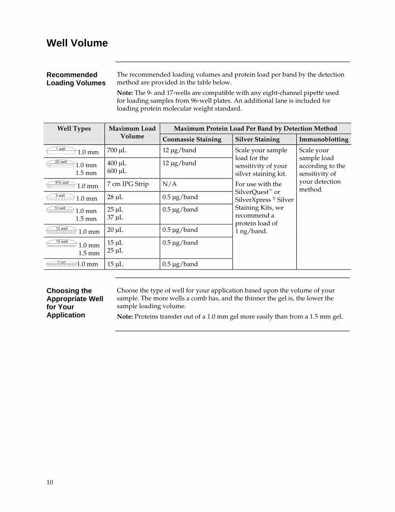
Well Volume
Recommended Loading Volumes
The recommended loading volumes and protein load per band by the detection method are provided in the table below.
Note: The 9- and 17-wells are compatible with any eight-channel pipette used for loading samples from 96-well plates. An additional lane is included for loading protein molecular weight standard.
Maximum Protein Load Per Band by Detection Method Well Types Maximum Load Volume Coomassie Staining Silver Staining Immunoblotting
������ 1.0 mm 700 μL 12 μg/band
������� 1.0 mm
1.5 mm
400 μL 600 μL
12 μg/band
������ 1.0 mm 7 cm IPG Strip N/A
������ 1.0 mm 28 μL 0.5 μg/band
� ����� 1.0 mm 1.5 mm
25 μL 37 μL
0.5 μg/band
������� 1.0 mm 20 μL 0.5 μg/band
������� 1.0 mm
1.5 mm
15 μL 25 μL
0.5 μg/band
������� 1.0 mm 15 μL 0.5 μg/band
Scale your sample load for the sensitivity of your silver staining kit.
For use with the SilverQuest™ or SilverXpress ® Silver Staining Kits, we recommend a protein load of 1 ng/band.
Scale your sample load according to the sensitivity of your detection method.
Choosing the Appropriate Well for Your Application
Choose the type of well for your application based upon the volume of your sample. The more wells a comb has, and the thinner the gel is, the lower the sample loading volume.
Note: Proteins transfer out of a 1.0 mm gel more easily than from a 1.5 mm gel.
10
Gel Staining
Staining NuPAGE® Gels
The NuPAGE® Novex® Pre-Cast Gels are compatible with most silver staining protocols. We recommend using the SilverQuest™ Silver Staining Kit or the SilverXpress® Silver Staining Kit (see pages 20–23) for silver staining of NuPAGE® Gels.
The NuPAGE® Novex® Pre-Cast Gels are compatible with any of the standard Coomassie staining procedures. The protocols that are accelerated by heat are preferable as the heat serves as a “fix” for proteins, especially smaller peptides. The SimplyBlue™ SafeStain and Novex® Colloidal Coomassie Blue Staining Kit (see pages 24–27) are recommended for staining NuPAGE® Gels.
The NuPAGE® Novex® Pre-Cast Gels are also compatible with copper or zinc staining, and fluorescent stains like the SYPRO® Ruby gel stain (see pages 28–30)
11
Methods
General Guidelines for Samples and Buffers
Introduction General information on the sample buffer and reducing agent is provided
below. For sample and buffer preparation protocols, see page 14. Instructions for preparing running buffers for denaturing and non-denaturing electrophoresis are provided on page 50–52.
Recommended Buffers
The recommended running buffer and sample buffer for each NuPAGE® Novex® Pre-Cast Gel is listed in the table below. Prepare your sample in the appropriate sample buffer so that the final concentration of the sample buffer is 1X. Running buffer must be diluted to 1X final concentration before use.
See page 48 for ordering information on pre-mixed buffers. See pages 50–52 for recipes if you are preparing your own buffers.
Gel Type Running Buffer Sample Buffer
NuPAGE® Novex® Bis-Tris Gel (SDS-PAGE)
NuPAGE® MES or MOPS SDS Running Buffer
NuPAGE® LDS Sample Buffer
NuPAGE® Novex® Tris-Acetate Gels (SDS-PAGE)
NuPAGE® Tris-Acetate Running Buffer
NuPAGE® LDS Sample Buffer
NuPAGE® Novex® Tris-Acetate Gels (Native-PAGE)
Novex® Tris-Glycine Native Running Buffer
Novex® Tris-Glycine Native Sample Buffer
NuPAGE® LDS Sample Buffer
Use the NuPAGE® LDS Sample Buffer for preparing samples when performing denaturing gel electrophoresis with NuPAGE® Gels. The slightly alkaline pH of the NuPAGE® LDS Sample Buffer (pH 8.4) provides the optimal conditions for reduction of protein disulfide bonds, and denaturation.
The NuPAGE® LDS Sample Buffer is a 4X concentrated solution containing twice as much dodecylsulfate as the 2X concentration of Novex® Tris-Glycine SDS or Tricine SDS Sample Buffer. The buffer also contains more glycerol, resulting in increased viscosity. Bring the NuPAGE® LDS Sample Buffer to room temperature (25°C) before use to make pipetting the buffer easier.
Tracking Dye The NuPAGE® LDS Sample Buffer uses Coomassie G250 and Phenol Red as
tracking dyes instead of bromophenol blue. Coomassie G250 gives a sharp dye front with both MES and MOPS SDS Running Buffers and migrates much closer to the moving ion front than bromophenol blue. Bromophenol blue runs more slowly than some peptides with the MES SDS Running Buffer. This ensures that small peptides do not run off the gel. The concentration of the tracking dye (Coomassie G250) is increased in the NuPAGE® LDS Sample Buffer to enhance viewing of the dye front.
Continued on next page
12
General Guidelines for Samples and Buffers, Continued
Reducing Agent Use the NuPAGE® Reducing Agent to prepare samples for reducing gel
electrophoresis. The NuPAGE® Reducing Agent (10X) contains 500 mM dithiothreitol (DTT) and is available in a ready-to-use, stabilized liquid form.
As an alternative, -mercaptoethanol can be used with NuPAGE® Gels at a final concentration of 2.5%. Add the reducing agent to the sample up to an hour before loading the gel. Avoid storing reduced samples for long periods, even if they are frozen. Reoxidation of samples occur during storage and produce inconsistent results.
NuPAGE® Antioxidant
Use the proprietary NuPAGE® Antioxidant in the running buffer of the Upper (cathode) Buffer Chamber when performing electrophoresis under reducing conditions to prevent sample reoxidation and maintain the proteins in a reduced state. DTT and -mercaptoethanol tend to remain at the top of the gel, and do not co-migrate with the sample in the neutral pH environment of NuPAGE® Gels. Disulfide bonds are less reactive at neutral pH and less likely to reoxidize than in higher pH systems, but some reoxidization may occur during electrophoresis in the absence of an antioxidant, and cause band diffusion.
The NuPAGE® Antioxidant migrates with the proteins during electrophoresis, and protects disulfide bonds and sensitive amino acids (e.g., methionine and tryptophan) from oxidizing. The NuPAGE® Antioxidant is NOT compatible with gel systems other than the NuPAGE® system because the antioxidant is not efficient at the higher pHs of other gel systems. For best results, use the NuPAGE® Antioxidant with reduced and alkylated samples.
��������
Do not use the NuPAGE® Antioxidant as a sample reducing agent. The antioxidant is not efficient in reducing disulfide bonds on its own, and using it to reduce samples results in substantial background smearing in the lane due to partially reduced bands.
NuPAGE® SDS Running Buffer
Three types of NuPAGE® Running Buffers are available for denaturing electrophoresis:
NuPAGE® MES SDS Running Buffer is used with NuPAGE® Novex® Bis-Tris Gels to resolve small molecular weight proteins
NuPAGE® MOPS SDS Running Buffer is used with NuPAGE® Novex® Bis-Tris Gels to resolve mid-size proteins
NuPAGE® Tris-Acetate SDS Running Buffer is used with NuPAGE® Novex®
Tris-Acetate Gels to resolve high molecular weight proteins
MES has a lower pKa than MOPS, making the NuPAGE® MES SDS Running Buffer faster than the NuPAGE® MOPS SDS Running Buffers. The difference in ion migration affects stacking and results in a difference in protein separation range between these buffers.
For native gel electrophoresis with NuPAGE® Novex® Tris-Acetate Gels, use the Novex® Tris-Glycine Native Running Buffer.
13
Preparing Buffers for Denaturing Electrophoresis
Materials Supplied by the User
The following reagents are needed to prepare samples for denaturing electrophoresis. Ordering information for pre-mixed buffers is on page 48. If you are preparing your own buffers, recipes are provided on page 50–52.
Protein sample
Deionized water
For Sample Preparation
NuPAGE® LDS Sample Buffer
NuPAGE® Reducing Agent
For Running Buffer Preparation
NuPAGE® MES SDS Running Buffer (for small proteins on NuPAGE® Bis-Tris Gels)
NuPAGE® MOPS SDS Running Buffer (for mid-sized proteins NuPAGE® Bis-Tris Gels)
NuPAGE® Tris-Acetate Running Buffer (for NuPAGE® Tris-Acetate Gels)
NuPAGE® Antioxidant
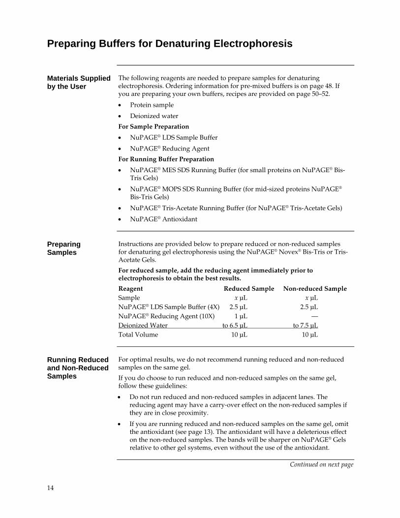
Preparing Samples
Instructions are provided below to prepare reduced or non-reduced samples for denaturing gel electrophoresis using the NuPAGE® Novex® Bis-Tris or Tris-Acetate Gels.
For reduced sample, add the reducing agent immediately prior to electrophoresis to obtain the best results.
Reagent Reduced Sample Non-reduced Sample Sample x μL x μL NuPAGE® LDS Sample Buffer (4X) 2.5 μL 2.5 μL NuPAGE® Reducing Agent (10X) 1 μL — Deionized Water to 6.5 μL to 7.5 μL Total Volume 10 μL 10 μL
Running Reduced and Non-Reduced Samples
For optimal results, we do not recommend running reduced and non-reduced samples on the same gel.
If you do choose to run reduced and non-reduced samples on the same gel, follow these guidelines:
Do not run reduced and non-reduced samples in adjacent lanes. The reducing agent may have a carry-over effect on the non-reduced samples if they are in close proximity.
If you are running reduced and non-reduced samples on the same gel, omit the antioxidant (see page 13). The antioxidant will have a deleterious effect on the non-reduced samples. The bands will be sharper on NuPAGE® Gels relative to other gel systems, even without the use of the antioxidant.
Continued on next page
14
Preparing Buffers for Denaturing Electrophoresis, Continued
Heating Samples Heat the sample for denaturing electrophoresis (reduced or non-reduced) at
70°C for 10 minutes for optimal results.
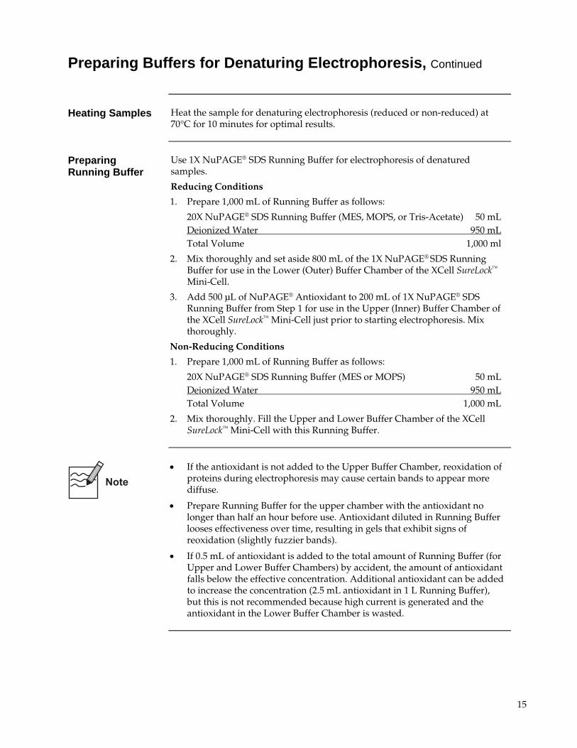
Preparing Running Buffer
Use 1X NuPAGE® SDS Running Buffer for electrophoresis of denatured samples.
Reducing Conditions
1. Prepare 1,000 mL of Running Buffer as follows:
20X NuPAGE® SDS Running Buffer (MES, MOPS, or Tris-Acetate) 50 mL Deionized Water 950 mL Total Volume 1,000 ml
2. Mix thoroughly and set aside 800 mL of the 1X NuPAGE® SDS Running Buffer for use in the Lower (Outer) Buffer Chamber of the XCell SureLock™ Mini-Cell.
3. Add 500 μL of NuPAGE® Antioxidant to 200 mL of 1X NuPAGE® SDS Running Buffer from Step 1 for use in the Upper (Inner) Buffer Chamber of the XCell SureLock™ Mini-Cell just prior to starting electrophoresis. Mix thoroughly.
Non-Reducing Conditions
1. Prepare 1,000 mL of Running Buffer as follows:
20X NuPAGE® SDS Running Buffer (MES or MOPS) 50 mL Deionized Water 950 mL Total Volume 1,000 mL
2. Mix thoroughly. Fill the Upper and Lower Buffer Chamber of the XCell SureLock™ Mini-Cell with this Running Buffer.
If the antioxidant is not added to the Upper Buffer Chamber, reoxidation of proteins during electrophoresis may cause certain bands to appear more diffuse.
Prepare Running Buffer for the upper chamber with the antioxidant no longer than half an hour before use. Antioxidant diluted in Running Buffer looses effectiveness over time, resulting in gels that exhibit signs of reoxidation (slightly fuzzier bands).
If 0.5 mL of antioxidant is added to the total amount of Running Buffer (for Upper and Lower Buffer Chambers) by accident, the amount of antioxidant falls below the effective concentration. Additional antioxidant can be added to increase the concentration (2.5 mL antioxidant in 1 L Running Buffer), but this is not recommended because high current is generated and the antioxidant in the Lower Buffer Chamber is wasted.
15
Preparing Buffers for Non-Denaturing Electrophoresis
Materials Supplied by the User
The following reagents are needed to prepare samples for non-denaturing electrophoresis with NuPAGE® Novex® Tris-Acetate Gels. Ordering information for pre-mixed buffers is on page 48. If you are preparing your own buffers, recipes are provided on page 51–52.
Protein sample
Deionized water
For Sample Preparation
Novex® Tris-Glycine Native Sample Buffer
For Running Buffer Preparation
Novex® Tris-Glycine Native Running Buffer
Preparing Samples for
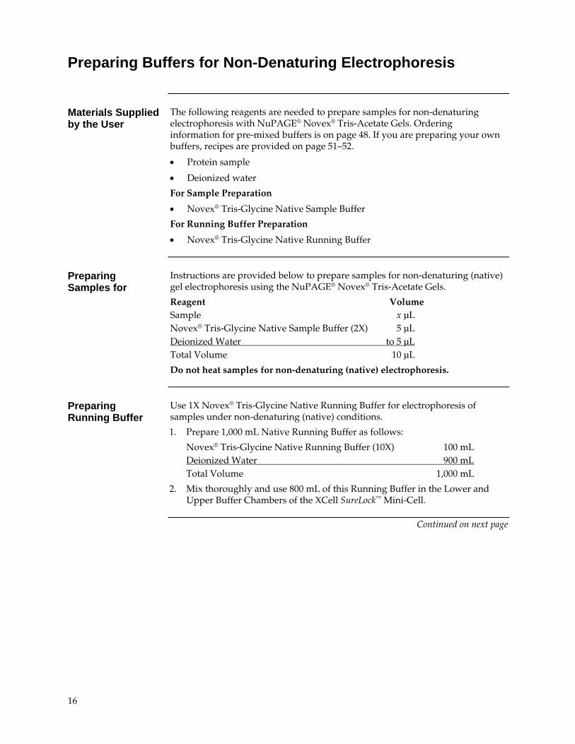
Instructions are provided below to prepare samples for non-denaturing (native) gel electrophoresis using the NuPAGE® Novex® Tris-Acetate Gels.
Reagent Volume Sample x μL Novex® Tris-Glycine Native Sample Buffer (2X) 5 μL Deionized Water to 5 μL Total Volume 10 μL
Do not heat samples for non-denaturing (native) electrophoresis.
Preparing Running Buffer
Use 1X Novex® Tris-Glycine Native Running Buffer for electrophoresis of samples under non-denaturing (native) conditions.
1. Prepare 1,000 mL Native Running Buffer as follows:
Novex® Tris-Glycine Native Running Buffer (10X) 100 mL Deionized Water 900 mL Total Volume 1,000 mL
2. Mix thoroughly and use 800 mL of this Running Buffer in the Lower and Upper Buffer Chambers of the XCell SureLock™ Mini-Cell.
Continued on next page
16
Electrophoresis of NuPAGE® Gels
Introduction Instructions are provided below for electrophoresis of NuPAGE® Gels using the
XCell SureLock™ Mini-Cell. For more information on the XCell SureLock™ Mini-Cell, refer to the manual (IM-9003). This manual is available on our website at www.invitrogen.com or contact Technical Support (see page 56).
If you are using any other electrophoresis mini-cell, follow the manufacturer’s recommendations.
��������
To ensure success with the NuPAGE® Electrophoresis System, remember the important points listed below:
Wear protective gloves and safety glasses when handling gels.
Under no circumstances should Tris-Glycine SDS buffers be used with NuPAGE® Gels for any denaturing gel electrophoresis (see page 46–47 for the outcome of your results using incorrect buffers).
Only use NuPAGE® SDS buffers (see page 12).
DO NOT BOIL samples. Heat samples at 70C for 10 minutes (see page 15).
Inner and Outer Buffer Chambers must be filled with the recommended amount of running buffer to prevent excessive heating (see below).
Procedure using XCell SureLock™ Mini-Cell
XCell SureLock™ Mini-Cell require 200 mL for the Upper Buffer Chamber and 600 mL for the Lower Buffer Chamber.
1. Remove the NuPAGE® Gel from the pouch.
2. Rinse the gel cassette with deionized water. Peel off the tape from the bottom of the cassette.
3. Gently pull the comb out of the cassette in one smooth motion.
4. Rinse the sample wells with 1X NuPAGE® SDS Running Buffer. Invert the gel and shake to remove the buffer. Repeat two more times.
5. Orient the two gels in the Mini-Cell such that the notched “well” side of the cassette faces inwards toward the Buffer Core. Seat the gels on the bottom of the Mini-Cell and lock into place with the Gel Tension Wedge. Refer to the XCell SureLock™ Mini-Cell manual (IM-9003) for detailed instructions.
Note: If you are running just one gel, use the plastic Buffer Dam in place of the second gel cassette to form the Upper Buffer Chamber.
6. Fill the Upper Buffer Chamber with a small amount of the Running Buffer to check for tightness of seal. If you detect a leak from Upper to the Lower Buffer Chamber, discard the buffer, reseal the chamber, and check the seal again.
7. Once the seal is tight, fill the Upper Buffer Chamber (Inner) with the appropriate 1X Running Buffer. The buffer level must exceed the level of the wells.
Continued on next page
17
Electrophoresis of NuPAGE® Gels, Continued
Procedure using XCell SureLock™ Mini-Cell, continued
Note: If you are running reduced samples, remember to fill the Upper Buffer Chamber with 200 mL of running buffer containing the NuPAGE® Antioxidant (see page 15).
8. Load an appropriate volume of sample at the desired protein concentration onto the gel (see page 10 for recommended loading volumes).
9. Load appropriate protein molecular weight markers (see page 48 for ordering information).
10. Fill the Lower (Outer) Buffer Chamber with 600 mL of the appropriate 1X Running Buffer.
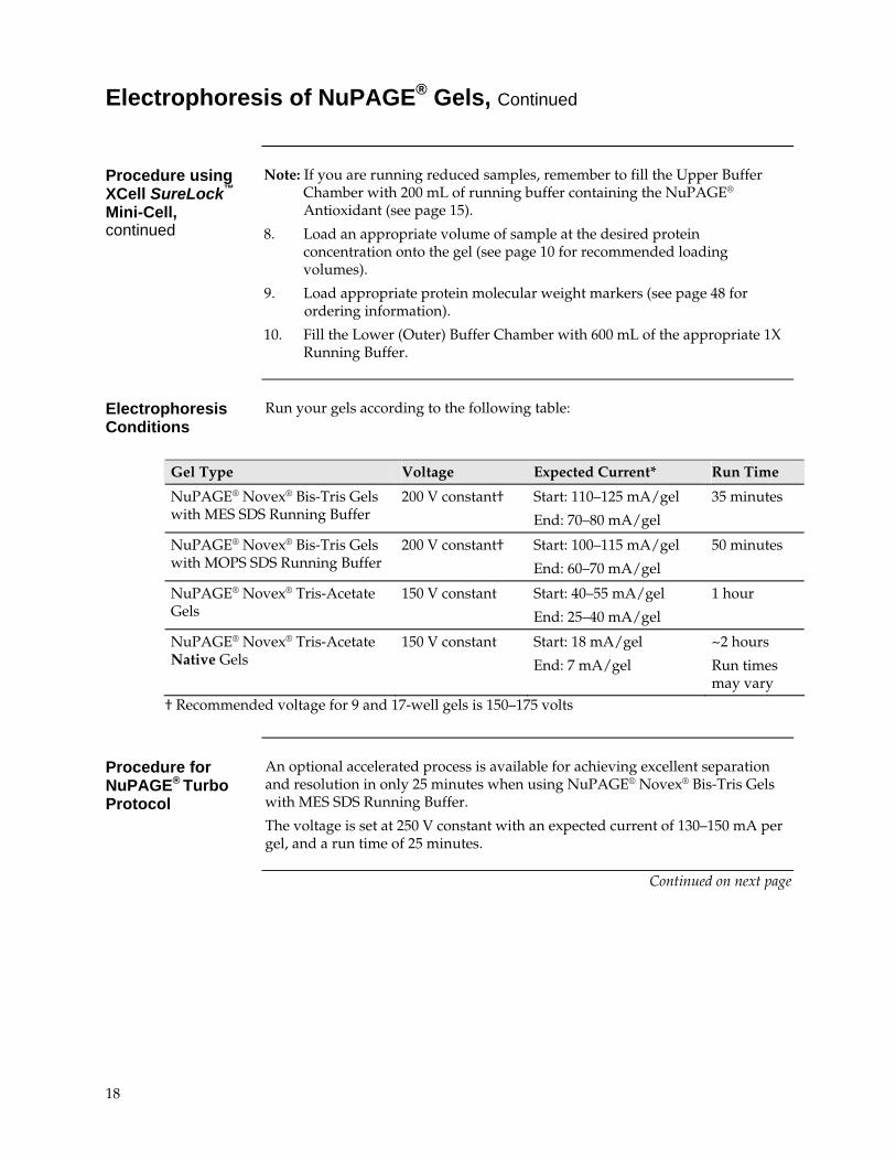
Electrophoresis Conditions
Run your gels according to the following table:
Gel Type Voltage Expected Current* Run Time
NuPAGE® Novex® Bis-Tris Gels with MES SDS Running Buffer
200 V constant† Start: 110–125 mA/gel
End: 70–80 mA/gel
35 minutes
NuPAGE® Novex® Bis-Tris Gels with MOPS SDS Running Buffer
200 V constant† Start: 100–115 mA/gel
End: 60–70 mA/gel
50 minutes
NuPAGE® Novex® Tris-Acetate Gels
150 V constant Start: 40–55 mA/gel
End: 25–40 mA/gel
1 hour
NuPAGE® Novex® Tris-Acetate Native Gels
150 V constant Start: 18 mA/gel
End: 7 mA/gel
~2 hours
Run times may vary
† Recommended voltage for 9 and 17-well gels is 150–175 volts
Procedure for NuPAGE® Turbo Protocol
An optional accelerated process is available for achieving excellent separation and resolution in only 25 minutes when using NuPAGE® Novex® Bis-Tris Gels with MES SDS Running Buffer.
The voltage is set at 250 V constant with an expected current of 130–150 mA per gel, and a run time of 25 minutes.
Continued on next page
18
Opening Novex® Pre-Cast Gel Cassettes
Removing the Gel after electrophoresis
1. After electrophoresis is complete, shut off the power, disconnect electrodes, and remove gel(s) from the XCell SureLock™ Mini-Cell.
2. Separate each of the three bonded sides of the cassette by inserting the Gel Knife into the gap between the two plastic plates that make up the cassette. The notched (“well”) side of the cassette should face up.
3. Push down gently on the knife handle to separate the plates. Repeat on each side of the cassette until the plates are completely separated.
Caution: Use caution while inserting the Gel Knife between the two plates to avoid excessive pressure on the gel.
4. Carefully remove and discard the top plate, allowing the gel to rest on the bottom (slotted) plate.
5. If blotting, proceed to page 34 without removing the gel from the bottom plate.
6. If staining, remove the gel from the plate by one of the methods:
Use the sharp edge of the Gel Knife to remove the gel foot from the bottom of the gel. Hold the Gel Knife at a 90° angle, perpendicular to the gel and the slotted half of the cassette. Push down on the knife, and then repeat the motion across the gel to cut off the entire foot. Hold the plate and gel over a container with the gel facing downward and use the knife to carefully loosen one lower corner of the gel and allow the gel to peel away from the plate.
Hold the plate and gel over a container with the gel facing downward. Gently push the Gel Knife through the slot in the cassette, until the gel peels away from the plate. Cut the gel foot off of the gel after fixing and staining, but before drying.
7. Fix and stain the gel as described on pages 20–27.
19
Silver Staining
Introduction Instructions are provided below for silver staining the NuPAGE® Gels using the
SilverQuest™ Silver Staining Kit and the SilverXpress® Silver Staining Kit (see page 49 for ordering information).
If you are using any other silver staining kit, follow the manufacturer’s recommendations.
The NuPAGE® system is more effective in reducing proteins and maintaining proteins in their reduced state. This may cause any minor contaminants present in the protein to be more visible under the sensitive silver staining techniques with the NuPAGE® system than in other systems.
Materials Supplied by the User
You will need following items for silver staining your gel (see pages 48–49 for ordering information on Invitrogen products):
Staining container
Rotary Shaker
Ultrapure water (>18 megohm/cm resistance recommended)
Teflon coated stir bars
Disposable 10 mL pipettes
Clean glass bottles for reagent preparation
Graduated glass cylinders
Protein molecular weight markers (Mark 12™ Unstained Standard, recommended)
For SilverQuest™ Silver Staining
Ethanol
Fixative (40% ethanol, 10% acetic acid) For SilverXpress® Silver Staining
Methanol
Acetic acid
����
�����
���
For optimal silver staining results, follow these guidelines:
Be sure to wear clean gloves that have been rinsed with deionized water while handling gels
Use clean containers and designate these containers for silver staining purposes only
Make sure the size of the container permits free movement of the gel during shaking and complete immersion in solution while staining
Do not touch the gel with bare hands or metal objects and do not put pressure on gels while handling or changing solutions
Use teflon coated stir bars and clean glass containers to prepare reagents
Avoid cross contamination of kit reagents
Use freshly made solutions
Continued on next page
20
Silver Staining, Continued
Preparing Solutions for SilverQuest™ Silver Staining
Use the reagents provided in the SilverQuest™ Silver Staining Kit to prepare the following solutions for staining:
Sensitizing solution
Ethanol 30 mL
Sensitizer 10 mL
Ultrapure water to 100 mL
Staining solution
Stainer 1 mL
Ultrapure water to 100 mL
Developing solution
Developer 10 mL
Developer enhancer 1 drop
Ultrapure water to 100 mL
Note: You may prepare all solutions immediately before starting the staining protocol or prepare them as you proceed to the next step.
SilverQuest™ Microwave Silver Staining Protocol
The microwave protocol for silver staining NuPAGE® Gels with SilverQuest™ Silver Staining Kit is provided below. For the Basic Protocol and more details on the staining procedure, refer to the SilverQuest™ Silver Staining Kit Manual (IM-6070). This manual is available on our website at www.invitrogen.com or contact Technical Support (see page 56).
Use 100 mL of each solution for each 1.0 mm thick, 8 cm × 8 cm NuPAGE® Gel.
Note: You may have to optimize the staining protocol, if the dimensions of your gel are not the same as mentioned above.
1. After electrophoresis, place the gel in a clean microwaveable staining tray of the appropriate size. Rinse the gel briefly with ultrapure water.
2. Place the gel in 100 mL of fixative and microwave at high power (700 watts) for 30 seconds. Remove the gel from the microwave and gently agitate it for 5 minutes at room temperature. Decant the fixative.
3. Wash the gel with 100 mL of 30% ethanol in a microwave at high power for 30 seconds. Remove the gel from the microwave and gently agitate it for 5 minutes at room temperature on a rotary shaker. Decant the ethanol.
4. Add 100 mL of Sensitizing solution to the washed gel. Microwave at high power for 30 seconds. Remove the gel from the microwave and place it on a rotary shaker for 2 minutes at room temperature. Decant the Sensitizing solution.
Continued on next page
21
Silver Staining, Continued
SilverQuest™ Microwave Silver Staining Protocol, continued
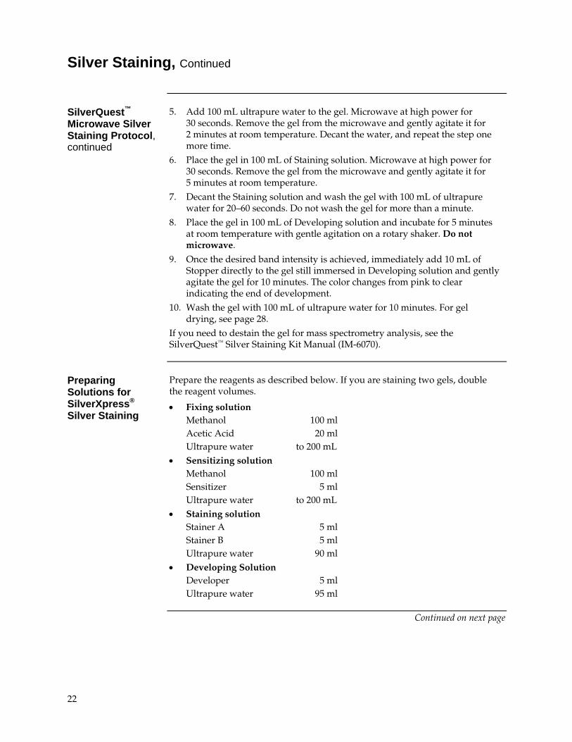
5. Add 100 mL ultrapure water to the gel. Microwave at high power for 30 seconds. Remove the gel from the microwave and gently agitate it for 2 minutes at room temperature. Decant the water, and repeat the step one more time.
6. Place the gel in 100 mL of Staining solution. Microwave at high power for 30 seconds. Remove the gel from the microwave and gently agitate it for 5 minutes at room temperature.
7. Decant the Staining solution and wash the gel with 100 mL of ultrapure water for 20–60 seconds. Do not wash the gel for more than a minute.
8. Place the gel in 100 mL of Developing solution and incubate for 5 minutes at room temperature with gentle agitation on a rotary shaker. Do not microwave.
9. Once the desired band intensity is achieved, immediately add 10 mL of Stopper directly to the gel still immersed in Developing solution and gently agitate the gel for 10 minutes. The color changes from pink to clear indicating the end of development.
10. Wash the gel with 100 mL of ultrapure water for 10 minutes. For gel drying, see page 28.
If you need to destain the gel for mass spectrometry analysis, see the SilverQuest™ Silver Staining Kit Manual (IM-6070).
Preparing Solutions for SilverXpress®
Silver Staining
Prepare the reagents as described below. If you are staining two gels, double the reagent volumes.
Fixing solution Methanol 100 ml Acetic Acid 20 ml Ultrapure water to 200 mL
Sensitizing solution Methanol 100 ml Sensitizer 5 ml Ultrapure water to 200 mL
Staining solution Stainer A 5 ml Stainer B 5 ml Ultrapure water 90 ml
Developing Solution Developer 5 ml Ultrapure water 95 ml
Continued on next page
22
Silver Staining, Continued
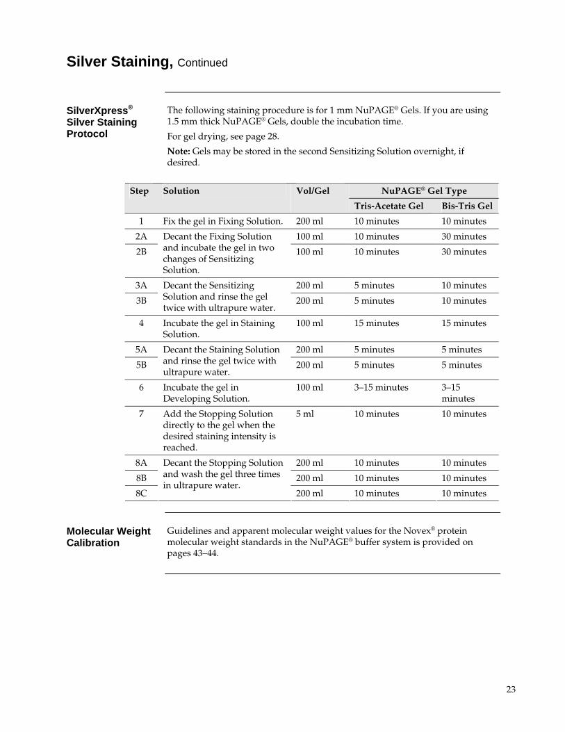
SilverXpress® Silver Staining Protocol
The following staining procedure is for 1 mm NuPAGE® Gels. If you are using 1.5 mm thick NuPAGE® Gels, double the incubation time.
For gel drying, see page 28.
Note: Gels may be stored in the second Sensitizing Solution overnight, if desired.
NuPAGE® Gel Type Step Solution Vol/Gel
Tris-Acetate Gel Bis-Tris Gel
1 Fix the gel in Fixing Solution. 200 ml 10 minutes 10 minutes
2A 100 ml 10 minutes 30 minutes
2B
Decant the Fixing Solution and incubate the gel in two changes of Sensitizing Solution.
100 ml 10 minutes 30 minutes
3A 200 ml 5 minutes 10 minutes
3B
Decant the Sensitizing Solution and rinse the gel twice with ultrapure water.
200 ml 5 minutes 10 minutes
4 Incubate the gel in Staining Solution.
100 ml 15 minutes 15 minutes
5A 200 ml 5 minutes 5 minutes
5B
Decant the Staining Solution and rinse the gel twice with ultrapure water.
200 ml 5 minutes 5 minutes
6 Incubate the gel in Developing Solution.
100 ml 3–15 minutes 3–15 minutes
7 Add the Stopping Solution directly to the gel when the desired staining intensity is reached.
5 ml 10 minutes 10 minutes
8A 200 ml 10 minutes 10 minutes
8B 200 ml 10 minutes 10 minutes
8C
Decant the Stopping Solution and wash the gel three times in ultrapure water.
200 ml 10 minutes 10 minutes
Molecular Weight Calibration
Guidelines and apparent molecular weight values for the Novex® protein molecular weight standards in the NuPAGE® buffer system is provided on pages 43–44.
23
Coomassie Staining
Introduction Instructions are provided below for Coomassie staining of NuPAGE® Gels
using the SimplyBlue™ SafeStain, Colloidal Blue Staining Kit, and Coomassie R-250.
If you are using any other Coomassie staining kit, follow the manufacturer’s recommendations.
If you are staining low molecular weight peptides (<2.5 kDa), we recommend fixing the gel in 5% glutaraldehyde and 50% methanol for one hour and then follow the instructions in the Colloidal Blue Staining Kit Manual (IM-6025) for small peptides.
Materials Supplied by the User
You will need the following items for staining your gel (see page 49 for ordering information on Invitrogen products):
SimplyBlue™ SafeStain
Colloidal Blue Staining Kit
Staining container
Ultrapure water or deionized water
Orbital Shaker
Protein molecular weight standards (see page 45 for ordering information)
Microwave oven and 20% NaCl (if using SimplyBlue™ SafeStain microwave protocol, see page 25)
Methanol and acetic acid (if using Colloidal Blue Staining Kit, see page 26)
For Coomassie R-250 staining
0.1% Coomassie R-250 in 40% ethanol and 10% acetic acid
Destaining Solution (10% ethanol and 7.5% acetic acid)
Microwave oven (if using Coomassie R-250 microwave protocol, see page 27)
Continued on next page
24
Coomassie Staining, Continued
SimplyBlue™ SafeStain Microwave Protocol
The microwave protocol for staining NuPAGE® Gels with SimplyBlue™ SafeStain is provided below. For the Basic Protocol and more details on the staining procedure, refer to the SimplyBlue™ SafeStain Manual (IM-6050). This manual is available on our website at www.invitrogen.com or contact Technical Support (see page 56).
The procedure is written for 1.0 mm thick mini-gels.
After electrophoresis, follow the instructions below:
Caution: Use caution while using the stain in a microwave oven. Do not overheat the staining solutions.
1. Place the gel in a loosely covered container containing 100 mL of ultrapure water and microwave on High (950 to 1100 watts) for 1 minute until the solution is close to boiling.
2. Gently shake the gel on an orbital shaker or rocker for 1 minute. Shake the gel for 2 minutes for 1.5 mm thick gels. Discard the water.
3. Repeat Steps 1 and 2 two more times.
4. Add 20 mL SimplyBlue™ SafeStain the mini-gel (the gel should be completely covered) and microwave on High for 45 seconds to 1 minute until the solution almost boils. For 1.5 mm thick gels, use 30 mL of stain and microwave for 1.5 minutes.
5. Shake the gel on an orbital shaker or rocker for 5 minutes. Shake the gel for 10 minutes for 1.5 mm thick gels. The detection limit after staining is 20 ng BSA.
6. Wash the gel in 100 mL of ultrapure water for 10 minutes on a shaker. The detection limit after washing is 10 ng BSA.
7. Add 20 mL of 20% NaCl for at least 5 minutes. The detection limit after the salt wash is 5 ng BSA. Gels can be kept for several weeks in the salt solution.
8. For gel-drying, see page 28.
Continued on next page
25
Coomassie Staining, Continued
Preparing Solutions for Colloidal Blue Staining
Prepare the reagents as described below. If you are staining two gels, double the reagent volumes. Note: Be sure to shake Stainer B prior to making the solution.
Fixing Solution (Bis-Tris Gels) Methanol 100 mL Acetic Acid 20 mL Ultrapure water to 200 mL
Staining Solution Tris-Acetate Gel Bis-Tris Gel Deionized water 55 mL 55 mL Methanol 20 mL 20 mL Stainer B 5 mL — Stainer A 20 mL 20 mL
Colloidal Blue Staining Kit Protocol
A brief staining protocol for staining NuPAGE® Gels with the Colloidal Blue Staining Kit is provided below. For more details on the staining procedure, refer to the Manual (IM-6025). This manual is available on our website at www.invitrogen.com or contact Technical Support (see page 56).
Colloidal Blue Staining Kit Protocol for NuPAGE® Novex® Tris-Acetate Gels
1. Incubate the gel in Staining Solution for a minimum of 3 hours and a maximum of 12 hours at room temperature with gentle shaking.
2. Decant the Staining Solution and add at least 200 mL of deionized water per gel to the staining container. Gently shake the gel in water for at least 7 hours. The gel background should be clear after 7 hours in water.
3. For gel-drying, see page 28.
Colloidal Blue Staining Kit Protocol for NuPAGE® Novex® Bis-Tris Gels
Note: If you are staining low molecular weight peptides (< 2.5 kDa), we recommend fixing the gel in 5% glutaraldehyde and 50% methanol for one hour and then follow the instructions in the Colloidal Blue Staining Kit Manual (IM-6025) for small peptides.
1. Incubate the gel in Fixing Solution for 10 minutes at room temperature with gentle shaking.
2. Incubate the gel in this Staining Solution (without Stainer B) for 10 minutes at room temperature with gentle shaking.
3. Add 5 mL Stainer B per gel to the Staining Solution from previous step. Continue staining for a minimum of 3 hours and a maximum of 16 hours.
4. Decant the Staining Solution and add 200 mL of deionized water per gel to the staining container. Gently shake the gel in water for at least 7 hours. The gel background should be clear after 7 hours in water.
5. For gelfdrying, see page 28.
Note: NuPAGE® Gels can be left in deionized water for up to 3 days without significant change in band intensity and background clarity. For long-term storage (over 3 days), keep the gel in a 20% ammonium sulfate solution at 4°C.
26
Coomassie Staining, Continued
Coomassie R-250 Microwave Staining Protocol
The Coomassie staining protocol described below is recommended for staining NuPAGE® Gels. You may use any Coomassie staining protocol of choice.
1. Prepare the staining solution containing 0.1% Coomassie R-250 in 40% ethanol, 10% acetic acid.
2. After electrophoresis, incubate 1 or 2 gels in a staining container containing 100 mL of staining solution prepared in Step 1.
3. Loosely cover the staining container and heat in a microwave oven at full power for 1 minute. To prevent hazardous, flammable vapors from forming, do not allow the solution to boil.
4. Remove the staining container from the microwave oven and gently shake the gel for 15 minutes at room temperature on an orbital shaker.
5. Decant the stain and rinse the gel once with deionized water.
6. Prepare a destain solution containing 10% ethanol and 7.5% acetic acid.
7. Place one or two stained gels in a staining container containing 100 mL of destain solution prepared in Step 6.
8. Loosely cover the staining container and heat in a microwave oven at full power for 1 minute.
9. Gently shake the gel at room temperature on an orbital shaker until the desired background is achieved.
Note: The NuPAGE® Gels destain faster than other Novex® Gels. To prevent over destaining of NuPAGE® Gels if destaining overnight, dilute the destain solution by adding 100 mL of deionized water to 100 mL of the destain solution in the staining container.
10. For-gel-drying, see page 28.
Molecular Weight Calibration
Guidelines and apparent molecular weight values for the Novex® protein molecular weight standards in the NuPAGE® buffer system is provided on pages 43–44.
27
SYPRO® Ruby Staining
Introduction Instructions are provided below for a basic and rapid protocol for Novex® Pre-
Cast Gels (NuPAGE® Novex® Bis-Tris and Tris-acetate gels) for the detection of proteins, including glycoproteins and phosphoproteins.
Advantages of SYPRO® Ruby Staining
SYPRO® Ruby provides the following advantages:
Linear quantitation range of over three orders of magnitude
Compatible with subsequent analysis of proteins by Edmanbased sequencing or mass spectrometry in 1D or 2D format
Compatible with nondenaturing gels and IEF gels (basic protocol)
Molecular Weight Calibration
Guidelines and apparent molecular weight values for Novex® protein molecular weight standards are provided on page 43.
Materials Supplied by the User
Staining containers, 1 per gel (see below for details)
Reagent-grade methanol
Reagent-grade glacial acetic acid
Trichloroacetic acid (for IEF gels only)
Ultrapure water (18 megohm-cm recommended)
Rotary shaker
Powder-free latex or vinyl gloves
Microwave oven (700–1200 W) (optional)
Water bath set at 80°C (optional)
����
�����
���
General considerations for the protocol include the following:
Perform all fixation, staining, and washing steps with continuous, gentle agitation (e.g., on an orbital shaker at 50 rpm)
We recommend polypropylene or polycarbonate containers for staining. Glass dishes are not recommended. Staining containers should be meticulously clean to minimize contamination and other artifacts
For convenience, gels may be left in fix solution overnight or longer
For convenience, gels may be left in SYPRO® Ruby stain indefinitely without overstaining, although speckling artifacts tend to increase over time
As with any fluorescent stain, cover the gel container during staining and subsequent wash steps to exclude light
Continued on next page
28
SYPRO® Ruby Staining, Continued
Preparing Solutions for SYPRO® Ruby Staining
Prepare the reagents as described below. If you are staining two gels, double the reagent volumes. Increase volumes 1.5-fold for 1.5mm thick gels.
Fix Solution
Methanol 100 mL Glacial Acetic Acid 14 mL Ultrapure water to 200 mL
Fix Solution for IEF Gels
Methanol 40 mL Trichloroacetic Acid 10 g Ultrapure water to 100 mL
Wash Solution
Methanol 10 mL Glacial Acetic Acid 7 mL Ultrapure water to 100 mL
SYPRO® Ruby Basic Protocol
The basic protocol results in the maximum signal strength and widest linear dynamic range for staining of denaturing gels, nondenaturing gels, and IEF gels. Sensitivity is in the 1 ng range for most proteins.
1. After electrophoresis, place the gel into a clean container with 100 mL of Fix Solution and agitate on an orbital shaker for 30 minutes. Pour off the used fix solution and repeat once more with fresh Fix Solution.
Note: For IEF Gels, place the gel into a clean container with 100 mL of IEF Fix Solution and agitate on an orbital shaker for 3 hours. After fixing, perform 3 washes in ultrapure water for 10 minutes each, before proceeding to the staining step.
2. Pour off the used fix solution.
3. Add 60 mL of SYPRO® Ruby gel stain to the tray containing the gel. Agitate on an orbital shaker overnight.
4. Transfer the gel to a clean container and wash in 100 mL of Wash Solution for 30 minutes. The transfer step helps minimize background staining irregularities and stain speckles on the gel.
5. Rinse the gel in ultrapure water for 5 minutes. Repeat the rinse a minimum of one more time to prevent possible corrosive damage to your imager.
Note: If you are staining two gels, double the reagent volumes. Increase volumes 1.5-fold for 1.5mm thick gels.
Visualization of SYPRO® Ruby Stained Gels
Proteins stained with SYPRO® Ruby protein gel stain are readily visualized using a UV or blue-light source. The use of a photographic camera or CCD camera and the appropriate filters is essential to obtain the greatest sensitivity.
Continued on next page
29
SYPRO® Ruby Staining, Continued
SYPRO® Ruby Rapid Protocol
The rapid protocol is optimized for Invitrogen NuPAGE® gels, and can be completed in 90 minutes. While the maximum fluorescence signal strength is lower than for the basic protocol, the results show excellent linearity and low background, with a lower limit of detection of 0.25 to 1 ng for most proteins.
1. After electrophoresis, place the gel into a clean a microwavable container with 100 mL of Fix Solution and agitate on an orbital shaker for 15 minutes. Pour off the used fix solution and repeat once more with fresh Fix Solution.
2. Pour off the used fix solution.
3. Add 60 mL of SYPRO® Ruby gel stain to the tray containing the gel.
4. Microwave 30 seconds, agitate 30 seconds to distribute heat evenly, then microwave another 30 seconds to reach 80–85°C, and agitate on an orbital shaker for 5 minutes.
5. Reheat the gel by microwaving a third time for 30 seconds and then agitate on an orbital shaker for 23 minutes for a total stain time of 30 minutes.
Note: An acceptable alternative to the microwave procedure is to incubate the gel at 80°C in a shaking water bath for a total of 30 minutes.
6. Transfer the gel to a clean container and wash in 100 mL of Wash Solution for 30 minutes. The transfer step is necessary to avoid heating the destain solution, which may reduce stain sensitivity, and also helps minimize background staining irregularities and stain speckles on the gel.
7. Rinse the gel in ultrapure water for 5 minutes. Repeat the rinse a minimum of one more time to prevent possible corrosive damage to your imager.
Note: If you are staining two gels, double the reagent volumes. Increase volumes 1.5-fold for 1.5mm thick gels.
Heat the gel in the microwave on full power in increments of 30–45 seconds, until the stain reaches 80–85°C.
Do not heat the fixative solution or other methanolic solutions in the microwave.
Although SYPRO® Ruby stain solution is not flammable, use caution when microwaving SYPRO® Ruby stain as the solution becomes very hot.
Using SYPRO®
Ruby Stain As a Post-Stain
SYPRO® Ruby stain can be used to post-stain gels stained with other gel stains such as Pro-Q® Diamond phosphoprotein gel stain, Pro-Q®Emerald 300 glycoprotein gel stain, Pro-Q® Sapphire or InVision™ oligohistidine-tag gel stains, Pro-Q® Amber transmembrane protein gel stain, or silver staining (such as Invitrogen SilverQuest™ or SilverXpress® stain).
Always use SYPRO® Ruby stain last, as the SYPRO® Ruby signal can dominate the signal from other stains. SYPRO® Ruby stain does not work well as a post-stain for colorimetric stains such as Coomassie and silver stains.
30
Gel Drying
Introduction Dry gels by passive evaporation (air-drying) or vacuum drying. Vacuum
drying is faster than passive air-drying methods but often results in cracked gels due to the speed of dehydration. We recommend drying Novex® Pre-Cast gels using passive air-drying methods such as DryEase® Mini-Gel Drying System (see below). For applications that require vacuum drying, follow the recommendations on page 33 to minimize cracking of the gels.
Do not leave Coomassie stained gels in Gel-Dry™ solution (or any equilibration solution containing >20% alcohol) for more than 5 minutes. Gels left in this solution for longer than 5 minutes lose band intensity and result in decreased sensitivity.
Materials Supplied by the User
You will need the following items for drying your gel (see page 48 for ordering information on Invitrogen products):
DryEase® Mini-Gel Drying System
Gel-Dry™ Drying Solution (or prepare your own gel drying solution containing 30% methanol and 5% glycerol)
StainEase® Gel Staining Tray
DryEase® Mini-Gel Drying System
A brief gel drying protocol using the DryEase® Mini-Gel Drying System is provided below. For more details on this system, refer to the DryEase® Mini-Gel Drying System manual (IM-2380). This manual is available for download from our website at www.invitrogen.com or contact Technical Support (see page 56).
Wear gloves while handling gels and gel drying solution.
1. After all staining and destaining steps are complete, wash the destained gel(s) three times for two minutes each time in deionized water (50 mL per mini-gel) on a rotary shaker.
2. Decant the water and add fresh Gel-Dry™ Drying Solution (35 mL per mini-gel).
3. Equilibrate the gel in the Gel-Dry™ Drying Solution by shaking the gel for 15–20 minutes in the StainEase® Gel Staining Tray or in a round container.
Note: Do not equilibrate Coomassie stained gels in the Gel-Dry™ Drying Solution for more than 5 minutes to avoid losing band intensity.
4. Cut any rough edges off the gel (including the wells and the gel foot) using the Gel Knife or a razor blade.
5. Remove 2 sheets of cellophane per gel from the package.
6. Immerse one sheet of cellophane in the Gel-Dry™ Drying Solution. Allow 15–20 seconds for complete wetting before adding additional sheets. Do not soak the cellophane sheets for more than 2 minutes.
Continued on next page
31
Gel Drying, Continued
DryEase® Mini-Gel Drying System, continued
7. Place one side of the DryEase® Gel Drying Frame with the corner pin facing up, on the DryEase® Gel Drying Base.
8. Center a piece of pre-wetted cellophane from Step 5 over the base/frame combination, so the cellophane lays over the inner edge of the frame.
9. Lay the gel on the center of the cellophane sheet making sure no bubbles are trapped between the gel and the cellophane. Add some Gel-Dry™ Drying Solution to the surface of the cellophane, if necessary.
10. Carefully lay the second sheet of cellophane over the gel so that no bubbles are trapped between the cellophane and the gel. Add some Gel-Dry™ Drying Solution if necessary. Gently smooth out any wrinkles in the assembly with a gloved hand.
11. Align the remaining frame so that its corner pins fit into the appropriate holes on the bottom frame. Push the plastic clamps onto the four edges of the frames.
12. Lift the frame assembly from the DryEase® Gel Drying Base and pour off the excess solution from the base.
13. Place the gel dryer assembly upright on a benchtop. Be careful to avoid drafts as they can cause an uneven rate of dying which leads to cracking. Drying takes between 12–36 hours depending on humidity and gel thickness.
14. When the cellophane is dry to touch, remove the gel/cellophane sandwich from the drying frame. Trim off the excess cellophane.
15. Press the dried gel(s) between the pages of a notebook under light pressure for approximately 2 days so they remain flat for scanning, photography, display, and overhead projection.
Continued on next page
32
Gel Drying, Continued
Vacuum Drying General guidelines are provided below to minimize cracking during vacuum
drying of gels. For detailed instructions, follow the manufacturer’s recommendations.
Handle Gels with Care:
Remove the gel from the cassette without breaking or tearing the edges. Small nicks or tears can act as a starting point for cracking. Remove the gel wells and foot off the bottom of the gel with a Gel Knife or a razor blade as described on page 19. Use the StainEase Staining Tray for staining and destaining gels. This tray is designed to facilitate the solution changing process without handling of gels.
Use a Gel Drying Solution:
We recommend equilibrating the gel in a gel drying solution such as Gel-Dry™ Gel Drying Solution for 10–30 minutes at room temperature with gentle shaking on an orbital shaker before drying the gel. Gel-Dry™ Gel Drying Solution contains a proprietary non-glycerol component to effectively regulate the rate of drying and prevent cracking. The gel drying solution does not interfere with autoradiography.
To prepare your own gel drying solution, prepare a solution containing 30% methanol and 5% glycerol.
Note: Do not incubate Coomassie stained gels in gel drying solution for more than 5 minutes as the bands may fade.
Remove Air Bubbles:
Remove any air bubbles that may be trapped between the paper, gel, and plastic wrap by rolling a small glass pipette over the gel. Use additional gel drying solution to remove any air bubbles.
Use Proper Gel Dryer Set-up:
Place gel on the gel dryer with the plastic wrap facing up. Make sure the vacuum pump is in working condition, and properly set up to form a tight seal when on. Use drying conditions for polyacrylamide gels, with the temperature increasing to a set value and holding for the duration of the drying cycle. We recommend drying mini-gels at 80C for 2 hours.
Ensure Gel is Completely Dry:
The gel will crack if the vacuum seal of the heated gel dryer is broken prior to complete drying of the gel. To ensure the gel is completely dried before releasing the vacuum seal, follow these tips :
Check the temperature of the gel
The temperature of the dried gel should be the same as the temperature of the surrounding gel drying surface. If the temperature of the dried gel is cooler, then the gel is not completely dried.
Check for moisture in the tubing connecting the gel dryer to the vacuum pump
The gel is not completely dried if there is residual moisture in the tubing and additional drying time is required.
33
Western Blotting
Introduction After performing electrophoresis, proteins can be transferred to membranes for
subsequent analysis. Methods of transfer include wet, semi-wet, semi-dry, and dry blotting.
Semi-dry blotting can be performed with the Novex® Semi-Dry Blotter or other semi-dry blotter. For details on performing semi-dry blotting, see page 39 or refer to the manual for the Novex® Semi-Dry Blotter (25-0911).
Dry blotting is performed with the iBlot® Gel Transfer Device. Refer to the manual for the iBlot® Dry Blotting System (25-0949) for details.
Instructions are provided below for semi-wet blotting of NuPAGE® Gels using the XCell II™ Blot Module. For more information on the XCell II™ Blot Module, refer to the manual (IM-9051) available at www.invitrogen.com or contact Technical Support (see page 56).
NuPAGE® Antioxidant
The NuPAGE® Antioxidant is added to the transfer buffer when blotting reduced proteins to prevent reoxidation and maintain proteins in a reduced state (see page 13).
The anode electrochemistry is the major cause of reoxidation during blotting, though proteins are oxidized at a slower rate in the neutral pH environment of the NuPAGE® blotting system compared to higher pH blotting systems.
NuPAGE® Transfer Buffer
We recommend using the NuPAGE® Transfer Buffer for western transfer of NuPAGE® Gels to maintain the neutral pH environment established during NuPAGE® electrophoresis.
The NuPAGE® Transfer Buffer protects against modification of amino acid side chains and is compatible with N-terminal protein sequencing using Edman degradation.
Materials Supplied by the User
In addition to the XCell II™ Blot Module, the following reagents are needed for blotting your gel (see page 48–49 for ordering information on Invitrogen products): Blotting membranes
Filter paper
Methanol (if using PVDF membranes)
XCell II™ Blot module
NuPAGE® Transfer Buffer
NuPAGE® Antioxidant for reduced samples
MagicMark™ Western Protein Standard
Deionized water
Continued on next page
34
Western Blotting, Continued
Preparing NuPAGE® Transfer Buffer
For blotting NuPAGE® Gels, use 1X NuPAGE® Transfer Buffer. If you are preparing your own transfer buffer see page 53 for a recipe.
Prepare 1,000 mL of Transfer Buffer (20X) as follows: Reduced Samples Non-Reduced Samples NuPAGE® Transfer Buffer (20X) 50 mL 50 mL NuPAGE® Antioxidant 1 mL — Methanol 100 mL * 100 mL * Deionized Water 849 mL 850 mL Total Volume 1,000 mL 1,000 mL
*NuPAGE® Transfer Buffer with 10% methanol provides optimal transfer of a single gel in the blot module. If you are transferring two gels in the blot module, increase the methanol content to 20% to ensure efficient transfer of both gels.
Preparing Blotting Pads
Use about 700 mL of 1X NuPAGE® Transfer Buffer to soak the pads until they are saturated. Remove the air bubbles by squeezing the pads while they are submerged in buffer. Removing the air bubbles is essential as they can interfere with the transfer of biomolecules if not removed.
Preparing Transfer Membrane and Filter Paper
Cut the transfer membrane and filter paper to the dimensions of the gel, or use Novex® pre-cut membrane/filter paper sandwiches (see page 48 for ordering information.
PVDF membrane—Pre-wet PVDF membrane for 30 seconds in methanol, ethanol (95%), or isopropanol. Briefly rinse in deionized water, then place in a shallow dish with 50 mL of 1X NuPAGE® Transfer Buffer for several minutes.
Nitrocellulose—Place the membrane directly into a shallow dish containing 50 mL of 1X NuPAGE® Transfer Buffer for several minutes.
Filter paper—Soak the filter paper briefly in 1X NuPAGE® Transfer Buffer immediately prior to use.
Gel—Use the gel immediately following the run. Do not soak the gel in transfer buffer.
Continued on next page
35
Western Blotting, Continued
Western Transfer Using the XCell II™ Blot Module
Wear gloves while performing the blotting procedure to prevent contamination of gels and membranes, and exposure to irritants commonly used in electrotransfer.
Transferring One Gel
1. After opening the gel cassette as described on page 19 remove wells with the Gel Knife.
2. Place a piece of pre-soaked filter paper on top of the gel, with the edge above the slot in the bottom of the cassette (leaving the foot of the gel uncovered). Keep the filter paper saturated with the transfer buffer and remove all trapped air bubbles by gently rolling over the surface using a glass pipette as a roller.
3. Turn the plate over so the gel and filter paper are facing downwards over a gloved hand or clean flat surface.
4. Use the Gel Knife to push the foot out of the slot in the plate, and separate the gel from the plate.
5. When the gel is on a flat surface, cut the foot off the gel with the Gel Knife.
6. Wet the surface of the gel with transfer buffer and position the pre-soaked transfer membrane on the gel, ensuring all air bubbles have been removed.
7. Place another pre-soaked filter paper on top of the membrane. Remove any trapped air bubbles.
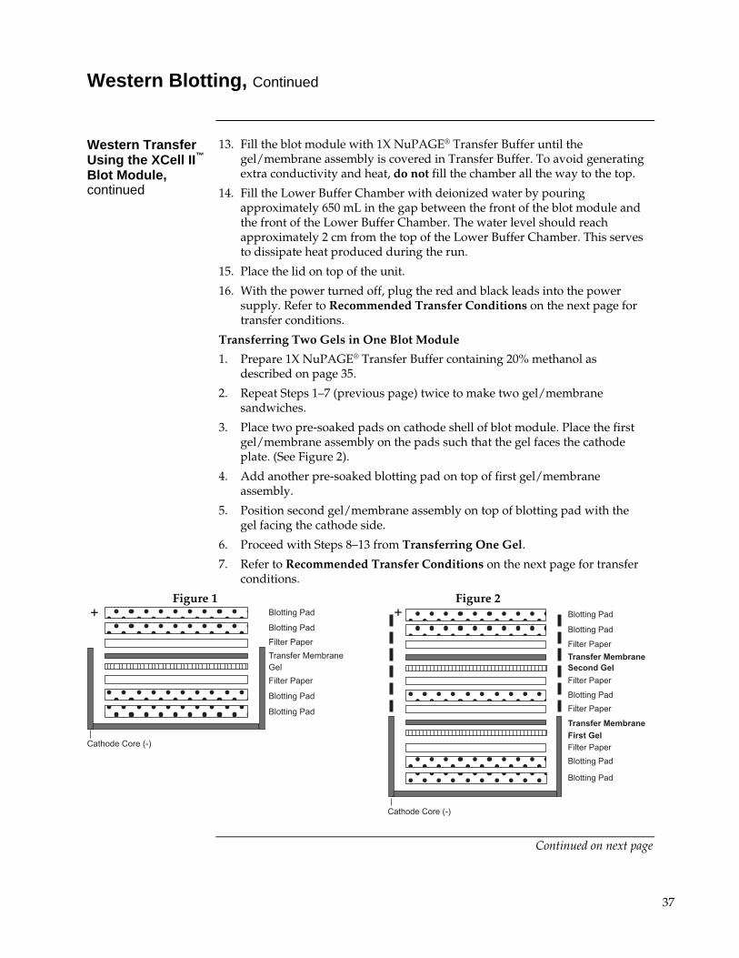
8. Place two soaked blotting pads into the cathode (–) core of the blot module. The cathode core is the deeper of the two cores and the corresponding electrode plate is a darker shade of gray. Carefully pick up the gel/membrane assembly and place on blotting pad such that the gel is closest to the surface of the cathode core (see Figure 1, next page).
9. Add enough pre-soaked blotting pads to raise the assembly 0.5 cm over the edge of cathode core. Place the anode (+) core on top of the pads. The gel/membrane assembly should be held securely between the two halves of the blot module ensuring complete contact of all components.
10. Position the gel/membrane assembly and blotting pads in the cathode core of the XCell II™ Blot Module to fit horizontally across the bottom of the unit. There should be a gap of approximately 1 cm at the top of the electrodes when the pads and assembly are in place.
11. Hold the blot module together firmly and slide it into the guide rails on the Lower Buffer Chamber. The blot module fits into the unit one way, with the (+) sign at the upper left hand corner of the blot module, and the inverted gold post fitting into the connector on the right side of the Lower Buffer Chamber.
12. Place the Gel Tension Wedge so that its vertical face is against the blot module. Lock the Gel Tension Wedge by pulling the lever forward.
Continued on next page
36
Western Blotting, Continued
Western Transfer Using the XCell II™ Blot Module, continued
13. Fill the blot module with 1X NuPAGE® Transfer Buffer until the gel/membrane assembly is covered in Transfer Buffer. To avoid generating extra conductivity and heat, do not fill the chamber all the way to the top.
14. Fill the Lower Buffer Chamber with deionized water by pouring approximately 650 mL in the gap between the front of the blot module and the front of the Lower Buffer Chamber. The water level should reach approximately 2 cm from the top of the Lower Buffer Chamber. This serves to dissipate heat produced during the run.
15. Place the lid on top of the unit.
16. With the power turned off, plug the red and black leads into the power supply. Refer to Recommended Transfer Conditions on the next page for transfer conditions.
Transferring Two Gels in One Blot Module
1. Prepare 1X NuPAGE® Transfer Buffer containing 20% methanol as described on page 35.
2. Repeat Steps 1–7 (previous page) twice to make two gel/membrane sandwiches.
3. Place two pre-soaked pads on cathode shell of blot module. Place the first gel/membrane assembly on the pads such that the gel faces the cathode plate. (See Figure 2).
4. Add another pre-soaked blotting pad on top of first gel/membrane assembly.
5. Position second gel/membrane assembly on top of blotting pad with the gel facing the cathode side.
6. Proceed with Steps 8–13 from Transferring One Gel.
7. Refer to Recommended Transfer Conditions on the next page for transfer conditions.
Figure 1 Figure 2
�����������
��������������������
�����������
����������
����������
����������
����������
!
"��#� ��"����$%&
����������
�����������
���� ��� ���� � ����� �
����������
�����������
����������
�����������
���� ��� ���� ������� ������������
����������
����������
!
"��#� ��"����$%&
Continued on next page
37
Western Blotting, Continued
Recommended Transfer Conditions
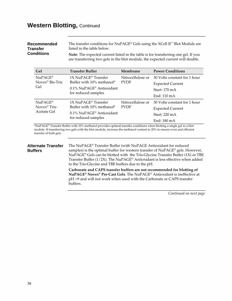
The transfer conditions for NuPAGE® Gels using the XCell II™ Blot Module are listed in the table below.
Note: The expected current listed in the table is for transferring one gel. If you are transferring two gels in the blot module, the expected current will double.
Gel Transfer Buffer Membrane Power Conditions
NuPAGE® Novex® Bis-Tris Gel
1X NuPAGE® Transfer Buffer with 10% methanol*
0.1% NuPAGE® Antioxidant for reduced samples
Nitrocellulose or PVDF
30 Volts constant for 1 hour
Expected Current
Start: 170 mA
End: 110 mA
NuPAGE® Novex® Tris-Acetate Gel
1X NuPAGE® Transfer Buffer with 10% methanol*
0.1% NuPAGE® Antioxidant for reduced samples
Nitrocellulose or PVDF
30 Volts constant for 1 hour
Expected Current
Start: 220 mA
End: 180 mA *NuPAGE® Transfer Buffer with 10% methanol provides optimal transfer conditions when blotting a single gel in a blot module. If transferring two gels with the blot module, increase the methanol content to 20% to ensure even and efficient transfer of both gels.
Alternate Transfer Buffers
The NuPAGE® Transfer Buffer (with NuPAGE Antioxidant for reduced samples) is the optimal buffer for western transfer of NuPAGE® gels. However, NuPAGE® Gels can be blotted with the Tris-Glycine Transfer Buffer (1X) or TBE Transfer Buffer (1/2X). The NuPAGE® Antioxidant is less effective when added to the Tris-Glycine and TBE buffers due to the pH. Carbonate and CAPS transfer buffers are not recommended for blotting of NuPAGE® Novex® Pre-Cast Gels. The NuPAGE® Antioxidant is ineffective at pH >9 and will not work when used with the Carbonate or CAPS transfer buffers.
Continued on next page
38
Western Blotting, Continued
Semi-Dry Blotting of NuPAGE® Novex® Bis-Tris Gels
The NuPAGE® Novex® Bis-Tris Gels do not transfer as efficiently with semi-dry blotting compared to blotting with the XCell II™ Blot Module. If you decide to use semi-dry blotting for NuPAGE® Novex® Bis-Tris Gels, follow the protocol provided below to ensure efficient transfer of proteins.
1. Prepare 100 mL of 2X NuPAGE® Transfer Buffer as follows:
NuPAGE® Transfer Buffer (20X) 10.0 mL NuPAGE® Antioxidant (for reduced sample) 0.1 mL Methanol 10.0 mL Deionized Water to 100 mL Total Volume 100 mL
2. Soak the filter paper and transfer membrane in 2X NuPAGE® Transfer Buffer.
If you are using Novex® pre-cut membrane/filter sandwiches, use three pieces of filter paper (0.4 mm/filter in thickness) on each side of the gel/membrane assembly.
If you are not using the Novex® pre-cut membrane/filter sandwiches, use two pieces of thick filter paper) on each side of the gel/membrane assembly.
3. Prepare 100 mL of 2X NuPAGE® Transfer Buffer ((without methanol)
4. Equilibrate the gel in 2X NuPAGE® Transfer Buffer (without methanol) for 10 minute with gentle agitation.
Note: For transfer of large proteins (>100 kDa), equilibrate the gel with 2X NuPAGE® Transfer Buffer (without methanol) with 0.02–0.04% SDS.
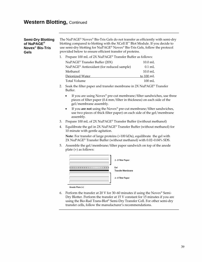
5. Assemble the gel/membrane/filter paper sandwich on top of the anode plate (+) as follows:
6. Perform the transfer at 20 V for 30–60 minutes if using the Novex® Semi-
Dry Blotter. Perform the transfer at 15 V constant for 15 minutes if you are using the Bio-Rad Trans-Blot® Semi-Dry Transfer Cell. For other semi-dry transfer cells, follow the manufacturer’s recommendations.
39
Using ZOOM® Gels
ZOOM® Gels ZOOM® Gels are used for 2D analysis of proteins following isoelectric focusing
of IPG strips. ZOOM® Gels are 1.0 mm thick, and contain an IPG well and a molecular weight marker well. The IPG well is designed to accommodate a 7.0 cm IPG strip.
Two types of ZOOM® Gels are available (see page 48 for ordering information)
NuPAGE® Novex® 4–12% Bis-Tris ZOOM® Gel
Novex® 4–20% Tris-Glycine ZOOM® Gel
Second Dimension Electrophoresis
The second dimension electrophoresis procedure involves reducing and alkylating the proteins focused on your IPG strip in equilibration buffer, loading the strip on your second dimension gel, and performing SDS-PAGE.
Materials Supplied by the User
You will need the following items for running ZOOM® Gels (see page 48–49 for ordering information on Invitrogen products):
4X NuPAGE® LDS Sample Buffer
NuPAGE® Sample Reducing Agent
NuPAGE® Novex® 4–12% Bis-Tris ZOOM® Gel or Novex® 4–20% Tris-Glycine ZOOM® Gel
Appropriate running buffer depending on the type of gel you are using
0.5% agarose solution
Iodoacetamide
Plastic flexible ruler or thin weighing spatula
15 mL conical tubes
Water bath set at 55C or 65C
XCell SureLock™ Mini-Cell
Protein molecular weight marker
Equilibrating the IPG Strip
1. Dilute 4X NuPAGE® LDS Sample Buffer to 1X with deionized water.
2. Add 500 μL of the NuPAGE® Sample Reducing Agent to 4.5 mL of the 1X NuPAGE® LDS Sample Buffer from Step 1 in a 15 mL conical tube. Place one IPG strip in this conical tube for equilibration.
3. Incubate for 15 minutes at room temperature. Decant the Reducing Solution.
4. Prepare 125 mM Alkylating Solution fresh by adding 116 mg of iodoacetamide to 5 mL of 1X NuPAGE® LDS Sample Buffer from Step 1.
5. Add 5 mL of Alkylating Solution (from Step 4) to the conical tube containing the IPG strip. Incubate for 15 minutes at room temperature.
6. Decant the Alkylating Solution and proceed to SDS-PAGE, next page. Use the equilibrated IPG strip immediately for second dimension.
Continued on next page
40
Using ZOOM® Gels, Continued
SDS-PAGE A protocol for separating proteins in an IPG strip by SDS-PAGE with ZOOM®
Gels and the XCell SureLock™ Mini-Cell is provided below. You may download the XCell SureLock™ Mini-Cell manual from our website at www.invitrogen.com or contact Technical Support (see page 56).
1. Prepare 0.5% agarose solution in the appropriate running buffer and keep it warm (55–65C) until you are ready to use the agarose solution.
2. If the molecular weight marker well is bent, straighten the well using a gel loading tip.
3. Cut the plastic ends of the IPG strip flush with the gel. Do not cut off any portions of the gel.
4. Slide the IPG strip into the ZOOM® Gel well.
5. Align the IPG strip properly in the ZOOM® Gel well using a thin plastic ruler or a weighing spatula. Avoid introducing any air bubbles while sliding the strip.
6. Pour ~ 400 μL of 0.5% agarose solution into the ZOOM® Gel well containing the IPG strip. Take care that the agarose solution does not overflow into the molecular weight marker well.
7. Assemble the gel cassette/Buffer Core sandwich as described in the XCell SureLock™ Mini-Cell manual.
Note: If you are running just one gel, use the plastic Buffer Dam in place of the second gel cassette to form the Upper Buffer Chamber.
Do not use the ZOOM® IPGRunner™ Core for electrophoresis of the second dimension gel. You must use the Buffer Core supplied with the XCell SureLock™ Mini-Cell.
8. Fill the Upper Buffer Chamber with a small amount of Running Buffer, and make sure there are no leaks.
9. Fill the Upper Buffer Chamber and Lower Buffer Chamber with the appropriate Running Buffer.
10. Load molecular weight standards in the marker well.
11. Place the XCell SureLock™ Mini-Cell lid on the Buffer Core. With the power on the power supply turned off, connect the electrode cords to the power supply [red to (+) jack, black to (–) jack].
12. Perform electrophoresis at 200 V for 40 minutes for NuPAGE® Novex® Bis-Tris ZOOM® Gels or at 125 V for 90 minutes for Novex® Tris-Glycine ZOOM® Gels.
13. At the end of electrophoresis, turn off the power and disassemble the gel cassette/Buffer Core sandwich assembly as described in the XCell SureLock™ Mini-Cell manual.
14. Proceed to staining the second dimension gel using an appropriate method for the type of gel and sample amount.
41
Calibrating Protein Molecular Weight
Introduction The molecular weight of a protein can be determined based upon its relative
mobility by constructing a standard curve with protein standards of known molecular weights.
The protein mobility in SDS-PAGE gels is dependent on the
Length of the protein in its fully denatured state,
SDS-PAGE buffer systems, and
Secondary structure of the protein.
The same molecular weight standard may have slightly different mobility, resulting in different apparent molecular weight when run in different SDS-PAGE buffer systems.
If you are using the Novex® protein molecular weight standards, see the apparent molecular weights of these standards in the NuPAGE® Gels listed on the next page to determine an apparent molecular weight of your protein.
Protein Secondary Structure
When using SDS-PAGE for molecular weight determination, slight deviations from the calculated molecular weight of a protein (calculated from the known amino acid sequence) can occur due to the retention of varying degrees of secondary structure in the protein, even in the presence of SDS. This phenomenon is observed in highly organized secondary structures (collagens, histones, or highly hydrophobic membrane proteins) and in peptides, where the effect of local secondary structure becomes magnified relative to the total size of the peptide.
Buffer Systems Slight differences in protein mobilities also occur when the same proteins are
run in different SDS-PAGE buffer systems. Each SDS-PAGE buffer system has a different pH, which affects the charge of a protein and its binding capacity for SDS. The degree of change in protein mobility is usually small in natural proteins but more pronounced with “atypical” or chemically modified proteins such as pre-stained standards.
Continued on next page
42
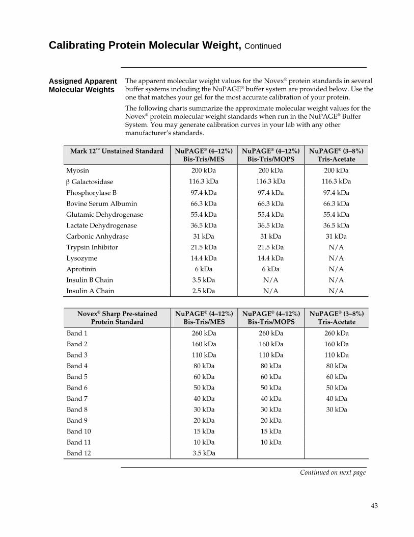
Calibrating Protein Molecular Weight, Continued
Assigned Apparent Molecular Weights
The apparent molecular weight values for the Novex® protein standards in several buffer systems including the NuPAGE® buffer system are provided below. Use the one that matches your gel for the most accurate calibration of your protein.
The following charts summarize the approximate molecular weight values for the Novex® protein molecular weight standards when run in the NuPAGE® Buffer System. You may generate calibration curves in your lab with any other manufacturer’s standards.
Novex® Sharp Pre-stained Protein Standard
NuPAGE® (4–12%)Bis-Tris/MES
NuPAGE® (4–12%) Bis-Tris/MOPS
NuPAGE® (3–8%) Tris-Acetate
Band 1 260 kDa 260 kDa 260 kDa
Band 2 160 kDa 160 kDa 160 kDa
Band 3 110 kDa 110 kDa 110 kDa
Band 4 80 kDa 80 kDa 80 kDa
Band 5 60 kDa 60 kDa 60 kDa
Band 6 50 kDa 50 kDa 50 kDa
Band 7 40 kDa 40 kDa 40 kDa
Band 8 30 kDa 30 kDa 30 kDa
Band 9 20 kDa 20 kDa
Band 10 15 kDa 15 kDa
Band 11 10 kDa 10 kDa
Band 12 3.5 kDa
Mark 12™ Unstained Standard NuPAGE® (4–12%)Bis-Tris/MES
NuPAGE® (4–12%) Bis-Tris/MOPS
NuPAGE® (3–8%) Tris-Acetate
Myosin 200 kDa 200 kDa 200 kDa
Galactosidase 116.3 kDa 116.3 kDa 116.3 kDa
Phosphorylase B 97.4 kDa 97.4 kDa 97.4 kDa
Bovine Serum Albumin 66.3 kDa 66.3 kDa 66.3 kDa
Glutamic Dehydrogenase 55.4 kDa 55.4 kDa 55.4 kDa
Lactate Dehydrogenase 36.5 kDa 36.5 kDa 36.5 kDa
Carbonic Anhydrase 31 kDa 31 kDa 31 kDa
Trypsin Inhibitor 21.5 kDa 21.5 kDa N/A
Lysozyme 14.4 kDa 14.4 kDa N/A
Aprotinin 6 kDa 6 kDa N/A
Insulin B Chain 3.5 kDa N/A N/A
Insulin A Chain 2.5 kDa N/A N/A
Continued on next page
43
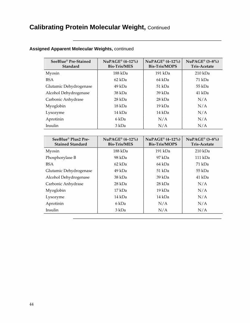
Calibrating Protein Molecular Weight, Continued
Assigned Apparent Molecular Weights, continued
SeeBlue® Pre-Stained Standard
NuPAGE® (4–12%) Bis-Tris/MES
NuPAGE® (4–12%) Bis-Tris/MOPS
NuPAGE® (3–8%) Tris-Acetate
Myosin 188 kDa 191 kDa 210 kDa
BSA 62 kDa 64 kDa 71 kDa
Glutamic Dehydrogenase 49 kDa 51 kDa 55 kDa
Alcohol Dehydrogenase 38 kDa 39 kDa 41 kDa
Carbonic Anhydrase 28 kDa 28 kDa N/A
Myoglobin 18 kDa 19 kDa N/A
Lysozyme 14 kDa 14 kDa N/A
Aprotinin 6 kDa N/A N/A
Insulin 3 kDa N/A N/A
SeeBlue® Plus2 Pre-Stained Standard
NuPAGE® (4–12%) Bis-Tris/MES
NuPAGE® (4–12%) Bis-Tris/MOPS
NuPAGE® (3–8%) Tris-Acetate
Myosin 188 kDa 191 kDa 210 kDa
Phosphorylase B 98 kDa 97 kDa 111 kDa
BSA 62 kDa 64 kDa 71 kDa
Glutamic Dehydrogenase 49 kDa 51 kDa 55 kDa
Alcohol Dehydrogenase 38 kDa 39 kDa 41 kDa
Carbonic Anhydrase 28 kDa 28 kDa N/A
Myoglobin 17 kDa 19 kDa N/A
Lysozyme 14 kDa 14 kDa N/A
Aprotinin 6 kDa N/A N/A
Insulin 3 kDa N/A N/A
44
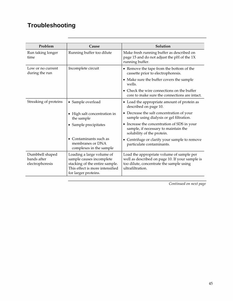
Troubleshooting
Problem Cause Solution
Run taking longer time
Running buffer too dilute Make fresh running buffer as described on page 15 and do not adjust the pH of the 1X running buffer.
Low or no current during the run
Incomplete circuit Remove the tape from the bottom of the cassette prior to electrophoresis.
Make sure the buffer covers the sample wells.
Check the wire connections on the buffer core to make sure the connections are intact.
Streaking of proteins Sample overload
High salt concentration in
the sample
Sample precipitates
Contaminants such as membranes or DNA complexes in the sample
Load the appropriate amount of protein as described on page 10.
Decrease the salt concentration of your sample using dialysis or gel filtration.
Increase the concentration of SDS in your sample, if necessary to maintain the solubility of the protein.
Centrifuge or clarify your sample to remove particulate contaminants.
Dumbbell shaped bands after electrophoresis
Loading a large volume of sample causes incomplete stacking of the entire sample. This effect is more intensified for larger proteins.
Load the appropriate volume of sample per well as described on page 10. If your sample is too dilute, concentrate the sample using ultrafiltration.
Continued on next page
45
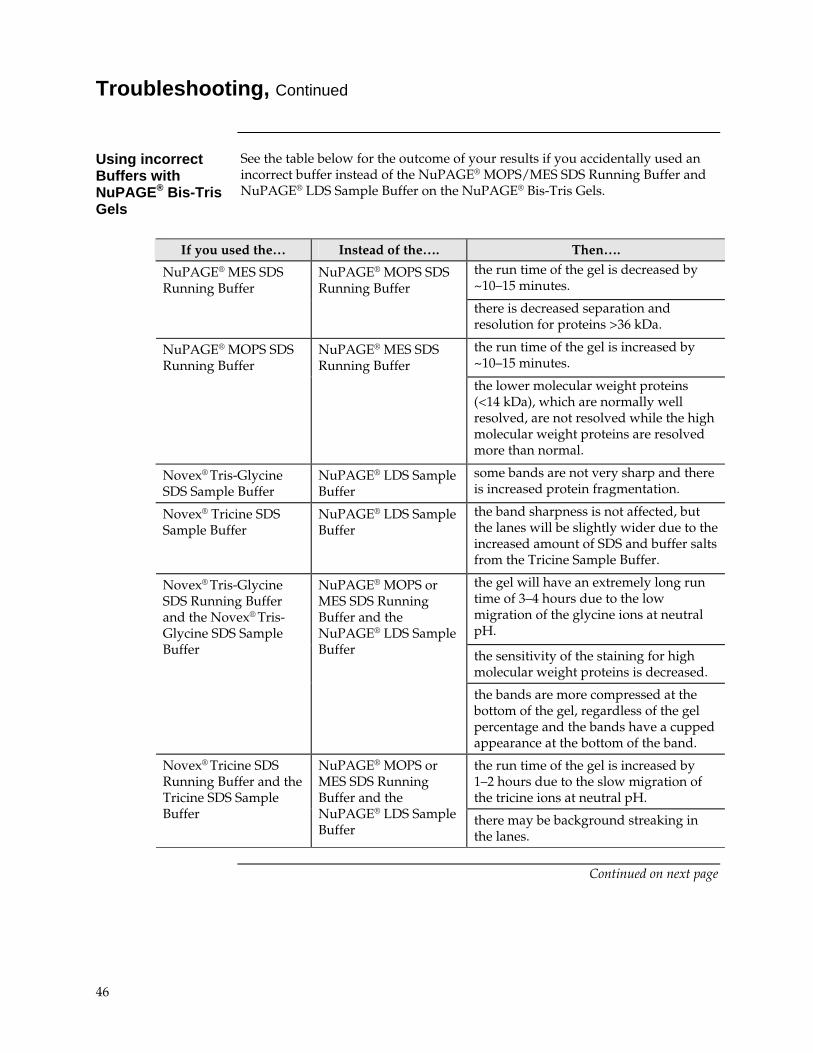
Troubleshooting, Continued
Using incorrect Buffers with NuPAGE® Bis-Tris Gels
See the table below for the outcome of your results if you accidentally used an incorrect buffer instead of the NuPAGE® MOPS/MES SDS Running Buffer and NuPAGE® LDS Sample Buffer on the NuPAGE® Bis-Tris Gels.
If you used the… Instead of the…. Then…. the run time of the gel is decreased by ~10–15 minutes.
NuPAGE® MES SDS Running Buffer
NuPAGE® MOPS SDS Running Buffer
there is decreased separation and resolution for proteins >36 kDa.
the run time of the gel is increased by ~10–15 minutes.
NuPAGE® MOPS SDS Running Buffer
NuPAGE® MES SDS Running Buffer
the lower molecular weight proteins (<14 kDa), which are normally well resolved, are not resolved while the high molecular weight proteins are resolved more than normal.
Novex® Tris-Glycine SDS Sample Buffer
NuPAGE® LDS Sample Buffer
some bands are not very sharp and there is increased protein fragmentation.
Novex® Tricine SDS Sample Buffer
NuPAGE® LDS Sample Buffer
the band sharpness is not affected, but the lanes will be slightly wider due to the increased amount of SDS and buffer salts from the Tricine Sample Buffer.
the gel will have an extremely long run time of 3–4 hours due to the low migration of the glycine ions at neutral pH.
the sensitivity of the staining for high molecular weight proteins is decreased.
Novex® Tris-Glycine SDS Running Buffer and the Novex® Tris-Glycine SDS Sample Buffer
NuPAGE® MOPS or MES SDS Running Buffer and the NuPAGE® LDS Sample Buffer
the bands are more compressed at the bottom of the gel, regardless of the gel percentage and the bands have a cupped appearance at the bottom of the band.
the run time of the gel is increased by 1–2 hours due to the slow migration of the tricine ions at neutral pH.
Novex® Tricine SDS Running Buffer and the Tricine SDS Sample Buffer
NuPAGE® MOPS or MES SDS Running Buffer and the NuPAGE® LDS Sample Buffer
there may be background streaking in the lanes.
Continued on next page
46
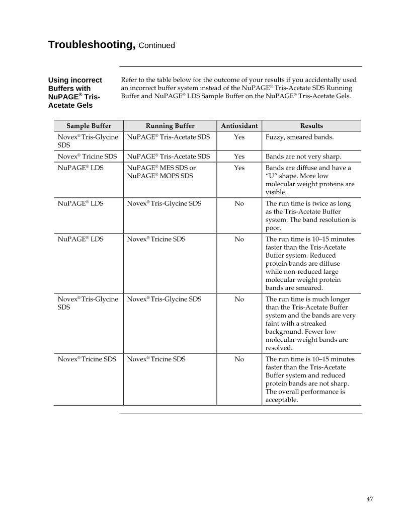
Troubleshooting, Continued
Using incorrect Buffers with NuPAGE® Tris-Acetate Gels
Refer to the table below for the outcome of your results if you accidentally used an incorrect buffer system instead of the NuPAGE® Tris-Acetate SDS Running Buffer and NuPAGE® LDS Sample Buffer on the NuPAGE® Tris-Acetate Gels.
Sample Buffer Running Buffer Antioxidant Results
Novex® Tris-Glycine SDS
NuPAGE® Tris-Acetate SDS Yes Fuzzy, smeared bands.
Novex® Tricine SDS NuPAGE® Tris-Acetate SDS Yes Bands are not very sharp.
NuPAGE® LDS NuPAGE® MES SDS or NuPAGE® MOPS SDS
Yes Bands are diffuse and have a “U” shape. More low molecular weight proteins are visible.
NuPAGE® LDS Novex® Tris-Glycine SDS No The run time is twice as long as the Tris-Acetate Buffer system. The band resolution is poor.
NuPAGE® LDS Novex® Tricine SDS No The run time is 10–15 minutes faster than the Tris-Acetate Buffer system. Reduced protein bands are diffuse while non-reduced large molecular weight protein bands are smeared.
Novex® Tris-Glycine SDS
Novex® Tris-Glycine SDS No The run time is much longer than the Tris-Acetate Buffer system and the bands are very faint with a streaked background. Fewer low molecular weight bands are resolved.
Novex® Tricine SDS Novex® Tricine SDS No The run time is 10–15 minutes faster than the Tris-Acetate Buffer system and reduced protein bands are not sharp. The overall performance is acceptable.
47
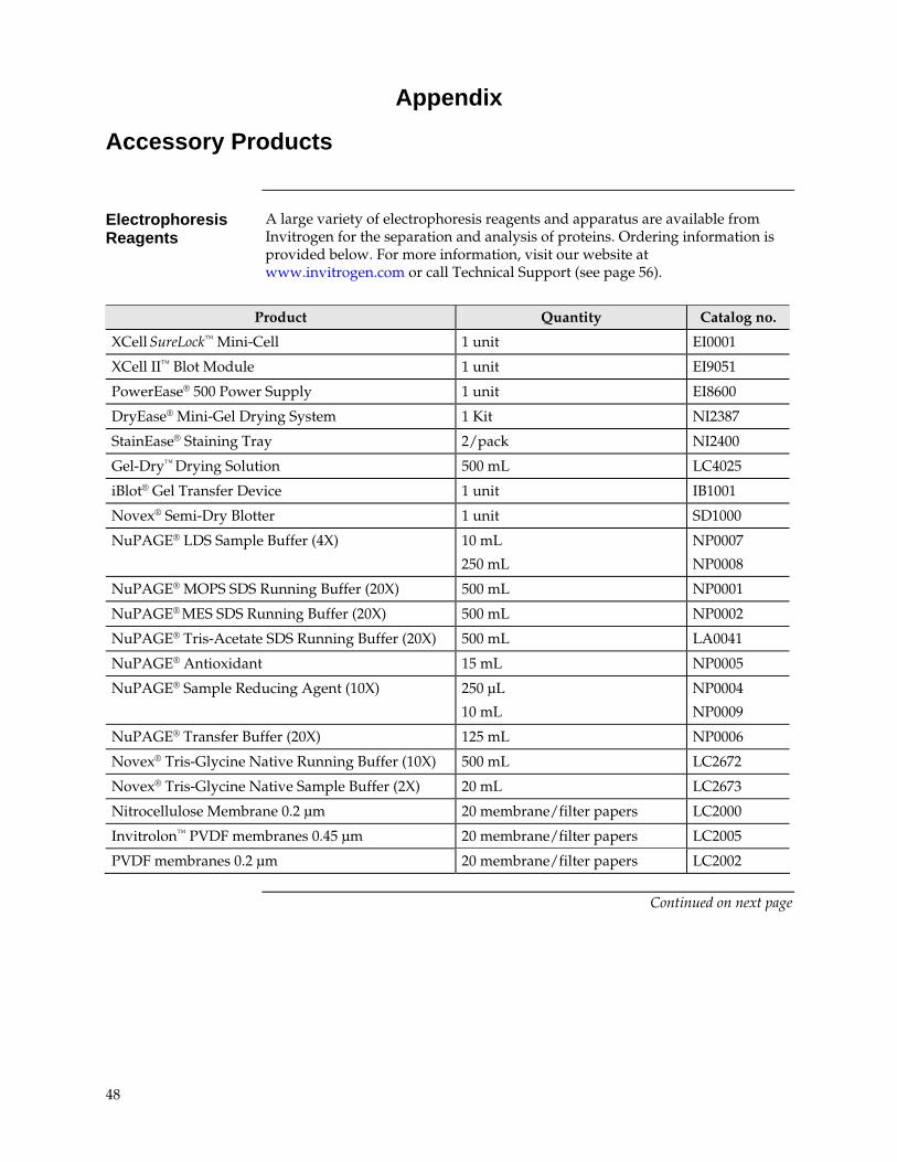
Appendix
Accessory Products
Electrophoresis Reagents
A large variety of electrophoresis reagents and apparatus are available from Invitrogen for the separation and analysis of proteins. Ordering information is provided below. For more information, visit our website at www.invitrogen.com or call Technical Support (see page 56).
Product Quantity Catalog no.
XCell SureLock™ Mini-Cell 1 unit EI0001
XCell II™ Blot Module 1 unit EI9051
PowerEase® 500 Power Supply 1 unit EI8600
DryEase® Mini-Gel Drying System 1 Kit NI2387
StainEase® Staining Tray 2/pack NI2400
Gel-Dry™ Drying Solution 500 mL LC4025
iBlot® Gel Transfer Device 1 unit IB1001
Novex® Semi-Dry Blotter 1 unit SD1000
NuPAGE® LDS Sample Buffer (4X) 10 mL
250 mL
NP0007
NP0008
NuPAGE® MOPS SDS Running Buffer (20X) 500 mL NP0001
NuPAGE® MES SDS Running Buffer (20X) 500 mL NP0002
NuPAGE® Tris-Acetate SDS Running Buffer (20X) 500 mL LA0041
NuPAGE® Antioxidant 15 mL NP0005
NuPAGE® Sample Reducing Agent (10X) 250 μL
10 mL
NP0004
NP0009
NuPAGE® Transfer Buffer (20X) 125 mL NP0006
Novex® Tris-Glycine Native Running Buffer (10X) 500 mL LC2672
Novex® Tris-Glycine Native Sample Buffer (2X) 20 mL LC2673
Nitrocellulose Membrane 0.2 μm 20 membrane/filter papers LC2000
Invitrolon™ PVDF membranes 0.45 μm 20 membrane/filter papers LC2005
PVDF membranes 0.2 μm 20 membrane/filter papers LC2002
Continued on next page
48
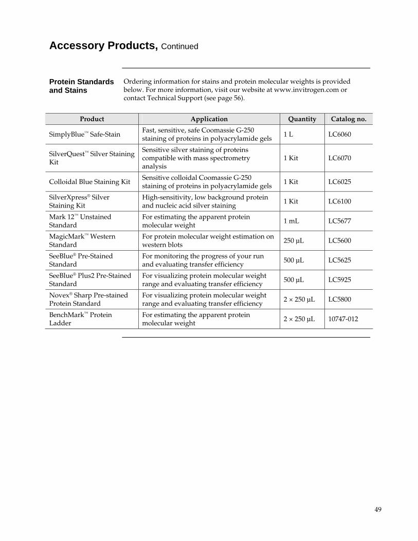
Accessory Products, Continued
Protein Standards and Stains
Ordering information for stains and protein molecular weights is provided below. For more information, visit our website at www.invitrogen.com or contact Technical Support (see page 56).
Product Application Quantity Catalog no.
SimplyBlue™ Safe-Stain Fast, sensitive, safe Coomassie G-250 staining of proteins in polyacrylamide gels 1 L LC6060
SilverQuest™ Silver Staining Kit
Sensitive silver staining of proteins compatible with mass spectrometry analysis
1 Kit LC6070
Colloidal Blue Staining Kit Sensitive colloidal Coomassie G-250 staining of proteins in polyacrylamide gels 1 Kit LC6025
SilverXpress® Silver Staining Kit
High-sensitivity, low background protein and nucleic acid silver staining 1 Kit LC6100
Mark 12™ Unstained Standard
For estimating the apparent protein molecular weight 1 mL LC5677
MagicMark™ Western Standard
For protein molecular weight estimation on western blots 250 μL LC5600
SeeBlue® Pre-Stained Standard
For monitoring the progress of your run and evaluating transfer efficiency 500 μL LC5625
SeeBlue® Plus2 Pre-Stained Standard
For visualizing protein molecular weight range and evaluating transfer efficiency 500 μL LC5925
Novex® Sharp Pre-stained Protein Standard
For visualizing protein molecular weight range and evaluating transfer efficiency 2 × 250 μL LC5800
BenchMark™ Protein Ladder
For estimating the apparent protein molecular weight 2 × 250 μL 10747-012
49
Recipes
NuPAGE® MOPS SDS Running Buffer
The NuPAGE® MOPS SDS Running Buffer (20X) is available from Invitrogen (see page 48).
50 mM MOPS 50 mM Tris Base 0.1% SDS 1 mM EDTA pH 7.7
1. To prepare 500 mL of 20X NuPAGE® MOPS SDS Running Buffer, dissolve the following reagents to 400 mL ultrapure water:
MOPS 104.6 g
Tris Base 60.6 g
SDS 10 g
EDTA 3.0 g
2. Mix well and adjust the volume to 500 mL with ultrapure water.
3. Store at +4C. The buffer is stable for 6 months when stored at +4C.
4. For electrophoresis, dilute this buffer to 1X with water (see page 15). The pH of the 1X solution is 7.7. Do not use acid or base to adjust the pH.
NuPAGE® MES SDS Running Buffer
The NuPAGE® MES SDS Running Buffer (20X) is available from Invitrogen (see page 48).
50 mM MES 50 mM Tris Base 0.1% SDS 1 mM EDTA pH 7.3
1. To prepare 500 mL of 20X NuPAGE® MES SDS Running Buffer, dissolve the following reagents to 400 mL ultrapure water:
MES 97.6 g
Tris Base 60.6 g
SDS 10 g
EDTA 3.0 g
2. Mix well and adjust the volume to 500 mL with ultrapure water.
3. Store at +4C. The buffer is stable for 6 months when stored at +4C.
4. For electrophoresis, dilute this buffer to 1X with water (see page 15). The pH of the 1X solution is 7.3. Do not use acid or base to adjust the pH.
Continued on next page
50
Recipes, Continued
NuPAGE® Tris-Acetate SDS Running Buffer
The NuPAGE® Tris-Acetate SDS Running Buffer (20X) is available from Invitrogen (see page 48).
50 mM Tricine 50 mM Tris Base 0.1% SDS pH 8.24
1. To prepare 500 mL of 20X NuPAGE® Tris-Acetate SDS Running Buffer, dissolve the following reagents to 400 mL ultrapure water:
Tricine 89.5 g
Tris Base 60.6 g
SDS 10 g
2. Mix well and adjust the volume to 500 mL with ultrapure water.
3. Store at +4C. The buffer is stable for 6 months when stored at +4C.
4. For electrophoresis, dilute this buffer to 1X with water (see page 15). The pH of the 1X solution is 8.24. Do not use acid or base to adjust the pH.
Tris-Glycine Native Running Buffer
The Tris-Glycine Native Running Buffer is available from Invitrogen (see page 48).
25 mM Tris Base 192 mM Glycine pH 8.3
1. To prepare 1,000 mL of 10X Tris-Glycine Native Running Buffer, dissolve the following reagents to 900 mL ultrapure water:
Tris Base 29 g
Glycine 144 g
2. Mix well and adjust the volume to 1,000 mL with ultrapure water.
3. Store at room temperature. The buffer is stable for 6 months when stored at room temperature.
4. For native electrophoresis, dilute this buffer to 1X with water (see page 51). The pH of the 1X solution is 8.3. Do not use acid or base to adjust the pH.
Continued on next page
51
Recipes, Continued
NuPAGE® LDS Sample Buffer
The NuPAGE® LDS Sample Buffer (4X) is available from Invitrogen (see page 48).
106 mM Tris HCl 141 mM Tris Base 2% LDS 10% Glycerol 0.51 mM EDTA 0.22 mM SERVA Blue G250 0.175 mM Phenol Red pH 8.5
1. To prepare 10 mL of 4X NuPAGE® LDS Sample Buffer, dissolve the following reagents to 8 mL ultrapure water:
Tris HCl 0.666 g
Tris Base 0.682 g
LDS 0.800 g
EDTA 0.006 g
Glycerol 4 g
SERVA Blue G250 (1% solution) 0.75 mL
Phenol Red (1% solution) 0.25 mL
2. Mix well and adjust the volume to 10 mL with ultrapure water.
3. Store at +4C. The buffer is stable for 6 months when stored at +4C.
4. For electrophoresis, prepare your samples in this buffer as described on page 14. The pH of the 1X solution is 8.5. Do not use acid or base to adjust the pH.
Tris-Glycine Native Sample Buffer
The Tris-Glycine Native Sample Buffer is available from Invitrogen (see page 48).
1X composition 100 mM Tris HCl 10% Glycerol 0.0025% Bromophenol Blue pH 8.6
1. To prepare 10 mL of 2X Tris-Glycine Native Sample Buffer, mix the following reagents :
0.5 M Tris HCl, pH 8.6 4 mL Glycerol 2 mL 0.1% (w/v) Bromophenol Blue 0.5 mL
2. Adjust the volume to 10 mL with ultrapure water.
3. Store at +4C. The buffer is stable for 6 months when stored at +4C.
4. Use this buffer to prepare samples for non-denaturing NuPAGE® Tris-Acetate gel electrophoresis (see page 52).
Continued on next page
52
Recipes, Continued
NuPAGE® Transfer Buffer
The NuPAGE® Transfer Buffer (20X) is available from Invitrogen (see page 48).
25 mM Bicine 25 mM Bis-Tris (free base) 1 mM EDTA pH 7.2
1. To prepare 125 mL of 20X NuPAGE® Transfer Buffer, dissolve the following reagents to 100 mL ultrapure water:
Bicine 10.2 g
Bis-Tris (free base) 13.1 g
EDTA 0.75 g
2. Mix well and adjust the volume to 125 mL with ultrapure water.
3. Store at +4C. The buffer is stable for 6 months when stored at +4C.
4. For western transfer, dilute this buffer to 1X with water (see page 35). The pH of the 1X solution is 7.2. Do not use acid or base to adjust the pH.
53
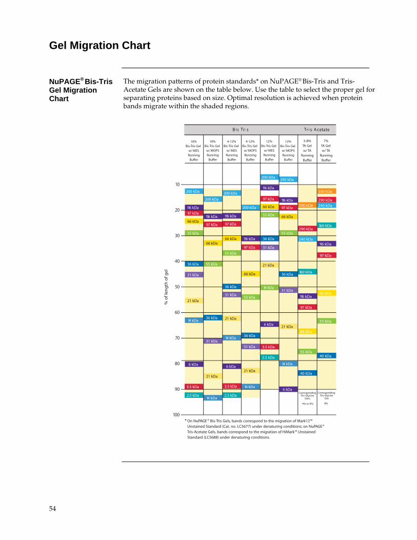
Gel Migration Chart
NuPAGE® Bis-Tris Gel Migration Chart
The migration patterns of protein standards* on NuPAGE® Bis-Tris and Tris-Acetate Gels are shown on the table below. Use the table to select the proper gel for separating proteins based on size. Optimal resolution is achieved when protein bands migrate within the shaded regions.
55 kDa
40 kDa
290 kDa
240 kDa
240 kDa
160 kDa
116 kDa
116 kDa
160 kDa
97 kDa
97 kDa
66 kDa
66 kDa
40 kDa
500 kDa290 kDa
500 kDa
% o
f len
gth
of g
el
10%Bis-Tris Gel
w/ MESRunning
Buffer
10%Bis-Tris Gel
w/ MOPSRunning
Buffer
4-12%Bis-Tris Gel
w/ MESRunning
Buffer
4-12%Bis-Tris Gel
w/ MOPSRunning
Buffer
200 kDa
200 kDa
200 kDa
200 kDa
116 kDa 116 kDa
116 kDa
97 kDa
97 kDa
97 kDa66 kDa
66 kDa
66 kDa
36 kDa
36 kDa
36 kDa
36 kDa
31 kDa
31 kDa31 kDa
31 kDa
21 kDa
21 kDa21 kDa
21 kDa14 kDa
14 kDa
14 kDa
6 kDa 6 kDa
3.5 kDa 3.5 kDa
2.5 kDa 2.5 kDa
55 kDa
55 kDa
55 kDa
55 kDa
10
20
30
40
50
60
70
80
90
100
14 kDa
116 kDa97 kDa
3-8%TA Gelw/ TA
RunningBuffer
7%TA Gelw/ TA
RunningBuffer
12%Bis-Tris Gel
w/ MESRunning
Buffer
12%Bis-Tris Gel
w/ MOPSRunning
Buffer
200 kDa200 kDa
116 kDa
116 kDa
97 kDa
97 kDa
66 kDa
66 kDa
36 kDa
36 kDa
31 kDa
31 kDa
21 kDa
21 kDa
14 kDa
6 kDa
3.5 kDa
2.5 kDa
55 kDa
55 kDa
14 kDa
* On NuPAGE ® Bis-Tris Gels, bands correspond to the migration of Mark12™Unstained Standard (Cat. no. LC5677) under denaturing conditions; on NuPAGE®Tris-Acetate Gels, bands correspond to the migration of HiMark™ UnstainedStandard (LC5688) under denaturing conditions.
6 kDa
66 kDa
etatecA-sirTsirT-siB
CorrespondingTris-Glycine
Gel:
CorrespondingTris-Glycine
Gels:
8%4% or 6%
55 kDa
etatecA-sirTsirT-siB
54
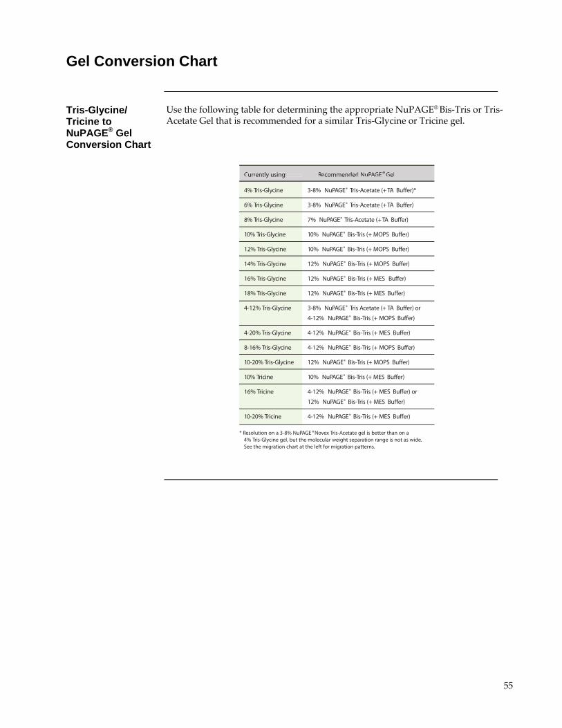
Gel Conversion Chart
Tris-Glycine/ Tricine to NuPAGE® Gel Conversion Chart
Use the following table for determining the appropriate NuPAGE® Bis-Tris or Tris-Acetate Gel that is recommended for a similar Tris-Glycine or Tricine gel.
Currently using: Recommended NuPAGE®Gel
4% Tris-Glycine 3-8% NuPAGE® Tris-Acetate (+TA Buffer)*
6% Tris-Glycine 3-8% NuPAGE® Tris-Acetate (+TA Buffer)
8% Tris-Glycine 7% NuPAGE® Tris-Acetate (+TA Buffer)
10% Tris-Glycine 10% NuPAGE® Bis-Tris (+ MOPS Buffer)
12% Tris-Glycine 10% NuPAGE® Bis-Tris (+ MOPS Buffer)
14% Tris-Glycine 12% NuPAGE® Bis-Tris (+ MOPS Buffer)
16% Tris-Glycine 12% NuPAGE® Bis-Tris (+ MES Buffer)
18% Tris-Glycine 12% NuPAGE® Bis-Tris (+ MES Buffer)
4-12% Tris-Glycine 3-8% NuPAGE® Tris Acetate (+ TA Buffer) or
4-12% NuPAGE® Bis-Tris (+ MOPS Buffer)
4-20% Tris-Glycine 4-12% NuPAGE® Bis-Tris (+ MES Buffer)
8-16% Tris-Glycine 4-12% NuPAGE® Bis-Tris (+ MOPS Buffer)
10-20% Tris-Glycine 12% NuPAGE® Bis-Tris (+ MOPS Buffer)
10% Tricine 10% NuPAGE® Bis-Tris (+ MES Buffer)
16% Tricine 4-12% NuPAGE® Bis-Tris (+ MES Buffer) or
12% NuPAGE® Bis-Tris (+ MES Buffer)
10-20% Tricine 4-12% NuPAGE® Bis-Tris (+ MES Buffer)
* Resolution on a 3-8% NuPAGE ®Novex Tris-Acetate gel is better than on a4% Tris-Glycine gel, but the molecular weight separation range is not as wide.See the migration chart at the left for migration patterns.
Currently using: Recommended NuPAPP GE®Gel
55

Technical Support
World Wide Web
Visit the Invitrogen website at www.invitrogen.com for:
Technical resources, including manuals, vector maps and sequences, application notes, SDSs, FAQs, formulations, citations, handbooks, etc.
Complete technical support contact information
Access to the Invitrogen Online Catalog
Additional product information and special offers
Contact Us For more information or technical assistance, call, write, fax, or email. Additional
international offices are listed on our website (www.invitrogen.com).
Corporate Headquarters: 5791 Van Allen Way Carlsbad, CA 92008 USA Tel: 1 760 603 7200 Tel (Toll Free): 1 800 955 6288 Fax: 1 760 602 6500 E-mail: [email protected]
Japanese Headquarters: LOOP-X Bldg. 6F 3-9-15, Kaigan Minato-ku, Tokyo 108-0022 Tel: 81 3 5730 6509 Fax: 81 3 5730 6519 E-mail: [email protected]
European Headquarters: Inchinnan Business Park 3 Fountain Drive Paisley PA4 9RF, UK Tel: +44 (0) 141 814 6100 Tech Fax: +44 (0) 141 814 6117 E-mail: [email protected]
SDS Safety Data Sheets (SDSs) are available at www.invitrogen.com/sds.
Certificate of Analysis
The Certificate of Analysis provides detailed quality control and product qualification information for each product. Certificates of Analysis are available on our website. Go to www.invitrogen.com/support and search for the Certificate of Analysis by product lot number, which is printed on the box.
Continued on next page
56
Technical Support, Continued
Limited Warranty Invitrogen (a part of Life Technologies Corporation) is committed to providing
our customers with high-quality goods and services. Our goal is to ensure that every customer is 100% satisfied with our products and our service. If you should have any questions or concerns about an Invitrogen product or service, contact our Technical Support Representatives. All Invitrogen products are warranted to perform according to specifications stated on the certificate of analysis. The Company will replace, free of charge, any product that does not meet those specifications. This warranty limits the Company’s liability to only the price of the product. No warranty is granted for products beyond their listed expiration date. No warranty is applicable unless all product components are stored in accordance with instructions. The Company reserves the right to select the method(s) used to analyze a product unless the Company agrees to a specified method in writing prior to acceptance of the order. Invitrogen makes every effort to ensure the accuracy of its publications, but realizes that the occasional typographical or other error is inevitable. Therefore the Company makes no warranty of any kind regarding the contents of any publications or documentation. If you discover an error in any of our publications, report it to our Technical Support Representatives. Life Technologies Corporation shall have no responsibility or liability for any special, incidental, indirect or consequential loss or damage whatsoever. The above limited warranty is sole and exclusive. No other warranty is made, whether expressed or implied, including any warranty of merchantability or fitness for a particular purpose.
57
Purchaser Notification
Limited Use Label License No. 5: Invitrogen Technology
The purchase of this product conveys to the buyer the non-transferable right to use the purchased amount of the product and components of the product in research conducted by the buyer (whether the buyer is an academic or for-profit entity). The buyer cannot sell or otherwise transfer (a) this product (b) its components or (c) materials made using this product or its components to a third party or otherwise use this product or its components or materials made using this product or its components for Commercial Purposes. The buyer may transfer information or materials made through the use of this product to a scientific collaborator, provided that such transfer is not for any Commercial Purpose, and that such collaborator agrees in writing (a) not to transfer such materials to any third party, and (b) to use such transferred materials and/or information solely for research and not for Commercial Purposes. Commercial Purposes means any activity by a party for consideration and may include, but is not limited to: (1) use of the product or its components in manufacturing; (2) use of the product or its components to provide a service, information, or data; (3) use of the product or its components for therapeutic, diagnostic or prophylactic purposes; or (4) resale of the product or its components, whether or not such product or its components are resold for use in research. For products that are subject to multiple limited use label licenses, the terms of the most restrictive limited use label license shall control. Life Technologies Corporation will not assert a claim against the buyer of infringement of patents owned or controlled by Life Technologies Corporation which cover this product based upon the manufacture, use or sale of a therapeutic, clinical diagnostic, vaccine or prophylactic product developed in research by the buyer in which this product or its components was employed, provided that neither this product nor any of its components was used in the manufacture of such product. If the purchaser is not willing to accept the limitations of this limited use statement, Life Technologies is willing to accept return of the product with a full refund. For information about purchasing a license to use this product or the technology embedded in it for any use other than for research use please contact Out Licensing, Life Technologies, 5791 Van Allen Way, Carlsbad, California 92008 ; Phone (760) 603-7200 or e-mail: [email protected].
58
59
References
Kubo, K. (1995) Effect of Incubation of Solutions of Proteins Containing Dodecyl Sulfate on the Cleavage
of Peptide Bonds by Boiling. Anal Biochem. 225, 351-353.
Laemmli, U. K., (1970) Cleavage of structural proteins during the assembly of the head of bacteriophage T4. Nature, 227, 680-685.
Moos, M Jr., Nguyen, N. Y., Liu, T.Y. (1988) Reproducible High Yield Sequencing of Proteins Electrophoretically Separated and Transferred to an Inert Support. J. Biol. Chem. 263, 6005-6008.
©2010 Life Technologies Corporation. All rights reserved. The trademarks mentioned herein are the property of Life Technologies Corporation or their respective owners.
Corporate Headquarters5791 Van Allen WayCarlsbad, CA 92008T: 1 760 603 7200F: 1 760 602 6500E: [email protected]
For country-specific contact information, visit our web site at www.invitrogen.com


![タンパク質電気泳動カタログ - Thermo Fisher Scientific · 1 day ago · Contents >LZ[LYU ^VYRÅV^ V]LY]PL^ 2 Separate NuPAGE プレキャストゲル 3 NuPAGE ゲル用泳動槽](https://img.pdfslide.us/doc/110x75/60c0ed0f17eb9f6d201f3017/ffeef-thermo-fisher-scientific-1-day-ago-contents.jpg)